Embed Size (px)
Citation preview
RESEARCH ARTICLE Open Access
Analysis of electric moments of RNA-bindingproteins: implications for mechanism and predictionShandar Ahmad1, Akinori Sarai2*
Abstract
Background: Protein-RNA interactions play important role in many biological processes such as gene regulation,replication, protein synthesis and virus assembly. Although many structures of various types of protein-RNAcomplexes have been determined, the mechanism of protein-RNA recognition remains elusive. We have earliershown that the simplest electrostatic properties viz. charge, dipole and quadrupole moments, calculated frombackbone atomic coordinates of proteins are biased relative to other proteins, and these quantities can be used toidentify DNA-binding proteins. Closely related, RNA-binding proteins are investigated in this study. In particular,discrimination between various types of RNA-binding proteins, evolutionary conservation of these bulk electrostaticfeatures and effect of conformational changes by complex formation are investigated. Basic binding mechanism ofa putative RNA-binding protein (HI1333 from Haemophilus influenza) is suggested as a potential application of thisstudy.
Results: We found that similar to DNA-binding proteins (DBPs), RNA-binding proteins (RBPs) also show significantlyhigher values of electric moments. However, higher moments in RBPs are found to strongly depend on theirfunctional class: proteins binding to ribosomal RNA (rRNA) constitute the only class with all three of the properties(charge, dipole and quadrupole moments) being higher than control proteins. Neural networks were trained usingleave-one-out cross-validation to predict RBPs from control data as well as pair-wise classification capacity betweenproteins binding to various RNA types. RBPs and control proteins reached up to 78% accuracy measured by thearea under the ROC curve. Proteins binding to rRNA are found to be best distinguished (AUC = 79%). Changes indipole and quadrupole moments between unbound and bound structures were small and these properties arefound to be robust under complex formation.
Conclusions: Bulk electric moments of proteins considered here provide insights into target recognition by RNA-binding proteins, as well as ability to recognize one type of RBP from others. These results help in understandingthe mechanism of protein-RNA recognition, and identifying RNA-binding proteins.
BackgroundProtein-RNA interactions have been identified as crucialfor a number of cellular processes [1-7]. However, themechanism of RNA recognition by proteins or viceversa has been poorly understood despite a recent surgein the study of protein-RNA interactions for specific sys-tems as well as their statistical analysis and prediction[8-12]. Most computational studies on protein-RNAinteractions have focused on classification, annotationand binding-site characterization [11,12]. A large
number of features have often been employed for accu-rate predictions of these RNA-binding proteins aswell as their interface residues [12]. Structure-basedpredictions and analysis of RBP’s have focused on high-resolution structures utilizing detailed structuralparameters such as solvent accessibility and detailedgeometrical features such as cleft and patch. In thesestudies, basic electrostatic features such as dipole andquadrupole moments are typically considered in combi-nation with many other parameters (e.g., 40 parametersby Shazman and Gutfreund [12]), which prevents usfrom looking at the role of individual physical propertiesof proteins in RNA-recognition and therefore some ofthe obvious role of electrostatic interactions may be lost
* Correspondence: [email protected] of Bioscience and Bioinformatics, Kyushu Institute ofTechnology, Iizuka, Fukuoka, 820-8502 JapanFull list of author information is available at the end of the article
Ahmad and Sarai BMC Structural Biology 2011, 11:8http://www.biomedcentral.com/1472-6807/11/8
© 2011 Ahmad and Sarai; licensee BioMed Central Ltd. This is an Open Access article distributed under the terms of the CreativeCommons Attribution License (http://creativecommons.org/licenses/by/2.0), which permits unrestricted use, distribution, andreproduction in any medium, provided the original work is properly cited.
in an effort to maximize prediction performance. Wehave earlier shown that simple electrostatic propertiesviz. net charge, dipole and quadrupole moments carrysignificant information useful to predict DNA-bindingproteins both from full atomic coordinates as well asmain chain atoms [13]. Subsequent studies confirmedthat low-resolution structures could be used to applythis method to the prediction of nucleic acid bindingfunction [14]. A Web-based tool to calculate dipole andquadrupole moments and reflections on their relation-ship to functional protein classes has also become avail-able and was published recently [15].Here, we carry out a systematic analysis of three bulk
electrostatic properties of RNA-binding proteins (RBPs)viz. net charge, dipole and quadrupole moments, all calcu-lated from low-resolution protein structures with onlymain-chain coordinates, in order to estimate how far thesesimple properties are able to identify RBPs from controlproteins. Simple statistical analysis of electric moments ineach category has been supplemented by all-against-allpair-wise recognition of various types, determined byneural network prediction. Our results show that thereexists a pattern of electric moments in RBPs, which is dif-ferent from the control data as well as within the proteinsbinding to various types of RNAs. One type of RNA-bind-ing proteins can be distinguished from the other on thebasis of these properties with various degrees of accuracy.Finally, we compile a data set of pairs of structures of thesame RBPs solved in monomer state as well as full pro-tein-RNA complex. Using this data set, we show that thecalculation of moments is rather robust against conforma-tional changes induced by complex formation. Finally, wediscuss possible implications of the present results for themechanism of protein-RNA interactions.
MethodsData set of RNA-binding proteinsPrimary source of RNA-binding proteins and theirannotations into various categories is SCOR database[16]. First, a list of all PDB codes present in SCOR wascompiled, resulting in 569 entries. All 569 PDB entrieswere scanned for RNA (998 chains) and proteins (1435chains). Protein chains were then scanned to be indirect contact with at least one RNA chain. Proteinswith at least 3 residues in contact were selected, result-ing in 1242 chains. FASTA-formatted protein sequenceswere generated from the PDB files and redundancy wasremoved by clustering them at 25% sequence identityusing BLASTCLUST [17]. This resulted in RBP_NR25database of 160 protein chains, to be subsequentlyreferred to as simply RBP. SCOR functional classifica-tion was used to annotate them as binding to mRNA(13 chains), tRNA (20 chains), rRNA (84 chains) or viralRNA (17 chains). Final list of selected protein chains,
their calculated moments, along with other data sets, isprovided in Additional File 1.
Development of control data setsFirst, a non-redundant list of all protein chains in PDBwas obtained from PDBselect [18]. The latest (May30-2010 version) PDBselect (25% sequence ID clusters) con-sisted of 4868 protein chains. From this, chains smallerthan 50 residues were removed, which resulted in 4133protein chains. Next, a keyword search using “Nucleicacid binding” was carried out in SWISSPROT and result-ing 20595 proteins chains were obtained in this way.Then, the 4133 chains selected from PDBselect werealigned against all the 20595 SWISSPROT sequences toobtain any similarity, using BLAST at e-values cutoff of0.01. These chains were excluded from PDB selectsequence database. Further PDB entry type was checkedand nucleic acid binding chains were removed, leaving2441 sequences with no similarity to RNA binding pro-teins with known or unknown structure were obtained.These 2441 protein chains were used as a control dataset for all our analysis (see Additional File 1).
Complex versus monomeric structure pairsSequence homologues of proteins used in the above dataset (RBP_NR25) were searched in PDB with at least 90%sequence identity and the best match was selected.Minimum alignment coverage was also set at 90% andonly those target sequences that occurred in monomericPDB entries were selected.
Calculations of electric momentsCharge, dipole moment and quadrupole moments werecalculated as described in our earlier study [13]. Accord-ing to that study, consideration of all-atom coordinatesdid not affect the overall results, as compared to thelow-resolution model with only backbone coordinates.Thus, in this study, side-chain coordinates of the pro-teins were ignored and the electric moments were basedon the main chain conformation determined by Ca-position of the residues. All Lys and Arg residues wereassigned a positive charge and Glu and Asp residueswere considered negative. All other residues were trea-ted as neutral: His was considered as neutral, as theconsideration of its charged states had negligible effects(see Results section). All water molecules, metals andligands were also ignored for these calculations.Components of dipole moments were calculated using
the expression
P R R q i o i (1)
where Ro is the reference point, which was taken asthe geometric center of all the residues (Ca-positions) in
Ahmad and Sarai BMC Structural Biology 2011, 11:8http://www.biomedcentral.com/1472-6807/11/8
Page 2 of 13
the structure, and i represents an atom in the proteinstructure. Net dipole moment was calculated by taking avector sum of these components.Quardupole moment is a tensor of rank 2 and a direct
calculation from the PDB coordinates gives nine compo-nents (Mxx, Mxy, Mxz, Myx, Myy, Myz, Mzx, Mzy and Mzz).Each of these components is calculated by the followingexpression
M r r qi . / 1 2 3 2 i i ir (2)
where ri is the relative position vector, i is the index ofcharge and summation is over all charges. The quadrupolemoment matrix can be diagonalized and the three eigenva-lues of the quadrupole moment matrix are represented asQ1, Q2 and Q3 in decreasing order. We used the largesteigenvalue Q1 for designating single quadrupole momentand all three eigen values for developing the predictor.All electric moment values were the absolute values
and normalized by the protein sequence length in a waysimilar to our earlier study [13]. Units are often omittedin describing quadrupole moments and net charge asthese values are measured in atomic units (using elec-tronic charge and Å as charge and distance units in cal-culations). Dipole moment values are quoted byconverting them to Debyes.Our method of computing electric moments is some-
what different from a similar approach adopted in arecently published dipole moment server [15]. First ofall, we use only the Ca atoms for assigning charges,whereas charges are assigned to specific atomic posi-tions in [15]. Secondly, we used geometric center of allCa atoms (including residues with zero charge assign-ments to compute the reference point and axes) andfinally, we obtain quadrupole moments by taking theireigen values, which is not provided in [15]. We find thatthere is a moderate correlation (~0.5) between thedipole moments computed by the two methods. Sinceour approach is more suitable for low resolution struc-tures (does not require side chain positions), we reportonly the results obtained by our procedure. For similarreasons, we did not try to predict protonation state ofresidues, which could sometimes be possible if side-chain coordinates are provided [19].
Statistical significance of differenceDistributions of moments between control andRNA-binding as well as between various classes ofRNA-binding proteins were compared by measuring thestatistical significance of difference between their means.A two-tailed Student t-test was conducted for all suchcomparisons using open-source statistical programminglanguage R http://r-project.org. Histograms of distribu-tions were also plotted in the same package.
Difference between bound and unbound pairsFor each protein chain in the RBP data set, a data set ofmonomeric proteins from PDB was scanned. Proteinswith more than 90% similarity and coverage values wereused as a pair of complexed and unbound monomers.Electric moments were then computed for both of themby the procedure described above. A total of 27 proteinswere found to occur both in monomeric as well asRNA-complexed forms.The difference between electric moments of a protein
in its complexed and unbound forms is measured usingEuclidean distance (ED) expression as follows:
ED X 1 2
NX bound X free[ ( ) ( )] (3)
Where X refers to dipole or quadrupole moment ofthe protein and summations is taken over all protein-pairs considered in a category (effectively a distance in27-dimensional space).
Neural network for predictionA neural network-based predictor, similar to our earlierimplementations (e.g. in [20]) was used to find a rela-tionship between input vectors composed here of fivedescriptors based on charge, dipole moment and threeeigenvalues of quadrupole moment and the functionalproperty of protein chain e.g., binding or non-binding(control). To account for any cooperative and non-linearcontribution of moments, a single hidden layer with 3nodes has been used. To avoid over-assessment of per-formance, the neural network was trained in a jackknifestyle, by optimizing the predictor for all but one data inthe training. Once the training is completed, predictionon the left-out protein is evaluated. After runningthrough all binding and control proteins, overall predic-tion performance on the left-out proteins is evaluated.Since the neural network returns a real value between 0and 1 for the target outputs 0 (non-binding) or 1 (bind-ing), ROC data between specificity and sensitivity is cal-culated and converted to the area under the curve(AUC) values, which reflects performance over theentire range of cutoffs. Other measures of performanceare as follows (T refers to true and F referes to false,whereas P is positive class and N is negative class):
Precision p TP TP FP
Recall r TP TP FN
Accuracy TP TN
/
/
/
/
TP TN FP FN
F measure 2pr p r
(4)
F-measure is the geometric mean of precision andrecall and can be computed by transforming real-valued
Ahmad and Sarai BMC Structural Biology 2011, 11:8http://www.biomedcentral.com/1472-6807/11/8
Page 3 of 13
outputs of neural network into binary class-label predic-tions at various cutoffs. Cutoffs at which F-measure hasthe highest value is used for reporting all class-wise per-formance measures, i.e. precision, recall, accuracy andF-measure.
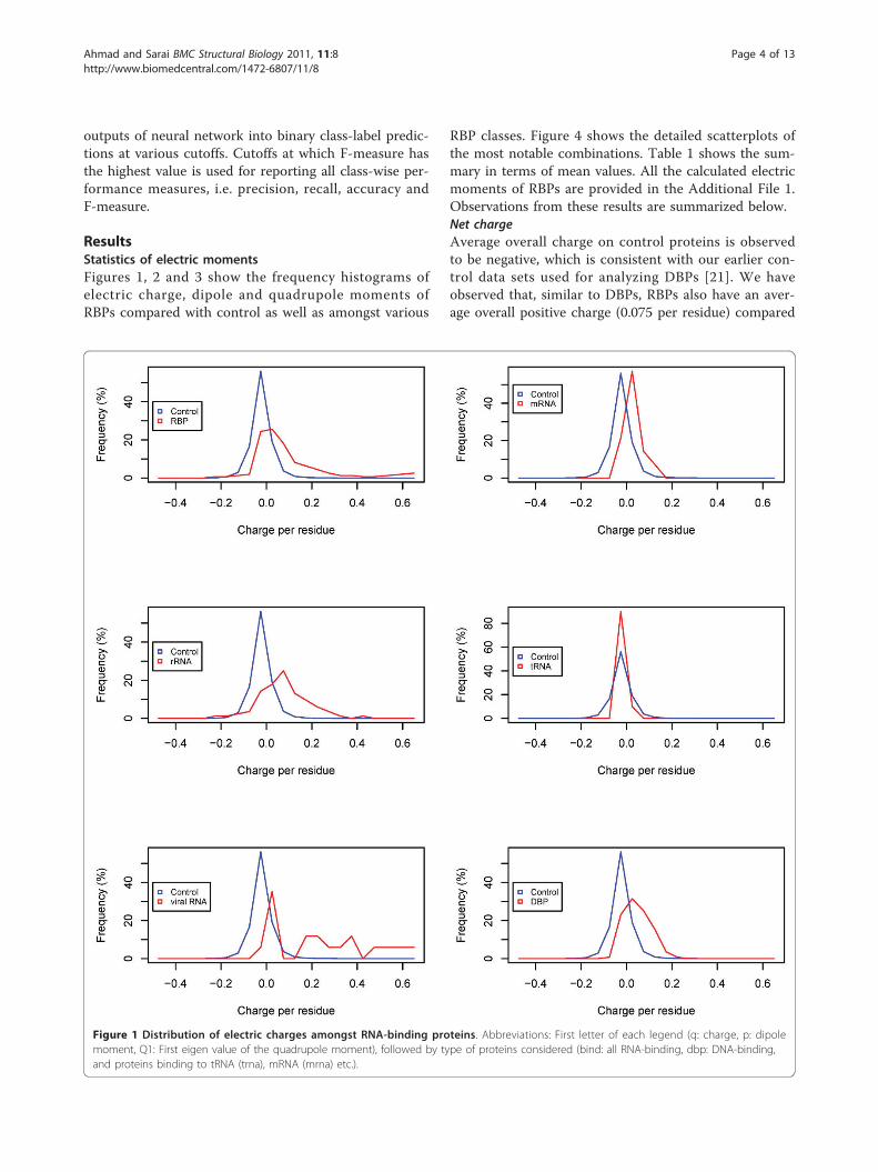
ResultsStatistics of electric momentsFigures 1, 2 and 3 show the frequency histograms ofelectric charge, dipole and quadrupole moments ofRBPs compared with control as well as amongst various
RBP classes. Figure 4 shows the detailed scatterplots ofthe most notable combinations. Table 1 shows the sum-mary in terms of mean values. All the calculated electricmoments of RBPs are provided in the Additional File 1.Observations from these results are summarized below.Net chargeAverage overall charge on control proteins is observedto be negative, which is consistent with our earlier con-trol data sets used for analyzing DBPs [21]. We haveobserved that, similar to DBPs, RBPs also have an aver-age overall positive charge (0.075 per residue) compared
Figure 1 Distribution of electric charges amongst RNA-binding proteins. Abbreviations: First letter of each legend (q: charge, p: dipolemoment, Q1: First eigen value of the quadrupole moment), followed by type of proteins considered (bind: all RNA-binding, dbp: DNA-binding,and proteins binding to tRNA (trna), mRNA (mrna) etc.).
Ahmad and Sarai BMC Structural Biology 2011, 11:8http://www.biomedcentral.com/1472-6807/11/8
Page 4 of 13
with negative (-0.020) values observed for control pro-teins. Statistical significance is also established by ap-value nearly zero (smaller than the precision limit ofthe software). However, the histogram in Figure 1 showsthat there are a significant number (~40%) of RNA-binding proteins with negative or near zero net chargesimilar to control proteins. Further look at class-widedistributions shows that tRNA-binding proteins havealmost no difference with control proteins in their dis-tribution of charges (p-value ~0.24). On the other hand,mRNA-binding proteins have small difference comparedto control data sets (p-values suggest that the difference
is significant). However, the most significantly positivelycharged proteins are rRNA-binding and viral RNA-bind-ing proteins (mean charge 0.077, 0.192 respectively), inwhich less than 20% proteins have negative or near zeronet charge. This is statistically confirmed by the corre-sponding p-values (nearly zero) in comparison to con-trol proteins (Table 2). When compared to DBPs (Table1), RBPs are found to have even higher average chargethan DBPs. However, looking at various RNA types, weobserve that the higher charge on the average in RBPcomes predominantly because of rRNA-binding proteinsas they are the most abundant in the data set and have
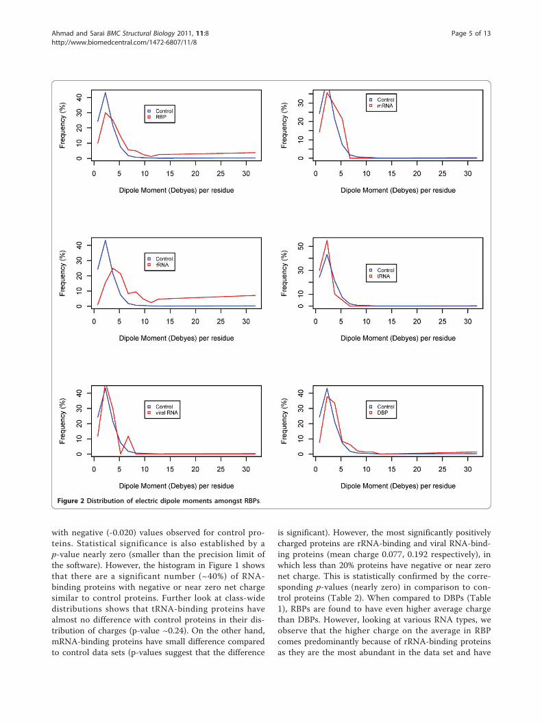
Figure 2 Distribution of electric dipole moments amongst RBPs.
Ahmad and Sarai BMC Structural Biology 2011, 11:8http://www.biomedcentral.com/1472-6807/11/8
Page 5 of 13
the highest positive charge. All other RBPs have signifi-cantly lower charge per residue than either the rRNA-binding proteins or DBPs.Dipole momentWe observe from Figure 2 that most RNA-binding pro-teins are distributed in the range of higher dipolemoments. Overall, mean dipole moment for all RBPs is4.6 units compared with 2.7 units for control proteins(with a highly significant p-value for the difference).Similar to the charge distribution, not all RBP typeshave higher dipole moments. However, interestingly, theclasses with higher dipole moments are slightly differentfrom those with higher positive charge. Although rRNA
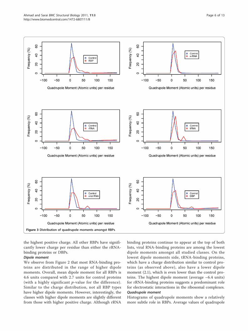
binding proteins continue to appear at the top of bothlists, viral RNA-binding proteins are among the lowestdipole moments amongst all studied classes. On thelowest dipole moments side, tRNA-binding proteins,which have a charge distribution similar to control pro-teins (as observed above), also have a lower dipolemoment (2.1), which is even lower than the control pro-teins. The highest dipole moment (average ~6.4 units)for rRNA-binding proteins suggests a predominant rolefor electrostatic interactions in the ribosomal complexes.Quadrupole momentHistograms of quadrupole moments show a relativelymore subtle role in RBPs. Average values of quadrupole
Figure 3 Distribution of quadrupole moments amongst RBPs.
Ahmad and Sarai BMC Structural Biology 2011, 11:8http://www.biomedcentral.com/1472-6807/11/8
Page 6 of 13
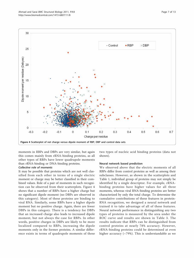
moments in RBPs and DBPs are very similar, but againthis comes mainly from rRNA-binding proteins, as allother types of RBPs have lower quadrupole momentsthan rRNA-binding or DNA-binding proteins.Collective role of momentsIt may be possible that proteins which are not well clas-sified from each other in terms of a single electricmoment or charge may be better classified in their com-bined values. Role of a pair of moments in such recogni-tion can be observed from their scatterplots. Figure 4shows that a number of RBPs have a higher charge butno significant dipole moment (no DBPs are observed inthis category). Most of these proteins are binding toviral RNA. Similarly, some RBPs have a higher dipolemoment but no positive charge. Again, there are fewerDBPs in this category. There is a tendency for DBPsthat an increased charge also leads to increased dipolemoment, but not always the case for RBPs. In otherwords, positive charges in DBPs are likely to be morelocalized compared to RBPs, increasing the dipolemoments only in the former proteins. A similar differ-ence exists in terms of quadrupole moments of these
two types of nucleic acid binding proteins (data notshown).
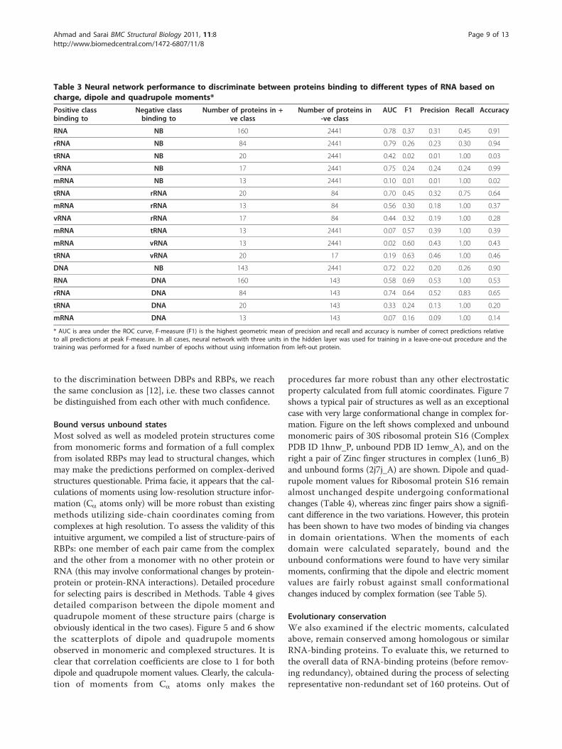
Neural network based predictionWe observed above that the electric moments of allRBPs differ from control proteins as well as among theirsubclasses. However, as shown in the scatterplots andTable 1, individual group of proteins may not simply beidentified by a single descriptor. For example, rRNA-binding proteins have higher values for all threemoments, whereas viral RNA-binding proteins are bettercharacterized by only the total charge. To determine thecumulative contributions of these features in protein-RNA recognition, we designed a neural network andtrained it to take advantage of all of these features.Neural network performance in distinguishing any twotypes of proteins is measured by the area under theROC curve and results are shown in Table 3. Theresults indicate that RBPs can be distinguished fromcontrol proteins at nearly 78% accuracy. However,rRNA-binding proteins could be determined at evenhigher accuracy (~79%). This is understandable as we
Figure 4 Scatterplot of net charge versus dipole moment of RBP, DBP and control data sets.
Ahmad and Sarai BMC Structural Biology 2011, 11:8http://www.biomedcentral.com/1472-6807/11/8
Page 7 of 13
show above that all three discussed properties in rRNA-binding proteins are significantly higher than any othercategory discussed, including DBPs. Some groups ofproteins such as tRNA-binding and mRNA-binding pro-teins could not be distinguished from control at all,showing over-fitting for training data and almost nogeneralization value in the trained neural network. Also,DBPs and RBPs, despite subtle differences in their distri-butions, show limited difference when all factors aretaken into account suggesting that the diversity in theirmoments is more than the amount of data and that manymore features will be needed for such a fine-tuning ofclassification. Results of this classification are consistent
with more detailed prediction obtained by up to 40descriptors [12]. Authors in that study report nearly 81%AUC for identifying RBPs from control data using 10electrostatic features. Our results, based on a much largerdata and just three features, reached a performance of78% AUC, which is comparable in performance, keepingin view that we do not apply the method to a specificpatch but use the whole protein, and thereby show thatthe method can be used in a more general frameworkwithout much loss of performance. We have also devel-oped pair-wise prediction models for various proteinclasses rather than just the comparison with control datasets, which has not been attempted earlier. With regards
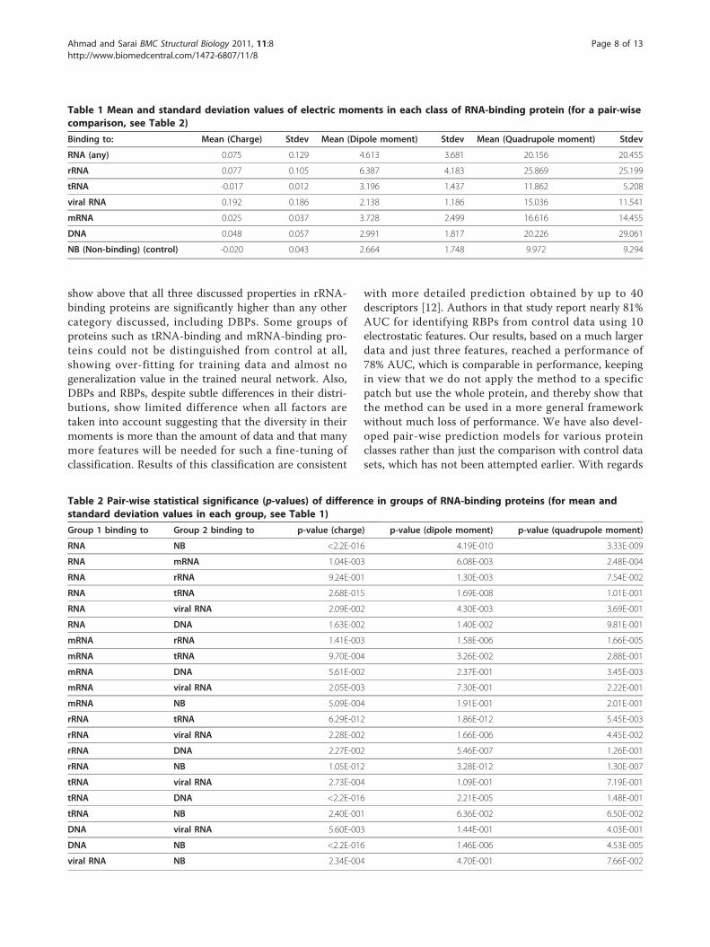
Table 2 Pair-wise statistical significance (p-values) of difference in groups of RNA-binding proteins (for mean andstandard deviation values in each group, see Table 1)
Group 1 binding to Group 2 binding to p-value (charge) p-value (dipole moment) p-value (quadrupole moment)
RNA NB <2.2E-016 4.19E-010 3.33E-009
RNA mRNA 1.04E-003 6.08E-003 2.48E-004
RNA rRNA 9.24E-001 1.30E-003 7.54E-002
RNA tRNA 2.68E-015 1.69E-008 1.01E-001
RNA viral RNA 2.09E-002 4.30E-003 3.69E-001
RNA DNA 1.63E-002 1.40E-002 9.81E-001
mRNA rRNA 1.41E-003 1.58E-006 1.66E-005
mRNA tRNA 9.70E-004 3.26E-002 2.88E-001
mRNA DNA 5.61E-002 2.37E-001 3.45E-003
mRNA viral RNA 2.05E-003 7.30E-001 2.22E-001
mRNA NB 5.09E-004 1.91E-001 2.01E-001
rRNA tRNA 6.29E-012 1.86E-012 5.45E-003
rRNA viral RNA 2.28E-002 1.66E-006 4.45E-002
rRNA DNA 2.27E-002 5.46E-007 1.26E-001
rRNA NB 1.05E-012 3.28E-012 1.30E-007
tRNA viral RNA 2.73E-004 1.09E-001 7.19E-001
tRNA DNA <2.2E-016 2.21E-005 1.48E-001
tRNA NB 2.40E-001 6.36E-002 6.50E-002
DNA viral RNA 5.60E-003 1.44E-001 4.03E-001
DNA NB <2.2E-016 1.46E-006 4.53E-005
viral RNA NB 2.34E-004 4.70E-001 7.66E-002
Table 1 Mean and standard deviation values of electric moments in each class of RNA-binding protein (for a pair-wisecomparison, see Table 2)
Binding to: Mean (Charge) Stdev Mean (Dipole moment) Stdev Mean (Quadrupole moment) Stdev
RNA (any) 0.075 0.129 4.613 3.681 20.156 20.455
rRNA 0.077 0.105 6.387 4.183 25.869 25.199
tRNA -0.017 0.012 3.196 1.437 11.862 5.208
viral RNA 0.192 0.186 2.138 1.186 15.036 11.541
mRNA 0.025 0.037 3.728 2.499 16.616 14.455
DNA 0.048 0.057 2.991 1.817 20.226 29.061
NB (Non-binding) (control) -0.020 0.043 2.664 1.748 9.972 9.294
Ahmad and Sarai BMC Structural Biology 2011, 11:8http://www.biomedcentral.com/1472-6807/11/8
Page 8 of 13
to the discrimination between DBPs and RBPs, we reachthe same conclusion as [12], i.e. these two classes cannotbe distinguished from each other with much confidence.
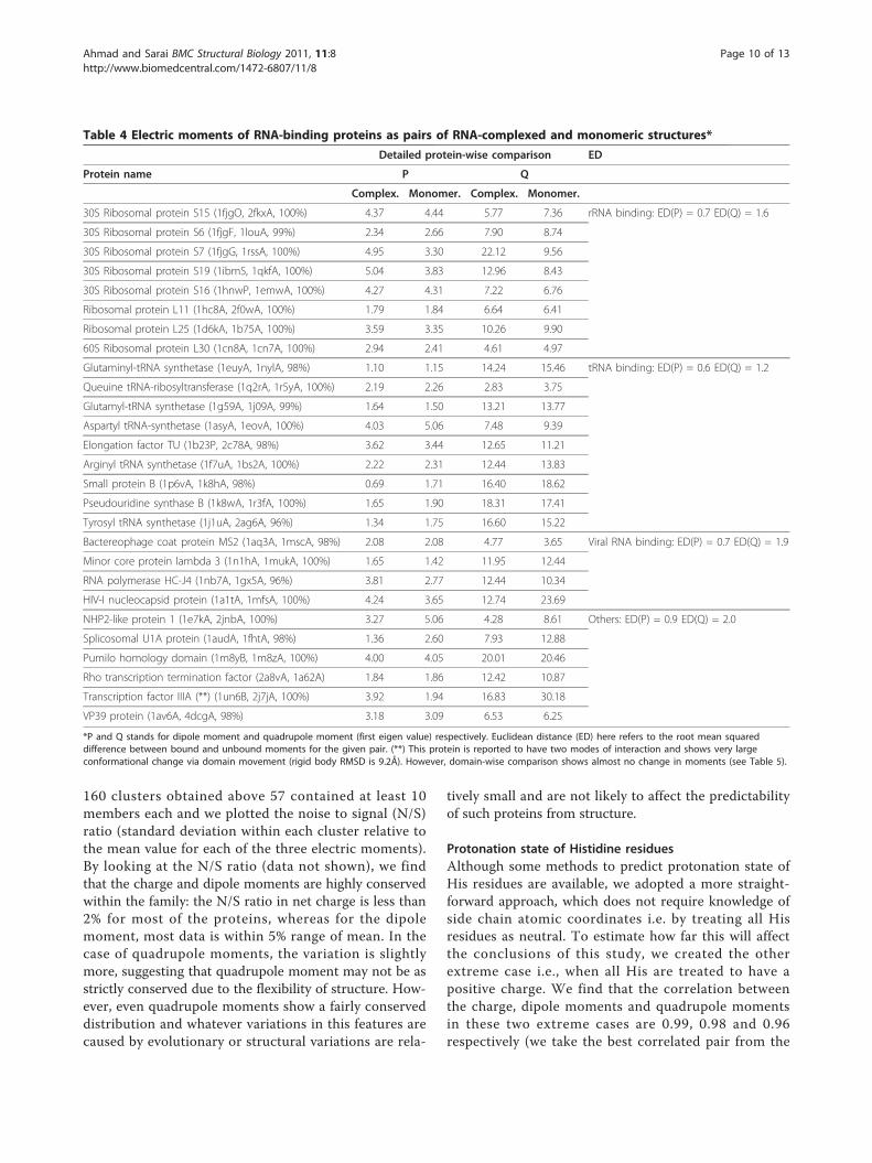
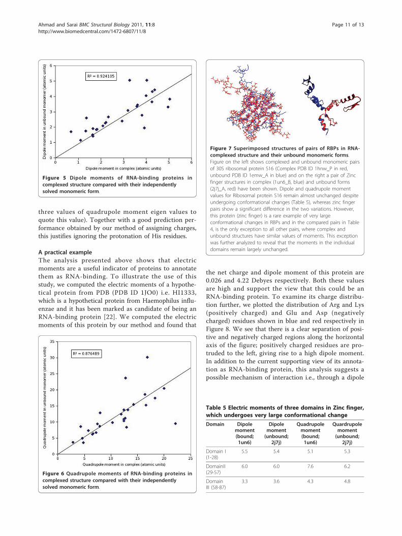
Bound versus unbound statesMost solved as well as modeled protein structures comefrom monomeric forms and formation of a full complexfrom isolated RBPs may lead to structural changes, whichmay make the predictions performed on complex-derivedstructures questionable. Prima facie, it appears that the cal-culations of moments using low-resolution structure infor-mation (Ca atoms only) will be more robust than existingmethods utilizing side-chain coordinates coming fromcomplexes at high resolution. To assess the validity of thisintuitive argument, we compiled a list of structure-pairs ofRBPs: one member of each pair came from the complexand the other from a monomer with no other protein orRNA (this may involve conformational changes by protein-protein or protein-RNA interactions). Detailed procedurefor selecting pairs is described in Methods. Table 4 givesdetailed comparison between the dipole moment andquadrupole moment of these structure pairs (charge isobviously identical in the two cases). Figure 5 and 6 showthe scatterplots of dipole and quadrupole momentsobserved in monomeric and complexed structures. It isclear that correlation coefficients are close to 1 for bothdipole and quadrupole moment values. Clearly, the calcula-tion of moments from Ca atoms only makes the
procedures far more robust than any other electrostaticproperty calculated from full atomic coordinates. Figure 7shows a typical pair of structures as well as an exceptionalcase with very large conformational change in complex for-mation. Figure on the left shows complexed and unboundmonomeric pairs of 30S ribosomal protein S16 (ComplexPDB ID 1hnw_P, unbound PDB ID 1emw_A), and on theright a pair of Zinc finger structures in complex (1un6_B)and unbound forms (2j7j_A) are shown. Dipole and quad-rupole moment values for Ribosomal protein S16 remainalmost unchanged despite undergoing conformationalchanges (Table 4), whereas zinc finger pairs show a signifi-cant difference in the two variations. However, this proteinhas been shown to have two modes of binding via changesin domain orientations. When the moments of eachdomain were calculated separately, bound and theunbound conformations were found to have very similarmoments, confirming that the dipole and electric momentvalues are fairly robust against small conformationalchanges induced by complex formation (see Table 5).
Evolutionary conservationWe also examined if the electric moments, calculatedabove, remain conserved among homologous or similarRNA-binding proteins. To evaluate this, we returned tothe overall data of RNA-binding proteins (before remov-ing redundancy), obtained during the process of selectingrepresentative non-redundant set of 160 proteins. Out of
Table 3 Neural network performance to discriminate between proteins binding to different types of RNA based oncharge, dipole and quadrupole moments*
Positive classbinding to
Negative classbinding to
Number of proteins in +ve class
Number of proteins in-ve class
AUC F1 Precision Recall Accuracy
RNA NB 160 2441 0.78 0.37 0.31 0.45 0.91
rRNA NB 84 2441 0.79 0.26 0.23 0.30 0.94
tRNA NB 20 2441 0.42 0.02 0.01 1.00 0.03
vRNA NB 17 2441 0.75 0.24 0.24 0.24 0.99
mRNA NB 13 2441 0.10 0.01 0.01 1.00 0.02
tRNA rRNA 20 84 0.70 0.45 0.32 0.75 0.64
mRNA rRNA 13 84 0.56 0.30 0.18 1.00 0.37
vRNA rRNA 17 84 0.44 0.32 0.19 1.00 0.28
mRNA tRNA 13 2441 0.07 0.57 0.39 1.00 0.39
mRNA vRNA 13 2441 0.02 0.60 0.43 1.00 0.43
tRNA vRNA 20 17 0.19 0.63 0.46 1.00 0.46
DNA NB 143 2441 0.72 0.22 0.20 0.26 0.90
RNA DNA 160 143 0.58 0.69 0.53 1.00 0.53
rRNA DNA 84 143 0.74 0.64 0.52 0.83 0.65
tRNA DNA 20 143 0.33 0.24 0.13 1.00 0.20
mRNA DNA 13 143 0.07 0.16 0.09 1.00 0.14
* AUC is area under the ROC curve, F-measure (F1) is the highest geometric mean of precision and recall and accuracy is number of correct predictions relativeto all predictions at peak F-measure. In all cases, neural network with three units in the hidden layer was used for training in a leave-one-out procedure and thetraining was performed for a fixed number of epochs without using information from left-out protein.
Ahmad and Sarai BMC Structural Biology 2011, 11:8http://www.biomedcentral.com/1472-6807/11/8
Page 9 of 13
160 clusters obtained above 57 contained at least 10members each and we plotted the noise to signal (N/S)ratio (standard deviation within each cluster relative tothe mean value for each of the three electric moments).By looking at the N/S ratio (data not shown), we findthat the charge and dipole moments are highly conservedwithin the family: the N/S ratio in net charge is less than2% for most of the proteins, whereas for the dipolemoment, most data is within 5% range of mean. In thecase of quadrupole moments, the variation is slightlymore, suggesting that quadrupole moment may not be asstrictly conserved due to the flexibility of structure. How-ever, even quadrupole moments show a fairly conserveddistribution and whatever variations in this features arecaused by evolutionary or structural variations are rela-
tively small and are not likely to affect the predictabilityof such proteins from structure.
Protonation state of Histidine residuesAlthough some methods to predict protonation state ofHis residues are available, we adopted a more straight-forward approach, which does not require knowledge ofside chain atomic coordinates i.e. by treating all Hisresidues as neutral. To estimate how far this will affectthe conclusions of this study, we created the otherextreme case i.e., when all His are treated to have apositive charge. We find that the correlation betweenthe charge, dipole moments and quadrupole momentsin these two extreme cases are 0.99, 0.98 and 0.96respectively (we take the best correlated pair from the
Table 4 Electric moments of RNA-binding proteins as pairs of RNA-complexed and monomeric structures*
Detailed protein-wise comparison ED
Protein name P Q
Complex. Monomer. Complex. Monomer.
30S Ribosomal protein S15 (1fjgO, 2fkxA, 100%) 4.37 4.44 5.77 7.36 rRNA binding: ED(P) = 0.7 ED(Q) = 1.6
30S Ribosomal protein S6 (1fjgF, 1louA, 99%) 2.34 2.66 7.90 8.74
30S Ribosomal protein S7 (1fjgG, 1rssA, 100%) 4.95 3.30 22.12 9.56
30S Ribosomal protein S19 (1ibmS, 1qkfA, 100%) 5.04 3.83 12.96 8.43
30S Ribosomal protein S16 (1hnwP, 1emwA, 100%) 4.27 4.31 7.22 6.76
Ribosomal protein L11 (1hc8A, 2f0wA, 100%) 1.79 1.84 6.64 6.41
Ribosomal protein L25 (1d6kA, 1b75A, 100%) 3.59 3.35 10.26 9.90
60S Ribosomal protein L30 (1cn8A, 1cn7A, 100%) 2.94 2.41 4.61 4.97
Glutaminyl-tRNA synthetase (1euyA, 1nylA, 98%) 1.10 1.15 14.24 15.46 tRNA binding: ED(P) = 0.6 ED(Q) = 1.2
Queuine tRNA-ribosyltransferase (1q2rA, 1r5yA, 100%) 2.19 2.26 2.83 3.75
Glutamyl-tRNA synthetase (1g59A, 1j09A, 99%) 1.64 1.50 13.21 13.77
Aspartyl tRNA-synthetase (1asyA, 1eovA, 100%) 4.03 5.06 7.48 9.39
Elongation factor TU (1b23P, 2c78A, 98%) 3.62 3.44 12.65 11.21
Arginyl tRNA synthetase (1f7uA, 1bs2A, 100%) 2.22 2.31 12.44 13.83
Small protein B (1p6vA, 1k8hA, 98%) 0.69 1.71 16.40 18.62
Pseudouridine synthase B (1k8wA, 1r3fA, 100%) 1.65 1.90 18.31 17.41
Tyrosyl tRNA synthetase (1j1uA, 2ag6A, 96%) 1.34 1.75 16.60 15.22
Bactereophage coat protein MS2 (1aq3A, 1mscA, 98%) 2.08 2.08 4.77 3.65 Viral RNA binding: ED(P) = 0.7 ED(Q) = 1.9
Minor core protein lambda 3 (1n1hA, 1mukA, 100%) 1.65 1.42 11.95 12.44
RNA polymerase HC-J4 (1nb7A, 1gx5A, 96%) 3.81 2.77 12.44 10.34
HIV-I nucleocapsid protein (1a1tA, 1mfsA, 100%) 4.24 3.65 12.74 23.69
NHP2-like protein 1 (1e7kA, 2jnbA, 100%) 3.27 5.06 4.28 8.61 Others: ED(P) = 0.9 ED(Q) = 2.0
Splicosomal U1A protein (1audA, 1fhtA, 98%) 1.36 2.60 7.93 12.88
Pumilo homology domain (1m8yB, 1m8zA, 100%) 4.00 4.05 20.01 20.46
Rho transcription termination factor (2a8vA, 1a62A) 1.84 1.86 12.42 10.87
Transcription factor IIIA (**) (1un6B, 2j7jA, 100%) 3.92 1.94 16.83 30.18
VP39 protein (1av6A, 4dcgA, 98%) 3.18 3.09 6.53 6.25
*P and Q stands for dipole moment and quadrupole moment (first eigen value) respectively. Euclidean distance (ED) here refers to the root mean squareddifference between bound and unbound moments for the given pair. (**) This protein is reported to have two modes of interaction and shows very largeconformational change via domain movement (rigid body RMSD is 9.2Å). However, domain-wise comparison shows almost no change in moments (see Table 5).
Ahmad and Sarai BMC Structural Biology 2011, 11:8http://www.biomedcentral.com/1472-6807/11/8
Page 10 of 13
three values of quadrupole moment eigen values toquote this value). Together with a good prediction per-formance obtained by our method of assigning charges,this justifies ignoring the protonation of His residues.
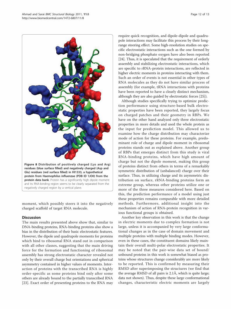
A practical exampleThe analysis presented above shows that electricmoments are a useful indicator of proteins to annotatethem as RNA-binding. To illustrate the use of thisstudy, we computed the electric moments of a hypothe-tical protein from PDB (PDB ID 1JO0) i.e. HI1333,which is a hypothetical protein from Haemophilus influ-enzae and it has been marked as candidate of being anRNA-binding protein [22]. We computed the electricmoments of this protein by our method and found that
the net charge and dipole moment of this protein are0.026 and 4.22 Debyes respectively. Both these valuesare high and support the view that this could be anRNA-binding protein. To examine its charge distribu-tion further, we plotted the distribution of Arg and Lys(positively charged) and Glu and Asp (negativelycharged) residues shown in blue and red respectively inFigure 8. We see that there is a clear separation of posi-tive and negatively charged regions along the horizontalaxis of the figure; positively charged residues are pro-truded to the left, giving rise to a high dipole moment.In addition to the current supporting view of its annota-tion as RNA-binding protein, this analysis suggests apossible mechanism of interaction i.e., through a dipole
Figure 5 Dipole moments of RNA-binding proteins incomplexed structure compared with their independentlysolved monomeric form.
Figure 6 Quadrupole moments of RNA-binding proteins incomplexed structure compared with their independentlysolved monomeric form.
Figure 7 Superimposed structures of pairs of RBPs in RNA-complexed structure and their unbound monomeric forms.Figure on the left shows complexed and unbound monomeric pairsof 30S ribosomal protein S16 (Complex PDB ID 1hnw_P in red,unbound PDB ID 1emw_A in blue) and on the right a pair of Zincfinger structures in complex (1un6_B, blue) and unbound forms(2j7j_A, red) have been shown. Dipole and quadrupole momentvalues for Ribosomal protein S16 remain almost unchanged despiteundergoing conformational changes (Table 5), whereas zinc fingerpairs show a significant difference in the two variations. However,this protein (zinc finger) is a rare example of very largeconformational changes in RBPs and in the compared pairs in Table4, is the only exception to all other pairs, where complex andunbound structures have similar values of moments. This exceptionwas further analyzed to reveal that the moments in the individualdomains remain largely unchanged.
Table 5 Electric moments of three domains in Zinc finger,which undergoes very large conformational change
Domain Dipolemoment(bound;1un6)
Dipolemoment(unbound;
2j7j)
Quadrupolemoment(bound;1un6)
Quardrupolemoment(unbound;
2j7j)
Domain I(1-28)
5.5 5.4 5.1 5.3
DomainII(29-57)
6.0 6.0 7.6 6.2
DomainIII (58-87)
3.3 3.6 4.3 4.8
Ahmad and Sarai BMC Structural Biology 2011, 11:8http://www.biomedcentral.com/1472-6807/11/8
Page 11 of 13
moment, which possibly steers it into the negativelycharged scaffold of target RNA molecule.
DiscussionThe main results presented above show that, similar toDNA-binding proteins, RNA-binding proteins also show abias in the distribution of their basic electrostatic features.However, the dipole and quadrupole moments for proteinswhich bind to ribosomal RNA stand out in comparisonwith all other classes, suggesting that the main drivingforce for the formation and functioning of ribosomalassembly has strong electrostatic character revealed notonly by their overall charge but orientations and sphericalasymmetry contained in higher values of moments. Inter-action of proteins with the transcribed RNA is highlyorder-specific as some proteins bind only after someothers are already bound to the partially transcribed RNA[23]. Exact order of presenting proteins to the RNA may
require quick recognition, and dipole-dipole and quadru-pole interactions may facilitate this process by their long-range steering effect. Some high-resolution studies on spe-cific electrostatic interactions such as the one formed bynon-bridging phosphate oxygen have also been reported[24]. Thus, it is speculated that the requirement of orderlyassembly and stabilizing electrostatic interactions, whichare specific to rRNA-protein interactions, are reflected inhigher electric moments in proteins interacting with them.Such an order of events is not essential in other types ofRNA molecules as they do not have similar process ofassembly (for example, tRNA interactions with proteinshave been reported to have a clearly distinct mechanism,although they are also guided by electrostatic forces [25]).Although studies specifically trying to optimize predic-
tion performance using structure-based bulk electro-static properties have been reported, they largely focuson charged patches and their geometry in RBPs. Wehave on the other hand analyzed only three electrostaticproperties in more details and used the whole protein asthe input for prediction model. This allowed us toexamine how the charge distribution may characterizemode of action for these proteins. For example, predo-minant role of charge and dipole moment in ribosomalproteins stands out as explained above. Another groupof RBPs that emerges distinct from this study is viralRNA-binding proteins, which have high amount ofcharge but not the dipole moment, making this groupof proteins distinct from others in terms of a remarkablysymmetric distribution of (unbalanced) charge over theirsurface. Thus, in utilizing charge and its asymmetric dis-tribution on surface, rRNA-binding proteins form anextreme group, whereas other proteins utilize one ormore of the three measures considered here. Based onthis, the prediction performance of a model using justthese properties remains comparable with more detailedmethods. Furthermore, additional insight into themechanism of action of RNA-protein recognition in var-ious functional groups is obtained.Another key observation in this work is that the change
in electric moments due to complex formation is notlarge, unless it is accompanied by very large conforma-tional changes as in the case of domain movement andmultiple proteins with multiple binding modes. However,even in these cases, the constituent domains likely main-tain their overall multi-polar electrostatic properties. Itmay be noted that the pair-wise data set of bound/unbound proteins in this work is somewhat biased as pro-teins whose structures change considerably are more likelyto be reported. This is confirmed by measuring theirRMSD after superimposing the structures (we find thatthe average RMSD of all pairs is 2.1Å, which is quite large;data not shown). Thus, despite these large conformationalchanges, characteristic electric moments are largely
Figure 8 Distribution of positively charged (Lys and Arg)residues (blue surface filled) and negatively charged (Asp andGlu) residues (red surface filled) in HI1333, a hypotheticalprotein from Haemophilus influenzae (PDB ID 1JO0) from theprotein data bank. Protein has a significantly high dipole momentand its RNA-binding region seems to be clearly separated from thenegatively charged region by a vertical plane.
Ahmad and Sarai BMC Structural Biology 2011, 11:8http://www.biomedcentral.com/1472-6807/11/8
Page 12 of 13
preserved, probably helping in long-range interactionsresulting in appropriate energy landscape for recognitionby steering.We observe that the three electric moments are fairly
conserved in evolution, and even sequence similaritybeing as low as 25%, RNA-binding proteins within thesame cluster seem to have very similar electricmoments, suggesting that the three properties may beuniversally employed for protein-RNA recognition.Finally, this method has been rigorously cross-
validated on known protein structures of RNA-bindingproteins. However, the most useful application of themethod would be to annotate proteins from their mod-eled structures. Unfortunately, a readily available publicdata of modeled structures with RNA-binding annota-tions was not available at the time of this study. Thus,all performance measures presented here correspond toreal structures (although with very lenient requirementsof resolution). Benchmarking performance on highthroughput modeled structures remains an area forfurther investigation.
ConclusionsRNA-binding proteins have distinct patterns of netcharge, dipole and quadrupole moments, which can beutilized to rapidly identify them and to some degreedetermine their structure class. This information is pre-sent even at a low-resolution level, as moments calcu-lated from only main-chain coordinates can be utilizedfor prediction. This method is also robust against con-formational changes, as well as evolutionary variationsin protein structures.
Additional material
Additional file 1: Electric Moments of RNA-binding proteins. Charge(q), dipole moment (p) and quadrupole moments (three eigen values,Q1, Q2, Q3) of RNA-binding proteins. DNA-binding proteins and controlproteins are also included.
AcknowledgementsA.S. acknowledges support by Grants-in-Aid for Scientific Research(20016022, 21310131) from Ministry of Education, Culture, Sports, Scienceand Technology in Japan. S.A. acknowledges support by Grants-in-Aid forScientific Research (Kaken-hi #22500277) from JSPS, Japan.
Author details1National Institute of Biomedical Innovation, 7-6-8, Saito-asagi, Ibaraki, Osaka,Japan. 2Department of Bioscience and Bioinformatics, Kyushu Institute ofTechnology, Iizuka, Fukuoka, 820-8502 Japan.
Authors’ contributionsThis project was jointly conceived by both authors (SA and AS), as part oftheir ongoing collaborations. Detailed experimental design andimplementation were carried out by SA. Manuscript preparation and analysisof results were carried out by SA in consultation with and suggestions fromAS. Both authors read and approved the manuscript.
Received: 29 June 2010 Accepted: 1 February 2011Published: 1 February 2011
References1. Draper D: Protein-RNA recognition. Annu Rev Biochem 1995, 64:593-620.2. de Guzman R, Turner R, Summers M: Protein-RNA recognition. Biopolymers
1998, 48:181-195.3. Jones S, Daley D, Luscombe N, Berman H, Thornton J: Protein-RNA
interactions: a structural analysis. Nucleic Acids Research 2001, 29:943-954.4. Chen Y, Varani G: Protein families and RNA recognition. FEBS J 2005,
272:2088-2097.5. Sanchez-Diaz P, Penalva L: Post-transcription meets post-genomic: the
saga of RNA binding proteins in a new era. RNA Biol 2006, 3:101-109.6. Keene J: RNA regulons: coordination of post-transcriptional events.
Nature Rev Genet 2007, 8:533-543.7. Lunde B, Moore C, Varani G: RNA-binding proteins: modular design for
efficient function. Nature Rev Mol Cell Biol 2007, 8:479-490.8. Yu X, Cao J, Cai Y, Shi T, Li Y: Predicting rrna-, rna-, and DNA-binding
proteins from primary structure with support vector machines. J TheorBiol 2006, 240:175-184.
9. Terribilini M, Sander J, Lee J, Zaback P, Jernigan R, Vasant H, Drena D:RNABindR: a server for analyzing and predicting RNA-binding sites inproteins. Nucleic Acids Research 2007, 35:W578-W584.
10. Chen YC, Lim C: Predicting RNA-binding sites from the protein structurebased on electrostatics, evolution and geometry. Nucleic Acids Research2008, 36(5):e29.
11. Ellis JJ, Broom M, Jones S: Protein-RNA interactions: structural analysisand functional classes. Proteins 2007, 66:903-911.
12. Shazman S, Gutfreund YM: Classifying RNA-Binding proteins based onelectrostatic properties. PLoS Comput Biol 2008, 4(8):e1000146.
13. Ahmad S, Sarai A: Moment-based prediction of DNA-binding proteins.J Mol Biol 2004, 341:65-71.
14. Szilagyi A, J S: Efficient prediction of nucleic acid binding function fromlow-resolution protein structures. J Mol Biol 2006, 358:922-933.
15. Felder CE, Prilusky J, Silman I, Sussman JL: A server and database fordipole moments of proteins. Nucleic Acids Research 2007, 35:W512-W521.
16. Klosterman PS, Tamura M, Holbrook SR, Brenner SE: SCOR: a structuralclassification of RNA database. Nucleic Acids Research 2002, 30:392-394.
17. Altschul SF, Gish W, Miller W, Myers EW, Lipman DJ: Basic local alignmentsearch tool. J Mol Biol 1990, 215:403-410.
18. Berman HM, Henrick K, Nakamura H: Announcing the worldwide ProteinData Bank. Nature Structural Biology 2003, 10(12):980.
19. Gordon JC, Myers JB, Folta T, Shoja V, Heath LS, Onufriev A: H++: a serverfor estimating pKas and adding missing hydrogens to macromolecules.Nucleic Acids Research 2005, 33:W368-W371.
20. Ahmad S, Gromiha MM, Sarai A: Analysis and prediction of DNA-bindingproteins and their binding residues based on composition, sequenceand structural information. Bioinformatics 2004, 20:477-486.
21. Ahmad S, Keskin O, Sarai A, Nussinov R: Protein-DNA interactions:structural, thermodynamic and clustering patterns of conserved residuesin DNA-binding proteins. Nucleic Acids Research 2008, 36(18):5922-5932.
22. Willis M, Krajewski W, Chalamasetty V, Reddy P, Howard A, Herzberg O:Structure of HI1333 (YhbY), a putative RNA-binding protein fromHaemophilus influenzae. Proteins 2002, 49(3):423-426.
23. Williamson JR: After the ribosome structures: How are the subunitsassembled. RNA 2003, 9:165-167.
24. Ghosh S, Joseph S: Non-bridging phosphate oxygen in 16S rRNAimportant for 30S subunit assembly and association with the 50Sribosomal subunit. RNA 2005, 11:657-667.
25. Tworowskia D, Feldmana AV, Safro MG: Electrostatic Potential ofAminoacyl-tRNA Synthetase Navigates tRNA on its Pathway to theBinding Site. Journal of Molecular Biology 2005, 350(5):866-882.
doi:10.1186/1472-6807-11-8Cite this article as: Ahmad and Sarai: Analysis of electric moments ofRNA-binding proteins: implications for mechanism and prediction. BMCStructural Biology 2011 11:8.
Ahmad and Sarai BMC Structural Biology 2011, 11:8http://www.biomedcentral.com/1472-6807/11/8
Page 13 of 13