Embed Size (px)
Citation preview

3435Research Article
IntroductionGap-junction-mediated intercellular communication facilitatesdirect communication among adjacent cells by allowingpassage of ions and small metabolites (White and Paul, 1999;Saez et al., 2003; Sohl and Willecke, 2004). Vertebrate gapjunctions, composed of integral membrane proteins from theConnexin gene family, are crucially important in regulatingembryonic development, coordinated contraction of excitablecells, tissue homeostasis, normal cell growth anddifferentiation (Saez et al., 2003; Sohl and Willecke, 2004).Furthermore, connexin mutations have been linked to severaldiseases (Bergoffen et al., 1993; Gong et al., 1997; Kelsell etal., 1997) including oculodentodigital dysplasia, a diseaselinked to Connexin43 (Cx43) mutations that can causeatrioseptal defects and arrhythmias (Paznekas et al., 2003).Twenty-one connexin genes have been identified in humans(Sohl and Willecke, 2004). During intercellular channelformation, six connexin proteins oligomerize into a hemi-channel or connexon; connexons are then transported to theplasma membrane by as yet unknown mechanisms. The intactchannel is formed when one hemi-channel docks with a secondin an opposing cell. Once assembled, groups of theseintercellular channels (termed gap junctional plaques) mediatethe diffusion of ions, amino acids, second messengers andother metabolites between the cytoplasms of the two cells(White and Paul, 1999; Sohl and Willecke, 2004). The channels
can be gated in response to various stimuli, including changesin voltage, pH and connexin phosphorylation. Regulation ofgap junctional communication could occur by controlling anyone of the steps mentioned above, however, many of theregulatory mechanisms underlying these events remain elusive.
Cx43, the most ubiquitously expressed connexin, isdifferentially phosphorylated at a dozen or more serineresidues throughout its life cycle (Lampe and Lau, 2004). Cx43from cultured cells commonly demonstrates multipleelectrophoretic isoforms when analyzed by SDS-PAGE: a fast-migrating form (sometimes referred to as P0 or NP) thatincludes the non-phosphorylated isoform, and multiple slow-migrating forms (sometimes termed P1 and P2) (Musiland Goodenough, 1991). Following alkaline phosphatasetreatment, the phosphorylated species collapse to the fastestmigrating form, suggesting that phosphorylation is the primarycovalent modification detected in SDS-PAGE analysis,although no assignment of specific phosphorylation sites to achange in Cx43 migration has been made. Pulse-chase studieswith Brefeldin A indicate some Cx43 phosphorylation occursprior to reaching the plasma membrane (Laird et al., 1995).This phosphorylation event might be necessary for maintaininghemi-channels in their closed state until docking occurs (Baoet al., 2004). In addition, studies investigating phosphorylationin normal rat kidney (NRK) cells show that, Cx43 acquiresresistance to Triton X-100 once it has been phosphorylated to
The functional consequences of Connexin43 (Cx43)phosphorylation remain largely unexplored. Using anantibody that specifically recognizes Cx43 phosphorylatedat serine residues 325, 328 and/or 330 (pS325/328/330-Cx43), we show that labeling of this form of Cx43 as wellas of total Cx43 is restricted to the intercalated disk regionof normal ventricular tissue. In ischemic heart, significantrelocalization of total Cx43 to the lateral edges of myocyteswas evident; however pS325/328/330-Cx43 remainedpredominately at the intercalated disk. Western blotsindicated a eightfold decrease in pS325/328/330-Cx43in ischemic tissue. Peptide-binding- and competition-experiments indicated that our antibody mainly detectedCx43 phosphorylated at S328 and/or S330 in heart tissue.To evaluate how this change in Cx43 phosphorylationcontributes to ischemia-induced downregulation of
intercellular communication, we stably transfected Cx43–/–
cells with a Cx43 construct in which serine residues 325,328 and 330 had been mutated to alanine (Cx43-TM).Cx43-TM was not efficiently processed to isoforms thathave been correlated with gap junction assembly.Nevertheless, Cx43-TM cells were electrically coupled,although development of coupling was delayed. Fullyopened channels were only rarely observed in Cx43-TMcells, and Lucifer-Yellow-dye-coupling was significantlyreduced compared with wild-type cells. These data suggestthat phosphorylation of Cx43 at serine residues 325, 328and/or 330 influences channel permselectivity andregulates the efficiency of gap junction assembly.
Key words: Connexin43, Gap junction, Heart, Ischemia,Phosphorylation
Summary
Analysis of Connexin43 phosphorylated at S325, S328and S330 in normoxic and ischemic heartPaul D. Lampe1,*, Cynthia D. Cooper1, Timothy J. King1,‡ and Janis M. Burt2
1Molecular Diagnostics Program, Fred Hutchinson Cancer Research Center and Department of Pathobiology, University of Washington, 1100Fairview Avenue N., M5C800, P.O. Box 19024, Seattle, WA 98109, USA2Department of Physiology, University of Arizona, Tucson, AZ*Author for correspondence (e-mail: [email protected])‡Present address: Hawaii Biotech, Inc., Aiea, HI, USA
Accepted 7 June 2006Journal of Cell Science 119, 3435-3442 Published by The Company of Biologists 2006doi:10.1242/jcs.03089
Jour
nal o
f Cel
l Sci
ence
3436
the slower migrating isoforms and assembled into gap junctionplaques (Musil and Goodenough, 1991). Thus, uncharacterizedphosphorylation events have been correlated with changes inassembly, acquisition of Triton-X-100-insolubility and,potentially, degradation of Cx43 gap junction channels.
In the normally functioning ventricle, Cx43 is localized tointercalated disks where it supports the longitudinal andtransverse spread of the action potential resulting incoordinated contractile activation. Myocardial ischemia leadsto Cx43 dephosphorylation and loss of localization to theintercalated disk, which probably contributes to contractilefailure and arrhythmias (Beardslee et al., 2000; Schulz et al.,2003). Casein kinase 1 (CK1) mediates phosphorylation ofCx43 at serine residues 325, 328 and/or 330 in vitro. Incultured cells these sites are routinely phosphorylated;inhibition of CK1 reduces phosphorylation at these sites andreduces gap junction assembly (Cooper and Lampe, 2002). Wesought to determine here whether phosphorylation at these sitesoccurs in heart tissue and whether this phosphorylation eventis affected during ischemia. Using an antibody specific forCx43 when phosphorylated at serine residues 325, 328 and/or330, we show that Cx43 localized to intercalated disks isphosphorylated at one or more of these residues (probablyS328 and S330) and that ischemia leads to loss of thisphosphorylation and relocalization of the protein. Furthermore,we show that mutation of these sites increases the migration ofthe phosphorylated Cx43 isoforms on SDS-PAGE, reduces theextent of gap junction formation and reduces gap junctionalcommunication.
ResultsIschemia-induced dephosphorylation of Cx43Previous studies have shown that the electrophoretic mobilityof Cx43 isolated from ischemic heart is increased, such thatmost of the protein migrates at a position that includesdephosphorylated Cx43 so the assumption has been made thatthese changes in electrophoretic mobility represent a reductionin phosphorylation at undefined sites (Beardslee et al., 2000;Jain et al., 2003). In addition, inhibition of CK1 has beenshown to reduce gap junction assembly and to phosphorylateCx43 in vitro at one or more of serines 325/328/330 (Cooperand Lampe, 2002). We hypothesized that phosphorylation atsites necessary for efficient assembly of channels might becompromised in the ischemic setting. To evaluate whetherthese phosphorylation events occur in control tissue andischemic heart tissue, we generated a polyclonal antibody thatuniquely detects Cx43 phosphorylated at serine residues 325,328 and/or 330 (pS325/328/330-Cx43) – the specificity ofwhich is described in the next section.
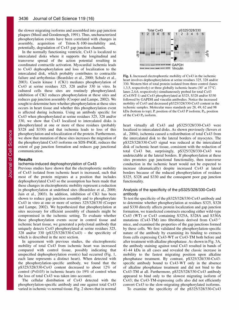
In agreement with previous studies, the electrophoreticmobility of total Cx43 from ischemic heart was increasedcompared with control tissue, possibly indicating thatunspecified dephosphorylation event(s) had occurred (Fig. 1,each lane represents a distinct heart). When detected withthe phosphorylation-specific antibody, we found that thepS325/328/330-Cx43 content decreased to about 12% ofcontrol (P<0.03) in ischemic hearts (to 19% of control whenthe loss of total Cx43 was taken into account).
The cellular distribution of Cx43 detected with thephosphorylation-specific antibody and one against total Cx43varied in ischemic vs normal tissue. Fig. 2 shows that in normal
Journal of Cell Science 119 (16)
heart virtually all Cx43 and pS325/328/330-Cx43 werelocalized to intercalated disks. As shown previously (Severs etal., 2004), ischemia caused a redistribution of total Cx43 fromthe intercalated disk to the lateral borders of myocytes. ThepS325/328/330-Cx43 signal was reduced at the intercalateddisk of ischemic heart tissue, consistent with the reduction oftotal Cx43 but, surprisingly, pS325/328/330-Cx43 wasundetectable at the lateral borders. If phosphorylation at thesesites promotes gap junctional functionality, then transverseconduction in the ischemic heart would not be expected toincrease (dramatically) despite increased Cx43 at lateralborders because of the reduced phosphorylation of residuesS325, S328 and S330 and the consequent poor gap junctionfunctionality.
Analysis of the specificity of the pS325/328/330-Cx43antibodyTo test the specificity of the pS325/328/330-Cx43 antibody andto determine whether phosphorylation at residues S325, S328and S330 directly affects protein localization and gap junctionformation, we transfected constructs encoding either wild-typeCx43 (WT) or Cx43 containing S325A, S328A and S330Amutations (Cx43-TM) into fibroblasts derived from Cx43–/–
mice, and examined the properties of the gap junctions formedby these cells. We first validated the phosphorylation-specificnature of the antibody by examining its binding to extractsfrom cells expressing Cx43-WT or Cx43-TM both before andafter treatment with alkaline phosphatase. As shown in Fig. 3A,the antibody staining against total Cx43 resulted in bands of41-44 kDa in all cases and revealed the classic increase inmobility to the fastest migrating position upon alkalinephosphatase treatment. By contrast, pS325/328/330-Cx43-specific antibody bound to Cx43-WT only in the absenceof alkaline phosphatase treatment and did not bind to theCx43-TM at all. Furthermore, pS325/328/330-Cx43 antibodyappeared to bind only to the slowest migrating isoform ofCx43; the Cx43-TM-expressing cells also did not efficientlyconvert Cx43 to the slow-migrating phosphorylated isoforms.
To examine the specificity of the pS325/328/330-Cx43
Fig. 1. Increased electrophoretic mobility of Cx43 in the ischemicheart involves dephosphorylation at serine residues 325, 328 and/or330. Western blot of total protein isolated from three control (lanes1,3,5, respectively) or three globally ischemic hearts (30’ at 37°C;lanes 2,4,6, respectively) simultaneously probed for total Cx43(Cx43NT-1) and Cx43 phosphorylated at S325, S328 and/or S330followed by GAPDH and vinculin antibodies. Notice the increasedmobility of Cx43 and decreased pS325/328/330-Cx43 content in theischemic samples. Molecular mass standards are 28, 49, 62 and 98kDa (bottom to top); P, position of the Cx43 P isoform; P0, positionof the Cx43 P0 isoform.
Jour
nal o
f Cel
l Sci
ence

3437Cx43 phosphorylation at S328 and S330
antibody and determine which sites are likely to bephosphorylated in vivo, we synthesized three peptides thatwere phosphorylated at one serine residue each, S325, S328 orS330 (pS325, pS328 or pS330, respectively), two peptides thatwere phosphorylated at two serine residues each, S325 andS328, as well as S328 and S330 (pS325/328, as well aspS328/330, respectively), and an immunogen peptide that wasphosphorylated at all three serine residues (pS325/328/330)and covalently bound these peptides to an activated 96-wellplate. The p325/328/330-Cx43 antibody was incubated in thewell and a standard enzyme-linked immunoabsorbent assay(ELISA) was performed (Fig. 3B, open bars). The pS328/330peptide bound almost as much total antibody as theimmunogen pS325/328/330 peptide, indicating that most of theantibody bound an epitope centered on these sites. The singlyphosphorylated peptides bound only a small amount ofantibody. To further answer the question of the specificity ofthis antibody preparation, we removed the antibody that boundto the p328/330 peptide and purified the one specific forpS325/328. Analysis of the binding of this preparation to thesix peptides in the ELISA format indicated that the pS325/328-
purified antibody indeed mostly bound to the pS325/328peptide, followed by the triply phosphorylated and the pS325singly phosphorylated peptides (data not shown). These resultsindicate that the antibody preparation is phosphorylation-specific although polyclonal with reactivity to all three sites.To examine the ability of the six peptides to block antibodybinding to Cx43, we preincubated these peptides with thepS325/328/330-Cx43 antibody prior to western blot analysisof heart lysates. Neither pS325 nor pS328 of the singlyphosphorylated peptides interfered significantly with antibodybinding to intact Cx43, leaving most of the singal as before,whereas the singly phosphorylated pS330 peptide and all of themore extensively phosphorylated peptides did reduce the signalon the western blot close to background levels (Fig. 3B, filledbars). Further, we tested the ability of the pS325/328-specificantibody and found that it could bind to Cx43 prepared fromheart in an immunoblot – albeit much less extensively than theantibody with the pS328/330 reactivity (data not shown).
Fig. 2. Differential phosphorylation of Cx43 localized at intercalateddisks vs lateral edges of myocytes in the normal and ischemic heart.The localization of total Cx43 (Sigma C6219) and pS325/328/330-Cx43 in normal and ischemic heart is shown. Notice the increase inCx43 at the lateral edges of the myocytes of the ischemic heart.Lateral edge Cx43 was not detectably phosphorylated at residuesS325, S328 and/or S330. By contrast, Cx43 at the intercalated disksof ischemic heart retained phosphorylation at residues S325, S328and/or S330 (indicated by arrowheads). Bar, 20 �m.
Fig. 3. The pS325/328/330-Cx43 antibody is specific for the P2isoform of Cx43 and pS328/330. (A) Equal amounts of protein lysatefrom fibroblasts expressing wild-type Cx43 (WT) and Cx43-TM(TM, clone A) were either treated with alkaline phosphatase (+AP)or untreated and probed in a western blot with Cx43NT1 (left panel,Total Cx43) and the pS325/328/330-Cx43 (right panel) antibodies onthe same blot. Molecular mass standards in kDa are marked on theleft side and the P2 isoform is marked with an asterisk. (B) Theamount of antibody bound to the different peptides representingsingly, doubly and triply phosphorylated Cx43 linked to an ELISAwell (open bars) is shown together with the ability of these peptidesto compete with Cx43 present in heart lysates in a westernimmunoblot format (filled bars). Error bars represent the mean ± s.d.
Jour
nal o
f Cel
l Sci
ence

3438
Combined, these antibody-binding and -competition resultsindicate that this polyclonal antibody can bind to Cx43phosphorylated at residues S325, S328 and S330, and also thatCx43 is phosphorylated at S328 and/or S330, and to a lesserextent at S325 in heart tissue.
The role of phosphorylation at S325, S328 and S330 ingap junction assemblyTo be more quantitative about the extent of gap junctionformation, we assayed the ability of three separate Cx43-TMclones to phosphorylate Cx43 to the P2 isoform andincorporate it into a Triton X-100-insoluble fraction, ahallmark of conventional gap junctions (Musil andGoodenough, 1991). Like most cell lines, fibroblastsexpressing wild-type Cx43 showed several isoforms of Cx43in the whole-cell lysate and a dramatic enrichment of the P2isoform in the Triton-insoluble fraction (Fig. 4, WT lanes).Transfectants expressing Cx43 with the TM mutations (clonesA-C) showed multiple differences in their Cx43 migration. Inthe whole-cell fraction, Cx43-TM migrated primarily at P0with a second band between P0 and P1 and no apparent P2.There was a significant reduction in the total amount of Triton-insoluble Cx43 in clone B and C, and essentially no bands thatco-migrated with P2 were observed in any of the clones.Variation in the levels of total Cx43 protein most probablyrepresents clonal heterogeneity among individual Cx43-expressing clones. Since cells expressing Cx43-TM clone Ashowed similar levels of total Cx43 compared with cellsexpressing Cx43-WT, we used clone A for most of thesubsequent studies. We conclude that phosphorylation of Cx43at S325, S328 and/or S330 is necessary for Cx43 to migrate asthe P2 isoform.
The localization of Cx43 in the clones that express Cx43-TM was also examined by immunofluorescence. In Fig. 5, weshow that fibroblasts transfected with Cx43-WT showed bothintracellular and punctate, gap junctional plaque-associatedCx43 [compare to the lack of signal in the knockout (KO)panel]. By contrast, the TM-mutant cell lines showedpredominately intracellular Cx43, although occasional,apparently appositional, labeling was observed (TM-A clone isshown).
We next determined whether cells expressing Cx43-TMsupported intercellular communication. Parental Cx43-knockout (KO) fibroblasts, Cx43-WT-expressing fibroblastsand the three individual Cx43-TM-expressing clones A-C weremicroinjected with Lucifer Yellow dye and evaluated for dyetransfer 3 minutes later. Digital images were taken and thenumber of recipient cells was quantified. As shown in Fig. 6,the number of cells receiving dye from donor cells was 60-85%lower in the Cx43-TM clones than in Cx43-WT-expressingcells and was comparable to the coupling observed in theparental fibroblasts that lacked Cx43. Although Cx43 proteinexpression levels varied between the clones (see Fig. 4), allthree Cx43-TM clones, including TM-A – the one thatexpressed similar levels of Cx43, showed significant reductionsin communication compared with the wild-type control(P<0.02). These data suggest that phosphorylation at S325,S328 and/or S330 is crucial for efficient dye transfer in mousefibroblasts.
We next evaluated whether junctional conductance andchannel behavior differed as a consequence of the Ser to Alamutations at serine residues 325, 328 and 330. Based on thedye-coupling data presented above, we expected junctionalconductance to be significantly reduced in the Cx43-TM cellclones. This was indeed the case for Cx43-TM cells plated for
Journal of Cell Science 119 (16)
Fig. 4. Cx43-TM-expressing cells do not phosphorylate Cx43 toyield the P2 isoform and contain less Triton-insoluble (TI) Cx43.Whole-cell (WC) and TI cellular lysates from cells expressing wild-type Cx43 (WT) or three Cx43-TM clones (TM-A, TM-B, and TM-C) were western blotted and probed for total Cx43 with theCx43NT1 antibody. A darker exposure of lysates of the TM-C cloneis shown. Migration positions of a 50- and 36-kDa marker areindicated with an asterisk and the vinculin loading controls areindicated with a V. The Triton-insoluble vinculin could be associatedwith the cytoskeleton or adhesive junctions.
Fig. 5. Comparison of Cx43 localization in parental knockout (KO)cells, wild-type Cx43 (WT) cells and Cx43-TM (TM-A)-expressingcells. Apparent gap junctions are marked by arrowheads in the WTpanel. Bar, 10 �m.
Fig. 6. Cells expressing Cx43 S325/328/330A are inefficient at dyetransfer. Wild-type (WT)-, parental knockout (KO)- and Cx43-TM(clones TM-A, TM-B and TM-C)-expressing cells weremicroinjected with Lucifer Yellow dye. After 3 minutes of transfer,digital images were taken and the number of recipient cells wasdetermined (n=the number of injected cells). Bars show the meanand error bars ± s.e.m. The extent of dye transfer was significantlydifferent (**P<0.02) for WT compared with all of the other celltypes.
Jour
nal o
f Cel
l Sci
ence

3439Cx43 phosphorylation at S328 and S330
an equivalent period of time as wild-type cells (2-5 hours) –junctional conductance in Cx43-TM cells was 3.5±0.7 nS(n=24) vs 6.2 ±1.1 nS (n=35, P<0.05) in Cx43-WT cells.Coupling between Cx43-TM cells improved with time suchthat 24-30 hours after plating, conductance was 4.9±0.8 nS(n=28), a value not significantly different from Cx43-WT cellsat 2-5 hours post plating. The profile of channel conductancesin the Cx43-TM cells was substantially different from thatobserved in wild-type cells, as shown in Fig. 7A andemphasized in the difference plot (Fig. 7B), with a severelydecreased frequency for activity of fully opened channels(~110 pS) and increased frequency of 60-85–pS channels.
DiscussionPrevious work has established that myocardial ischemiarapidly induces uncharacterized changes in thephosphorylation and localization of Cx43, however, nomechanistic connections between specific phosphorylationevents and changes in conduction or localization have beenidentified. Here, we show that phosphorylation of Cx43 atS325, S328, and/or S330 was drastically reduced in theischemic heart. Further, we demonstrate that Cx43 thatlocalized to the lateral borders of myocytes was not
phosphorylated at these sites, whereas Cx43 that remained atthe intercalated disk retained these modifications. Finally, weshow that, when these sites were not phosphorylated, the eventfrequency for fully opened channels, the overall junctionalconductance and the extent of coupling to Lucifer Yellow dyewere all significantly reduced.
Although direct Cx43 phosphorylation on serine residueshas long been correlated with the formation and functionof gap junctions under basal conditions (Musil andGoodenough, 1991), few mechanistic connections to specificphosphorylation events have been drawn. For instance,phosphorylation at S364 was shown to be important for cAMPinduced upregulation of gap junction assembly. However, cellsexpressing Cx43 with a S364A mutation assembled Cx43 intojunctions almost as well as cells expressing wild-type Cx43,suggesting S364 is not necessary for assembly in homeostaticcells (TenBroek et al., 2001). Similarly, several serines areknown to be involved in mitogen-induced downregulation ofcommunication, including serine residues 255, 279 and 282(MAPK) and serine residues 262 and 368 (protein kinase C),but these phosphorylation events have been reported tonegatively affect channel-gating properties (Lampe et al., 2000;Cottrell et al., 2003). S325, S328 and/or S330 were previouslyshown to be substrates for CK1 phosphorylation, a kinaseimplicated in the regulation of gap junction assembly (Cooperand Lampe, 2002). Our phosphorylation-specific antibodycould react with Cx43 phosphorylated at all three sites in hearttissue. We probed heart lysates with seven other Cx43phosphorylation-specific antibodies, that we had either createdor are available from commercial sources, and that we find tobe specific for phosphorylation at S255, S262, S279-S282,S364, S368 or S372. S325/328/330-Cx43 antibody was theonly one that gave a significant decrease in phosphorylation(results not shown). The antibody against pS368 showedincreased binding upon hypoxia that we have investigatedseparately (Ek-Vitorin et al., 2006). Similar to a previous report(Jeyaraman et al., 2003), we found (data not shown) thatischemia led to increased binding of an anti-Cx43 antibody weprepared against a nonphosphorylated peptide correspondingto residues 360-382 of Cx43 that appears to be identical toZymed 13800, an antibody prepared to a similar epitope thathas been reported to bind dephosphorylated Cx43 (Nagy et al.,1997).
We probed the functional consequences of thesephosphorylation events by expressing Cx43 with Ser to Alaconversions at all three sites (i.e. the Cx43-TM mutant).This mutant, rather than double- or single-site mutants, wasselected for study for two reasons: First, all three sitesappear to be phosphorylated in heart tissue. Second, thefunctional consequences of phosphorylation at clusters of(phosphorylated) serines can be misinterpreted from single-sitemutants because either the remaining sites partially or fullycompensate for the missing site or the remaining sites are notphosphorylated because phosphorylation is sequential with themutated site necessary for subsequent phosphorylation events(as can be the case for CK1) (Flotow et al., 1990).Microinjected Cx43-TM cells and parental (Cx43–/–)fibroblasts were occasionally dye-coupled to one or two cells.By contrast, although electrical coupling between Cx43-TMcells was reduced compared with wild-type cells in the first fewhours after plating, significant coupling was routinely observed
Fig. 7. Histogram of channel events observed in Cx43-TM (gray) andCx43-WT-expressing cells. (A) Event frequency as percent of totalevents in each bin. (B) Difference induced by mutation of serineresidues 325, 328 and 330. Notice the reduced incidence of eventscorresponding to fully opened channels (106±1 pS) and increasedincidence of 75±2 pS. The 54±1 pS population was not differentfrom wild-type and mutant data sets. Difference data were fit usingOrigin software yielding Chi2 and R2 for fit are 5.677 and 0.94,respectively (–WT, 1851 events, n=13; TM, 418 events, n=7).
Jour
nal o
f Cel
l Sci
ence

3440
24-30 hours after plating. This coupling was mediated bychannels with conductances of 70-90 pS, a value intermediateto the fully opened channel (~110 pS) and the previouslyreported, phosphorylation-induced 50-60–pS substate (Lampeet al., 2000). These results are consistent with at least twoexplanations. First, phosphorylation at residues S325, S328and/or S330 is necessary for Cx43 channels to open to theirfully opened state. If correct, the intermediate conductancesubstate of Cx43 observed in the Cx43-TM cells would haveto be poorly permeated by Lucifer Yellow to accommodate thelow levels of dye-coupling observed for these cells.Interestingly, this intermediate conductance state is commonlyobserved in rat insulinoma cells expressing Cx43-WT, whichnormally do not express Cx45 (Ek-Vitorin et al., 2006), and thejunctions formed by these cells are poorly permeated by dye.Second, because Cx45 is expressed at low levels in the parentalfibroblasts (Martyn et al., 1997), these intermediate channelscould reflect the activity of heteromeric Cx43-Cx45 channels(Martinez et al., 2002). This seems unlikely for two reasons.First, both single-channel data sets (Cx43-TM and Cx43-WT)were obtained from cells whose macroscopic levels of couplingwere comparable and, second, heteromeric Cx43-Cx45channels are not permeated by Lucifer Yellow (Martinez et al.,2002). Regardless of which of these possibilities is correct, thedata indicate that loss of the capacity for phosphorylation atS325, S328 and/or S330 residues renders the junctionsinefficient at dye transfer and less conductive.
Which mechanisms might control the level ofphosphorylation at S328 and/or S330 during hypoxia? Severalgroups have shown that phosphatase inhibitors block thechange in electrophoretic mobility of Cx43 (Duthe et al., 2001;Jeyaraman et al., 2003; Turner et al., 2004). PP1 has beenreported to be the main phosphatase that is active against Cx43in cardiomyocytes and it appears to be constitutively active(Duthe et al., 2001; Jeyaraman et al., 2003; Turner et al., 2004).We have shown that CK1 can phosphorylate Cx43 at residuesS325, S328, and/or S 330, and CK1 inhibition leads to loss ofphosphorylated Cx43, which supports the role of this kinase inthe phosphorylation of these sites (Cooper and Lampe, 2002).However, a constant kinase-phosphatase cycling processhas been reported to occur during hypoxia, with Cx43dephosphorylation resulting from reduced cellular ATP levelswithout kinase or phosphatase activity changes or Cx43synthesis (Duthe et al., 2001; Turner et al., 2004). Given thatkinase or phosphatase localization could also be crucial, theroles that kinase activity, phosphatase activity and ATPconcentration play on the control of the level ofphosphorylation at residues S325, S328 and S330 duringhypoxia remain to be determined.
In summary, data presented here indicate that Cx43 presentin cardiac tissue is phosphorylated at serine residues 325, 328and/or 330. During ischemia, the amount of total Cx43 isreduced at the intercalated disk and Cx43 relocates to thelateral edges of the myocytes. Cx43 at the intercalated diskretained phosphorylation at S325, S328 and/or S330, whereasCx43 present at the lateral edges did not. We have shown thatphosphorylation at these residues was necessary to form the P2isoform, and these residues are important in gap junctionassembly and stability. Thus, the expected net effect ofischemia would be a measured reduction in longitudinalconduction velocity, due to decreased localization at
intercalated disks (Poelzing and Rosenbaum, 2004), without aconcomitant increase in transverse conduction velocity, despiteincreased localization at lateral borders. Whether reducedphosphorylation at S325, S328 and/or S330 triggers therelocalization of Cx43 to the lateral edges is not known.However, the spatially specific retention of phosphorylation atthese sites at the intercalated disk and loss at the lateral edgeswould be expected to help retain the anisotropy of conductionin the heart during ischemia.
Materials and MethodsAntibodies and reagentsAll general chemicals, unless otherwise noted, were purchased from FisherScientific. Mouse anti-Cx43 antibodies, Cx43CT1, Cx43CT2, and Cx43IF1 wereprepared against amino acid residues 360-382 of Cx43 and antibody Cx43NT1against amino acids residues 1-20 (Cx43NT1) of Cx43 at the Fred HutchinsonCancer Research Center Hybridoma Development Facility (Seattle, WA). A rabbitantibody against Cx43 was purchased from Sigma (St Louis, MO, C6219). We madea rabbit anti-pS325/328/330-Cx43 antibody by custom commercial preparation(ProSci Inc., Poway, CA; 13-week schedule) against a synthetic peptide that wasphosphorylated at all three serines [CQAGS(P)TIS(P)NS(P)HAQ-amide] that hadbeen linked via the N-terminal cysteine to maleimide-activated KLH (PierceBiotechnology, Rockford, IL) according to manufacturer’s instructions.Phosphorylation-specific antibody was affinity purified by linking to thephosphorylated peptide and the corresponding nonphosphorylated version toSulfoLink Coupling Gel (Pierce) and passing the serum first over a 3-ml columnprepared with the nonphosphorylated peptide gel followed by a phosphopeptide-SulfoLink column according to the manufacturer’s instructions. After washing,phosphorylation-specific antibody was eluted from the column with 5% acetic acidand fractions were neutralized with ammonium acetate. Antibody specific forphosphorylation at S325 and S328 was affinity purified by linking peptidesphosphorylated at S325-S328 and S328-S330 (CGQAGS(P)TIS(P)NSH-amide andCGSTIS(P)NS(P)HAQ-amide) to SulfoLink beads to make 1.5-ml columns of each.The polyclonal antibody was then run first over the pS328/330 column and theunbound antibody was run over the pS325/328 column. The pS325/328-specificantibody that bound was then eluted as described above. All antibodies werealiquoted and stored frozen at –80°C.
Cell cultureCx43 knockout fibroblasts lacking Cx43 (clone 23-3) or exogenously expressingwild-type Cx43 (Cx43-WT; clone 22C-3) were provided by Erica TenBroek(University of Minnesota). Normal rat kidney cells (E51) were obtained fromAmerican Tissue Type Collection (Manassas, VA). Cells were cultured inDulbecco’s Modified Essential Medium (DMEM, Fisher Scientific, Pittsburgh PA)supplemented with 5-10% fetal calf serum and antibiotics (100 U/ml penicillin Gand 100 �g/ml streptomycin) in a humidified 5% CO2 environment. Cx43 withserine residues 325, 328 and 330 mutated to alanine (Cx43-TM), kindly providedin pcDNA3.1 by Steven Taffet (SUNY Health Science Center, Syracuse NY), wassubcloned into pIREShygro. To make Cx43-TM-expressing fibroblasts,pIREShygro Cx43-TM was transfected into Cx43-knockout fibroblasts (clone 23-3) using Lipofectamine Plus transfection reagent (GibcoBRL/Invitrogen, Carlsbad,CA) according to the manufacturer’s instructions. Cells were selected at 500 �g/mlHygromycin B and were dilution-cloned in DMEM supplemented with hygromycinand endothelial cell growth supplement (ECGS, 5 mg/mL) to facilitate selection andgrowth of cells. Three separate clones that stably expressed Cx43-TM were isolated(TM-A, TM-B, TM-C).
Immunodetection of Cx43 in heartMouse studies were conducted under FHCRC Institutional Animal Care and UseCommittee approval. Inbred mice (11 months of age in a FVB/N:C57BL6background) were anaesthetized (avertin, 0.1 ml per 3 g body weight) and heartswere excised and placed either in ice-cold PBS (Ca2+ and glucose-free) for 30seconds (control group) or incubated without coronary perfusion in non-oxygenated,glucose-free PBS for 30 minutes at 37°C. After 30 seconds (control) or 30 minutes(ischemic) of incubation, hearts were longitudinally bisected and either sonicatedin Laemmli sample buffer (for western analysis) or fixed overnight at 4°C in 10%neutral-buffered formalin (for immunohistochemistry). Although not thoroughlycharacterized, the 30-minute ischemic period in the presence or absence of 1 mMCa2+ reproduces the effects of ischemia on Cx43 electrophoretic mobility and gapjunction remodeling (see Results) demonstrated in better characterized models ofischemia.
Formalin-fixed tissue was processed, sectioned, immunostained andmicroscopically analyzed as previously described (King and Lampe, 2004). Briefly,tissue sections were de-paraffinized, antigen retrieved, blocked and detected withrabbit anti-Cx43 (1:250, Sigma) or rabbit anti-pS325/328/330-Cx43 antibodies
Journal of Cell Science 119 (16)
Jour
nal o
f Cel
l Sci
ence
3441Cx43 phosphorylation at S328 and S330
(1:1000). Slides were washed and incubated with a biotinylated anti-rabbitsecondary antibody (1:250) and detected with ABC-avidin–biotin conjugate(Vectastain, Vector Labs, Burlingame, CA).
Immunoblotting and immunofluorescenceApproximately the same amount of cardiac tissue for each treatment was sonicatedin sample buffer supplemented with 50 mM NaF, 1 mM Na3VO4, 5% �-mercaptoethanol, 1 mM PMSF and 1� complete protease inhibitors (RocheMolecular Biochemicals, Alameda, CA). Insoluble material was removed bycentrifugation, and the soluble fraction was separated on 10% SDS-PAGE. Afterimmunoblotting, protein was detected with a rabbit antibody against pS325/328/330-Cx43 and a mouse anti-Cx43 antibody (Cx43NT). The blots were also probed withanti-GAPDH (Ambion, Austin, TX) and anti-vinculin (Sigma) to confirm consistentloading. Primary antibodies were visualized with fluorescent-dye-labeled secondaryantibodies [(Alexa Fluor-680 goat anti-rabbit, Molecular Probes, Eugene, OR) andIRDye800-conjugated donkey anti-mouse IgG (Rockland Immunochemicals)] anddirectly quantified using the LI-COR Biosciences Odyssey infrared imaging system(Lincoln, NE) and associated software (inverted images are shown).
To test the phosphorylation specificity of the p325/328/330 antibody, we treatedwith alkaline phosphatase or left untreated equal amounts of protein lysates fromfibroblasts expressing Cx43-WT and Cx43-TM (clone A) as previously reported(Lampe et al., 1998), ran the lysates in SDS-PAGE and performed a western blot withCx43NT1 and the pS325/328/330-Cx43 antibodies on the same blot as describedabove. To determine the specificity of binding of the antibody, peptides representingsingly phosphorylated S325, S328 and S330 (CGQAGS(P)TISN-amide,CAGSTIS(P)NSHA-amide, CGSTISNS(P)HAQP-amide, respectively), doublyphosphorylated S325-S328 and S328-S330 (CGQAGS(P)TIS(P)NSH-amide andCGSTIS(P)NS(P)HAQ-amide) and the triply phosphorylated immunizing peptidewere synthesized and individually incubated (at 200 �g/ml) with pS325/328/330-Cx43 antibody for 30 minutes prior to probing western blots of whole-cell lysatesfrom mouse heart (prepared and immunodetected as described above) using a Surf-Blot apparatus (Idea Scientific, Minneapolis, MN) for the primary antibody incubationstep. The level of antibody binding was determined as described above by LI-CORdensitometry and normalized to the signal in the absence of peptide. The amount ofantibody that can bind to the different peptides was determined by linking the sixpeptides to Reacti-Bind Maleimide-activated 96-well plates (Pierce) at saturatingconcentrations. The plates were washed, blocked and incubated with pS325/328/330-Cx43 antibody or the further purified pS325/328 specific antibody in quadruplicateaccording to manufacturer’s instructions and with peroxidase labeled donkey anti-rabbit secondary antibody (Chemicon, Temecula, CA). Peroxidase levels weredetected using ABTS Peroxidase substrate (KPL, Gaithersburg, MD). The amount ofantibody bound was determined by normalizing to the signal at 410 nm for theimmunizing peptide and mean and standard deviation (s.d.) were calculated.
To test the effect of the mutation at residues S325, S328 and S330, fibroblastsexogenously expressing wild-type Cx43 or Cx43-TM were grown to 90-95%confluency, and were harvested in 1% Triton X-100 in PBS supplemented with 50mM NaF, 1 mM Na3VO4 and protease inhibitors. These samples were separated intoTriton-soluble and -insoluble fractions by centrifugation at 13,000 g at 4°C for 10minutes. Triton-insoluble fractions (pellets) were resuspended in Laemmli samplebuffer and sonicated. Duplicate parallel cultures were lysed in 1� Laemmli SDS-sample buffer supplemented with phosphatase-protease inhibitors and 5% �-mercaptoethanol (whole-cell lysate), followed by brief sonication. Protein assaysfor equal loading, electrophoresis and immunodetection using Cx43CT2 antibodywas performed as previously described (Cooper and Lampe, 2002).
Immunofluorescence was performed as previously described (Solan et al., 2003).Cx43 was detected with anti-Cx43 antibody (C6219) and Alexa Fluor-594 anti-rabbit secondary antibody. DAPI was added to visualize nuclei. The coverslips weremounted onto slides with DABCO antifade medium [25 mg/ml of 1,4-Diazobicyclo-(2,2,2)octane (Sigma) diluted in Spectroglycerol (Eastman Kodak Co.) and 10%PBS pH 8.6] and viewed with a Nikon Diaphot TE300 fluorescence microscope,equipped with a 40� (1.3 n.a.) oil objective and a Princeton Instruments digitalcamera driven by an attached PC and Metamorph imaging software.
Dye injectionCx43-WT- or Cx43-TM-expressing cells were grown in 35-mm dishes to 70-80%confluency. Donor cells were microinjected with Lucifer Yellow (1 mg/ml dissolvedin 0.15 M LiCl) and allowed to transfer dye for 3 minutes. Digital images werecollected at identical camera settings on the Nikon Diaphot TE300 fluorescencemicroscope described above. Following imaging, the number of recipient cells forboth conditions was quantified in a blind manner.
Junctional conductance and single-channel activityConfluent cells were trypsinized and replated at low-density on glass coverslips.After 2-5 hours or 24-30 hours coverslips were mounted in a custom-made chamberand cells were visualized on an inverted (Olympus IMT2) microscope equipped forphase-contrast observation. Cells were bathed in solution containing (in mM): 142.5NaCl, 4 KCl, 1 MgCl2, 5 glucose, 2 sodium pyruvate, 10 HEPES, 1 BaCl2, 1 CaCl2,15 CsCl, and 10 TEACl, pH 7.2 and osmolarity 315 mOsM. Junctional conductance
was determined using dual whole-cell voltage-clamp techniques as previouslydescribed (Cottrell et al., 2003). The pipette solution contained (in mM): 120 KCl,14 CsCl, 3 MgCl2, 5 glucose, 9 HEPES, 9 EGTA, 9 TEACl, 5 Na2ATP, 30 KOH toadjust pH to 7.2, 315 mOsM. Because Cx45 is sometimes detected at low levels inthe parental Cx43–/– cell line (Martyn et al., 1997), we first evaluated each cell pairfor voltage-dependent loss of junctional conductance. Junctions displayingsignificant voltage-dependent gating at transjunctional voltages of 40 mV (>10-15%decrease in conductance in 5-10 seconds) were not studied further. For voltage-insensitive junctions, junctional conductance was evaluated with 10 mVtransjunctional pulses and single-channel events were visualized with atransjunctional driving force of 40 mV following superfusion with 4 mM halothane(Burt and Spray, 1989). Event amplitudes were measured by hand for each cell pairand binned into 6.25-pS bins; the binned data from all pairs were pooled, and therelative frequencies of each bin calculated and plotted. Data were fit using Originsoftware as previously described (Cottrell et al., 2003).
These studies were supported by Grants from the National Institutesof Health: GM055632 (to P.D.L.) and HL058732 (to J.M.B.).
ReferencesBao, X., Reuss, L. and Altenberg, G. A. (2004). Regulation of purified and reconstituted
connexin 43 hemichannels by protein kinase C-mediated phosphorylation of Serine368. J. Biol. Chem. 279, 20058-20066.
Beardslee, M. A., Lerner, D. L., Tadros, P. N., Laing, J. G., Beyer, E. C., Yamada, K.A., Kleber, A. G., Schuessler, R. B. and Saffitz, J. E. (2000). Dephosphorylation andintracellular redistribution of ventricular connexin43 during electrical uncouplinginduced by ischemia. Circ. Res. 87, 656-662.
Bergoffen, J., Scherer, S. S., Wang, S., Oronzi Scott, M., Bone, L. J., Paul, D. L.,Chen, K., Lensch, M. W., Chance, P. F. and Fishbeck, K. H. (1993). Connexinmutations in X-linked Charcot-Marie-Tooth disease. Science 262, 2039-2042.
Burt, J. M. and Spray, D. C. (1989). Volatile anesthetics block intercellularcommunication between neonatal rat myocardial cells. Circ. Res. 65, 829-837.
Cooper, C. D. and Lampe, P. D. (2002). Casein kinase 1 regulates connexin43 gapjunction assembly. J. Biol. Chem. 277, 44962-44968.
Cottrell, G. T., Lin, R., Warn-Cramer, B. J., Lau, A. F. and Burt, J. M. (2003).Mechanism of v-Src- and mitogen-activated protein kinase-induced reduction of gapjunction communication. Am. J. Physiol. Cell Physiol. 284, C511-C520.
Duthe, F., Plaisance, I., Sarrouilhe, D. and Herve, J. C. (2001). Endogenous proteinphosphatase 1 runs down gap junctional communication of rat ventricular myocytes.Am. J. Physiol. Cell Physiol. 281, C1648-C1656.
Ek-Vitorin, J. F., King, T. J., Heyman, N. S., Lampe, P. D. and Burt, J. M. (2006).Selectivity of connexin 43 channels is regulated through protein kinase C-dependentphosphorylation. Circ. Res. 98, 1498-1505.
Flotow, H., Graves, P. R., Wang, A. Q., Fiol, C. J., Roeske, R. W. and Roach, P. J.(1990). Phosphate groups as substrate determinants for casein kinase I action. J. Biol.Chem. 265, 14264-14269.
Gong, X., Li, E., Klier, G., Huang, Q., Wu, Y., Lei, H., Kumar, N. M., Horwitz, J.and Gilula, N. B. (1997). Disruption of alpha3 connexin gene leads to proteolysis andcataractogenesis in mice. Cell 91, 833-843.
Jain, S. K., Schuessler, R. B. and Saffitz, J. E. (2003). Mechanisms of delayed electricaluncoupling induced by ischemic preconditioning. Circ. Res. 92, 1138-1144.
Jeyaraman, M., Tanguy, S., Fandrich, R. R., Lukas, A. and Kardami, E. (2003).Ischemia-induced dephosphorylation of cardiomyocyte connexin-43 is reduced byokadaic acid and calyculin A but not fostriecin. Mol. Cell. Biochem. 242, 129-134.
Kelsell, D. P., Dunlop, J., Stevens, H. P., Lench, N. J., Laing, J. N., Parry, G., Mueller,R. F. and Leigh, I. M. (1997). Connexin 26 mutations in hereditary non-syndromicsensorineural deafness. Nature 387, 80-83.
King, T. J. and Lampe, P. D. (2004). The gap junction protein connexin32 is a mouselung tumor suppressor. Cancer Res. 64, 7191-7196.
Laird, D. L., Castillo, M. and Kasprzak, L. (1995). Gap junction turnover, intracellulartrafficking, and phosphorylation of connexin43 in Brefeldin A-treated rat mammarytumor cells. J. Cell Biol. 131, 1193-1203.
Lampe, P. D. and Lau, A. F. (2004). The effects of connexin phosphorylation on gapjunctional communication. Int. J. Biochem. Cell Biol. 36, 1171-1186.
Lampe, P. D., Kurata, W. E., Warn-Cramer, B. and Lau, A. F. (1998). Formation ofa distinct connexin43 phosphoisoform in mitotic cells is dependent upon p34cdc2 kinase.J. Cell Sci. 111, 833-841.
Lampe, P. D., TenBroek, E. M., Burt, J. M., Kurata, W. E., Johnson, R. G. and Lau,A. F. (2000). Phosphorylation of connexin43 on serine368 by protein kinase C regulatesgap junctional communication. J. Cell Biol. 126, 1503-1512.
Martinez, A. D., Hayrapetyan, V., Moreno, A. P. and Beyer, E. C. (2002). Connexin43and connexin45 form heteromeric gap junction channels in which individualcomponents determine permeability and regulation. Circ. Res. 90, 1100-1107.
Martyn, K. D., Kurata, W. E., Warn-Cramer, B. J., Burt, J. M., TenBroek, E. andLau, A. F. (1997). Immortalized connexin43 knockout cell lines display a subset ofbiological properties associated with the transformed phenotype. Cell Growth Differ.8, 1015-1027.
Musil, L. S. and Goodenough, D. A. (1991). Biochemical analysis of connexin43intracellular transport, phosphorylation and assembly into gap junctional plaques. J.Cell Biol. 115, 1357-1374.
Jour
nal o
f Cel
l Sci
ence

3442
Nagy, J. I., Li, W. E. I., Roy, C., Doble, B. W., Gilchrist, J. S., Kardami, E. andHertzberg, E. L. (1997). Selective monoclonal antibody recognition and cellularlocalization of an unphosphorylated form of connexin43. Exp. Cell Res. 236, 127-136.
Paznekas, W. A., Boyadjiev, S. A., Shapiro, R. E., Daniels, O., Wollnik, B., Keegan,C. E., Innis, J. W., Dinulos, M. B., Christian, C., Hannibal, M. C. et al. (2003).Connexin 43 (GJA1) mutations cause the pleiotropic phenotype of oculodentodigitaldysplasia. Am. J. Hum. Genet. 72, 408-418.
Poelzing, S. and Rosenbaum, D. S. (2004). Nature, significance, and mechanisms ofelectrical heterogeneities in ventricle. Anat. Rec. A Discov. Mol. Cell. Evol. Biol. 280,1010-1017.
Saez, J. C., Berthoud, V. M., Branes, M. C., Martinez, A. D. and Beyer, E. C. (2003).Plasma membrane channels formed by connexins: their regulation and functions.Physiol. Rev. 83, 1359-1400.
Schulz, R., Gres, P., Skyschally, A., Duschin, A., Belosjorow, S., Konietzka, I. andHeusch, G. (2003). Ischemic preconditioning preserves connexin 43 phosphorylationduring sustained ischemia in pig hearts in vivo. FASEB J. 17, 1355-1357.
Severs, N. J., Dupont, E., Coppen, S. R., Halliday, D., Inett, E., Baylis, D. andRothery, S. (2004). Remodelling of gap junctions and connexin expression in heartdisease. Biochim. Biophys. Acta 1662, 138-148.
Sohl, G. and Willecke, K. (2004). Gap junctions and the connexin protein family.Cardiovasc. Res. 62, 228-232.
Solan, J. L., Fry, M. D., TenBroek, E. M. and Lampe, P. D. (2003). Connexin43phosphorylation at S368 is acute during S and G2/M and in response to protein kinaseC activation. J. Cell Sci. 116, 2203-2211.
TenBroek, E. M., Lampe, P. D., Solan, J. L., Reynhout, J. K. and Johnson, R. G.(2001). Ser364 of connexin43 and the upregulation of gap junction assembly by cAMP.J. Cell Biol. 155, 1307-1318.
Turner, M. S., Haywood, G. A., Andreka, P., You, L., Martin, P. E., Evans, W. H.,Webster, K. A. and Bishopric, N. H. (2004). Reversible connexin 43dephosphorylation during hypoxia and reoxygenation is linked to cellular ATP levels.Circ. Res. 95, 726-733.
White, T. and Paul, D. (1999). Genetic diseases and gene knockouts reveal diverseconnexin functions. Annu. Rev. Physiol. 61, 283-310.
Journal of Cell Science 119 (16)
Jour
nal o
f Cel
l Sci
ence