Embed Size (px)
Citation preview

Analysis of a genomic island housing genes for DNA S-modification
system in Streptomyces lividans 66 and its counterparts in other
distantly related bacteria
Xinyi He1, Hong-Yu Ou1, Yu Qing1, Xiufen Zhou1, Jun Wu1, Jingdan Liang1, Wei
Zhang2, Kumar Rajakumar3,4 and Zixin Deng1*
1 Laboratory of Microbial Metabolism and School of Life Science & Biotechnology,
Shanghai Jiaotong University, Shanghai 200030, People’s Republic of China. 2 Division of Biology, Illinois Institute of Technology, Chicago, Illinois 60616, United
States of America. 3 Department of Infection, Immunity and Inflammation, Leicester Medical School,
University of Leicester, Leicester LE1 9HN, United Kingdom. 4 Department of Clinical Microbiology, University Hospitals of Leicester NHS Trust,
Leicester LE1 5WW, United Kingdom.
*For Correspondence.
E-mail [email protected]; Tel. (+86) 21 62933404; Fax (+86) 21 6293 2418.
1

Summary
The complete sequence (92,770-bp) of a genomic island (GI) named SLG
from Streptomyces lividans 66, encoding a novel DNA S-modification system (dnd),
was determined. Its overall G+C content was 67.8%, lower than those of three
sequenced Streptomyces genomes. Among eighty-five predicted open reading frames
(ORFs) in SLG, twenty-two ORFs showed little homology with previously known
proteins. SLG displays a mosaic structure composed of four modules, indicative of
multiple recombination events in its formation. Spontaneous excision and
circularization of SLG was observed, and the excision rate appeared to be induced at
least five-fold by MNNG exposure. Using constructed mini-islands of SLG, we
demonstrated that Slg01, a P4-like integrase, was sufficient to promote SLG
integration, excision and circularization. Eleven counterpart dnd clusters, which also
mapped to GIs in ten chromosomes and a plasmid, were found in taxonomically
unrelated bacterial species from various geographic niches. Additionally, ca. 10% of
actinomycetes were found to possess a dnd cluster in a survey involving 74 strains.
Comparison of dnd clusters in the twelve bacteria strongly suggests that these
dnd-bearing elements might have evolved from a common ancestor similar to
plasmid-originated chromosome II of Pseudoalteromonas haloplanktis TAC125.
2

Introduction
Streptomyces species are soil-dwelling filamentous bacteria that produce most
known natural antibiotics as well as many other secondary metabolites and secreted
enzymes of economic and industrial importance (Hopwood, 2007). Considerable
phenotypic variation is commonly observed in many genera of actinomycetes. This is
predominantly attributed to high levels of chromosomal instability caused by
homologous or illegitimate recombination events that frequently result in deletions,
insertions, amplifications and/or rearrangements (Gunes et al., 1999; Volff and
Altenbuchner, 1998). Horizontal gene transfer events contribute to further genome
instability via the integration of unstable alien mobile genetic elements into the
Streptomyces linear chromosome. Examples of acquired elements include ISs,
transposons, prophages, conjugative plasmids and genomic islands (GIs) (Bentley et
al., 2002; Choulet et al., 2006; Ikeda et al., 2003). The ‘mobilome’ (mobile genome)
(Ou et al., 2005) of an individual organism has been hypothesized to reflect the
bacterium’s lifestyle, pathogenicity, adaptation to particular ecological niches and
evolutionary history (Dobrindt et al., 2004). Interestingly, a subset of GIs associated
with secondary metabolism has recently been identified in many bacterial species and
these are proposed as important players in the moulding of natural product
biosynthesis (Dobrindt et al., 2004; Piel et al., 2004). Most integrases encoded by GIs
recognize the 3’ termini of tRNA genes as integration hotspots (Ou et al., 2006),
whereas some integrases target other conserved loci such as thdF (in Salmonella SGI1)
(Doublet et al., 2005) and bacA (in Streptomyces scabies pathogenicity island PAI)
(Kers et al., 2005). Island-borne integrases or ‘cross-talking’ homologues typically
mediate the process of site-specific integration and excision (Hochhut et al., 2006). In
some cases, additional excisionases or other auxiliary factors are required (Doublet et
al., 2005). In addition, some GIs are readily transmissible under standard laboratory
conditions and are consequently thought to have been recently acquired. Examples of
mobile GIs include the 500-kb symbiosis island in Mesorizobium loti (Sullivan and
Ronson, 1998), SXT genomic island in Vibrio cholerae (Hochhut and Waldor, 1999)
and clc genomic island of Pseudomonas sp. strain B-13 (Sentchilo et al., 2003a;
Sentchilo et al., 2003b). In contrast, many chromosomally integrated GIs seem to
have lost transmissibility. Several of these ‘immobile’ structures exhibit highly mosaic
content, indicating the likely occurrence of multiple recombination events (Hsiao et
3

al., 2003; Mantri and Williams, 2004).
The sizes of linear Streptomyces chromosomes range from the 8.7-Mb
chromosome in S. coelicolor A3(2) (Bentley et al., 2002) to the 10.1-Mb replicon in S.
scabies (http://www.sanger.ac.uk/Projects/S_scabies/). These linear Streptomyces
chromosomes appear to be compartmentalized into ‘core’ and ‘arm’ regions. The
central core region contains the essential genes, whereas the arms carry conditionally
adaptive genes and species-specific DNA (Bentley et al., 2002). Despite the high level
of synteny between the core regions of actinomycete chromosomes, several GIs have
been found in these regions (Bentley et al., 2002; Choulet et al., 2006; Ikeda et al.,
2003). For example, 12 large insertions were identified in the core region of the S.
coelicolor chromosome following comparison of its genome with that of a close
relative, S. ambofaciens (Choulet et al., 2006). Most of these were probably acquired
recently because they contain discernible mobility-associated features. Application of
the Islander algorithm, which detects GIs adjacent to tRNA sites bounded by direct
repeats and containing an integrase gene homologue (Mantri and Williams, 2004), led
to the identification of five and three islands in the chromosomes of S. coelicolor A3(2)
and S. avermitilis MA-4680, respectively. A large (325–660-kb), mobile PAI was
reported to be conserved among three plant pathogenic Streptomyces species (S.
acidoscabies, S. scabies and S. turgidiscabies), which encodes a pathogenicity
determinant, the phytotoxin thaxtomin (Kers et al., 2005).
Another important example is the ca. 90-kb S. lividans SLG, found inserted
into the 3’-end of the chromosomal murA1 gene, and absent from S. coelicolor A3(2)
(Zhou et al., 2004). The SLG island contains a phage φHAU3 resistance gene
(φHAU3r) (Zhou et al., 1994b) and a 8.2-kb five-gene cluster involved in site-specific
incorporation of sulphur (Zhou et al., 2005) into S. lividans DNA (Dyson et al., 1998,
Liang et al., 2007). We named this SLG-mediated in vivo DNA modification the Dnd
phenotype. Although the role of this modification is not yet known, modified DNA is
sensitive to in vitro oxidative double-strand cleavage and degradation during normal
and pulsed-field gel electrophoresis (PFGE) (Zhou et al., 1988).
In order to better understand the function and evolution of SLG in
4

Streptomyces, we sequenced and characterized SLG in S. lividan 66. We found that
SLG has a mosaic composition and shows features of multiple horizontal acquisition
events. We demonstrated that an SLG-encoded P4-type integrase mediated
site-specific integration, excision, and circularization of SLG, while native SLG itself
is apparently non-transmissible. The dnd cluster is widely distributed among distantly
related bacterial species. Most strikingly, the identified dnd-clusters appear to be
borne on large integrated elements, supporting the existence of a diverse 'family' of
GIs that carry this novel and intriguing DNA modification system.
Results SLG, a mosaic-like region characteristic of an unusual genomic island in S. lividans
The 93-kb DNA is thought to be inserted into the S. lividans 66 chromosome at
the 3’ terminus of murA1 with a recognizable flanking direct repeat (DR) that matched
the 15-bp 3’-end of murA1 (Zhou et al., 2004). Analysis of the SLG sequence
revealed an intact integrase gene homologue (slg01) and a truncated transposase gene
(slg02) in the murA1-proximal end (Zhou et al., 2004). Eighty-five putative
protein-coding genes, slg01-slg85, were predicted with FramePlot beta 3.0 (Ishikawa
and Hotta, 1999), and found to occupy 76% of the island sequence (Fig. 1B). The
results of a BLASTP (Schaffer et al., 2001),Pfam (Finn et al., 2006) and TMHMM
(Krogh et al., 2001) searches for predicted proteins and its putative functions are
shown in Table S1 (supplementary material). Twenty-two of them showed little
homology with proteins in databases. SLG has a markedly lower G+C content (67.8%)
than the genome of S. coelicolor A3(2) (72.1%), a very close relative of S. lividans 66
(Kieser et al., 1992; Leblond et al., 1993), suggesting that SLG could have been
acquired laterally from elsewhere. A distinct dinucleotide bias of the island region
compared with the complete sequence of S. coelicolor A3(2) also strongly supported a
foreign origin of SLG (Table 1).
For the convenience of description and discussion, the SLG island was divided
into four putative modules defined on the basis of G+C content, clustered functional
organization, sequence homology and/or AT-rich valleys (Fig. 1): module I (11.9-kb,
64.3% G+C) harbours a putative recombination hotspot (Fig. 1A; Fig. S1), module II
(20.4-kb, 65.4% G+C) putatively encodes a nucleotide-related metabolic pathway
5

(green in Fig. 1B; Table S1), module III (46.2-kb, 68.8% G+C) seemed to cover a
large syntenic DNA fragment (slg45-71) with 95% full-length nucleotide identity to a
region in S. coelicolor A3(2) (SCO3509-34, highlighted in red in Fig. 1B) and module
IV (14.2-kb, 65.1% G+C) encompasses the dnd gene cluster (blue in Fig. 1B; Table
S1). (Detailed bioinformatics analysis of these four modules is provided in the
supplementary results.)
SLG can spontaneously excise from the S. lividans chromosome As a putative GI, can SLG excise from the S. lividans chromosome? The failure
to detect a putatively excised DNA band specific to SLG by PFGE analysis of total
DNA of S. lividans 66 (Table 2) prompted us to examine this possibility with a more
sensitive PCR method using total DNA of S. lividans 66 as template, and
oligonucleotides flanking both sides of attL (Lp1F and Lp1R) and attR (Rp2F and
Rp1R) as primers (Table S2, Fig. 2). If SLG excises from the chromosome and
circularizes between the attL and attR, primers Rp2F and Lp1R would only amplify a
product of 467-bp; Likewise, another pair of primer Lp1F and Rp1R targeting to
chromosome fusion between attL and attR would amplify a 725-bp coupled with the
SLG excision. Two PCR fragments with the expected sizes were obtained (Fig. 2),
demonstrating that the chromosomally-integrated SLG was capable of spontaneous
excision from the chromosome of the wild-type 66 at low frequency.
To determine the frequency of spontaneous excision of SLG, we randomly
selected 1,015 colonies of S. lividans HXY1 (dndA::aadA) (Zhou et al., 2005) and
patched onto spectinomycin-containing medium after growth on SFM medium to
identify putative SLG-minus derivatives. None of the colonies exhibited
spectinomycin sensitivity, indicating no SLG was excised. However, when spores
were treated with MNNG at a concentration of 1 mg/ml, pH 8 at 30°C for 1 hour, as
described in (Kieser et al., 2000), we found that 1 out of 200 (0.5%) randomly
selected S. lividans HXY1 colonies lost SLG (Fig. S2) via excision at the attB site
coupled with the loss of resistance to spectinomycin. Therefore, the exposure to
MNNG might have increased the frequency of SLG excision in comparison to growth
in standard condition. To estimate the rate of SLG excision, we used quantitative
real-time PCR analysis and calculated the ratios of the cells containing excised-SLG
verse all cells in the different media including TSBY (34% sucrose), YEME (34%
6

sucrose), and YMG. The copy number of the excised SLG (as determined by PCR
using primers P21F and P21R, Table S2) was compared with the copy number of
reference locus (3196th-nt - 3343th-nt upstream of attL, whose copy number was
arbitrarily set to 100%) (determined by PCR using primers LC2F and LC2R, Table
S2). We observed that the ratios of cells harbouring the excised SLG ranged from
0.016% to 0.027% (Fig. 2C) in different media.
Role of an integrase in site-specific excision and integration of SLG
To localize SLG-borne features that might be required for site-specific integration
into and/or excision/self-circularization from the chromosome, we constructed a
thermosensitive Streptomyces replicon pSG5-derived (Maas et al., 1998) plasmid
(pJTU1514) carrying the attL/attR sites, an intact integrase gene (slg01) and a
truncated transposase gene (slg02), but with replacement of all other SLG internal
genes by pUC18 carrying an E. coli replicon and an apramycin resistance gene
(aac(3)IV) (Fig. 3A), using an approach based on the method proposed by (Ubeda et
al., 2003). As a result, we generated a smaller circular molecule with precise
site-specific excision via attL/attR as a circular SLG-derived mini-island by simple
loop-out of pJTU1515 (Fig. 3B) from pJTU1514. (see supplementary results.)
Next, we tested whether pJTU1515 could be site-specifically integrated into the
S. lividans ZX1 core chromosome (“backbone”) at the attB site to form a strain
identical to HXY10 (Table 2). By protoplast transformation, pJTU1515 was
introduced into ZX1, and a strain (HXY10-1) selected by apramycin resistance was
subjected to PCR using five primers designed to anneal in the vicinity of the attL and
attR sites: Lp1F and Rp1R annealed to backbone DNA, whereas Lp1R, Rp1F and
Rp2F bound to sequences in pJTU1515 (in the directions indicated in Fig. 4). Using
primers Lp1F and Lp1R targeting the left junction, a 0.8-kb specific product was
amplified from HXY10-1 (Fig. 4A). Similarly, using primers Rp1F and Rp1R that
target the right junction, a 0.7-kb specific product was amplified (Fig. 4C). The data
suggested that the mini-island had integrated site-specifically at the attB site. In
addition, the circularly excised mini-island was detected by PCR with primers Rp2F
and Lp1R in HXY10-1-derived DNA (Fig. 4B), this suggested that the mini-island
existed dynamically in chromosomally-integrated and non-replicative, episomal forms
in the host strain.
7

Finally, the three elements at the murA1-proximal end of SLG were
investigated for their requirement for island excision and integration, including: (i)
slg01 (int), encoding an intact integrase homologue; (ii) slg02 (tnp), encoding a likely
truncated transposase; and (iii) the 15-bp predicted attP site. Plasmids derived from
pJTU1515 (Fig. 4; Table 2) were constructed and analyzed: (i) pJTU1520, in which
the 5’ 590 nucleotides of slg01 and the entire slg02 were removed from pJTU1515,
failed to integrate into ZX1 by transformation, but its integration/excision capability
could be restored in trans by firstly introducing pJTU1522 carrying an intact slg01
(int), resulting in strain HXY12 (Fig. 4). After a round of non-selective growth of
HXY12, the replicative but highly unstable pHZ1358-derived pJTU1522 was cured
from the mycelium, leading to HXY13 (Fig. 4). PCR with primers Lp1R and Rp2F
failed to detect free pJTU1520 in HXY13, although all the expected PCR fragments
could be detected in HXY11, HXY12 and HXY13 by PCR using the same primers
used for HXY10-1 (Fig. 4A-C). (ii) pJTU1516, in which the 3’ 306 nucleotides of
slg02 (tnp) were deleted from pJTU1515, was introduced by transformation into ZX1
to generate HXY11, the resultant plasmid was found to be capable of integration,
excision and circularization, as pJTU1515 (in HXY10-1). (iii) pJTU1517 was
constructed by removing a 175-bp attP region from pJTU1515, and was found to be
still able to integrate into the ZX1 at a similar rate as pJTU1515, the exconjugants
were confirmed by PCR using primer pairs: P1F&R, P2F&R and aac(3)IV-T F&R
(Table S2, data not shown), suggesting the availability of a secondary attP site other
than attP (15-bp) locating within the pJTU1517. The secondary attachment site is yet
to be determined. Hence, the functional integrase encoded by slg01 (int) on SLG was
found to be necessary to mediate site-specific integration, excision, and circularisation
of the mini-island.
The native SLG seems to be non-transmissible in Streptomyces
To determine whether the SLG island is transmissible between{Schwartz,
2000 #109} Streptomyces strains, an inter-species mating experiment was first
performed with S. lividans HXY1 (dndA::aadA, resistant to streptomycin) haboring a
helper conjugative plasmid pIJ101 (Kieser et al., 1982) as a donor, and S. coelicolor
M145-derived recipient strain ZH3 (SCO3930::aac(3)IV, resistant to apramycin) (Li
et al., 2006) as a recipient. No ZH3-derived exconjugants with acquired SLG,
8

however, could be obtained, as monitored from an intensive screening of ca. 100
streptomycin-apramycin double resistant colonies (with production of pigmental
actinorhodin characteristic of the recipient strain ZH3) by PCR amplification using a
pair of primers (M1F & M1R) targeting to module I of SLG (exemplified in Fig. S3).
Meanwhile, in a second intra-specific mating experiment involving use of S. lividans
HXY18 (dndA::aadA, slg10::tsr) with the helper conjugative plasmid pIJ101 as a
donor, and HXY19 (ZX1 derivative with aac(3)IV inserted at 17090th nt upstream of
the attL) as a recipient, no HXY19-specific exconjugants with acquired SLG was
detected. It seemed, therefore, that the native SLG may have lost its capabilities of
inter-, and even intra-species transmission between Streptomyces strains.
Widespread distribution of dnd clusters among distantly related bacterial species Interrogation of bacterial genome sequences (available in GenBank up to October
2006) identified additional likely homologues of the dnd cluster in another 11 strains
belonging to phylogenetically diverse bacterial species (Fig. 5; Fig. S4). These strains
represented species and genera of variable origin and diverse habitats, such as
GC-rich S. lividans versus AT-rich Pelagibacter ubique, soil-dwelling organisms
versus marine microbes, non-pathogenic saprophytes versus human pathogens.
Eight of the 12 genomes bearing the dnd cluster contained homologues of five
genes. Examination of gene order and spacing suggested that dndA and dndB-E
comprised independent transcription units (Fig. 5). Indeed, reverse transcription-PCR
on S. lividans 66 confirmed that dndB-E formed a single operon (Liang et al.,
unpublished data). DndA protein could function as an IscS-like cysteine desulfurase
(Schwartz et al., 2000; You et al., 2007) and is essential for Dnd phenotype (Zhou et
al., 2005). The dndA gene is divergently transcribed relative to dndB-E in Strain A, B,
D, I and J (Fig. 5) but lies downstream of and in the same orientation as dndB-E in H,
K and L (Fig. 5). No putative dndA ortholog was identified in the immediate vicinity
of dndB-E in C, E, F and G (Fig. 5). However, an iscS homologue was present
elsewhere in these bacterial genomes and the cognate proteins may have served as
functional homologues of DndA. We are currently exploiting these data to further
examine the functional nature of the Dnd proteins.
Notably, 5 of the 12 strains with a genomic G+C content of less than 56% all
9

contain another highly conserved three-gene cluster tightly linked with the dnd cluster
(Fig. 5D-H). Additionally, the gene designated PSHAb0092 (Fig. 5D) shares 35%
amino-acid identity with Pfl_0743 (Fig. 5C). SAV2928 (Fig. 5B) shares 38% identity
with RD1_0805 (Fig. 5K), 28% identity with Meso_4564 (Fig. 5L). Finally, at the
right flanking region of the dnd loci, Slg81 (Fig. 5A) resembles SAV2934 (Fig. 5B)
with 72% amino-acid identity.
To test the prevalence of the dnd cluster in the same genera, we performed a Dnd
phenotype survey on 74 actinomycete strains collected from geographically distinct
regions. The DNA of five Streptomyces strains were found to possess Dnd phenotype
as that of S. lividans 66 and S. avermitilis MA-4680 (S. acrimycini 2236, S. caneuscus,
S. griseoplanes 92023, S. verticillus ATCC 15003 and Streptomyces sp. As-01) (data
not shown). We also investigated the linkage between dnd and integrase in these
surveyed actinomyctes strains by Southern hybridization using slg01 as a probe. Five
newly identified Dnd+ strains showed a negative signal, whereas one Dnd- strain
(Streptomyces sp. 30214) produced a positive result (data not shown). This
observation suggests that dnd-bearing GIs do not necessarily have to be linked with a
specific integrase gene, but can be equipped with one or more alternatives (Fig. 5).
Dnd phenotype has also been discovered recently in other bacterial genus or species
including two E. coli isolates causing blood stream infections in humans (Rajakumar
et al., personal communication). Two isolates of Salmonella enterica serovar
Livingstone and two isolates of S. enterica serovar Cerro have been identified by
normal agarose gel electrophoresis to display the Dnd phenotype (Murase et al., 2004),
whose counterpart gene cluster had been isolated (Tiegang Xu, personal
communication). Similarly, 11 out of 34 clinical isolates of Pseudomonas aeruginosa
have the Dnd phenotype (Romling and Tummler, 2000). These findings suggested that
dnd clusters are more common among different strains than had been anticipated
based on available genome sequence data.
All dnd-clusters in other 11 sequenced genomes locate on mobile elements As mentioned above, the S. lividans 66 dnd cluster lies in module IV of the large,
mosaic SLG. This discovery prompted us to examine further the genomic context of
the dnd clusters in the other 11 completely or partially sequenced bacterial strains.
Remarkably, all eleven other identified dnd clusters seemed to lie on mobile genetic
10

elements, ten within the chromosome and one on a large plasmid (Fig. 5L, Table 1).
Sequence analysis showed that these putative dnd-encoding islands shared key
features typical of GIs on the basis of G+C content (Column 4, Table 1), dinuleotide
bias (Column 5&6, Table 1), context homology and genomic island characteristic
elements, such as integration into tRNA, or possession of DRs, integrase and/or
transposase (Column 7, Table 1; Fig. 5). The prediction that the dnd-bearing
fragments are on genomic islands in E. coli B7A and Hahella chejuensis KCTC 2396
agrees well with the predication (Ou et al., 2007) and (Jeong et al., 2005), respectively.
The fragment encoding the dnd homologue in S. avermitilis MA-4680 is also a
putative island as predicted by ISLANDPATH (Hsiao et al., 2003). In addition, dnd
cluster is also found on the plasmid in Mesorhizobium sp. BNC1 (Fig. 5L) or on
plasmid-derived chromosome in the Pseudoalteromonas haloplanktis TAC125 (Fig.
5D). Collectively, we demonstrated that the dnd clusters identified in all known cases
up to now are located on the mobile elements.
Discussion
The 93-kb SLG of S. lividans 66, absent from its close relative S. coelicolor A3(2)
was found to integrate into the 3’ terminus of murA1, a well conserved locus in three
sequenced Streptomyces genomes. Like many functional GIs (Williams, 2002), the
direct relevance of att and int for mediating site-specific excision and integration of
SLG into the S. lividans 66 chromosome was demonstrated using a constructed
mini-island with int sandwiched between attL and attR in a temperature-sensitive
plasmid. Across the mosaic-like, four-module SLG, the dnd gene cluster is present on
module IV, a relatively small portion of this large island. We propose that module I,
flanked by att sites and including int, is the basic mobile entity of SLG. Module II
encodes hypothetical proteins involved in a putative nucleotide-related metabolic
pathway. Module III shows greater than 95% nucleotide identity to a contiguous
region of S. coelicolor A3(2); however, two internal ORFs are replaced by a putative
IS element, including a transposase gene and two tightly linked genes, ea31 and ea59
(known as φHAU3r, Zhou et al., 1994a) in six bacterial species (Fig. S5), such
organization is reminiscent of the IS element containing ea31 and ea59 in
Pseudomonas syringae pv. tomato DC3000 (Alfano et al., 2000). Such a mosaic
compilation of diverse functional modules could involve an initial invasion by a
11

mobile element, containing, for example, a component of module I and followed by
recombinational promiscuity in disparate organisms, and suggests that SLG could be
an example of how a bacterial host has successfully captured peripatetic genetic
information from multiple sources. Thus, given the highly mosaic structure of SLG,
we suggest that the present entity represents a relic of multiple past recombination
events that may have since lost its native capability of self-transmission.
Horizontal transfer of GIs is often initiated by excision of a linear form from the
chromosome to form a circular, mobilizable episome. This process can be induced by
a degradable substrate in the growth medium, as in activation of clc excision
(Sentchilo et al., 2003a); regulated in a cell density-dependent manner, as in excision
of ICEMlSymR7A (Ramsay et al., 2006); or more often by activation of the promoter
of an independent excisionase or integrase, which also leads to GI integration into the
genome of the recipient strain (Doublet et al., 2005; Ramsay et al., 2006; Sentchilo et
al., 2003a). Apparently, the excision of SLG falls into the last case as a result of the
absolute necessity of the int gene (Fig. 4). The currently reported excision rate of the
SLG in different media, which was kept at a stable level (0.016%~0.027%) after
standard 36 hours of incubation, is a suggestion that the regulation on the integrase is
not so dependent on properties of the medium component, such as osmotic pressure
(34% sucrose), and/or the availability of the iron (YMG is a low-iron medium
compared with YEME). By contrast, the likely up-regulation of GI integrase activity,
as detected by the increased SLG excision after MNNG exposure, is reminiscent of
increased prophage induction from lysogens when hosts are subjected to UV
irradiation (Tomizawa and Ogawa, 1967), but its exact mechanisms have not been
investigated any further.
Horizontal gene transfer of the excised GI usually involves process of conjugation,
transformation, or phage transduction. Conceivably, the conjugation would need
genes related to DNA transfer, whose presence was apparently not detected
bioinformatically in SLG, in agreement with the detected absence of its
self-transmissibility. However, the lack of the self-transmissibility of a GI could also
result from the deletion of its transfer functions after conjugation, a case of which had
been described in E. coli ECOR31 that a 35-kb transfer region of a conjugative
plasmid was deleted from the Yps HPI but present on the Escherichia coli HPI
12

(Schubert et al., 2004). As well, the failed detection of the mobilized transfer of the
SLG by the conjugative plasmid pIJ101 does not necessarily conclude that the SLG is
non-mobilizable as transfer systems between GI and the helper plasmid have to match
well each other, and some of the GIs may need phage transduction system(s) other
than conjugal system(s) of the helper plasmid(s) for its/their mobilization. Examples
of the mobilization by phage transduction systems had been reported by Ruzin et al.,
(2001) for phage SaPI1, and by O’Shea and Boyd, (2002) for phage VPI, respectively.
In addition, even the mobilization of the GIs by the phage transduction systems has
their matching specificities. For example, phages 13 and 80α were reported able to
mobilize SaPI1 (Ruzin et al., 2001), but failed to mobilize SaPIbov in Staphylococcus
aureus (Fitzgerald et al., 2001). Furthermore, while SaPIbov2 was reported capable of
excision from the chromosome Staphylococcus aureus, its mobility is still unclear till
now (Ubeda et al., 2003). Given the above considerations, and the mosaic structure of
the SLG, it is tempting to speculate that SLG may have lost genes required for its
natural and/or mobilized transfer in order to maintain a relatively stable inheritance
with the host during evolution. To our knowledge, no other Streptomyces GIs had
been described or demonstrated in details of being able to integrate into and/or excise
from their chromosomes, as demonstrated here as a result of a specific control by a
discrete int gene via a localized 15-bp direct repeats, in SLG.
Although it is yet undefined how the dnd gene cluster evolved and was
disseminated across different bacterial species based on the limited genome sequence
data, the diversity of the dnd-bearing hosts, the markedly different dnd sequence
signatures and the lack of a functionally mobile dnd GI, collectively suggest that the
dnd cluster was organized into a functional locus on a conjugative plasmid and/or
other mobile element in very ancient times, prior to extensive spread and sequence
diversification over the eons. Plasmids have been proposed to play a role in the
evolution and dissemination of some GIs, such as the exoU locus of Pseudomonas
aeruginosa (Kulasekara et al., 2006). In this study, the dnd cluster islands were found
in Mesorhizobium sp. BNC1 plasmid 3 (Fig. 5L) and the plasmid-derived
Chromosome II of Pseudoalteromonas haloplanktis TAC125 (Fig. 5D). In addition,
the degree of gene organization conservation extends beyond the dnd clusters for
several of these GIs (Fig. 5D-H), suggesting these elements might have been evolved
from a common ancient ancestor. Similarly, we hypothesize that the plasmid-derived,
13

mosaic-like small chromosome of P. haloplanktis TAC125 could have served as a
‘natural depository’ for various accessory genetic elements, such as the dnd loci and
its linked genes that could potentially be sourced from diverse bacterial or phage
donors (Fig. 5D).
The widespread occurrence of highly conserved dnd gene clusters in the bacterial
kingdom that are however only sporadically represented amongst members of a
species, is reminiscent of classical DNA methylation-based restriction-modification
systems, which frequently play a key role in preventing the uptake of foreign DNA or
in altering the way in which the genetic blueprint of an organism is decoded and
translated into proteins (Brezellec et al., 2006). It is reasonable to assume that the
sulphur-based DNA modification system in widespread bacterial species conferred by
dnd clusters on GIs may play a role in DNA maintenance (Bolden et al., 1984),
replication (Schmidt et al., 1985) and/or code for novel DNA uptake barrier systems
(Bair and Black, 2006) that ultimately help promote the survival and dispersal of
‘selfish islands’. We are currently actively exploring these hypotheses.
Experimental procedures
Bacterial strains and plasmids, growth conditions, and genetic manipulations
The construction of plasmids and strains are listed in Table 2. Culture and
standard bacteriological methods were generally as described by (Sambrook et al.,
1989) and (Kieser et al., 2000). E. coli strains were grown at 37°C in Luria-Bertani
(LB) medium. Streptomyces strains were routinely grown at 28°C on SFM (Kieser et
al., 2000) agar for sporulation or for conjugation between E. coli ET12567/pUZ8002
and Streptomyces; on R2YE (Kieser et al., 2000) for protoplast transformation; on
solid MM (Kieser et al., 2000) medium for thermo-sensitivity tests; or in YEME and
TSBY (Kieser et al., 2000) liquid media supplemented with 34% (w/v) sucrose or
YMG (glucose 4.0, malt extract 10.0, yeast extract 4.0, pH 7.2.) for mycelial growth.
Plasmid and total DNA was prepared from Streptomyces strains according to
(Kieser et al., 2000). Unmethylated DNA was prepared from E. coli ET12567. In vivo
generation of targeted mutations in Streptomyces was achieved by conjugation
between E. coli ET12567 containing the RP4 derivative pUZ8002 (Flett et al., 1997)
14

and S. lividans according to (Kieser et al., 2000).
DNA sequencing and sequence analysis
Four overlapping cosmids, 16C3, 16C2, 16E3 and 16H2 (Zhou et al., 2004),
were purified (Sambrook et al., 1989) and sheared with a 550 Sonic Dismembrator
(Fisher Scientific) into ca. 2-3-kb fragments. These random fragments were blunted
with T4 polymerase (Fermentas) at 11°C for 20 min, gel purified and ligated into
pUC18 for shotgun sequencing. Some gaps were filled using PCR amplifications as
detailed in (Zhang et al., 1999). The assembled sequence covers the complete
92,770-bp SLG, flanked by 24,872-bp and 19,223-bp at the left and right boundary,
respectively.
The nucleotide sequence of SLG and the flanking regions has been deposited in
GenBank under accession number EF210454. Putative protein coding sequences
larger than 150-bp were predicted using FramePlot beta 3.0 (Ishikawa and Hotta,
1999), with a 120-bp sliding window and a step of 15-bp. Homology searches were
performed by using BLAST at National Center for Biotechnology Information
(http://www.ncbi.nlm.nih.gov). The G+C plot (Fig.1A) was drawn using the Freak
software within the EMBOSS package (Altschul et al., 1997) with a 300-bp sliding
window and a step of 10-bp. Dinucleotide bias analysis was performed using the
method proposed by Karlin (Karlin, 2001). The dinucleotide relative abundance value
(Karlin, 2001) was calculated with the δρ–web program (http://deltarho.amc.nl/)
(van Passel et al., 2005).
∗δ
PCR primers and PCR reaction conditions
Primers used in this study are listed in Table S2. PCR reactions (50µl) containing
5 ng template DNA, 25pM of each primer, 1.5mM MgCl2, 100µM dNTPs, 1 unit Taq
polymerase, in 1×PCR buffer (Sangon, Shanghai, China) were performed. PCR
cycling conditions were as follows: initial denaturation at 94°C for 180 s, followed by
30 cycles of denaturation at 94°C for 30 s, annealing at a primer-specific temperature
for 30 s, and final extension at 72°C for a duration dependent on the length of the
expected amplicon (50 s kb-1). PCR products were purified from 0.8% agarose gels
using the DNA Gel Extraction Kit (V-gene Biotechnology Limited, China) and
subsequently inserted into pMD18-T vector (TaKaRa, Dalian, China) for sequencing.
15

Real-time PCR
Real-time PCR was performed using a Mastercycler® ep realplex 4S (Eppendorf)
in the presence of SYBR-green. The PCR was performed using SYBR® Premix
ExTaqTM (TakaRa Biotechnology (Dalian) Co., ltd) according to the manufacturer
instruction. Cells of S. lividans 1326 were harvested after 36 hours of standard growth
in TSBY (34%), YEME (34%) (Kieser et al., 2000) and YMG to investigate whether
there exists variation of the excision rate influenced by the media components, i.e.
osmotic pressure, ion. Amplification products designed to be less than 200-bp in size
and primers are listed in Table S2. The left flanking fragment (nt-3343-nt-3196; LC2F
& LC2R) of SLG was used as the reference loci. Primer P21F & P21R
(nt-92686-nt-56) is targeting for quantification of ratios of cells bearing excised SLG;
Reactions was performed in 20 µl volumes. PCR condition was set as follows:
initial denaturation at 95°C for 5 minutes, followed by 40 amplification cycles at 95°C
for 15s, and final extension at 60°C for 30s. Melting-curves were analyzed at the end
of each elongation step to validate the amplification specificity. All PCR
amplifications were performed in duplicates on different days to validate the
reproducibility of the assays. The relative copy number for each size of DNA
molecule was calculated using the comparative Ct method with the formula 2-∆∆Ct
(∆∆Ct=∆Ct sample-∆Ct reference). The copy number for reference fragment was
assigned a value of 100%, others were presented as calculated percentage relative to
the copy numbers of the reference locus.
Determination of integrase activity through excision of the mini-island from
pJTU1514
pJTU1514 was introduced into S. lividans ZX1, a dnd-mutant of S. lividans 66
(Zhou et al., 2004), by conjugation according to (Kieser et al., 2000). After cultivation
on SFM at 28°C for 16 hours, exconjugant HXY7 was transferred to grow at 42°C for
72 hours on MM agar supplemented with apramycin (30 μg ml -1). These
exconjugants were propagated for sporulation on SFM agar at 28°C for 4 days. Total
DNA was prepared from mycelium of HXY10 and used to transform E. coli DH10B
by electroporation. Plasmid DNA was extracted from E. coli transformants exhibiting
resistance to apramycin and digested with ApaI, NheI, SspI, and BglII to determine
16
which part was deleted. Subsequently, the fusion junction was amplified by PCR with
primers P-pJTU1515F and P-pJTU1515R and the resulting product inserted into
pMD18-T, leading to pJTU1511.
Conjugal mating experiments between Streptmyces
Conjugal mating experiments were carried out using the S. lividans HXY1
(dndA::aadA) or S. lividans HXY18 (dndA::aadA, slg10::tsr) as the SLG donor strain
and the S. coelicolor M145 mutant ZH3 (SCO3930::aac(3)IV) as a recipient for the
inter-specific conjugal mating or S. lividans HXY19 (ZX1 derivative with aac(3)IV
inserted at 17090th nt upstream of the attL) as a recipient for the intra-species conjugal
mating. Spores of both strains were mixed and co-cultured on the SFM medium at
28°C for 20 hours. The mating plates were overlaid with 50 μg ml-1 streptomycin, 100
μg ml-1 apramycin and/or 15 μg ml-1 thiostrepton to select the potential exconjugants.
The donor-to-recipient ratios range from 10:1, 1:1 and 1:10. The number of either
donor or recipient spores was approximately 107-109 cfu per 90 mm plate on SFM
agar.
Acknowledgements
We thank Prof. Sir. D. A. Hopwood, FRS for critical reading of the manuscript and
many valuable comments. We are grateful to Dr. James M. Fleckenstein of University
of Tennessee Health Sciences Center, USA, for providing Escherichia coli B7A and
the Institute for Genomic Research (TIGR) for their policy of making preliminary
sequence data publicly available and acknowledge the use in this study of unpublished
genome data corresponding to E. coli B7A. This work received support from the
National Science Foundation of China, the 863 and 973 programs from the Ministry
of Science and Technology, the Funds from the Ministry of Education and the
Shanghai Municipal Council of Science and Technology.
References
Alfano, J.R., Charkowski, A.O., Deng, W.L., Badel, J.L., Petnicki-Ocwieja, T., van
Dijk, K., and Collmer, A. (2000) The Pseudomonas syringae Hrp pathogenicity
island has a tripartite mosaic structure composed of a cluster of type III secretion
17
genes bounded by exchangeable effector and conserved effector loci that contribute
to parasitic fitness and pathogenicity in plants. Proc Natl Acad Sci U S A 97:
4856-4861.
Altschul, S.F., Madden, T.L., Schaffer, A.A., Zhang, J., Zhang, Z., Miller, W., and
Lipman, D.J. (1997) Gapped BLAST and PSI-BLAST: a new generation of protein
database search programs. Nucleic Acids Res 25: 3389-3402.
Bair, C.L., and Black, L.W. (2006) A Type IV Modification Dependent Restriction
Nuclease that Targets Glucosylated Hydroxymethyl Cytosine Modified DNAs. J
Mol Biol.
Bao, K., Hu, Z., Zhou, X., Zhou, Q., and Deng, Z. (1997) A bifunctional cosmid
vector for the mobilized conjugal transfer of DNA from E. coli to Streptomyces sp.
Prog. Nat. Sci. 7: 568-72.
Bentley, S.D., Chater, K.F., Cerdeno-Tarraga, A.M., Challis, G.L., Thomson, N.R.,
James, K.D., Harris, D.E., Quail, M.A., Kieser, H., Harper, D., Bateman, A., Brown,
S., Chandra, G., Chen, C.W., Collins, M., Cronin, A., Fraser, A., Goble, A., Hidalgo,
J., Hornsby, T., Howarth, S., Huang, C.H., Kieser, T., Larke, L., Murphy, L., Oliver,
K., O'Neil, S., Rabbinowitsch, E., Rajandream, M.A., Rutherford, K., Rutter, S.,
Seeger, K., Saunders, D., Sharp, S., Squares, R., Squares, S., Taylor, K., Warren, T.,
Wietzorrek, A., Woodward, J., Barrell, B.G., Parkhill, J., and Hopwood, D.A. (2002)
Complete genome sequence of the model actinomycete Streptomyces coelicolor
A3(2). Nature 417: 141-147.
Bierman, M., Logan, R., O'Brien, K., Seno, E.T., Rao, R.N., and Schoner, B.E. (1992)
Plasmid cloning vectors for the conjugal transfer of DNA from Escherichia coli to
Streptomyces spp. Gene 116: 43-49.
Bolden, A., Ward, C., Siedlecki, J.A., and Weissbach, A. (1984) DNA methylation.
Inhibition of de novo and maintenance methylation in vitro by RNA and synthetic
polynucleotides. J Biol Chem 259: 12437-12443.
Brezellec, P., Hoebeke, M., Hiet, M.S., Pasek, S., and Ferat, J.L. (2006) DomainSieve:
a protein domain-based screen that led to the identification of dam-associated genes
with potential link to DNA maintenance. Bioinformatics 22: 1935-1941.
Choulet, F., Aigle, B., Gallois, A., Mangenot, S., Gerbaud, C., Truong, C., Francou,
F.X., Fourrier, C., Guerineau, M., Decaris, B., Barbe, V., Pernodet, J.L., and
Leblond, P. (2006) Evolution of the terminal regions of the Streptomyces linear
chromosome. Mol Biol Evol 23: 2361-2369.
18

Dobrindt, U., Hochhut, B., Hentschel, U., and Hacker, J. (2004) Genomic islands in
pathogenic and environmental microorganisms. Nat Rev Microbiol 2: 414-424.
Doublet, B., Boyd, D., Mulvey, M.R., and Cloeckaert, A. (2005) The Salmonella
genomic island 1 is an integrative mobilizable element. Mol Microbiol 55:
1911-1924.
Dyson, P., and Evans, M. (1998) Novel post-replicative DNA modification in
Streptomyces: analysis of the preferred modification site of plasmid pIJ101. Nucleic
Acids Res 26: 1248-1253.
Feitelson, J.S., and Hopwood, D.A. (1983) Cloning of a Streptomyces gene for an
O-methyltransferase involved in antibiotic biosynthesis. Mol Gen Genet 190:
394-398.
Finn, R.D., Mistry, J., Schuster-Bockler, B., Griffiths-Jones, S., Hollich, V.,
Lassmann, T., Moxon, S., Marshall, M., Khanna, A., Durbin, R., Eddy, S.R.,
Sonnhammer, E.L., and Bateman, A. (2006) Pfam: clans, web tools and services.
Nucleic Acids Res 34: D247-251.
Fitzgerald, J.R., Monday, S.R., Foster, T.J., Bohach, G.A., Hartigan, P.J., Meaney,
W.J., and Smyth, C.J. (2001) Characterization of a putative pathogenicity island
from bovine Staphylococcus aureus encoding multiple superantigens. J Bacteriol
183: 63-70.
Flett, F., Mersinias, V., and Smith, C.P. (1997) High efficiency intergeneric conjugal
transfer of plasmid DNA from Escherichia coli to methyl DNA-restricting
Streptomycetes. FEMS Microbiol Lett 155: 223-229.
Gunes, G., Smith, B., and Dyson, P. (1999) Genetic instability associated with
insertion of IS6100 into one end of the Streptomyces lividans chromosome.
Microbiology 145 ( Pt 9): 2203-2208.
Hanahan, D. (1983) Studies on transformation of Escherichia coli with plasmids. J
Mol Biol 166: 557-580.
Hochhut, B., and Waldor, M.K. (1999) Site-specific integration of the conjugal Vibrio
cholerae SXT element into prfC. Mol Microbiol 32: 99–110.
Hochhut, B., Wilde, C., Balling, G., Middendorf, B., Dobrindt, U., Brzuszkiewicz, E.,
Gottschalk, G., Carniel, E., and Hacker, J. (2006) Role of pathogenicity
island-associated integrases in the genome plasticity of uropathogenic Escherichia
coli strain 536. Mol Microbiol 61: 584-595.
Hopwood, D.A. (2007) Streptomyces in Nature and Medicine:The Antibiotic Makers.
19

New York: Oxford University Press.
Hsiao, W., Wan, I., Jones, S.J., and Brinkman, F.S. (2003) IslandPath: aiding detection
of genomic islands in prokaryotes. Bioinformatics 19: 418-420.
Ikeda, H., Ishikawa, J., Hanamoto, A., Shinose, M., Kikuchi, H., Shiba, T., Sakaki, Y.,
Hattori, M., and Omura, S. (2003) Complete genome sequence and comparative
analysis of the industrial microorganism Streptomyces avermitilis. Nat Biotechnol
21: 526-531.
Ishikawa, J., and Hotta, K. (1999) FramePlot: a new implementation of the frame
analysis for predicting protein-coding regions in bacterial DNA with a high G + C
content. FEMS Microbiol Lett 174: 251-253.
Jeong, H., Yim, J.H., Lee, C., Choi, S.H., Park, Y.K., Yoon, S.H., Hur, C.G., Kang,
H.Y., Kim, D., Lee, H.H., Park, K.H., Park, S.H., Park, H.S., Lee, H.K., Oh, T.K.,
and Kim, J.F. (2005) Genomic blueprint of Hahella chejuensis, a marine microbe
producing an algicidal agent. Nucleic Acids Res 33: 7066-7073.
Karlin, S. (2001) Detecting anomalous gene clusters and pathogenicity islands in
diverse bacterial genomes. Trends Microbiol 9: 335-343.
Kers, J.A., Cameron, K.D., Joshi, M.V., Bukhalid, R.A., Morello, J.E., Wach, M.J.,
Gibson, D.M., and Loria, R. (2005) A large, mobile pathogenicity island confers
plant pathogenicity on Streptomyces species. Mol Microbiol 55: 1025-1033.
Kieser, T., Hopwood, D.A., Wright, H.M., and Thompson, C.J. (1982) pIJ101, a
multi-copy broad host-range Streptomyces plasmid: functional analysis and
development of DNA cloning vectors. Mol Gen Genet 185: 223-228.
Kieser, H.M., Kieser, T., and Hopwood, D.A. (1992) A combined genetic and physical
map of the Streptomyces coelicolor A3(2) chromosome. J Bacteriol 174:
5496-5507.
Kieser, T., Bibb, M.J., Chater, K.F., Butter, M.J., and Hopwood, D.A. (2000) Practical
Streptomyces genetics. A Laborratory Manual: John Innes Foundation, Norwich,
UK.
Krogh, A., Larsson, B., von Heijne, G., and Sonnhammer, E.L. (2001) Predicting
transmembrane protein topology with a hidden Markov model: application to
complete genomes. J Mol Biol 305: 567-580.
Kulasekara, B.R., Kulasekara, H.D., Wolfgang, M.C., Stevens, L., Frank, D.W., and
Lory, S. (2006) Acquisition and evolution of the exoU locus in Pseudomonas
aeruginosa. J Bacteriol 188: 4037-4050.
20

Leblond, P., Redenbach, M., and Cullum, J. (1993) Physical map of the Streptomyces
lividans 66 genome and comparison with that of the related strain Streptomyces
coelicolor A3(2). J Bacteriol 175: 3422-3429.
Liang, J., Wang, Z., He, X., Li, J., Zhou, X., and Deng, Z. (2007) DNA modification
by sulfur: analysis of the sequence recognition specificity surrounding the
modification sites. Nucleic Acids Res 35: 2944-2954.
Li, W., Wu, J., Tao, W., Zhao, C., Wang, Y., He, X., Chandra, G., Zhou, X., Deng, Z.,
Chater, K.F., and Tao, M. (2006) A genetic and bioinformatic analysis of
Streptomyces coelicolor genes containing TTA codons, possible targets for
regulation by a developmentally significant tRNA. FEMS Microbiol Lett.
Maas, R.M., Gotz, J., Wohlleben, W., and Muth, G. (1998) The conjugative plasmid
pSG5 from Streptomyces ghanaensis DSM 2932 differs in its transfer functions
from other Streptomyces rolling-circle-type plasmids. Microbiology 144 ( Pt 10):
2809-2817.
Mantri, Y., and Williams, K.P. (2004) Islander: a database of integrative islands in
prokaryotic genomes, the associated integrases and their DNA site specificities.
Nucleic Acids Res 32: D55-58.
Medigue, C., Krin, E., Pascal, G., Barbe, V., Bernsel, A., Bertin, P.N., Cheung, F.,
Cruveiller, S., D'Amico, S., Duilio, A., Fang, G., Feller, G., Ho, C., Mangenot, S.,
Marino, G., Nilsson, J., Parrilli, E., Rocha, E.P., Rouy, Z., Sekowska, A., Tutino,
M.L., Vallenet, D., von Heijne, G., and Danchin, A. (2005) Coping with cold: the
genome of the versatile marine Antarctica bacterium Pseudoalteromonas
haloplanktis TAC125. Genome Res 15: 1325-1335.
Murase, T., Nagato, M., Shirota, K., Katoh, H., and Otsuki, K. (2004) Pulsed-field gel
electrophoresis-based subtyping of DNA degradation-sensitive Salmonella enterica
subsp. enterica serovar Livingstone and serovar Cerro isolates obtained from a
chicken layer farm. Vet Microbiol 99: 139-143.
O'Shea, Y.A., and Boyd, E.F. (2002) Mobilization of the Vibrio pathogenicity island
between Vibrio cholerae isolates mediated by CP-T1 generalized transduction.
FEMS Microbiol Lett 214: 153-157.
Ou, H.Y., Smith, R., Lucchini, S., Hinton, J., Chaudhuri, R.R., Pallen, M., Barer, M.R.,
and Rajakumar, K. (2005) ArrayOme: a program for estimating the sizes of
microarray-visualized bacterial genomes. Nucleic Acids Res 33: e3.
Ou, H.Y., Chen, L.L., Lonnen, J., Chaudhuri, R.R., Thani, A.B., Smith, R., Garton,
21

N.J., Hinton, J., Pallen, M., Barer, M.R., and Rajakumar, K. (2006) A novel strategy
for the identification of genomic islands by comparative analysis of the contents
and contexts of tRNA sites in closely related bacteria. Nucleic Acids Res 34: e3.
Ou, H.Y., He, X., Harrison, E.M., Kulasekara, B.R., Thani, A.B., Kadioglu, A., Lory,
S., Hinton, J.C., Barer, M.R., Deng, Z., and Rajakumar, K. (2007)
MobilomeFINDER: web-based tools for in silico and experimental discovery of
bacterial genomic islands. Nucleic Acids Res.
Piel, J., Hofer, I., and Hui, D. (2004) Evidence for a symbiosis island involved in
horizontal acquisition of pederin biosynthetic capabilities by the bacterial symbiont
of Paederus fuscipes beetles. J Bacteriol 186: 1280-1286.
Ramsay, J.P., Sullivan, J.T., Stuart, G.S., Lamont, I.L., and Ronson, C.W. (2006)
Excision and transfer of the Mesorhizobium loti R7A symbiosis island requires an
integrase IntS, a novel recombination directionality factor RdfS, and a putative
relaxase RlxS. Mol Microbiol 62: 723-734.
Romling, U., and Tummler, B. (2000) Achieving 100% typeability of Pseudomonas
aeruginosa by pulsed-field gel electrophoresis. J Clin Microbiol 38: 464-465.
Ruzin, A., Lindsay, J., and Novick, R.P. (2001) Molecular genetics of SaPI1--a
mobile pathogenicity island in Staphylococcus aureus. Mol Microbiol 41: 365-377.
Sambrook, J., Fritsch, E.F., and Maniatis, T. (1989) Molecular Cloning: a Laboratory
Manual, 2nd ed. Cold Spring Harbor, NY: Cold Spring Harbor.
Schaffer, A.A., Aravind, L., Madden, T.L., Shavirin, S., Spouge, J.L., Wolf, Y.I.,
Koonin, E.V., and Altschul, S.F. (2001) Improving the accuracy of PSI-BLAST
protein database searches with composition-based statistics and other refinements.
Nucleic Acids Res 29: 2994-3005.
Schmidt, M., Wolf, S.F., and Migeon, B.R. (1985) Evidence for a relationship
between DNA methylation and DNA replication from studies of the
5-azacytidine-reactivated allocyclic X chromosome. Exp Cell Res 158: 301-310.
Schubert, S., Dufke, S., Sorsa, J., and Heesemann, J. (2004) A novel integrative and
conjugative element (ICE) of Escherichia coli: the putative progenitor of the
Yersinia high-pathogenicity island. Mol Microbiol 51: 837-848.
Schwartz, C.J., Djaman, O., Imlay, J.A., and Kiley, P.J. (2000) The cysteine
desulfurase, IscS, has a major role in in vivo Fe-S cluster formation in Escherichia
coli. Proc Natl Acad Sci U S A 97: 9009-9014.
Sentchilo, V., Ravatn, R., Werlen, C., Zehnder, A.J., and van der Meer, J.R. (2003a)
22

Unusual integrase gene expression on the clc genomic island in Pseudomonas sp.
strain B13. J Bacteriol 185: 4530-4538.
Sentchilo, V., Zehnder, A.J., and van der Meer, J.R. (2003b) Characterization of two
alternative promoters for integrase expression in the clc genomic island of
Pseudomonas sp. strain B13. Mol Microbiol 49: 93-104.
Sullivan, J.T., and Ronson, C.W. (1998) Evolution of rhizobia by acquisition of a
500-kb symbiosis island that integrates into a phe-tRNA gene. Proc Natl Acad Sci
U S A 95: 5145-5149.
Sun, Y., Zhou, X., Liu, J., Bao, K., Zhang, G., Tu, G., Kieser, T., and Deng, Z. (2002)
'Streptomyces nanchangensis', a producer of the insecticidal polyether antibiotic
nanchangmycin and the antiparasitic macrolide meilingmycin, contains multiple
polyketide gene clusters. Microbiology 148: 361-371.
Tomizawa, J., and Ogawa, T. (1967) Effect of ultraviolet irradiation on bacteriophage
lambda immunity. J Mol Biol 23: 247-263.
Tsai, J.F., and Chen, C.W. (1987) Isolation and characterization of Streptomyces
lividans mutants deficient in intraplasmid recombination. Mol Gen Genet 208:
211-218.
Ubeda, C., Tormo, M.A., Cucarella, C., Trotonda, P., Foster, T.J., Lasa, I., and
Penades, J.R. (2003) Sip, an integrase protein with excision, circularization and
integration activities, defines a new family of mobile Staphylococcus aureus
pathogenicity islands. Mol Microbiol 49: 193-210.
van Passel, M.W., Luyf, A.C., van Kampen, A.H., Bart, A., and van der Ende, A.
(2005) Deltarho-web, an online tool to assess composition similarity of individual
nucleic acid sequences. Bioinformatics 21: 3053-3055.
Volff, J.N., and Altenbuchner, J. (1998) Genetic instability of the Streptomyces
chromosome. Mol Microbiol 27: 239-246.
Williams, K.P. (2002) Integration sites for genetic elements in prokaryotic tRNA and
tmRNA genes: sublocation preference of integrase subfamilies. Nucleic Acids Res
30: 866-875.
You, D., Wang, L., Yao, F., Zhou, X., and Deng, Z. (2007) A novel DNA
modification by sulfur: DndA is a NifS-like cysteine desulfurase capable of
assembling DndC as an iron-sulfur cluster protein in Streptomyces lividans.
Biochemistry 46: 6126-6133.
Zhang, J., Voss, K.O., Shaw, D.F., Roos, K.P., Lewis, D.F., Yan, J., Jiang, R., Ren, H.,
23
Hou, J.Y., Fang, Y., Puyang, X., Ahmadzadeh, H., and Dovichi, N.J. (1999) A
multiple-capillary electrophoresis system for small-scale DNA sequencing and
analysis. Nucleic Acids Res 27: e36.
Zhou, X., Deng, Z., Firmin, J.L., Hopwood, D.A., and Kieser, T. (1988) Site-specific
degradation of Streptomyces lividans DNA during electrophoresis in buffers
contaminated with ferrous iron. Nucleic Acids Res 16: 4341-4352.
Zhou, X., Deng, Z., Hopwood, D.A., and Kieser, T. (1994a) Characterization of
φHAU3, a broad-host-range temperate Streptomyces phage, and development of
phasmids. J Bacteriol 176: 2096-2099.
Zhou, X., Deng, Z., Hopwood, D.A., and Kieser, T. (1994b) Streptomyces lividans 66
contains a gene for phage resistance which is similar to the phage lambda ea59
endonuclease gene. Mol Microbiol 12: 789-797.
Zhou, X., He, X., Li, A., Lei, F., Kieser, T., and Deng, Z. (2004) Streptomyces
coelicolor A3(2) lacks a genomic island present in the chromosome of Streptomyces
lividans 66. Appl Environ Microbiol 70: 7110-7118.
Zhou, X., He, X., Liang, J., Li, A., Xu, T., Kieser, T., Helmann, J.D., and Deng, Z.
(2005) A novel DNA modification by sulphur. Mol Microbiol 57: 1428-1438.
24

Figure Legends
Fig. 1. (A) G+C plot of the SLG DNA sequence. The numbers next to the troughs in
the plot represent the four low G+C content valleys. (B) Organizational map of the 85
predicted ORFs on the chromosomally integrated SLG element extending from the
left junction attL to the right junction attR. The four modules (I-IV) referred to in the
text are shown as thick lines; boxes above and below the axis represent ORFs in the
forward and reverse frame, respectively. The ORFs shown in green are predicted to be
involved in nucleotide metabolism and biosynthetic pathways; red boxes indicate
ORFs with >95% DNA identity to syntenic genes in S. coelicolor A3(2); and blue
boxes represent homologues of S. avermitilis. The inset schematically shows an
expanded view of the S. coelicolor A3(2) region related to SLG-formation (slg 45-71)
and highlights the limited nature of the modular swap that has occurred. There are no
S. coelicolor A3(2) SCO3521 and SCO3522 homologues in S. lividans 66, while there
is another copy of this tandem gene-pair (SCO0874-0875) in S. coelicolor A3(2).
Fig. 2. Spontaneous excision of SLG from the chromosome of S. lividans 66. Thick
straight lines represent the S. lividans 66 chromosome; attL and attR are shown as
grey arrowheads, whilst primer annealing sites are represented by short arrows. (A)
PCR analysis of S. lividans 66 genomic DNA using primers Lp1F and Rp1R detected
the 467-bp fusion fragment between attL and attR, confirming the existence of
excised and circularized forms of SLG. (B) PCR analysis of S. lividans 66 genomic
DNA using primers Rp2F and Lp1R, which would amplify across the predicted
deletion points, detected a 725-bp junction fragment, supporting the existence of an
SLG-minus variant in the population. (C) Real-time PCR analysis of the ratios of the
cells harboring the excised SLG. All values were shown as the relative copy numbers
to the reference fragment. DNA was prepared from three different media to monitor
the difference of excision rates. All data represent the mean ±SD.
Fig. 3. Integration and excision of a SLG mini-island in S. lividans 66. The
mini-island was introduced on a thermo-sensitive plasmid, pJTU1514; derivatives that
had undergone an island-swapping event were selected by growth at 42°C in the
presence of apramycin, resistance to which was encoded by the mini-island-borne
25

aac(3)IV gene. (A) Schematic representation of the SLG mini-island used in this study
and its cognate wild-type island in the chromosome of S. lividans 66. The attL and
attR boundaries are shown as grey triangles, S. lividans 66 DNA as black lines or
arrows, and plasmid DNA or mini-island-specific components (aac(3)IV and pUC18
ori) as grey lines or arrows. Dashed lines indicate restriction sites outside the
mini-island, while solid lines denote restriction sites within the boundaries of the
mini-island. (B) Gel electrophoresis of undigested pJTU1514 (lane 1) and the
spontaneously excised and circularized mini-island pJTU1515 (lanes 2-7). The latter
plasmid was originally derived from the S. lividans ZX1-derivative HXY7 but was
passaged through E. coli DH10B to allow for pUC18 ori-based amplification. (C)
Map of pJTU1515 showing the positions of primer binding sites for P-pJTU1515F
and P-pJTU1515R (short arrows). (D) PCR analysis using the above primers spanning
the re-circularization junction of the excised mini-island confirmed the existence of
this entity in S. lividans HXY10 (lanes 2 and 3); pJTU1515 plasmid DNA was used as
a positive control (lane 4).
Fig. 4. Localization of essential elements for SLG excision and integration by
constructing SLG derivatives. The symbols are the same as in Fig. 3. Primer
annealing sites are indicated by short arrows. pJTU1515, pJTU1516, pJTU1517 and
pJTU1520 are shown in the linear form; dotted lines indicate regions deleted. The
replicative plasmid pJTU1522 carried an intact copy of the SLG integrase gene.
HXY10-1, HXY11 and HXY13 are ZX1 derivatives containing an integrated copy of
pJTU1515, pJTU1516 and pJTU1520, respectively. HXY12, the parent strain of
HXY13, contains an integrated copy of pJTU1520 and episomal pJTU1522. (A) PCR
analysis using primers Lp1F and Lp1R and genomic DNA as indicated, targeting the
left junctions of integrated mini-islands. (B) PCR analysis using primers Rp2F and
Lp1R, targeting the fused junctions of excised circular mini-islands. No PCR product
was observed with HXY13 DNA. (C) PCR analysis using primers Rp1F and Rp1R,
targeting the right junctions of integrated mini-islands.
Fig. 5. Maps of putative dnd-encoding GIs and backbone in 11 bacterial
chromosomes and one plasmid. The dnd gene clusters are shown as blue arrows, with
the labels referring to the matching dnd gene in S. lividans 66. Red, light purple and
26

yellow arrows indicate genes coding for functionally similar proteins that do not
necessarily exhibit sequence similarity, while the remaining filled arrows indicate sets
of genes sharing sequence homology. Unfilled arrows indicate genes coding for
hypothetical proteins. Hatched boxes represent flanking genomic backbone. The G+C
content of islands and chromosomes and the taxonomic division to which the
organisms belong are shown in curved and square parentheses, respectively. GenBank
accession numbers are shown.
27

Table 1. dnd clusters identified in twelve bacterial strains present in putative GIs
Species & strain
designation
DNA coordinates
(predicted ORFs)
[contig designation]
Size
(kb)
G+C
content
(%)
[genome
G+C
content]
310×∗δ a
Genome
fragmen
ts
with
lower
(%)∗δ b
Other features
Streptomyces
lividans 66 SLG
1-92770
(slg01-slg85)
92.7 67.8
[ca. 70]
47.5 c 100 c
Ia 1-8310
(slg01-slg05)
8.3 62.1 79.4 c 98.4 c Integrase (slg01) gene, transposase
pseudogene (slg02) and SLG-associated
DR(1-15-bp)
II 8311-28683
(slg06-slg25)
20.4 65.4 80.8 c 98.4 c Putative nucleotide biosynthetic
pathway
III 28684-74926
(slg26-slg71)
46.2 68.8 41.1 c 95.2 c Genomic rearrangement, transposase
gene (slg57) and φHAU3r
IV 74927-89159
(slg72-slg81)
14.2 65.1 86.7 c 99.7 c dnd cluster
Ib 89160-92770
(slg82-slg85)
3.6 66.5 77.5 c 86.4 c SLG-associated DR (92,756-92,770-bp)
Streptomyces
avermitilis
MA-4680
3655943-3684643
(SCO2922-2937)
28.7 62.8
[70.7]
76.8 99.4 tRNA-Arg gene, integrase gene
(SAV2736) and integrase pseudogene
(SAV2737)
Pseudomonas
fluorescens PfO-1
865302 – 879219
(Pfl_0738-0747)
14.9 51.4
[60.5]
100.9 99.1 tmRNA gene and two integrase genes
(Pfl_0746, Pfl_0747)
Pseudoalteromonas
haloplanktis
TAC125(ChrII)
101629-119302
(PSHAb0089-0099)
17.4 36.1
[39.4]
78.1 100 Four conserved genes linked with dnd
clusters
Escherichia coli
B7A
9671-27566
[NZ_AAJT01000066]
1-33601
[NZ_AAJT01000025]
54.3 49.9
[ca. 50.8]
56.6 d 100 d Insertion sequence elements related to
IS3 and IS66, KPLE2 phage-like
element, iron-dicitrate transport gene
cluster and three conserved genes linked
with dnd clusters
Hahella chejuensis
KCTC 2396
7145272 – 7193152
(HCH_07029-07068)
47.9
47.0
[53.9]
67.5 100 Tn7-like tandem transposase genes
Oceanobacter sp.
RED65
38472-57669
[NZ_AAQH01000003.1]
19.2
40.7 [ca.
45]
76.4 e 100 e Three conserved genes linked with dnd
clusters
Bacillus cereus
E33L
930024-963899
(BCZK0812-0835)
33.9 32.6
[35.4]
60.4 99.4 Three conserved genes linked with dnd
clusters
Geobacter
uraniumreducens
Rf4
1-12996
[NZ_AAON01000085]
13.6 51.4
[54.2]
65.2 f 92.8 f Flanking DRs, an island-borne integrase
gene and a second identical integrase
gene immediately adjacent to the island
Pelagibacter
ubique HTCC1002
59035-97062
(NZ_AAPV01000002)
37.9 28.0
[ca. 29.6]
39.5 g 100 g dnd cluster
28

Roseobacter
denitrificans OCh
114
768003 – 792504
(RD1_0795-0808)
24.5 52 [59%] 114.8 100 dnd-cluster, two clustered transposition
protein at right boundary
a Dinucleotide bias analysis was adapted from the method proposed by (Karlin et al., 2001). The value denotes
the dinucleotide relative abundance difference between the island fragment and the reference genome. The
value was calculated with the δρ–web program (http://deltarho.amc.una.nl) (van Passel et al., 2005). The high
values of these fragments indicate a likely heterologous origin.
∗δ
∗δ∗δ
b The percentage distribution of is plotted using the δρ–web tool with random host genomic fragments of
equal length as input sequences (van Passel et al., 2005).
∗δ
c The complete sequence of S. coelicolor A3(2) was employed as the reference genome as its genome is closely
related to that of S. lividans 66 (Kieser et al., 1992). d The complete sequence of E. coli K-12 MG1655 was employed as the reference genome as sequencing of the E.
coli B7A genome is ongoing and 198 contigs are available to date. e The contig sequence of the ongoing sequenced Oceanobacter sp. RED65 genome (NZ_AAQH01000003.1;
215045-bp) was employed as the reference. The RED65 genome has an estimated size of 3.53 Mb. f The complete sequence of Geobacter metallireducens GS-15 was employed as the reference genome as
sequencing of the Geobacter uraniumreducens Rf4 genome is ongoing and 189 contigs are available to date. g The sequence of the largest Pelagibacter ubique HTCC1002 contig (NZ_AAPV01000001; 1012886-bp) among
four available contigs was employed as the reference. The HTCC genome has an estimated size of 1.33 Mb.
29

Table 2. Bacterial strains and plasmids used in this study
Strain or plasmid
Relevant properties a Comment Source or Reference
S. lividans 66 Wild type, Dnd+, φHAU3r, SLP2+, SLP3+ (Feitelson &
Hopwood, 1983; Zhou et al., 2004)
JT46 rec-46, str-6, pro-2, Dnd+, φHAU3r (Tsai and Chen, 1987)
ZX1 JT46 derivative, selected for DNA stability during electrophoresis, dnd, φHAU3s
(Zhou et al., 2004)
HXY1 1326 derivatives with insertion of a aadA gene into dndA, Spc/StrR
(Zhou et al., 2005)
HXY7 ZX1 derivative obtained following conjugal acquisition at 28°C of pJTU1514
Fig. 3A This study
HXY10 /HXY10-1
ZX1 derivative containing a chromosomally integrated copy of pJTU1515
Fig. 3C & Fig. 4
This study
HXY11 ZX1 derivative containing a chromosomally integrated copy of pJTU1516
Fig. 4 This study
HXY12 ZX1 derivative containing episomoal pJTU1522 and a chromosomally integrated copy of pJTU1520
Fig. 4 This study
HXY13 HXY12 derivative obtained following spontenous loss of pJTU1522
Fig. 4 This study
HXY16 S. lividans 66 derivative with SLG precisely excised at the attB site
Fig. S2 This study
HXY18 S. lividans HXY1-derivied mutant, slg10 is disrupted by ori (pUC18)-tsr mediated by pJTU1519
This study
HXY19 ZX1 derivative with aac(3)IV cassette inserted at 17090th nt upstream of the attL (the distal end to dnd cluster) of SLG
This study
S.coelicolor M145
Wild type Sequenced strain
(Kieser et al., 2000)
ZH3 M145 derivative with SCO3930 partially replaced with aac(3)IV
(Li et al., 2006)
Escherichia coli
DH5α F- recA lacZ △M15 General cloning host
(Hanahan, 1983)
ET12567 /pUZ8002
Strain used for conjugation between E. coli and Streptomyces spp. recF, dam, dcm, hsdS, Cmlr, Strr, Tetr, Kmr
(Flett et al., 1997)
16C3 SuperCos1-derived cosmid with insert from S. lividans 66 carrying dnd and attR
Genomic library cosmid
(Zhou et al., 2004)
16H2 SuperCos1-derived cosmid with insert from S. lividans 66 carrying attL
Genomic library cosmid
This study
16C2 SuperCos1-derived cosmid with insert from S. Genomic This study
30

lividans 66 library cosmid 6E3 SuperCos1-derived cosmid with insert from S.
lividans 66 Genomic library cosmid
This study
pHZ132 Bifunctional pSG5 derivative, vph, oriT, ori(pAT153), bla, tsr, cos, rep, ori(pSG5)
(Bao et al., 1997)
pSET152 aac(3)IV, lacZ, reppUC, attФC31, oriT (Bierman et al., 1992)
pOJ260 aac(3)IV, oriT, reppUC, lacZ (Kieser et al., 2000)
pMD18-T pUC18 derivative TaKaRa pJTU412 Shuttle cosmid derived from pHZ1358 (Sun et
al., 2002), oriT, ori(ColE1), bla, tsr, cos, rep(pIJ101), ori(pIJ101)
Sun et al. unpublished data
pJTU1510 pMD18-T containing 722-bp PCR product carrying attR from 16C3
Fig. 3A This study
pJTU1511 pMD18-T containing the PCR amplicon generated from pJTU1515 using primers P-pJTU1515F and P-pJTU1515R
Fig. 3C This study
pJTU1512 A 3575-bp ApaI fragment carrying attL-int-tnp from 16H2 was cloned into the unique ApaI site of pSET152
This study
pJTU1513 pJTU1512 containing an insert bearing the oriT and thermo-sensitive ori of pSG5
This study
pJTU1514 Mini-island-carrying vector constructed by ligation of a HincII-BamHI fragment carrying attR from pJTU1510 with EcoRV-BamHI-cut pJTU1513
Fig. 3A This study
pJTU1515 The excised and re-circularized mini-island derived from HXY10 DNA following replicative passage in E. coli DH10B
Fig. 3C This study
pJTU1516 pJTU1515 mini-island derivative lacking tnp Fig. 4 This study pJTU1517 The region from 148nt upstream to 12nt
downstream of the attP was removed from pJTU1515 with PstI and NheI digestion, blunted and self-relagated.
Fig. 4 This study
pJTU1518 SmaI DNA fragment (10364-14128) was recoverd from 16H2 and ligated with pSET152 that was digested with EcoRV and FspI
This study
pJTU1519 6173-bp tsr-bla-ori (pAT153) containing NotI fragment from pHZ132 was inserted into NotI site of pJTU1518
This study
pJTU1520 pJTU1515 mini-island derivative lacking tnp and int
Fig. 4 This study
pJTU1521 pOJ260 (Streptomyces suicide plasmid) containing an intact copy of the int gene from pJTU1515
This study
pJTU1522 pJTU412 containing an intact int gene from pJTU1521
Fig. 4 This study
a oriT, origin of transfer of plasmid RK2; tsr, thiostrepton resistance gene; aac3(IV), apramycin resistance gene; aadA, streptomycin/spectinomycin (str/spc) resistance
31
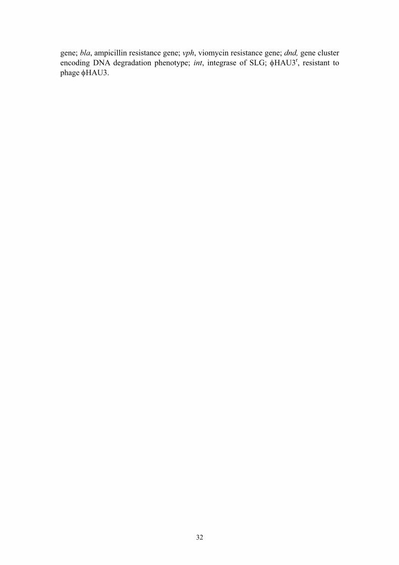
gene; bla, ampicillin resistance gene; vph, viomycin resistance gene; dnd, gene cluster encoding DNA degradation phenotype; int, integrase of SLG; φHAU3r, resistant to phage φHAU3.
32
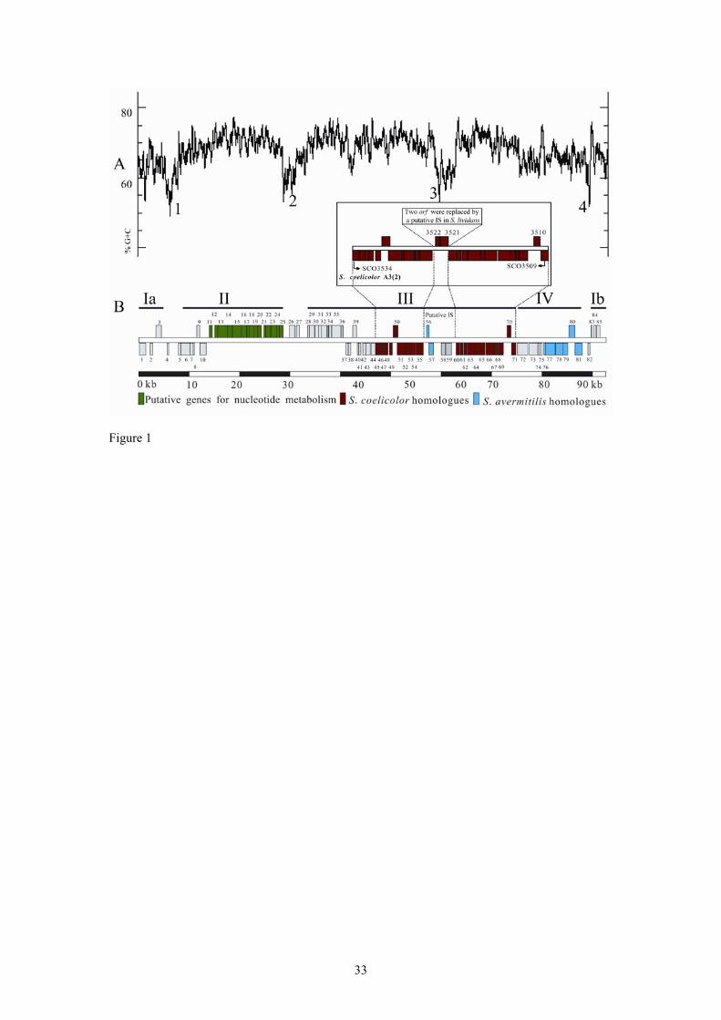
Figure 1
33

Figure 2
34

Figure 3
35

Figure 4
36

Figure 5
37