Embed Size (px)
Citation preview

Anaesthesia for non-cardiac surgery in patients left ventricular outflow
tract obstruction (LVOTO)
Dr. Siân JaggarConsultant AnaesthetistRoyal Brompton Hospital
London
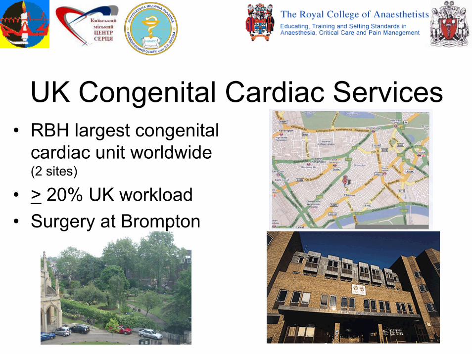
UK Congenital Cardiac Services• RBH largest congenital
cardiac unit worldwide (2 sites)
• > 20% UK workload• Surgery at Brompton

LVOTO & Anaesthesia• Why does it matter?• What is it?
– Fixed / Dynamic• Anaesthetic management
– Pre-op– Per-op

Why does LVOTO matter?
• Common• ↑ed LV afterload → LV hypertrophy• Untreated results in LV dilatation & failure• High risk infective endocarditis
• Anaesthesia → ↑ed risk • ∴ recognise prior to anaesthesia
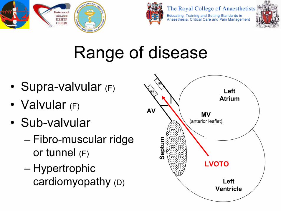
Range of disease
• Supra-valvular (F)
• Valvular (F)
• Sub-valvular– Fibro-muscular ridge
or tunnel (F)
– Hypertrophic cardiomyopathy (D)
Left Atrium
Left Ventricle
LVOTO
AV MV (anterior leaflet)
Sept
um
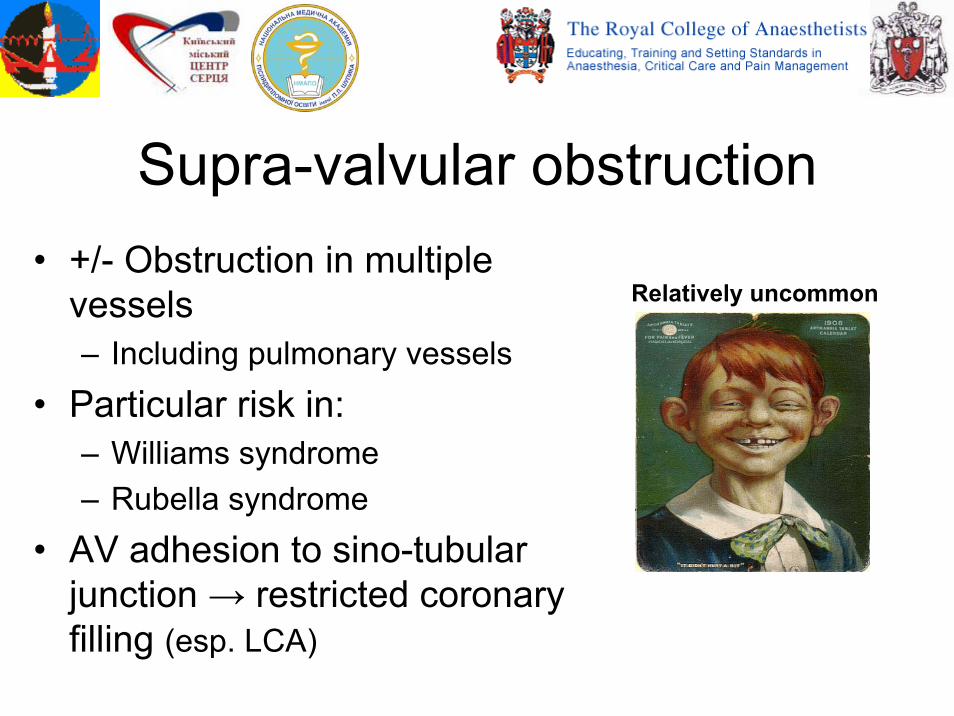
Supra-valvular obstruction• +/- Obstruction in multiple
vessels– Including pulmonary vessels
• Particular risk in:– Williams syndrome– Rubella syndrome
• AV adhesion to sino-tubular junction → restricted coronary filling (esp. LCA)
Relatively uncommon
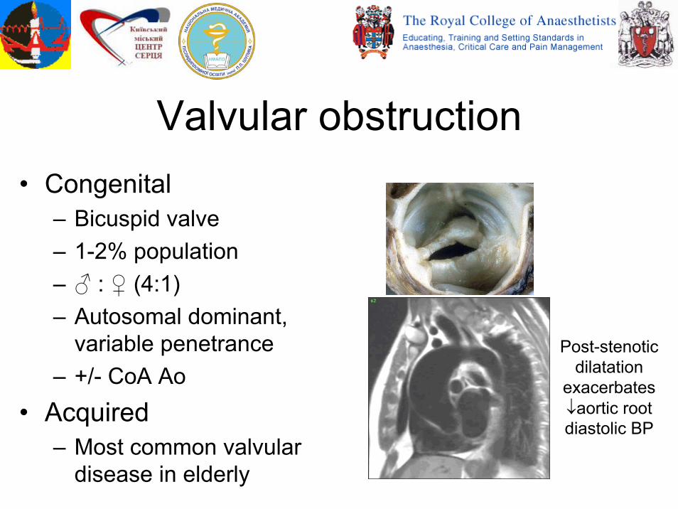
Valvular obstruction• Congenital
– Bicuspid valve– 1-2% population– ♂ : ♀ (4:1)– Autosomal dominant,
variable penetrance– +/- CoA Ao
• Acquired– Most common valvular
disease in elderly
Post-stenoticdilatation
exacerbates ↓aortic root diastolic BP
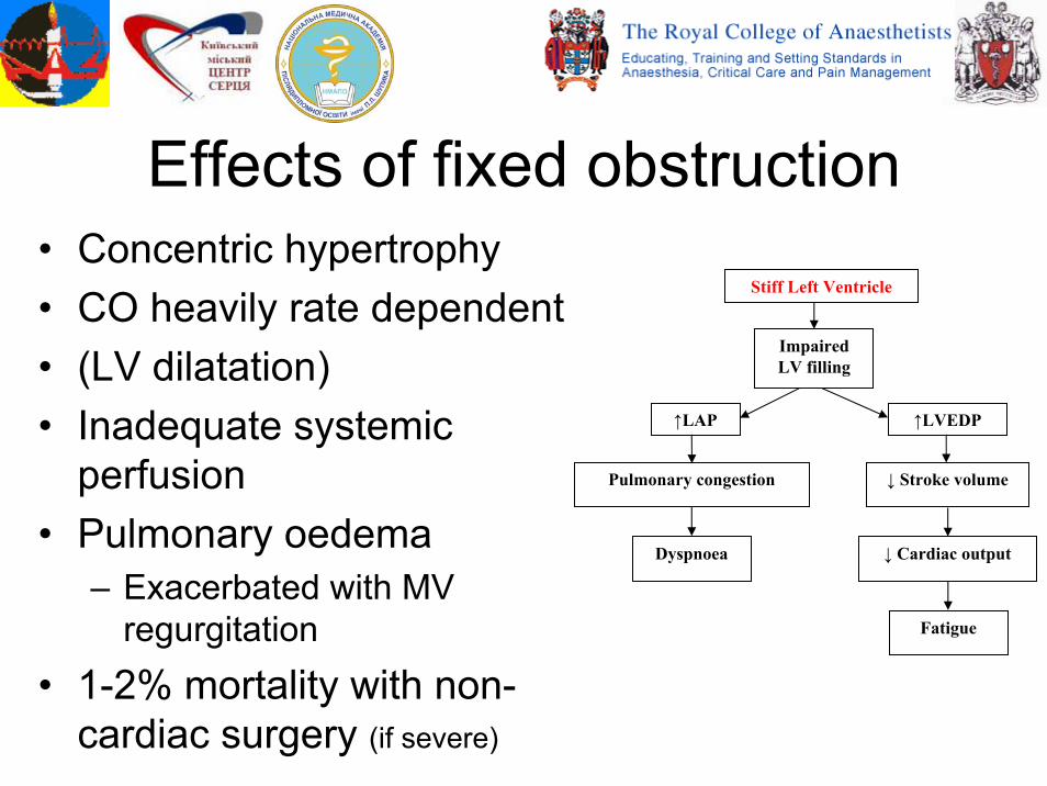
Effects of fixed obstruction• Concentric hypertrophy• CO heavily rate dependent • (LV dilatation)• Inadequate systemic
perfusion• Pulmonary oedema
– Exacerbated with MV regurgitation
• 1-2% mortality with non-cardiac surgery (if severe)
Stiff Left Ventricle
Impaired LV filling
↑LAP ↑LVEDP
Pulmonary congestion ↓ Stroke volume
↓ Cardiac outputDyspnoea
Fatigue
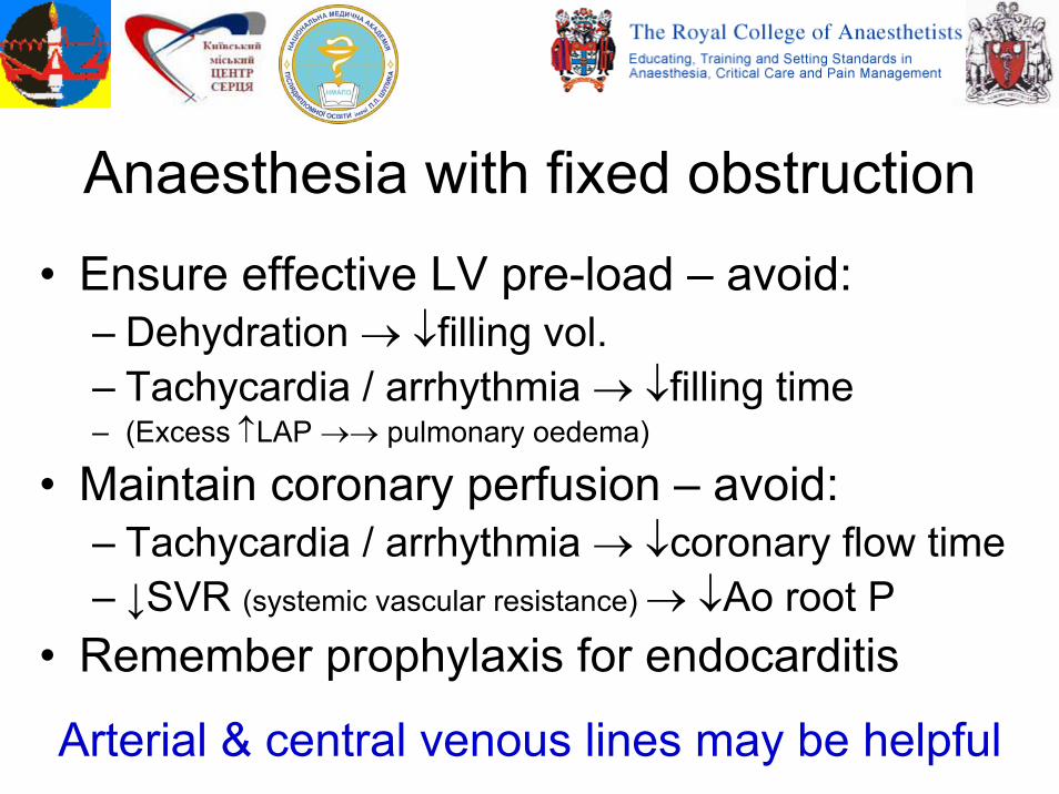
Anaesthesia with fixed obstruction• Ensure effective LV pre-load – avoid:
– Dehydration → ↓filling vol.– Tachycardia / arrhythmia → ↓filling time– (Excess ↑LAP →→ pulmonary oedema)
• Maintain coronary perfusion – avoid:– Tachycardia / arrhythmia → ↓coronary flow time– ↓SVR (systemic vascular resistance) → ↓Ao root P
• Remember prophylaxis for endocarditis
Arterial & central venous lines may be helpful
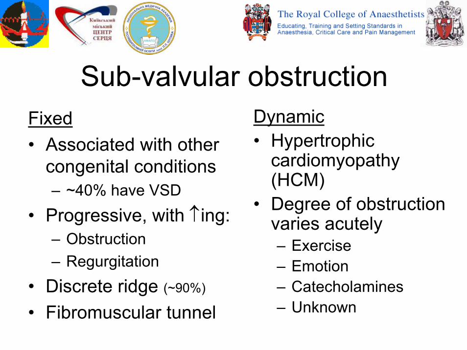
Sub-valvular obstructionFixed• Associated with other
congenital conditions– ~40% have VSD
• Progressive, with ↑ing:– Obstruction– Regurgitation
• Discrete ridge (~90%)
• Fibromuscular tunnel
Dynamic• Hypertrophic
cardiomyopathy(HCM)
• Degree of obstruction varies acutely– Exercise– Emotion– Catecholamines– Unknown
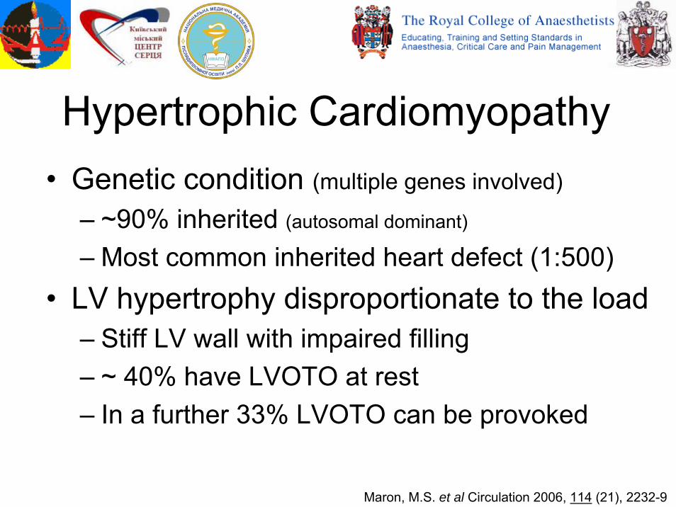
Hypertrophic Cardiomyopathy• Genetic condition (multiple genes involved)
– ~90% inherited (autosomal dominant)
– Most common inherited heart defect (1:500)• LV hypertrophy disproportionate to the load
– Stiff LV wall with impaired filling– ~ 40% have LVOTO at rest– In a further 33% LVOTO can be provoked
Maron, M.S. et al Circulation 2006, 114 (21), 2232-9
Clinical symptoms of HCM• Nil • May only be provoked by exercise:
– Dyspnoea– Syncope– Angina– Arrhythmias
• Sudden death most common in young• Hypertrophic Cardiomyopathy
Association (HCMA) – Provides information for medical staff &
families– http://www.4hcm.org/

Obstruction in HCM• Not affect all individuals • Mainly sub-aortic
– Systolic anterior motion of mitral valve (SAM)
– Mid-systolic contact between mitral valve & septum
• Diagnose with gradient > 30mmHg– Obstructive at rest – With provocation
Maron, B.J. et al JACC 2009, 54 (3), 191-200
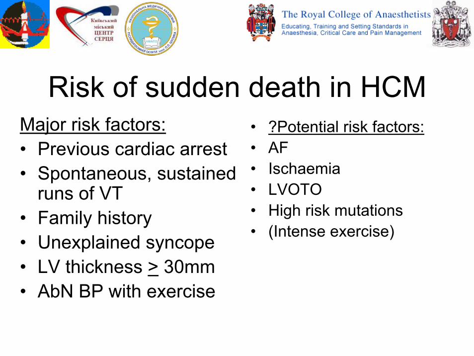
Risk of sudden death in HCMMajor risk factors:• Previous cardiac arrest• Spontaneous, sustained
runs of VT• Family history• Unexplained syncope• LV thickness > 30mm• AbN BP with exercise
• ?Potential risk factors:• AF• Ischaemia• LVOTO• High risk mutations• (Intense exercise)
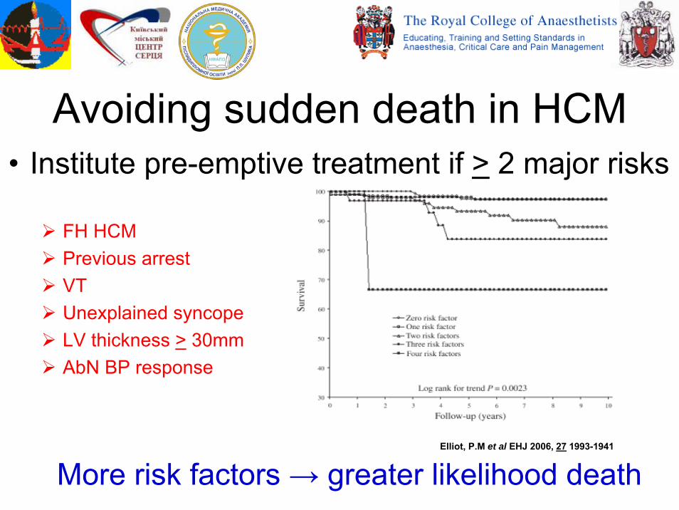
Avoiding sudden death in HCM
More risk factors → greater likelihood death
FH HCMPrevious arrestVTUnexplained syncopeLV thickness > 30mmAbN BP response
• Institute pre-emptive treatment if > 2 major risks
Elliot, P.M et al EHJ 2006, 27 1993-1941

Clinical signs of HCM• Nil• Rapid up- & down-stroke of
arterial pulse• Prominent jugular vein• Systolic murmur at left
sternal edge– ↑s with ↓blood in LV (e.g. Valsalva)
• ↓ (or no ↑) BP with exercise
Left Atrium
Left Ventricle
LVOTO
AV MV (anterior leaflet)
Sept
um
Optimum treatment of HCMDrug Treatment:• Symptoms of failure:
– β-blockers– Ca2+ channel blockers– Disopyramide
• Arrhythmia prevention:– β-blockers– Amiodaroneo ? need anti-coagulation
Interventions:• Myomectomy
– Surgical– Alcohol septal ablation
• Consider AICD
ACC/ESC consensus document 2003 (JACC)

Anaesthesia & dynamic obstructionAvoid: • ↑ dynamic obstruction
– Dehydration (empty LV cavity)– +ve inotropes
• ↓ ΔP coronary perfusion– ↓ diastolic pressure– ↑ LV wall pressure (+ve inotropes)
• ↓ coronary perfusion time– +ve chronotropes
• Arrhythmias– Metabolic derangement (K+, Mg2+)– Excess LA dilatation
↑LVOTO
Arrhythmias Altered walldynamics
Ischaemia
Hypotension
Unstable myocardium

Anaesthesia with LVOTO Summary
• LVOTO may be congenital or acquired – Always take a family history
• Obstruction may be fixed or dynamic• If fixed, avoid:
– Dehydration, tachycardia & ↓SVR• If dynamic, avoid:
– Dehydration, tachycardia, ↓SVR & +ve inotropes