Embed Size (px)
Citation preview

REVIEW ARTICLE
An Updated Overview on Therapeutic Drug Monitoring of RecentAntiepileptic Drugs
Shery Jacob1 • Anroop B. Nair2
Published online: 20 October 2016
� The Author(s) 2016. This article is published with open access at Springerlink.com
Abstract Given the distinctive characteristics of both
epilepsy and antiepileptic drugs (AEDs), therapeutic drug
monitoring (TDM) can make a significant contribution to
the field of epilepsy. The measurement and interpretation
of serum drug concentrations can be of benefit in the
treatment of uncontrollable seizures and in cases of clinical
toxicity; it can aid in the individualization of therapy and in
adjusting for variable or nonlinear pharmacokinetics; and
can be useful in special populations such as pregnancy.
This review examines the potential for TDM of newer
AEDs such as eslicarbazepine acetate, felbamate, gaba-
pentin, lacosamide, lamotrigine, levetiracetam, peram-
panel, pregabalin, rufinamide, retigabine, stiripentol,
tiagabine, topiramate, vigabatrin, and zonisamide. We
describe the relationships between serum drug concentra-
tion, clinical effect, and adverse drug reactions for each
AED as well as the different analytical methods used for
serum drug quantification. We discuss retrospective studies
and prospective data on the serum drug concentration–ef-
ficacy of these drugs and present the pharmacokinetic
parameters, oral bioavailability, reference concentration
range, and active metabolites of newer AEDs. Limited data
are available for recent AEDs, and we discuss the con-
nection between drug concentrations in terms of clinical
efficacy and nonresponse. Although we do not propose
routine TDM, serum drug measurement can play a
beneficial role in patient management and treatment indi-
vidualization. Standardized studies designed to assess, in
particular, concentration–efficacy–toxicity relationships for
recent AEDs are urgently required.
Key Points
Seizures occur sporadically, so antiepileptic drug
therapy is generally experiential and prophylactic.
Therapeutic drug monitoring can help establish an
individual’s optimal serum/plasma concentration
range and benchmark the serum concentrations at
which seizures are restrained or at which
antiepileptic drug-specific adverse effects occur.
Therapeutic drug monitoring enables more decisive
and effective optimization of therapy and disease
management.
1 Introduction
The fundamental objective of therapeutic drug monitoring
(TDM) for antiepileptic drugs (AEDs) is the prevention of
seizures and the minimization of negative effects on gen-
eral well-being, including cognition, mood, and endocrine
function. The International League Against Epilepsy
(ILAE) determines seizure type on the basis of clinical
outcomes and electroencephalograms. Around 34 AEDs
have been prescribed to manage seizures over the last
century. Many enzyme-inducing AEDs are cytochrome
P450 (CYP) mixed function oxidase, glucuronyl trans-
ferase, or epoxy hydrolysis enzyme inducers. First-
& Shery Jacob
1 Department of Pharmaceutics, College of Pharmacy, Gulf
Medical University, University Street, P.O.Box No.4184,
Ajman, UAE
2 Department of Pharmaceutics, College of Clinical Pharmacy,
King Faisal University, Al-Ahsa, Saudi Arabia
Drugs R D (2016) 16:303–316
DOI 10.1007/s40268-016-0148-6

generation ‘old’ AEDs such as carbamazepine, phenobar-
bital, and phenytoin are enzyme inducers. Table 1 lists the
pharmacokinetic parameters of the newer or ‘second-gen-
eration’ AEDs (e.g., lamotrigine, gabapentin), the newest
or ‘third-generation’ AEDs (e.g., retigabine, perampanel),
and the newest orphan drugs (e.g., rufinamide, stiripentol).
1.1 Factors Influencing the Selection
of Antiepileptic Drugs
The selection of AEDs for a particular seizure type depends
on drug-specific (e.g., adverse effects, toxicity, drug
interactions), patient-specific (e.g., sex, age, use of con-
traception, genetics), and country-specific (e.g., availabil-
ity, cost) variables [1].
1.2 Therapeutic Drug Monitoring or Target
Concentration Interpretation
The aim of TDM is to contribute a ‘reference concentration
range’ that laboratories can cite and clinicians can use as a
benchmark. The ‘therapeutic concentration range’ of an
individual patient is the range that achieves the best pos-
sible response and should be selected on the basis of
symptoms and associated risks. The flaw of this strategy is
that, in a few patients, optimal benefit will only be attained
above minimum toxic concentrations, with associated risks
of adverse reactions. Serum drug concentrations (SDC)
achieved over months or years can provide invaluable
information regarding the clinical scenario for each patient
to enable interpretation of a change in response [2, 3].
Serum samples collected within a few hours of a sudden
seizure can provide information that may lead to clinical
reasons or a definite cause of the seizure [4]. SDC mea-
surement can distinguish between highly variable concen-
trations as a result of poor compliance and low
concentrations because of erratic absorption, rapid meta-
bolism, or drug interactions. The ability to discriminate
between insufficient seizure control as a result of inade-
quate dosing or drug overload is invaluable [4].
Polypharmacy can also lead to complex intoxication
despite concentrations being within the therapeutic window
[5]. SDC is particularly relevant in pediatric and psychi-
atric patients in whom clinical assessment can be difficult.
It is also useful for dosage adjustment in complex epilepsy
that may need multiple drug therapy. The reference con-
centration range may not always be ‘therapeutic’, ‘effec-
tive’, or ‘target’, so the reporting method should be factual,
with a statement such as ‘the SDC lies between/
above/below the reference range’. Clinical judgment
should not be based solely on SDC, because adverse drug
reactions can occur even at low concentrations.
2 Newest Orphan Antiepileptic Drugs
2.1 Rufinamide
Rufinamide is a well-tolerated and effective treatment for
drug-resistant epilepsies and is also used as a second-line
adjuvant for routine neurologic practice [6]. A recently
published clinical report [7] also indicated that rufinamide
was successful in the treatment of super-refractory tonic-
status epilepticus. Rufinamide was granted orphan drug
status by the European Medicines Agency (EMA) and the
US FDA in 2004 for the treatment of Lennox–Gastaut
syndrome (LGS). Mean serum drug concentration was
found to be 10 ± 6.5 mg/l in children aged \12 years,
10.6 ± 7.0 mg/l in adolescents aged 12–17.9 years, and
15.9 ± 8.5 mg/l in adults aged C18 years [8]. Approxi-
mately 85 % of the metabolites are eliminated via the
kidneys, with an average half-life of 8 h. Metabolism is
induced by enzyme inducers such as carbamazepine and
rifampin [8]. Impaired renal function does not affect
clearance, so patients undergoing hemodialysis may
require larger doses. Rufinamide exhibits dose-dependent
gastrointestinal (GI) absorption with good bioavailability
(&85 %) and high peak plasma drug concentration (Cmax)
especially when administered with food [9]. Therefore,
patients should be adequately counselled to ensure they
understand the need to follow the strict dosage regimen
with respect to meals. Serum rufinamide concentration was
high (&22 %) when co-administered with valproic acid
(VPA). Reductions in seizure frequency have been found to
be dose dependent. Mean concentration was 10 ± 6.5 mg/l
in children aged\12 years, 10.6 ± 7.0 mg/l in those aged
12–17.9 years, and 15.9 ± 8.5 mg/l in patients
aged C18 years [10]. Patients receiving a dose of 40 mg/
kg were found to have a serum concentration range of
0–45 lg/ml, indicating considerable pharmacokinetic
variability. Therefore, TDM may be beneficial in clinical
practice, albeit no reference range is available. Adverse
reactions such as dizziness, fatigue, nausea, vomiting,
diplopia, and somnolence have also been found to increase
with concentration [8]. Enzyme-inducing AEDs such as
oxcarbazepine, and particularly methsuximide, have been
demonstrated to decrease the serum concentration of rufi-
namide [11]. TDM for rufinamide correlates well with
seizure control and therefore can be helpful in treatment
individualization [12]. Monitoring is useful for patients
receiving enzyme inducers or those who are concurrently
undergoing hemodialysis. Information regarding the phar-
macokinetics of rufinamide in pregnancy and patients with
hepatic impairment is inadequate. Drug interactions data
are comparable to those for (PER) perampanel. A current
report suggests a reference range of 126–168 lmol/l
304 S. Jacob, A. B. Nair
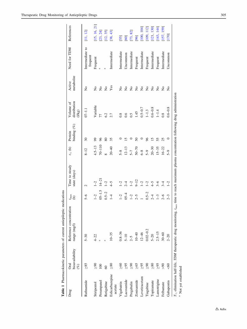
Table
1Pharmacokinetic
param
etersofcurrentantiepilepticmedications
Drug
Oral
bioavailability
(%)
Reference
concentration
range(m
g/l)
t max
(h)
Tim
eto
steady
state(days)
t �(h)
Protein
binding(%
)
Volumeof
distribution
(l/kg)
Active
metabolite
NeedforTDM
References
Rufinam
ide
C85
5–30
5–6
28–12
30
07–1.1
No
Interm
ediate
to
frequent
[11,12]
Stiripentol
C90
4–22
1–2
1–2
4.5–13
99
Variable
No
Frequent
[15,16,21]
Peram
panel
100
a05–1.5
14–21
70–110
96
77
No
a[23,24]
Retigabine
60
a0.5–2
1–2
880
6.2
No
a[12,35]
Eslicarbazepine
acetate
C80
10–35
1–4
4–5
20–40
35
2.7
Yes
Interm
ediate
[38,43]
Vigabatrin
C60
0.8–36
1–2
1–2
5–8
00.8
No
Interm
ediate
[55]
Lacosamide
C95
5–10
05–4
2–4
12–13
15
0.6
No
Uncommon
[60]
Pregabalin
C90
2–5
1–2
1–2
5–7
00.5
No
Interm
ediate
[73,82]
Zonisam
ide
C65
10–40
2–5
9–12
50–70
50
1.45
No
Frequent
[96]
Levetiracetam
C95
12–46
11–2
6–8
00.5–0.7
No
Interm
ediate
[100,104]
Tiagabine
C90
0.02–0.2
0.5–2
1–2
5–9
96
1–1.3
No
Frequent
[109,112]
Topiram
ate
C80
5–20
2–4
4–5
20–30
15
0.6–0.8
No
Interm
ediate
[121,130]
Lam
otrigine
C95
2.5–15
1–3
3–6
15–35
55
1–1.4
No
Frequent
[143,144]
Felbam
ate
[90
30–60
2–6
3–4
16–22
25
0.8
No
Interm
ediate
[157,159]
Gabapentin
\60
2–20
2–3
1–2
5–9
00.6–0.8
No
Uncommon
[170]
T�elim
inationhalf-life,TDM
therapeuticdrugmonitoring,t m
axtimeto
reachmaxim
um
plasm
aconcentrationfollowingdrugadministration
aNotyet
established
Therapeutic Drug Monitoring of Antiepileptic Drugs 305

(30–40 mg/l) in LGS, which presumably would be lower
for other seizure types [7]. High-performance liquid chro-
matography (HPLC) [13] and liquid chromatography–mass
spectrometry (LC–MS) [14] can be used to determine
plasma/serum concentrations.
2.2 Stiripentol
Stiripentol (STP) has been granted orphan drug status by
EMEA for PGTCS in 2001 and approved additionally for
the treatment of severe myoclonic epilepsy in infancy
(SMEI) or Dravet’s syndrome [15, 16]. Stiripentol has
been reported to elevate brain c-aminobutyric acid
(GABA) levels and to interfere with uptake and metabo-
lism [17]. It is bound strongly to plasma proteins
(&99 %) with extensive hepatic metabolism and low
bioavailability. Major metabolites are excreted renally.
The half-life of stiripentol increases with dose because of
its dose-dependent pharmacokinetics. Stiripentol exhibits
a significant reduction in clearance with dose escalation
[18]. The reference serum concentration for stiripentol is
not distinct, but a range of 4–22 mg/l may be associated
with control of absence seizures in children, and a range
of 8–12 mg/l may be associated with control of Dravet
syndrome [19]. The pharmacokinetics of stiripentol are
complex as a result of non-linearity, strong protein
binding, and considerable metabolism [12]. Stiripentol
inhibits many CYPs (CYP3A4, 1A2, 2C19) and interacts
with many drugs, including AEDs. Stiripentol signifi-
cantly decreases phenobarbital and phenytoin concentra-
tions, whereas clobazam moderately increased stiripentol
serum concentrations [20]. No data are available regard-
ing the pharmacokinetics of stiripentol in pregnancy, or in
patients with hepatic and renal impairment. TDM seems
to be beneficial when using stiripentol because of the
broad fluctuation in its concentration–dose ratio, age-de-
pendent pharmacokinetics, non-linear relationship,
extensive clearance, and drug–drug interactions [21].
Quantification of plasma/serum stiripentol via HPLC has
been reported [22].
3 Newest Antiepileptic Drugs
3.1 Perampanel
Perampanel is prescribed for refractory partial onset sei-
zures, tonic–clonic seizures in patients aged[12 years, and
PGTCS [23]. Perampanel received regulatory approval from
the FDA and the EMA in 2012. The minimum effective
dose is 4 mg once daily, and larger doses provide a greater
therapeutic effect with a comparable increase in adverse
events [24]. Perampanel is not an enzyme inducer and acts
as a selective non-competitive antagonist of ionotropic
glutamate receptors [25]. Oral absorption of the drug is
delayed up to 2 h with food. The high protein binding
(96 %) and volume of distribution (77 l/kg) may lead to
displacement interactions with other AEDs. The drug has a
long half-life of 70–110 h, and steady state may be achieved
after 14 days. Its primary route of hepatic metabolism is via
enzyme CYP3A4. The bulk of the dose is excreted in the
feces and the rest in the urine, with\2 % excreted
unchanged in the urine. Although the drug is well tolerated,
the prevalence of adverse events rises with increasing
dosages. Serious psychiatric and behavioral reactions such
as dizziness, fatigue, irritability, obesity, vertigo, ataxia, gait
disturbance, anxiety, blurred vision, dysarthria, asthenia, and
hypersomnia have been reported [26]. A recent investigation
demonstrated a linear dose–concentration relationship with
serum perampanel concentrations, independent of age and
sex [27]. Carbamazepine and oxcarbazepine can signifi-
cantly and dose-dependently reduce perampanel concentra-
tions, presumably through CYP3A4-induced metabolism.
Perampanel dosage should be monitored carefully dur-
ing pregnancy and after childbirth, with adjustments made
on a clinical basis. Anticonvulsant effects are possibly
antagonized by antipsychotics and antimalarials such as
mefloquine. Antifungals such as ketoconazole can elevate
plasma concentrations, and anxiolytics and hypnotics such
as midazolam can reduce them. Concomitant administra-
tion with orlistat, an obesity drug, may lead to an increased
risk of convulsions [28]. No adequate well-controlled
studies have investigated the pharmacokinetics of peram-
panel in pregnancy, possible drug interactions, or TDM,
and a reference range has not yet been established. HPLC
with fluorescence detection and HPLC–MS has been used
to assay serum perampanel [29].
3.2 Retigabine
Retigabine (also known as ezogabine) is used as an
adjunctive treatment for partial epilepsies in adults and
was approved by both the EMA and the FDA in 2011
[12, 30]. The mechanism of action of retigabine is due to
the activation of voltage-gated potassium channels in the
brain [31]. A placebo-controlled clinical trial has sug-
gested that seizure frequency may be significantly reduced
at higher doses [32]. The most frequently used dose in an
open-label add-on study was 600 mg per day [33]. Ezo-
gabine/retigabine was demonstrated to be effective as
adjunctive therapy to specified monotherapies such as
carbamazepine/oxcarbazepine, lamotrigine, levetiracetam,
and VPA using a flexible dosing regimen in adults with
partial-onset seizures. Retigabine increases lamotrigine
(LTG) metabolism, whereas carbamazepine and phenytoin
enhance the clearance of retigabine [12]. The drug and
306 S. Jacob, A. B. Nair

metabolites are excreted almost completely by the kidneys
[12]. Determination of retigabine and its acetyl metabolite
via solid-phase extraction LC–MS has been reported [34].
A recent post-authorization safety study recognized the
importance of TDM for adverse episodes of retinal pig-
mentation and alteration of vision in addition to established
risks of urinary retention, central nervous system effects,
and QTc prolongation [35]. An observational study sug-
gested that retigabine is useful in patients with treatment-
refractory seizures as a drug of reserve [36]. Data are
limited regarding the pharmacokinetics of retigabine in
pregnancy, possible drug interactions, and TDM. Identifi-
cation and quantification of retigabine via HPLC has been
described [37].
3.3 Eslicarbazepine Acetate
Eslicarbazepine acetate (ESL) is a prodrug that is struc-
turally related to oxcarbazepine; it was approved by the
FDA as a monotherapy and adjunct treatment for partial
onset seizures in 2009 [38]. It was recently proposed that
oxcarbazepine be switched to ESL with a dose ratio of
1–1.5:1 and that carbamazepine be switched to ESL with
a dose ratio of 1–1.3:1 [39, 40]. ESL is extensively
metabolized to S-licarbazepine (95 %), which, similar to
oxcarbazepine, inhibits voltage-gated sodium channels
[41]. A linear relationship exists between dose and serum
concentration. It also induces CYPs and increases clear-
ance (12–16 %) of carbamazepine, lamotrigine, and
topiramate. A stable dose–response relationship was
recognized between ESL serum concentrations and
reductions in seizure frequency that were not altered by
other AEDs. ESL has been reported as having minimum
drug–drug interactions [42]. The pharmacokinetics of
ESL are not influenced by enzyme-inducing AEDs or
VPA, and ESL does not change the metabolism of lam-
otrigine [43]. ESL metabolites are primarily eliminated
renally, and dose adjustment is particularly necessary in
patients with a clearance rate of\ 60 ml/min.
Hemodialysis efficiently removes ESL and its metabo-
lites from serum [44]. Moderate hepatic impairment has
limited clinical effects [10]. This was endorsed in a sin-
gle-dose study of ESL 800 mg once daily over a period of
1 week, which demonstrated no change in pharmacoki-
netic parameters [45]. Concomitant administration of
drugs that prolong the PR interval should be avoided.
ESL adversely interacts with oral contraceptives. There is
a possible risk of developing Stevens–Johnson syndrome
in the presence of human leukocyte antigen (HLA)-B
1502 allele [46]. No clinically relevant data are available
with respect to pregnancy, and no data support the use-
fulness of TDM in ESL although it is expected to be
similar to oxcarbazepine. An enantioselective HPLC–UV
detector [47] and LC [48] have been used to analyze ESL
and its metabolites.
3.4 Vigabatrin
Vigabatrin has been indicated as adjunct therapy for adults
and children aged[10 years with refractory complex
partial seizures and to control infantile spasms. Vigabatrin
was granted initial approval from the FDA in 2009 and
received orphan drug status from the EMA in 2000.
Vigabatrin is currently available in more than 50 countries.
Its mechanism of action is based on the irreversible inhi-
bition of GABA-transaminase (GABA-T), thus elevating
the concentration of GABA in the brain [49]. Vigabatrin S
(?) isomer is pharmacologically active and associated with
irreversible visual field defects in 44 % of patients with
epilepsy, which has resulted in prescription limitations
[49, 50]. Clearance in children is greater than in adults, and
therefore higher doses are required to attain comparable
serum concentrations [51]. As the drug is eliminated ren-
ally, toxicity might occur in patients with renal impair-
ment; therefore dosage adjustment is required [49].
Pharmacokinetic interactions are minimal since vigabatrin
is neither metabolized nor protein bound. Due to irre-
versible inhibition of GABA-T, there is no rationale for
TDM of vigabatrin, although it may be helpful in assessing
compliance [52]. Serum GABA and GABA-T concentra-
tions are not an indicator/marker for clinical response
because the blood–brain barrier is relatively impermeable
to GABA, and serum concentrations do not necessarily
reflect cerebral spinal fluid concentrations [53]. Informa-
tion about the pharmacokinetics of vigabatrin in pregnancy
is inadequate. The use of nuclear magnetic resonance
(NMR) spectroscopy to analyze brain GABA concentra-
tions with vigabatrin treatment has been described [54].
Compliance can be verified by checking the anticipated
trough serum vigabatrin target range (6–278 mol/l) at a
dose level of 1000–3000 mg/day [55]. Serum vigabatrin
has been quantified with HPLC, LC–MS [56], and gas
chromatography (GC)-MS [51, 57]. Simultaneous HPLC
analysis of vigabatrin, gabapentin, pregabalin, and topira-
mate has been reported as useful for monitoring polyther-
apy regimens including these drugs [58, 59].
3.5 Lacosamide
Lacosamide has been approved as an adjunctive treatment
for partial-onset seizures and focal epilepsies in adult
patients [60]. It was approved by both the FDA and the
EMA in 2008. It enhances the inactivation of voltage-gated
sodium channels, leading to the stabilization of hyper-ex-
citable neuronal membranes [61]. Lacosamide has been
proposed for the treatment of neuropathic pain of various
Therapeutic Drug Monitoring of Antiepileptic Drugs 307

etiologies in patients who do not respond to standard
treatments [62]. Lacosamide has a high level of oral
bioavailability (&100 %) and a short time to Cmax (tmax), is
unaffected by food, and has linear pharmacokinetics, which
are all advantages in the clinical use of lacosamide [61].
The presence of high free drug concentrations of lacosa-
mide led to the hypothesis that the drug should be effec-
tively eliminated by hemodialysis, although published data
to support this are limited [63]. It is primarily metabolized
via the hepatic route by demethylation to an inactive
O-desmethyl metabolite by CYP2C19 (30 %) [64].
Approximately 40 % of lacosamide is eliminated unchan-
ged via renal excretion mechanisms. Population pharma-
cokinetic data from phase II clinical studies of lacosamide
have shown that lamotrigine, levetiracetam, topiramate,
oxcarbazepine, and VPA have no significant effects on the
pharmacokinetics of lacosamide [65]. Enzyme inducers
such as carbamazepine and phenytoin can significantly
reduce plasma lacosamide concentrations and thereby
enhance clearance [66, 67]. Although data are limited,
overall, lacosamide does not affect the pharmacokinetics of
many AEDs [60]. Frequent adverse effects include
diplopia, nausea, vomiting, dizziness, abnormal coordina-
tion, and blurred vision [42]. Daytime fluctuations can be
lowered by increasing the frequency of administration from
twice daily to three times daily [68]. Lacosamide serum
levels are elevated in patients with hepatic or renal
impairment. Dose-dependent escalation of serum lacosa-
mide concentrations have been reported to be independent
of age and higher in women than in men [69]. No clinically
relevant information is available regarding serum lacosa-
mide concentrations in pregnancy. A rapid sensitive HPLC
and GC–MS method for the measurement of lacosamide
and desmethyl lacosamide has been reported [70, 71].
4 Newer Antiepileptic Drugs
4.1 Pregabalin
Pregabalin is a short-acting (elimination half-life [t�]
5–7 h) analogue of gabapentin used as an adjunct treatment
for focal epilepsies, neuropathic pain, fibromyalgia, and
anxiety disorder in adult patients [72, 73]. Initial approval
was granted by the FDA and EMA in 2004. Although
pregabalin is patent protected in the USA until 2018, a
generic version is currently available in the UK and
Canada. Pregabalin has been investigated for use in cancer-
induced bone pain [74] and as a premedication to provide
postoperative analgesia [75, 76]. Pregabalin can be an
effective treatment for peripheral neuropathic pain pro-
vided adverse reactions are carefully monitored and dosa-
ges are correctly determined [77]. It demonstrates good
bioavailability (&98 %), minimal drug interactions, and
low protein binding [78]. The poor binding of pregabalin
means it can be effectively cleared by hemodialysis tech-
niques [79]. A major portion of the drug is excreted
unchanged in the urine, with a clearance equal to the
glomerular filtration rate. Thus, the dosage regimen should
be adjusted in geriatric patients as well as those with renal
impairment [80]. TDM of pregabalin is restricted to dosage
adjustment and to assess compliance. Although no specific
reference concentration range has been established, a
comparative value of 3–8 mg/l has been suggested [81].
Pregabalin trough concentrations are subject to high inter-
subject variability [82]. Significant adverse drug reactions
are sedation, obesity, cerebellar symptoms, and peripheral
edema. Enzyme-inducing AEDs can moderately lower
pregabalin concentrations [80]. An increased risk of major
birth defects after first-trimester exposure to pregabalin has
been recently documented [83]. An initial clinical study
has demonstrated the use of pregabalin as an adjuvant to
labor pain relief associated with late termination of preg-
nancy [84]. Plasma drug concentrations can be determined
via HPLC [85], LC–MS/MS [85, 86], GC–MS [87], or a
fluorometric method [88].
4.2 Zonisamide
Zonisamide is used as an adjunct treatment for partial
seizure epilepsy and off label for bipolar disorder [89],
chronic pain, migraine [90], and myoclonic dystonia [91].
It was initially approved by the FDA in 2000, and the EMA
granted a marketing authorization valid throughout the EU
in 2005. It exhibits low capacity strong binding to red cell
components and carbonic anhydrase enzymes. The dual
mechanism of action is due to weak inhibition of enzymes
and modulation of GABAergic and glutamatergic neuro-
transmission via alteration of voltage sensitive sodium and
calcium channels [92]. Zonisamide exhibits linear phar-
macokinetics with metabolism via phase I and phase II
biotransformation pathways to produce inactive metabo-
lites [93]. The t� decreased from 60 h to 30 h in patients
concomitantly receiving enzyme inducers. However,
enzyme inhibitors such as cimetidine, erythromycin, keto-
conazole, and VPA can extend the half-life of zonisamide
[93]. Inter-individual variability in patients who are also
receiving CYP enzyme inducers or inhibitors should be
monitored. Zonisamide is cleared efficiently from serum
via hemodialysis [94]. In general, children need larger
doses than adults, calculated on the basis of weight [95]. A
serum/plasma target range of 10–40 mg/l has been sug-
gested for seizure management [96]. Many analytical
methods to assay zonisamide in biological fluids have been
reported, including HPLC [97], LC–MS [98], and solid-
phase extraction techniques [99].
308 S. Jacob, A. B. Nair

4.3 Levetiracetam
Levetiracetam is an enantioselective (S) isomer used for
focal epilepsies in adult patients as monotherapy and as
adjunctive treatment in children. It is also used for the
management of juvenile myoclonic and generalized tonic–
clonic seizures in patients aged C12 years [100]. Leve-
tiracetam obtained initial approval from the FDA in 1999.
It may be effective for prophylaxis of early traumatic brain
injury [101]. Its t� is increased in patients with renal
impairment; therefore dosage adjustments will be neces-
sary. The clearance value of the drug is greater in children
than in adults [102]. Correlation between efficacy and
serum levels of levetiracetam has been demonstrated in
children with refractory epilepsy [103]. Levetiracetam has
all the optimal characteristics of an AED and follows linear
pharmacokinetics [104]. It undergoes minimal metabolism,
meaning drug interactions with enzyme inducers or inhi-
bitors are unlikely. SDC monitoring is not yet established
for levetiracetam by virtue of its wide therapeutic index
and minimal side effects. For a daily dosage of 1–3 g, a
target reference range of 12–46 mg/l is suggested for
monitoring for compliance, drug overdose, and dose
adjustment. A reduction in SDC of %60 % has been
reported in pregnancy [105]. A 12-year comparative study
of AED use in pregnancy indicated that levetiracetam was
more effective and better tolerated than older AEDs and
lamotrigine [106]. Levetiracetam can be measured in bio-
logical fluids using either HPLC [107], GC [108], or GC/
MS [71].
4.4 Tiagabine
Tiagabine is used as add-on treatment for focal epilepsies
not responding to other AEDs [109] and was approved by
the FDA in 1997. Tiagabine follows linear pharmacoki-
netics with extensive oxidative hepatic metabolism [110].
The half-life of tiagabine decreases from 7 h to 3 h in
patients receiving concomitant enzyme inducers [111].
Higher clearance was observed in children than in adults
[109]. Metabolism is gradual in patients with hepatic
impairment; consequently, the half-life in these patients
is &14 h [112]. Therefore, dose individualization may be
necessary in hepatic dysfunction but not in renal dysfunc-
tion [112]. A placebo-controlled clinical trial demonstrated
that the frequency of complex partial seizures was con-
centration dependent [113]. Accordingly, a broad reference
range of 20–200 ng/ml (53–532 nmol/l) has been proposed
[114]. It has been demonstrated that tiagabine may induce
generalized non-convulsive status epilepticus (NCSE) in
patients with focal lesional epilepsy in addition to those
with generalized syndromes [115]. Data regarding the
pharmacokinetics of tiagabine during pregnancy are lim-
ited. TDM may be helpful in cases of noncompliance and
toxicity, provided assay techniques are sensitive and robust
[116]. Strong protein binding and high inter-subject vari-
ability due to hepatic metabolism makes TDM necessary.
Ideally, the serum sample for TDM should be taken before
the morning dose because the half-life of the drug is short.
Tiagabine in serum can be quantified with HPLC [117],
LC/MS [118], and GC/MS [118, 119].
4.5 Topiramate
Topiramate is licensed for the treatment of epilepsies in
adults and children, for adjunct treatment in polytherapy,
LGS, and migraine prophylaxis [120, 121]. The FDA ini-
tially approved topiramate in 1996 and endorsed it for
migration prophylaxis in 2003. The mechanism of action
includes sodium channel blockade, GABAergic, anti-glu-
tamergic, and carbonic anhydrase inhibition. Increased
clearance of the drug was found in children aged\10 -
years and in the presence of enzyme inducers [95, 122].
The half-life of topiramate is 25 h, which reduces to 12 h
in patients receiving concomitant enzyme-inducing AEDs
[123]. Topiramate clearance is decreased by lithium, pro-
pranolol, amitriptyline, and sumatriptan, thereby elevating
serum topiramate concentrations [61]. Food can delay oral
absorption of the drug (&2 h), but serum topiramate con-
centrations are unaffected [124]. Average drug concentra-
tions did not differ significantly between responders, non-
responders, and patients with adverse reactions [125], but
did decrease during pregnancy [126]. A higher topiramate
concentration of[10 mg/l [127] and according to another
study an average serum concentration of 7 mg/l has been
reported to result in marked improvements in seizure fre-
quency [128]. A topiramate concentration of 2.4–8 mg/l
was reported with dose ranges between 125 and 400 mg in
combination with other AEDs [20]. Serum concentrations
between 5 and 20 mg/l have also been reported with
therapeutic doses [129]. The available literature indicates
routine TDM is only necessary in patients with hepatic and
renal impairment; it may also be used to differentiate
between non-convulsive epilepsy and drug intoxication
[130]. Salivary drug concentrations can be considered an
alternative TDM method in patients receiving chronic
therapy [131]. Fluorescence polarization immunoassay
(FPIA) is commercially available to estimate serum con-
centrations [132]. Many analytical methods are available
for estimating serum topiramate concentrations, including
GC [133], HPLC [114], LC/MS [134], and immunoassay
[135]. Simultaneous HPLC–MS/MS analysis of topira-
mate, zonisamide, lamotrigine, carbamazepine, and leve-
tiracetam in serum has been discussed [136].
Therapeutic Drug Monitoring of Antiepileptic Drugs 309

4.6 Lamotrigine
Lamotrigine is effective as monotherapy or adjunctive
therapy in partial, generalized tonic–clonic, and absence
seizures [137], and in LGS [138]. It has also been inves-
tigated for off-label use in treatment-resistant depressive
disorders [139]. Lamotrigine received initial FDA approval
in 1994 and approval for bipolar disorder in 2003. Estra-
diol-containing oral contraceptives and pregnancy can
drastically decrease serum lamotrigine concentrations
[140]; however, this effect was not observed in women also
receiving VPA [141]. Transient declines in lamotrigine
serum concentrations has also been observed after epilepsy
surgery; thus, TDM will be helpful in the prevention of
early postoperative seizures [142]. TDM appears beneficial
both for the finalization of individual reference ranges and
for the optimization of individual dosage regimens [143].
TDM and dose adjustments may be necessary in patients
undergoing hemodialysis or in those with severe liver
impairment [144, 145]. Lamotrigine should be introduced
with slow dose titrations because of the possibility of
severe dermatological reactions, including Stevens–John-
son syndrome and toxic epidermal necrolysis. Enzyme-
inducing AEDs can halve the t� of lamotrigine (8–20 h),
resulting in decreased lamotrigine concentrations. The half-
life of lamotrigine increased to 60 h when combined with
VPA [146]. In polytherapy regimens with VPA, its inhi-
bitory effect counteracts the inducing effect of enzyme-
inducing AEDs [147]. Co-medication with methsuximide
lowers lamotrigine concentration by 70 % [148]. Lamot-
rigine serum concentrations have been shown to increase
by 90 % from pre-pregnancy baseline to third-trimester
[149]; however, this was not observed in patients receiving
concomitant VPA [148]. Therefore, TDM is important in
the prevention of seizure episodes during pregnancy, and
dose adjustments are based on trough serum concentrations
at least once a month [148]. Trans-placental transfer of
lamotrigine in maternal blood, amniotic fluid, and cord
blood has been measured and correlated [150]. Lamotrigine
clearance has been reported to be higher in children and the
elderly [95]. TDM is valuable for dosage adjustments in
pregnancy, and to check inter-individual variability and
drug interactions. In general, reference concentrations of
2.5–15 mg/l have been suggested in patients receiving
therapeutic doses [2, 3]. The prevalence of toxicity
increases significantly with concentrations[15 mg/l [143].
TDM of lamotrigine may be further complicated by phar-
macodynamic interactions as demonstrated for co-medi-
cation with carbamazepine [95]. Assay of lamotrigine
based on HPLC [151, 152], turbidimetric immunoassay
[153], GC–MS [154], and GC and densitometric methods
[155] have been reported.
4.7 Felbamate
Felbamate is used as add-on therapy in LGS in patients
aged[4 years [156]. It was approved by the FDA in 1993.
The half-life decreases from &19 to &14 h in patients
receiving concomitant enzyme inducers [157]. Felbamate
serum concentrations vary widely between patients and are
affected by age; they are higher in patients with renal
impairment, and the longer half-life depends on the degree
of renal function [158]. The clearance value is quite high in
children (20–65 %) compared with adults [95].
Atropaldehyde, an intermediate metabolite, is responsible
for serious progressive organ toxicity [156]. The drug is
contraindicated in patients with liver impairment [3]. Data
on the pharmacokinetics of felbamate during pregnancy are
insufficient. Life-threatening adverse reactions such as
liver toxicity and aplastic anemia strongly restrict the use
of this drug. Serum concentrations of felbamate can be
decreased by enzyme-inducing AEDs [159] and increased
by enzyme inhibitors such as VPA [159]. Clinical inves-
tigators have divided felbamate concentrations into lower
(9–36 mg/l), intermediate (37–54 mg/l), and higher
(54–134 mg/l) ranges [160], and a therapeutic reference
range of 210–462 lmol/l has been proposed [159] in
patients with epilepsy. The drug is less well-tolerated in
elderly populations. The serum concentration of felbamate
can be determined via HPLC–MS [161, 162], HPLC–UV
[163], LC–MS [164], and GC with flame ionization
detection [162] methods.
4.8 Gabapentin
Gabapentin is approved as an adjunctive in the manage-
ment of focal epilepsies in patients aged[6 years and as
monotherapy in patients aged[12 years. It is also used for
the management of peripheral neuropathy in adults [165].
Gabapentin was granted initial FDA approval in 1993.
Gabapentin is rapidly absorbed (tmax 2–3 h) from the GI
tract by capacity-limited L-amino acid transport systems
[166]. It has an indirect effect on voltage-gated calcium
channels and increases brain GABA concentrations [167].
This was demonstrated by linearity of concentration with
doses up to 1800 mg/day and non-linearity at higher doses
[168]. The half-life of gabapentin (5–9 h) increases with
renal impairment [169]. Dose adjustment is mandatory in
patient with creatinine clearance\60 ml/min and in the
elderly because of reduced renal function. The concentra-
tion/dose ratio progresses with age, and inter-subject
variability is common at any dose [170]. Capacity-limited
GI transport means gabapentin can be administered safely
and effectively either twice or three times daily as steady
state fluctuations are minor. Data are limited regarding the
310 S. Jacob, A. B. Nair

pharmacokinetics of gabapentin during pregnancy and in
terms of drug–drug interactions. Antacids reduce gaba-
pentin absorption from the GI tract, and H-2 receptor
blockers decrease the renal clearance of gabapentin [171].
The main adverse reactions are sedation, dizziness, head-
ache, nausea, and obesity. Therapeutic concentrations of
gabapentin in treatment-refractory patients with partial
seizures range from 2 to 20 mg/l [55]. HPLC [58], LC/MS-
coupled GC [172], and GC-electron ionization-MS [173]
methods have been used to quantify serum gabapentin.
5 Conclusion
TDM is a valuable tool in the optimization of individual
dosage regimens to maximize clinical efficacy while min-
imizing adverse drug reactions. Although reference con-
centration ranges indicate the range in which most patients
display therapeutic response, many patients may require
target concentrations outside the reference range. When
interpreting SDC, situations or factors that may modify the
relationship between serum AED and clinical effect should
be considered, such as type and severity of epilepsy,
pathological and physiological states, drug interactions,
and the variability of pharmacogenetics. It is worthwhile
remembering that clinical decisions should not be made on
the basis of drug concentrations alone. In short, dosage
regimens must be individualized based on patient history,
clinical signs and symptoms, pharmacogenetics, and
interpretation of clinical laboratory data.
Compliance with Ethical Standards Shery Jacob and Anroop Nair
have no conflicts of interest that are directly related to the content of
this work.
Funding No sources of funding were used to conduct this study or
prepare this manuscript.
Open Access This article is distributed under the terms of the
Creative Commons Attribution-NonCommercial 4.0 International
License (http://creativecommons.org/licenses/by-nc/4.0/), which per-
mits any noncommercial use, distribution, and reproduction in any
medium, provided you give appropriate credit to the original
author(s) and the source, provide a link to the Creative Commons
license, and indicate if changes were made.
References
1. Perucca E, Dulac O, Shorvon S, Tomson T. Harnessing the
clinical potential of antiepileptic drug therapy: dosage optimi-
sation. CNS Drugs. 2001;15(8):609–21.
2. Perucca E. Is there a role for therapeutic drug monitoring of new
anticonvulsants? Clin Pharmacokinet. 2000;38(3):191–204.
doi:10.2165/00003088-200038030-00001.
3. Johannessen SI, Tomson T. Pharmacokinetic variability of
newer antiepileptic drugs: when is monitoring needed? Clin
Pharmacokinet. 2006;45(11):1061–75. doi:10.2165/00003088-
200645110-00002.
4. Perucca E, Gram L, Avanzini G, Dulac O. Antiepileptic drugs as
a cause of worsening seizures. Epilepsia. 1998;39(1):5–17.
5. Perucca E. Overtreatment in epilepsy: adverse consequences and
mechanisms. Epilepsy Res. 2002;52(1):25–33.
6. Verrotti A, Loiacono G, Ballone E, Mattei PA, Chiarelli F,
Curatolo P. Efficacy of rufinamide in drug-resistant epilepsy: a
meta-analysis. Pediatr Neurol. 2011;44(5):347–9. doi:10.1016/j.
pediatrneurol.2010.12.005.
7. Thompson AG, Cock HR. Successful treatment of super-re-
fractory tonic status epilepticus with rufinamide: first clinical
report. Seizure. 2016;39:1–4. doi:10.1016/j.seizure.2016.04.003.
8. Perucca E, Cloyd J, Critchley D, Fuseau E. Rufinamide: clinical
pharmacokinetics and concentration-response relationships in
patients with epilepsy. Epilepsia. 2008;49(7):1123–41. doi:10.
1111/j.1528-1167.2008.01665.x.
9. Gall Z, Vancea S, Szilagyi T, Gall O, Kolcsar M. Dose-de-
pendent pharmacokinetics and brain penetration of rufinamide
following intravenous and oral administration to rats. Eur J
Pharmaceut Sci. 2015;68:106–13. doi:10.1016/j.ejps.2014.12.
012.
10. Almeida L, Potgieter JH, Maia J, Potgieter MA, Mota F, Soares-
da-Silva P. Pharmacokinetics of eslicarbazepine acetate in
patients with moderate hepatic impairment. Eur J Clin Phar-
macol. 2008;64(3):267–73. doi:10.1007/s00228-007-0414-1.
11. May TW, Boor R, Rambeck B, Jurgens U, Kon-Merker E,
Brandt C. Serum concentrations of rufinamide in children and
adults with epilepsy:the influence of dose, age and comedica-
tion. Ther Drug Monit. 2011;33(2):214–21. doi:10.1097/FTD.
0b013e31820fa9ad.
12. Luszczki JJ. Third-generation antiepileptic drugs: mechanisms
of action, pharmacokinetics and interactions. Pharmacol Rep.
2009;61(2):197–216.
13. Gall Z, Vancea S, Dogaru MT, Szilagyi T. Liquid chromatog-
raphy-mass spectrometric determination of rufinamide in low
volume plasma samples. J Chromatogr B Analyt Technol
Biomed Life Sci. 2013;940:42–6. doi:10.1016/j.jchromb.2013.
07.014.
14. la Marca G, Malvagia S, Filippi L, Innocenti M, Rosati A, Falchi
M, et al. Rapid assay of rufinamide in dried blood spots by a new
liquid chromatography-tandem mass spectrometric method.
J Pharm Biomed Anal. 2011;54(1):192–7. doi:10.1016/j.jpba.
2010.07.015.
15. Chiron C. Stiripentol and vigabatrin current roles in the treat-
ment of epilepsy. Expert Opin Pharmacother. 2016;. doi:10.
1517/14656566.2016.1161026.
16. Verrotti A, Prezioso G, Stagi S, Paolino MC, Parisi P. Phar-
macological considerations in the use of stiripentol for the
treatment of epilepsy. Expert Opin Drug Metab Toxicol.
2016;12(3):345–52. doi:10.1517/17425255.2016.1145657.
17. Trojnar MK, Wojtal K, Trojnar MP, Czuczwar SJ. Stiripentol. A
novel antiepileptic drug. Pharmacol Rep. 2005;57(2):154–60.
18. Levy RH, Lin HS, Blehaut HM, Tor JA. Pharmacokinetics of
stiripentol in normal man: evidence of nonlinearity. J Clin
Pharmacol. 1983;23(11–12):523–33.
19. Farwell JR, Anderson GD, Kerr BM, Tor JA, Levy RH.
Stiripentol in atypical absence seizures in children: an open trial.
Epilepsia. 1993;34(2):305–11.
20. May TW, Boor R, Mayer T, Jurgens U, Rambeck B, Holert N,
et al. Concentrations of stiripentol in children and adults with
epilepsy: the influence of dose, age, and comedication. Ther Drug
Monit. 2012;34(4):390–7. doi:10.1097/FTD.0b013e31825dc4a6.
21. Verdier MC, Tribut O, Bentue-Ferrer D. Therapeutic drug
monitoring of stiripentol. Therapie. 2012;67(2):157–60. doi:10.
2515/therapie/2012014.
Therapeutic Drug Monitoring of Antiepileptic Drugs 311

22. Darwish HW, Abdelhameed AS, Attia MI. A stability-indicating
HPLC-DAD method for determination of stiripentol: develop-
ment, validation, kinetics, structure elucidation and application
to commercial dosage form. J Anal Methods Chem.
2014;2014:638951. doi:10.1155/2014/638951.
23. French JA, Krauss GL, Wechsler RT, Wang XF, DiVentura B,
Brandt C, et al. Perampanel for tonic-clonic seizures in idio-
pathic generalized epilepsy. A randomized trial. Neurology.
2015;85(11):950–7. doi:10.1212/wnl.0000000000001930.
24. Patsalos PN, Gougoulaki M, Sander JW. Perampanel serum
concentrations in adults with epilepsy: effect of dose, age,
gender and concomitant antiepileptic drugs. Ther Drug Monit.
2015;. doi:10.1097/FTD.0000000000000274.
25. Rogawski MA, Hanada T. Preclinical pharmacology of peram-
panel, a selective non-competitive AMPA receptor antagonist.
Acta Neurol Scand Suppl. 2013;197:19–24. doi:10.1111/ane.
12100.
26. Rugg-Gunn F. Adverse effects and safety profile of perampanel:
a review of pooled data. Epilepsia. 2014;55(Suppl 1):13–5.
doi:10.1111/epi.12504.
27. Patsalos PN, Gougoulaki M, Sander JW. Perampanel serum
concentrations in adults with epilepsy: effect of dose, age, sex,
and concomitant anti-epileptic drugs. Ther Drug Monit.
2016;38(3):358–64. doi:10.1097/ftd.0000000000000274.
28. Baxter K, Sharp J. Orlistat and possible drug interactions that
can affect over-the-counter sales. Pharmaceut J.
2010;284(7599):431.
29. Mano Y, Takenaka O, Kusano K. High-performance liquid
chromatography-tandem mass spectrometry method for the
determination of perampanel, a novel alpha-amino-3-hydroxy-5-
methyl-4-isoxazolepropionic acid receptor antagonist in human
plasma. J Pharm Biomed Anal. 2015;107:56–62. doi:10.1016/j.
jpba.2014.12.018.
30. Splinter MY. Ezogabine (retigabine) and its role in the treatment
of partial-onset seizures: a review. Clin Ther.
2012;34(9):1845–56.e1. doi:10.1016/j.clinthera.2012.07.009.
31. Ihara Y, Tomonoh Y, Deshimaru M, Zhang B, Uchida T, Ishii
A, et al. Retigabine, a Kv7.2/Kv7.3-channel opener, attenuates
drug-induced seizures in knock-in mice harboring Kcnq2
Mutations. PLoS One. 2016;11(2):e0150095. doi:10.1371/
journal.pone.0150095.
32. Plosker GL, Scott LJ. Retigabine: in partial seizures. CNS
Drugs. 2006;20(7):601–8; discussion 9–10.
33. Lerche H, Daniluk J, Lotay N, DeRossett S, Edwards S, Brandt
C. Efficacy and safety of ezogabine/retigabine as adjunctive
therapy to specified single antiepileptic medications in an open-
label study of adults with partial-onset seizures. Seizure.
2015;30:93–100. doi:10.1016/j.seizure.2015.06.002.
34. Knebel NG, Grieb S, Leisenheimer S, Locher M. Determination
of retigabine and its acetyl metabolite in biological matrices by
on-line solid-phase extraction (column switching) liquid chro-
matography with tandem mass spectrometry. J Chromatogr B
Biomed Sci Appl. 2000;748(1):97–111.
35. Daniluk J, Cooper JA, Stender M, Kowalczyk A. Survey of
physicians’ understanding of specific risks associated with reti-
gabine. Drugs Real World Outcomes. 2016;3(2):155–63. doi:10.
1007/s40801-016-0068-3.
36. Nass RD, Kurth C, Kull A, Graf W, Kasper B, Hamer HM, et al.
Adjunctive retigabine in refractory focal epilepsy: postmarket-
ing experience at four tertiary epilepsy care centers in Germany.
Epilepsy Behav. 2016;56:54–8. doi:10.1016/j.yebeh.2015.12.
034.
37. Dousa M, Srbek J, Radl S, Cerny J, Klecan O, Havlıcek J, et al.
Identification, characterization, synthesis and HPLC quantifica-
tion of new process-related impurities and degradation products
in retigabine. J Pharm Biomed Anal. 2014;94:71–6. doi:10.
1016/j.jpba.2014.01.042.
38. Shirley M, Dhillon S. Eslicarbazepine acetate monotherapy: a
review in partial-onset seizures. Drugs. 2016;76(6):707–17.
doi:10.1007/s40265-016-0570-7.
39. Poza-Aldea JJ. A proposal for a model to replace carbamazepine
or oxcarbazepine by eslicarbazepine acetate in clinical practice.
Rev Neurol. 2016;63(5):219–23.
40. Schmid E, Kuchukhidze G, Kirschner M, Leitinger M, Hofler J,
Rohracher A, et al. Overnight switching from oxcarbazepine to
eslicarbazepine acetate: an observational study. Acta Neurolog
Scand. 2016;. doi:10.1111/ane.12645.
41. Ambrosio AF, Silva AP, Malva JO, Soares-da-Silva P, Carvalho
AP, Carvalho CM. Inhibition of glutamate release by BIA 2-093
and BIA 2-024, two novel derivatives of carbamazepine, due to
blockade of sodium but not calcium channels. Biochem Phar-
macol. 2001;61(10):1271–5.
42. Bialer M, Johannessen SI, Levy RH, Perucca E, Tomson T,
White HS. Progress report on new antiepileptic drugs: a sum-
mary of the Ninth Eilat Conference (EILAT IX). Epilepsy Res.
2009;83(1):1.
43. Johannessen Landmark C, Svendsen T, Dinarevic J, Kufaas RF,
Reimers A, Brodtkorb E, et al. The impact of pharmacokinetic
interactions with eslicarbazepine acetate versus oxcarbazepine
and carbamazepine in clinical practice. Ther Drug Monit.
2016;38(4):499–505. doi:10.1097/ftd.0000000000000306.
44. Maia J, Almeida L, Falcao A, Soares E, Mota F, Potgieter MA,
et al. Effect of renal impairment on the pharmacokinetics of
eslicarbazepine acetate. Int J Clin Pharmacol Ther.
2008;46(3):119–30.
45. Almeida L, Falcao A, Maia J, Mazur D, Gellert M, Soares-da-
Silva P. Single-dose and steady-state pharmacokinetics of esli-
carbazepine acetate (BIA 2-093) in healthy elderly and young
subjects. J Clin Pharmacol. 2005;45(9):1062–6. doi:10.1177/
0091270005279364.
46. Rocamora R. A review of the efficacy and safety of eslicar-
bazepine acetate in the management of partial-onset seizures.
Ther Adv Neurolog Disord. 2015;8(4):178–86. doi:10.1177/
1756285615589711.
47. Alves G, Fortuna A, Sousa J, Direito R, Almeida A, Rocha M,
et al. Enantioselective assay for therapeutic drug monitoring of
eslicarbazepine acetate: no interference with carbamazepine and
its metabolites. Ther Drug Monit. 2010;32(4):512–6. doi:10.
1097/FTD.0b013e3181e5c855.
48. Servais A-C, Janicot B, Takam A, Crommen J, Fillet M. Liquid
chromatography separation of the chiral prodrug eslicarbazepine
acetate and its main metabolites in polar organic mode. Appli-
cation to their analysis after in vitro metabolism. J Chromatogr
A. 2016;1467:306–11. doi:10.1016/j.chroma.2016.07.022.
49. Rey E, Pons G, Olive G. Vigabatrin. Clinical pharmacokinetics.
Clin Pharmacokinet. 1992;23(4):267–78. doi:10.2165/
00003088-199223040-00003.
50. Maguire MJ, Hemming K, Wild JM, Hutton JL, Marson AG.
Prevalence of visual field loss following exposure to vigabatrin
therapy: a systematic review. Epilepsia. 2010;51(12):2423–31.
doi:10.1111/j.1528-1167.2010.02772.x.
51. Chang SY, Lin W-C. Determination of vigabatrin by capillary
electrophoresis with laser-induced fluorescence detection.
J Chromatogr B Analyt Technol Biomed Life Sci.
2003;794(1):17–22. doi:10.1016/S1570-0232(03)00396-9.
52. Patsalos PN. New antiepileptic drugs. Ann Clin Biochem.
1999;36(Pt 1):10–9.
53. Erdal J, Gram L, Alving J, Loscher W. Changes in plasma
GABA concentration during vigabatrin treatment of epilepsy: a
prospective study. Epilepsy Res. 1999;34(2–3):145–50.
312 S. Jacob, A. B. Nair

54. Mueller SG, Weber OM, Duc CO, Weber B, Meier D, Russ W,
et al. Effects of vigabatrin on brain GABA?/CR signals in
patients with epilepsy monitored by 1H-NMR-spectroscopy:
responder characteristics. Epilepsia. 2001;42(1):29–40.
55. Lindberger M, Luhr O, Johannessen SI, Larsson S, Tomson T.
Serum concentrations and effects of gabapentin and vigabatrin:
observations from a dose titration study. Ther Drug Monit.
2003;25(4):457–62.
56. Kostic N, Dotsikas Y, Jovic N, Stevanovic G, Malenovic A,
Medenica M. Vigabatrin in dried plasma spots: validation of a
novel LC-MS/MS method and application to clinical practice.
J Chromatogr B Anal Technol Biomed Life Sci.
2014;962:102–8. doi:10.1016/j.jchromb.2014.05.037.
57. Borrey DCR, Godderis KO, Engelrelst VIL, Bernard DR, Lan-
glois MR. Quantitative determination of vigabatrin and gaba-
pentin in human serum by gas chromatography–mass
spectrometry. Clin Chim Acta. 2005;354(1–2):147–51. doi:10.
1016/j.cccn.2004.11.023.
58. Chollet DF, Goumaz L, Juliano C, Anderegg G. Fast isocratic
high-performance liquid chromatographic assay method for the
simultaneous determination of gabapentin and vigabatrin in
human serum. J Chromatogr B Biomed Sci Appl.
2000;746(2):311–4.
59. Martinc B, Roskar R, Grabnar I, Vovk T. Simultaneous deter-
mination of gabapentin, pregabalin, vigabatrin, and topiramate
in plasma by HPLC with fluorescence detection. J Chromatogr B
Anal Technol Biomed Life Sci. 2014;962:82–8. doi:10.1016/j.
jchromb.2014.05.030.
60. Chung SS. Lacosamide: new adjunctive treatment option for
partial-onset seizures. Expert Opin Pharmacother.
2010;11(9):1595–602. doi:10.1517/14656566.2010.488639.
61. Patsalos PN, Berry DJ. Pharmacotherapy of the third-generation
AEDs: lacosamide, retigabine and eslicarbazepine acetate.
Expert Opin Pharmacother. 2012;13(5):699–715. doi:10.1517/
14656566.2012.667803.
62. Alcantara-Montero A, Sanchez-Carnerero CI. Lacosamide and
neuropathic pain, a review. Rev Neurol. 2016;62(5):223–9.
63. Lacerda G, Krummel T, Sabourdy C, Ryvlin P, Hirsch E.
Optimizing therapy of seizures in patients with renal or hepatic
dysfunction. Neurology. 2006;67(12 Suppl 4):S28–33.
64. Cawello W, Nickel B, Eggert-Formella A. No pharmacokinetic
interaction between lacosamide and carbamazepine in healthy
volunteers. J Clin Pharmacol. 2010;50(4):459–71. doi:10.1177/
0091270009347675.
65. Thomas D, Scharfenecker U, Schiltmeyer B, Doty P, Cawello
W, Horstmann R. 640 Low potential for drug-drug-interaction of
lacosamide. Eur J Pain. 2006;10(S1):S167c–8c. doi:10.1016/
S1090-3801(06)60643-5.
66. Contin M, Albani F, Riva R, Candela C, Mohamed S, Baruzzi
A. Lacosamide therapeutic monitoring in patients with epilepsy:
effect of concomitant antiepileptic drugs. Ther Drug Monit.
2013;35(6):849–52. doi:10.1097/FTD.0b013e318290eacc.
67. Cawello W, Rosenkranz B, Schmid B, Wierich W. Pharmaco-
dynamic and pharmacokinetic evaluation of coadministration of
lacosamide and an oral contraceptive (levonorgestrel plus
ethinylestradiol) in healthy female volunteers. Epilepsia.
2013;54(3):530–6. doi:10.1111/epi.12085.
68. SattlerA,SchaeferM,MayTW,RambeckB,BrandtC.Fluctuationof
lacosamide serum concentrations during the day and occurrence of
adverse drug reactions–first clinical experience. Epilepsy Res.
2011;95(3):207–12.doi:10.1016/j.eplepsyres.2011.03.019.
69. Markoula S, Teotonio R, Ratnaraj N, Duncan JS, Sander JW,
Patsalos PN. Lacosamide serum concentrations in adult patients
with epilepsy: the influence of gender, age, dose, and con-
comitant antiepileptic drugs. Ther Drug Monit.
2014;36(4):494–8. doi:10.1097/ftd.0000000000000051.
70. Payto D, Foldvary-Schaefer N, So N, Bruton M, Wang S. A
sensitive and rapid method for quantification of lacosamide and
desmethyl lacosamide by LC-MS/MS. Bioanalysis.
2014;6(23):3161–8. doi:10.4155/bio.14.158.
71. Nikolaou P, Papoutsis I, Spiliopoulou C, Voudris C, Athanaselis
S. A fully validated method for the determination of lacosamide
in human plasma using gas chromatography with mass spec-
trometry: application for therapeutic drug monitoring. J Sep Sci.
2015;38(2):260–6. doi:10.1002/jssc.201400858.
72. Selak I. Pregabalin (Pfizer). Curr Opin Investig Drugs.
2001;2(6):828–34.
73. Clair A, Emir B. The safety and efficacy of pregabalin for
treating subjects with fibromyalgia and moderate or severe
baseline widespread pain. Curr Med Res Opin.
2016;32(3):601–10. doi:10.1185/03007995.2015.1134463.
74. Raman S, DeAngelis C, Bruera E, Chow R, Lechner B, Chow E.
Does pregabalin still have a role in treating cancer-induced bone
pain? J Clin Oncol. 2016;34(6):524–6. doi:10.1200/jco.2015.64.
7545.
75. Rajappa GC, Vig S, Bevanaguddaiah Y, Anadaswamy TC.
Efficacy of pregabalin as premedication for post-operative
analgesia in vaginal hysterectomy. Anesthesiol Pain Med.
2016;6(3):e34591. doi:10.5812/aapm.34591.
76. Sebastian B, Talikoti AT, Nelamangala K, Krishnamurthy D.
Effect of oral pregabalin as preemptive analgesic in patients
undergoing lower limb orthopedic surgeries under spinal
anaesthesia. J Clin Diagn Res. 2016;10(7):UC01–UC4. doi:10.
7860/JCDR/2016/18854.8081.
77. Otsuki T, Higuchi T, Yamazaki T, Okawa E, Okada K, Abe M.
Efficacy and safety of pregabalin for the treatment of neuro-
pathic pain in patients undergoing hemodialysis. Clin Drug
Invest. 2016;. doi:10.1007/s40261-016-0464-1.
78. Ben-Menachem E. Pregabalin pharmacology and its relevance
to clinical practice. Epilepsia. 2004;45(Suppl 6):13–8. doi:10.
1111/j.0013-9580.2004.455003.x.
79. Randinitis EJ, Posvar EL, Alvey CW, Sedman AJ, Cook JA,
Bockbrader HN. Pharmacokinetics of pregabalin in subjects
with various degrees of renal function. J Clin Pharmacol.
2003;43(3):277–83.
80. Haslam C, Nurmikko T. Pharmacological treatment of neuro-
pathic pain in older persons. Clin Int Aging. 2008;3(1):111–20.
81. Patsalos PN, Berry DJ, Bourgeois BF, Cloyd JC, Glauser TA,
Johannessen SI, et al. Antiepileptic drugs–best practice guide-
lines for therapeutic drug monitoring: a position paper by the
subcommission on therapeutic drug monitoring. ILAE Com-
mission on Therapeutic Strategies. Epilepsia.
2008;49(7):1239–76. doi:10.1111/j.1528-1167.2008.01561.x.
82. May TW, Rambeck B, Neb R, Jurgens U. Serum concentrations
of pregabalin in patients with epilepsy: the influence of dose,
age, and comedication. Ther Drug Monit. 2007;29(6):789–94.
doi:10.1097/FTD.0b013e31815d0cd5.
83. Winterfeld U, Merlob P, Baud D, Rousson V, Panchaud A,
Rothuizen LE, et al. Pregnancy outcome following maternal
exposure to pregabalin may call for concern. Neurology.
2016;86(24):2251–7. doi:10.1212/wnl.0000000000002767.
84. Lavand’homme PM, Roelants F. Evaluation of pregabalin as an
adjuvant to patient-controlled epidural analgesia during late
termination of pregnancy. Anesthesiology.
2010;113(5):1186–91. doi:10.1097/ALN.0b013e3181f5d32d.
85. Berry D, Millington C. Analysis of pregabalin at therapeutic
concentrations in human plasma/serum by reversed-phase
HPLC. Ther Drug Monit. 2005;27(4):451–6.
86. Kostic N, Dotsikas Y, Jovic N, Stevanovic G, Malenovic A,
Medenica M. Quantitation of pregabalin in dried blood spots and
dried plasma spots by validated LC-MS/MS methods. J Pharm
Biomed Anal. 2015;109:79–84. doi:10.1016/j.jpba.2015.02.023.
Therapeutic Drug Monitoring of Antiepileptic Drugs 313

87. Hlozek T, Bursova M, Coufal P, Cabala R. Gabapentin, prega-
balin and vigabatrin quantification in human serum by GC-MS
after hexyl chloroformate derivatization. J Anal Toxicol. 2016;.
doi:10.1093/jat/bkw070.
88. Yoshikawa N, Naito T, Yagi T, Kawakami J. A validated flu-
orometric method for the rapid determination of pregabalin in
human plasma applied to patients with pain. Ther Drug Monit.
2016;38(5):628–33. doi:10.1097/ftd.0000000000000325.
89. Dauphinais D, Knable M, Rosenthal J, Polanski M, Rosenthal N.
Zonisamide for bipolar disorder, mania or mixed states: a ran-
domized, double blind, placebo-controlled adjunctive trial.
Psychopharmacol Bull. 2011;44(1):5–17.
90. Ashkenazi A, Benlifer A, Korenblit J, Silberstein SD. Zon-
isamide for migraine prophylaxis in refractory patients. Cepha-
lalgia. 2006;26(10):1199–202. doi:10.1111/j.1468-2982.2006.
01191.x.
91. Hainque E, Vidailhet M, Cozic N, Charbonnier-Beaupel F,
Thobois S, Tranchant C, et al. A randomized, controlled, dou-
ble-blind, crossover trial of zonisamide in myoclonus-dystonia.
Neurology. 2016;86(18):1729–35. doi:10.1212/wnl.
0000000000002631.
92. Hamer H, Baulac M, McMurray R, Kockelmann E. Retention,
dosing, tolerability and patient reported seizure outcome of
Zonisamide as only add-on treatment under real-life conditions
in adult patients with partial onset seizures: results of the
observational study ZOOM. Seizure. 2016;34:66–73. doi:10.
1016/j.seizure.2015.12.001.
93. Perucca E, Bialer M. The clinical pharmacokinetics of the newer
antiepileptic drugs. Focus on topiramate, zonisamide and tia-
gabine. Clin Pharmacokinet. 1996;31(1):29–46. doi:10.2165/
00003088-199631010-00003.
94. Ijiri Y, Inoue T, Fukuda F, Suzuki K, Kobayashi T, Shibahara N,
et al. Dialyzability of the antiepileptic drug zonisamide in
patients undergoing hemodialysis. Epilepsia. 2004;45(8):924–7.
doi:10.1111/j.0013-9580.2004.30603.x.
95. Perucca E. Clinical pharmacokinetics of new-generation
antiepileptic drugs at the extremes of age. Clin Pharmacokinet.
2006;45(4):351–63. doi:10.2165/00003088-200645040-00002.
96. Mimaki T. Clinical pharmacology and therapeutic drug moni-
toring of zonisamide. Ther Drug Monit. 1998;20(6):593–7.
97. Antonilli L, Brusadin V, Filipponi F, Guglielmi R, Nencini P.
Development and validation of an analytical method based on
high performance thin layer chromatography for the simulta-
neous determination of lamotrigine, zonisamide and levetirac-
etam in human plasma. J Pharm Biomed Anal.
2011;56(4):763–70. doi:10.1016/j.jpba.2011.07.018.
98. Subramanian M, Birnbaum AK, Remmel RP. High-speed
simultaneous determination of nine antiepileptic drugs using
liquid chromatography-mass spectrometry. Ther Drug Monit.
2008;30(3):347–56. doi:10.1097/FTD.0b013e3181678ecb.
99. Shimoyama R, Ohkubo T, Sugawara K. Monitoring of zon-
isamide in human breast milk and maternal plasma by solid-
phase extraction HPLC method. Biomed Chromatogr.
1999;13(5):370–2.
100. Ito S, Yano I, Hashi S, Tsuda M, Sugimoto M, Yonezawa A,
et al. Population pharmacokinetic modeling of levetiracetam in
pediatric and adult patients with epilepsy by using routinely
monitored data. Ther Drug Monit. 2016;. doi:10.1097/FTD.
0000000000000291.
101. Patanwala AE, Kurita A, Truong E. Low-dose levetiracetam for
seizure prophylaxis after traumatic brain injury. Brain Inj.
2016;30(2):156–8. doi:10.3109/02699052.2015.1089596.
102. Johannessen Landmark C, Baftiu A, Tysse I, Valso B, Larsson
PG, Rytter E, et al. Pharmacokinetic variability of four newer
antiepileptic drugs, lamotrigine, levetiracetam, oxcarbazepine,
and topiramate: a comparison of the impact of age and
comedication. Ther Drug Monit. 2012;34(4):440–5. doi:10.
1097/FTD.0b013e31825ee389.
103. Sheinberg R, Heyman E, Dagan Z, Youngster I, Kohn E, Gan-
delman-Marton R, et al. Correlation between efficacy of leve-
tiracetam and serum levels among children with refractory
epilepsy. Pediatr Neurol. 2015;52(6):624–8. doi:10.1016/j.
pediatrneurol.2015.01.012.
104. Patsalos PN. Pharmacokinetic profile of levetiracetam: toward
ideal characteristics. Pharmacol Ther. 2000;85(2):77–85.
105. Tomson T, Palm R, Kallen K, Ben-Menachem E, Soderfeldt B,
Danielsson B, et al. Pharmacokinetics of levetiracetam during
pregnancy, delivery, in the neonatal period, and lactation.
Epilepsia. 2007;48(6):1111–6. doi:10.1111/j.1528-1167.2007.
01032.x.
106. Martinez Ferri M, Pena Mayor P, Perez Lopez-Fraile I, Escartin
Siquier A, Martin Moro M, Forcadas Berdusan M. Comparative
study of antiepileptic drug use during pregnancy over a period of
12 years in Spain. Efficacy of the newer antiepileptic drugs
lamotrigine, levetiracetam, and oxcarbazepine. Neurologia.
2016;. doi:10.1016/j.nrl.2016.05.004.
107. Pucci V, Bugamelli F, Mandrioli R, Ferranti A, Kenndler E,
Raggi MA. High-performance liquid chromatographic determi-
nation of Levetiracetam in human plasma: comparison of dif-
ferent sample clean-up procedures. Biomed Chromatogr.
2004;18(1):37–44. doi:10.1002/bmc.289.
108. Vermeij TAC, Edelbroek PM. High-performance liquid chro-
matographic and megabore gas–liquid chromatographic deter-
mination of levetiracetam (ucb L059) in human serum after
solid-phase extraction. J Chromatogr B Biomed Appl.
1994;662(1):134–9.
109. Gustavson LE, Boellner SW, Granneman GR, Qian JX, Guen-
ther HJ, el-Shourbagy T, et al. A single-dose study to define
tiagabine pharmacokinetics in pediatric patients with complex
partial seizures. Neurology. 1997;48(4):1032–7.
110. Uthman BM, Rowan AJ, Ahmann PA, Leppik IE, Schachter SC,
Sommerville KW, et al. Tiagabine for complex partial seizures:
a randomized, add-on, dose-response trial. Arch Neurol.
1998;55(1):56–62.
111. Wang X, PATSALOS PN. The pharmacokinetic profile of tia-
gabine. Rev Contemp Pharmacother. 2002;12(5):225–33.
112. Lau AH, Gustavson LE, Sperelakis R, Lam NP, El-Shourbagy T,
Qian JX, et al. Pharmacokinetics and safety of tiagabine in
subjects with various degrees of hepatic function. Epilepsia.
1997;38(4):445–51.
113. Mozayani A, Carter J, Nix R. Distribution of topiramate in a
medical examiner’s case. J Anal Toxicol. 1999;23(6):556–8.
114. Bahrami G, Mirzaeei S, Kiani A. Sensitive analytical method for
Topiramate in human serum by HPLC with pre-column fluo-
rescent derivatization and its application in human pharma-
cokinetic studies. J Chromatogr B Anal Technolog Biomed Life
Sci. 2004;813(1–2):175–80. doi:10.1016/j.jchromb.2004.09.
054.
115. Vinton A, Kornberg AJ, Cowley M, Matkovic Z, Kilpatrick C,
O’Brien TJ. Tiagabine-induced generalised non convulsive sta-
tus epilepticus in patients with lesional focal epilepsy. J Clin
Neurosci. 2005;12(2):128–33. doi:10.1016/j.jocn.2004.03.027.
116. Williams J, Bialer M, Johannessen SI, Kramer G, Levy R,
Mattson RH, et al. Interlaboratory variability in the quantifica-
tion of new generation antiepileptic drugs based on external
quality assessment data. Epilepsia. 2003;44(1):40–5.
117. Gustavson LE, Chu SY. High-performance liquid chromato-
graphic procedure for the determination of tiagabine concen-
trations in human plasma using electrochemical detection.
J Chromatogr. 1992;574(2):313–8.
118. Wang X, Ratnaraj N, Patsalos PN. The pharmacokinetic inter-
relationship of tiagabine in blood, cerebrospinal fluid and brain
314 S. Jacob, A. B. Nair
extracellular fluid (frontal cortex and hippocampus). Seizure.
2004;13(8):574–81. doi:10.1016/j.seizure.2004.01.007.
119. Chollet DF, Castella E, Goumaz L, Anderegg G. Gas chro-
matography-mass spectrometry assay method for the therapeutic
drug monitoring of the antiepileptic drug tiagabine. J Pharm
Biomed Anal. 1999;21(3):641–6.
120. Motaghinejad M, Motevalian M, Shabab B. Neuroprotective
effects of various doses of topiramate against methylphenidate
induced oxidative stress and inflammation in rat isolated hip-
pocampus. Clin Exp Pharmacol Physiol. 2016;43(3):360–71.
doi:10.1111/1440-1681.12538.
121. LaRoche SM, Helmers SL. The new antiepileptic drugs: clinical
applications. JAMA. 2004;291(5):615–20. doi:10.1001/jama.
291.5.615.
122. Ferrari AR, Guerrini R, Gatti G, Alessandri MG, Bonanni P,
Perucca E. Influence of dosage, age, and co-medication on
plasma topiramate concentrations in children and adults with
severe epilepsy and preliminary observations on correlations
with clinical response. Ther Drug Monit. 2003;25(6):700–8.
123. May TW, Brandt C, Helmer R, Bien CG, Cawello W. Com-
parison of lacosamide concentrations in cerebrospinal fluid and
serum in patients with epilepsy. Epilepsia. 2015;56(7):1134–40.
doi:10.1111/epi.13022.
124. Doose DR, Walker SA, Gisclon LG, Nayak RK. Single-dose
pharmacokinetics and effect of food on the bioavailability of
topiramate, a novel antiepileptic drug. J Clin Pharmacol.
1996;36(10):884–91.
125. Froscher W, Schier KR, Hoffmann M, Meyer A, May TW,
Rambeck B, et al. Topiramate: a prospective study on the
relationship between concentration, dosage and adverse events
in epileptic patients on combination therapy. Epileptic Disord.
2005;7(3):237–48.
126. Ohman I, Sabers A, de Flon P, Luef G, Tomson T. Pharma-
cokinetics of topiramate during pregnancy. Epilepsy Res.
2009;87(2–3):124–9. doi:10.1016/j.eplepsyres.2009.08.004.
127. Penovich PE, Schroeder M, Gates JR, Morriatry GL. Clinical
experience with topiramate: correlation of serum levels with
efficacy and adverse effects. Epilepsia. 1997;38(8):181.
128. Lhatoo SD, Wong IC, Sander JW. Prognostic factors affecting
long-term retention of topiramate in patients with chronic epi-
lepsy. Epilepsia. 2000;41(3):338–41.
129. Johannessen SI, Battino D, Berry DJ, Bialer M, Kramer G,
Tomson T, et al. Therapeutic drug monitoring of the newer
antiepileptic drugs. Ther Drug Monit. 2003;25(3):347–63.
130. Brandt C, Elsner H, Furatsch N, Hoppe M, Nieder E, Rambeck
B, et al. Topiramate overdose: a case report of a patient with
extremely high topiramate serum concentrations and noncon-
vulsive status epilepticus. Epilepsia. 2010;51(6):1090–3. doi:10.
1111/j.1528-1167.2009.02395.x.
131. Miles MV, Tang PH, Glauser TA, Ryan MA, Grim SA,
Strawsburg RH, et al. Topiramate concentration in saliva: an
alternative to serum monitoring. Pediatr Neurol.
2003;29(2):143–7.
132. Berry DJ, Patsalos PN. Comparison of topiramate concentra-
tions in plasma and serum by fluorescence polarization
immunoassay. Ther Drug Monit. 2000;22(4):460–4.
133. Tang PH, Miles MV, Glauser TA, Coletta L, Doughman N,
Doose D, et al. An improved gas chromatography assay for
topiramate monitoring in pediatric patients. Ther Drug Monit.
2000;22(2):195–201.
134. Britzi M, Soback S, Isoherranen N, Levy RH, Perucca E, Doose
DR, et al. Analysis of topiramate and its metabolites in plasma
and urine of healthy subjects and patients with epilepsy by use
of a novel liquid chromatography-mass spectrometry assay.
Ther Drug Monit. 2003;25(3):314–22.
135. Snozek CL, Rollins LA, Peterson PW, Langman LJ. Compar-
ison of a new serum topiramate immunoassay to fluorescence
polarization immunoassay. Ther Drug Monit.
2010;32(1):107–11. doi:10.1097/FTD.0b013e3181c4cebb.
136. Carlow DC, Shi H, Schofield RC. Simultaneous quantitation of
lamotrigine, levetiracetam, 10-hydroxycarbazepine, topiramate,
and zonisamide in serum using HPLC-MS/MS. Methods Mol
Biol. 2016;1383:29–37. doi:10.1007/978-1-4939-3252-8_4.
137. Yasumoto S, Shimizu M, Sato K, Kurata A, Numachi Y.
Lamotrigine monotherapy for newly diagnosed typical absence
seizures in children: a multi-center, uncontrolled, open-label
study. Brain Dev. 2016;38(4):407–13. doi:10.1016/j.braindev.
2015.10.007.
138. Warshavsky A, Eilam A, Gilad R. Lamotrigine as monotherapy
in clinical practice: efficacy of various dosages in epilepsy.
Brain Behav. 2016;6(3):e00419. doi:10.1002/brb3.419.
139. Nakamura A, Mihara K, Nagai G, Kagawa S, Suzuki T, Nemoto
K, et al. Prediction of an optimal dose of lamotrigine for aug-
mentation therapy in treatment-resistant depressive disorder
from plasma lamotrigine concentration at week 2. Ther Drug
Monit. 2016;38(3):379–82. doi:10.1097/ftd.0000000000000279.
140. Christensen J, Petrenaite V, Atterman J, Sidenius P, Ohman I,
Tomson T, et al. Oral contraceptives induce lamotrigine meta-
bolism: evidence from a double-blind, placebo-controlled trial.
Epilepsia. 2007;48(3):484–9. doi:10.1111/j.1528-1167.2007.
00997.x.
141. Tomson T, Luef G, Sabers A, Pittschieler S, Ohman I. Valproate
effects on kinetics of lamotrigine in pregnancy and treatment
with oral contraceptives. Neurology. 2006;67(7):1297–9. doi:10.
1212/01.wnl.0000238522.79277.9c.
142. Paul F, Veauthier C, Fritz G, Lehmann TN, Aktas O, Zipp F,
et al. Perioperative fluctuations of lamotrigine serum levels in
patients undergoing epilepsy surgery. Seizure.
2007;16(6):479–84. doi:10.1016/j.seizure.2007.03.006.143. Sondergaard Khinchi M, Nielsen KA, Dahl M, Wolf P.
Lamotrigine therapeutic thresholds. Seizure. 2008;17(5):391–5.
doi:10.1016/j.seizure.2007.11.023.
144. Kaufman KR. Lamotrigine and hemodialysis in bipolar disorder:
case analysis of dosing strategy with literature review. Bipolar
Disord. 2010;12(4):446–9. doi:10.1111/j.1399-5618.2010.
00818.x.
145. Marcellin P, de Bony F, Garret C, Altman C, Boige V, Castelnau
C, et al. Influence of cirrhosis on lamotrigine pharmacokinetics.
Br J Clin Pharmacol. 2001;51(5):410–4.
146. Biton V. Pharmacokinetics, toxicology and safety of lamotrigine
in epilepsy. Expert Opin Drug Metab Toxicol.
2006;2(6):1009–18. doi:10.1517/17425255.2.6.1009.
147. Fitton A, Goa KL. Lamotrigine. An update of its pharmacology
and therapeutic use in epilepsy. Drugs. 1995;50(4):691–713.
148. May TW, Rambeck B, Jurgens U. Influence of oxcarbazepine
and methsuximide on lamotrigine concentrations in epileptic
patients with and without valproic acid comedication: results of
a retrospective study. Ther Drug Monit. 1999;21(2):175–81.
149. Pennell PB, Hovinga CA. Antiepileptic drug therapy in preg-
nancy I: gestation-induced effects on AED pharmacokinetics.
Int Rev Neurobiol. 2008;83:227–40. doi:10.1016/S0074-
7742(08)00013-5.
150. Paulzen M, Lammertz SE, Veselinovic T, Goecke TW, Hiemke
C, Grunder G. Lamotrigine in pregnancy—therapeutic drug
monitoring in maternal blood, amniotic fluid, and cord blood. Int
Clin Psychopharmacol. 2015;30(5):249–54. doi:10.1097/yic.
0000000000000088.
151. Croci D, Salmaggi A, de Grazia U, Bernardi G. New high-
performance liquid chromatographic method for plasma/serum
analysis of lamotrigine. Ther Drug Monit. 2001;23(6):665–8.
Therapeutic Drug Monitoring of Antiepileptic Drugs 315
152. Morgan PE, Fisher DS, Evers R, Flanagan RJ. A rapid and
simple assay for lamotrigine in serum/plasma by HPLC, and
comparison with an immunoassay. Biomed Chromatogr.
2011;25(7):775–8. doi:10.1002/bmc.1515.
153. Westley IS, Morris RG. Seradyn quantitative microsphere sys-
tem lamotrigine immunoassay on a Hitachi 911 analyzer com-
pared with HPLC-UV. Ther Drug Monit. 2008;30(5):634–7.
doi:10.1097/FTD.0b013e31818580b3.
154. Dasgupta A, Hart AP. Lamotrigine analysis in plasma by gas
chromatography–mass spectrometry after conversion to a tert.-
butyldimethylsilyl derivative. J Chromatogr B Biomed Sci Appl.
1997;693(1):101–7. doi:10.1016/S0378-4347(97)00064-9.
155. Wyszomirska E, Czerwinska K. Identification and assay of
lamotrigine in human milk with gas chromatography and den-
sitometry. Acta Pol Pharm. 1999;56(2):101–5.
156. van Rijckevorsel K. Treatment of Lennox-Gastaut syndrome:
overview and recent findings. Neuropsychiatr Dis Treat.
2008;4(6):1001–19.
157. Kelley MT, Walson PD, Cox S, Dusci LJ. Population pharma-
cokinetics of felbamate in children. Ther Drug Monit.
1997;19(1):29–36.
158. Glue P, Sulowicz W, Colucci R, Banfield C, Pai S, Lin C, et al.
Single-dose pharmacokinetics of felbamate in patients with
renal dysfunction. Br J Clin Pharmacol. 1997;44(1):91–3.
159. Troupin AS, Montoris G, Hussein G. Felbamate: therapeutic
range and other kinetic information. J Epilepsy.
1997;10(1):26–31. doi:10.1016/S0896-6974(96)00063-1.
160. Harden CL, Trifiletti R, Kutt H. Felbamate levels in patients
with epilepsy. Epilepsia. 1996;37(3):280–3.
161. Contin M, Balboni M, Callegati E, Candela C, Albani F, Riva R,
et al. Simultaneous liquid chromatographic determination of
lamotrigine, oxcarbazepine monohydroxy derivative and felba-
mate in plasma of patients with epilepsy. J Chromatogr, B: Anal
Technol Biomed Life Sci. 2005;828(1–2):113–7. doi:10.1016/j.
jchromb.2005.09.009.
162. Rifai N, Fuller D, Law T, Mikati M. Measurement of felbamate
by wide-bore capillary gas chromatography and flame ionization
detection. Clin Chem. 1994;40(5):745–8.
163. Tang PH. Drug monitoring and toxicology: a simple procedure
for the monitoring of felbamate by HPLC-UV detection. J Anal
Toxicol. 2008;32(5):373–8.
164. Thompson CD, Barthen MT, Hopper DW, Miller TA, Quigg M,
Hudspeth C, et al. Quantification in patient urine samples of
felbamate and three metabolites: acid carbamate and two mer-
capturic acids. Epilepsia. 1999;40(6):769–76.
165. Atalay H, Solak Y, Biyik Z, Gaipov A, Guney F, Turk S. Cross-
over, open-label trial of the effects of gabapentin versus pre-
gabalin on painful peripheral neuropathy and health-related
quality of life in haemodialysis patients. Clin Drug Invest.
2013;33(6):401–8. doi:10.1007/s40261-013-0080-2.
166. Vollmer KO, von Hodenberg A, Kolle EU. Pharmacokinetics
and metabolism of gabapentin in rat, dog and man. Arzneimit-
telforschung. 1986;36(5):830–9.
167. Petroff OA, Hyder F, Rothman DL, Mattson RH. Effects of
gabapentin on brain GABA, homocarnosine, and pyrrolidinone
in epilepsy patients. Epilepsia. 2000;41(6):675–80.
168. Stewart BH, Kugler AR, Thompson PR, Bockbrader HN. A
saturable transport mechanism in the intestinal absorption of
gabapentin is the underlying cause of the lack of proportionality
between increasing dose and drug levels in plasma. Pharmaceut
Res. 1993;10(2):276–81.
169. McLean MJ. Gabapentin in the management of convulsive
disorders. Epilepsia. 1999;40(Suppl 6):S39–50 (discussionS73–4).
170. Armijo JA, Pena MA, Adin J, Vega-Gil N. Association between
patient age and gabapentin serum concentration-to-dose ratio: a
preliminary multivariate analysis. Therap Drug Monitor.
2004;26(6):633–7.
171. McLean MJ. Clinical pharmacokinetics of gabapentin. Neurol-
ogy. 1994;44(6 Suppl 5):S17–22 (discussion S31–2).172. Ifa DR, Falci M, Moraes ME, Bezerra FA, Moraes MO, de
Nucci G. Gabapentin quantification in human plasma by high-
performance liquid chromatography coupled to electrospray
tandem mass spectrometry. Application to bioequivalence study.
J Mass Spectrometr. 2001;36(2):188–94. doi:10.1002/jms.120.
173. Ikeda K, Ikawa K, Yokoshige S, Yoshikawa S, Morikawa N.
Gas chromatography-electron ionization-mass spectrometry
quantitation of valproic acid and gabapentin, using dried plasma
spots, for therapeutic drug monitoring in in-home medical care.
Biomed Chromatogr. 2014;28(12):1756–62. doi:10.1002/bmc.
3217.
316 S. Jacob, A. B. Nair