Embed Size (px)
Citation preview

An Update on the Diagnosis, Grading, and Staging of
Appendiceal Mucinous Neoplasms Reet Pai, MD
University of Pittsburgh Medical Center Pittsburgh, PA
Session Outline Topic 1: Classification and Staging of Low-
Grade Appendiceal Mucinous Neoplasm • Peritoneal Surface Oncology Group
International (PSOGI) classification proposal • AJCC 8th Edition staging update
Topic 2: Classification and Grading of
Mucinous Adenocarcinoma • PSOGI and AJCC 8th Edition Terminology and
Grading Schemes • “Grey zone areas” in classifying peritoneal
disease

The Problem of Terminology • Peritoneal Surface Oncology Group International
(PSOGI) recognized persistent lack of uniform diagnostic terminology in appendiceal mucinous neoplasia • A working group of 71 participants (surgical pathology, surgical oncology, medical oncology) from 13 countries on appendiceal mucinous neoplasia led by Dr. Norman Carr of Basingstoke Hospital in the United Kingdom
• Adopted a consensus on diagnostic terminology published in the American Journal of Surgical Pathology in 2016

No. of Responses
Confined to mucosa
Dissecting Mucin
Pushing invasion Infiltrative Invasive
Signet Ring Cells
11 ? Low-grade mucinous neoplasm (LAMN)
Mucinous adenocarcinoma
5 Adenoma Low-grade mucinous neoplasm (LAMN)
Mucinous adenocarcinoma
8 ? ? Low-grade mucinous
adenocarcinoma
High-grade mucinous
adenocarcinoma
High-grade mucinous
adenocarcinoma with signet ring
cells
6 ? ? Low-grade mucinous
adenocarcinoma
High-grade mucinous adenocarcinoma
2 Adenoma Adenocarcinoma
The Problem of Terminology Classification schemes used by participants prior to PSOGI consensus proposal.
• Low-grade appendiceal mucinous neoplasm (LAMN)
• Mucinous adenocarcinoma – >50% mucin, <50% signet ring cells
• Signet ring cell carcinoma – >50% signet ring cells
• Non-mucinous adenocarcinoma – similar to conventional colorectal carcinoma
• Precursor lesions (conventional adenoma, SSA/P, TSA, HP)
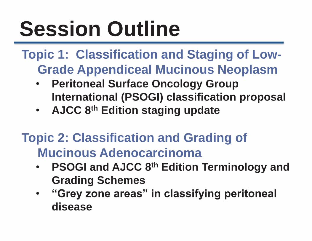
WHO (2010) Diagnostic Terminology for Primary Appendiceal Neoplasms (excluding neuroendocrine tumors)

Non-Invasive Neoplasms – Low-grade appendiceal mucinous neoplasm
(LAMN)
– High-grade appendiceal mucinous neoplasm (HAMN) (new diagnostic category; rare)
– Serrated polyp with or without dysplasia – Conventional adenoma, resembling colorectal
type (rare)
Carr N et al. Am J Surg Pathol 2016;40:14.
PSOGI (2016) Diagnostic Terminology for Primary Appendiceal Neoplasms

Invasive Neoplasms – Mucinous adenocarcinoma
• Mucinous adenocarcinoma with signet ring cells (≤50% signet ring cells)
– Signet ring cell carcinoma (>50% signet ring cells)
– Non-mucinous adenocarcinoma
Carr N et al. Am J Surg Pathol 2016;40:14.
PSOGI (2016) Diagnostic Terminology for Primary Appendiceal Neoplasms
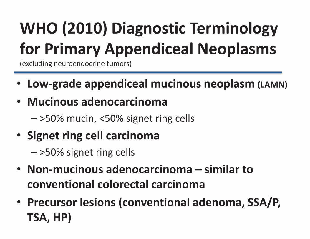
Definition of LAMN (PSOGI 2016) • Mucinous neoplasm with low-grade cytology and
any of the following: • Loss of muscularis mucosae • Fibrosis of submucosa • Undulating or flattened epithelial growth • “Pushing invasion” (expansile or diverticulum like
growth) • Dissection of acellular mucin in the wall • Mucin and/or neoplastic cells outside of the appendix
• Typically see circumferential involvement of the mucosa by a mucin-rich epithelium involving at least 1 segment of the appendix
• Use of the term “mucinous adenoma” was not supported by the majority of the panel.

LAMN: Loss of lamina propria and muscularis mucosae with submucosal fibrosis
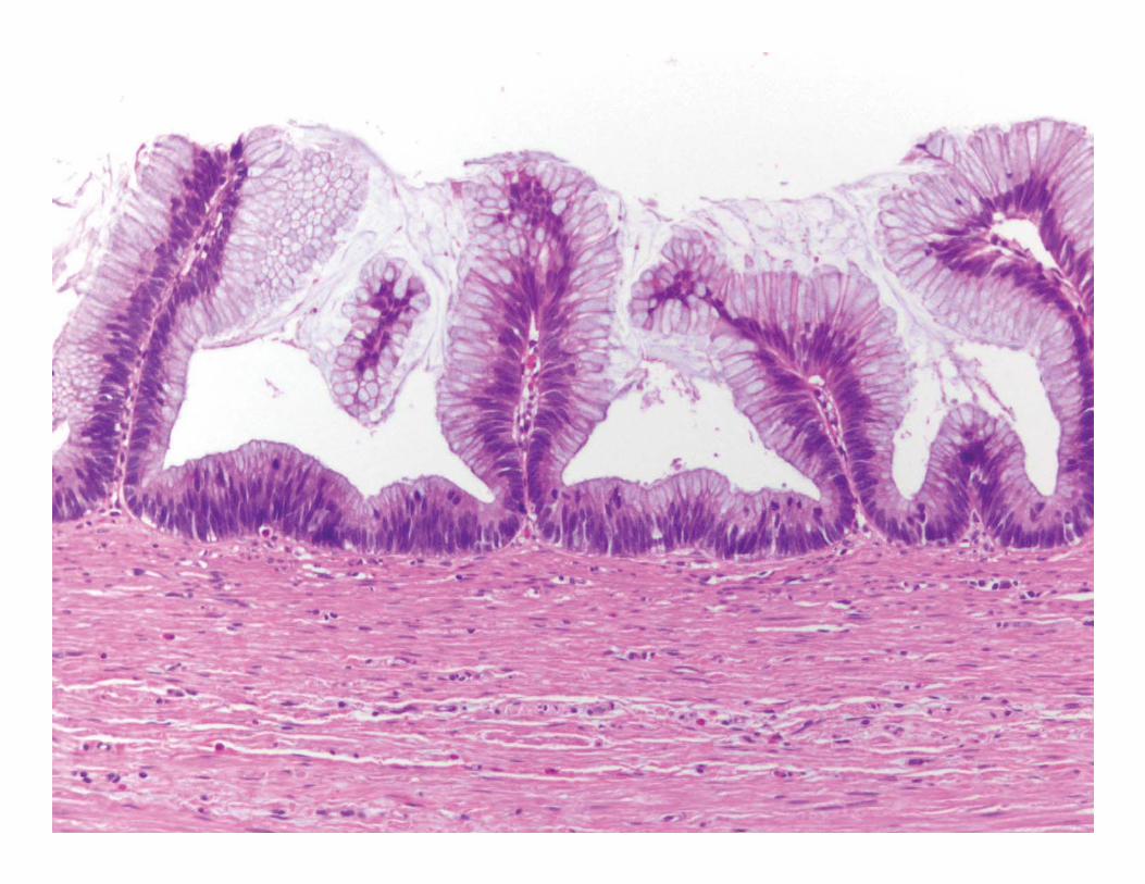

Flattened Epithelium Undulating growth
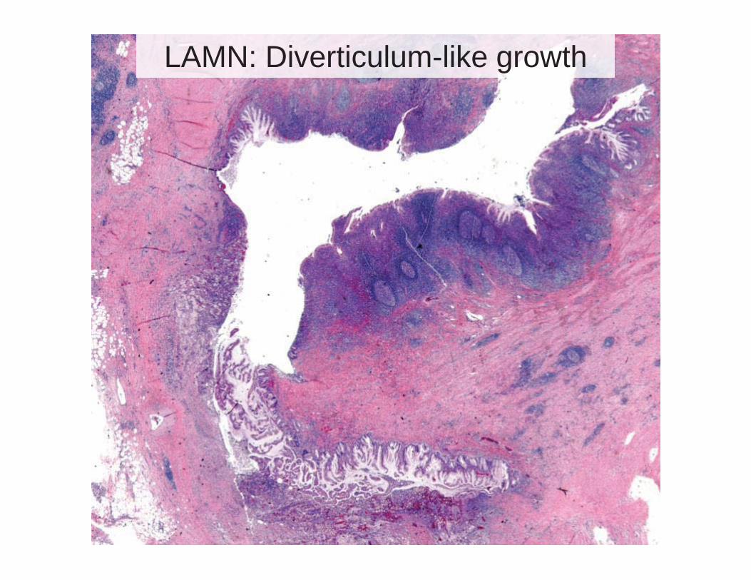
LAMN: Diverticulum-like growth
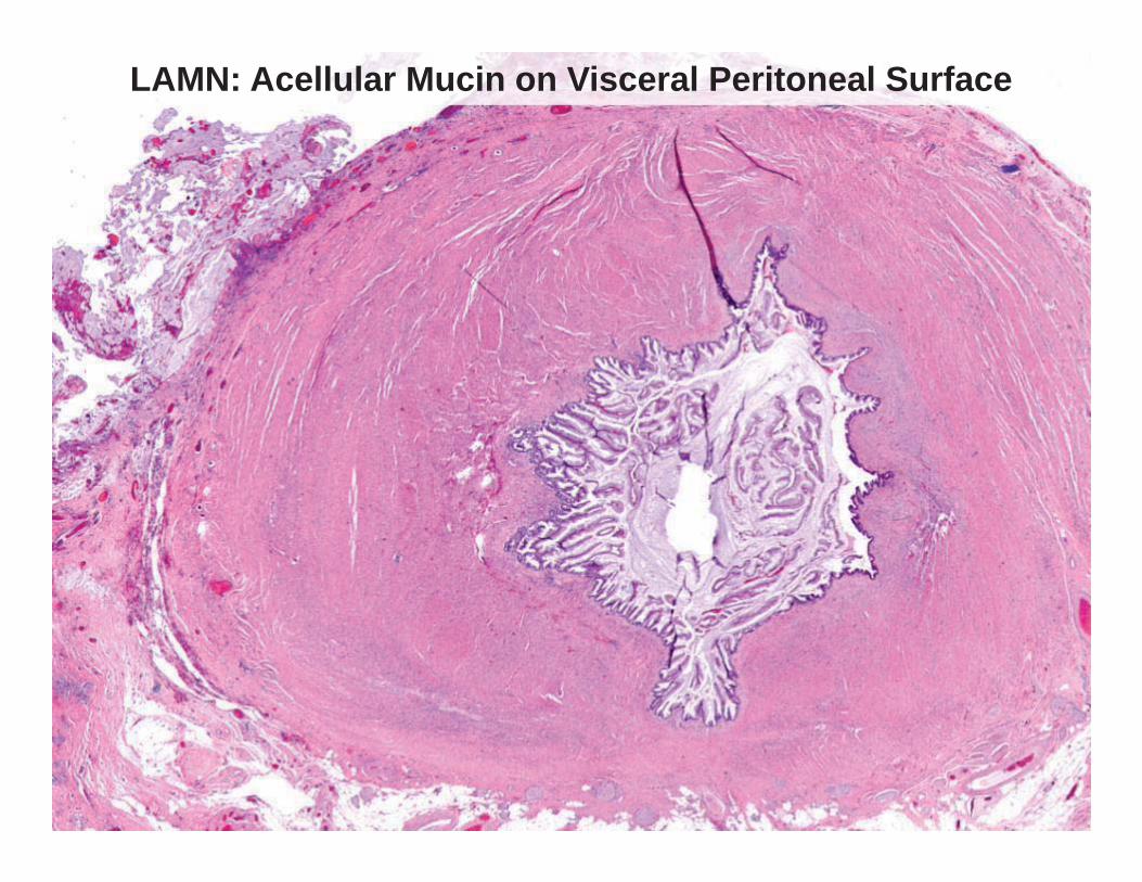
LAMN: Acellular Mucin on Visceral Peritoneal Surface

High-Grade Appendiceal Mucinous Neoplasm (HAMN, New diagnostic category)
• Mucinous neoplasms with high-grade cytology but with the neoplasm confined to the appendix without invasion.
• Very rare neoplasm – must entirely submit the appendix to evaluate for invasion.
• Two-thirds of patients with high-grade cytology without invasion in the primary appendix developed recurrent adenocarcinoma in the peritoneum (including all of the cases reported in the literature1,2, none of the cases had the entire appendix submitted).
( g g y)
1. Misdraji, et al. Am J Surg Pathol 2003; 27:1103. 2. Pai RK, et al. Am J Surg Pathol 2009;33: 1425.

High-Grade Appendiceal Mucinous Neoplasm (HAMN)Low-power features of LAMN
High-Grade Appendiceal Mucinous Neoplasm (HAMN): Low-power features of LAMN
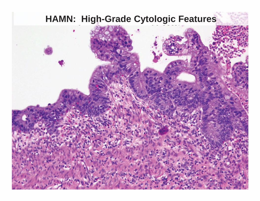
HAMN: High-Grade Cytologic Features

HAMN: High-Grade Cytologic Features

Mimics of Appendiceal Mucinous Neoplasms
• Appendiceal serrated polyps • Ruptured Appendiceal Diverticula • Endometriosis with intestinal metaplasia • Acute appendicitis with mucosal
hyperplasia

Serrated Polyp without Dysplasia
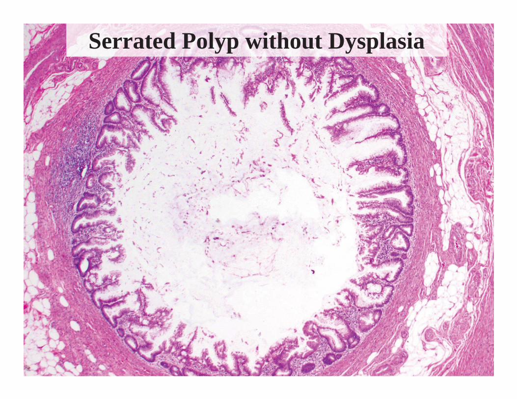
Serrated Polyp without Dysplasia
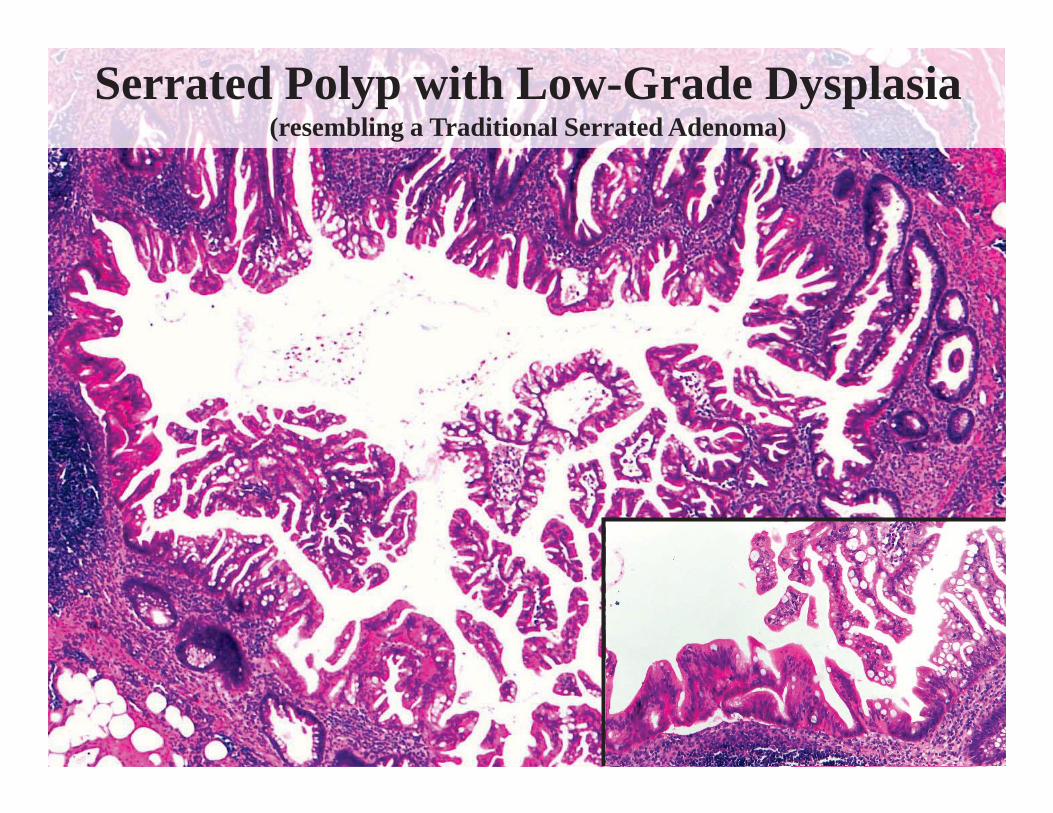
Serrated Polyp with Low-Grade Dysplasia (resembling a Traditional Serrated Adenoma)

LAMN: Should it be staged? • PSOGI Participants:
39 of 60 respondents within the group responded “Yes”.
• When staging LAMN, do you stage
neoplastic epithelium, mucin, or both?
• The AJCC 8th edition provides some clarification
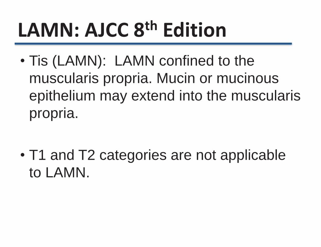
LAMN: AJCC 8th Edition • Tis (LAMN): LAMN confined to the
muscularis propria. Mucin or mucinous epithelium may extend into the muscularis propria.
• T1 and T2 categories are not applicable
to LAMN.
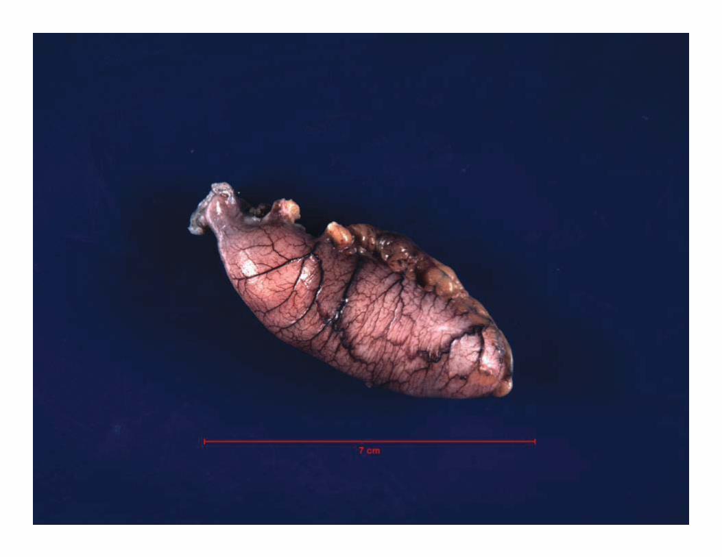
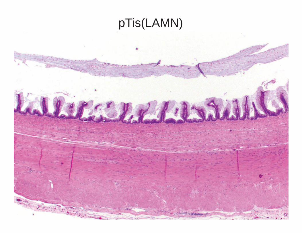
pTis(LAMN)

pTis (LAMN): Pushing into muscularis propria
AJCC 8th Edition: pTis (LAMN) • Most are incidental lesions; patients present
with acute appendicitis (~60%) • Requires that the entire appendix be
submitted for histologic examination. • Literature evidence indicates that patients
with pTis(LAMN) do not develop tumor recurrence and are essentially cured by appendectomy.

LAMN: AJCC 8th Edition T Category Description
T3 Tumor* extends through the muscularis propria into the subserosa or mesoappendix.
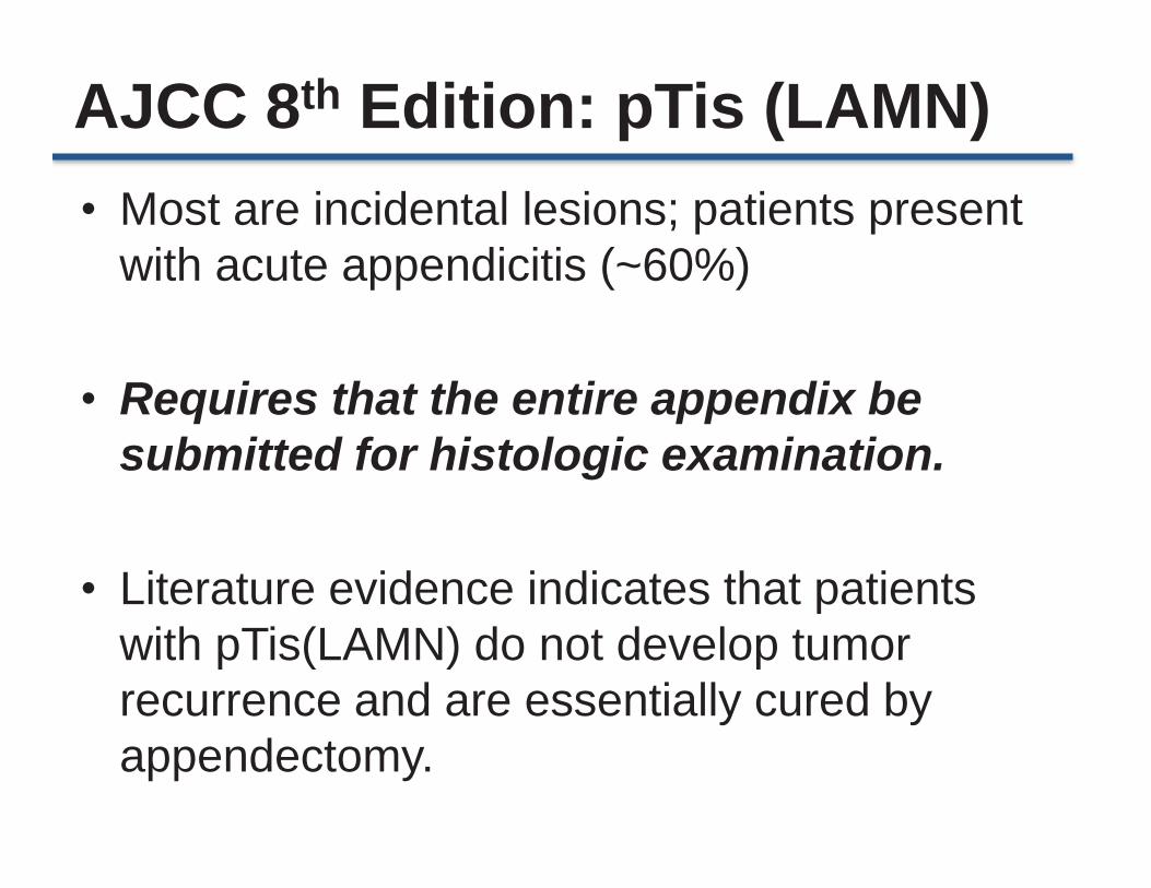
T4a Tumor penetrates the visceral peritoneum, including acellular mucin or mucinous epithelium involving the serosa of the appendix.
T4b Tumor directly involves adjacent organs or structures, including acellular mucin or mucinous epithelium (does not include luminal or mural spread into adjacent cecum).
*For T3 category, “tumor” is not defined. Does acellular mucin count?

Acellular mucin in mesoappendix (likely not pT3 - will have to wait for clarification by authors)
pT4a LAMN Due to Acellular Mucin
pT4a LAMN Due to Cellular Mucin

pT4a LAMN Due to Acellular Mucin • Acellular mucin is present on the visceral
peritoneal surface. • Potential for over-staging: acellular mucin
may be seen on the serosal surface due to “carry-over” related to specimen handling.
• Of the cases reported in the literature1,2 , ~5% patients with acellular disease localized to the right lower quadrant have developed recurrence.
1. Yantiss RK, et al. Am J Surg Pathol 2009; 33:248. 2. Pai RK, et al. Am J Surg Pathol 2009;33: 1425.

Mucin on visceral peritoneal surface with inflammatory reaction and neovascularization

Mucin on visceral peritoneal surface due to “carry-over” from sectioning
Potential for over-staging LAMN, as knife artifact can “carry-over” mucin onto the surface of the serosa.

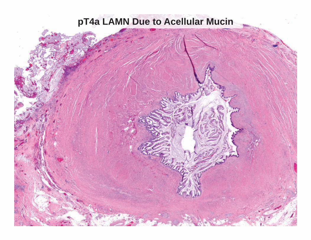
pT4a LAMN Due to Cellular Mucin
• Extra-appendiceal neoplastic epithelium is present within mucin on the visceral peritoneal surface of the appendix.
• For tumors localized to the right lower quadrant at initial presentation, 42% of patients with cellular disease on the visceral peritoneal surface of the appendix have developed recurrence1,2.
1. Yantiss RK, et al. Am J Surg Pathol 2009; 33:248. 2. Pai RK, et al. Am J Surg Pathol 2009;33: 1425.
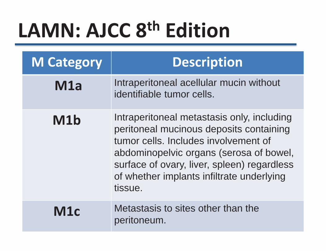
LAMN: AJCC 8th Edition M Category Description
M1a Intraperitoneal acellular mucin without identifiable tumor cells.
M1b Intraperitoneal metastasis only, including peritoneal mucinous deposits containing tumor cells. Includes involvement of abdominopelvic organs (serosa of bowel, surface of ovary, liver, spleen) regardless of whether implants infiltrate underlying tissue.
M1c Metastasis to sites other than the peritoneum.
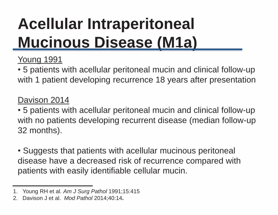
Acellular Intraperitoneal Mucinous Disease (M1a)
1. Young RH et al. Am J Surg Pathol 1991;15:415 2. Davison J et al. Mod Pathol 2014;40:14.
Young 1991 • 5 patients with acellular peritoneal mucin and clinical follow-up with 1 patient developing recurrence 18 years after presentation Davison 2014 • 5 patients with acellular peritoneal mucin and clinical follow-up with no patients developing recurrent disease (median follow-up 32 months).
• Suggests that patients with acellular mucinous peritoneal disease have a decreased risk of recurrence compared with patients with easily identifiable cellular mucin.

Invasive Neoplasms – Mucinous adenocarcinoma
• Mucinous adenocarcinoma with signet ring cells (≤50% signet ring cells)
– Signet ring cell carcinoma (>50% signet ring cells)
– Non-mucinous adenocarcinoma
Carr N et al. Am J Surg Pathol 2016;40:14.
PSOGI (2016) Diagnostic Terminology for Primary Appendiceal Neoplasms

Mucinous Adenocarcinoma • Defined by infiltrative destructive invasion
• High-grade cytologic features present, at least
focally, and may have areas of both low and high cytologic grades.
• May have a signet ring cell component
• PSOGI and AJCC 8th edition advocate a three-tier grading of mucinous neoplasia.
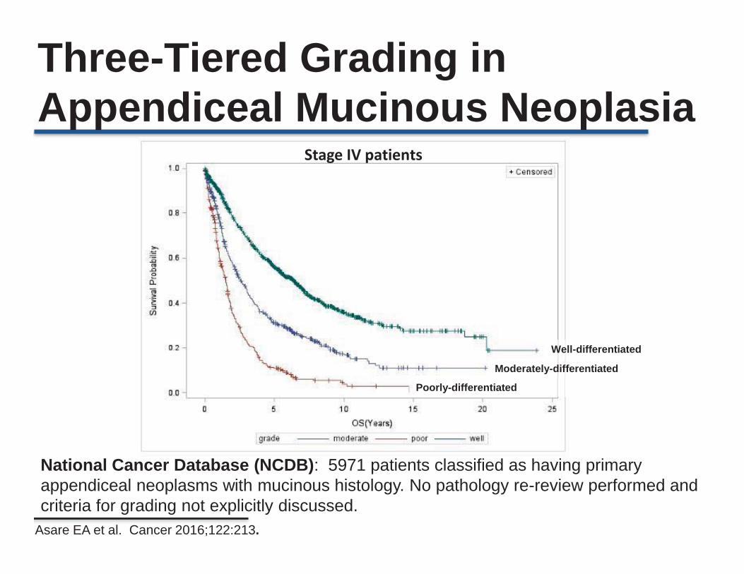
Asare EA et al. Cancer 2016;122:213.
National Cancer Database (NCDB): 5971 patients classified as having primary appendiceal neoplasms with mucinous histology. No pathology re-review performed and criteria for grading not explicitly discussed.
Well-differentiated
Moderately-differentiated
Poorly-differentiated
Stage IV patients
Three-Tiered Grading in Appendiceal Mucinous Neoplasia
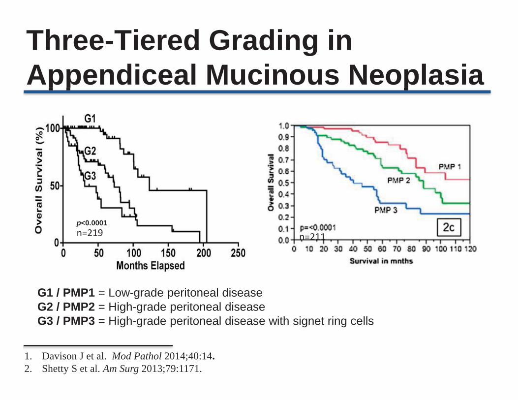
Three-Tiered Grading in Appendiceal Mucinous Neoplasia
p<0.0001
1. Davison J et al. Mod Pathol 2014;40:14. 2. Shetty S et al. Am Surg 2013;79:1171.
G1 / PMP1 = Low-grade peritoneal disease G2 / PMP2 = High-grade peritoneal disease G3 / PMP3 = High-grade peritoneal disease with signet ring cells
n=219 n=211

AJCC (8th Edition) Grade
Characteristics
G1, well differentiated Low cytologic grade (corresponds to LAMN)
G2, moderately differentiated
High cytologic grade without signet ring cells
G3, poorly differentiated High cytologic grade, usually with signet ring cells
1. Davison J et al. Mod Pathol 2014;40:14.
AJCC 8th Edition - Mucinous Neoplasia
• Modification of the scheme proposed by Davison et al. • G2 and G3 mucinous adenocarcinomas are considered
high-grade.

Two-Tiers: Therapeutic Decision Making • Patients with disseminated low-grade (G1) disease benefit from cytoreductive surgery with hyperthermic intraperitoneal chemotherapy (CRS-HIPEC) with no role for systemic chemotherapy.
• Patients with disseminated high-grade (G2 and G3) disease are often treated with systemic chemotherapy with the option of CRS-HIPEC at some institutions. The role of CRS-HIPEC is not entirely well-delineated although is used aggressively at many institutions with evidence of survival benefit.
Two-Tier versus Three-Tier Grading Schemes: Which is Better?
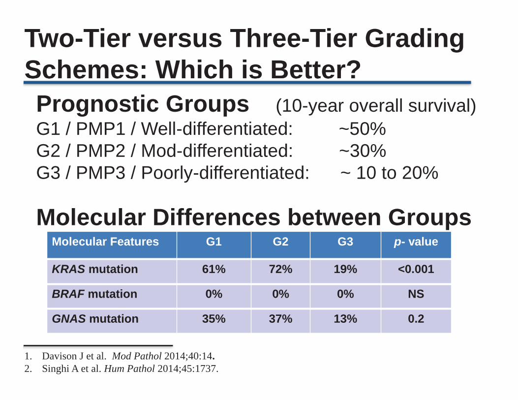
Prognostic Groups (10-year overall survival) G1 / PMP1 / Well-differentiated: ~50% G2 / PMP2 / Mod-differentiated: ~30% G3 / PMP3 / Poorly-differentiated: ~ 10 to 20%
Molecular Features G1 G2 G3 p- value
KRAS mutation 61% 72% 19% <0.001
BRAF mutation 0% 0% 0% NS
GNAS mutation 35% 37% 13% 0.2
Molecular Differences between Groups
1. Davison J et al. Mod Pathol 2014;40:14. 2. Singhi A et al. Hum Pathol 2014;45:1737.
Two-Tier versus Three-Tier Grading Schemes: Which is Better?
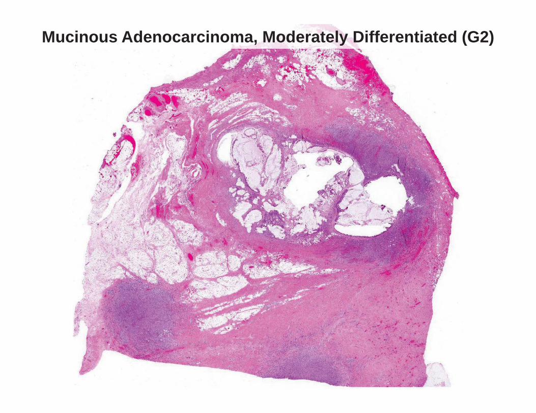
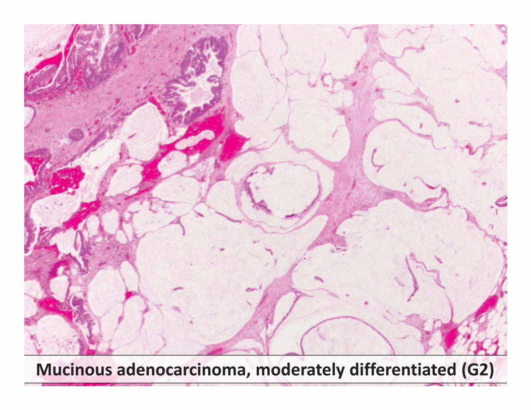
Mucinous Adenocarcinoma, Moderately Differentiated (G2)

Mucinous Adenocarcinoma, Moderately Differentiated (G2)

Mucinous Adenocarcinoma, Moderately Differentiated (G2)
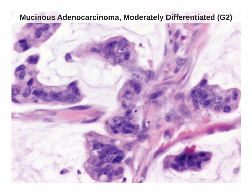
Mucinous Adenocarcinoma, Moderately Differentiated (G2)

Signet Ring Cell Carcinoma (>50% signet ring cells), Poorly Differentiated (G3)
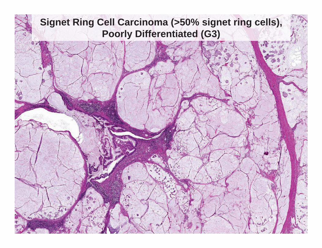
Signet Ring Cell Carcinoma (>50% signet ring cells), Poorly Differentiated (G3)

Signet Ring Cell Carcinoma (>50% signet ring cells), Poorly Differentiated (G3)

AJCC: The Problem of Terminology • Throughout the AJCC 8th edition chapter the terms
“well-differentiated mucinous adenocarcinoma” and “low-grade appendiceal mucinous neoplasm” are used interchangeably.
• In the section on histologic grading, the AJCC states “G1 tumors with peritoneal involvement may be categorized as LAMN with peritoneal involvement”.
• The proposed CAP protocol also states that “in
cancer protocols the histologic type of G1 tumors with peritoneal involvement is best recorded as LAMN”.
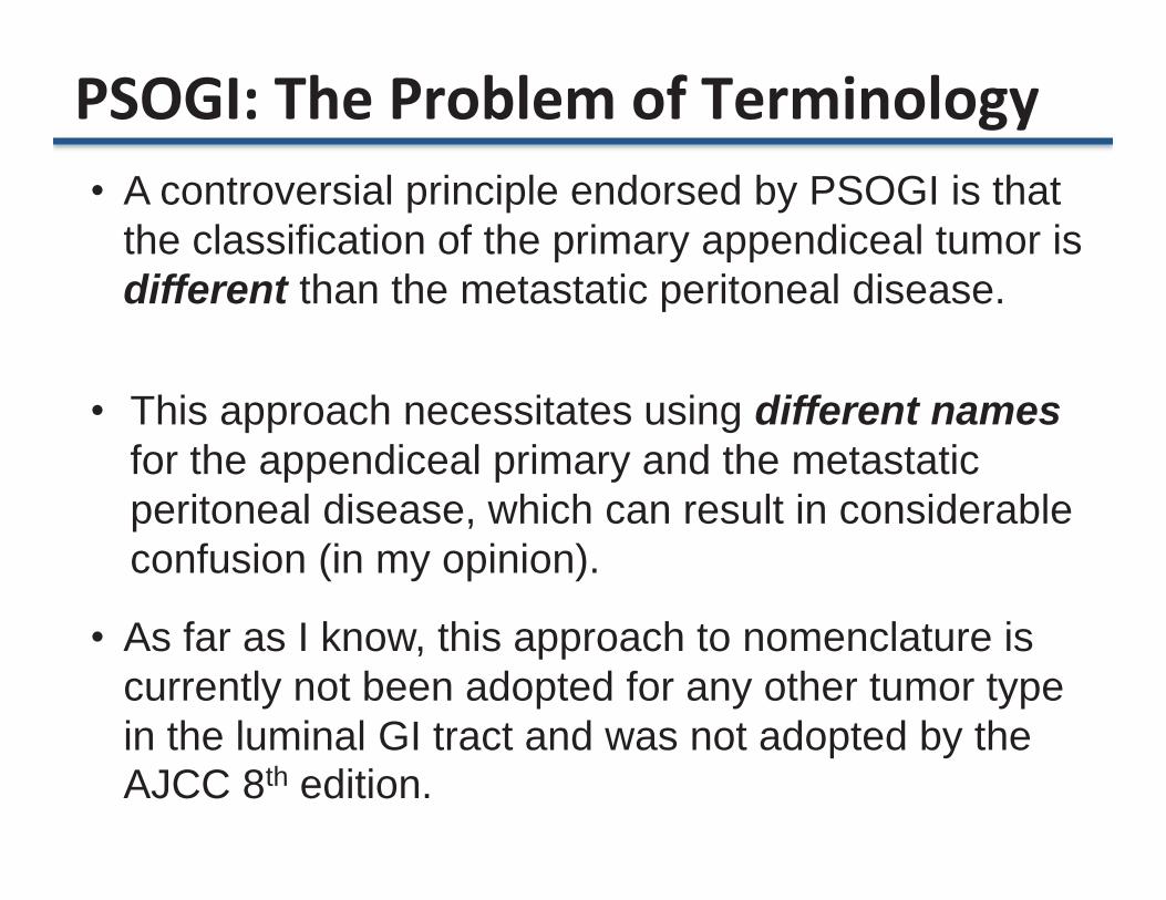
PSOGI: The Problem of Terminology • A controversial principle endorsed by PSOGI is that
the classification of the primary appendiceal tumor is different than the metastatic peritoneal disease.
• This approach necessitates using different names for the appendiceal primary and the metastatic peritoneal disease, which can result in considerable confusion (in my opinion).
• As far as I know, this approach to nomenclature is
currently not been adopted for any other tumor type in the luminal GI tract and was not adopted by the AJCC 8th edition.

Primary Neoplasm PSOGI Terminology for Peritoneal Disease
AJCC Terminology for Primary and Peritoneal
Disease
Low-grade appendiceal
mucinous neoplasm
Low-grade mucinous carcinoma peritonei
Low-grade appendiceal mucinous neoplasm (G1)
Mucinous adenocarcinoma
without signet ring cells
High-grade mucinous carcinoma peritonei
Mucinous adenocarcinoma,
moderately differentiated (G2)
Mucinous adenocarcinoma
with signet ring cells
High-grade mucinous carcinoma peritonei with signet ring cells
Mucinous and/or signet ring cell adenocarcinoma, poorly differentiated (G3)
My opinion: once you decide what to call the primary appendiceal neoplasms, their metastases should align with that name (some exceptions).
AJCC vs. PSOGI: Terminology
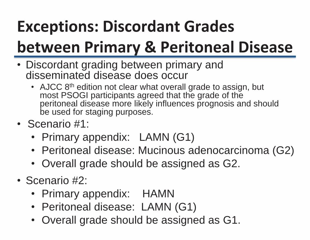
Exceptions: Discordant Grades between Primary & Peritoneal Disease • Discordant grading between primary and
disseminated disease does occur • AJCC 8th edition not clear what overall grade to assign, but
most PSOGI participants agreed that the grade of the peritoneal disease more likely influences prognosis and should be used for staging purposes.
• Scenario #1: • Primary appendix: LAMN (G1) • Peritoneal disease: Mucinous adenocarcinoma (G2) • Overall grade should be assigned as G2.
• Scenario #2:
• Primary appendix: HAMN • Peritoneal disease: LAMN (G1) • Overall grade should be assigned as G1.
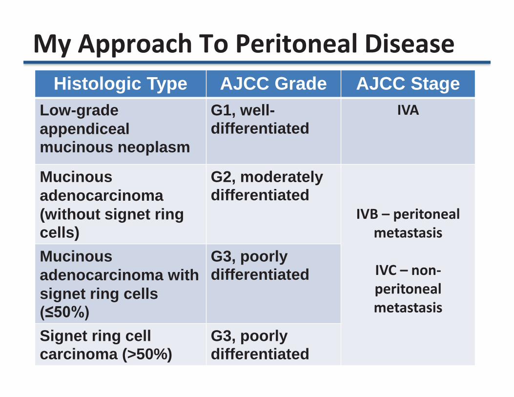
Histologic Type AJCC Grade AJCC Stage Low-grade appendiceal mucinous neoplasm
G1, well-differentiated
IVA
Mucinous adenocarcinoma (without signet ring cells)
G2, moderately differentiated
IVB – peritoneal metastasis
IVC – non-peritoneal metastasis
Mucinous adenocarcinoma with signet ring cells (≤50%)
G3, poorly differentiated
Signet ring cell carcinoma (>50%)
G3, poorly differentiated
My Approach To Peritoneal Disease
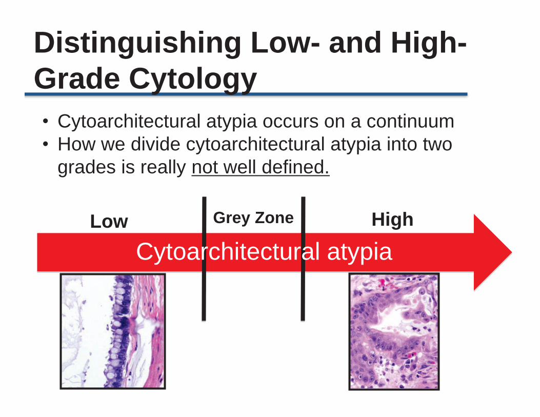
• Cytoarchitectural atypia occurs on a continuum • How we divide cytoarchitectural atypia into two
grades is really not well defined.
Cytoarchitectural atypia Low High
Distinguishing Low- and High- Grade Cytology
Grey Zone

Histologic Features of Low-Grade (G1) Peritoneal Disease • Low cytologic grade • Low cellularity at low-power (2x objective)
magnification • Lacks the following features:
– Any high-grade cytology – Destructive stromal and/or organ invasion – Desmoplasia – Lymph node metastases (rare cases reported) – Signet ring cells

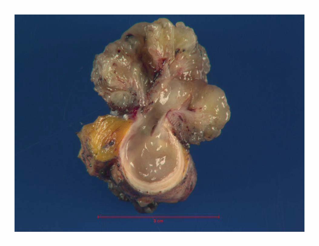
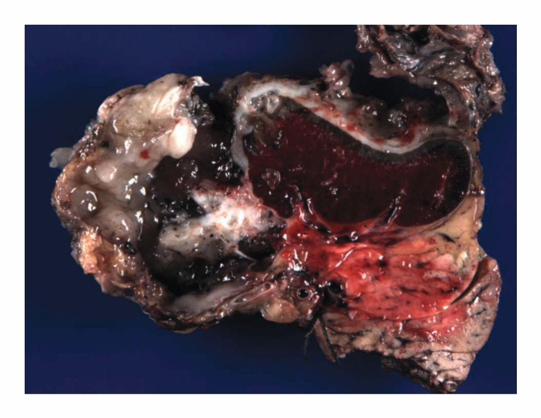
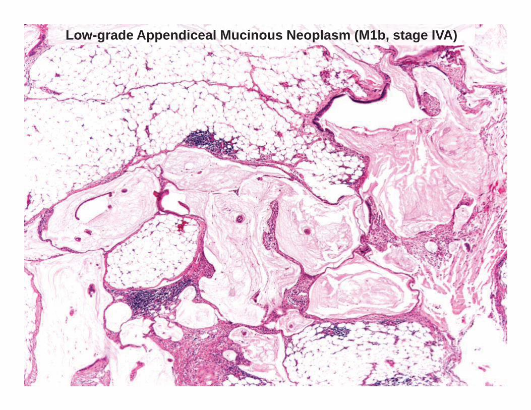
Low-grade Appendiceal Mucinous Neoplasm (M1b, stage IVA)

Low-grade Appendiceal Mucinous Neoplasm (M1b, stage IVA)

Mucinous Adenocarcinoma, Moderately Differentiated (G2) • High-grade cytology
– Mix of low-grade and high-grade areas (common) – Diffuse high-grade cytology
• Destructive stromal and/or organ invasion
• High cellularity at low-power (2x objective)
magnification (~75% of cases)
• Lymph node metastases in ~20% of cases

Criteria for High-Grade Cytology in Peritoneal Disease
• Use criteria similar to that employed for the rest of the luminal GI tract: – Marked nuclear enlargement and rounding of
the nuclei – Nuclear hyperchromasia – Irregular chromatin – Macronucleoli – Increased mitotic activity – Loss of nuclear polarity
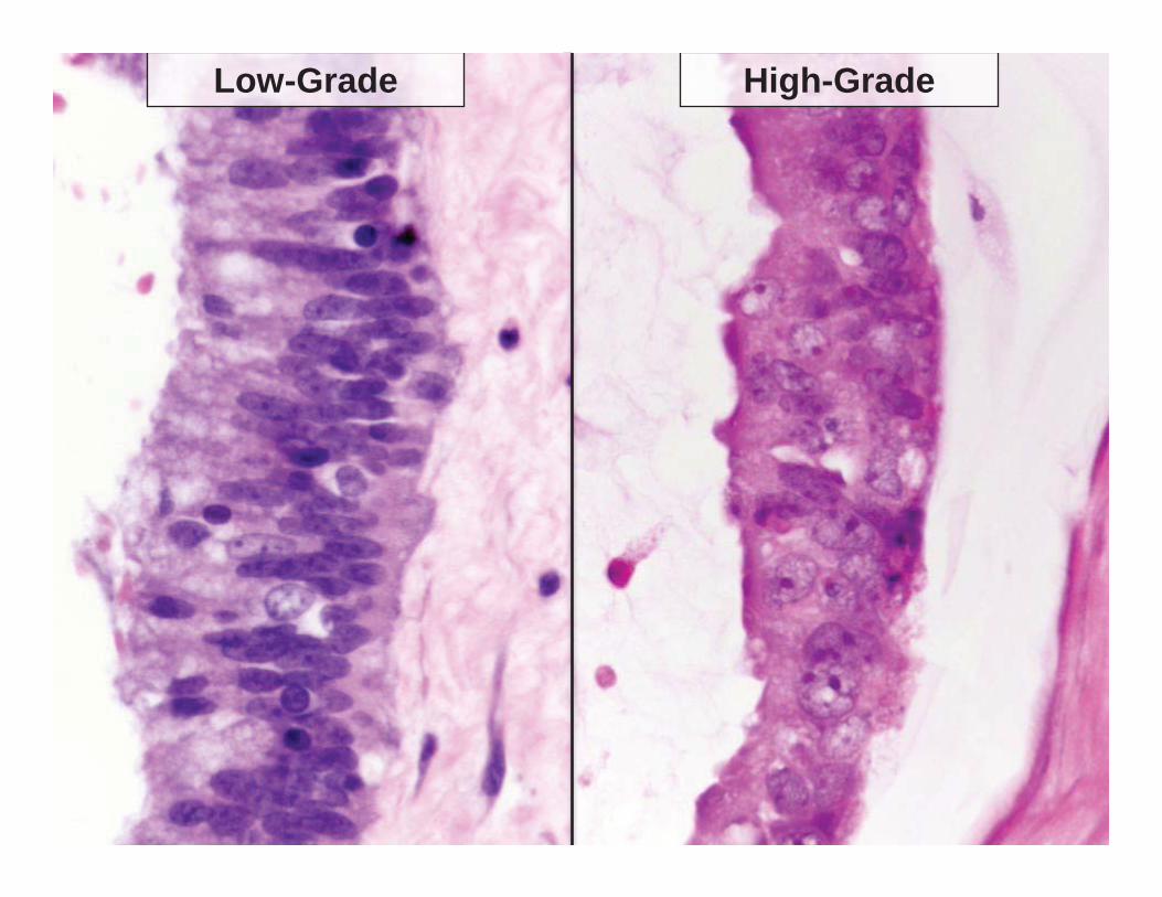
Low-Grade High-Grade
Mucinous adenocarcinoma, moderately differentiated (G2)

Grade Heterogeneity

Patterns of Stromal/Organ Invasion
• Numerous small epithelial clusters floating in small pools of mucin (common pattern of invasion)
• Tubule formation within desmoplastic stroma
• Predominance of complex architectural growth (cribriform)

Small Mucin Pool Pattern of Invasion

Small Mucin Pool Pattern of Invasion with Diffuse High-Grade Cytology
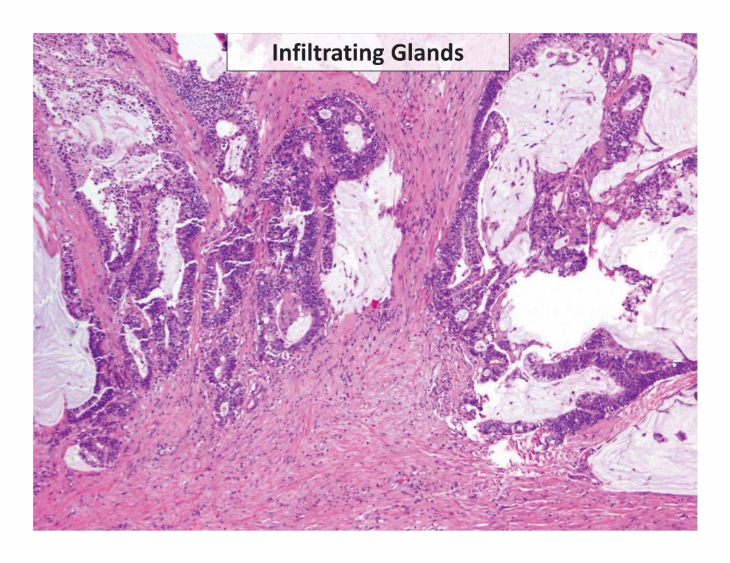
Infiltrating Glands
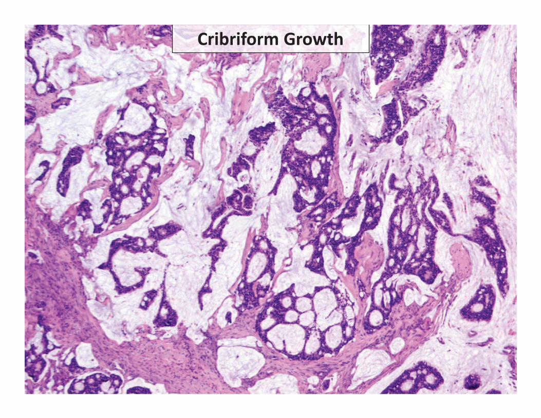
Cribriform Growth
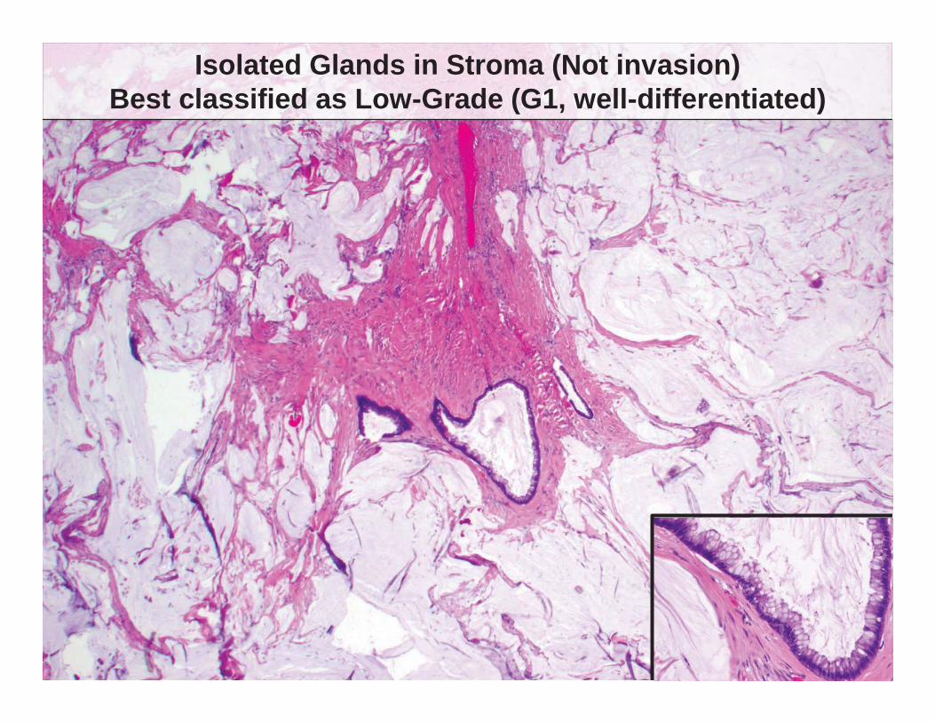
Isolated Glands in Stroma (Not invasion)Best classified as Low-Grade (G1, well-differentiated)
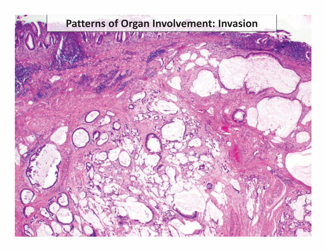
Patterns of Organ Involvement: Invasion
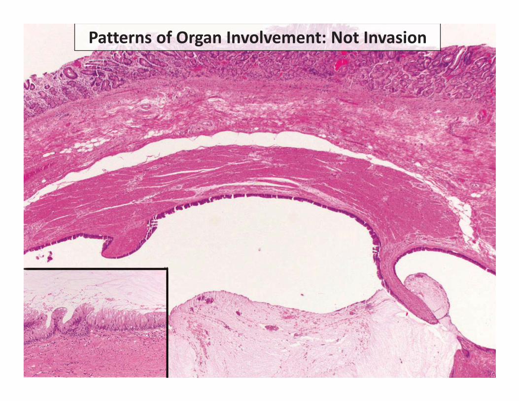
Patterns of Organ Involvement: Not Invasion
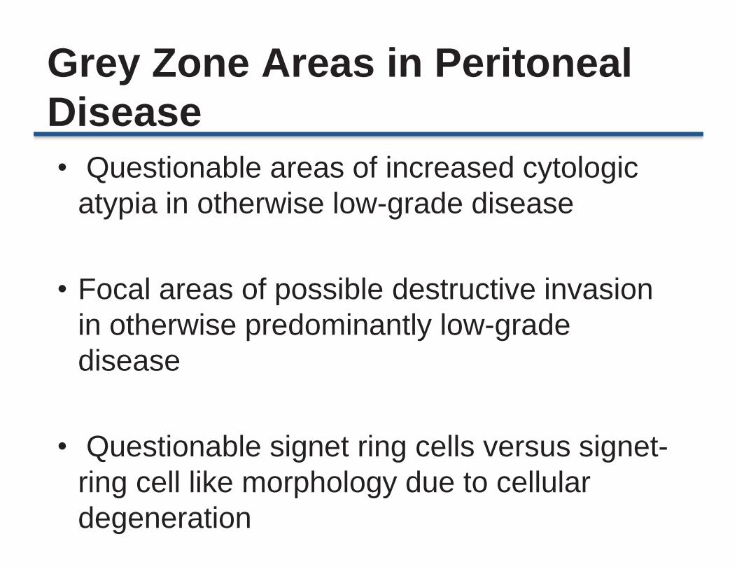
Grey Zone Areas in Peritoneal Disease • Questionable areas of increased cytologic
atypia in otherwise low-grade disease
• Focal areas of possible destructive invasion in otherwise predominantly low-grade disease
• Questionable signet ring cells versus signet-ring cell like morphology due to cellular degeneration

Predominantly Low-grade with areas of increased proliferation
Microscopic area (single field & <10% of tumor) with increased cytologic atypia
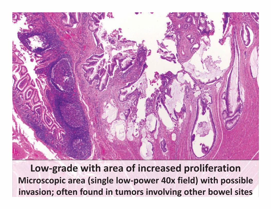
Low-grade with area of increased proliferation Microscopic area (single low-power 40x field) with possible invasion; often found in tumors involving other bowel sites

Predominantly Low-Grade Disease with Areas of Increased Proliferation
• Eight patients: none progressed to high-grade disease on follow-up and none died of disease at last clinical follow-up (median follow-up 94 months)
• Trend to accelerated time to progression compared to other patients with low-grade neoplasms (HR 2.4, 0.82-7.2 but not statistically significant)

Distinguishing Between Grade G2 and Grade G3 in Mucinous Neoplasia • In general, G3 tumors are defined by the
presence of signet ring cells
• How many signet ring cells are required to classify a lesion as G3?
• Is there a difference between signet ring cells floating within mucin and infiltrating signet ring cells?

Floating Signet Ring Cells Infiltrating Signet Ring Cells
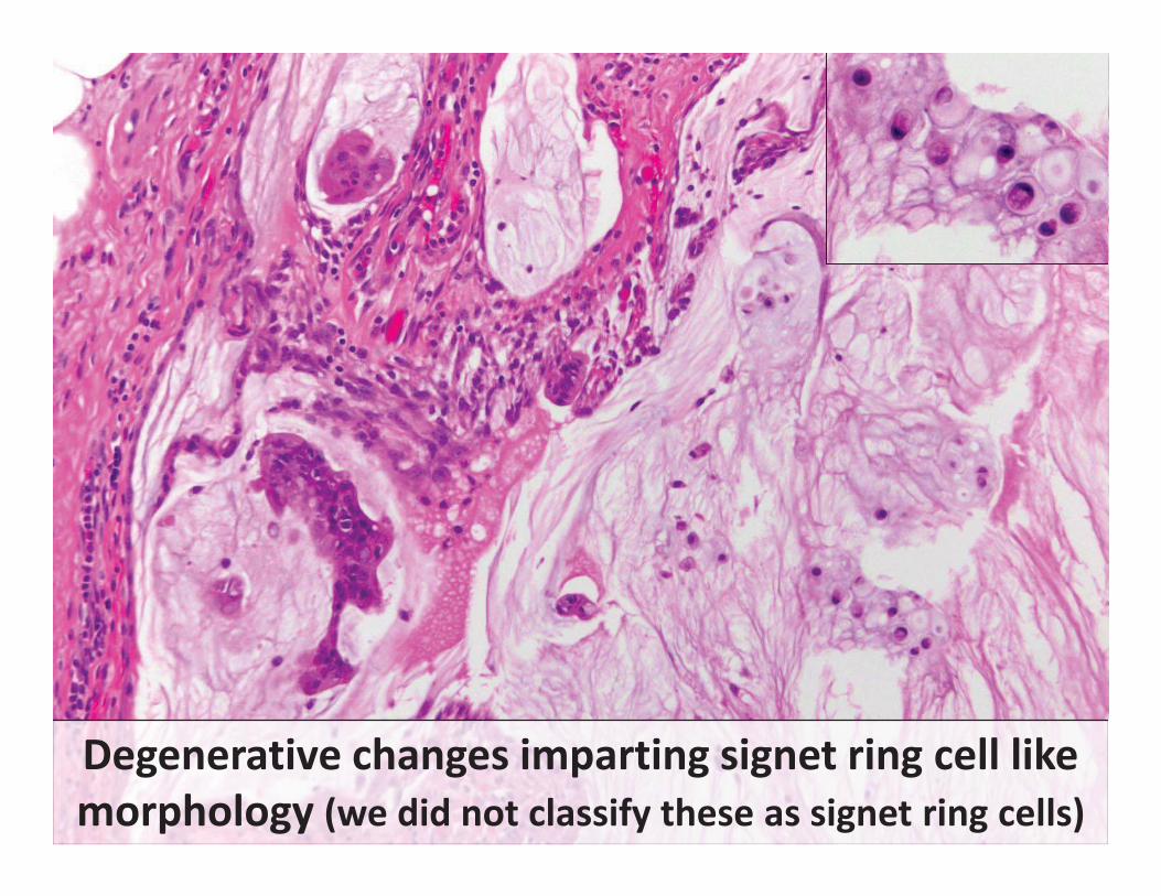
Degenerative changes imparting signet ring cell like morphology (we did not classify these as signet ring cells)
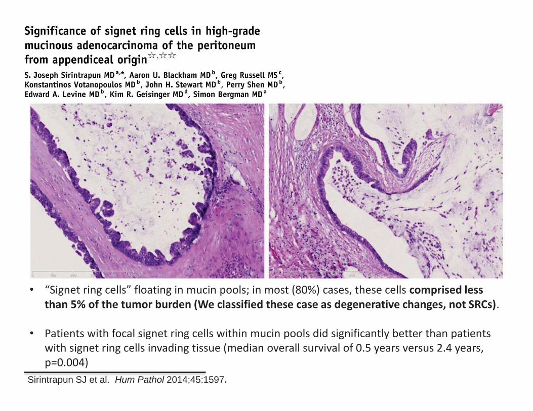
Sirintrapun SJ et al. Hum Pathol 2014;45:1597.
• “Signet ring cells” floating in mucin pools; in most (80%) cases, these cells comprised less than 5% of the tumor burden (We classified these case as degenerative changes, not SRCs).
• Patients with focal signet ring cells within mucin pools did significantly better than patients
with signet ring cells invading tissue (median overall survival of 0.5 years versus 2.4 years, p=0.004)

Additional Issues with Grading
• Grade progression can occur • Patients with low-grade disease can recur with high-grade
(G2) disease
• Assessing grade on small biopsy samples • G2 tumors can show a mix of low and high cytologic
grades. • A small biopsy may only sample low-grade areas.

Conclusions
• PSOGI provided diagnostic criteria for low-grade appendiceal mucinous neoplasms and the AJCC 8th edition clarified staging of LAMN.
• AJCC and PSOGI diagnostic terminology for appendiceal mucinous neoplasms. – Importance of distinguishing between low-grade
mucinous neoplasm and high-grade mucinous adenocarcinoma with or without signet ring cells.
• “Grey zone” cases and discordant grading exist resulting in issues with reproducibility in grade assessment