Embed Size (px)
Citation preview
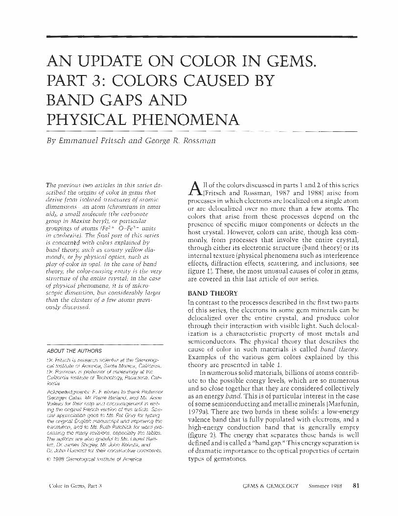
AN UPDATE ON COLOR IN GEMS. PART 3: COLORS CAUSED BY BAND GAPS AND PHYSICAL PHENOMENA By Emmanuel Fritsch and George R. Rossman
The previous two articles in this series de- scribed the origins of color in gems that derive from isolated structures of atomic dimensions-an atom (chromium in emer- ald), a small molecule (the carbonate group in Maxixe beryl), or particular groupings of atoms (Fez +-0-Fe:f + units in cordieiite). The final part of this series is concerned with colors explained by band theoiy, such as canary yellow &a- monds, o r b y physical optics, such as play-of-color in opal. In the case of band theory, the color-causing entity is the very structure of the entire crystal; in the case of physical phenomena, it is of inicro- scopic dimension, but considerably larger than the clusters of a few atoms previ- ously discussed.
ABOUT THE AUTHORS
Dr. Fritsch is research scientist at the Gemologi- cal Institute 01 America, Santa Monica, Calilornia. Dr. Rossman is prolessor of mineralogy at the California Institute of Technology, Pasadena, Cali- lornia.
Acknowledgments: E . F. wishes to thank Professor Georges Calas, Mr. Pierre Bariand, and Ms. Anne Voileau for their help and encouragement in writ- ing the original French version of this article. Spe- cial appreciation goes to Ms. Pat Gray lor typing the original English manuscript and improving the translation, and to Ms. Ruth Patchick for word pro- cessing the many revisions, especially the tables. The authors are also grateful to Ms, Laurel Bart- let!, Dr. James Shigley, Mr. John Koivula, and Dr. John Hummel tor their constructive comments.
0 1988 Gemological Institute of America
A 11 of the colors discussed in parts 1 and 2 of this series (Fritsch and Rossman, 1987 and 1988) arise from
processes in which electrons are localized on a single atom or are delocalized over no more than a few atoms. The colors that arise from these processes depend on the presence of specific minor components or defects in the host crystal. However, colors can arise, though less com- monly, from processes that involve the entire crystal, through either its electronic structure (band theory) or its internal texture (physical phenomena such as interference effects, diffraction effects, scattering, and inclusions; see figure 1). These, the most unusual causes of color in gems, are covered in this last article of our series.
BAND THEORY In contrast to the processes described in the first two parts of this series, the electrons in some gem minerals can be delocalized over the entire crystal, and produce color through their interaction with visible light. Such delocal- ization is a characteristic property of most metals and semiconductors. The physical theory that describes the cause of color in such materials is called band theory. Examples of the various gem colors explained by this theory are presented in table 1.
In numerous solid materials, billions of atoms contrib- ute to the possible energy levels, which are so numerous and so close together that they are considered collectively as an energy band. This is of particular interest in the case of some semiconducting and metallic minerals (Marfunin, 1979a). There arc two bands in these solids: a low-energy valence band that is fully populated with electrons, and a high-energy conduction band that is generally empty (figure 2). The energy that separates these bands is well defined and is called a "band gap." This energy separation is of dramatic importance to the optical properties of certain types of gemstones.
Color in Gems, Part 3 GEMS & GEMOLOGY Summer 1988 81
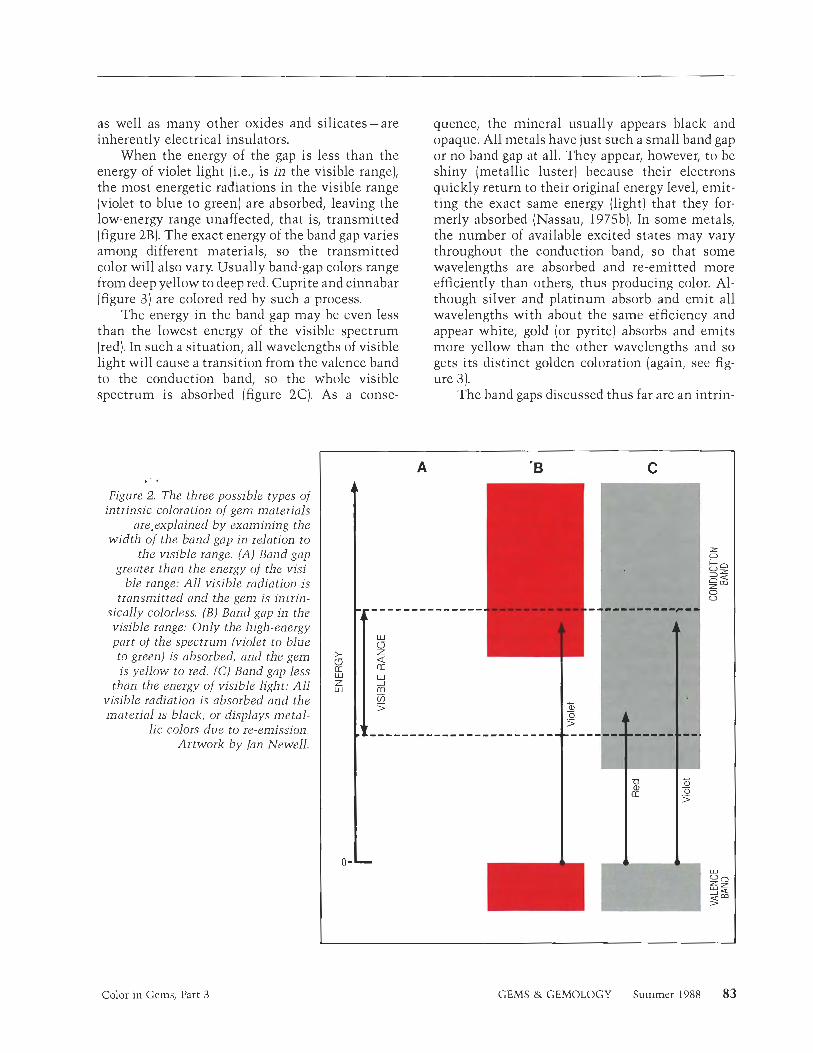
For these gemstones, transitions between bands rather than between energy levels of single atoms are responsible for the color. These "inter- band transitions" occur when electrons from the valence band receive sufficient energy by absorb- ing light to "jump" over the band gap and reach the conduction band. As illustrated in figure 2, three different scenarios are possible for interband tran- sitions.
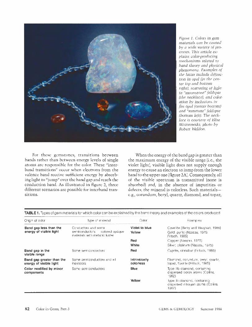
Figure 1. Colors in gem materials can be caused by a wide variety of pro- cesses. This article ex- plains color-producing mechanisms related to band theory and physical phenomena. Examples of the latter include diffrac- tion in opal (in the cen- ter top and bottom right), scattering of light in "moonstone" feldspar (the necklace), and color- ation by inclusions in fire opal (center bottom) and "siinstone" feldspar (bottom left). The neck- lace is courtesy of Elise Misiorowski; photo by Robert Weldon.
When the energy of the band gap is greater than the maximum energy of the visible range (i.e., the violet light), visible light does not supply enough energy to cause an electron to jump from the lower band to the upper one (figure 2A). Consequently, all of the visible spectrum is transmitted (none is absorbed) and, in the absence of impurities or defects, the mineral is colorless. Such materials - e.g., corundum, beryl, quartz, diamond, and topaz,
TABLE 1. Types of gem materials for which color can be explained by the band theory and examples of the colors produced,
Origin of color Type of material Color Examples
Band gap energy of
less than the Conductors and some Violet to blue visible light semiconductors = colored opaque Yellow
materials with metallic luster
Red White
Red Band gap in the Some semiconductors visible range
Band gap greater than the Some semiconductors and all energy of visible light insulators
Color modified by minor components
Some semiconductors
Intrinsically colorless
Blue
Yellow
Covellite (Berry and Vaughan, 1985) Gold, pyrite (Nassau, 1975; Fritsch, 1985) Copper (Nassau, 1975) Silver, platinum (Nassau, 1975)
Cuprite, cinnabar (Fritsch, 1985)
Diamond, corundum, beryl, quartz, topaz, fluorite (Fritsch, 1985)
Type llb diamond, containing dispersed boron atoms (Collins, 1982) Type Ib diamond, containing dispersed nitrogen atoms (Collins, 1982)
82 Color in Gems, Part 3 GEMS & GEMOLOGY Summer 1988
as well as many other oxides and silicates-are inherently electrical insulators.
When the energy of the gap is less than the energy of violet light (i.e., is in the visible range), the most energetic radiations in the visible range (violet to blue to green] are absorbed, leaving the low-energy range unaffected, that is, transmitted (figure 2B). The exact energy of the band gap varies among different materials, so the transmitted color will also vary. Usually band-gap colors range from deep yellow to deep red. Cuprite and cinnabar (figure 3) are colored red by such a process.
The energy in the band gap may be even less than the lowest energy of the visible spectrum (red). In such a situation, all wavelengths of visible light will cause a transition from the valence band to the conduction band, so the whole visible spectrum is absorbed (figure 2C). As a conse-
quence, the mineral usually appears black and opaque. All metals have just such a small band gap or no band gap at all. They appear, however, to be shiny (metallic luster) because their electrons quickly return to their original energy level, emit- ting the exact same energy (light) that they for- merly absorbed (Nassau, 1975b). In some metals, the number of available excited states may vary throughout the conduction band, so that some wavelengths are absorbed and re-emitted more efficiently than others, thus producing color. Al- though silver and platinum absorb and emit all wavelengths with about the same efficiency and appear white, gold (or pyrite) absorbs and emits more yellow than the other wavelengths and so gets its distinct golden coloration (again, see fig- ure 3).
The band gaps discussed thus far are an intrin-
. . Figure. 2..The three possible types of
intrinsic coloration of gem materials are.explained by examining the
width of the band gap in relation to the visible range. (A) Band gap
greater than the energy of the visi- ble range: All visible radiation i s
transmitted and the gem is intrin- sically colorless. (B ) Band gap in the visible range: Only the high-energy part of the spectrum (violet to blue to green) is absorbed, and the gem is yellow to red. (C) Band gap less
than the energy o f visible light: All visible radiation is absorbed and the material is black, or displays metal-
lic colors due to re-emission. Artwork by Ian Newell.
Color in Gems, Part 3 GEMS & GEMOLOGY Summer 1988 83
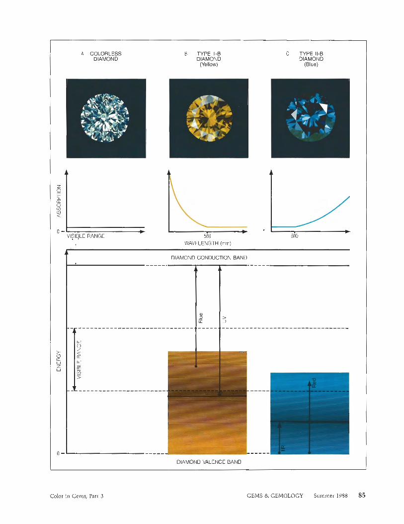
sic property of the material; they are ultimately directly related to its chemical composition and atomic structure. I11 some semiconductors, how- ever, color is caused by small amounts of impurity atoms that normally do not produce color in intrinsically colorless minerals. Specifically, these atoms can introduce electronic energy levels at an energy between the valence band and the conduc- tion band of the host mineral (see figure 4). Some of the most striking examples are canary yellow and fancy blue diamonds, which contain isolated nitro- gen and boron atoms, respectively. Although pure (colorless) diamonds are insulators, they may also be considered semiconductors with a band gap so large that they have neither color nor appreciable electrical conductivity (figure 4A). Nitrogen can easily substitute for carbon, which it follows in the periodic table of elements. Because nitrogen pos- sesses one more electron than carbon, however, it becomes an electron "donor" when i t substitutes for carbon in diamond. This additional electron contributes an additional energy level situated above the diamond valence band, but below the diamond conduction band (figure 4B). However, because this donor level has a finite width, light of a variety of wavelengths extending from the ultra- violet into the visible range up to 560 nm (green) will be absorbed, creating a strong yellow color. This type of coloration occurs only in type Ib diamonds, in which isolated nitrogen atoms sub- stitute for carbon atoms in the proportion of about 1 to 100,000 (Collins, 1982). This color is distinct from the yellow color commonly caused by the
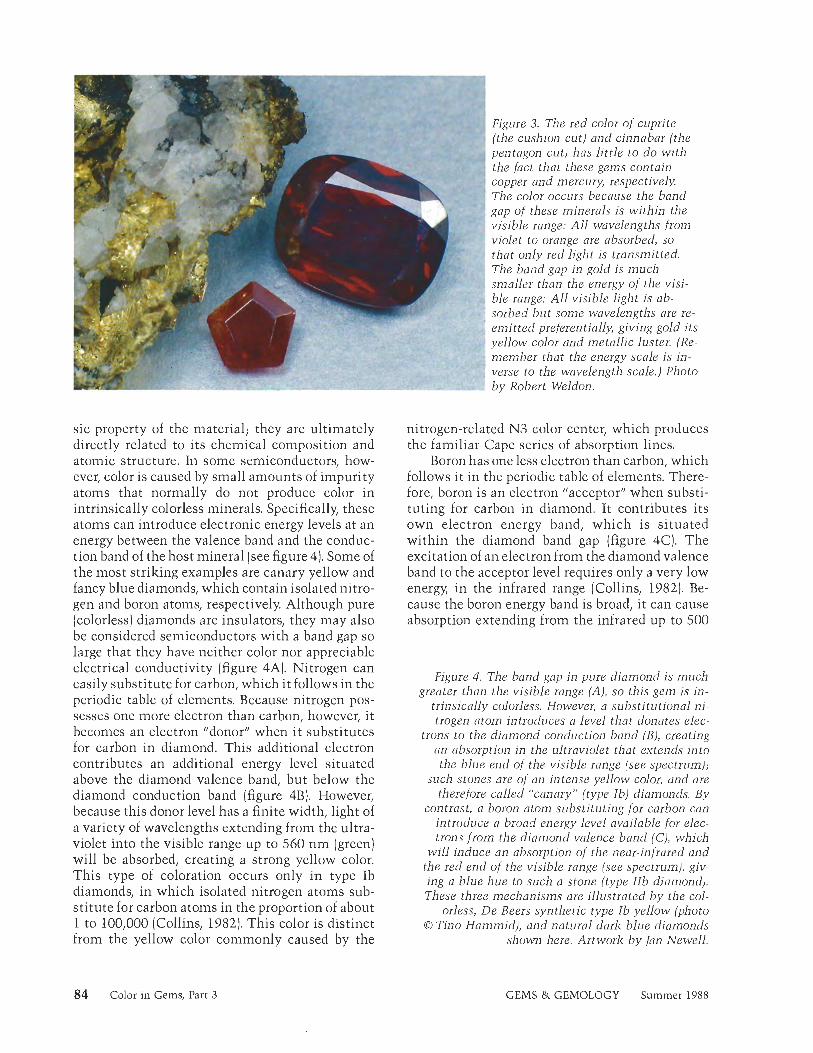
Figure 3. The red color of cuprite ( the cushion cut) and cinnabar ( the pentagon cut) has little t o d o wi th the fact that these gems contain copper and mercury, respectively. The color occurs because the band gap of these minerals i s within the visible range: All wavelengths from violet t o orange are absorbed, so that only red light is transmitted. The band gap in gold is much smaller than the energy of the visi- ble range: All visible light i s ab- sorbed but some wavelengths are re- emit ted preferentially, giving gold i t s yellow color and metallic luster. (Re- member that the energy scale i s in - verse to the wavelength scale.) Photo b y Robert Weldon.
nitrogen-related N3 color center, which produces the familiar Cape series of absorption lines.
Boron has one less electron than carbon, which follows it in the periodic table of elements. There- fore, boron is an electron "acceptor" when substi- tuting for carbon in diamond. It contributes its own electron energy band, which is situated within the diamond band gap (figure 4C). The excitation of an electron from the diamond valence band to the acceptor level requires only a very low energy, in the infrared range (Collins, 1982). Be- cause the boron energy band is broad, it can cause absorption extending from the infrared up to 500
Figure 4. The band gap i n pure diamond i s much greater than the visible range (A), so this gem is in-
trinsically colorless. However, a substitution a1 ni- trogen atom introduces a level that donates elec-
trons t o the diamond conduction band (B), creating an absorption in the ultraviolet that extends in to the blue end of the visible range (see spectrum);
such stones are of an intense yellow color, and are therefore called "canary" (type Ib) diamonds. By
contrast, a boron atom substituting for carbon can introduce a broad energy level available for elec- trons f rom the diamond valence band (C), which
will induce an absorption of the near-infrared and the red end o f the visible range (see spectrum), giv- ing a blue hue to such a stone (type l l b diamond). These three mechanisms are illustrated b y the col-
orless, De Beers synthetic type Ib yellow (photo 0 Tino Hammid) , and natural dark blue diamonds
shown here. Artwork b y Jan Newell.
84 Color in Gems, Part 3 GEMS & GEMOLOGY Summer 1988
A COLORLESS DIAMOND
0 - VISIBLE RANGE
B TYPE 1-B DIAMOND
(Yellow)
--
'I- ^ C TYPE 11-0
DIAMOND (Blue)
. I WAVELENGTH (nm)
t DIAMOND CONDUCTION BAND
DIAMOND VALENCE BAND
Color in Gems, Part 3 GEMS &. GEMOLOGY Summer 1988 85
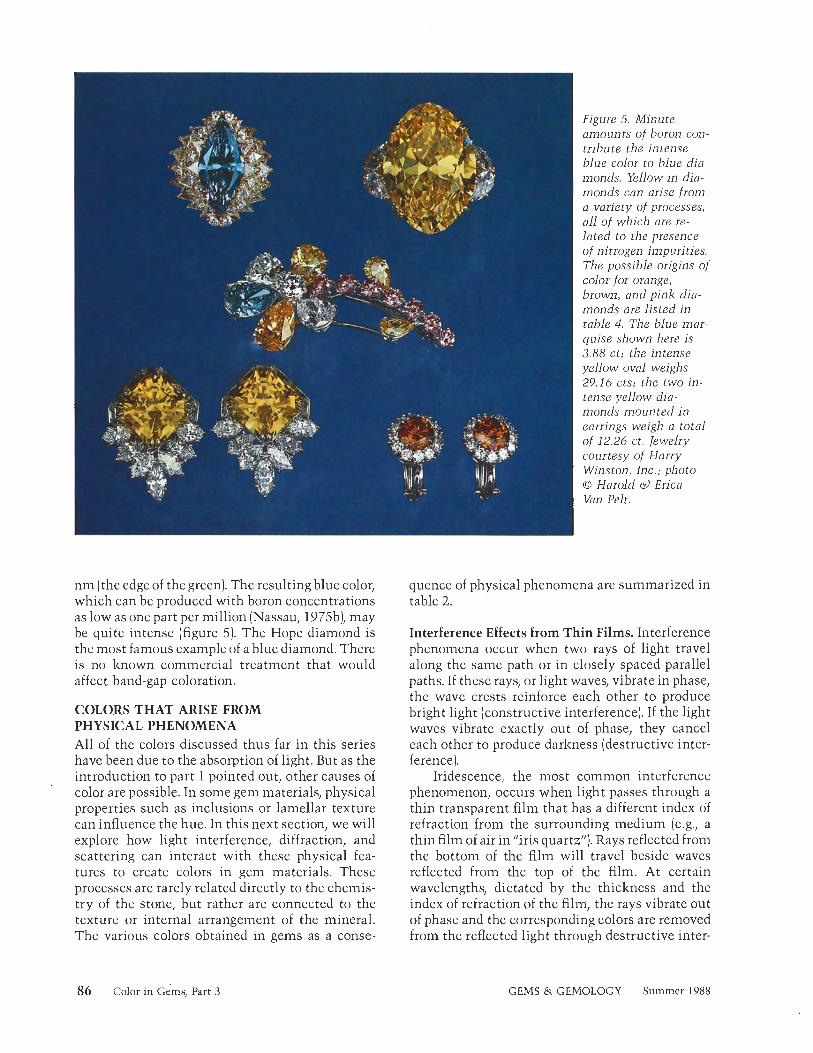
n m (the edge of the green). The resulting blue color, which can be produced with boron concentrations as low as one part per million (Nassau, 1975b), may be quite intense (figure 5). The Hope diamond is the most famous example of a blue diamond. There is no known commercial treatment that would affect band-gap coloration.
COLORS THAT ARISE FROM PHYSICAL PHENOMENA All of the colors discussed thus far in this series have been due to the absorption of light. But as the introduction to part 1 pointed out, other causes of color are possible. In some gem materials, physical properties such as inclusions or lamellar texture can influence the hue. In this next section, we will explore how light interference, diffraction, and scattering can interact with these physical fea- tures to create colors in gem materials. These processes are rarely related directly to the chemis- try of the stone, but rather are connected to the texture or internal arrangement of the mineral. The various colors obtained in gems as a conse-
Figure 5 . Minute amounts of boron con- tribute the intense blue color to blue dia- monds. Yellow in dia- monds can arise from a variety of processes, 011 of which are re- lated to the presence of nitrogen impurities. The possible origins of color for orange, brown, and pink dia- monds are listed in table 4. The blue mar- quise shown here is 3.88 ct; the intense yellow oval we igh 29.16 cts; the two in- tense yellow dia- monds mounted in earrings weigh a total of 12.26 ct. Jewelry courtesy of Harry Winston, Inc.; photo 0 Harold d Erica Van Pelt.
quence of physical phenomena are summarized in table 2.
Interference Effects from Thin Films. Interference phenomena occur when two rays of light travel along the same path or in closely spaced parallel paths. If these rays, or light waves, vibrate in phase, the wave crests reinforce each other to produce bright light (constructive interference). If the light waves vibrate exactly out of phase, they cancel each other to produce darkness (destructive inter- ference).
Iridescence, the most common interference phenomenon, occurs when light passes through a thin transparent film that has a different index of refraction from the surrounding medium (e.g., a thin film of air in "iris quartz"). Rays reflected from the bottom of the film will travel beside waves reflected from the top of the film. At certain wavelengths, dictated by the thickness and the index of refraction of the film, the rays vibrate out of phase and the corresponding colors are removed from the reflected light through destructive inter-
86 Color in Gems, Part 3 GEMS & GEMOLOGY Summer 1988
ference. The remaining wavelengths produce the familiar colored effects that appear when a drop of oil expands as a thin film on water. The possible colors in iridescence are illustrated in figure 6. None of these colors is a pure spectral color.
In gemoloey, examples can be found as inter- ference color in cracks (again, "iris quartz"), or in tarnish films on oxidized cut stones and sulfide crystals, such as pyrite and bornite (Nassau, 1975b). Iridescent cracks are sometimes created by heating and rapidly cooling a stone ("quench craclz- ling"), especially quartz. The color observed in many pearls is also due in part to interference effects (again, see figure 6). Pearls are constructed from concentric alternating layers of aragonite and conchiolin, two substances of different refractive indices. Incoming overhead light is reflected from the surfaces between those successive layers. The reflected light interferes with the incoming light to create delicate iridescent colors, called orient. Mother-of-pearl and some abalone pearls exhibit similar interference effects, but the colors gener- ally are stronger and less subtle (figure 7). In addition* to "quench crackling," interference ef- fects can also be generated by coating thin films of various substances on the surface of a gem.
Diffraction Effects. Diffraction effects are special types of interference phenomena. The most impor- tant of these for gem materials is that caused by a regular stacking of alternating layers that have different indices of refraction. This diffraction effect produces pure spectral colors, in contrast to iridescence, which gives rise to colors that are a combination of several spectral colors (again, see figure 6).
Opal is one of the very few gems that can exhibit all colors of the spectrum in a single stone. It is interesting to note in play-of-color opal that although the pattern may be quite irregular, within each color region the color is homogeneous (see, for example, figure 1 of this article and the cover of this issue). The color of any one patch depends on the orientation of the overhead light source; when the stone moves, the color changes, giving "life" to the opal. If the light emerging from one of the color patches is analyzed, it appears to be a pure spectral color, that is, essentially of only one wavelength. These properties are characteristic of the diffraction effect created by the interaction of white light with a regularly layered structure (figure 8).
TABLE 2. Physical phenomena and examples of the colors they cause in various gem materials.
Process Color and
gem material
Interference on a thin film
Diffraction
Scattering Rayleigh scattering
Mie scattering
Scattering from structures larger than visible wavelengths
Presence of colored inclusions
Various (nonspectral) colors: iris quartz, iridescent coatings and tarnish, "orient" in pearls, mother-of-pearl (Nassau, 1975) All (spectral) colors: play-of-color opal (Darragh and Sanders, 1965), labradorite/spectrolite (Ribbe, 1972), some rare andradites (Hirai and Nakazawa, 1982)
Blue: feldspar/moonstone (Lehmann, 1978), some blue quartz (Zolensky et al,, 1988), some opal (Lehmann, 1978) Violet: fluorite, scattering by calcium microcrystals (Braithwaite et al., 1973) Red: ruby glass, scattering by copper or gold microcrystals (Nassau, 1983) White: milky quartz (Fritsch, 1985)
Blue: dumorlierite inclusions in quartz (J. Koivula, pers. comm., 1988) Green: nickel-bearing clays in chrysoprase and prase opal (A. Manceau, pers. comm., 1987; Koivula and Fryer, 1984), chromian mica (fuchsite) in aventurine quartz (Lehmann, 1978) Orange: hydrous iron oxides in carnelian agate and fire opal (J. Koivula, pers. comm., 1988) Red: hematite platelets in orthoclase (Andersen, 1915), hematite or copper platelets in sunstone feldspar (Lehmann, 1978), cordierite/"bloodshot iolite" (Gubelin and Koivula, 1987)
The structure of opal was first revealed with scanning electron microscopy more than 20 years ago (Darragh and Sanders, 1965). It is an extraordi- narily regular stacking of parallel layers of small spheres composed of hydrous silica. Color phe- nomena occur when the diameter of these spheres is less than the wavelengths of visible light. The conditions for diffraction of a given color are met when the distance between two successive layers is approximately equal to the wavelength of that color divided by the index of refraction of the spheres. The exact conditions are described in Nassau (1983). Consequently, the diffracted wave- length is proportional to the size of the particle. For example, the intense red is selected by spheres of about 250 nm in diameter (Darragh and Sanders,
Color in Gems, Part 3 GEMS & GEMOLOGY Summer 1988 87
1965). The other colors are diffracted by smaller spheres, down to 140 nin in diameter.
As stated earlier, the color of the diffracted light varies with the angle between the direction of illumination and the direction of observation. The observed wavelength is at a maximum (e.g., red) when those two directions are perpendicular. When the stone is rocked away from this position, the observed wavelength decreases (e.g., goes to orange; Lehmann, 1978).
For the more commonplace play-of-color opals, those with mostly blue and green patches, the remainder of the incoming light (i.e., yellow to red) is transmitted so that an orange coloration is seen in transmitted light. Fire opal, however, probably owes its yellow-to-red body color (figure 9) to both diffraction and body absorption by Fe3+ -rich sub- microscopic to microscopic inclusions between the silica spheres (J. Koivula, pers. comm., 1988).
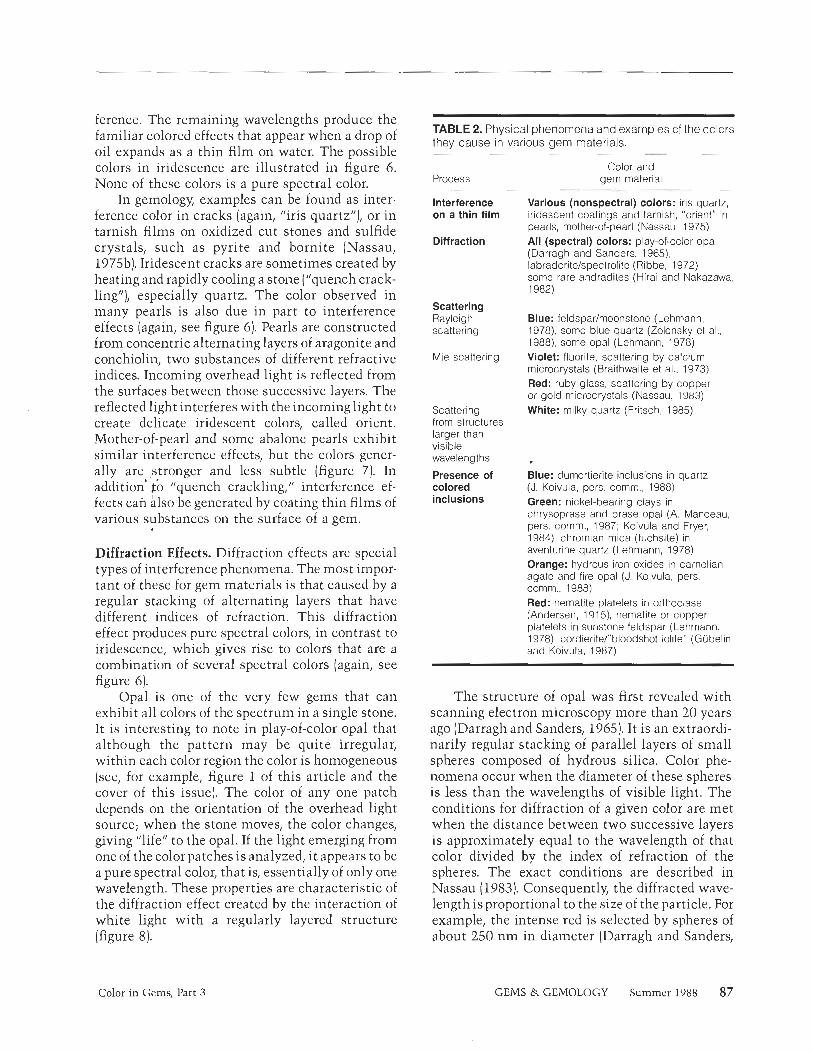
Figure 6. The colors produced by interference on a thin film are the same as those observed on this quartz wedge (top left) in polarized light. None of them is a pure spectral color. Notice that the "higher orders" on the right produce mostly pink and green. Interference colors are caused in pearls by light passing through and reflected back by al- ternating concentric layers of aragonite and conchiolin, which are readily visible on the elec- tron photomicrograph of a pearl section (left). The resulting "high order" interference colors (mostly green and pink) are called orient and overtone (the latter, when they are stronger and homogeneous all over the pearl). They are readily apparent in these black pearl cufflinks (top right). Photomicro- graph courtesy of B. Lasnier, Gemology Laboratory, Nantes University, France. Jewelry courtesy of Harry Winston, Inc. Color photos by Robert Weldon.
Similar effects are encountered in some feld- spars belonging to the plagioclase series. These feldspars display regions of color, often violet to green, against a generally black background. Finn- ish "Spectrolite," a variety of labradorite, appears to show every color of the spectrum. This phenom- enon is called "labradorescence," after the classic occurrence of these stones on the Isle of Paul, Labrador, although varieties of plagioclase feld- spars other than labradorite may display this effect. The diffracting objects in labradorescence are alternating layers, known as exsolution lamellae, of two feldspars with different chemical compositions. One layer is calcium rich and the other is calcium poor. The color created by the lamellar structures depends on their respective thicknesses and indices of refraction (figure 10). Another gemstone that occasionally shows diffrac- tion colors is andradite from Hermosillo, Mexico
88 Color in Gems, Part 3 GEMS & GEMOLOGY Summer 1988
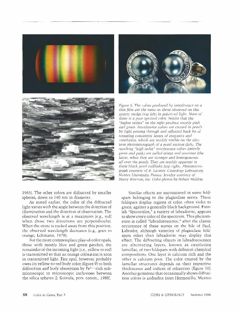
ire ', . . 'ink and green interference colors '<e a spectacular display in this abalone rl. Courtesy of Lowell Jones, St. Louis,
MU; photo @ Tino Hammid.
Scattering. When the internal structure of the stone is irregular and/or the size of the components is outside the very narrow range needed for diffrac- tion (approximately 100-400 nm), visible light cannot be diffracted. It can, however, still be scattered, the process by which light entering a stone in a given direction is deflected in different directions through interaction with the scattering centers. This creates both striking color effects and optical phenomena. The exact phenomenon de- pends on the size and shape of the scattering centers. When the scattering centers are smaller than the wavelength of visible light [including down to molecular dimensions) and not regularly distributed, the process is called Rayleigh scatter- ing; when the scattering centers are comparable in size to visible wavelengths, the process is called Mie scattering. (The names are derived from the mathematical theories used to describe scattcr- ing.) A third type of scattering occurs when the centers are larger than visible wavelengths.
Rayleigh scattering. When the incoming light ray encounters randomly distributed objects smaller
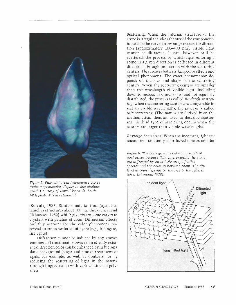
Figure 8. The homogeneous color in a patch of opal arises because light rays entering the stone are diffracted b y an orderly array of silica spheres and the holes in between them. The dif- fracted color depends on the size of the spheres (of ter Lehmann, 1978).
(Koivula, 1987). Similar material from Japan has lamellar structures about 100 nm thick (Hirai and Nakazawa, 1982), which give rise to some very rare crystals with patches of color. Diffraction effects probably account for the color phenomena ob- served in some varieties of agate (e.g., iris agate, fire agate).
Diffraction cannot be induced by any known commercial treatment. However, an already exist- ing diffraction color can be enhanced by inducing a dark background (sugar and smoke treatment of opals, for example, as well as doublets], or by reducing the scattering of light in the matrix through impregnation with various kinds of poly- mers.
Incident light /\
Color in Gems, Part 3 GEMS & GEMOLOGY Summer 1988 89
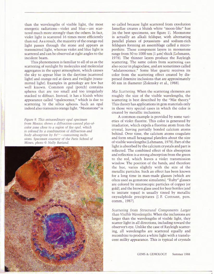
than the wavelengths of visible light, the most energetic radiations -violet and blue- are scat- tered much more strongly than the others. In fact, violet light is scattered 16 times more efficiently than red. As a result, the majority of the orange-red light passes through the stone and appears as transmitted light, whereas violet and blue light is scattered and can be observed at right angles to the incident beam.
This phenomenon is familiar to all of us as the scattering of sunlight by molecules and molecular aggregates in the upper atmosphere, which causes the sky to appear blue in the daytime (scattered light) and orange-red at dawn and twilight (trans- mitted light). Examples in gemology are few but well known. Common opal (potch) contains spheres that are too small and too irregularly stacked to diffract. Instead, it has a bluish white appearance called "opalescence," which is due to scattering by the silica spheres. Such an opal indeed also transmits orange light. "Moonstonef1 is
F I ~ U I ~ 9. This extraordinary opal specimen from Mesdco shows a difiactim-caused play-of- color zoae close to a region of fire opal, which is colored.by a combination of diffraction sad. body a bsqption by F E ~ + -containing inclu- sions. Specimen courtesy of the Paris School of Minei; photo 0 Nelly Hariand
so called because light scattered from exsolution lamellae creates a bluish white "moon-like" hue [in the best specimens; see figure 1). Moonstone is actually an alkali feldspar, with alternating parallel planes of potassium- and sodium-rich feldspars forming an assemblage called a micro- perthite. These component layers in moonstone range from 50 to 1000 run (1 pm) thick (Lehmann, 1978). The thinner layers produce the Rayleigh scattering. The same colors from scattering can also occur in plagioclase, and are sometimes called "adularescenc6.~ Some blue quartz receives its color from the scattering effect created by dis- persed ilmenite inclusions that are approximately 60 nm in diameter (Zolensky et al., 1988).
Alia Scattering. When the scattering elements are roughly the size of the visible wavelengths, the scattering is best described by the "Mie theory." This theory has applications in gem materials only in those very special cases in which the color is created by metallic inclusions.
A common example is provided by some vari- eties of violet fluorite. This color is generated by irradiation, which expels a fluorine atom from the crystal, leaving partially bonded calcium atoms behind. Over time, the calcium atoms coagulate and form small hexagonal platelets about the size of visible wavelengths (Lehmann, 1978). Part of the light is absorbed by the calcium crystals and part is reflected. The combined effect of this absorption and reflection is a strong absorption from the green to the red, which leaves a violet transmission window. The position of the bands, and therefore the hue, varies slightly with the size of the metallic particles. Such an effect has been known for a long time in man-made glasses (which are often used as gemstone simulants). "Ruby" glasses are colored by microscopic particles of copper (or gold), and the brown glass used for beer bottles (and to imitate topaz] is usually tinted by metallic oxysulphide precipitates (J. F. Cottrant, pers. comrn., 1987).
Scattering from Structural Components Larger than Visible Wavelengths. When the inclusions are larger than the wavelengths of visible light, they scatter light in all directions, including toward the observer's eye. Unlike the case of Rayleigh scatter- ing, all wavelengths are scattered equally and recombine to produce a white light with a translu- cent milky appearance. This is typical of crystals
GEMS & GEMOLOGY Summer 1988
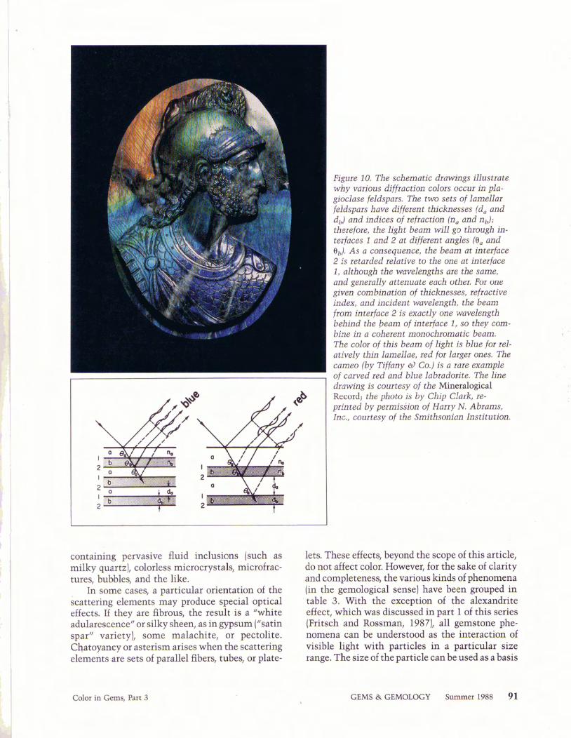
Figure 10. The schematic drawings illu9trats why vdtious difflaation colors occur ia pla- gioclase feldspars. The two sets of lameLlar feldspars have different thicfenes$m (dy and dJ and indices of reftacficos (n, and nd; therefore, the light beam will go through in- terfaces 1 mid 2 at differant angles (OÃ and 9J, As a consequence, the beam at interface 2 is retarded dat ive to the one at iatetface 1, although the wavelengths p the same, and gsneiaQy attenuate each other, For mfc givexi combinaticte of thicknesses, refractive index, and indent warnlength, the beam from interface 2 is exactly one wuveleogth behind the beam of interface 1, so they ewi- bine in a cobereat moaochiosaiatie bearn. The cote of this beam of light is blue for rd- a t i d y thin lamellw, red for lwei ones. The cameo (by Tiffany Co.) is a lars exampls
- of carved zed and blue labiadon'te. The h e
containing pervasive fluid inclusions (such as milky quartz), colorless microcr ystals, microfrac- m e s , bubbles, and the like.
In some cases, a particular orientation of the scattering elements may produce special optical effects. If they are fibrous, the result is a "white adulirescencet' or silky sheen, as in gypsum ("satin spar" variety), some malachite, or pectolite. Chatoyancy or asterism arises when the scattering elements are sets of parallel fibers, tubes, or plate-
chdwing is courtesy of the Mineralogical Record; the photo is by Chip Clffrk, ie-
printed by permission of H q N. A brams, Inc., courtesy of the Sm'tbsaaicso. Ins£1tution
lets. These effects, beyond the scope of this article, do not affect color. However, for the sake of clarity and completeness, the various kinds of phenomena (in the gemological sense) have been grouped in table 3. With the exception of the alexandrite effect, which was discussed in part 1 of this series (Fritsch and Rossman, 1987), all gemstone phe- nomena can be understood as the interaction of visible light with particles in a particular size range. The size of the particle can be used as a basis
GEMS & GEMOLOGY Summer 1988 9 1
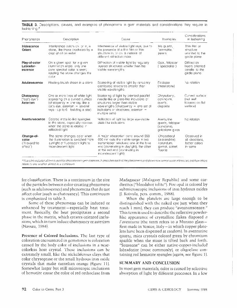
TABLE 3. Descriptions, causes, and examples of phenomena in gem materials and considerations they require in fa~hioning.~
Considerations in fashioning
Thin film or structure oriented to the girdle plane
Diffraction layers oriented parallel to the girdle plane
Cause Examples
Iris quartz, 'ammolite," pearls
Phenomenon Description
Iridescence Orient
Interference colors on, or in, a stone, like those produced by a drop of oil on water
Interference of visible light rays, due to the presence of a thin film or thin structure in, or on, a material of different refractive index
Play-of-color Labrador- escence
On a given spot, for a given illumination angle, only one pure spectral color is seen; rotating the stone changes the color
Floating bluish sheen in a stone
Diffraction of visible light by regularly layered structures smaller than the visible wavelengths
Opal, feldspar ("spectrolite")
Adularescence Scattering of visible light by randomly distributed structures smaller than visible wavelengths
Feldspar (moonstone)
No relation
Chatoyancy ("cat's-eye") Asterism
One or more lines of white light appearing on a curved surface (chatoyancy = one ray, like a cat's eye; asterism = several rays-up to 6- building a star)
Scattering of light by oriented parallel needle-like or plate-like inclusions or structures larger than visible wavelengths (chatoyancy = one set of inclusions or structures; asterism = multiple sets)
Chrysoberyl, corundum, quartz, diopside
Curved surface (not well focused on flat surface)
Aventurescence Colored metallic-like spangles in the stone, especially obvious when the stone is rotated in reflected light
Reflection of light by large eye-visible plate-like inclusions
Aventurine quartz, feldspar (sunstone), goldstone glass
Chrysoberyl (alexandrite), corundum, garnet, spinel, fluorite
No relation
Change-of- color ("Alexandrite effect")
The stone changes color when the illumination is switched from sunlight or fluorescent light to incandescent light
A major absorption band around 550- 600 nm cuts the visible range in two transmission windows: one at the blue end (dominating in daylight), the other at the red end (dominating in incandescent light)
Observed in all directions, better colors in some
¡'Thi table includes all terms used lor phenomena in gem materials It describes each of the phenomena andshows how similar some 01 them are and how others relate to one another almost in a continuum
for classification. There is a continuum in the size of the particles between color-creating phenomena (such as adularescence) and phenomena that do not affect color (such as chatoyancy). This continuum is emphasized in table 3 .
Some of these phenomena can be induced or enhanced by treatment-especially heat treat- ment. Basically, the heat precipitates a second phase in the matrix, which creates oriented inclu- sions, which in turn induce chatoyancy or asterism (Nassau, 1984).
Madagascar (Malagasy Republic) and some cor- dierites ("bloodshot iolite"). Fire opal is colored by submicroscopic inclusions of iron hydrous oxides (J. Koivula, pers. comm., 1988).
When the platelets are large enough to be distinguished with the naked eye (say, when they reach 1 mm), they can produce "aventurescence." This term is used to describe the reflective powder- like appearance of crystalline flakes disposed a l'oventurra (the term refers to a Murano glass- first made in Venice, Italy- in which copper plate- lets have been dispersed at random). In aventurine quartz, mica crystals colored green by chromium sparkle when the stone is tilted back and forth. "Sunstonefl can be either native-copper-included labradorite (most commonly), or oligoclase con- taining red hematite spangles (again, see figure 1).
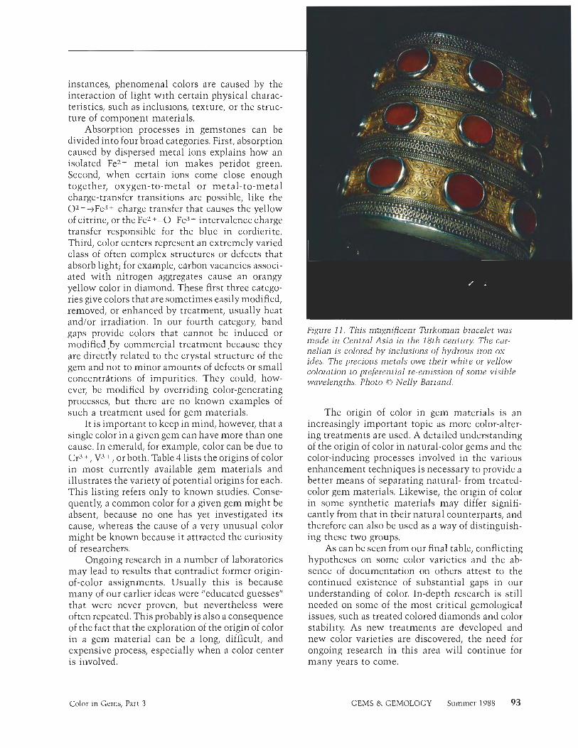
Presence of Colored Inclusions. The last type of coloration encountered in gemstones is coloration caused by the body color of inclusions in a near- colorless host crystal. These inclusions can be extremely small, like the niclzelifero~~s clays that color chrysoprase or the small hydrous iron oxide crystals that make carnelian orange (figure 11). Somewhat larger but still microscopic inclusions of hematite cause the color of red orthoclase from
SUMMARY AND CONCLUSION In most gem materials, color is caused by selective absorption of light by different processes. In a few
92 Color in Gems, Part 3 GEMS & GEMOLOGY Summer 1988
instances, phenomenal colors are caused by the interaction of light with certain physical charac- teristics, such as inclusions, texture, or the struc- ture of component materials.
Absorption processes in gemstones can be divided into four broad categories. First, absorption caused by dispersed metal ions explains how an isolated Fez+ metal ion makes peridot green. Second, when certain ions come close enough together, oxygen-to-metal or metal-to-metal charge-transfer transitions are possible, like the 02-+Fe3+ charge transfer that causes the yellow of citrine, or the Fez+-0-Fe^ intervalence charge transfer responsible for the blue in cordierite. Third, color centers represent an extremely varied class of often complex structures or defects that absorb light; for example, carbon vacancies associ- ated with nitrogen aggregates cause an orangy yellow color in diamond. These first three catego- ries give colors that are sometimes easily modified, removed, or enhanced by treatment, usually heat and/or irradiation. In our fourth category, band gaps provide colors that cannot be induced or modified by commercial treatment because they are directly related to the crystal structure of the gem and not to minor amounts of defects or small concentriions of impurities. They could, how- ever, be modified by overriding color-generating processes, but there are no known examples of such a treatment used for gem materials.
It is important to keep in mind, however, that a single color in a given gem can have more than one cause. In emerald, for example, color can be due to Cr3+, V3+, or both. Table 4 lists the origins of color in most currently available gem materials and illustrates the variety of potential origins for each. This listing refers only to known studies. Conse- quently, a common color for a given gem might be absent, because no one has yet investigated its cause, whereas the cause of a very unusual color might be known because it attracted the curiosity of researchers.
Ongoing research in a number of laboratories may lead to results that contradict former origin- of-color assignments. Usually this is because many of our earlier ideas were "educated guesses" that were never proven, but nevertheless were often repeated. This probably is also a consequence of the fact that the exploration of the origin of color in a gem material can be a long, difficult, and expensive process, especially when a color center is involved.
Figure 1 1 . This m'agnificent Turkoman bracelet was made in Central Asia in the 18th century. The car- nelian is colored by inclusions of hydrous iron ox- ides. The precious metals owe their white or yellow coloration to preferential re-emission of some visible wavelengtl1s. Photo 0 Nelly Bariand.
The origin of color in gem materials is an increasingly important topic as more color-alter- ing treatments are used. A detailed understanding of the origin of color in natural-color gems and the color-inducing processes involved in the various enhancement techniques is necessary to provide a better means of separating natural- from treated- color gem materials. Likewise, the origin of color in some synthetic materials may differ signifi- cantly from that in their natural counterparts, and therefore can also be used as a way of distinguish- ing these two groups.
As can be seen from our final table, conflicting hypotheses on some color varieties and the ab- sence of documentation on others attest to the continued existence of substantial gaps in our understanding of color. In-depth research is still needed on some of the most critical gemological issues, such as treated colored diamonds and color stability. As new treatments are developed and new color varieties are discovered, the need for ongoing research in this area will continue for many years to come.
Color in Gems, Part 3 GEMS & GEMOLOGY Summer 1988 93
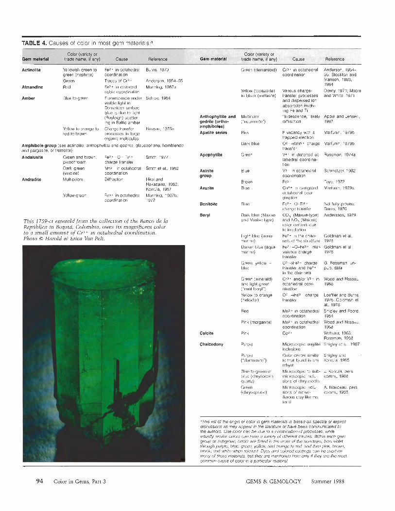
TABLE 4. Causes of color in most gem materia1s.a
Actinollte Yellowish green to Fez+ in octahedral Burns, 1970 green (nephrite) coordination Green Traces of Cr3 + Anderson, 1954-55
Almandlne Red Fez* in distorted Manning, 1967a cubic coordination
Amber Blue to green Fluorescence under Schlee, 1984 visible light in Dominican amber; blue is due to light (Rayleigh) scatter- ing in Baltic amber
Yellow to orange to Charge-transfer Nassau, 1975a red to brown processes in large
organic molecules
Amphibole group (see actinolile. anthophyllite and gedrite. glaucophane, hornblende and pargasite, or tremolite)
Andalusite Green and brown, Fez* -0- Ti4+ Smith, 1977 pleochroism charge transfer
Dark green Mn3+ in octahedral Smith et al., 1982 (viridine) coordination
Andradite Multicolors Diffraction Hirai and Nakazawa, 1982; Koivula, 1987
Yellow-green Fez+ in octahedral Manning, 1967b. coordination 1972
This 1759-ct emerald from the collection of the Bunco de la Republics in Bogota, Colombia, owes its magnificent color to a small amount of Cr3+ in octahedral coordination. Photo ec) Harold &> Erica Van Pelt.
Green (demantoid)
Yellow (topazolite) to black (melanite)
Anthophylllte and Multicolor gedrite (ortho- ("nuurnmite") amphiboles)
Apatite series Pink
Apophyllite
Azurite
Benitoite
Beryl
Calcite
Chalcedony
Dark blue
Green
Blue
Brown
Blue
Blue
Dark blue (Maxixe and Maxixe-type)
Light blue (aqua- marine)
Darker blue (aqua- marine)
Green: yellow + blue
Green (emerald) and light green ("mint beryl")
Yellow to orange (heliodor)
Red
Pink (morganite)
Pink
Purple
Purple ("damsonite")
Blue to greenish blue (chrysocolla quartz)
Green (chrysoprase)
Cr3* in octahedral Anderson. 1954- coordination 55; Stockton and
Manson, 1983. 1984
Various charge- Dowly. 1971; Moore transfer processes and White. 1971 and dispersed ion absorption involv- ing Fe and Ti "Iridescence," likely Appel and Jensen, diffraction 1987
F vacancy with a Marfunin. 1979b trapped electron 0 2 - -)MnS+ charge Marfunin, 1979b transfer
V4+ in distorted oc- Rossman, l974a tahedral coordina- tion V3+ in octahedral Schmelzer. 1982 coordination
Fez + Faye, 1972
Cuz+ in elongated Marfunin. 1979a octahedral coor- dination Fez + . Not fully proven: charge transfer Burns. 1970
CO; (Maxixe-type) Andersson, 1979 and NO3 (Maxixe) color centers due to irradiation Fe2+ in the chan- Goldrnan et al., nels 01 the structure 1978
FeZ+-O-Fe=* inter- Goldman el al., valence charge 1978 transfer
--)Fe3+ charge G. Rossman, un- transfer and Fez* pub data in the channels Cr3+ andlor V3* in Wood and Nassau, octahedral coor- 1968 dination
O%-+Fe3* charge Loeffler and Burns, transfer 1976. Goldman el
al., 1978 Mna+ in octahedral Shigley and Foord, coordination 1984
Mnz+ in octahedral Wood and Nassau. coordination 1968
co2 + Webster. 1983; Rossman. 1988
Microscopic sugilite Shigley el al., 1987 inclusions
Color center similar Shigley and to that found in am- Koivula, 1985 ethyst
Microscopic to sub- J. Koivula, pers. microscopic inclu- comrn., 1988 sions of chrysocolla
Microscopic inclu- A. Manceau, pers. sions of nickel- comrn., 1985 ilerous clay-like ma- lerial
'This list of the origin of color in gem materials is based on spectra or explicit discussions as they appear in the literature or have been communicated to the authors One color can be due to a combination of processes, while visually similar colors can have a variety of different causes Within each gem group or subgroup, colors are listed in the order of the spectrum. from violet through purple, blue, green, yellow, arid orange to red, and then pink, brown, black, and white when relevant Dyes and colored coatings can be used on many of these materials, but they are mentioned here only if they are the most common cause o l color in a particular material
94 Color in Gems, Part 3 GEMS & GEMOLOGY Summer 1988
Qpfw (variety or Gem mat#rial A IM6 W e . if '$0~)
Orange to red (car- nelian, jasper)
Submicroscopic to microscopic inclu- sions of hydrous Fe oxides Fe3* in octahedral coordination Cr3* in octahedral coordination Cu'+ in octahedral coordination Cr3- in octahedral coordination
Organic pigment from the carolenoid lamily
J Koivula, pers. comm., 1988
Chrysoberyl Yellow Loeftler and Burns. 1976 Farrell and Newn- ham. 1965 Lehmann. 1978
Color-change (alexandrite) Blue Chryaocolla
Green (tawmawite) Schmetzer. 1982
Dele-Dubois and Merlin. 1981
Conch "pearl" and Pink shell
Copal (same as amber) Coral Blue
(Heliopora cae- rulea) Red to pink
An organic pigment Fox et al., 1983 of the bilins family. helioporobilin Organic pigments Delb-Dubois ana from the carotenoid Merlin, 1981 family, at least lor Corallurn rubrurn Various organic ma- Rolandi. 1981 terials ol unknown nature Fe7* -0-Fex* Faye el al 1968, charge transfer Smith, 1977.
Goldman et al , 1977
Black
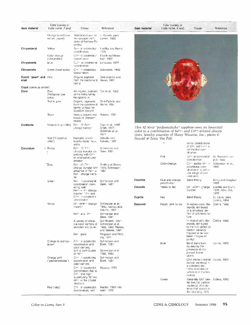
Cordlerlte Violet to blue (iolite) This 32.50-ct "pi~dparadschi~" sapphire owes its beautiful color to a combination of Fe3 + - and Cr3+-related absorp- tions. Jewelry courtesy of Harry Winston, Inc.; photo 0
Red ("bloodshot iolite")
Hematite andlor lepidocrocite inclu- sions Fe? + -&Ti4 +
charge transfer co- existing with Cr3* in octahedral coor- dination Fe2 4 -0-Tià charge transfer with influence of Fez * -Ã
Fe3* charge trans- fer Fe3+ in octahedral coordination coex- isting with FeZ*-ÈTi * charge transfer, Ti3* and Cr3* in octahedral coordination O^+Fe3+ charge transfer
Gubelin and Koivula, 1987
~ a r o l d a) ~ r i c a Van Pelt. minor contributions of V3+ and Fe3* in octahedral coor- dination
Corundum Schmetzer and Bank. 1981
t Purple
I
Pink
Color-change
G. Rossman, un- pub data
Cr3* in octahedral coordination Cr3* andlor V3* in octahedral coor- dination in a partic- ular range of con- centration
Blue and orange, Band theory pleochroism Yellow to red 02-+Cr6+ charge
transfer
Schnietzer et at., 1980
Smith and Sirens, 1976; Schmetzer, 1987
Green Covelllte
Crocoite
Berry and Vaughan, 1985 Loeffler and Burns, 1976: Abou-Eid, 1976 G. Calas, pers. comm., 1984 Collins, 1982
Schmelzer and Bank. 1981
Cuprite
Diamond
Red Band theory
Yellow Schmetzer el al., 1982; Nassau and Valente, 1987 Schmetzer and Bank, 1981 Schittmann, 1981: Schmetzer et al., 1982, 1983; Nassau and Valenle. 1987 Ferguson and Field- ing, 1971 Schmetzer and Bank, 1981
Purple, pink to red In natural-color dia- monds. attributed to a structural de- fect of unknown na- ture In treated pink dia- monds, attributed to the N-V defect (a carbon vacancy trapped at an iso- lated nitrogen im- purity) Band transitions caused by the presence of dis- persed boron atoms GRt center (neutral carbon vacancy) in a colorless dia- mond irradiated in nature or in the lab- oratory
Generally GR1 cen- ter (neutral carbon vacancy), plus d e fects that absorb in Ihe blue (e.g., N3)
Fe3* and Ti2 *
Collins, 1982 A variety of unsta- ble color centers of unknown structure
Fe3* pairs
Orange to orange- brown
Cr3* in octahedral coordination and color centers; with a contribution of Fe3 Cr3* in octahedral coordination and color centers Cr4* in octahedral coordination due to Cr-' ' and Mg2+ substituting for two Al3* in the crystal structure Cr3* in octahedral coordination, with
Blue Collins, 1982
Schmetzer et al., 1982, 1983
Orangy pink ("padparadscha")
Schmetzer and Bank, 1981 Collins. 1982
Nassau, 1983
Green Collins, 1982
Red (ruby) Harder. 1969; Gu- belin, 1975
Color in Gems, Part 3 GEMS & GEMOLOGY Summer 1988 95
Color (variety or Gem material trade name, if any) Cause Reference
Dlopslde
Dloptase
Dravite
Elbaite and llddlcoatite
Yellow
Orange
Brown
Green (chrome diopside)
Yellowish green
Green
Green ("chrome tourmaline")
Yellow to brown
Red
Blue (indicolite)
Green
More rarely, due to very strong green fluorescence under visible light ("green transmitter" effect) of the H3 andlor H4 defect (a carbon vacancy trapped at an aggregate of two or four nitrogen atoms)
Most commonly due to Ihe N3 de- fect, an aggregate of three nitrogen atoms (color cen- ter) at a carbon va- cancy More rarely, due to band transitions caused by the presence of iso- lated nitrogen atoms
Most commonly H3 center (a carbon vacancy trapped at an aggregate of two nitrogen atoms), in natural and treated orange diamonds More rarely, origi- nating from a color center of unknown nature
Color center of un- known nature, with various other color centers adding or- ange, yellow, pink, or green
Cr3- in octahedral coordination. V3* in octahedral coordination
Fez-*- in octahedral coordination Cu7+ in octahedral coordination
V3+ generally with minor amounts of Cr3+, both in octa hedral coordination
Related to titanium due to Fez * -0- Tid* charge trans- fer, with those low in iron yellow and those rich in iron brown
Fe3* pairs
Fe7* in octahedral coordination with possible influence of some iron-related charge-transfer pro- cesses Fez+ and Ti4 + in octahedral coor- dination. The influ- ence of various charge transfer pro- cesses involving iron is a distinct possibility Mnz*-0-Ti4 * charge transfer
Collins, 1982
Collins, 1982, Lowlher, 1984
Collins, 1982
Cottrant and Calas, 1981
Collins, 1982
Collins. 1982
Rossman, 1980
Schmetzer. 1982
Burns. 1970
Lehmann, 1978
Schmetzer and Bank, 1979
Smith, 1977: Ross- man, as cited in Dietrich. 1985
Mattson and Ross- man. 1984
See Dietrich, 1985
Malison, as ciled in Dietrich, 1985
Rossman and Malison, 1986
Greenish yellow Mn7* in octahedral Rossrnan and coordination (rare) Malison, 1986
Orange Yellow t pink See Dietrich, 1985 Pink to red Related lo man- Manning, 1973, De (rubellite) ganese, generally Camargo and Iso-
believed to be due tam. 1988 to Mn3* in octa- hedral coordination. sometimes created by irradiation
Brown Fez* -)TI"- charge Rossman and transfer Malison, 1986
Enstatlte Greenish brown Fez * Rossman, 1980
Green Fe7 * with rninor Anderson, 1954-55 Cr3 *
Epidote group (see clinozoisite, epidote, piemontile, or zoisite)
Epidote Green and brown, Fe3+ in distorted Burns, 1970 pleochroism octahedral coor-
dination
Euclase Blue Fez * -0-Fe3 * Mattson and Ross- charge transfer man, 1987
Green Cr3* in octahedral Anderson, 1954-55 coordination
Feldspar group (see labradorite. microcline, oligoclase, orthoclase, or plagioclase series)
Fluorite Violet Mie scattering on Braithwaite el al., calcium micro- 1973, Lehmann, crystalliles 1978
Blue Y3+ t F vacancy Bill and Calas, + 2 electrons 1978
'Emerald" green Sm2 * Bill and Calas. ("chrome fluorite") 1978: Rossman.
1981
Yellowish green Color center con- Bill and Calas. taming Y and Ce 1978 associated with an F vacancy
Yellow 0; color center = Bill and Calas. 0, substituting for 1978 fluorite
Pink YO, color center Bill and Calas, (Y3+ t O;-) 1978
Color change Y3+-associated Bill and Calas, color center and 1978: Schmetzer et Smz*. with minor at., 1980 influence of a Ce3 *-associated color center
Gahnite and Blue Fez* in tetrahedral Dickson and Smith. "gahnospinel" coordination 1976
Garnet group (see almandine, andradite, grossular, hydrogrossular group, pyrope, spessartine, or uvarovite: also rliodolite)
Glass Yellowish green Fep* in octahedral Pye el al , 1984 '1
(natural) (moldavite, tektite) coordination
Brown Fe3+ in octahedral Pye et al., 1984 coordination
Glaucophane Blue FeS t 4 - ~ ~ 3 + Smith and Strens, charge transfer 1976
Grossular Green (tsavorite) V3.- in octahedral Gubelin and coordination Weibei, 1975
Orange Mn3+ in distorted Manning. 1970 (hessonite) cubic coordination:
Fea Manson and Stock- ton, 1986
Gypsum All (alabaster) Color usually due to J. Koivula, pers. dyes exclusively, comm., 1988 except for some brown staining due to hydrous iron oxides
Hematite Gray in reflection, Fe3* Bell el al.. 1975 red in transmission
Hornblende Green to brown Fez+ in various Rossman. 1988 and pargaslte sites
Howlite Blue Dyes exclusively
96 Color in Gems, Part 3 GEMS & GEMOLOGY Summer 1988
Hydrogrossular Green ("Transvaal group lade")
Pink
Cr3* in octahedral coordination Mnz* in octahedral coordination Cr3- in octahedral coordination Fe3* in octahedral cwrdination Few -O-Fe3 *
charge transfer: synthetic is colored by Mns* Cr3- in octahedral coordination V3* in octahedral coordination Fez t -0-Ti4 *
charge transfer. Fez + -0-Fe3 ' charge transfer. Fez* and Fe3* in octahedral coor- dination can all be involved: with contribution from Cr3- in octa- hedral coordination
V3- in octahedral coordination; Fe3* in octahedral coordination
Cr3 ' in octa- hedral coordination Diffraction of light by the internal lamellar structure
Manning and Owens, 1977 Manning and Owens, 1977 Rossrnan, 1981
Rossrnan, 1981
Rossman, 1974b
Nassau and Shigley. 1987 Schrnetzer, 1982
Schrnetzer 1982
Parkin el al., 1977
Jadeite 'Emerald green (chrome ladeite) Yellowish green
Violet ("lavender jadeite")
Kornerupine Blue
Green
Kyanite Blue
Bosshart et al.. 1982
Green Schrnetzer. 1982
G. Rossrnan. un- pub. data
Color change
" Mullicolors
Bosshart et al., 1982
Ribbe, 1972: Lehrnann. 1978
Labradorlte
Red (in the material Irorn Oregon)
Subrnicroscopic metallic copper particles Could be Cu * IVCT or CuO pairs Fez * -0-Fe3 * charge transfer
83 (charge transfer) Mn2+ in octahedral cwrdination, Mna* in octahedral coordination
Holrneister and Rossrnan, 1985b
Green and orange- pink, pleochroisrn Blue
Holrneister and Rossrnan, 1985b Arnthauer and Rossrnan. 1984 Loefller and Burn- 1978 Faye, 1968
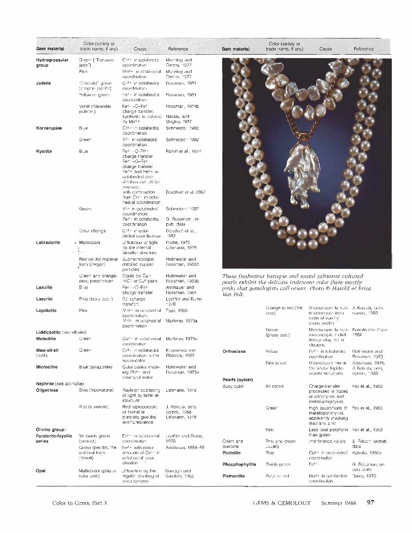
These freshwater baroque and round saltwater cultured pearls exhibit the delicate iridescent color (here mostly pink) that gemologists call orient. Photo 0 Harold ei) Erica Van Pelt.
Lazurite Blue (lapis lazuli)
Orange to red (lire Microscopic to sub- opal) microscopic inch-
sions of iron hy- drous oxides
Green Microscopic to sub- (prase opal) microscopic nickel-
iferous clay-like in- clusions
Yellow Fe3- in tetrahedral coordination
Pink lo red Microscop~c hema- tite and/or lepido- crocite inclusions
J Koivula, pers comm . 1980 Pink
Marfunin, 1979a
Koivula and Frypr 1984 Liddlcoatite (see elbaite)
Malachite Green Cuz* in octahedral coordination
Cr31 in octahedral coordination in the kosrnochlor Color center involv- ing Pb3 and slructural water
Marlunin, 1979a
Maw-sit-sit (rock)
Green Khornenko and Platonov, 1985
Orthoclase Hofmeister and Rossman, 1983 Andersen, 1915, J Koivula, pers cornm , 1908
Blue (arnazonite) Hofrneister and Rossman. 1985a
Pearls (oyster) Body color Nephrite (see actinol~le)
Oligoclase Blue (moonstone) All colors Charge-transfer
processes in traces of porphyrins and metalloporphyrins
Fox el al.. 1903 Rayleigh scattering of light by lamellar structure Red lepidocrocite or hematite platelets give the aventurescence
Lehrnann, 1978
Red (sunstone) J. Koivula, pers, cornrn., 1988; Lehrnann, 1978
Green Fox et al., 1983 High proportions of metalloporphyrins. apparently involving lead and zinc
Pink Less total porphyrin than green
Orient and Pink and green Interference colors overtone usually Pectolite Blue Cu2+ in octahedral
coordination Phosphophylllte Bluish green Fe2 *
Ollvine group: Forsterite-fayalite Yellowish green Fe7- in octahedral Loeffler and Burns, series (peridot) coordination 1976
Green (peridot, the Fez+ with minor Anderson, 1954-55 material from amounts of Cr<- ' in Hawaii) octahedral coor-
dination '
Fox el al , 1903
E. Fntsch. unpub data Koivula. 1986a
G. Rossman. un- pub. data
Burns. 1970 Opal Multicolors (play-01- Diffraction by the Darragh and
color opal) regular stacking of Sanders, 1965 silica spheres
Piemontite Purplish red Mn3* in octahedral coordination
Color in Gems, Part 3 GEMS & GEMOLOGY
Plagloclase series Blue Color center involv- Hofmeister and ing Pb and water Rossman. 1986
Yellow W+ in tetrahedral Hofmeister and coordination and Rossman, 1983 Fez* in octahedral coordination
Green Fe ̂ -0-Fe3 + G. Rossman, un- (chlorastrolite) charge transfer pub. data
plus FW* Brownish red Fez* in distorted Manning, 1967a
cubic coordination Red Fez+ in distorted Anderson, 1954-
cubic site plus 55; Manning, 1967a Cr3* in octahedral coordination
Color change (in V3+ and/or Cr3+ in Schmetzer et al., pyrope and pyrope- octahedral coor- 1980 spessartine) dination
Pyrope-Almandlne Reddish purple Fez+ in distorted Manning, 1967a (rhodolite) cubic coordination
Pyroxene group (see enstatite, diopside, jadeite, kosmochlor in maw-sit-sit, or spodumene)
Smoky (smoky Color center related Partlow and Cohen, quartz) to the Al3+ impurity 1986 Pink (rose quartz) Charge transfer be- Cohen and Makar,
tween a substitu- I985 tional Tid* and an interstitial Ti3+; unstable color Maschrneyer and center 0- Lehmann, 1983 ion bridging between substitu- tional aluminum and substitutional phosphorus atom; dumortierite inclu- Applin and Hicks, sions 1987
White (milky quartz) Scattering of light Fritsch, 1985 by inclusions larger than the visible wavelengths Mn2+ in octahedral Rossman, 1988 coordination
Pumpellyite
PyroPe
Rhodochroslte Pink to red
Rhodollte (see pyrope-almandine) Rhodonlte Pink
0 2 -->Fed+ charge transfer, due to irra- diation Inclusions of blue dumortierite Inclusions ol tourmaline
Inclusions of ilmenite of a diame- ter smaller than vis- ible wavelengths Fe2 +
Mnz* in octahedral Marshall and coordination, with Runcinam, 1975 minor Fez ; Mn3+ in octahedral Gibbons et al., coordination 1974 Band transition due G. Rossman, un- to the presence of pub. data Ti3 +
Due to color cen- Marlunin, 1979b ters related to irra- diation of Cl. Cog- or SCI- groups present in the large voids of the crystal structure
Yellow Fe Not proven: Gunawardene, 1986
Green (williamsite) Cr3+ around chro- J. Koivula, pers. mite inclusions comm,, 1988
Blue Cu2 + Fleischer, 1987 Pink (see conch shell)
Black A violet organic Fox el al., 1983 pigment, hal- iotiviolin, has been recovered from the shell of the black abalone, Haliotis cracherodii Fez + -O-T~- Rossman et al., charge transfer, 1982 probably similar to blue kyanite Fe3+ or Cr3* in Rossman et al., tetrahedral coor- 1982 dination Fe features of yel- Rossman el al., low sillimanite plus 1982 inclusions of iron- rich phase 0Z-+Fe3* charge Farrell and transfer and Fez+ Newnharn, 1965 in octahedral coor- dination Cup+ G. Rossman. un-
pub. data CoZ * G. Rossman, un-
pub. data Interstitial oxygen Pizani et al., 1985 ion 0 - near Al or Si
Pink (hackmanite) Unstable electron Pizani et al., 1985 substituting for Cl- in a tetrahedron of Na* ions
Quartz Violet to purple (amethyst)
Blue C. Fryer, pers. comm., 1988 Gubelin and Koivula, 1987
Zolensky el al., 1988
Rutile Blue (synthetic rutile)
Scapolite series Various colors
Green ("greened amethyst" or prasiolite) Green (aventurine quartz) Greenish yellow
Nassau. 1980
Chromian mica Lehmann. 1978 (fuchsite) inclusions Color center Nassau and
Prescolt, 1977 0 2 - +Fe3* charge Balitsky and transfer Balitskaya, 1986 Various AP+-re- Samoilwich el al., lated color centers 1969
Scheellte
Serpentine
Yellow to orange (citrine) Shattuckite
Shell
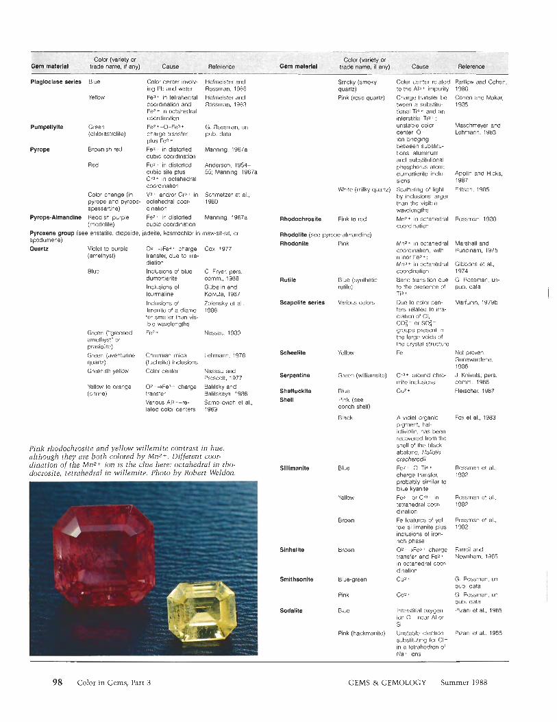
Pink rhodochrosite and yellow willemite contrast in hue, although they are both colored by Mn2+. Different coor- dination of the Mn2+ ion is the clue here: octahedral in rho- docrosite, tetrahedral in willemite. Photo by Robert Weldon.
Blue
Yellow
Brown
Sinhalite Brown
Smithsonlte Blue-green
Pink
Blue Sodalite
98 Color in Gems, Part 3 GEMS & GEMOLOGY Summer 1988
Spessartine Orange Mn2 in distorted cubic coordination
Sphalerite Yellow to black Iron + sulfur charge transfer
Green Co2+ in tetrahedral coordination
Spinel group (see gahnite and gahnospinel, or spinel)
Manning, 1967a
Madunin, t979a
Marfunin, 1979a
Spinel Violet to purple Cra+ in octahedral coordination and Fez+ in tetrahedral coordination Co2+ and Fez* in tetrahedral coor- dination Fe3+ and Fe7* in tetrahedral coor- dination Cr3* in octahedral coordination Cra- in octahedral coordination
G, Rossrnan, un- pub, dala
Cobalt blue Shigley and Stockton. 1984
Bluish green G. Rossman, un- pub. data
Green (synthetic spinel) Pink to red
Vogel, 1934
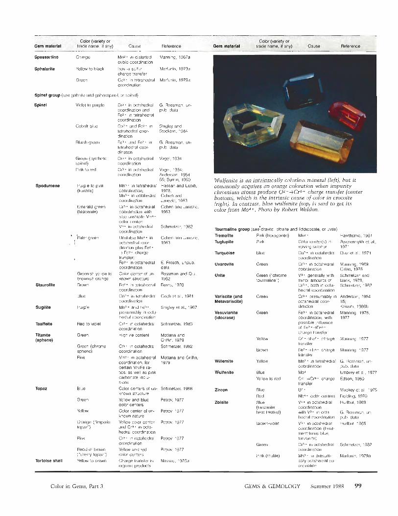
Vogel. 1934; Anderson. 1954- 55; Surnin, 1950 Wulfenite is an intrinsically colorless mineral (left), but it
commonly acquires an orange coloration when impurity chromium atoms produce 02-+Cr6+ charge transfer (center bottom), which is the intrinsic cause of color in cmcoite (right). In contrast, blue wulfenite (top) is said to get its color from Mo4+. Photo by Robert Weldon.
Spodumene Purple to pink (kunzite)
Mn3* in tetrahedral coordination. Mn3+ in octahedral coordination Cr3* in octahedral coordination with also unstable Mn4 color center. V3- in octahedral coordination Unstable Mn4* in octahedral coor- dination plus Fez* ) Fe3* charge
transfer Fe3 + in octahedral coordination Color center of un- known structure Fez+ in tetrahedral coordination Co-"+ in tetrahedral coordination Mn3* and FeJ*. presumably in octa- hedral coordination
Cr3+ in octahedral coordination High Fe content
Cr3* in octahedral coordination Mn2+ in octahedral coordination, for certain MnIFe ra- tios, as well as pink carbonate inclu- sions Color centers of un- known structure Yellow and blue color centers Color center of un- known nature Yellow color center and Cr3* in octa- hedral coordination Cr3* in octahedral coordination Yellow and red color centers Charge transfer in organic products
Hassan and Labib, 1978; Cohen and Janezic. 1983 Cohen and Janezic, 1983
Schmetzer, 1982
Cohen and Janezic, 1983
Emerald green (hiddenile)
Tourmaline group (see'drauite, elbaite and liddicoalile, or uvile) t '
Paler green Pink (hexagonite) Pink
Mn3 *
Color center(s) in- volving sulphur Cuz* in octahedral coordination CrJ* in octahedral coordination Va* generally with minor amounts of Cr3* both in octa hedral coordination Cra* presumably in octahedral coor- dination Fe3* in octahedral coordination with possible influence of Fez * +Fe3 * charge transfer 0 2 - +Fez* charge transfer
Fe7' -)Ti4 + charge transfer Mn7* in tetrahedral coordination
Mod * 07 ->Crb+ charge transfer
u4 * Nbd* color centers V-1- in octahedral coordination with V3+ in ocla hedral coordination V3* in octahedral coordination (treat- ment lurns blue. tanzanite) Cr3- in octahedral coordination Mn3- in presum- ably octahedral co- ordination
Hawthorne. 1981 Povarennykh el al., 1971 Diaz et al., 1971
Tremolite Tugtupite
Turquoise
Uvarovlte
Uvite
Blue
E. Frilsch, unpub. data Rossman and Oiu, 1982 Burns. 1970
Green Manning, 1969, Calas, 1978 Schmetzer and Bank, 1979. Schmetzer, 1982
Greenish yellow to brownish orange Brown
Green ("chrome tourmaline")
Staurolite
Variscite (and Metavariscite)
Blue Cech el al., 1981 Green Anderson, 1954- 55: Koivula. 1986b Sugilite Purple Shigley et al., 1987
Vesuvianite (idocrase)
Green Manning, 1976, 1977
Red to violet Schrnetzer, 1983
Yellow Manning. 1977 Titanite (sphene)
Green Mottana and Griffin. 1979 Schmetzer, 1982 Green (chrome
sphene) Pink
Brown Manning, 1977
Moltana and Gnlfin, 1979 Willemite
Wulfenite
Yellow G Rossman, un- pub. data Embrey el al., 1977 EdSOn, 1980
Blue Yellow to red
Topaz Blue Schmelzer. 1986
Petrw, 1977
Petrov. 1977
Petrw. 1977
Zircon
Zolsite
Blue
Red Blue (tanzanite- heat treated)
Mackey el al,, 1975 Fielding, 1970 Hurlbut, 1969
Green
Yellow G . Rossrnan, un pub. data Hurlbut, 1965 Orange ("imperial
topaz")
Pink Petrov, 1977
Petrov, 1977
Nassau. 1975a
Green
Pink (thulite)
Schrnetzer, 1982
Marfunin, 1979a
Reddish brown ("sherry topaz")
Tortoise shell Yellow to brown
Color in Gems, Part 3 GEMS & GEMOLOGY Summer 1988 99
REFERENCES Abou-Eid R.M. (1976) Absorption spectra of transition metal
bearing minerals at high pressure. In R. J. G. Strens, Ed., The Physics and Chemistry of Minerals and Rocks, John Wiley & Sons, New York, pp. 641-675.
Amthaucr G., Rossman G.R. (1984) Mixed valence of iron in minerals with cation clusters. Physics and Chemistry of Minerals, Vol. 11, pp. 37-51.
Andersen 0 . (1915) On adventurine feldspar. American lournal of Science, Vol. 40, pp. 351-399.
Anderson B.W. (1954-55) The spectroscope and its applications to gemmology, parts 10 to 17. The Gemmologist, Vol. 23, Nos. 275-282.
Andersson L.O. (1979) The difference between Maxixe beryl and Maxixe-type beryl: An electron paramagnetic reso- nance investigation. Journal of Cemmolosv, Vol. 16, No. 5, - -.
pp. 313-317, Aooel EW. Iensen A. (19871 A new gem material from Green- . . . ,
land: Iridescent orthoamphibole. Gems a) Gemolosv, Vol. -,
23, No. 1, pp. 3 6 4 2 . Applin K.R., Hicks B.D. (1987) Fibers of dumortierite in quartz.
American Mineralogist, Vol. 72, pp. 170-1 72. Balitsky VS., Balitskaya 0.V (1986) The amethyst-citrine di-
chromatism in quartz and its origin. Physics and Chemis- try of Minerals, Vol. 13, pp. 4 15421 .
Bell EM., Mao H.K., Rossman G.R. (1975) Absorption spectros- copy of ionic and molecular units in crystals and glasses. In C. Karr, Jr., Ed., Infrared and Roman Spectroscopy of Lunar and Terrestrial Minerals, Academic Press, New York, pp. 1-38.
Berry F.J., Vaughan DJ . (1985) Chemical Bonding and Spectros- copy in Mineral Chemistry. Chapman and Hall, New York.
Bill H., Calas G. (1978) Color centers, associated rare-earth ions and the origin of coloration in natural fluorites. Physics and Chemistry of Minerals, Vol. 3, pp. 117-131.
Bosshart G,, Frank E., Hanni H.A., Barot N, (1982) Blue color- changing kyanite from East Africa. Journal of Gemmology, Vol. 18, No. 3, pp. 205-212.
Braithwaite R.S.W., Flowers W.T, Hazeldine R.N., Russel M, (1973) The cause of the colour of Blue John and other purple fluorites. Mineralogical Magazine, Vol. 39, pp. 4 0 1 4 1 1.
Burns R.G. (1970) Mineralogical Applications of Crystal Field Theory. Cambridge Earth Science Series, Cambridge Uni- versity Press.
Calas G. (1978) Le chrome et la couleur des mineraux: Un example "pkdagogique." Revue de Gemmologie a.f.g,, Vol. 54, pp. 6-8,
Cech F.. Povondra E, Vrana S. (1981 1 Cobaltoan staurolite from . , Zambia. ~ul le t in de Miniralogie, Vol. 104, pp. 526-529.
Cohen A.J., Janezic G.G. (1983) The crystal-field spectra of the 3d3, Cr3+ and Mn4+ in green spodumenes. In The Signifi- cance of Trace Elements in Solving Petrogenetic Problems and Controversies, Theophrastus Publications, Athens, Greece.
Cohen A.J., Makar L.N. (1985) Dynamic biaxial absorption spectra of Ti3+ and Fe^ in a natural rose quartz crystal. Mineralogical Magazine, Vol. 49, pp. 709-715.
Collins A.T. (1982) Colour centres in diamond. lournal of Gemmology, Vol. 18, No. 1, pp. 37-75.
Cottrant J.-F., Calas G. (1981) Etude de la coloration de quelques diamants du Museum dlHistoire Naturelle. Revue de Gemmologie a.f.g., No. 67, pp. 2-5.
Cox R.T. (1977) Optical absorption of the d4 ion Fe4+ in pleochroic amethyst quartz. Iouinal of Physics, Vol. C10, pp. 46314643.
Darragh EJ., Sanders J.V (1965) The origin of colour in opal. Australian Gemmologist, No. 46, pp. 9-12.
De Camargo M.B., Isotani S. (19881 Optical absorption of
natural and irradiated pink tourmaline. American Miner- alogist, Vol. 73, pp. 172-180.
Dele-Dubois M.-L., Merlin J.-C. (1981) Etude par spectroscopie Raman de la pigmentation du squelette calcaire du corail. Revue de Gemmologie a.f.g., No. 68, pp. 10-13.
Diaz J., Farach H.A., Poole C.R Jr. (1971) An electron spin resonance and optical study of turquoise. Ainericcin Min- eralogist, Vol. 56, pp. 773-781.
Dickson B.L., Smith G. (1976) Low temperature optical absorp- tion and Mossbauer spectra of staurolite and spinel. Canadian Mineralogist, Vol. 14, pp. 206-215.
Dietrich R.V (1985) The Tourmaline Croup. Von Nostrand Reinhold Co., New York.
Dowty E. (1971) Crystal chemistry of titanian and zirconian garnet: I. Review and spectral studies. American Miner- alogist, Vol. 56, pp. 1983-2009.
Edson G.M. (1980) The Red Cloud mine, Yuma County, Arizona. Mineralogical Record, Vol. 11, No. 3, pp. 141-152,
Embrey EG., Dunn P.J., Clark A.M. (1977) Blue wulfenite from Tsumeb. Mineralogical Record, Vol. 8, No. 3, pp. 86-87.
Farrell E.F., Newnham R.E. (19651 Crystal field spectra of chrysoberyl, alexandrite, peridot and sinhalite. American Mineralogist, Vol. 50, pp. 1972-1981,
Faye G.H. (1968) The optical absorption spectra of certain transition metal ions in muscovite, lepidolite and fuchsite. Canadian Journal of Earth Sciences, Vol. 5, pp. 31-38.
Faye G.H. (1972) Relationship between crystal field splitting parameter "Avi" and M,,osr-O bond distance as an aid in the interpretation of absorption spectra of Fe^-bearing mate- rials. Canadian Mineralogist, Vol. 11, pp. 4 7 3 4 7 .
Faye G .H. Manning EG,, Nickel E.H. (1968) The polarized optical absorption spectra of tourmaline, cordierite, chlo- ritoid and vivianite: ferrous-ferric electronic interaction as a source of pleochroism. American Mineralogist, Vol. 53, pp. 1174-1201,
Ferguson J., Fielding RE. (1971) The origins of the colours of yellow, green and blue sapphires. Chemical Physics Let- ters, Vol. 10, No. 3, pp. 262-265.
Fielding RE. (1970) The distribution of uranium, rare earths, and color centers in a crystal of natural zircon. American Mineralogist, Vol. 55, pp. 428440 .
Fleischer M. (1987) Glossary of Mineral Species, 5th ed. Mineralogical Record, Inc., Tucson, AZ.
Fox D.L., Brown F.A., Losey G.S. (1983) Coloration, biological. New Encyclopedia Brittanica, Encyclopedia Brittanica, Chicago, IL, pp. 91 1-929,
Fritsch E. (1985) La couleur des min6raux et des gemmes: Des couleurs du diamant aux etoiles du saphir. Monde es) Minttraux, No. 70, pp. 12-22.
Fritsch E., Rossman G.R. (1987) An update on color in gems. Part 1: Introduction and colors caused by dispersed metal ions. Gems etJ Gemology, Vol. 23, No. 3, pp. 126-139.
Fritsch E., Rossman G.R. (1988) An update on color in gems. Part 2: Colors involving multiple atoms and color centers. Gems &> Gemology, Vol. 24, No. 1, pp. 3-15.
Gibbons R.V, Ahrens T.J., Rossman G.R. (1974) A spec- trographic interpretation of shock-produced color change in rhodonite (MnSi03): The shock-induced reduction of Mn(II1) to Mn(I1). American Mineralogist, Vol. 59, pp. 177-182.
Goldman D.S., Rossman G.R., Dollase W.A. (1977) Channel constituents in cordieritc. American Mineralogist, Vol. 62, pp. 1 144-1 157.
Goldman D.S., Rossman G.R., Parkin K.M. (1978) Channel constituents in beryl. Physics and Chemistry of Minerals, Vol. 3, pp. 225-235,
Giibelin E.J. (1975) The Color Treasury of Gemstones. Elsevier
100 Color in Gems, Part 3 GEMS â‚ GEMOLOGY Summer 1988
Phaidon, London. Gubelin E.J., Koivula J.1. (1987) Photoat-/as of Inclusions in
Gemstones, ABC Edition, Zurich. Gubelin E.J., Weibel M. (1975) Green vanadiumgrossulargarnet
from Lualenyi, near Voi, Kenya. Lapidary Journal, Vol. 29, pp. 402-426.
Giinawardene M. (1986) Colombage-Ara scheelite. Gems ei) Gemo/ogy, Vol. 22, No. 3, pp. 166-169.
Hassan F., Labib M. (1978) Induced color centers in a-spodun~ene called kunzite. Neues fahrbtich fur Miner- cilogie, Abhandlungen, Vol. 134, No. 1, pp. 104-1 15.
Harder H. (1969) Farbgebende Spurenelemente in den naturlichen Koriinden. Neiies Jcihrb~icli fiir Mineralogie, Abhaiiclhingen, Vol. 110, pp. 128-141.
Hawthorne F. (198 1) Amphibole spectroscopy. In D. R. Veblen, Ed., Reviews in Mineralogy, Vol. 9A: Amphiboles and Other Hydrous Pyriboles-Mineralogy, Mineralogical So- ciety of America, Washington, DC, pp. 103-137.
Hirai H., Nakazawa H. (1982) Origin of iridescence in garnet: An optical interference study. Physics and Chemistry of Minerals, Vol. 8, pp. 25-28.
Hofnleister A.M., Rossman G.R. (1983) Colors in feldspars. In l? H. Ribbe, Ed., Reviews in Mi~~eralogy, Vol. 2, 2nd ed.: Feldspar Minerals, Mineralogical Society of America, Washington, DC, pp. 271-280.
Hofnleister A.M., Rossman G.R. (1985a) A spectroscopic study of irradiation coloring of amazonite: Structurally hydrous, Pb-bearing feldspar. American Mineralogist, Vol. 70, pp. 794-804.
Hofnieister A.M., Rossman G.R. (1985b) Exsolution of n~etallic copper from Lake County labradorite. Geology, Vol. 13, pp. 644-647.
~ofmeis tk r A.F., Rossman G.R. (1986) A spectroscopic study of blue radiation coloring plagioclase. American Mineralo- gist, Vol. 71, pp. 95-98.
Hurlbut C.S. (1969) Gem zoisite from Tanzania. American Mineralogist, Vol. 54, pp. 702-709.
Khomenko VM., Platonov A.N. (1985) Electronic absorption spectra of Cr3+ ions in natural clinopyroxenes. Physics and Chemistry of Minerals, Vol. 11, pp. 261-265.
Koivula 1.1. (1986a) Gem news: Pectolite. Gems a) Gemology, Vol. 22, No. 3, pp. 187-188.
Koivula J.I. (1986b) Gem news: Metavariscite. Gems @I Gemol- ogy, Vol. 22, No. 4, pp. 247-248.
Koivula J.I. (1987) Gem news: Iridescent andradite garnets. Gems a) Gemology, Vol. 23, No. 3, pp. 173-1 75.
Koivula J.I., Fryer C.W (1984) Green opal from East Africa. Gems a) Gemology, Vol. 20, No. 4, pp, 226-227.
Lehn~ann G. (1978) Farben von Mineralen [sic] und ihre Ursachen. Fortschrit te der Mineralogie, Vol. 56, No. 2, pp. 172-252.
Loeffler B.M., Burns R.G. (1976) Shedding light on the color of gems and minerals. American Scier~tist, Vol. 64, pp. 636-647.
Lowther J.E. (1984) The form of different charge states of the vacancy in diamond. Journal of Physics and Chemistry of Solids, Vol. 45, pp. 127-131.
Mackey D.J., Runcinam WA., Vance E.R. (1975) Crystal-field calculations for energy levels of U4+ in ZrSi04. Physical Review B, Vol. 11, pp. 21 1-218.
Manning P.G. (1967a) The optical absorption spectra of the garnets aln~andine-pyrope, pyrope and spessartine and some structural interpretations of n~ineralogical signifi- cance. Canadian Mineralogist, Vol. 9, pp. 237-25 1.
Manning RG. (1967b) The optical absorption spectra of some andradite and the identification of the 6Al-PAl "£10 transition in octahedrally bonded Fe"+. Canadian Journal of Earth Sciences, Vol. 4, pp. 1039-1047.
Manning EG. (1969) Optical absorption studies of grossular,
andradite (var. colophonite) and uvarovite. Canadian Min- eralogist, Vol. 9, pp. 723-729.
Manning EG. (1970) Racah parameters and the relationship to lengths and covalence of Mn2+- and Fe3+-oxygen bonds in silicates. Canadian Mineralogist, Vol. 10, pp. 677-688.
Manning EG. (1972) Optical absorption spectra of Fe3+ in octahedral and tetrahedral sites in natural garnets. Cana- dian Mineralogist, Vol. l l, pp. 826-839.
Manning EG. (1973) Effect of second nearest-neighbour interac- tion on Mn3+ absorption in pink and black tourmaline. Canadian Mineralogist, Vol. 11, pp. 971-977.
Manning EG. (1976) Ferrous-ferric interaction on adjacent face- sharing antiprismatic sites in vesuvianite: Evidence for ferric iron in eight coordination. Canadian Mineralogist, Vol. 14, pp. 216-220.
Manning EG. (1977) Charge transfer interaction and the origin of color in brown vesuvianite. Canadian Mineralogist, Vol. 15, pp. 508-51 1.
Manning EG., Owens D.R. (19771 Electron microprobe, X-ray diffraction, and spectral studies of South African and British Columbian "jades." Canadian Mineralogist, Vol. 15, pp. 512-517.
Manson D.V, Stockton C.M. (1982) Gem-quality grossular garnets. Gems 01 Gemology, Vol. 18, No. 4, pp. 204-213.
Marfunin A.S. (1979a) Physics of Minerals and Inorganic Materials, An Introduction. Trans. by N. G. Egorova and A. G. Mishchenko, Springer Verlag, Berlin.
Marfunin A.S. (1979b) Spectroscopy, Luminescence and Radia- tion Centers inMinerals. Pans. by V V Schiffer, Springer Verlag, Berlin.
Marshall M., Runcinam WA. (1975) The absorption spectrum of rhodonite. American Mineralogist, Vol. 60, pp. 88-97.
Maschmeyer D., Lehmann G. (1983) A trapped-hole center causing rose coloration of natural quartz. Zeitschrift fiir Kristallograpbie, Vol. 163, pp. 181-196.
Mattson S.M., Rossman G.R. (1984) Ferric iron in tourmaline. Physics and Chemistry of Minerals, Vol. 11, pp. 225-234.
Mattson S.M., Rossman G.R. (1987) Identifying characteristics of charge transfer transitions in minerals. Physics and Chemistry of Minerals, Vol. 14, pp. 94-99.
Moore R.K., White WB. (1971) Intervalence electron transfer effects in the spectra of the melanite garnets. American Mineralogist, Vol. 56, pp. 826-840.
Mottana A., Griffin WL. (1979) Pink titanite (greenovite) from St. Marcel, Valle D'Aosta, Italy. Rendiconti Societa ltal- iana di Mineralogia e Petrologia, Vol. 35, pp. 135-143.
Nassau K. (1975a) The origin of color in gems and minerals. Part 2. Gems a) Gemology, Vol. 15, No. 1, pp. 2-11.
Nassau K. (1975b) The origin of color in gems and minerals. Part 3. Gems a) Gemohgy, Vol. 15, No. 2, pp. 34-43.
Nassau K. (1980) Gems Made by Man. Chilton Book Co., Radnor, PA.
Nassau K. (1983) The Physics and Chemistry of Color: The Fifteen Causes of Color. John Wiley & Sons, New York.
Nassau K. (1984) Gemstone Enhancement. Butterworths, Stoneham, MA.
Nassau K., Prescott E. (1977) Smoky, blue, greenish yellow and other irradiation-related colors in quartz. Mineralogical Magazine, Vol. 41, pp. 301-312.
Nassau K., Shigley J.E. (1987) A study of the General Electric synthetic jadeite. Gems a) Gemology, Vol. 23, No. 1, pp. 27-35.
Nassau K., Valente G.K. (1987) Thc seven types of yellow sapphire and their stability to light. Gems &) Gemology, Vol. 23, No. 4, pp. 222-231.
Parkin K.M., Loeffler B.M. Burns R.G. (1977) Mossbauer spectra of kyanite, aquamarine, and cordierite showing intervalence charge transfer. Physics and Chemistry of Minerals, Vol. 1, pp. 301-31 1.
Color in Gems, Part 3 GEMS & GEMOLOGY Summer 1988 101
Partlow D.l?, Cohen A.J. (1986) Optical studies of biaxial Al- related color centers in smoky quartz. American Miner- cilogist, Vol. 71, pp. 589-598.
Petrov I. (1977) Farbuntersuchungen an Topas. Neues Jahrbuch fiir Mineralogie Abhandlungen, Vol. 130, No. 3, pp. 288-302.
Pizani PS., Terrile M.C., Farach H.A., Poole C.l? Jr. (1985) Color centers in sodalite. American Mineralogist, Vol. 70, pp. 1186-1 192.
Povarennykh A.S., Platonov A.M., Tarashchan A.M., Be- lichenko V P (1971) The colour and luminescence of tugtupite (beryllosodalite) from Ilimaussaq, South Green- land. Meddelelser om Granland, Vol. 181, pp. 1-12.
Pye L.D., O'Keefe J.A., Frekchette VD., Eds. (1984) Natural Glasses. North Holland Publishing, Amsterdam.
Ribbe PH. (1972) Interference colors in oil slicks and feldspars. Mineralogical Record, Vol. 3, pp. 18-22.
Rolandi V (1981) Les gemmes du regne animal: Etude gem- rnologique des secretions des Cnidaires. Revue de Gem- mologie a.fx., Vol. 66, pp. 3-9.
Rossman G.R. (1974a) Optical spectroscopy of green vanadium apophyllite from Poona, India. American Mineralogist, Vol. 59, pp. 621-622.
Rossman G.R. (1974b) Lavender jade. The optical spectrum of Fe^ and Fe2+-Fe3+ intervalence charge transfer in jad- eite from Burma. American Mineralogist, Vol. 59, pp. 868-870.
Rossman G.R. (1980) Pyroxene spectroscopy. In C. T. Prewitt, Ed., Reviews in Mineralogy, Vol. 7: Pyroxenes, Mineralogi- cal Society of America, Washington, DC, pp. 93-113.
Rossman G.R. (1981) Color in gems: The new technologies. Gems s) Gemology, Vol. 17, No. 2, pp. 60-71.
Rossman G.R. (1988) Optical spectroscopy In F. C. Hawthorne, Ed., Reviews in Mineralogy, Vol. 18: Spectroscopic Methods in Mineralogy and Geology, Mineralogical Soci- ety of America, Washington, DC, pp. 207-254.
Rossrnan G.R., Mattson S.M. (1986) Yellow, manganese-rich elbaite with manganese-titanium intervalence charge transfer. American Mineralogist, Vol. 71, pp. 599-602.
Rossman G.R., Qiu Y. (1982) Radioactive irradiated spodu- mene. Gems e) Cenwiogy, Vol. 18, No. 2, pp. 87-90.
Rossman G.R., Grew E.S., Dollase W.A. (1982) The colors of sillimanite. American Mineralogist, Vol. 67, pp. 749-761.
Samoilovich M.I., Tsinober L.I., Kreishop VN. (1969) The nature of radiation-produced citrine coloration in quartz. Soviet Physics- Crystallography, Vol. 13, pp. 626-628.
Schiffmann C.A. (1981) Unstable colour in a yellow sapphire from Sri Lanka. Journal of Gemmology, Vol. 17, No. 8, pp. 615-618.
Schlee D. (1984) Ungewohnliche Farbvarianten des Baltischen Bernsteins: Blau, grau, orange und "gold" als Folge von Rissesystemen. Bernstein-Neiiigkeiten, Stuttgarter Be- itrdge zur Naturkunde, Series C, No. 18, pp. 2-8.
Schmetzer K. (1982) Absorptionsspektroskopie und Farbe von V3+-haltigen natiirlichen Oxiden und Silikaten-ein Be- itrag zur Kristallchemie des Vanadiums. Neues Jahrbuch fur Mineralogie, Abhandl~ingen, Vol. 144, pp. 73-106.
Schmetzer K. (1983) Crystal chemistry of natural Be-Mg-Al- oxides: Taaffeite, taprobanite, musgravite. Neues Jahr- buch fur Mineralogie, Abhandlungen, Vol. 146, pp. 15-28.
Schmetzer K. (1986) Farbung und Bestrahlungschaden in elek- tronenbestrahlten blauen Topasen. Zeitschrift der Deut- schen Gemmologischen Gesellschaft, Vol. 35, No. 112, pp. 27-38.
Schmetzer K. (1987) Zur Deutung der Farbursache blauer Saphire-eine Diskussion. Neiies Jahrbuch fur Miner- dogie, Monatshefte, No. 8, pp. 337-343.
Schmetzer K., Bank H. (1979) East African tourmalines and their non~enclature. Journal of Gemmology, Vol. 16, pp. 310-311.
Schmetzer K., Bank H. (1981) The colour of natural corundum. Neues Jahrbuch fur Mineralogie, Monatshefte, Vol. 11, No. 2, pp. 59-68.
Schmetzer K., Bank H., Glibelin E. (1980) The alexandriteeffect in minerals: Chrysoberyl, garnet, corundum, fluorite. Neiies Jahrbuch fur Mineralogie, Abhandlungen, Vol. 138, pp. 147-164.
Schmetzer K., Bosshart G., Hanni H.A. (1982) Naturfarbene und behandelte gelbe und orange braune Sapphire. Zeitschrift der Deiitschen Cemmologischen Ce- sellschaft, Vol. 31, No. 4, pp. 265-279.
Schmetzer K., Bosshart G., Hiinni H.A. (1983) Naturally- coloured and treated yellow and orange brown sapphires. Journal of Gemmology, Vol. 18, No. 7, pp. 607-621.
Shigley J.E., Foord E.E. (1984) Gem-quality red beryl from the Wah-Wah Mountains, Utah. Gems fs) Gemology, Vol. 20, -.
No. 4, pp. 208-221. Shigley J.E., Koivula J.I. (1985) Amethystine chalcedony. Gems
e) Gemoloxy, Vol. 21, No. 4, pp. 219-223. Shigley J.E., ~ o i v u l a J.l., ~ r ~ e r C.W. (1987) The occurrence and
gen~ological properties of Wessels Mine sugilite. Gems e) Gemology, Vol. 23, No. 2, pp 78-90.
Shigley J.E., Stockton C.M. (1984) "Cobalt-blue" gem spinels. Gems e) Gemology, Vol. 20, No. 1, pp. 3 4 4 1.
Smith G. (1977) Low temperature optical studies of metal-me- tal charge transfer transitions in various minerals. Cana- d i m Mineralogist, Vol. 10, pp. 500-507.
Smith G., Strens R.G.J. (1976) Intervalence transfer absorption in some silicate, oxide and phosphate minerals. In R. G. J. Strens, Ed., The Physics and Chemistry of Minerals and Rocks, John Wiley & Sons, New York, pp. 583-612.
Smith G., Hilenius U., Langer K. (1982) Low temperature spectral studies of Mn3+-bearing andalusite and epidote type minerals in the range 30000-5000 cm- 1. Physics and Chemistry of Minerals, Vol. 8, pp. 136-142.
Stockton C.M., Manson D.V (1983) Gem andradite garnets. Gems a) Gemology, Vol. 19, No. 4, pp. 202-208.
Stockton C.M., Manson D.V. (1984) Editorial forum: "Fine green" demantoids. Gems e) Gemology, Vol. 20, No. 3, p. 179.
Sumin N.G. (1950) Accessory elements in spinels. Trudy Mineralog. Museya, Akad. Nauk S.S.S.R., No. 2, pp. 113-123.
Vogel l? (1934) Optische Untersuchungen am Smaragd und einigen anderen durch Chrom gefarbten Mineralien. Neues Jahrbuch fur Mineralogie, Geologic und Paleon- tologje, Vol. A68, pp. 401438.
Webster R., rev. by B. W. Anderson (1983) Gems, Their Sources, Descriptions and Identification, 4th ed. Butterworths, London.
Wood D.L., Nassau K. (1968) The characterization of beryl and emerald by visible and infrared absorption spectroscopy. American Mineralogist, Vol. 53, pp. 777-800.
Zolensky M.E., Sylvester PI., Paces J.B. (1988) Origin and significance of blue coloration in quartz from Llano rhyolite (llanite), north-central Llano County, Texas. American Mineralogist, Vol. 73, pp. 313-323.
102 Color in Gems, Part 3 GEMS & GEMOLOGY Summer 1988