Embed Size (px)
Citation preview
An Unusual Sugar Conformation in the Structure of anRNA/DNA Decamer of the Polypurine Tract May AffectRecognition by RNase H
Mary L. Kopka1*, Laurence Lavelle2, Gye Won Han1, Ho-Leung Ng1
and Richard E. Dickerson1,2
1Molecular Biology InstituteUniversity of California, LosAngeles, CA 90095-1570, USA
2Department of Chemistry andBiochemistry, University ofCalifornia, Los Angeles, CA90095-1570, USA
Retroviral conversion of single-stranded RNA into double-stranded DNArequires priming for each strand. While host cellular t-RNA serves asprimer for the first strand, the viral polypurine tract (PPT) is primer forthe second. Therefore, polypurine tracts of retroviruses are essential forviral replication by reverse transcriptase (RT). These purine tracts areresistant to cleavage during first strand synthesis. In obtaining the primerfor second strand synthesis, the RNase H function of RT must cleave thePPT exactly for in vivo transcription to proceed efficiently and properintegration to occur. At the RNase H active site the protein makes contactsprimarily along the backbone, with hydrogen bonds to the sugar–phos-phate oxygen atoms. A high-resolution structure (1.10 A) of the first tenbase-pairs of the RNA/DNA hybrid PPT, r-(c-a-a-a-g-a-a-a-a-g)/d-(C-T-T-T-T-C-T-T-T-G), contains the highly deformable r-(a-g-a) steps found inretroviral polypurine tracts. This r-(a-g-a) motif is utilized in the“unzipping” or unpairing of bases that occurs when RT binds a malleablePPT. Another unusual feature found in our high-resolution PPT structureis the sugar switch at RNA adenine 2. All the RNA sugars are theexpected C30-endo, except sugar 2, which is C20-endo, characteristic ofB-form sugars. This local A-to-B conversion adversely affects the patternof hydrogen bonds from protein to sugar–phosphate backbone, disrupt-ing the catalytic site. Disruption could cause the enzyme to pause at the50-end of the PPT, leaving it intact. Pyrimidine–purine (YR) steps aremost deformable and the T-A step especially can undergo A-to-Btransitions readily.
q 2003 Elsevier Ltd. All rights reserved.
Keywords: polypurine tract; RNA/DNA hybrid; RNase H; recognition;U-box*Corresponding author
Introduction
Before the human immunodeficiency virus (HIV)can successfully infect its host, the viral RNA mustbe copied into DNA. To accomplish this task thevirus utilizes a multi-functional enzyme, reverse
transcriptase (RT), that converts single-strandedviral RNA into double-stranded DNA (polymerasefunction) and then degrades the RNA template(RNase H function). The three X-ray structures ofHIV-RT complexed with a drug, a bound nucleicacid and the unliganded enzyme1 – 3 showed themolecule to have a “hand” motif similar to thatfound in other polymerases, with fingers, palmand thumb to guide the nucleic acid to the twoactive sites. Two “primer grip” regions of themolecule have been defined, the polymerase gripand the RNase H primer grip.4 – 6 These gripregions are situated adjacent to both active sites.
The RNase H domain has both endonucleaseand 30,50-exonuclease activity.7 During (2 )-strand
0022-2836/$ - see front matter q 2003 Elsevier Ltd. All rights reserved.
L.L. & G.W.H. contributed equally to this work.Present address: H.- L. Ng, Department of Molecular
and Cell Biology, University of California, Berkeley, CA94720-3206, USA.
E-mail address of the corresponding author:[email protected]
Abbreviations used: PPT, polypurine tract; RT, reversetranscriptase.
doi:10.1016/j.jmb.2003.09.057 J. Mol. Biol. (2003) 334, 653–665

synthesis, RNase H degrades the RNA template(exonuclease activity) as the DNA strand is beingcopied, but leaves intact (endonuclease activity) apurine-rich run of 15 bases (Figure 1(A)) known asthe polypurine tract.8 This PPT serves as the primerfor second or (þ )-strand DNA synthesis. TheRNase H subunit, consisting of approximately120 þ amino acid residues, resides in the p66C-terminal domain of the p66/p51 RT heterodimer,and its crystal structure has been reported.9 TheRNase H primer grip region makes extensive con-tacts with the DNA primer,5 and mutations ofamino acids in both the active-site RNase H gripregions have been examined.10 – 12 The structure ofRT complexed with an RNA/DNA primer-template containing the PPT5 has shown thatcontacts between the RNA/DNA hybrid bases,and amino acid residues in the RNase H primergrip and its catalytic center, occur primarily withthe sugar–phosphate backbone. There arerelatively few hydrogen bonds with the hybridbases. Only three direct protein contacts(H-bonds) to the bases occur, two of which arefound in the RNase H active site where the scissilebond is cleaved.
Knowledge of the structure of nucleic acidduplexes utilized by RT is important to under-standing the multi-step process of reverse tran-scription. Structural biologists have investigatedby NMR and crystallography various duplex struc-tures that the enzyme contacts. To initiate copyingof the minus strand at the start of reverse transcrip-tion, the viral RNA recruits a host tRNA formingan RNA/RNA duplex. However once (2 )-strandDNA synthesis begins, a chimeric hybrid, anOkazaki fragment, is formed at the junction of thegrowing DNA chain with the tRNA/RNA. Thisjunction has been studied by both crystallographyand NMR,13,14 as has the Okazaki fragmentgenerated at the completion of synthesis when the(2 )-strand primer is removed from the RNA/
DNA junction.15 Another decamer RNA/DNAhybrid beginning one RNA nucleotide 50 of thecleavage site has been looked atcrystallographically.16 All of these structures arerelated to the template-primer for (2 )-strandDNA synthesis.
Unusual backbone conformations have beenseen by both crystal and solution studies in base-pairs at these Okazaki junctions recognized byRT.13 – 15 The NMR structures have a bend in thejunction region.14,15 This conformational backboneflexibility, primarily at one base removed in the 50
direction from the junction, has been proposed asa recognition signal for RNase H endolyticcleavage.13 Szyperski et al.14 agree that the back-bone conformations in this region are important.They make particular note of a sugar pucker vari-ation at the hybrid junction and a surroundingbase-step, where two of the DNA sugars inter-convert between C30-endo and C20-endo.
The template-primers for both plus and minusstrand are resistant to hydrolytic cleavage untilDNA synthesis of the two strands is near com-pletion, prior to integration. To obtain the properprimer for (þ )-strand synthesis, the RNase Hmust cleave the PPT accurately for complete tran-scription to proceed efficiently.8,17 – 19 The bindingand endolytic cleavage of the PPT by RNase Hhave been the subject of many reports.5,11,12,17,20 – 22
Only one PPT sequence has been investigatedstructurally (by NMR), the 30-end of the PPT alongwith four bases in the immediate flankingsequence. However, these investigators23 have sub-stituted an adenine base, e.g. gagg for gggg, in acritical region in the guanine tract that is essentialfor proper extension, cleavage and primerremoval.20
In NMR studies of hybrids the DNA strandshave B-like sugar conformations, e.g. O40-endo,C10-exo, C20-endo, while RNA sugars are C30-endo.14,15,21,24,25 This has not been the case for thecrystal structures. Generally, these structures havebeen A-like helices with both RNA and DNAstrands having C30-endo sugar conformations.However, exceptions in sugar conformation atindividual base-steps have been seen crystallo-graphically, as well as other conformationalvariations along the backbones, such as abg crank-shaft transitions.13,16,26 The NMR solution studiesnote a difference in groove widths from standardA and B-form helices, with most hybrids having aminor groove width intermediate between A andB. The crystal structures, on the other hand, tendto have A-like minor groove widths of9–10 A.13,16,26
This report focuses on the questions: whatspecific features of the hybrid RNA/DNA PPTmake it resistant to RNase H digestion, and howdoes RNase H recognize the proper 50-cleavagesite to define adequately the 50-end of the PPT? Inthe RT-hybrid complex, the “unzipping” of the 50
A-tract5 and a narrower minor groove in theA-tracts5,21,27 have been reported as features of the
Figure 1. (A) The HIV-1 hybrid polypurine tractsequence (bold). RNA is shown in lower case, DNA inupper case. Also shown are the 50 leading “U-tract “,and two base-pairs 30 to the PPT. Bold arrows indicatethe position where RNase H cuts to remove the PPT.Grey arrow indicates start of “U- or T-box”. (B) Thesequence of the RNA/DNA decamer analyzed in thisreport, where the first adenine is replaced by a cytosineto improve crystal packing. Numbering of bases isindicated.
654 Structural Basis for PPT Recognition
PPT important in recognition. In the high-resolution (1.10 A) structure reported here, welook at the first ten bases of the polypurine tract(Figure 1(B)) in which the first 50-adenine base hasbeen replaced by cytosine to improve crystalpacking.
Results
The high-resolution (1.10 A) crystals of thehybrid r-(c-a-a-a-g-a-a-a-a-g)·d-(C-T-T-T-T-C-T-T-T-G) using two cations, Mg and Ca, at two pHvalues, and solved by two different methods,molecular replacement (this work) and directmethods28 provide a look at the first ten bases ofthe PPT at atomic resolution. These crystals containthe two A-tracts, but not the G-tracts found at the30-end of the PPT. As is commonly done tofacilitate good stacking and packing contacts inthe crystal, there is a substitution of cytosine forthe first adenine base at the 50-end (for a view ofpacking contacts, see Figure 5 of Han28).
The Ca-crystal solved by molecular replacementwill be the structure to which the other twostructures are compared. All three structures areessentially the same. The rms fit betweenstructures is quite small, 0.231 A for Mg-crystaland 0.137 A for the direct method solution of theCa-crystal (Table 1). Both molecular replacementsolutions exhibited alternate conformations alongthe backbone at sugar–phosphate 2–3 and 19–20.The Ca-crystal also had an alternate conformationat O50 on base 11. The unusual C20-endo sugarpucker at sugar 2 on the RNA purine strand andthe deformable a-g-a steps are the defining struc-tural features of this hybrid helix (Figure 2). Onecan see that the hybrid is A-like with a high degreeof bending on the bottom half of the helix. Twosugars, adenine 2 and guanine 20, switch confor-mation from C30-endo, as found elsewhere through-out the structure, to a C20-endo conformation. The
sugar switch at sugar 2 is very apparent in theglobal view of the hybrid, where the gold-coloredO20 atom (Figure 2) now sits in the major groove,not readily accessible for recognition by RNase H.
Helix parameters
Nucleic acid helix parameters are indicative ofhelical type, determining whether a helix falls intothe A, B or Z family. With the PPT hybrid, mosthelical values are typical of an A-form helix,which has a large roll, inclination and minorgroove width, with negative slide and negativex-displacement. The following are average valuesfor this hybrid, with A-helix values given inparentheses for comparison: roll ¼ þ6.658 (þ98 toþ108), inclination ¼ þ17.28 (þ208), slide ¼ –1.35 A(21.5 to 21.7 A), x-displacement ¼ 24.2 A(24.5 A), rise ¼ 2.7 A (2.7–2.9 A). The minorgroove width across the phosphate groups, sub-tracting 5.8 A for two phosphate group radii, is9.95 A (11.8 A). The distance measured across thegroove from O40 to O40 on the sugar rings is muchless, 6.1 A (7.5 A). This alternation in groovedimensions, wide across the phosphate groupsand narrow between the sugar O40 atoms, is typicalof A-form helices. Thus far the parameters of thesefirst ten bases of the PPT point to an A-type helix.But other parameters, the base-pair normal vectorsand sugar–phosphate conformations, indicatelocal differences.
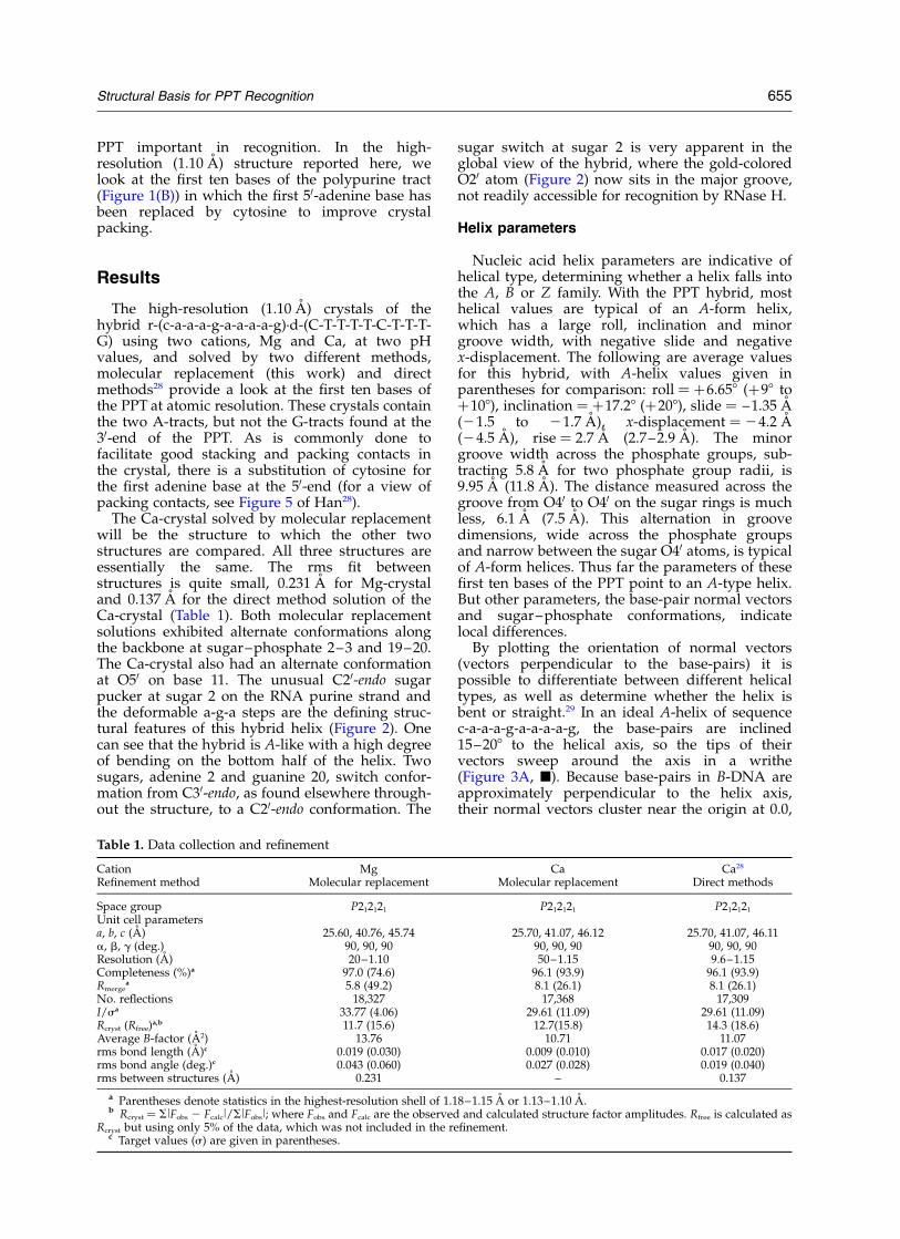
By plotting the orientation of normal vectors(vectors perpendicular to the base-pairs) it ispossible to differentiate between different helicaltypes, as well as determine whether the helix isbent or straight.29 In an ideal A-helix of sequencec-a-a-a-g-a-a-a-a-g, the base-pairs are inclined15–208 to the helical axis, so the tips of theirvectors sweep around the axis in a writhe(Figure 3A, B). Because base-pairs in B-DNA areapproximately perpendicular to the helix axis,their normal vectors cluster near the origin at 0.0,
Table 1. Data collection and refinement
Cation Mg Ca Ca28
Refinement method Molecular replacement Molecular replacement Direct methods
Space group P212121 P212121 P212121
Unit cell parametersa, b, c (A) 25.60, 40.76, 45.74 25.70, 41.07, 46.12 25.70, 41.07, 46.11a, b, g (deg.) 90, 90, 90 90, 90, 90 90, 90, 90Resolution (A) 20–1.10 50–1.15 9.6–1.15Completeness (%)a 97.0 (74.6) 96.1 (93.9) 96.1 (93.9)Rmerge
a 5.8 (49.2) 8.1 (26.1) 8.1 (26.1)No. reflections 18,327 17,368 17,309I/sa 33.77 (4.06) 29.61 (11.09) 29.61 (11.09)Rcryst (Rfree)
a,b 11.7 (15.6) 12.7(15.8) 14.3 (18.6)Average B-factor (A2) 13.76 10.71 11.07rms bond length (A)c 0.019 (0.030) 0.009 (0.010) 0.017 (0.020)rms bond angle (deg.)c 0.043 (0.060) 0.027 (0.028) 0.019 (0.040)rms between structures (A) 0.231 – 0.137
a Parentheses denote statistics in the highest-resolution shell of 1.18–1.15 A or 1.13–1.10 A.b Rcryst ¼ SlFobs 2 Fcalcl/SlFobsl; where Fobs and Fcalc are the observed and calculated structure factor amplitudes. Rfree is calculated as
Rcryst but using only 5% of the data, which was not included in the refinement.c Target values (s) are given in parentheses.
Structural Basis for PPT Recognition 655

0.0, as shown in the Han et al.27 structure of thesame sequence (Figure 3A, O). In this structure,the A-tracts exhibit little or no bend (points 2–4and 6–9 on the graph). Our PPT hybrid plot(Figure 3A, V) reveals: (1) that its two A-tracts (a2-a3-a4 and a6-a7-a8-a9) are almost as straight as inthe B-DNA helix of the same sequence with itspoints 2–4 and 6–9 clustered at opposite ends ofthe plot; and (2) that a large bend of 14.58 occursover the a-g-a steps, points 4–6. Roll values (Vrol)are very high at these steps, with the a4-g5 andg5-a6 steps having positive roll angles of 15.978and 10.278, respectively. Buckle at g5-a6 and a6-a7is also high, 18.468 and 16.488, respectively (seeTable 2). The a4-g5 purine step is unstacked,
which was not the case in the DNA/DNAB-helix.27 This destacking, high propeller, rolland buckle occur in the region of the hybridwhere the helix unzips in the Arnold andco-workers protein–PPT complex.5 The overallwrithe in the helix is 448. Both the bend at thea-g-a steps and the overall writhe are veryapparent in Figure 2.
Figure 3B reveals how each strand contributes tothe overall normal vector curve. The two indi-vidual strands, rR and dY, are shown as dottedlines. Surprisingly, the DNA strand (V) writhes ina way that smoothes the bend occurring at thea-g-a steps on the RNA strand, and compensatesfor the straight A-tracts at the two ends of the
Figure 2. A, Stereo view of minor groove of RNA/DNA hybrid. B, Axial view of hybrid. Red spheres along the RNAstrand are O30 phosphate oxygens, gold are O20 hydroxyls. Arrows indicate C20-endo O20 hydroxyl on sugar 2. Figuregenerated using PYMOL (http://pymol.sourceforge.net/).
656 Structural Basis for PPT Recognition
RNA (O). Adenine-over-adenine stacking is thepredominant interaction that makes A-tractsstraight. The large disjuncture at the a-g-a stepscontributes to the overall writhe.
Circular dichroism
The CD spectra for the DNA duplex and thehybrid are compared in Figure 4. The DNA duplexhas a typical B-DNA spectrum with positive peaksat 217 nm and 275 nm, and a negative band at248 nm.30 The hybrid duplex has spectral com-ponents associated with both A and B-type confor-mations. A-form characteristics are a large peak at271 nm, and the trough at 208 nm characteristic ofan r-purine rich strand in a hybrid duplex.20,30 B-form character is shown by a peak at 221 nm anda negative band at 248 nm. The PPT sequence withtwo short A-tracts has 70% AT base-pairs. Boththe DNA duplex and the hybrid have a shoulderat 264 nm that we interpret as a spectral feature ofA-tracts. Sen & Grasslund31 in their CD spectra ofA-tract duplexes have a shoulder at 264 nm, butdo not discuss this feature. This shoulder is unli-kely to be a structural feature of AT base-pairs inmixed sequences, because d(AAG)8·d(CTT)8 with67% AT and no A-tracts has a classical B-DNA CDspectrum with no shoulder in this region.30 Norcan this shoulder be attributed to nearest-neighborinteractions, because d(C-A-A-A-G-A-A-A-A-G)·d(C-T-T-T-T-C-T-T-T-G) and d(AAG)8·d(CTT)8
have similar nearest-neighbor interactions.
Backbone and sugar conformation
The single most striking feature of the hybridhelix is the adenine 2 ribose sugar switch fromO30-endo to O20-endo. This B-DNA sugar confor-mation was found independently in all three struc-ture analyses, the Ca and Mg crystals solved bymolecular replacement and the Ca-crystal solved apriori by direct methods.28 In the Conn et al.26
hybrid model used for molecular replacement, allsugars were in the C30-endo conformation.Difference maps (Fo 2 Fc) indicated clearly thatthe ribose O20 atom on adenine 2 had to be moved(Figure 5A). In repositioning the O20 oxygen atom
Figure 3. A, Normal vector plots of (B) RNA/RNAideal A-helix, (O) DNA/DNA B-helix27 and, (V) RNA/DNA hybrid helix. B, Repetition of the overall RNA/DNA hybrid helix curve (V) along with curves for eachindividual strand (dotted lines): (X) DNA strand (dY),(O) RNA strand (rR).
Figure 4. CD spectra of decamer duplexes of the PPTsequence. Thin line ¼ RNA/DNA hybrid (this work).Thick line ¼ DNA/DNA analog.27
Table 2. Helix parameters (8)
DNA Hybrid
Propeller Vrol Buckle Propeller Vrol Buckle
C 215.57 28.51 2.23 212.44 7.95 27.53A 211.99 6.49 2.75 27.66 0.82 24.07A 215.47 0.68 21.52 221.19 7.42 26.84A 22.62 2.83 28.01 224.76 15.97 20.92G 25.52 22.04 210.02 24.78 10.27 18.46A 211.99 21.36 21.65 24.93 6.42 16.48A 217.82 23.32 22.42 26.09 5.30 4.76A 217.01 21.92 0.02 212.53 4.68 1.39A 212.48 9.74 24.54 23.98 1.03 5.00G 214.26 – 29.71 26.59 – 26.64
Nucleic acid parameters calculated with FREEHELIX.29,42 TheRT-PPT parameters are not included in this Table because theavailable helix analysis programs do not allow for unpairedbases, instead matching each base with its supposed partnerwhether that partner is paired with it or not. Most global par-ameters in analysis of the RT-PPT helix would be correct. Base-pair parameters in the region of mispairing are in question.
Structural Basis for PPT Recognition 657

into the density, the sugar pucker converted fromC30 to C20-endo (Figure 5B). This C20-endo pucker isnot caused by crystal packing contacts. On theopposing DNA strand, all sugars are in the C30-endo conformation except guanine sugar 20, whichhas a C20-endo pucker.
At high resolution alternate conformations areoften indicated in the electron density. In this struc-ture, alternate conformations are found along thebackbone at RNA bases 2, 3 and 8, and DNA base20. The major conformers at these positions havethe following characteristics: The phosphate back-bone angles, 1 and z, at RNA sugar 2 are in theB II conformation. Adoption of the B II confor-mation requires that the sugar ring have a C20-endo conformation.32,33 Sugar 20 on the DNA strandalso has a C20-endo conformation. Along the back-bone at bases 3 (RNA) and 20 (DNA) the alternateconformers try to achieve an extended confor-mation (t, t, t) for the abg angles, with two anglestrans and the third angle tending towards transbut still in the gþ region. RNA sugar 8 has a com-pletely extended backbone in which the abgangles, the crankshaft linkage32,33 are t, t, t ratherthan the standard g2, t, gþ. Horton & Finzel16
observed crystallographically, and Cheatham &Kollman34 by molecular dynamics, that an unusualbackbone conformation on one strand is matchedfrequently by an extended conformation one base-pair away on the opposite strand. If the strain isnot released on the opposite strand, bending torelieve the strain is a possibility. In our PPT hybrid,the conformer switch from C30 to C20-endo atadenine sugar 2 is matched by a similar reversalat guanine sugar 20. The position of the phosphategroup within a two base-pair frame, Zp, alsoindicates a B-like conformation at base-pairs1–20 (0.23 A) and 2–19 (0.93 A), with A-formvalues . 1.5 A.35
Deformability of the a-g-a steps
In DNA helices, the six-membered rings of pur-ine bases are known to stack ring-over-ring. Thisis particularly true of runs of adenine bases. Intro-duction of a single guanine base (Figure 6A) intothe stack does not alter this pattern, as shown inthe crystal structure of the DNA/DNA analog ofthe PPT.27 However, a discontinuity in the stacking
occurs in the RNA/DNA hybrid PPT: where a4 isnot stacked on g5, but g5 does stack with its six-membered ring on the five-membered ring of a6(Figure 6B). Although the stacking in the hybridPPT bound to RT5 is similar to that of theuncomplexed hybrid reported here (Figure 6C),the complex also has unpaired bases. Helixparameters, propeller, roll and buckle, are utilizedto bring about this destacking. In Table 2, whichcompares these parameters at each base-step forthe DNA and the hybrid helices, one can see thatfor the DNA analog all changes occur at the A-Gstep, which is at the center of the decamer. In con-trast, the hybrid spreads out radical changes inparameters over three base-steps, a-a-g-a. Allthese parameter changes contribute to the deform-ability of this sequence.
In the RT-PPT structure Arnold and co-workers5
reported an “unzipping” of a seven base-pairregion, a-a-a-a-g-a-a, of the PPT. This unzipping orunpairing of base-pairs involves one RNA purineand one DNA pyrimidine two base-pair stepsapart on opposite strands. Their RNA base, a3(numbered as in our RNA/DNA helix), isunpaired and an unpaired DNA base, C16, isfound on the pyrimidine strand (Figure 7A). Thisunzipping is not found in our hybrid (Figure 7B).The deformability of the purine a-g-a steps appar-ently contributes to the destacking and unpairingthat occurs during RT-hybrid recognition.
Hydration and cations
Along the bottom half of the molecule betweenbases 6 to 8, O20 hydroxyl groups on the sugarsare linked together by a string of water molecules.The string is broken in the top half of the moleculebecause of the sugar switch on base 2 from C30 toC20-endo. This places the O20 hydroxyl group closerto the major groove rather than the minor groovewhere it usually resides. Because the ends of onehelix pack into the minor groove of another, theminor groove hydration pattern is broken at base-pair 1–20 as well as bases 4, 5 and 6 where packingcontacts occur. The phosphate groups are highlyhydrated, but again hydration patterns are brokenby packing contacts. Water molecules on thephosphate groups have somewhat longer H-bonds(between 3.1 A and 4.0 A), whereas water molecules
Figure 5. A, Electron density ofadenine sugar 2. Blue, 2Fo 2 Fc den-sity (1s) and pink, Fo 2 Fc density(3s), both indicating the incorrect-ness of the C30-endo sugar confor-mation. B, Density after changingto a C20-endo sugar and refinement.
658 Structural Basis for PPT Recognition

linking the O20 hydroxyl atoms are shorter (2.6–3.5 A). The wide major groove, of course, is filledwith water.
All three structure solutions, Ca-direct methods(Ca-DM), Ca-molecular replacement (Ca-MR) andMg-molecular replacement (Mg-MR), contain fourcations at the same position with the followingexceptions: 2-methyl-2,4-pentanediol (MPD) wasmodeled for one of the calcium ions in the Ca-DMsolution, while in the Ca-MR refinement a calciumion having two alternate positions was modeledfor this density. The Mg-MR located an additionaltwo magnesium ions that are not at 100%occupancy. All cations are hydrated, andmagnesium ions maintain octahedral geometry.
Hydrated calcium ions are known to have irregularcoordination geometry with six, seven or eveneight water molecules in the coordination sphere.In this structure, calcium coordination numbers ofsix or seven are found. In some cases, a phosphateoxygen atom replaces one of the coordinationwater molecules, anchoring the cation complex tothe hybrid.
Discussion
Both polymerases and nucleases perform theirenzyme action on the sugar-phosphate backboneof the nucleic acid duplexes to which they bind.
Figure 6. Stacking of the A-G-A and a-g-a steps in (A) the DNA/DNA analog27 and (B) the RNA/DNA hybrid.Grey ¼ A4-T17 (nearest viewer), black ¼ G5-C16, white ¼ A6-T15 (farthest pair). In A the six-membered rings ofpurines stack atop one another, and an exocyclic oxygen of thymine or cytosine stacks over the following pyrimidinering. In B the purine a4 is completely destacked, but its base-pair thymine stacks its exocyclic oxygen over the C16ring below it. At the next base-pair step the six-membered ring of g5 stacks over the five-membered ring of a6 andC16 does not stack on T17 at all. C, The “unzipped” RT-bound hybrid helix,5 where mispairing occurs. (Base number-ing is that of our hybrid helix, Figure 1B.) a4 (grey) is no longer paired with its mate T17, but with the neighboring T18(gold). g5 (black) is mispaired with T17 (grey). C16 (black) is unpaired and the A6–T15 base-pair (white) is back inproper register. In this structure the a4–T18 base-pair is destacked, g5 stacks on a6, and the remaining pyrimidines(T17, C16, T15) stack atop one another. Figure generated using PYMOL (http://pymol.sourceforge.net/).
Structural Basis for PPT Recognition 659

The importance of the 20-OH as the single dis-tinguishing feature of hybrid helices that confersselectivity was recognized by Horton & Finzel inthe first crystal structure of an RNA/DNAhybrid.16 Earlier X-ray work on chimeric duplexes,where a few RNA bases are interspersed in aDNA strand, observed that enzymes that recognizechimeric sequences must perceive the presence ofthe 20-hydroxyl group.36 Other features such asgroove width and bending play a role, but theseare sequence-dependent. The sugar switch from30-endo to a 20-endo pucker on adenine 2 of thehybrid duplex reported here changes the patternof sugar oxygen atoms on the floor of the minorgroove. Other local backbone changes occur atbase-pairs 1–20 and 2–19 where the A-form helixbecomes more B-like, as judged on sugar puckerand Zp, crankshaft transitions and B I versus B IIphosphate groups. B-to-A transitions have beenfound recently in several nucleic acid structures,including an A-B intermediate,37 methylated andbrominated hexamers38,39 and an RNA tetraplex.40
Protein–nucleic acid complexes analyzed byOlson and co-workers35 demonstrated that B-to-Atransitions occur with enzymes that perform cut-
ting or sealing operations at the O30 phospho-diester bond. B-to-A transitions selectively exposefor enzymatic attack sugar phosphate atoms, suchas the 30-oxygen atom, that ordinarily are buried.The reverse transition, A-to-B, conceals the 30-phos-phate oxygen atom from attack.
In the structure reported here, an unusual sugarswitch on the RNA strand from C30-endo to C20-endo at the 50-end of the PPT may play a role inrejection of the PPT by RNase H. Before consider-ing evidence implicating this switch in recognition,it is important to see what evidence exists for C20-endo sugar puckers on RNA strands. Zimmerman& Pheiffer41 in their X-ray diffraction study onpoly(rA)·poly(dT) state that poly(rA)·poly(dT) isunique in its ability to adopt either A or B-formhelices. They note that by increasing thehumidity of the fibers that initially gave anA-form diffraction pattern, “the helical parametersderived from wetted fibers of poly(rA)·poly(dT)are similar but not identical with those of wettedDNA fibers”. This makes poly(rA)·poly(dT) uniqueamong hybrids, in that it can adopt either A orB-form.
Molecular dynamic simulations34 have shown
Figure 7. A, 2Fo 2 Fc map of the PPT sequence, r-(a-a-a-g-a)/d-(T-C-T-T-T) as found in the Sarafianos et al.5 PPT/RTcomplex, contoured at 0.9s. B, The same hybrid sequence from our RNA/DNA structure, contoured at 1.2s. In A thefirst base-pair a2-T19 is paired, a3 is unpaired, a4 is mispaired with T18, g5 is mispaired with T17, C16 is unpairedand the helix comes back into register with the last base-pair a6-T15. In B all bases are properly paired. The differencein map-cage density occurs because A is a 3.0 A structure and B is a 1.1 A structure. Base numbering as in Figure 1.Figure generated using PYMOL (http://pymol.sourceforge.net/).
660 Structural Basis for PPT Recognition

that “B-RNA” is energetically possible and can beconverted to A-RNA by a change in sugar puckerfrom C20-endo to C30-endo. Sugar repuckering fromC20 to C30-endo and back occurs at a much lowerrate than in DNA simulations. For this reason, thebarrier to repuckering is adjudged to be higher inRNA than DNA, but is longer-lived when it occurs.As for the phosphate groups in the simulations, B Ito B II transitions are observed in both DNA andRNA strands in “B-RNA” and hybrid helices.Although not conventional dogma, the authorsconclude, “It was previously thought that theunacceptable stereochemistry of the O20 hydroxyl“bumping” into the following phosphate group,sugar ring and base would destabilize the B-formgeometry and make B-RNA unfavorable… theinteraction is not unfavorable.”
It appears from the preceding arguments that a
B-form sugar pucker (C20-endo) in an RNA strandof a hybrid is acceptable geometry. This sugaroccurs on the RNA strand at a c-a step, which is apyrimidine-purine step (YR). YR steps, includingT-A, C-A and U-A, are the most deformable,13,42
and it has been shown in protein/DNA complexesthat the T-A step can adopt either A or Bconformation.35
The RNase H catalytic pocket is composed ofpositively charged amino acid residues, a con-served histidine residue and divalent cations,either manganese or magnesium, that are requiredfor catalysis.43,44 Catalysis occurs by deprotonationof water to form a nucleophilic hydroxide groupthat attacks the scissile phosphate group on RNA.To illustrate the role the adenine 2 sugar confor-mation plays in RNase H recognition at the 50-endof the PPT, Figure 8 shows a least-squares fit of
Figure 8. A, Least square superposition of bases 1 to 4 of the RNA strand of the RNA/DNA decamer, c-a-a-a, (white)onto the Sarafianos et al.5 backbone (black, red, yellow) at the RNase H active site (rms ¼ 1.19 A). Two magnesium ions,pink (crystallographic B ¼ 23.5) and white (crystallographic B ¼ 79.9) in the Huang et al.6 RT structure are least squarefitted onto the Sarafianos structure5 (rms ¼ 1.10 A). B, Least square superposition of bases 2 to 5 (white), a-a-a-g, ontothe backbone (rms ¼ 1.25 A). Positively-charged amino acid side-chains, R448, N474, and Q475, (from top to bottomleft) are blue-green. Negatively charged E478, D443 and D549 (from left to right), with D498 below D443, are in red.H539 at lower right is magenta. Figure generated using PYMOL (http://pymol.sourceforge.net/).
Structural Basis for PPT Recognition 661

the backbone atoms of the RNA/DNA hybrid (inwhite) to the Arnold and co-workers5 RNA/DNAPPT structure at the RNase H active site. Threeamino acid residues capable of recognizing thepolyanionic backbone of the nucleic acid, R448,N474 and Q475, “read” the 20-hydroxyl groups ofthe first four bases of the hybrid decamer RNAstrand (white). N474 and Q475 have beensuggested as critical in determining RNase Hspecificity.11 These residues make hydrogen bondsfrom N and O atoms on their amino acid side-chains to 20-oxygen atoms on the RNA sugars(Figure 8A). The second sugar, with the 20-endopucker, is not synchronous with the readingframe, having a distance of 6.70 A from its 20-hydroxyl group to an arginine NH2. At the bottomof Figure 8A are the acidic amino acid residues,D443, E478, D498 and D549, of the RNase Hcatalytic site along with H539, which is implicatedin the cutting mechanism. RNase H cuts at a 30-OH. Two magnesium ions, pink (B ¼ 23 A3) andwhite (B ¼ 79 A3), found in the structure byHarrison and co-workers6 have been super-imposed. In Figure 8B the 20-endo sugar hasmoved into the RNase H catalytic pocket. Here,the reading frame has been disrupted andH-bonds to the 20-hydroxyl group of the C20-endosugar are larger than 6 A. The contacts to the O1and O2 phosphate atoms are also too long (3.82and 5.96 A), and the scissile 30OH group on theRNA strand (white) has been moved out of reachof the catalysis mechanism.
If sugar pucker and phosphate backbone confor-mation are important for recognition, the questionremains, what keeps the sugar in this C20-endo con-formation? An interesting report, investigating therole of the “U-box” in replication, observes thatneither the mechanism for PPT resistance toRNase H, nor the requirements for its 50-end cuthave been established.45 A string of five uridinebases in the RNA strand (thymine in the finaltranscript) immediately upstream, i.e. 50, to thePPT is highly conserved in retroviruses. Mutationsin this region can block viral replication in cellsand the authors45 pinpoint the blocking event asoccurring between the first strand jump and thestart of second strand synthesis. It is possible this
string of uridine bases plays a structural role.Stacking of five uridine bases upon four adeninebases would have quite different stackinggeometry than that of random sequence. Inrelieving the strain at the juncture between thepyrimidine (Y) stack and the purine (R) stack, thebackbone may adjust at this YR step with a C30-endo to C20-endo sugar switch.
The unzipping of the protein-bound hybrid helixfound by Sarafianos et al.5 occurs in the five base-pair region, a-a-a-g-a, of the PPT that includes thea-g-a steps. This five base motif is found on RNAstrands in a number of retroviruses includingCAEV, FIV, and SIV (see Table 2 of Iliyinski &Desrosiers45). Some retroviruses do not have thefull five base motif, but have a shortened motif,e.g. FeLV has a-a-g-a. This sequence, a-a-a-g-a,appears to be important in retroviral recognitionand may be used by other polymerases thatrecognize hybrids.
In the Nucleic Acid Database46 three hybridstructures (AH0001, AH0005 and DR0003) arefound that have an RNA strand with only purinesand a DNA strand entirely of pyrimidines.26,47,48
All of these sequences, r-(50g-a-a-g-a-g-a-a-g-c30),r-(50g-a-a-g-a-a-g-a-g30) and r-(50g-a-a-g-a-a-g-a-a30),contain deformable a-g-a steps. The Conn et al.26
sequence crystallizes in the same space group,P212121, as the hybrid reported here; the other twocrystallize in space group P61. Each structure hastwo a-g-a steps (Table 3). At the 50-end of theConn et al. P212121 structure, adenine stacksover guanine, but not as in a DNA helix, wheresix-membered rings lie over each other. Thesix-membered ring of the first adenine stacks overthe five-membered ring of guanine. The guaninesix-membered ring then stacks only partially overthe following five-membered adenine ring. At thea-g-a steps at the 30-end, six-membered over five-membered stacking predominates. At the 50-end ofboth Xiong & Sundaralingam crystal structures47,48
the a-g step is de-stacked as in the hybrid reportedhere (Figure 6B), while at both 30-ends, the a-gsteps are stacked with the six-membered ring overthe five. This leads to the conclusion that a-g-asteps in a hybrid helix are deformable, and thisdeformability appears to be context-dependent.
Table 3. The a-g-a steps in hybrid crystal structures
AuthorNDB orPDB No. Sequence (RNA strand)
Spacegroup (A) Resolution Stacking of a-g step
This work UH0005 c-a-a-a-g-a-a-a-a-g P212121 1.15 UnstackedUH0006
Han28 AH0012Conn et al.26 AH0001 g-a-a-g-a-g-a-a-g-c P212121 2.5 50
-a-g stacked30
-a-g partially stackedXiong &Sundaralingam48
DR0003 g-a-a-g-a-a-g-a-g P61 1.8 50
-a-g unstacked30
-a-g stackedXiong &Sundaralingam47
AH0005 g-a-a-g-a-a-g-a-a P61 1.8 50
-a-g unstacked30
-a-g stackedSarafianos et al.5 1HYS a-a-a-a-g-a-a-a-a-g (seq. located
within a 31-mer)P3212 3.0 Unstacked with mispaired bases
before and after the unstacking
662 Structural Basis for PPT Recognition

Only one protein-hybrid structure in the PDBdatabase49 contains the a-a-a-g-a sequence, theSarafianos et al.5 RT-PPT structure. In fact, onlyfive structures report utililization of these fivebases in their recognition sequence. Three of theseare proteins bound to DNA targets, one is thehybrid RT-PPT structure, and the last is aribosomal protein bound to RNA.
Conclusions
Two sugar switches have been found in nucleicacid sequences utilized for both plus and minusstrand initiation. Both occur at a YR step locatedone base-step away from the scissile bond. In theNMR structure of the Okazaki junction formed atminus-strand initiation, a sugar switch from C30-endo to C20-endo is found one step away from thescissile bond. This cut removes the tRNALys primerthat is annealed to the viral RNA.14 A narrowminor groove is found in the hybrid portion ofthis junction as well. Sarafianos et al.5 also found anarrow minor groove in the PPT bound to RT.Neither the crystal structure of (2 )-strand Okazakifragment13 nor the PPT decamer sequence reportedhere have a narrow minor groove. Possibly aninduced fit between protein and the malleablenucleic acid with concomitant groove narrowing isoccurring. The narrow minor groove, the sugarswitch to C20-endo on the RNA strand one baseremoved from the scissile bond, along with theunzipping of the bases found in the PPT bound toprotein, facilitated by the deformable a-g-a steps,appear to be important signals for RNase Hrecognition.
In conclusion, the RT–nucleic acid structures todate have all been at or near 3.0 A resolution. Atthis resolution, sugar conformation is indetermi-nate. To be able to release sugar constraints duringrefinement, data at or near 2.3 A or less are needed.High-resolution structures of hybrids containinguridine tracts elucidating their stacking propertieswould also be helpful, as well as a structure of the30 end of the PPT which contains the six guaninebases.
Materials and Methods
Crystallization and structure solution
The deoxy-pyrimidine strand was synthesized bysolid-phase phosphoramidite methods and purifiedby anion-exchange chromatography on a WhatmanDE-52 column. The ribonucleotide purine strandwas purchased from Yale University’s Keck Oligo-nucleotide Synthesis facility and purified by PAGE on50 cm £ 33 cm £ 0.3 cm gels. The RNA was eluted fromthe gel using a Schleicher & Schuell Elutrap Electroeluter.Gel-purification of the RNA strand was essential forobtaining hybrid crystals that diffract to high resolution(1.10 A). Hybrid crystals that resulted when the RNAstrand was purified by chromatography diffracted only
between 2.5 A and 4.5 A. The two strands were annealedin a waterbath at 65 8C and allowed to cool slowly over-night to room temperature. Crystals were grown at roomtemperature by vapor-diffusion in micro-bridges(Hampton Research) using two different cations, mag-nesium (Mg-crystal) and calcium (Ca-crystal), and twopH values, 5.8 and 6.8. The Ca-crystal was grown from0.3 mM hybrid duplex, 12 mM calcium acetate, 0.6 mMspermidine hydrochloride (pH 6.8), 0.075% b-octylgluco-side, 12 mM sodium cacodylate (pH 6.8) and 12% (v/v)MPD versus 40% (Ca-crystal) and 50% (Mg-crystal)MPD in the crystallization reservoirs. All crystallizationcomponents were the same for the Mg-crystal with theexception of substituting 12 mM magnesium acetate forthe calcium acetate and 12 mM sodium cacodylate atpH 5.8. The two crystals were isomorphous. Data werecollected at the National Synchrotron Light Source atBrookhaven National Laboratories at 21808C. The MPDused in crystallization was a sufficient cryoprotectantfor both crystals. Data were processed using DENZOand SCALEPACK.50 The Ca-crystal was solved indepen-dently by direct methods.28 Comparison data statisticsfor all three structure solutions are shown in Table 1.
The Mg and Ca structures were solved by molecularreplacement with EPMR51 using a hybrid duplex model,NDB AH0001,26 consisting of an RNA purine strandr-(g-a-a-g-a-g-a-a-g-c) and a DNA pyrimidine strandd-(G-C-T-T-C-T-C-T-T-C). (Statistics for the Ca and Mgdata sets are reported together, separated by a slashbetween values, e.g. Ca/Mg.) Using data from 8–3 A inEPMR for the Ca-crystal and 15–4 A for the Mg-crystal,the starting Rxtal was (Ca/Mg) 54.8/55.8%. Rigid-bodyrefinement in CNS52 dropped (Ca/Mg) Rxtal to 45.9/50.3% and Rfree to 44.2/49.4%. Simulated annealing plusB-factor refinement at 2.0 A/1.5 A (Ca/Mg) gaveRxtal ¼ 31.3/35.0%, Rfree ¼ 38.8/38.2%. SHELXL-9753
refinement was begun (Ca/Mg) 8–2 A/8–1.6 A followedby anisotropic refinement when the majority of the datahad been added. Final anisotropic R-factors were (Ca/Mg) Rxtal ¼ 12.7/11.7% and Rfree ¼ 15.8/15.6%. The Ca-structure has four hydrated calcium ions, with oneshowing 50% occupancy at two positions, and 127 watermolecules. The Mg-structure has six hydrated mag-nesium ions with two at lower occupancy (70% and30%) and 128 water molecules. Because negativedifference density was observed on two of the mag-nesium cations, it became apparent that this high-qualitydata required a reduced scattering factor for Mg2þ. Forthis reason, Ne scattering factors, isoelectronic withMg2þ, were used. Water molecules were positionedwhere peaks greater than 1–1.25s in the 2Fo 2 Fc and atleast 3s in the Fo 2 Fc maps appeared simultaneously.
CD spectroscopy
Microcrystals were dissolved in double-distilled waterand the solution desalted in a Centricon 3 filter (threewashes). The final solutions contained each strand at aconcentration of 4.8 mM in a buffer of 150 mM NaCl,5 mM MgCl2, 10 mM sodium cacodylate (pH 7.0). Spectrawere recorded at 5 8C on a computer-driven AVIV 62DSCD spectrophotometer using a 1.0 nm band-width. Thecell compartment was purged continuously with N2. Thedata were least-squares fit with a polynomial of seventhorder. Extinction coefficients used were: r/d(C-A-A-A-G-A-A-A-A-G) 1 ¼ 115.3 cm21 mM21, d(C-T-T-T-T-C-T-T-T-G) 1 ¼ 83.94 cm21 mM21
Structural Basis for PPT Recognition 663

Coordinates accession number
Atomic coordinates and structure factors have beendeposited in the Protein Data Bank (PDB)† and theNucleic Acid Database (NDB) (PDB ID code 1PJG and1PJO; NDB ID code UH0005 and UH0006).
Acknowledgements
We thank Dr Duilio Cascio for data collectionassistance, Dr Ann Maris for technical assistanceand discussion, and Dr Michael Sawaya for assist-ance with graphics. This study was supported byNational Institute of Healths grant GM31299.
References
1. Kohlstaedt, L. A., Wang, J., Friedman, J. M., Rice, P. A.& Steitz, T. A. (1992). Crystal structure at 3.5 A resol-ution of HIV-1 reverse transcriptase complexed withan inhibitor. Science, 256, 1783–1790.
2. Jacobo-Molina, A., Ding, J., Nanni, R. G., Clark, A. D.,Jr., Lu, X., Tantillo, C. et al. (1993). Crystal structure ofhuman immunodeficiency virus type 1 reverse tran-scriptase complexed with double-stranded DNA at3.0 A resolution shows bent DNA. Proc. Natl Acad.Sci. USA, 90, 6320–6324.
3. Rodgers, D. W., Gamblin, S. J., Harris, B. A., Ray, S.,Culp, J. S., Hellmig, B. et al. (1995). The structure ofunliganded reverse transcriptase from the humanimmunodeficiency virus type 1. Proc. Natl Acad. Sci.USA, 92, 1222–1226.
4. Ding, J., Das, K., Hsiou, Y., Sarafianos, S. G., Clark,A. D., Jr, Jacobo-Molina, A., Tantillo, C. et al. (1998).Structure and functional implications of the poly-merase active site region in a complex of HIV-1 RTwith a double-stranded DNA template-primer andan antibody Fab fragment at 2.8 A resolution. J. Mol.Biol. 284, 1095–1111.
5. Sarafianos, S., Das, K., Tantillo, C., Clark, A. D., Jr,Ding, J., Whitcomb, J. M. et al. (2001). Crystal structureof HIV-1 reverse transcriptase in complex with a poly-purine tract RNA:DNA. EMBO J. 20, 1449–1461.
6. Huang, H., Chopra, R., Verdine, G. L. & Harrison,S. C. (1998). Structure of a covalently trapped catalyticcomplex of HIV-1 reverse transcriptase: implicationsfor drug resistance. Science, 282, 1669–1675.
7. Le Grice, S. J. (1993). Human immunodeficiencyvirus reverse transcriptase. In Reverse Transcriptase(Skalka, A. M. & Goff, S. P., eds), pp. 175–191, ColdSpring Harbor Laboratory Press, Plainview, NY.
8. Coffin, J. M., Hughes, S. H. & Varmus, H. E. (1997).Chapter 4. In Retroviruses, Cold Spring HarborLaboratory Press, Plainview, NY pp. 129–130.
9. Davies, J. F., II, Hostomska, Z., Hostomsky, Z.,Jordan, S. R. & Matthews, D. A. (1991). Crystal struc-ture of the ribonuclease H domain of HIV-1 reversetranscriptase. Science, 252, 88–95.
10. Tisdale, M., Schulze, T., Larder, B. A. & Moelling, K.(1991). Mutations within the RNase H domain ofhuman immunodeficiency virus type 1 reverse tran-
scriptase abolish virus infectivity. J. Gen. Virol. 72,59–66.
11. Julias, J. G., McWilliams, M. J., Sarafianos, S. G.,Arnold, E. & Hughes, S. H. (2002). Mutations in theRNaseH domain of HIV-1 reverse transcriptase affectthe initiation of DNA synthesis and the specificity ofRNase H cleavage in vivo. Proc. Natl Acad. Sci. USA,99, 9515–9520.
12. Rausch, J. W., Lener, D., Miller, J. T., Julias, J. G.,Hughes, S. H. & Le Grice, S. F. J. (2002). Altering theRNase H primer grip of human immunodeficiencyvirus reverse transcriptase modifies cleavagespecificity. Biochemistry, 41, 4856–4865.
13. Mueller, U., Maier, G., Onori, A. M., Cellai, L.,Heumann, H. & Heinemann, U. (1998). Crystalstructure of an eight base pair duplex containing the30-DNA–RNA-50 junction formed during initiationof minus strand synthesis of HIV replication.Biochemistry, 37, 12005–12011.
14. Szyperski, T., Gotte, M., Billeter, M., Perola, E., Cellai,L., Heumann, H. & Wuthrich, K. (1999). NMR struc-ture of the chimeric hybrid duplex r(gcaguggc)·r(gc-ca)d(CTGC) comprising the tRNA-DNA junctionformed during initiation of HIV-1 reverse transcrip-tion. J. Biomol. NMR, 13, 343–355.
15. Fedoroff, O. Y., Salazar, M. & Reid, B. R. (1996).Structural variation among retroviral primer–DNAjunctions: solution structure of the HIV-1 (2)-strandOkazaki fragment r(gcca)d(CTGC)·d(GCAGTGGC).Biochemistry, 35, 11070–11080.
16. Horton, N. C. & Finzel, B. C. (1996). The Structure ofan RNA/DNA hybrid: a substrate of the ribo-nuclease activity of HIV-1 reverse transcriptase.J. Mol. Biol. 264, 521–533.
17. Powell, M. D., Ghosh, M., Jacques, P. S., Howard,K. J., Le Grice, S. F. J. & Levin, J. G. (1997). Alanine-scanning mutations in the “primer grip” of p66 HIV-1reverse transcriptase result in selective loss of RNApriming activity. J. Biol. Chem. 272, 13262–13269.
18. Randolph, C. A. & Champoux, J. J. (1994). The use ofDNA and RNA oligonucleotides in hybrid structureswith longer polynucleotide chains to probe the struc-tural requirements for Moloney murine leukemiavirus plus strand priming. J. Biol. Chem. 269,19207–19215.
19. Fuentes, G. M., Rodriguez-Rodriguez, L., Fay, P. J. &Bambara, R. A. (1995). Use of an oligoribonucleotidecontaining the polypurine tract sequence as a primerby HIV reverse transcriptase. J. Biol. Chem. 47,28169–28176.
20. Powell, M. D. & Levin, J. G. (1996). Sequence andstructural determinants required for priming ofplus-strand DNA synthesis by the human immuno-deficiency virus type 1 polpurine tract. J. Virol. 70,5288–5296.
21. Fedoroff, O. Y., Salazar, M. & Reid, B. R. (1993). Struc-ture of a DNA:RNA hybrid duplex. Why RNase Hdoes not cleave pure RNA. J. Mol. Biol. 233, 509–523.
22. Schultz, S. J., Zhang, M., Kelleher, C. D. &Champoux, J. J. (1999). Polypurine tract primergeneration and utilization by Moloney murineleukemia virus reverse transcriptase. J. Biol. Chem.274, 34547–34555.
23. Fedoroff, O. Y., Ge, Y. & Reid, B. R. (1997). Solutionstructure of r(gaggacug):d(CAGTCCTC) hybrid:implications for the initiation of HIV-1 (þ)-strandsynthesis. J. Mol. Biol. 269, 225–239.
24. Gao, X., Jeffs, P. W. & Jeffs, P. W. (1994). Sequence-dependent conformational heterogeneity of a hybrid† www.rcsb.org
664 Structural Basis for PPT Recognition

DNA·RNA dodecamer duplex. J. Biomol. NMR, 4,367–384.
25. Gyi, J. I., Lane, A., Conn, G. L. & Brown, T. (1998).Solution structures of DNA·RNA hybrids withpurine-rich and pyrimidine-rich strands: comparisonwith the homologous DNA and RNA duplexes.Biochemistry, 37, 73–80.
26. Conn, G. L., Brown, T. & Leonard, G. A. (1999). Thecrystal structure of the RNA/DNA hybrid r(GAAGAGAAGC)·d(GCTTCTCTTC) shows significantdifferences to that found in solution. Nucl. Acids Res.27, 555–561.
27. Han, G. W., Kopka, M. L., Cascio, D., Grzeskowiak,K. & Dickerson, R. E. (1997). Structure of a DNAanalog of the primer for HIV-1 RT second strandsynthesis. J. Mol. Biol. 269, 811–826.
28. Han, G. W. (2001). Direct-methods determination ofan RNA/DNA hybrid decamer at 1.15 A resolution.Acta Crystallog. sect. D, 57, 213–218.
29. Dickerson, R. E. & Chiu, T. K. (1998). Helix bendingas a factor in protein/DNA recognition. Biopolymers(Nucl. Acid Sci.), 44, 361–403.
30. Hung, S. H., Yu, Q., Gray, D. M. & Ratliff, R. L.(1994). Evidence from CD spectra that d(purine)·r(pyrimidine) and r(purine)·d(pyrimidine hybridsare in different structural classes. Nucl. Acids Res. 22,4326–4334.
31. Sen, A. & Graslund, A. (2000). Structural constraintsregulating triple helix formation by A-tracts. Biophys.Chem. 88, 69–80.
32. Fratini, A. V., Kopka, M. L., Drew, H. R. & Dickerson,R. E. (1982). Reversible bending and helix geometryin a B-DNA dodecamer: CGCGAATTBRCGCG.J. Biol. Chem. 257, 14686–14707.
33. Dickerson, R. E., Kopka, M. L. & Drew, H. R. (1983).Structural correlations in B-DNA. In Structure andDynamics: Nucleic Acids and Proteins (Clementi, E. &Sarma, R. H., eds), pp. 149–179, Adenine Press,Guilderland, NY.
34. Cheatham, T. E., III & Kollman, P. A. (1997). Molecu-lar dynamics simulations highlight the structuraldifferences among DNA:DNA, RNA:RNA, andDNA:RNA hybrid duplexes. J. Am. Chem. Soc. 119,4805–4825.
35. Lu, X.-J., Shakked, Z. & Olson, W. K. (2000). A-formconformational motifs in ligand-bound DNA struc-tures. J. Mol. Biol. 300, 819–840.
36. Egli, M., Usman, N. & Rich, A. (1993). Conformationalinfluence of the ribose 20-hydroxyl group: crystal struc-tures of DNA–RNA chimeric duplexes. Biochemistry,32, 3221–3237.
37. Ng, H.-L., Kopka, M. L. & Dickerson, R. E. (1999).The structure of a stable intermediate in the A $ BDNA helix transition. Proc. Natl Acad. Sci. USA, 97,2035–2039.
38. Vargason, J. M., Eichman, B. F. & Ho, P. S. (2000). Theextended and eccentric E-DNA structure induced bycytosine methylation or bromination. Nature Struct.Biol. 7, 758–761.
39. Vargason, J. M., Henderson, K. & Ho, P. S. (2001). A
crystallographic map of the transition from B-DNAto A-DNA. Proc. Natl Acad. Sci. USA, 98, 7265–7270.
40. Deng, H., Xiong, Y. & Sundaralingam, M. (2001).X-ray analysis of an RNA tetraplex (UGGGGU)4
with divalent Sr2þ ions at subatomic resolution(0.61 A). Proc. Natl Acad. Sci. USA, 98, 13665–13670.
41. Zimmerman, S. B. & Pheiffer, B. H. (1981). A RNA–DNA hybrid that can adopt two conformations: anX-ray diffraction study of poly(rA)·poly(dT) in con-centrated solution or in fibers. Proc. Natl Acad. Sci.USA, 78, 78–82.
42. Dickerson, R. E. (1998). DNA bending: theprevalence of kinkiness and the virtues of normality.Nucl. Acids Res. 26, 1906–1926.
43. Goedken, E. R. & Marqusee, S. (2001). Co-crystals ofEscherichia coli RNase H with Mn2þ ions reveals twodivalent metals bound in the active site. J. Biol.Chem. 10, 7266–67211.
44. Keck, J. L., Goedken, E. R. & Marqusee, S. (1998).Activation/attenuation model for RNaseH. J. Biol.Chem. 273, 34128–34133.
45. Iliyinski, P. O. & Desrosiers, R. C. (1998). Identifi-cation of a sequence element immediately upstreamof the polypurine tract that is essential for replicationof simian immunodeficiency virus. EMBO J. 17,3766–3774.
46. Berman, H. M., Olson, W. K., Beveridge, D. L.,Westbrook, J., Gelbin, A., Demeny, T., Hsieh, S.-H.,Srinivasan, A. R. & Schneider, B. (1992). The NucleicAcid Database: a comprehensive relational databaseof three-dimensional structures of nucleic acids.Biophys. J. 63, 751–759.
47. Xiong, Y. & Sundaralingam, M. (1998). Crystal struc-ture and conformation of a DNA–RNA hybridduplex with a polypurine RNA strand:d(TTCTTBr5CTTCC)-r(GAAGAAGAA). Structure, 6,1493–1501.
48. Xiong, Y. & Sundaralingam, M. (2000). Crystal struc-ture of a DNA·RNA hybrid duplex with a poly-purine RNA r(gaagaagag) and a complementarypolypyrimidine DNA d(CTCTTCTTC). Nucl. AcidsRes. 28, 2171–2176.
49. Berman, H. M., Westbrook, J., Feng, Z., Gilliland, G.,Bhat, T. N., Weissig, H. et al. (2000). The ProteinData Bank. Nucl. Acids Res. 28, 235–242.
50. Otwinowski, Z. & Minor, W. (1996). Macromolecularcrystallography. In Methods Enzymol. (Carter, J. W. &Sweet, R. M., eds), vol. 276, Academic Press, NewYork, pp. 307–326.
51. Kissinger, C. R., Gelhaar, D. K. & Fogel, D. B. (1999).Rapid automated molecular replacement by evol-utionary search. Acta Crystallog. sect. D, 55, 484–491.
52. Brunger, A. T., Adams, P. D., Clore, G. M., DeLano,W. L., Gros, P., Grosse-Kuntsleve, W. et al. (1998).Crystallography & NMR system: a new softwaresuite for macromolecular structure determination.Acta Crystallog. sect. D, 54, 905–921.
53. Sheldrick, G. M. & Schneider, T. R. (1997). SHELXL:high resolution refinement. In Methods Enzymol.(Carter, J. W. & Sweet, R. M., eds), vol. 277, AcademicPress, New York, pp. 319–343.
Edited by K. Morikawa
(Received 17 July 2003; received in revised form 17 September 2003; accepted 25 September 2003)
Structural Basis for PPT Recognition 665


![Human exonuclease 1 (EXO1) activity characterization and its … · 2017-10-04 · EXO1 to the DNA [8], while the I-domain exhibits multiple cysteine and glutamate residues that are](https://img.pdfslide.us/doc/110x75/5f8ce5575c41787f96248c61/human-exonuclease-1-exo1-activity-characterization-and-its-2017-10-04-exo1-to.jpg)