Embed Size (px)
Citation preview
Proc. Natl. Acad. Sci. USAVol. 90, pp. 7119-7123, Augustcvs 1993Biochemistry
An erythromycin analog produced by reprogramming ofpolyketide synthesis
(enoyl reductase/macrolide/metabolic engineering/Streptomyces)
STEFANO DONADIO*, JAMES B. MCALPINE, PAUL J. SHELDONt, MARIANNA JACKSON, AND LEONARD KATZPharmaceutical Products Division, Abbott Laboratories, Abbott Park, IL 60064
Communicated by Richard Losick, March 24, 1993
ABSTRACT The polyketide-derived macrolactone of theantibiotic erythromycin is made through successive condensa-tion and processing of seven three-carbon units. The fourthcycle involves complete processing of the newly formed 13-ketogroup (.3keto reduction, dehydration, and enoyl reduction) toyield the methylene that will appear at C-7 of the lactone ring.Synthesis of this molecule in Saccharopolyspora erythraea isdetermined by the three large eryA genes, organized in sixmodules, each governing one condensation cycle. Two aminoacid substitutions were introduced in the putative NAD(P)Hbinding motif in the proposed enoyl reductase domain encodedby eryAMi. The metabolite produced by the resulting strain wasidentified as A6"7-anhydroerythromycin C resulting from fail-ure ofenoyl reduction during the fourth cycle ofsynthesis ofthemacrolactone. This result demonstrates the involvement of atleast the enoyl reductase from the fourth module in the fourthcycle and indicates that a virtually complete macrolide can beproduced through reprogramming of polyketide synthesis.
A wide variety of natural compounds, exhibiting antibacte-rial, antihelminthic, antitumor, and immunosuppressive ac-tivities, contain a polyketide-derived skeleton. Biosynthesisof polyketides is mechanistically equivalent to formation oflong-chain fatty acids (1), where the fatty acid synthase (FAS)condenses the extender unit malonate with the starter unitacetate and the resulting (3-keto group undergoes three pro-cessing steps, 3-keto reduction, dehydration, and enoylreduction, to yield a fully saturated butyryl unit. The C4 chainis elongated through repeated addition of two carbon atoms(derived from malonate) and fully processed at each cycle,until the proper length of a symmetrical chain has beenreached. Many polyketides, in contrast, retain ketone, hy-droxyl, or olefinic functions and contain methyl or ethyl sidegroups interspersed along an acyl chain oflength comparableto that of common fatty acids. This asymmetry in structureimplies that the polyketide synthase (PKS), the enzymesystem responsible for formation of these molecules, al-though mechanistically equivalent to FAS, must somehow beprogrammed to produce the correct molecular structure.The current model (2) for biosynthesis of complex poly-
ketides (defmed as compounds whose synthesis requireseach FAS-like cycle to be usually different from the previousone) is exemplified, for the erythromycin aglycone DEB (Fig.1), in Fig. 2. In Saccharopolyspora erythraea, three eryAgenes govern the synthesis of this molecule and consist of sixrepeated units, termed modules, encoding six synthase units(SUs). We have proposed that each SU, which comprises aseries of putative FAS-like activities, is responsible for oneof the six elongation cycles required for DEB formation (2).A total of28 distinct enzymatic activities, each unique for onecatalytic event, reside in three large, multifunctional pro-
The publication costs of this article were defrayed in part by page chargepayment. This article must therefore be hereby marked "advertisement"in accordance with 18 U.S.C. §1734 solely to indicate this fact.
teins-EryAl, EryAII, and EryAIII-encoded by eryA (Fig.2). Thus, the noniterative processive synthesis ofasymmetricacyl chains found in complex polyketides is accomplishedthrough the use ofa programmed protein template, where thenature of the chemical reactions occurring at each point isdetermined by the specificities of the domains contained ineach SU.The involvement of a distinct enzymatic activity in each
synthesis step implies that a modification affecting a singleactivity should perturb only the corresponding step. Suchmodifications offer the potential for reprogramming the PKS,with the consequent production of novel polyketide struc-tures. If each enzymatic activity can be assigned to a specificstep in the biosynthetic pathway, the structure of the newlyformed polyketide can be predicted. For DEB synthesis, theorder in which the modules are arranged in the Sac. erythraeachromosome has been proposed to match the sequence inwhich the corresponding SUs are employed in the six elon-gation cycles (2). Previous work showed that deletion of thesequence in eryAI corresponding to the KR domain of thefifth SU resulted in formation of a DEB derivative retainingthe keto group introduced during the fifth cycle at the C-5position, in accordance with the prediction from the model(2). Here we extend our previous finding that modifiedpolyketides ofpredicted structure can be produced by geneticintervention, by demonstrating that polyketide synthesis canbe reprogrammed by abolishing another activity involved in13-carbon processing, resulting in the production of a novelcomplete macrolide.
MATERIALS AND METHODSBacterial Strains, Culture Conditions, and Plasmids. Sac.
erythraea strains ER720 (3) and EER4S, described below,were cultivated as described (2, 4). Conditions for isolation ofgenomic DNA (5), integrative transformation (4, 6, 7), andgene replacement (8) have been reported. Sac. erythraeametabolites were isolated and analyzed by TLC (9). Plasmidswere propagated and isolated from Escherichia coli DH5a(GIBCO/BRL) by following the supplier's conditions fortransformation. Plasmids of the pUC series (10) were rou-tinely employed for subcloning, and the vector pWHM3,which replicates poorly in Sac. erythraea (11), was used forintegrative transformation. The eryA DNA has been de-scribed (2, 9).
Purification of A6,7-Anhydroerythromycin C. Spores ofSac. erythraea strain EER4S were grown as described (12).
Abbreviations: ACP, acyl carrier protein; DEB, 6-deoxyerythro-nolide B; DH, dehydratase; ER, enoyl reductase; FAS, fatty acidsynthase; KR, 3-keto reductase; PKS, polyketide synthase; SU,synthase unit.*Present address: Lepetit Research Center, via R. Lepetit 34, 21040Gerenzano (VA), Italy.
tPresent address: Bioprocess Technology Institute, University ofMinnesota, St. Paul, MN 55108.
7119
Dow
nloa
ded
by g
uest
on
Dec
embe
r 29
, 201
9
7120 Biochemistry: Donadio et al.
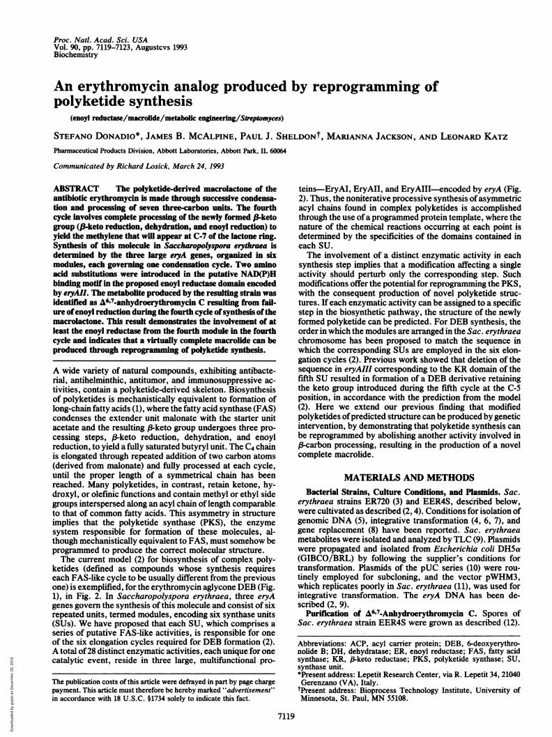
6-Deoxyerythronolide B CH3 CH(DEB) Erythromycin A A6,7-Anhydroerythromycin C
FIG. 1. Structures of 6-deoxyerythronolide B (DEB), erythromycin A, and t6,7-anhydroerythromycin C.
After 7 days, the pH was adjusted to 9 with NaOH and thebeer was extracted with 2 half-volumes of methylene chlo-ride. The combined concentrated extracts were digested in 1liter of methylene chloride and extracted with 1 volume of0.33 M citric acid. The aqueous layer was readjusted to pH9 and extracted with 1 volume of methylene chloride. Thissecond methylene chloride extract, after water washes andconcentration to a residue (176 mg), was chromatographed onan Ito multilayered Planet Coil centrifuge in a system con-sisting of carbon tetrachloride, methanol, 0.01 M potassiumphosphate (pH 7.2), 1:1:1 (vol/vol), with the lower phasestationary. The eluate was monitored by TLC and selectedfractions were diluted with an equal volume of water,adjusted to pH 9 with NH40H, and extracted twice withequal volumes of methylene chloride, and the residue ob-tained after drying the combined organic layers was digestedin C2HCl3 and subjected to 'H NMR analysis. Fractionsfound to contain macrolides were combined and similarlyextracted to yield 45 mg of a solid residue, which waschromatographed on an Ito multilayered Planet Coil centri-fuge in a system consisting of heptane, benzene, 2-propanol,acetone, 0.01 M potassium phosphate (pH 7.0), 5:10:3:2:5,with the upper phase mobile. On the basis of TLC and 'HNMR analysis, fractions were combined, concentrated, andpartitioned between water (adjusted to pH 9) and methylenechloride. The organic phase was separated and concentratedto give a solid residue of A6'7-anhydroerythromycin C.
Spectral Determinations. NMR spectra were obtained from2HC13 solutions with a General Electric GN500 spectrometerwith data acquired at 500 MHz (1H) and 125 MHz (13C). Massspectrometry was performed on a Kratos Analytical Instru-
ments MS50 instrument operating in the fast-atom-bombardment positive-ion mode.
RESULTSRationale. According to the model for DEB formation (Fig.
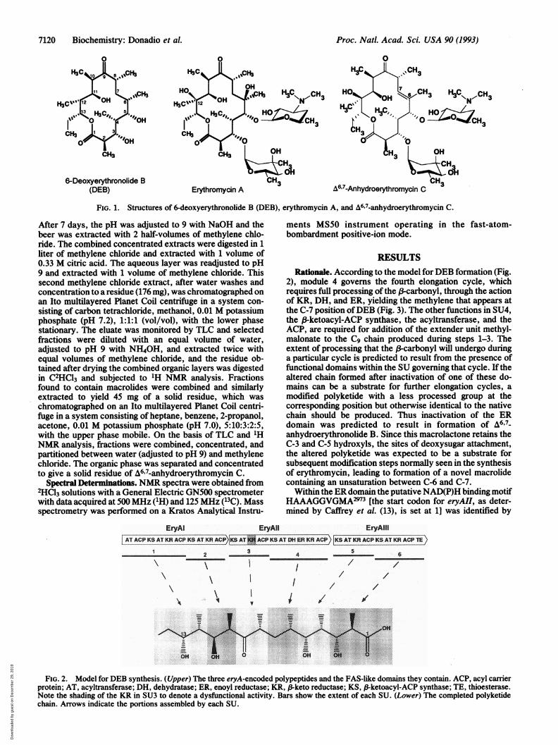
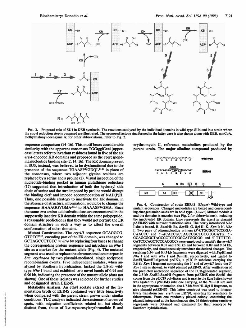
2), module 4 governs the fourth elongation cycle, whichrequires full processing of the B-carbonyl, through the actionof KR, DH, and ER, yielding the methylene that appears atthe C-7 position ofDEB (Fig. 3). The other functions in SU4,the ,B-ketoacyl-ACP synthase, the acyltransferase, and theACP, are required for addition of the extender unit methyl-malonate to the C9 chain produced during steps 1-3. Theextent of processing that the f-carbonyl will undergo duringa particular cycle is predicted to result from the presence offunctional domains within the SU governing that cycle. If thealtered chain formed after inactivation of one of these do-mains can be a substrate for further elongation cycles, amodified polyketide with a less processed group at thecorresponding position but otherwise identical to the nativechain should be produced. Thus inactivation of the ERdomain was predicted to result in formation of A6'7-anhydroerythronolide B. Since this macrolactone retains theC-3 and C-5 hydroxyls, the sites of deoxysugar attachment,the altered polyketide was expected to be a substrate forsubsequent modification steps normally seen in the synthesisof erythromycin, leading to formation of a novel macrolidecontaining an unsaturation between C-6 and C-7.Within the ER domain the putative NAD(P)H binding motif
HAAAGGVGMA2973 [the start codon for eryAII, as deter-mined by Caffrey et al. (13), is set at 1] was identified by
EryAl EryAll EryAlliAT ACPKSATKRACPKSATK CPKSAT '--ACP KS AT DH ER KR ACP KSATKRACP KS AT KR ACP TE )
1 2 3 4 5 6\ I // /
I\ I
.................. .
... ........ " .. .
..... ......... ,. .... .
.. , .....:*:: :::::::::-: :-: :-: :.::+:::+::+::*:z:.-+:t:4:.::::-::t:*:-: :-- :-:j:-:4 als
/ /
FIG. 2. Model for DEB synthesis. (Upper) The three eryA-encoded polypeptides and the FAS-like domains they contain. ACP, acyl carrierprotein; AT, acyltransferase; DH, dehydratase; ER, enoyl reductase; KR, /3keto reductase; KS, f3ketoacyl-ACP synthase; TE, thioesterase.Note the shading of the KR in SU3 to denote a dysfunctional activity. Bars show the extent of each SU. (Lower) The completed polyketidechain. Arrows indicate the portions assembled by each SU.
Proc. Natl. Acad. Sci. USA 90 (1993)
\I
Dow
nloa
ded
by g
uest
on
Dec
embe
r 29
, 201
9
Proc. Natl. Acad. Sci. USA 90 (1993) 7121
[2H]
ERKS -SH
[2H]A
6ACP-S l\R51
6/
V R/K- KS
0 0
AT HOKk%9S-ACP
(2R)-mmCoA
R=OH OH O
FIG. 3. Proposed role of SU4 in DEB synthesis. The reactions catalyzed by the individual domains in wild-type SU4 and in a strain wherethe enoyl reduction step is bypassed are illustrated. The proposed lactone ring formed in the latter case is also shown along with DEB. mmCoA,methylmalonyl-coenzyme A; for other abbreviations, refer to Fig. 2.
sequence comparison (14-16). This motif bears considerablesimilarity with the apparent consensus TGGtgglGxel (upper-case letters refer to invariant residues) found in five of the sixeryA-encoded KR domains and proposed as the correspond-ing nucleotide binding site (2, 14, 16). The KR domain presentin SU3, instead, was believed to be dysfunctional due to thepresence of the sequence TGAASPVGDQL'147 in place ofthe consensus, where two adjacent glycine residues arereplaced by a serine and a proline (2). Visual inspection ofthenucleotide-binding pocket in human glutathione reductase(17) suggested that introduction of both the hydroxyl sidechain of serine and the turn imposed by proline would disruptthe binding cleft and impede accommodation of NAD(P)H.Thus, one possible strategy to inactivate the ER domain, inthe absence of structural information, would be to change thesequence HAAAGGVGMA2973 to HAAASPVGMA. Sincethe same two amino acid substitutions are encountered in thesupposedly inactive KR domain within the same polypeptide,a reasonable prediction is that they would not perturb the ERdomain structure in such a way as to affect the overallconformation of other domains.Mutant Construction. The eryAIl sequence GCAGGCG-
GTGTC8910, encoding part of the ER domain, was changed toGCTAGCCCTGTC in vitro by replacing four bases to changethe corresponding protein sequence and introduce an Nhe Isite as a marker for the mutant allele (Fig. 4). The mutatedsegment was used to replace the chromosomal counterpart inSac. erythraea by two plasmid-mediated, single reciprocalrecombination events. Five independent isolates, when an-
alyzed by Southern hybridization, lacked the 1.8-kb wild-type Nhe I band and exhibited two novel bands of 0.94 and0.90 kb, indicating the presence of the mutant allele (data notshown). One ofthese isolates was selected for further studiesand designated strain EER4S.
Metabolite Analysis. An ethyl acetate extract of the fer-mentation broth of EER4S contained very little bioactivitywhen compared with the parent strain grown under similarconditions. TLC analysis indicated the existence oftwo novelspots, with migration coefficients related to, but clearlydistinct from, those of 3-a-mycarosylerythronolide B and
erythromycin C, reference metabolites produced by theparent strain. The major alkaline compound produced by
CACGCAGCGGCAGGCGGTGTCGGCH A A A G G V G
NhelCACGCAGCGGCqtG3CITGTCGGCH A A A S P V G
wild type
EER4S
K N Bs l11 B rN G
HKSH AT HDHF-H ~X IKRHACP
FIG. 4. Construction of strain EER4S. (Upper) Wild-type andmutant sequences. Changed nucleotides are boxed and correspond-ing changed amino acids are in bold type. (Lower) Mutant module 4and the domains it encodes (see Fig. 2 for abbreviations), includingthe inactivated ER domain. Line represents the insert in plasmidpAER4S5 with relevant restriction sites. The newly introduced NheI site is boxed. B, BamHI; Bs, BspEI; G, Bgl II; K, Kpn I; N, NheI. Two pairs of oligonucleotide primers (5'-CTGCGGTTCCGGA-CAACCC and 5'-ACACCGCTAGCCGCTGCGTGGATG; 5'-GCAGCGGCTAGCCCTGTCGGCATGGCGG and 5'-TTTTTG-GATCCCAGCTCCCACGCC) were employed to amplify the eryAIIsegments between 8.37 and 8.91 kb and between 8.89 and 9.34 kb,respectively, and simultaneously introduce the desired changes. Theresulting 0.54- and 0.45-kb fragments were digested with BspEI andNhe I and with Nhe I and BamHI, respectively, and ligated toBspEI/BamHI-digested pAIK3, a pUC19 subclone carrying the3.6-kb Kpn I fragment comprising the 3' end of eryAII (only the 5'Kpn I site is shown), to yield plasmid pAER4S. After verification ofthe predicted nucleotide sequence of the PCR-generated segment,the 2.5-kb EcoRI-BamHI fragment from pAER4S (the EcoRI sitecomes from the pUC19 polylinker and is next to the Kpn I site shown)was ligated to a pWHM3 subclone carrying, at the BamHI site andin the appropriate orientation, the 1.7-kb BamHI-Bgl II fragment, togive plasmid pAER4S5. This latter construct was used to integra-tively transform Sac. erythraea ER720, selecting for resistance tothiostrepton. From one randomly picked colony, containing theplasmid integrated at the homologous site, 16 thiostrepton-sensitivesegregants were obtained and examined for their genotype bySouthern hybridization.
Biochemistry: Donadio et al.
Dow
nloa
ded
by g
uest
on
Dec
embe
r 29
, 201
9
7122 Biochemistry: Donadio et al.
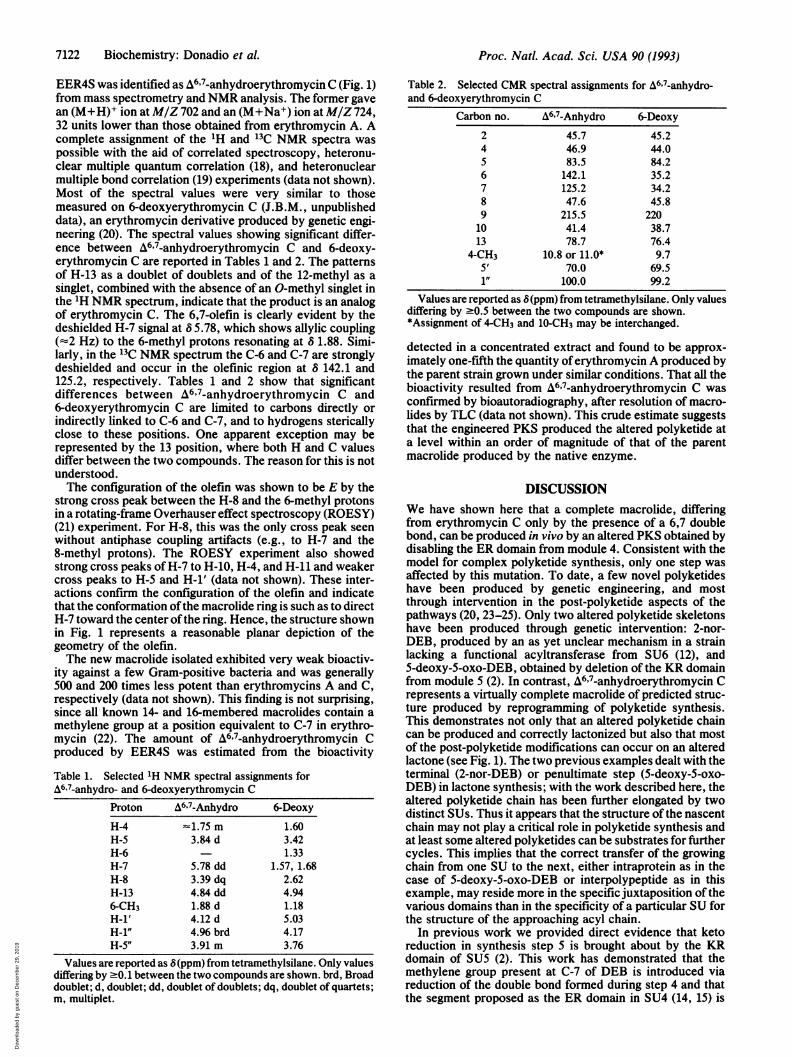
EER4S was identified as A6'7-anhydroerythromycin C (Fig. 1)from mass spectrometry and NMR analysis. The former gavean (M+H)+ ion at M/Z 702 and an (M+Na+) ion at M/Z 724,32 units lower than those obtained from erythromycin A. Acomplete assignment of the 'H and 13C NMR spectra waspossible with the aid of correlated spectroscopy, heteronu-clear multiple quantum correlation (18), and heteronuclearmultiple bond correlation (19) experiments (data not shown).Most of the spectral values were very similar to thosemeasured on 6-deoxyerythromycin C (J.B.M., unpublisheddata), an erythromycin derivative produced by genetic engi-neering (20). The spectral values showing significant differ-ence between A6'7-anhydroerythromycin C and 6-deoxy-erythromycin C are reported in Tables 1 and 2. The patternsof H-13 as a doublet of doublets and of the 12-methyl as asinglet, combined with the absence of an 0-methyl singlet inthe 'H NMR spectrum, indicate that the product is an analogof erythromycin C. The 6,7-olefin is clearly evident by thedeshielded H-7 signal at 6 5.78, which shows allylic coupling(-2 Hz) to the 6-methyl protons resonating at 8 1.88. Simi-larly, in the 13C NMR spectrum the C-6 and C-7 are stronglydeshielded and occur in the olefinic region at 8 142.1 and125.2, respectively. Tables 1 and 2 show that significantdifferences between A6'7-anhydroerythromycin C and6-deoxyerythromycin C are limited to carbons directly orindirectly linked to C-6 and C-7, and to hydrogens stericallyclose to these positions. One apparent exception may berepresented by the 13 position, where both H and C valuesdiffer between the two compounds. The reason for this is notunderstood.The configuration of the olefin was shown to be E by the
strong cross peak between the H-8 and the 6-methyl protonsin a rotating-frame Overhauser effect spectroscopy (ROESY)(21) experiment. For H-8, this was the only cross peak seenwithout antiphase coupling artifacts (e.g., to H-7 and the8-methyl protons). The ROESY experiment also showedstrong cross peaks of H-7 to H-10, H-4, and H-11 and weakercross peaks to H-5 and H-i' (data not shown). These inter-actions confirm the configuration of the olefin and indicatethat the conformation ofthe macrolide ring is such as to directH-7 toward the center ofthe ring. Hence, the structure shownin Fig. 1 represents a reasonable planar depiction of thegeometry of the olefin.The new macrolide isolated exhibited very weak bioactiv-
ity against a few Gram-positive bacteria and was generally500 and 200 times less potent than erythromycins A and C,respectively (data not shown). This finding is not surprising,since all known 14- and 16-membered macrolides contain amethylene group at a position equivalent to C-7 in erythro-mycin (22). The amount of A6'7-anhydroerythromycin Cproduced by EER4S was estimated from the bioactivity
Table 1. Selected 'H NMR spectral assignments forA6'7-anhydro- and 6-deoxyerythromycin C
Proton A6'7-Anhydro 6-Deoxy
H-4 -1.75 m 1.60H-5 3.84 d 3.42H-6 1.33H-7 5.78 dd 1.57, 1.68H-8 3.39 dq 2.62H-13 4.84 dd 4.946-CH3 1.88 d 1.18H-l' 4.12 d 5.03H-i" 4.96 brd 4.17H-5" 3.91 m 3.76
Values are reported as 8(ppm) from tetramethylsilane. Only values
Table 2. Selected CMR spectral assignments for A6'7-anhydro-and 6-deoxyerythromycin C
Carbon no. A6'7-Anhydro 6-Deoxy
2 45.7 45.24 46.9 44.05 83.5 84.26 142.1 35.27 125.2 34.28 47.6 45.89 215.5 22010 41.4 38.713 78.7 76.4
4-CH3 10.8 or 11.0* 9.75' 70.0 69.51" 100.0 99.2
Values are reported as 8(ppm) from tetramethylsilane. Only valuesdiffering by -0.5 between the two compounds are shown.*Assignment of 4-CH3 and 10-CH3 may be interchanged.
detected in a concentrated extract and found to be approx-imately one-fifth the quantity oferythromycin A produced bythe parent strain grown under similar conditions. That all thebioactivity resulted from A6'7-anhydroerythromycin C wasconfirmed by bioautoradiography, after resolution of macro-lides by TLC (data not shown). This crude estimate suggeststhat the engineered PKS produced the altered polyketide ata level within an order of magnitude of that of the parentmacrolide produced by the native enzyme.
DISCUSSIONWe have shown here that a complete macrolide, differingfrom erythromycin C only by the presence of a 6,7 doublebond, can be produced in vivo by an altered PKS obtained bydisabling the ER domain from module 4. Consistent with themodel for complex polyketide synthesis, only one step wasaffected by this mutation. To date, a few novel polyketideshave been produced by genetic engineering, and mostthrough intervention in the post-polyketide aspects of thepathways (20, 23-25). Only two altered polyketide skeletonshave been produced through genetic intervention: 2-nor-DEB, produced by an as yet unclear mechanism in a strainlacking a functional acyltransferase from SU6 (12), and5-deoxy-5-oxo-DEB, obtained by deletion of the KR domainfrom module 5 (2). In contrast, A6'7-anhydroerythromycin Crepresents a virtually complete macrolide of predicted struc-ture produced by reprogramming of polyketide synthesis.This demonstrates not only that an altered polyketide chaincan be produced and correctly lactonized but also that mostof the post-polyketide modifications can occur on an alteredlactone (see Fig. 1). The two previous examples dealt with theterminal (2-nor-DEB) or penultimate step (5-deoxy-5-oxo-DEB) in lactone synthesis; with the work described here, thealtered polyketide chain has been further elongated by twodistinct SUs. Thus it appears that the structure ofthe nascentchain may not play a critical role in polyketide synthesis andat least some altered polyketides can be substrates for furthercycles. This implies that the correct transfer of the growingchain from one SU to the next, either intraprotein as in thecase of 5-deoxy-5-oxo-DEB or interpolypeptide as in thisexample, may reside more in the specific juxtaposition of thevarious domains than in the specificity of a particular SU forthe structure of the approaching acyl chain.
In previous work we provided direct evidence that ketoreduction in synthesis step 5 is brought about by the KRdomain of SU5 (2). This work has demonstrated that themethylene group present at C-7 of DEB is introduced viareduction of the double bond formed during step 4 and thatthe segment proposed as the ER domain in SU4 (14, 15) is
differing by :0.1 between the two compounds are shown. brd, Broaddoublet; d, doublet; dd, doublet of doublets; dq, doublet of quartets;m, multiplet.
Proc. Natl. Acad. Sci. USA 90 (1993)
Dow
nloa
ded
by g
uest
on
Dec
embe
r 29
, 201
9
Proc. Natl. Acad. Sci. USA 90 (1993) 7123
responsible for this reduction. Thus, one domain each fromSU4 (ER), SU5 (KR), and SU6 (acyltransferase) has beendirectly demonstrated to be involved in the correspondingsynthesis step, strengthening the argument of colinearitybetween genetic order of the modules and sequence ofcondensation cycles. Within the ER domain, the predictedNAD(P)H-binding motif must be essential for activity, sinceas few as two amino acid substitutions abolish this function.Because these same two amino acid changes are also en-countered in the putative NAD(P)H-binding motif in the KRdomain of SU3, the findings presented here provide addi-tional support to the original notion that this KR is inactive(2), thus accounting for retention ofa keto group during cycle3 (Fig. 2), and that the lack of nucleotide binding is, at leastin part, responsible for the inactivity.Recent analysis ofgenes involved in avermectin formation
has indicated that the corresponding PKS is organized sim-ilarly to that for the DEB PKS (26, 27). These two enzymesystems, recently referred to as modular PKSs (28) to dis-tinguish them from those operating in processes that aremostly iterative, present a certain flexibility in the primarystructure of SUs introducing a common degree of 3-carbonprocessing. In avermectin synthesis, where five doublebonds are retained, the DH-containing SUs completely lackER domains (27). In the erythromycin PKS, a small changein the ER domain is sufficient to eliminate this activity, buta functional PKS deleted of the entire ER domain has notbeen successfully generated (S.D., unpublished work). Sim-ilarly, a keto group is retained in avermectin synthesis by aSU completely lacking a KR domain (27); for erythromycin,KR function can be lost either through the presence of a fewpoint mutations, as in SU3, or after purposeful removal ofthis domain without noticeable effects on the other activitiesresiding in the PKS (2).Formation of A6,7-anhydroerythromycin C indicates that
the lactone ring carrying the 6,7 unsaturation is a substrate forthe post-polyketide modification enzymes: mycarosyl- anddesosaminyltransferases and C-12 hydroxylase. These threereactions have thus far been observed with several ringstructures in genetically modified Sac. erythraea (2, 12, 20),even when the alterations were very close to the site ofmodification. On the other hand, the isolation of a macrolidewith an unmodified mycarose moiety in the present studyindicates that the O-methyltransferase is unable to methylatethe 3" position of the unsaturated macrolide. This finding wasnot totally unexpected, as- this enzyme is quite sensitive tosmall perturbations in substrate structure (29).The erythromycin PKS represents a good model system for
reprogramming polyketide synthesis. The entire sequence ofthe corresponding genes is known (2, 15, 30) and a role foreach enzymatic domain has been proposed (2). In addition,methodologies for gene transfer in Sac. erythraea are avail-able and the order of occurrence of the post-polyketidemodifications is known. Thus, engineering the erythromycinpathway through PKS reprogramming offers the challengingopportunity of understanding the rules governing complexpolyketide synthesis while producing, in vivo, novel mac-rolide structures.
We are grateful to Michael Staver for confirmatory sequencing,Bill Kohl for fermentation, Susan Swanson for technical advice, andJoyti Patel for providing oligonucleotides. We thank Dick Hutchin-
son for critical reading of the manuscript and Doug MacNeil forcommunicating information prior to publication.
1. Hopwood, D. A. & Sherman, D. H. (1990) Annu. Rev. Genet.24, 37-66.
2. Donadio, S., Staver, M. J., McAlpine, J. B., Swanson, S. J. &Katz, L. (1991) Science 262, 675-679.
3. DeWitt, J. P. (1985) J. Bacteriol. 164, 969-971.4. Donadio, S., Shafiee, A. & Hutchinson, C. R. (1990) J. Bac-
teriol. 171, 350-360.5. Hopwood, D. A., Bibb, M. J., Chater, K. F., Kieser, T.,
Bruton, C. J., Kieser, H. M., Lydiate, D. J., Smith, C. P.,Ward, J. M. & Schrempf, H. (1985) Genetic Manipulation ofStreptomyces: A Laboratory Manual (John Innes Foundation,Norwich, U.K.).
6. Kieser, T. & Hopwood, D. A. (1991) Methods Enzymol. 204,430-458.
7. Weber, J. M. & Losick, R. (1988) Gene 68, 173-180.8. Donadio, S. & Hutchinson, C. R. (1991) Gene 100, 231-235.9. Tuan, J. S., Weber, J. M., Staver, M. J., Leung, J. O., Don-
adio, S. & Katz, L. (1990) Gene 90, 21-29.10. Vieira, J. & Messing, J. (1982) Gene 19, 259-268.11. Vara, J. A., Lewandoska-Skarbek, M., Wang, Y.-G., Donadio,
S. & Hutchinson, C. R. (1989) J. Bacteriol. 170, 5872-5881.12. McAlpine, J. B., Tuan, J. S., Brown, D. P., Grebner, K. D.,
Whittern, D. N., Buko, A. & Katz, L. (1987) J. Antibiotics 40,1115-1122.
13. Caffrey, P., Bevitt, D. J., Staunton, J. & Leadlay, P. F. (1992)FEBS Lett. 304, 225-228.
14. Donadio, S. & Katz, L. (1992) Gene 111, 51-60.15. Bevitt, D. J., Cortes, J., Haydock, S. F. & Leadlay, P. F.
(1992) Eur. J. Biochem. 204, 39-49.16. Witkowski, A., Rangan, V. S., Randhawa, Z. I., Amy, C. M.
& Smith, S. (1991) Eur. J. Biochem. 198, 571-579.17. Scrutton, N. S., Berry, A. & Perham, R. N. (1990) Nature
(London) 343, 38-44.18. Muller, L. (1979) J. Am. Chem. Soc. 101, 4481-4484.19. Bax, A. & Summers, M. F. (1986) J. Am. Chem. Soc. 108,
2093-2094.20. Weber, J. M., Leung, J. O., Swanson, S. J., Idler, K. B. &
McAlpine, J. B. (1991) Science 252, 114-117.21. Bothner-By, A. A., Stephens, R. L., Lee, J. M., Warren,
C. D. & Jeanloz, R. W. (1984) J. Am. Chem. Soc. 106, 811-813.22. Omura, S. (1984) in Macrolide Antibiotics: Chemistry, Biology
and Practice, ed. Omura, S. (Academic, Orlando, FL).23. Epp, J. K., Huber, M. L. B., Turner, J. R., Goodson, T. &
Schoner, B. E. (1989) Gene 85, 293-301.24. Hara, 0. & Hutchinson, C. R. (1992) J. Bacteriol. 174, 5141-
5144.25. Hopwood, D. A., Malpartida, F., Kieser, H. M., Ikeda, H.,
Duncan, J., Fujii, I., Rudd, B. A., Floss, H. G. & Omura, S.(1985) Nature (London) 314, 642-644.
26. MacNeil, D. J., Occi, J. L., Gewain, K. M., MacNeil, T.,Gibbons, P. H., Ruby, C. L. & Danis, S. J. (1992) Gene 115,119-125.
27. MacNeil, D. J., Occi, J. L., Gewain, K. M., MacNeil, T.,Gibbons, P. H., Foor, F. & Morin, N. (1993) in Genetics andMolecular Biology of Industrial Microorganisms, eds. Baltz,R. H., Ingolia, T. & Hegeman, G. (Am. Soc. Microbiol.,Washington, DC), in press.
28. Hopwood, D. A., Khosla, C., Sherman, D. H., Bibb, M. J.,Ebert-Khosla, S., Kim, E.-S., McDaniel, R., Revill, W. P.,Torres, R. & Yu, T.-W. (1993) in Genetics and MolecularBiology of Industrial Microorganisms, eds. Baltz, R. H., In-golia, T. & Hegeman, G. (Am. Soc. Microbiol., Washington,DC), in press.
29. Paulus, T. J., Tuan, J. S., Luebke, V. E., Maine, G. T., De-witt, J. P. & Katz, L. (1990) J. Bacteriol. 172, 2541-2546.
30. Cortes, J., Haydock, S. F., Roberts, G. A., Bevitt, D. J. &Leadlay, P. F. (1991) Nature (London) 346, 176-178.
Biochemistry: Donadio et al.
Dow
nloa
ded
by g
uest
on
Dec
embe
r 29
, 201
9


![Doc1 - CDC...126 Gr. A Streptococci erythromycin 2001 2003 [24] 53% faecalis erythromycin chloram- phenicol ciprofloxacin gentamicin avoparcin ' E. faecalis vancomycin](https://img.pdfslide.us/doc/110x75/6118145c1932226e937f5e05/doc1-cdc-126-gr-a-streptococci-erythromycin-2001-2003-24-53-faecalis-erythromycin.jpg)