Embed Size (px)
Citation preview
An Ovine Model of Intramedullary Nailing Through the Pediatric Distal
Femoral Physis
Derrick M. Knapik, MD, Raymond W. Liu, MD
SIGN Fracture Care International Conference
September 23, 2016
Rainbow Babies and Children’s Hospital
Case Western Reserve University
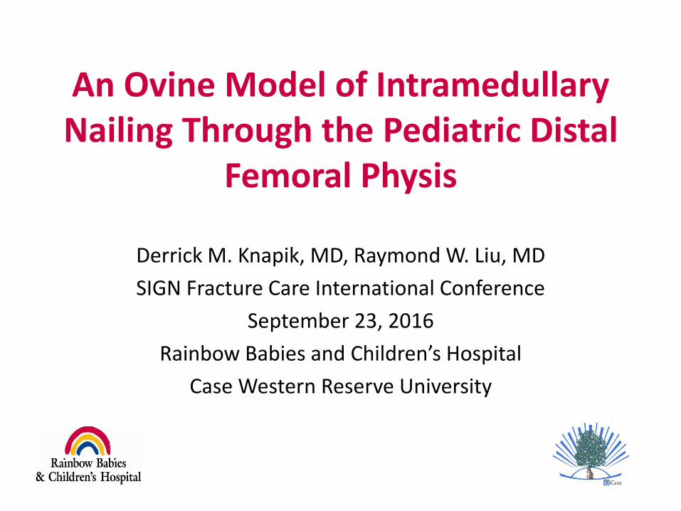
Femur Fractures
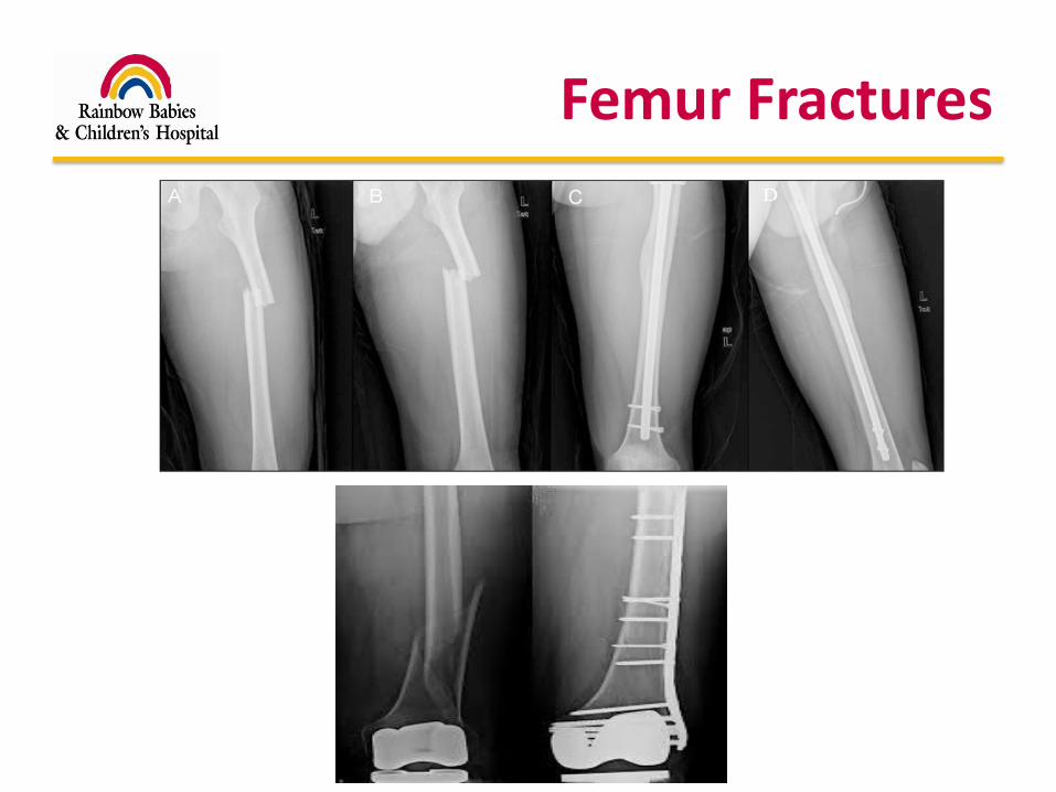
Resource Poor Hospital
Operating Room Waiting Area Operating Room

Significant Impact of Injury
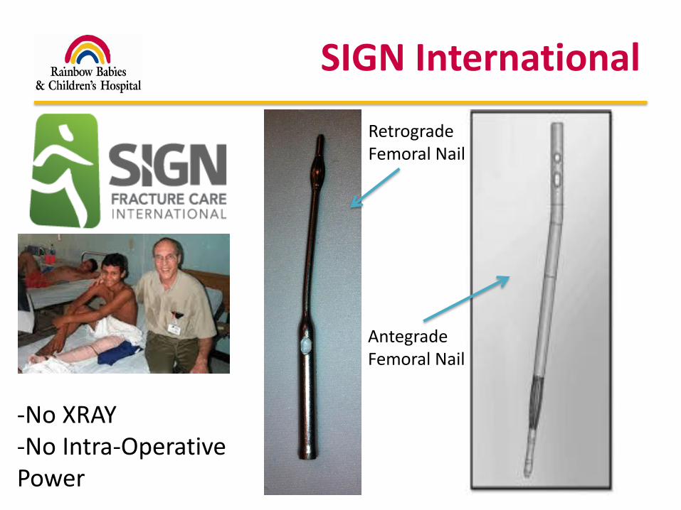
SIGN International
Retrograde Femoral Nail
Antegrade Femoral Nail
-No XRAY-No Intra-Operative Power
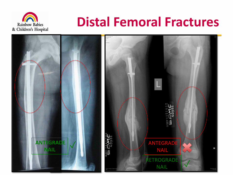
Distal Femoral Fractures
RETROGRADE NAIL
ANTEGRADENAIL
ANTEGRADENAIL
✓
✓
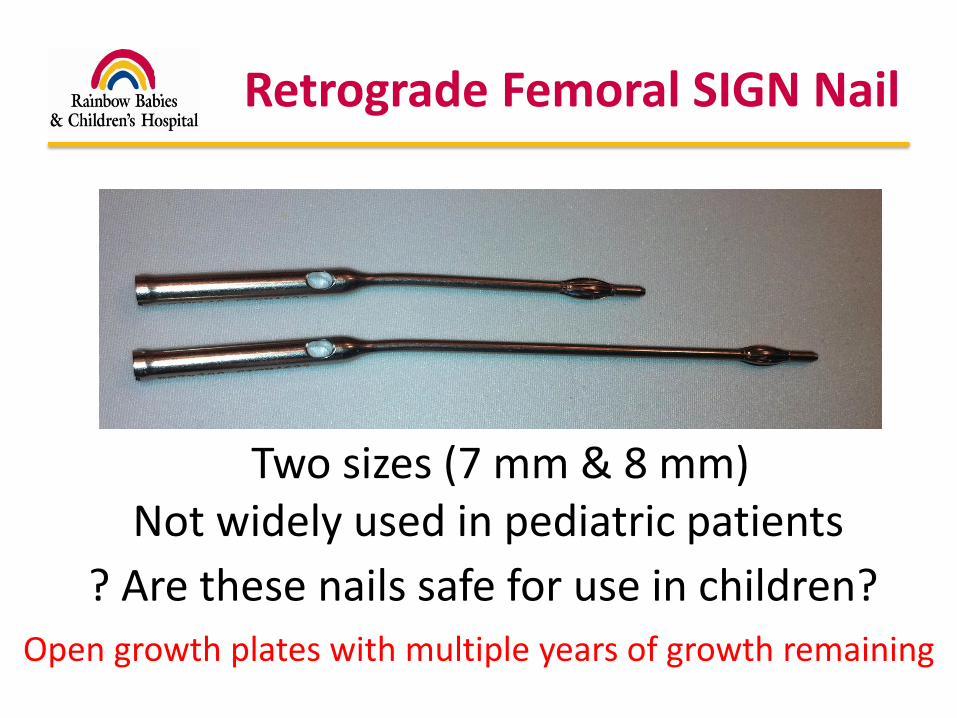
Retrograde Femoral SIGN Nail
? Are these nails safe for use in children?
Two sizes (7 mm & 8 mm)
Open growth plates with multiple years of growth remaining
Not widely used in pediatric patients
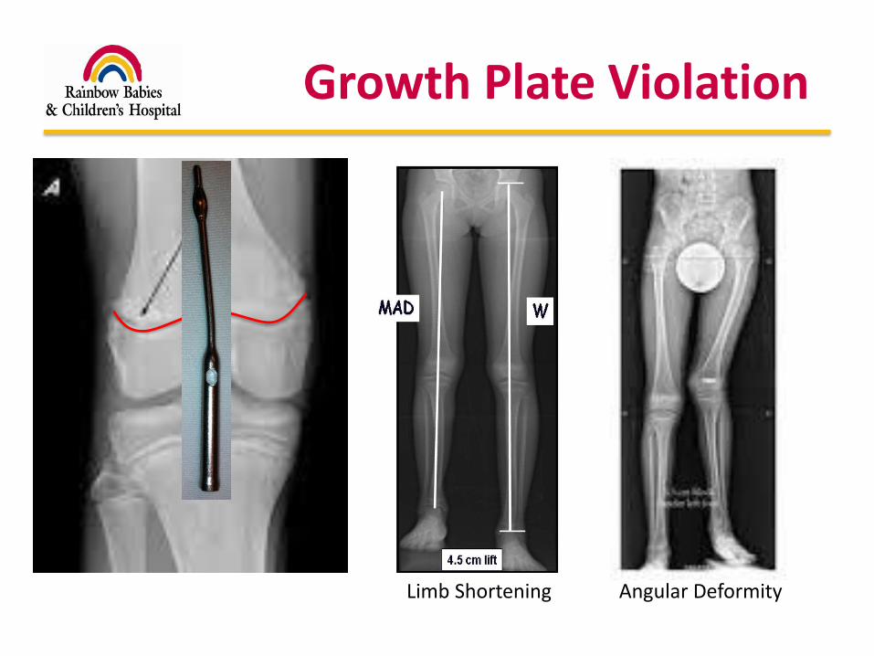
Growth Plate Violation
Limb Shortening Angular Deformity
Purpose
What magnitude of an immature, distal femoral growth plate’s cross sectional area can be violated with a retrograde, centrally located and perpendicularly placed implant without causing significant growth arrest?

Hypothesis
With increasing implant diameter, once a certain area of the growth plate is violated, growth arrest will occur
Rabbit Model3% no inequality in femoral lengths7% femoral shortening
JBJS (Br.), 1988
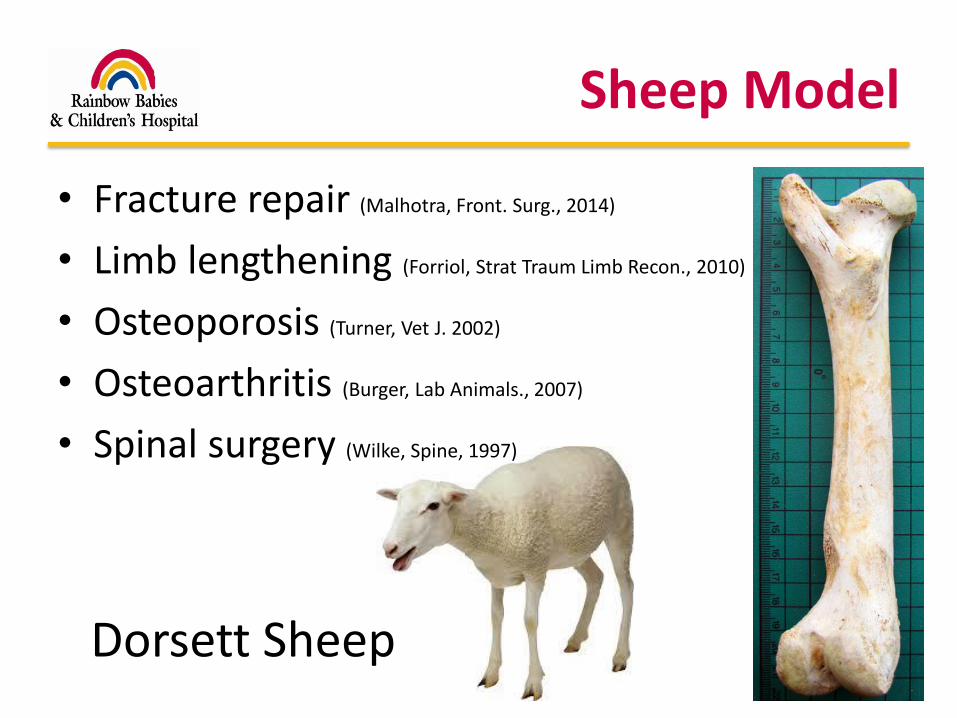
Sheep Model
• Fracture repair (Malhotra, Front. Surg., 2014)
• Limb lengthening (Forriol, Strat Traum Limb Recon., 2010)
• Osteoporosis (Turner, Vet J. 2002)
• Osteoarthritis (Burger, Lab Animals., 2007)
• Spinal surgery (Wilke, Spine, 1997)
Dorsett Sheep
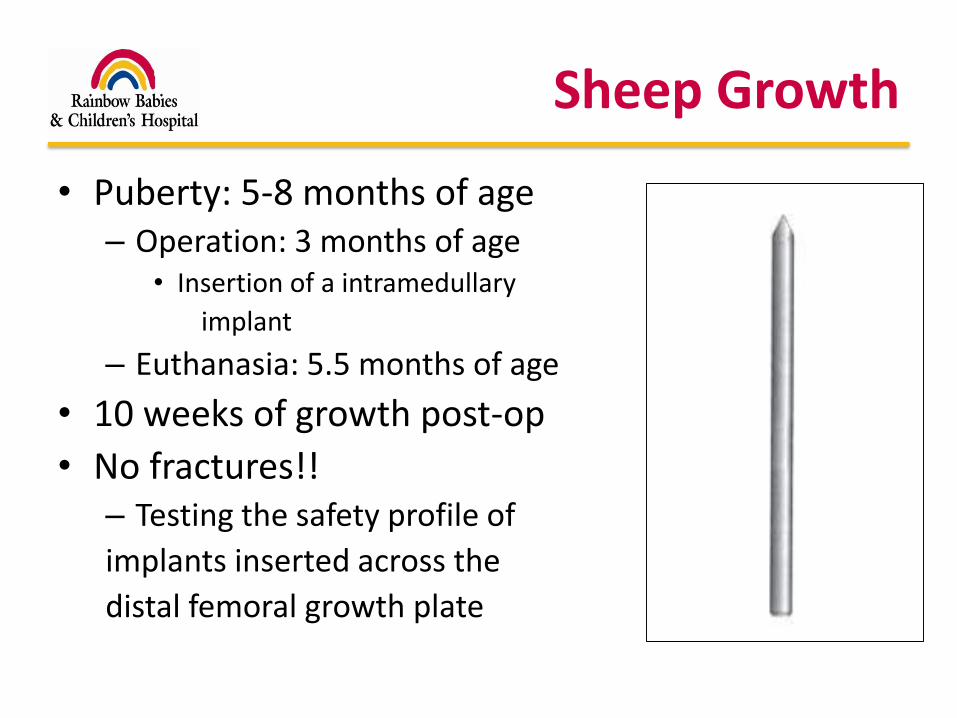
Sheep Growth
• Puberty: 5-8 months of age– Operation: 3 months of age
• Insertion of a intramedullary
implant
– Euthanasia: 5.5 months of age
• 10 weeks of growth post-op
• No fractures!!– Testing the safety profile of
implants inserted across the
distal femoral growth plate
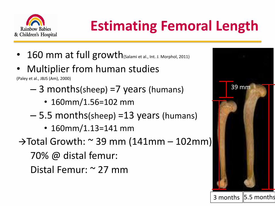
Estimating Femoral Length
• 160 mm at full growth(Salami et al., Int. J. Morphol, 2011)
• Multiplier from human studies(Paley et al., JBJS (Am), 2000)
– 3 months(sheep) =7 years (humans)
• 160mm/1.56=102 mm
– 5.5 months(sheep) =13 years (humans)
• 160mm/1.13=141 mm
Total Growth: ~ 39 mm (141mm – 102mm)
70% @ distal femur:
Distal Femur: ~ 27 mm
3 months 5.5 months
39 mm
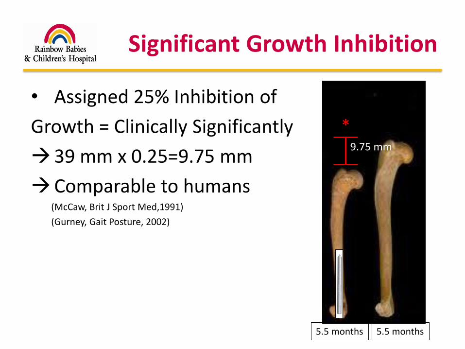
Significant Growth Inhibition
• Assigned 25% Inhibition of
Growth = Clinically Significantly
39 mm x 0.25=9.75 mm
Comparable to humans(McCaw, Brit J Sport Med,1991)
(Gurney, Gait Posture, 2002)
5.5 months 5.5 months
9.75 mm
*
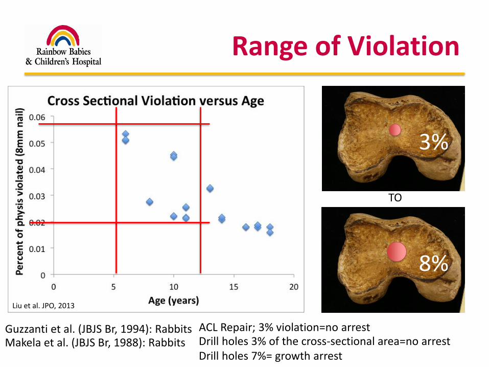
Range of Violation
Makela et al. (JBJS Br, 1988): Rabbits Drill holes 3% of the cross-sectional area=no arrestDrill holes 7%= growth arrest
3%
8%
TO
Liu et al. JPO, 2013
Guzzanti et al. (JBJS Br, 1994): Rabbits ACL Repair; 3% violation=no arrest

Power Analysis
• Power analysis: 3 limbs per increment
• 3 sheep @ 3%
• 3 sheep @ 4%
• 3 sheep @ 5%
• 3 sheep @ 6%
• 3 sheep @ 7%
• 3 sheep @ 8%
1 sheep= 1 operative limb, 1 control limb
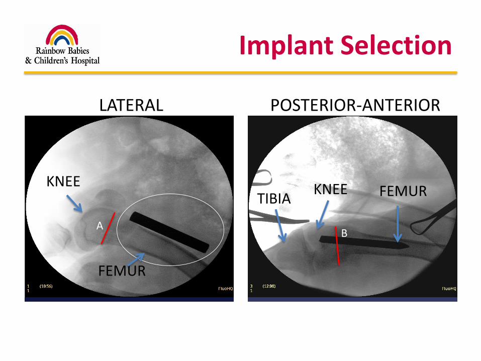
Implant Selection
AB
LATERAL POSTERIOR-ANTERIOR
KNEE KNEETIBIA FEMUR
FEMUR

Implant Selection
Area (Circle): πr2
A
B
Area (Ellipse): πAB
? %AreaSteinman Pin (πr2)
AreaPhysis (πab)
x (ab)√rSteinman Pin ? %
(3-8%)
(3-8%)
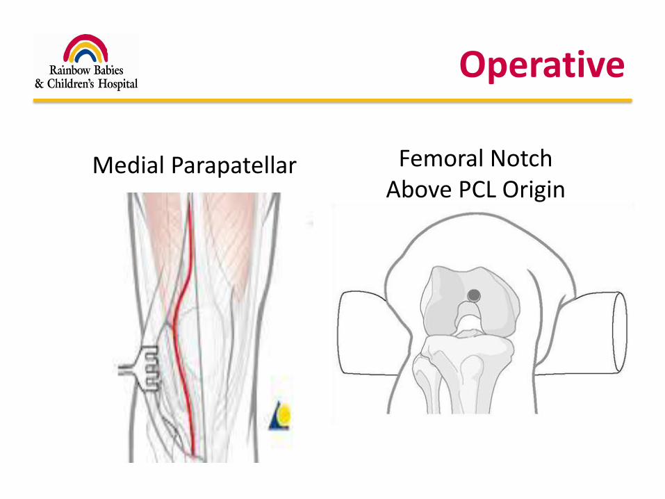
Operative
Femoral NotchAbove PCL Origin
Medial Parapatellar
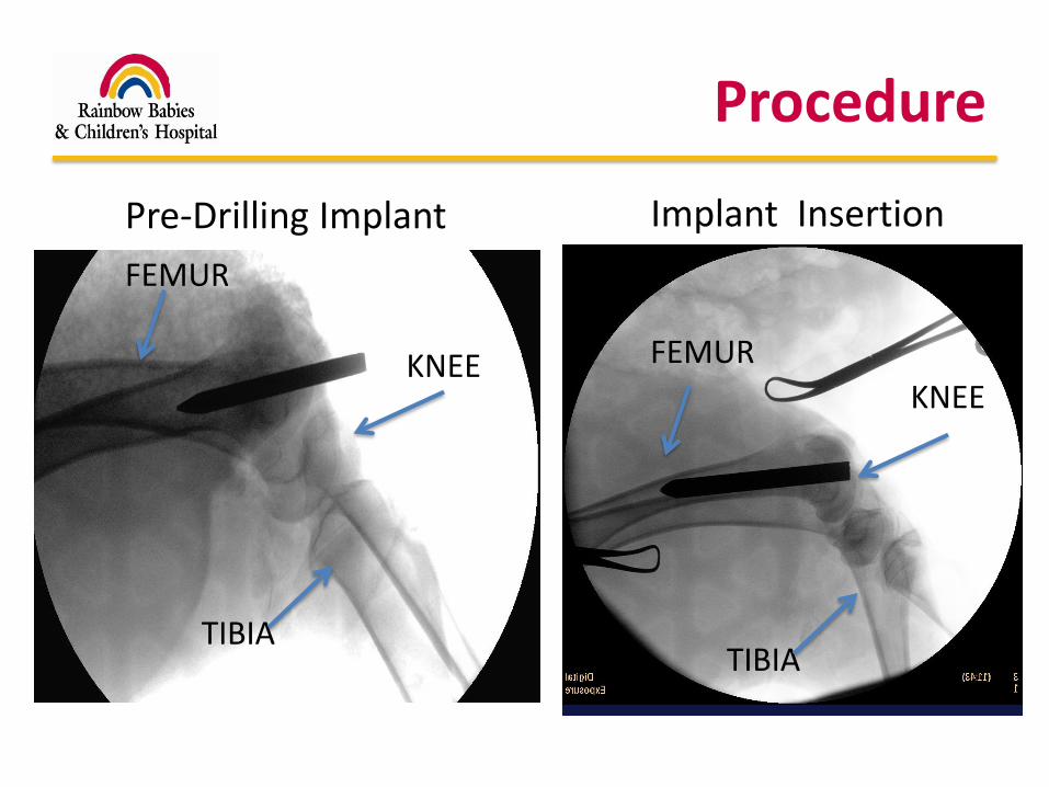
Procedure
Pre-Drilling Implant
KNEE
TIBIA
FEMUR
KNEE
TIBIA
FEMUR
Implant Insertion
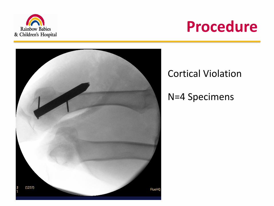
Procedure
Cortical Violation
N=4 Specimens
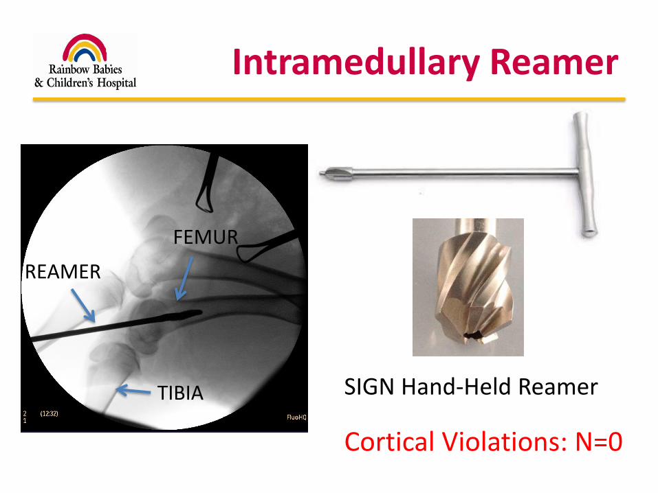
Intramedullary Reamer
TIBIA
FEMUR
SIGN Hand-Held Reamer
REAMER
Cortical Violations: N=0
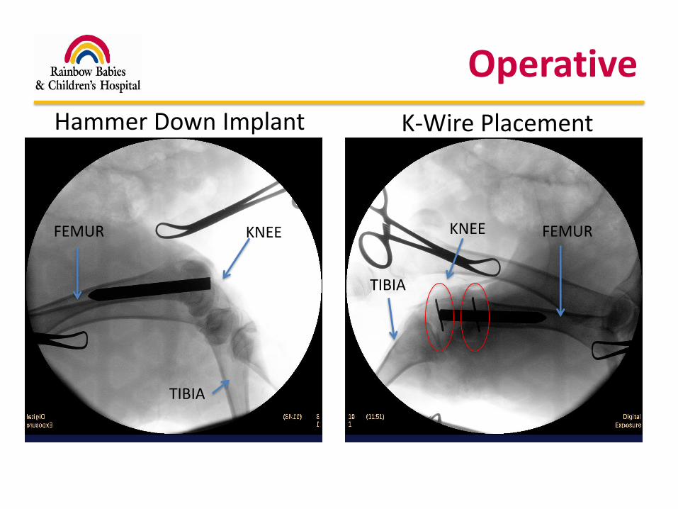
Operative
Hammer Down Implant K-Wire Placement
KNEE
TIBIA
FEMURKNEE
TIBIA
FEMUR
Post-Operative Course
• No immobilization, no weight-bearing restrictions
• Repeat X-rays to assess distal femoral growth
– 4 weeks post-op
– 8 weeks post-op
– 10 weeks post-op
• Euthanasia (10 weeks) Harvesting bilateral femurs
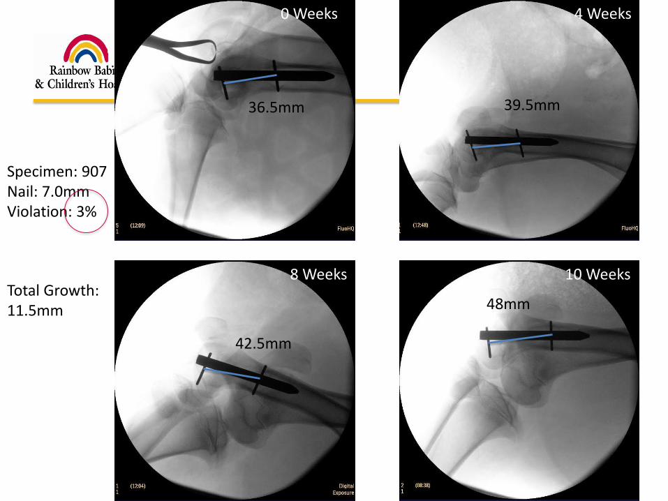
0 Weeks 4 Weeks
8 Weeks 10 Weeks
36.5mm
48mm
42.5mm
39.5mm
Specimen: 907Nail: 7.0mmViolation: 3%
Total Growth: 11.5mm
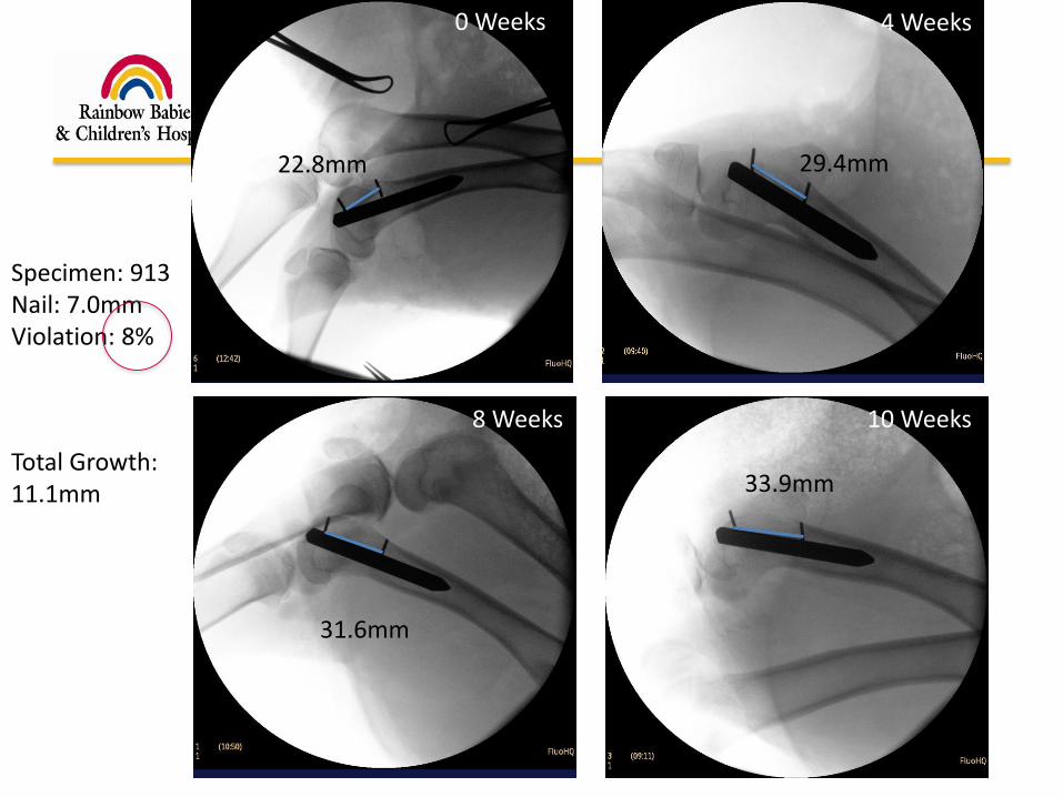
0 Weeks 4 Weeks
8 Weeks 10 Weeks
22.8mm
33.9mm
31.6mm
29.4mm
Specimen: 913Nail: 7.0mmViolation: 8%
Total Growth: 11.1mm
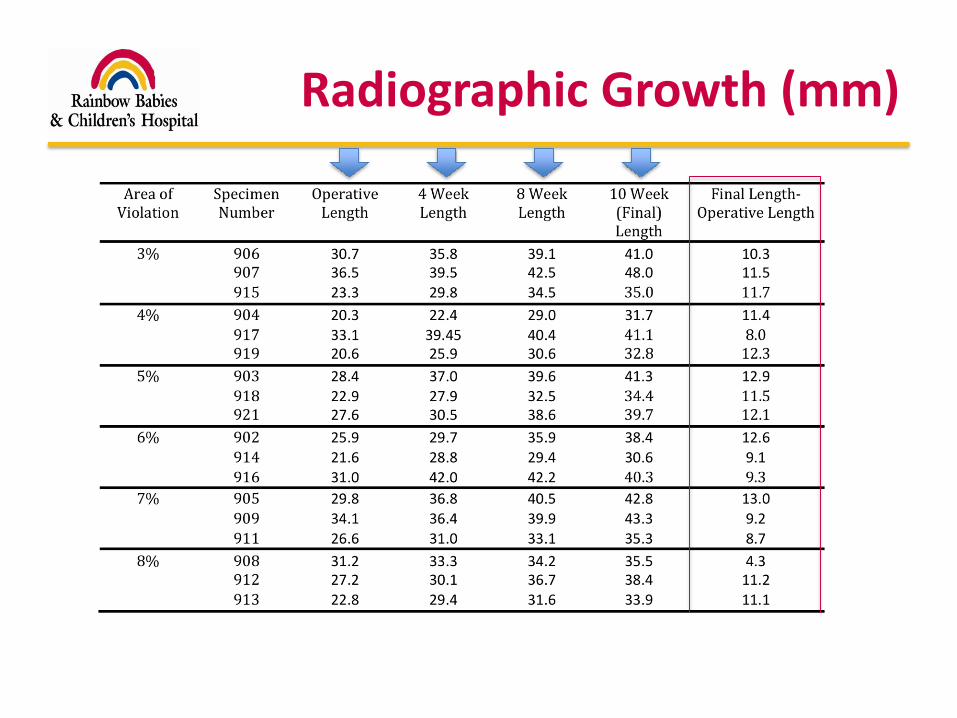
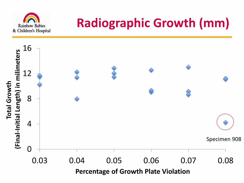
Radiographic Growth (mm)
0
4
8
12
16
0.03 0.04 0.05 0.06 0.07 0.08
Tota
l Gro
wth
(Fin
al-I
nit
ial L
en
gth
) in
mili
met
ers
Percentage of Growth Plate Violation
Radiographic Growth (mm)
Specimen 908
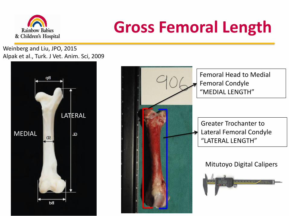
Gross Femoral LengthWeinberg and Liu, JPO, 2015Alpak et al., Turk. J Vet. Anim. Sci, 2009
Femoral Head to Medial Femoral Condyle“MEDIAL LENGTH”
Greater Trochanter to Lateral Femoral Condyle“LATERAL LENGTH”
LATERAL
MEDIAL
Mitutoyo Digital Calipers
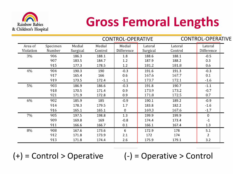
Gross Femoral Lengths
(+) = Control > Operative (-) = Operative > Control
CONTROL-OPERATIVE CONTROL-OPERATIVE
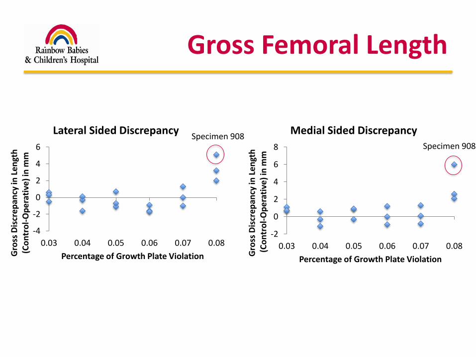
Gross Femoral Length
-4
-2
0
2
4
6
0.03 0.04 0.05 0.06 0.07 0.08
Gro
ss D
iscr
ep
ancy
in L
en
gth
(C
on
tro
l-O
pe
rati
ve)
in m
m
Percentage of Growth Plate Violation
Lateral Sided Discrepancy
-2
0
2
4
6
8
0.03 0.04 0.05 0.06 0.07 0.08G
ross
Dis
cre
pan
cy in
Le
ngt
h(C
on
tro
l-O
pe
rati
ve)
in m
mPercentage of Growth Plate Violation
Medial Sided DiscrepancySpecimen 908
Specimen 908
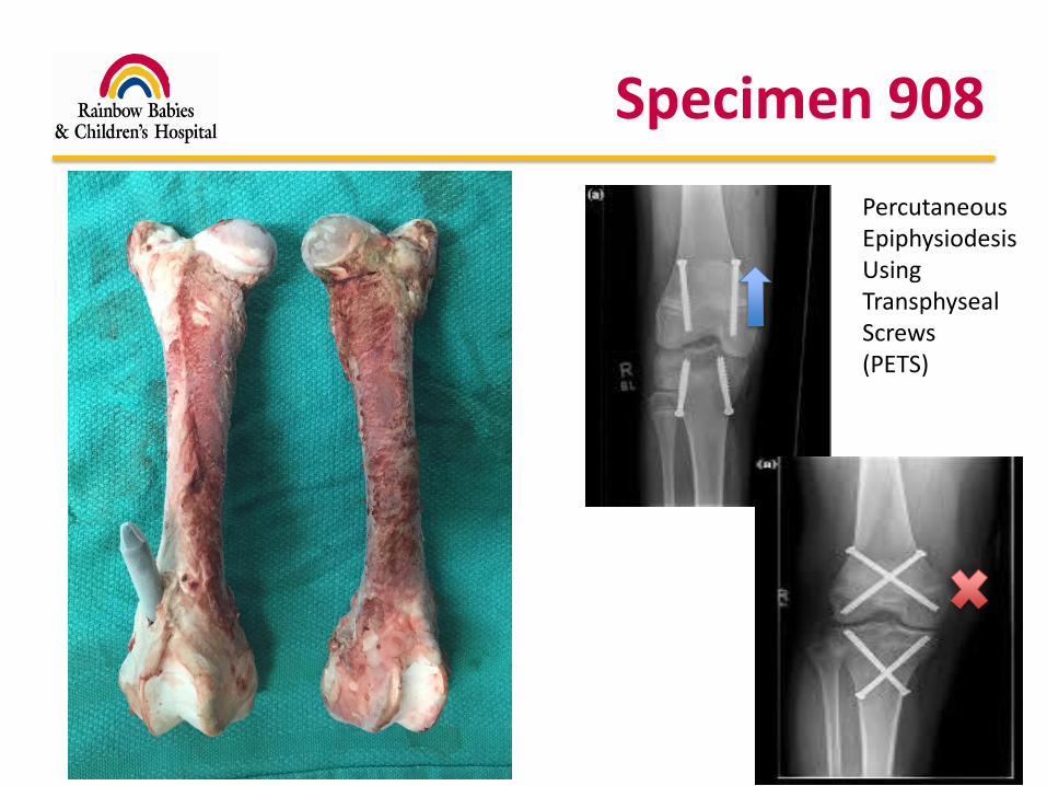
Specimen 908
Percutaneous EpiphysiodesisUsing TransphysealScrews(PETS)
Limitations
• Femoral length >160mm in all specimens
– No standardized value for Dorsett sheep
• Multiplier method unreliable?
– Needed to measure femoral length every month
• Distal femoral growth plate in sheep does not grow like in humans?
– More like a humerus where the majority of growth occurs proximally?

Growth Inhibition?FE
MO
RA
L G
RO
WTH
RAT
E (m
m/d
ay)
TIME (days)
NORMAL FEMORAL GROWTHINSERTION OF IMPLANT
RATE OF INHIBITION?
PLATEAU?
IMMEDIATEDECLINE?
Conclusions
• Growth appears to continue at the distal femoral physis when 3 to 7% of the growth plate is violated
– No evidence of significant growth arrest, but trending towards arrest with violation at 8%
• Insertion of a nail across the distal femoral growth is safe in the normal size range (3-7%)
– Violating 8% of the growth plate in children is extreme and unlikely to be performed
Clinical Recommendations
• Implant Insertion
– Smallest hand reamer for pilot hole to avoid large oblique passage
– 8 mm reamer (*do not over-ream!)
– 8 mm SIGN nail
• Safe for use in all children
• For Age 12 and up, a 10mm nail can be safely considered
Clinical Recommendations
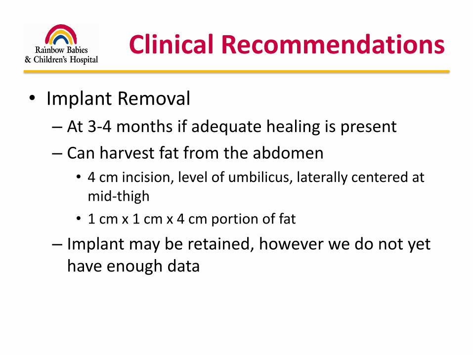
• Implant Removal
– At 3-4 months if adequate healing is present
– Can harvest fat from the abdomen
• 4 cm incision, level of umbilicus, laterally centered at mid-thigh
• 1 cm x 1 cm x 4 cm portion of fat
– Implant may be retained, however we do not yet have enough data
Clinical Recommendations
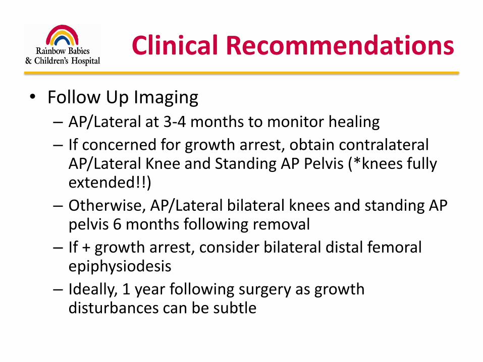
• Follow Up Imaging– AP/Lateral at 3-4 months to monitor healing
– If concerned for growth arrest, obtain contralateral AP/Lateral Knee and Standing AP Pelvis (*knees fully extended!!)
– Otherwise, AP/Lateral bilateral knees and standing AP pelvis 6 months following removal
– If + growth arrest, consider bilateral distal femoral epiphysiodesis
– Ideally, 1 year following surgery as growth disturbances can be subtle

Thank You
Questions?