Embed Size (px)
Citation preview
i
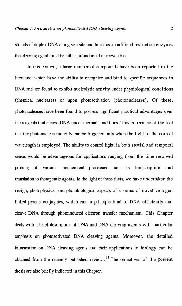
Chapter 1
AN OVERVIEW ON PHOTOACTIVATED DNA CLEAVINGAGENTS
1.1. Introduction
Design of small molecules that selectively recogmze nucleic acids and
induce strand scission in presence of light is an active area of research that has
important biochemical and therapeutic applications. 1-3 While natural enzymes have
been extremely useful in many applications, their large size and/or limited range
of sequence-recognition capabilities prevent their general use. For example, type II
restriction enzymes which cleave within or near specific recognition sites, usually
require Mg2+ ions as cofactor. Moreover, efficient cleavage is only observed with
DNA containing two or more recognition sites, suggesting that the active complex
binds at two sites.4,s An application, which demands cleavage with a higher level
of sequence selectivity or at single recognition site requires a new cleavage agent
with expanded recognition features. Such shortcomings of natural nucleic acid
cleaving agents provide opportunities for chemists to design novel
chemotherapeutics targeted to DNA and in the development of probes useful in
biology and medicine. In order for a single molecule to recognize and cleave both
Chapter I: An overview on photoactivated DNA cleaving agents 2
strands of duplex DNA at a given site and to act as an artificial restriction enzyme,
the cleaving agent must be eithr-r bifunctional or recyclable.
In this context, a large number of compounds have been reported in the
literature, which have the ability to recognize and bind to specific sequences in
DNA and are found to exhibit nucleolytic activity under physiological conditions
(chemical nucleases) or upon photoactivation (photonucleases). Of these,
photonucleases have been found to possess significant practical advantages over
the reagents that cleave DNA under thermal conditions. This is because of the fact
that the photonuclease activity can be triggered only when the light of the correct
wavelength is employed. The ability to control light, in both spatial and temporal
sense, would be advantageous for applications ranging from the time-resolved
probing of various biochemical processes such as transcription and
translation to therapeutic agents. In the light of these facts, we have undertaken the
design, photophysical and photobiological aspects of a series of novel viologen
linked pyrene conjugates, which can in principle bind to DNA efficiently and
cleave DNA through photoinduced electron transfer mechanism. This Chapter
deals with a brief description of DNA and DNA cleaving agents with particular
emphasis on photoactivated DNA cleaving agents. Moreover, the detailed
information on DNA cleaving agents and their applications in biology can be
obtained from the recently published reviews. 1•3 The objectives of the present
thesis are also briefly indicated in this Chapter.

Chapter 1: An overview on photoactivated DNA cleaving agents 3
1.2. Structure of DNA
The primary structure of DNA has a string of nucleosides each joined to its
two neighbours through phosphodiester linkages as shown in Figure 1.1A.6.7 Each
B
~J-1l--~"()~ H3
~i->"""Hy
Adenine (A) Thymine (T)
~ rO-·····H-I~-tt----0N1-H·····ry
HGuanine (G) Cytosine (C)
1'
o 4o 3'
oJ,'o
~'
4' l'
0q 3' 2'
od'~ A
0-- '0
5' ~'4' l'
) 3' 2'
3'
Figure 1.1. (A) Schematic representation of the primary structure of DNA
and (B) Watson-Crick base pairing of adenine-thym~ne(A-T) and guanine
cytosine (G-C).
regular 5'-hydroxyl group is linked through a phosphate to a 3'-hydroxyl group
and the uniqueness of any primary structure depends only on the sequence of
bases present in its chain. DNA secondary structure consists of two chains, which
run in opposite directions (anti-parallel) and are coiled around each other to form a
double helix. These two chains are linked together by a large number of weak

Chapter 1: An overview on photoactivated DNA ~leaving agents 4
hydrogen bonds formed between the complementary bases (Figure 1.IB). The
complementary base pairs in the case of DNA are adenine-thymine and guanine
cytosine. The bases, which are hydrophobic and paired by hydrogen bonding lie
inside and perpendicular to the helix axis, whereas the hydrophilic and negatively
charged sugar-phosphate backbone face out into the aqueous medium. The double
helical structure of DNA is stabilized by hydrogen bonding between the
complementary base pairs and also by hydrophobic interactions between the
stacked bases.
DNA adopts different secondary helical structures based on the
environmental conditions such as humidity and salt concentration. A-DNA and
B-DNA are the predominant DNA secondary structures (Figure 1.2) with right
Minor Groovr' ;UiIo,.~~~
A-DNA B-DNA Z-DNA
Figure 1.2. The structures of various forms of duplex DNA.
,: ' '.� C,, ,_,::,, (
Chapter 1: An overvie� �h���d Jwlt cleaving agents 5
'-,.· . ',-;: ,t�: •. -�#
handed double helices anci'W��ck base pairing, whereas Z-DNA is a left
handed double helical structure that is stabilized by high concentrations of MgCh
and NaCl.8 Only B-DNA is the one that exists under physiological pH conditions,
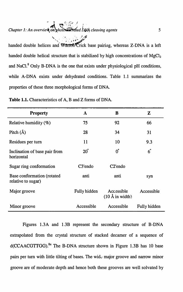
while A-DNA exists under dehydrated conditions. Table 1.1 summarizes the
properties of these three morphological forms of DNA.
Table 1.1. Characteristics of A, B and Z forms of DNA.
Property A B z
Relative humidity (%) 75 92 66
Pitch (A) 28 34 31
Residues per turn 11 10 9.3
Incli,:iation of base pair from 20· o· 6" horizontal
Sugar ring conformation C3'endo C2'endo
Base conformation (rotated anti anti syn relative to sugar)
Major groove Fully hidden Accessible Accessible (10 A in width)
Minor groove Accessible Accessible Fully hidden
Figures 1.3A and 1.3B represent the secondary structure of B-DNA
extrapolated from the crystal structure of stacked decamer of a sequence of
d(CCAACGTTGG).8a The B-DNA structure shown in Figure 1.38 has 10 base
pairs per tum with little tilting of bases. The wid� major groove and narrow minor
groove are of moderate depth and hence both these grooves are well solvated by

Chapter 1: An overview on photoactivated DNA cleaving agents 6
water molecules. Another promising feature of B-DNA is that its structure is
sufficiently flexible to permit a conformational response in the backbone to
particular base sequences.
A B
Figure 1.3. (A) Double helix model for the B-form of DNA and (B) twelve
base pairs of B-DNA extrapolated from the crystal structure of stacked
decamer of sequence d(CCAACGTIGG).
1.3. Ligand-DNA Interactions
DNA secondary structure possesses a variety of sites for the ligands to
interact including the sugar-phosphate backbone, the central base core and the
major and minor grooves. Figure 1.4 schematically represents the possible sites of
ligand binding to B-DNA structure. The DNA structure by itself is stabilized by a
'spine of hydration' on the grooves and stacked cations around the anionic sugar-
phosphate backbone. Therefore, there exist a number of possible pathways for the

Chapter 1: An overview on photoactivated DNA cleaving agents 7
ligands to interact with DNA through either covalent or non-covalent interactions.
Although, DNA can be modified without any specific interactions with ligand, the
selectivity and efficiency of modification is marginal for such ligands. This
Chapter presents mainly photocleaving agents, which bind to DNA through non-
covalent interactions (reversible interactions). Moreover, the important examples of
Sugar-phosphate
back bone II, Pl~am>,~'1:#","
Central core ofbase pairs.'•
Figure 1.4. Schematic representation of B-DNA and probable ligand
binding sites.
molecules that cleave DNA without any specific interactions as well as
covalent interactions can be obtained from the published reviews.9•10
1.4. DNA Cleaving Agents
Cellular DNA is by no means inert, stable structure holding genetic
information in dead storage. It continually suffers assault from exogenous and
endogenous agents, which result in a wide variety of modifications. These inciude

Chapter J: An overview on photoactivated DNA cleaving agents 8
base modifications, abasic sites, strand breaks and DNA protein cross links.11
When these lesions escape repair by cellular mechanisms, they lead to
mutagenesis, carcinogenesis, aging and cell death.12 While cleavage of DNA is
generally considered to be deleterious, synthetic DNA cleaving agents have been
developed in recent years that have been extremely useful in the treatment of
diseases and also as probes for understanding DNA-protein interactions and
macromolecular structure.14 Hydrolysis of phosphodiester bonds and oxidation of
sugar and nucleobases can cause DNA cleavage. Most enzymatic nucleases (e.g.
repair enzymes) cleave DNA by the hydrolysis of phosphodiester bonds, whereas
the synthetic reagents typically involve an initial oxidation reaction of either sugar
or nucleobase, which indirectly results in phosphodiester cleavage. This chapter
deals with a brief description on small and hybrid molecules that can cleave DNA
upon photoexcitation.
1.5. Photoactivated DNA Cleaving Agents
Photonucleases are those compounds that react directly with the nucleic
acids while in an electronically excited state and cause an immediate scission of
the nucleic acid chain. In the strictest sense, the cleaving agent should not be
consumed in the process, permitting it to react catalytically with the nucleic acid,
similar to the reaction of enzymes. But in reality, only very few compounds meet
this criteria. Thus photonucleases can be referred to as those compounds, whose
excited states can initiate a series of chemical reactions, which ultimately lead to

Chapter 1: An overview on photoactivated DNA cleaving agents 9
nucleic acid cleavage. One appealing aspect of the photoactivated cleaving agent
is that it allows the reaction to be controlled spatially and temporally by
combining all the components of the reaction mixture before the irradiation.
Excitation of the reaction mixture with an appropriate light source initiates the
reaction, which continues until the light is shut off. Several types of photoactive
compounds are reported in the literature,la.S but very few compounds cleave DNA
catalytically. Hence majority of these compounds can be referred to as
"photoactivated DNA cleaving agents," and are defined as those compounds
whose excited states can initiate a series of chemical reactions, which ultimately
lead to the nucleic acid cleavage. The ability to control light, in both spatial and
temporal sense, would be advantageous for applications such as the time-resolved
probing of various biochemical processes. These include transcription or
translation, to therapeutic agents, which are activated under in vivo conditions by
laser sources coupled into the body through fiber optics. Perhaps the most
important feature of these reagents is the fact that light can be a very selective
cofactor in a chemical reaction. Moreover, if the photocleaver is ser:.sitive to light
at wavelengths longer than 360 nm, selective excitation of the photocleavage agent
is possible. This is critical in limiting the number of side reactions in the system as
well as in analyzing the reaction mechanisms.
The mechanisms of DNA cleavage through photosensitization can be
broadly classified into (i) by generation of diffus~ole (singlet oxygen) and non
diffusible (hydroxyl radicals) reactive intermediates, (ii) hydrogen atom
Chapter 1: An overview on photoactivated DNA cleaving agents 10
abstraction and (iii) electron transfer mechanism. The energy transfer from the
excited state of photosensitizer to the ground state of triplet molecular oxygen can
yield singlet oxygen. Hydroxyl radical intermediates, on the other hand, can be
generated by direct homolysis of the peroxide bond in the excited state of the
substrate or by an energy/electron transfer mediated heterolytic cleavage.
Alternatively, photosensitizers can also initiate formation of hydroxyl radicals by
electron transfer to superoxide radical anion to yield hydrogen peroxide, which
can in tum generate hydroxyl radicals through Fenton reaction. 13
Photosensitizers can also initiate hydrogen atom abstraction by mechanisms
not involving hydroxyl radicals. Many n, 1r* excited states of photosensitizers
readily abstract hydrogen atoms, and radicals derived from them are capable of
hydrogen· atom abstraction from the deoxyribose sugar.2b In the case of electron
transfer mechanism, the first step in DNA cleavage involves the oxidation of
nucleobases by the excited state of the photosensitizer. The free energy for
electron transfer from a nucleic acid base to an excited state sensitizer will depend
on the oxidation potential of the ground state base and the reduction potential and
excited state energy of the sensitizer. Of all the nucleobases, guanine has the
lowest oxidation potential, 14 and hence guanine is implicated as the base in an
electron transfer mechani.;m in DNA.
A variety of compounds, from ketones to organometallic complexes to
polycyclic heteroaromatic drugs have been reported in the literature, which
photosensitize the cleavage of.DNA. Some of these involve direct reaction of an
Chapter 1: An overview on photoactivated DNA cleaving agents 11
excited state of the photosensitizer or its radical with the nucleic acids, while
others generate intermediates such as singlet oxygen or hydroxyl radical. Some
photosensitizers initiate photocleavage by more than one mechanism. Here we
present salient features of some selected photoactivated cleaving agents, which
cleave DNA through different mechanisms. For convenience, these compounds
have been divided into two groups on the basis of their likely sites of reactivity in
DNA, namely, sugar or nucleobase.
1.5.1. Photoactivated Cleaving Agents Selective for Oxidation of
Sugar Moiety in DNA
Oxidation of deoxyribose due to hydropn atom abstraction from the sugar
furanose is quite often the key step in DNA cleavage. The resulting sugar radicals
can decompose by a variety of pathways to yield small molecules and DNA
fragments. 2b Since identical deoxyribose residues are found at every step along the
DNA duplex, the cleavage by hydrogen abstraction is in:.1erently non-selective
with respect to the sequence. The sequence selectivity depends on the local
structure of the DNA and the physicochemical properties of the hydrogen
abstracting sensitizer. The ability to alter the sequence selectivity of cleavage by
varying the structure of the photocleavage agent is a key component in the design
of new DNA cleavage agents. Therefore, this section deals with the reagents,
which cleave DNA by .the oxidation of sugar moiety.
Chapter 1: An overview on photoactivated DNA cleaving agents 12
1.s.1.1. DNA Photocleavage Induced by Metal Complexes
The first DNA photocleaving agent based on uranyl ion (UO/+) was
introduced by Nielsen and co-workers in 1988. 15 The photoactivated uranyl salts
were found to spontaneously cleave both supercoiled and linear DNA targets
resulting in nicks in the DNA. Mechanistic studies indicated that the cleavage
occurs through abstraction of a hydrogen atom. The intact nucleobases are
observed as byproducts of the photocleavage and the quantum yield for their
production ( -104) matches the quantum yield for plasmid nicking, consistent with
a mechanism in which the damage is targeted to the sugar and not the nucleobase.
uo/+ binds to the DNA by coordinating with phosphate backbone across the
minor groove with an affinity of the order of 1010 M-1, making it a valuable probe
of local structure. Subsequently, the uranyl salts have been successfully employed
as photofootprinting agents to probe a wide variety of nucleic acid structures and
protein-nucleic acid complexes including imaging of DNA triplexes and gene
expression regulator/DNA complexes. 16 The uranyl salts are the most useful
photocleavage agents developed so far, however, there are a few disadvantages
associated with these salts. One of the drawbacks of these salts is that they need
either neutral or acidic pH, otherwise uranyl ion forms insoluble uranyl hydroxide
aggregates, which makes it inefficient in cleaving DNA.
Biological applications of rhodium complexes, such as DNA and RNA
photocleavage, have been extensively reviewed. 1a Photocleavage of DNA by

Chapter J: An overview on photoactivated DNA cleaving agents 13
rhodium (II) complexes (Chart 1.1) containing the phi ligand (phi = 9,10-
phenanthrenequinone diimine) (1) has been studied by Barton and co
workers.17·18 Irradiation of solutions containing Rh(phi) complexes in the presence
1
'.:::::
N"H
H .. [, ••.. I ..... X� _.....·,h,N
H -..:
Chart 1.1
of DNA results in spontaneous cleavage. Analysis of the product mixture revealed
the presence of free bases and base propenoic acids, while DNA fragments are
terminated by 3'-phosphates. These products correspond to those expected from
hydrogen abstraction from the 3'-carbon of the deoxyribose. The Rh(phi)
complexes bind to B-form DNA through intercalation of the phi ligand from the
major groove and the excited state of the complex abstracts a hydrog?11 atom from
C-3' of sugar at the intercalation site.
It has also been reported that rhodium complexes with different ligands can
generate complexes with varying selectivities for DNA. For example, the complex
bis(phenanthrenequinone diimine)bipyridylrhodium(III) (Rh(phih(bpy)3+) (2,
Chart 1.1) is a sequence neutral complex in its reactions with DNA. The role of
ancillary (non-intercalating) ligands on the DNA cleavage by Rh(phi) complexes

Chapter J: An overview on photoactivated DNA cleaving agents 14
is quite interesting. 18 For example, the A-isomer of [Rh(en)zphi]3+ (3, Chart 1.2)
(en = ethylenediamine) cleaves DNA with high selectivity for 5'-GG-3'
dinucleotide steps, whereas the A-isomer (4, Chart 1.2) exhibits cleavage at all
sites, with some preference for Alf sites. Thus, the Rh(phi) complexes represent
excellent examples of how the cleavage selectivity can be determined through the
binding selectivity of the photocleaver. The ability to alter the cleavage selectivity
by varying the ancillary ligands of the complexes provides control, unavailable in
most other systems.
3,A 4,A
Chart 1.2
Bleomycin, a product of streptomyces fermentation, cleaves DNA via an
iron-oxo intermediate and has played a heuristic role in our understanding of the
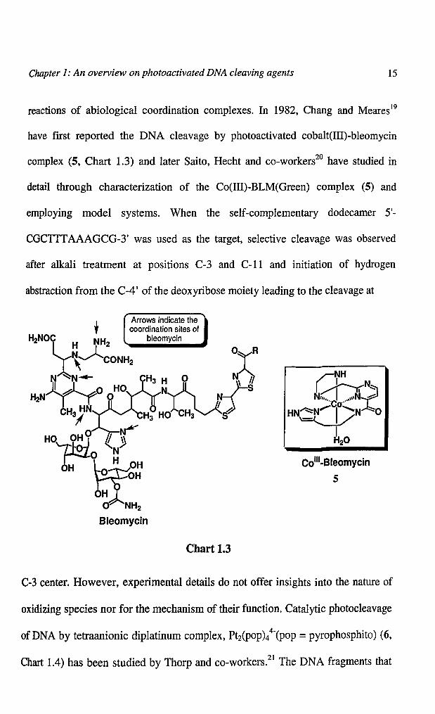
Chapter J: An overview on photoactivated DNA cleaving agents 15
reactions of abiological coordination complexes. In 1982, Chang and Meares19
have fIrst reported the DNA cleavage by photoactivated cobalt(III)-bleomycin
complex (5, Chart 1.3) and later Saito, Hecht and co-workers20 have studied in
detail through characterization of the Co(Ill)-BLM(Green) complex (5) and
employing model systems. When the self-complementary dodecamer 5'-
CGCTITAAAGCG-3' was used as the target, selective cleavage was observed
after alkali treatment at positions C-3 and C-ll and initiation of hydrogen
abstraction from the C-4' of the deoxyribose moiety leading to the cleavage at
Colli-Bleomycin
5
Chart 1.3
C-3 center. However, experimental details do not offer insights into the nature of
oxidizing species nor for the mechanism of their function. Catalytic photocleavage
of DNA by tetraanionic diplatinum complex, Pt2(POP)/(pop =pyrophosphito) (6,
Chart 1.4) has been studied by Thorp and co-workers.21 The DNA fragments that

Chapter 1: An overview on photoactivated DNA cleaving agents 16
arise from the cleavage possess 3'-phosphate and 3'-phosphoglycolate as well as
5'-phosphate and 5'-aldehyde termini. This indicates that the cleavage by platinum
complex is the result of hydrogen abstraction from both C-4' and C-5' of
deoxyribose, respectively. Zaleski and co-workers22 reported that bis(9-diazo-4,5-
diazafluorene)copper (II) nitrate complex (7, Chart 1.4) cleaves DNA in the
presence of visible light under anaerobic conditions and act as a transition metal
kinamycin model. On the other hand, irradiation of the Fe(III) complex (8) in the
9
%N2
03N,,, .... Ju/ ,,,,- 'Noa
�
N2
Chart 1.4
11
presence of plasmid DNA affords both single- and double-strand cleavage,23 with
significantly greater efficiency than photolysis of free ligand in threefold higher
concentration. From a mechanistic perspective, detection of Fe(II) as a reaction
product raises important questions concerning the mode of activation of the
Chapter 1: An overview on photoactivated DNA cleaving agents 17
kinamycins and 6-diazo-5-oxo-L-norleucine. However, ligand to metal charge
transfer (LMCT) activated Nz release from Fe(III) complex produces localized
ligand radical intermediates capable of cleaving DNA and represents a new
chemical approach for the design of photoactivated cleaving agents.
The photolysis of CpW(CO)3Me (9, Chart 1.4) has been shown to produce
methyl radicals and to cleave DNA in a single-stranded manner. The experimental
evidence suggests the involvement of carbon-centered radical in this process.Z4 In
this work, the mechanism of strand scission was determined to occur by hydrogen
atom abstraction from the 4'- and 5'-positions of the deoxyribose moiety of the
backbone of DNA. Additionally, in a side reaction that does not lead to frank
strand scissioJl., all four bases of DNA are methylated under these conditions;
however, none of these bases or backbone modifications lead to the formation of
abasic sites.
Chen and co-workersz5 have reported that vanadyl (V) complexes serve as
efficient reagents for cleaving supercoiled plasmid DNA by photoinitiation.
Complex (10, Chart 1.4), derived from 2-hydroxy-l-naphthaldehyde aad L
phenylalanine, exhibits a unique wedge feature, inducing a site-selective
photocleavage at the C22-T23 of the bulge backbone for a HIV-27 DNA system at
0.1-5 pM. Transient absorption experiments indicate the involvement of ligand to
metal charge transfer (LMCT) with concomitant tautomerization. This results in
the formation of an ortho-quinone-methide V-bound hydroxyl species, which is
found to be responsible for the cleavage profiles. Fujii and co-workersz6 reported

Chapter 1: An overview on photoactivated DNA cleaving agents 18
photoinduced cleavage of DNA by a cationic Schiff base complex of manganese
(11, Chart 1.4). The cationic complex effectively cleaved T-site of DNA in ca.
88% selectivity upon visible light irradiation. The high selectivity observed for the
cationic complex suggests that the cationic organic radical formed by photolysis of
11 selectively attacks thymine or thymidine, thereby cleaving DNA at the T site.
1.5.1.2. DNA Photocleavage Induced by Organic Compounds
Several photoactivated organic compounds have been found to cause the
sugar oxidation followed by DNA cleavage either by hydrogen abstraction in the
excited state or they generate free radicals and hydroxyl radicals in situ, which in
tum abstract hydrogen atom from the deoxyribose. These include enediyenes (12
and 13),27 nitroaromatics, triazoles (14), anthraquinones (15 and 16), flavin-
oligopyrroles, halogenated bithiazoles, N-hydroxypyridones (20), hydroperoxides,
(18 and 21) etc (Charts 1.5 and 1.6). This section deals with salient features of
some of these compounds.
Enediyne antitumor antibiotics are natural products and are believed to
destroy cancer cells by damaging DNA. These compounds bind to DNA in the
minor groove and, upon activation, undergo rearrangements to produce
diradicals.2b·27 These are potent DNA cleavage agents, capable of producing single
and double strand breaks, suggesting their use in a variety of diagnostic and
therapeutic applications. Compounds that are active and involve diradical
intermediates in situ include neoc.arzinostatin, esperamicin A 1, dynemicin A,

Chapter J: An overview on photoactivated DNA cleaving agents 19
Cl027 (12), calicheamicin etc.2b These compounds are normally activated by
reduction, but irradiation also leads to the cleavage of supercoiled DNA.
Due to the structural complexity of the natural enediynes, a large number of
simplified model compounds designed to generate diradicals have been studied as
photocleavers. Goldberg and co-workers28 have demonstrated that the enediyne
antibiotic C1027 (12, Chart 1.5) produces sequence specific covalent DNA drug
15
e @ Oa Na
Chart 1.5
e 2CI
13
16
adducts and DNA interstrand cross-links under anaerobic conditions. After
binding to DNA by intercalation, the enediyne core of Cl027 (12) chromophore
Chapter J: An overview on photoactivated DNA cleaving agents 20
rearranges to the 3,6-diradical form. The diradical is positioned in such a way that
one of the radicals abstracts a hydrogen atom from C-4' position of the Al
nucleotide (A = adenine) and the other radical, from either C-1' of A2 or C-5' of
A3, ultimately leading to DNA cleavage.
Wender and co-workers29 used a similar strategy in developing a series of
triazole compounds as new photocleavers. Irradiation of triazole (14, Chart 1.5) in
presence of ethanol led to formation of hydrogen abstraction products, consistent
with diradical intermediates. The triazoles photonick plasmid DNA and
radiolabelled restriction fragments spontaneously. It was found that there is some
preference for cleavage at GG sites as observed for many photocleavers that react
with DNA nucleobases by electron transfer. However, the spontaneous nature of
the cleavage and a distinct preference for cleavage at 3'-G of the GG steps by the
triazoles argues against cleavage by an electron transfer pathway.
Schuster and co-workers30 showed that anthraquinones (AQ) photocleave
DNA by three distinct pathways, which are differentiated by the binding mode of
the AQ as well as the composition of the buffer (Chart 1.5). One mechanism
involves an electron transfer from the DNA nucleobases to an intercalated AQ and
the other two pathways involve hydrogen atom abstraction from deoxyribose.31
Anthraquinones possessing electron withdrawing substituents are capable of
oxidizing organic substrates by hydrogen atom abstraction, since the excited
singlet state of these molecules is n-ttr' in character. AQ derivatives (15 and 16,
Chart 1.5) intercalate into DNA and cleave DNA through hydrogen abstraction.
Chapter 1: An overview on photoactivated DNA cleaving agents 21
However, if the AQ is present in excess relative to intercalation sites, spontaneous
sequence-neutral cleavage of the DNA results.30
The preferential binding of monosubstituted AQ derivatives to DNA by
intercalation limits their ability to abstract hydrogen atoms from deoxyribose due
to rapid electron transfer from the nucleobases, which form the intercalation site,
requiring saturation of the intercalation sites before H-abstraction by non
intercalated AQ can occur. However, AQ, (15 and 16, Chart 1.5), binds to DNA in
one of the grooves or by electrostatic association with the backbone phosphates.3 1
It has been observed that mode of their interaction has profound effect on the
photochemical reaction with DNA. Laser studies of AQ derivatives 15 and 16
showed that rates oz electron transfer from the nucleobases to AQ decreased by ca.
50-fold when compared to AQ bound through intercalative mode. Spontaneous
sequence-neutral cleavage occurs even when AQ is present at non-saturating
concentrati0ns. Thus, it can be concluded that mode of binding in DNA can play a
vital role in the cleavage mechanism of photocleavage agents.
In addition, Nielsen and co-workers32 reported the photocleavage activity of
a series of acridine-linked nitrobenzamides and Shibuya and colleagues33 have
demonstrated the photoactivity of nitro substituted oligopyrroles, which mimic the
chemistry of netropsin. Recently, Saito and co-workers34 reported the DNA
photocleavage properties of a family of nitro-substituted naphthalimide
derivatives. The authors propose that the oxidation reaction is due to photoinduced
hydrogen abstraction from the methyl of the thymine by the excited state nitro
Chapter 1: An overview on photoactivated DNA cleaving agents 22
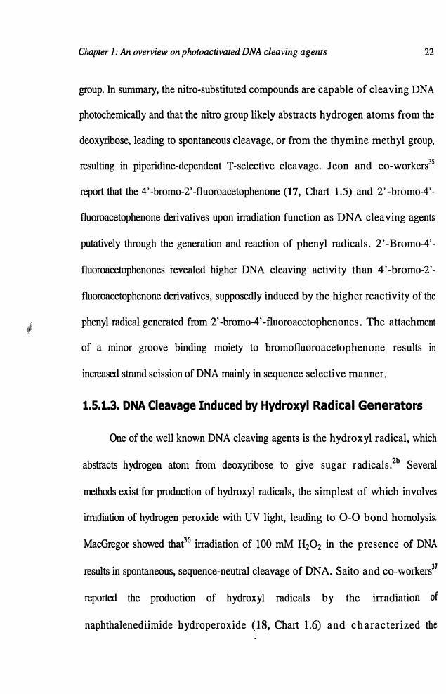
group. In summary, the nitro-substituted compounds are capable of cleaving DNA
photochemically and that the nitro group likely abstracts hydrogen atoms from the
deoxyribose, leading to spontaneous cleavage, or from the thymine methyl group,
resulting in piperidine-dependent T-selective cleavage. Jeon and co-workers35
report that the 4'-bromo-2'-fluoroacetophenone (17, Chart 1.5) and 2'-bromo-4'-
fluoroacetophenone derivatives upon irradiation function as DNA cleaving agents
putatively through the generation and reaction of phenyl radicals. 2'-Bromo-4'-
fluoroacetophenones revealed higher DNA cleaving activity than 4' -bromo-2' -
fluoroacetophenone derivatives, supposedly induced by the higher reactivity of the
· phenyl radical generated from 2' -bromo-4' -fluoroacetophenones. The attachment'!''
of a minor groove binding moiety to bromofluoroacetophenone results in
increased strand scission of DNA mainly in sequence selective manner.
1.5.1.3. DNA Cleavage Induced by Hydroxyl Radical Generators
One of the well known DNA cleaving agents is the hydroxyl radical, which
abstracts hydrogen atom from deoxyribose to give sugar radicals.2b Several
methods exist for production of hydroxyl radicals, the simplest of which involves
irradiation of hydrogen peroxide with UV light, leading to 0-0 bond homolysis.
MacGregor showed that36 irradiation of 100 mM H202 in the presence of DNA
results in spontaneous, sequence-neutral cleavage of DNA. Saito and co-workers37
reported the production of hydroxyl radicals by the irradiation of
naphthalenediimide hydroperoxide (18, Chart 1.6) and character ized the

Chapter 1: An overview on photoactivated DNA cleaving agents 23
hydroxyl radicals by DMPO trapping and ESR spectroscopic studies. Further,
photonicking of supercoiled plasmid DNA by this hydroperoxide was found to be inhibited
by 1 mM sodium benzoate, consistent with hydroxyl radical mediated cleavage
mechanism. When the cleavage selectivity was studied with radiolabelled
restriction fragments, damage was found to occur with higher selectivity at 5'-G of
GG sites and required piperidir.� treatment. This observation with the restricted
fragment is inconsistent with the hydroxyl radical mediated damage, unless the
compounds bind to DNA sequence selectively.
18
Chart 1.6
Other compounds that are known to produce hydroxyl radicals are the
heterocyclic N-oxide derivatives38 (19), N-hydroxypyridones (20) and

Chapter 1: An overview on photoactivated DNA cleaving agents 24
furocoumarin hydroperoxides (21-23) (Chart 1.6). 39 For example, irradiation of N-
oxide in water results in the production of 2 equivalents of hydroxyl radicals per
N-oxide precursor, but radicals are not produced if the irradiation is performed in
acetonitrile. The N-oxide (19) was able to photonick plasmid DNA in a process
that could be inhibited by the radical scavenger DMSO. However, production of
hydroxyl radicals in the presence of DNA was not demonstrated. Furocoumarin
hydroperoxide (Chart 1.6) (21) upon photoactivation with UV light, cleave
plasmid DNA by in situ generation of hydroxyl radicals.39 The analysis of DNA
damage by employing various endonucleases indicated that the damage profiles
are similar to that of y-radiation induced damage. The damage was inhibited by
hydroxyl radical quenchers such as tert-butanol and DMSO and the involvement
of hydroxyl radicals in these reactions was further confirmed by isolation of
products and also by trapping with phenol, adamantane and DMP0.40
An alternative source of hydroxyl radicals is through superoxide dismutase
reaction, producing hydrogen peroxide and oxygen (eq 1.1). The hydrogen
peroxide then can undergo homolysis in the presence of UV light or after
(1.1)
reduction by metal ions (Fenton reaction)13 to produce hydroxyl radicals. Thus,
any agent that is capable of reducing oxygen to superoxide can produce hydroxyl
radicals and the radical of transient metals.41 There are many examples of natural
compounds42 which photocleave DNA by processes which are partially inhibited

Chapter 1: An overview on photoactivated DNA cleaving agents 25
by radical scavangers. These photocleavers are also susceptible to inhibition by the
enzyme catalase, which decomposes hydrogen peroxide to water and oxygen.
Superoxide dismutase (SOD) detects superoxide in the reaction by catalysing the
reaction indicated in the equation 1.1 i.e. hydrogen peroxide production can be
faster in the presence of SOD than in its absence. Thus, inhibition of DNA
cleavage by SOD without catala~.~ means that superoxide plays a significant and
complex role in the cleavage mechanism than serving as a precursor to hydrogen
peroxide.
1.5.2. Photoactivated Cleaving Agents Selective for Oxidation of
Nucleobases in DNA
Cleavage of DNA strand as a result of chemical reactions initially occurring
on the nucleobase usually requires a second reaction step such as heat, alkali, or
enzymatic treatment to effect strand scission.2c Hence, compounds that react with
the nucleobases of DNA will not be inducing spontaneous cleavage in comparison
with systems that react with 'sugar residues. This class of photocleavage agents
function by three distinct processes: (i) direct electron transfer from +he
nucleobase to the excited state of the photocleaver; (ii) triplet energy transfer from
the excited photocleaver to molecular oxygen producing singlet oxygen, which
then reacts with nucleobases and (iii) formation of an adduct with the nucleobase.
Figure 1.5 shows the reaction of a nucleobase, for example, guanine with singlet
oxygen, hydroxyl radicals and ionizing radiation/electron acceptors. Hydroxyl
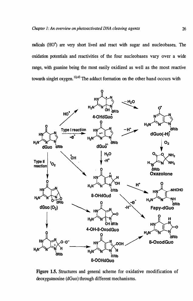
Chapter 1: An overview on photoactivated DNA cleaving agents 26
radicals (HO•) are very short lived and react with sugar and nucleobases. The
oxidation potentials and reactivities of the four nucleobases vary over a wide
range, with guanine being the most easily oxidized as well as the most reactive
towards singlet oxygen. 12.43 The adduct formation on the other hand occurs with
Type II102 reaction
•
N
H,N� hRib
dGuo(-H)
i o
,
0�vNH2
H'N
�NH2�Rib
Oxazolone
w 0
� HN�HCHO
�� e- H2N � \� • URlb
-W Fapy-dGuo
Figure 1.5. Structures and general scheme for oxidative modification of deoxyguanosine ( dGuo) through different mechanisms.
Chapter 1: An overview on photoactivated DNA cleaving agents 27
different selectivities, depending on the reaction mechanism. With nucleobases
they add to the double bond thereby generating piperidine dependent cleavage sites.
Superoxide radical anion formed in situ by electron transfer mechanisms does not
react directly with DNA to produce damage, but reacts through an iron-catalyzed
process to yield hydroxyl radicals (Fenton reaction). 13
1.5.2.1. Singlet Oxygen Generators
Electronically excited compounds that undergo efficient intersystem
crossing to the triplet state and have sufficiently high triplet energy can generate
singlet oxygen by energy transfer to molecular oxygen. Singlet oxygen is a highly
reactive species that prefe,:entially adds to guanine.43•44 The resulting oxidized
guanine is sensitive to piperidine treatment which induces a strand scission.33
Performing experiments in D20 rather than in H20 can lead to a substantial
increase in the cleavage efficiency since the lifetime of singlet oxygen is
significantly longer in D20 as compared to H20.45
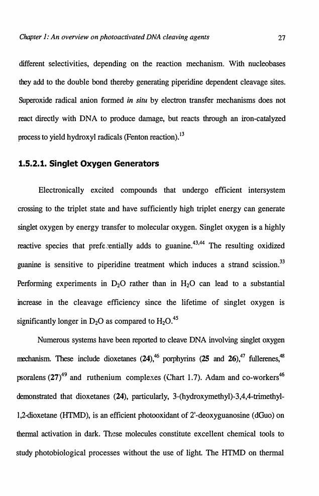
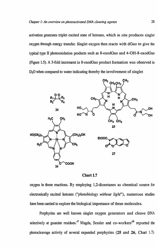
Numerous systems have been reported to cleave DNA involving singlet oxygen
mechanism. These include dioxetanes (24),46
porphyrins (25 and 26),47 fullerenes,48
psoralens (27)49 and ruthenium complexes (Chart 1. 7). Adam and co-workers46
demonstrated that dioxetanes (24), particularly, 3-(hydroxymethyl)-3,4,4-trimethyl
l,2-dioxetane (HTMD), is an efficient photooxidant of 2'-deoxyguanosine (dGuo) on
thermal activation in dark. These molecules constitute excellent chemical tools to
study photobiolo�ical processes without the use of light. The HTMD on thermal
Chapter 1: An overview on photoactivated DNA cleaving agents 28
activation generates triplet excited state of ketones, which in situ produces singlet
oxygen through energy transfer. Singlet oxygen then reacts with dGuo to give the
typical type II photooxidation products such as 8-oxodGuo and 4-0H-8-oxodGuo
(Figure 1.5). A 3-fold increment in 8-oxodGuo product formation was observed in
D20 when compared to water indicating thereby the involvement of singlet
24
I ' EtOOC
;QCQ
'<:::::
0 �
27
Chart 1.7
oxygen in these reactions. By employing 1,2-dioxetanes as chemical source for
electronically excited ketones ("photobiology without light"), numerous studies
have been carried to explore the biological importance of these molecules.
Porphyrins are well known singlet oxygen generators and cleave DNA
selectively at guanine residues.47 Magda, Sessler and co-workers48 reported the
photocleavage activity of several expanded porphyrins (25 and 26, Chart 1.7).

Chapter 1: An overview on photoactivated DNA cleaving agents 29
These compounds were found to initiate photonicking of supercoiled plasmid
DNA upon irradiation at wavelengths above 700 nm. Sodium azide strongly
inhibited the cleavage, which is indicative of a singlet oxygen mechanism in these
reactions. The significance of this work lies in the use of long wavelength of
irradiation, which is essential for in vivo photodynamic therapy applications.
Porphyrins can also be used to probe nonduplex structures in nucleic acids.
Tetracationic porphyrins5o (Chart 1.7) for example, were found to bind with high
selectivity at a DNA three way junction, then initiate photocleavage at guanine
residues adjacent to the junction.
Helene and co-workers51 demonstrated that irradiation of 3-carbethoxy
psoralen (27) in the presence of DNA and followed by piperidine treatment
resulted in selective cleavage at GG sites with modest preference for 5'-G. When
the experiments were carried out in D20, an enhancement in cleavage yield was
observed, suggesting the involvement of singlet oxygen. Thus, while the cleavage
pattern was inconsistent with a singlet oxygen process, the mechanistic
experiments were. Detailed studies indicated that both singlet oxygen and electron
transfer pathways were operative in the case of 3-carbethoxypsoralene.
1.5.2.2. DNA Photocleavage Through Electron Transfer Mechanism
Recently, a number of small molecules that oxidize DNA by photoinduced
electron transfer mechanism have been reported. The most important systems
include ethidium bromide (28), riboflavin52 (29), naphthalimides and

Chapter 1: An overview on photoactivated DNA cleaving agents 30
anthraquinone derivative� (30)3°·31 (Chart 1.8). A unique feature of the cleavage
induced by these systems is that it predominates at the 5'G of the GG step with a
ratio of approximately 5: 1. Oxidation of guanine by an excited state photocleaver
(P) produces the G-radical cation and the P radical anion as shown in equation 1.2.
* .- •+ P +G---;,.__- P + G (1.2)
The subsequent reactivity of o•+ is complex and highly dependent on the
secondary structure of the DNA, leading to both 8-0xodG and oxazolone
decomposition products (Figure 1.5).53•54 8-0xodG is detected in conjunction with
photocleavage at G by electron transfer agents, but Cunis and co-workers
questioned whether this lesion is the one which is actually cleaved by piperidine
treatment or is merely incidental.55 Photocleavage of B-form DNA by electron-
transfer agents is highly selective for guanines but c an be distinguished from that
of singlet oxygen based on the difference in the efficiency of cleavage at different
G sites. In particular, guanines located on the 5'-side of at least one other Gare
strongly preferred over all other cleavage sites. Kawanishi and co-workers
reported that irradiation of riboflavin (29) (Chart 1.8) with UV light in the
presence of DNA led to piperidine dependent cleavage at the 5'-G of GG steps.52
8-0xodG was detected as a byproduct of the chemistry. An electron transfer
mechanism was suggested but the 5'-preference for the cleavage could not be
explained.

Chapter 1: An overview on photoactivated DNA cleaving agents 31
Saito and co-workers56 performed mechanistic experiments for
understanding the factors leading to cleavage at GG sites and particularly
preference for the cleavage at the 5'-G of the GG steps. The quantum yield for
cleavage of the hexamer duplex 5'-TTGGTA-3'.5'-TACCAA-3' by lysine-
naphthalimide was determined to be 3 x 10⁴ with a 8:2 preference for the 5'-G.
H ~ 0
~ # 2 H3CXXNyO aM-...::::He- I I I e9\.,.sC( HC~ N 'H ~ .6 N~NHB\lH2CH3 3 I 3 r
H
28 29 30R =D·ribo·2,3,4,5·tetrahydroxypentyl
32
Chart 1.8
33
Laser experiments demonstrated the production of imide radical anion with
the same microsecond time scale kinetics as quenching of the imide triplet
state, providing evidence for a photoinduced electron transfer reaction. Ab initio
calculations have been used to explain the intriguing selectivity for cleavage at GG
steps by electron transfer agents. Results of calculations of all 10 of the possible
stacked base paired dinucleotide steps revealed that GG steps were by far the most
easily oxidized of the 10 and that the highest occupied molecular orbital (HOMO)
for GG was localized almost exclusively on the 5'-G, both in the neutral and
radical cation forms.

Chapter J: An overview on photoactivated DNA cleaving agents 32
Schuster and co-workers30 reported that the anthraquinone derivative (15)
(Chart 1.5) cleaves DNA exclusively through electron transfer mechanism. In
addition to other experimental evidence, they have made use of laser spectroscopy
to understand the mechanism. AQ in the presence of DNA produced a radical
anion of 30 by electron transfer from a nucleobase within 20 ps after excitation
with a laser pulse, effectively precluding reaction by other pathways. This
radical anion subsequently decayed by approximately 30% over the next 100-200
ps and is then stable for several microseconds, until it transfers an electron to
oxygen, thereby producing superoxide radical anion and recycling of AQ.
One of the key components to successful cleavage of DNA by an electron
transfer mechanism is inhibiting the exergonic back electron transfer.53'57 To
circumvent the back electron transfer, Kochevar and co-workers58 have adopted a
co-sensitizer approach. The photosensitization involving the co-sensitizer that is
bound far away from the sensitizer is expected to inhibit the back electron transfer
and thereby increase the DNA modifications. For example, ethidium bromide (28,
Chart 1.8), which is a very good intercalator and viologen, a groove binder were
simultaneously bound to duplex and then irradiated with visible light to selectively
excite ethidium bromide. Ethidium chromophore does not react appreciably with
DNA by electron transfer, but it can be oxidized by the surface bound viologen
producing the ethidium radical cation and the viologen radical cation. The
oxidized ethidium can then accept an electron from one of the nucleobases in
DNA, retarding back electron transfer. Meanwhlle, the reduced viologen can give

Chapter 1: An overview on photoactivated DNA cleaving agents 33
the electron to oxygen, further separating the hole from the electron. This process
led to the G-selective cleavage of duplex DNA. Recently, Fukuzumi and co
workers59 reported the direct detection of nucleotide radical cations through the
photoinduced electron-transfer mediated oxidation of DNA bases by charge
separated state of 9-mesityl-1O-methylacridinium ion, resulting in efficient DNA
cleavage in the absence of oxygen..
Ihmels and co-workers investigated various photoprocesses of the
photoactivated acridizinium salts (32) in the presence of DNA and evaluated their
relevance for the subsequent DNA damage. Under aerobic conditions, triplet
sensitization leads to formation of 102, which causes oxidative base modifications
in DNA. Under anaerobic conditions, an electron transfer reaction predominates
resulting in hydroxyl radicals, which abstract hydrogen atoms from the DNA
backbone and induces a direct strand cleavage. It has also been observed that the
intercalated acridizinium salt undergoes initially photoinduced electron transfer
reaction with the DNA bases; however, due to the fast back electron transfer
processes, the contribution from the excited state of the intercalated dye was found
to be negligible to the overall DNA damage induced by this system. Camptothecin
(CPT) (33) is an anticancer drug that inhibits topoisomerase I (Topo I), an enzyme
closely linked to cell division, by forming a ternary DNA-CPT-Topo I complex.
However, Valko and co-workers6o have argued against this hypothesis. They have
demonstrated that the photoactivation of CPT in the absence of Topo I generates
Chapter J: An overview on photoactivated DNA cleaving agents 34
significant amount of oxidative DNA damage due to the generation of free radicals
and formation of such free radicals were confirmed through ESR studies.
1.5.3. Photoactivated DNA Cleaving Agents Selective for DNA
Sequences
DNA double helix can be subjected to several sequence restrictions and
upon interacting with a third strand of oligonucleotide can lead to the formation of
DNA triple helix. 1b This feature has been exploited as a potential strategy for
regulating gene expression. Association of a third strand with a duplex is
thermodynamically weaker and kinetically a slower process than the duplex
formatio_n itself. Their instability under normal physiological conditions is a
critical limitation that restricts the use of triplex DNA under in vivo conditions .
Various approaches are being explored to improve their stability. Since triplex
formation allows small molecules to recognize and associate specifically with
selective sequences of duplex DNA, there have been efforts to develop ligands,
which can discriminate between duplex and triplex DNA helices.
Oligonucleotides can recognize single- and double-stranded nucleic acid
targets by forming Watson-Crick base paired duplexes or Hoogsteen base paired
triplexes, respectively. 1b There are two advantages for tethering a photocleaver to
an oligonucleotide: (i) depending on the cleavage mechanism, the opportunity
exists for sequence-specific cleavage of the nucleic acid target, and (ii) restricting

Chapter 1: An overview on photoactivated DNA cleaving agents 35
the photocleavage agent to one (or only a few) binding sites can greatly facilitate
elucidation of the cleavage mechanism.
Helene and co-workers reported the G-selective photocleavage by
porphyrin-oligonucleotide conjugates (34 and 35, Chart 1.9).61 DNA oligomers
consisting of seven consecutive thymine residues and a porphyrin photosensitizer
located on either 3'- or 5'-end of these conjugates were synthesized by solid-phase
methods. Hybridization of the oligomers with a DNA 27mer single-strand
containing an A 7 recognition site and irradiation with visible light led to extensive
cross-linking of the two strands. Further treatment with piperidine led to a
significant decrease in the amount of cross-linked material and the appearance of
e H O H
'5-'T--T--T--T--T--T-'Tr1:�1 HO
34 35
Chart 1.9
cleavage bands particularly at guanine sites. Significant cleavage bands were
found in the direction where the porphyrin was expected to be positioned (i.e. 5'-
end for the 3'-linked porphyrin and vice versa) and the intensity of the cleavage
was greater at site G that is closest to the porphyrin chromophore. The
Chapter 1: An overview on photoactivated DNA cleaving agents 36
directionality and base selectivity of the cleavage are consistent with production of
singlet oxygen by the excited porphyrin chromophore and its reaction with DNA
within the vicinity of the porphyrin. Similarly, Sessler and co-workers62 reported
the enhanced cleavage selectivity with the expanded porphyrins linked to various
oligonucleotide sequences.
There are also reports that fullerene C60 cleaves DNA selectively at site G
by singlet oxygen. Miyata and co-workers63 reported the photocleavage activity of
C60 and acridine-C60 conjugate (36, Chart 1.10). It was observed that the irradiation of
C60 led to about 25% conversion of supercoiled DNA to nicked form within one hour
at 35-40 °c. The acridine-C60 conjugate, on the other hand, showed a stronger DNA
cleaving activity which was attributed to the intercalating affinity of the acridine
moiety and generation of singlet oxygen within the matrix. However, a recent study
by Foote and co-workers questioned this mechanism through C60 linked
oligonucleotide (38, Chart 1.10).64 In such systems, they observed photocleavage
primarily at G residues, but neither enhancement in D20 nor inhibition by azide,
was observed. On the other hand, eosin-oligonucleotide (39, Chart 1.10) conjugate
showed G-selective cleavage, but in this case, the cleavage was enhanced by D20
and inhibited by azide, as expected for singlet oxygen mediated cleavage. It seems
unlikely that D20 and azide would have such different effects if singlet oxygen
was involved in the cleavage induced by C60 and eosin derivatives. These results
clearly indicate that the cleavage mechanism induced by C60 involves both electron
transfer processes as well as singlet oxygen. y-Cyclodextrin-bicapped C6o

Chapter 1: An overview on photoactivated DNA cleaving agents 37
(C60"r-cD) (37, Chart 1.10),65 on the other hand, shows an efficient DNA
cleaving-activity in the presence of NADH (P-nicotinamide adenine dinucleotide,
reduced form) in an Orsaturated aqueous solution under visible-light irradiation.
No significant DNA cleavage has been observed without NADH under similar
experimental conditions. Though singlet oxygen ( 102) formation has been
established through ESR studies, but the detailed results using various additives
indicate that neither triplet excited state of C6ofy.CD nor 102 is involved in t�e
DNA damage induced by C6ofy.CD.
36 37
Br
Br
38
Chart 1.10
Hybridization of azidoproflavine derivative (40, Chart l.lQ) linked to a
oligonucleotide T 9 sequence with its complementary sequence in a 2: 1

Chapter 1: An overview on photoactivated DNA cleaving agents 38
stoichiometry, yields triple-stranded structure.66 Irradiation of this triple helix led
to photocross-linking as well as piperidine dependent strand breaks at both ends of
the target region, indicating that two T 9 strands bind in opposite directions.
However, the addition of this conjugate to a duplex of A9.T9 sequence and
followed by irradiation led to the piperidine dependent cleavage on both strands of
the duplex, but significantly only at one end of the recognition site. These results
demonstrate that T 9 strand binds to the duplex target through one preferred
orientation, namely the parallel alignment to the third A9 strand, indicating the
importance of orientation of triple helices in the damage induction.
1.6. Objectives of the Present Investigation
Since photoactivated DNA cleaving agents possess significant practical
advantages over the reagents that cleave under thermal conditions, one of our
objectives was to design bifunctional molecules that cleave DNA purely through
photoinduced electron transfer mechanism. Our strategy was to construct molecules
in which both an intercalating functionality and the electron acceptor moiety are
connected by a flexible linker chain. The intercalator was so chosen as to act as a
sensitizer with absorption in the UV A region (A > 360 nm) and is capable of
transferring electrons, uoon excitation to the acceptor moiety. The important
productive reaction in such systems is the photoinduced one electron oxidation of
DNA, which subsequently results in the cleavage of DNA. In this context, we have
recently reported DNA binding and cleaving efficiencies of a few acridinium and

Chapter 1: An overview on photoactivated DNA cleaving agents 39
bisacridinium systems and conjugates consisting of acridine chromophore as
sensitizer and the viologen moiety as co-sensitizer.67•68 These molecules exhibited
high affinity for DNA and induced DNA damage that is characteristic of an electron
transfer mechanism involving two different pathways (Figure 1.6). One of these
pathways follows the oxidation of DNA by the excited state of acridine (path A);
whereas the other one involves the oxidation of DNA by the charge separated
�: +G
Acr!.
A-1_..,,.
T '-. A-2/ CS1 '"'
��
�
PaU
�
:Acrt
ES B-�
�J9
Path B �-
2
CS3 GS
A y+
Acr.j.
CS2
Figure 1.6. Schematic representation of pathways A and B for the
oxidative DNA damage induced by the photoactivated viologen linked
acridine derivatives (Acr = Acridine, V = Viologen moiety, GS = Ground
state complex, ES= Excited state complex, CS= Charge-separated state).
viologen linked acridine (path B). Eventhough both these pathways lead to the
oxidation of DNA, back-electron transfer from the excited acridine to DNA
involved in the former pathway reduces the efficiency of the DNA damage.
Chapter J: An overview on photoactivated DNA cleaving agents 40
Progress in this area would require new strategies to reduce the back electron
transfer between the donor-acceptor dyads and DNA bases and for the efficient
oxidation of DNA through co-sensitization mechanism.
In the present investigation, we have designed a new series of novel
bifunctional conjugates consisting of intercalating pyrene chromophore as a
sensitizer and the viologen moiety as an electron acceptor cum co-sensitizer. Both
the sensitizer and cosensitizer can bind with DNA through non-covalent inttractions
and the binding affinity in these systems can be tuned by varying the length of the
linking spacer group. Similarly, the spacer group so varied that it can also control
the electron transfer processes between the sensitizer and cosensitizer. Another
objective of our investigations was to evaluate how efficiently these molecules
interact with various nucleosides and DNA and cleave DNA upon photoexcitation.
We have investigated the interactions of these bifunctional molecules with
nucleosides, calf thymus DNA and polyoligonucleotides through photophysical and
biophysical techniques and investigated their efficiency of plasmid DNA cleavage
employing various restriction enzymes. Yet another objective of our investigations
has been to evaluate the cytotoxicity of the bifunctional conjugates in the dark as
well as under irradiation conditions so that they can have potential biological
applications. It was also our interest to investigate the interaction of a few viologen
linked acridine derivatives containing aliphatic and aromatic spacer groups with
organized media. In this context, we have examined the photophysical properties of
these systems in presence of �-cyclodextrin, evaluated the intramolecular electron

Chapter 1: An overview on photoactivated DNA cleaving agents 41
transfer processes and characterized the inclusion complexes through various
photophysical, electrochemical, chiroptical and microscopic techniques.
1.7. References
1. (a) Armitage, B. Chem. Rev. 1998, 98, 1171-1200. (b) David, S. S.;
Williams, S. D. Chem. Rev. 1998, 98, 1221-1261. (c) Kuimelis, R. G.;
McLaughlin, L. W. Chem. Rev. 1998, 98, 1027-1044. (d) Trawick, B. N.;
Daniher, A. T.; Bashkin, J. K. Chem. Rev. 1998, 98, 939-960. (e) Oivanen,
M.; Kuusela, S.; Lonnberg, H. Chem. Rev. 1998, 98, 961-990. (f) Schuster,
G. B. Acc. Chem. Res. 2000, 33, 253-260.
2. (a) Sigman, D. S.; Mazumdar, A.; Perrin, D. M. Chem. Rev. 1993, 93,
2295-2316. (b) Pogozelski, W. K.; Tullius, T. D. Chem. Rev. 1998, 98,
1089-1107. (c) Burrows, C. J.; Muller, J. G. Chem. Rev. 1998, 98, 1109-
1151. (d) McMillin, D. R.; McNett, K. M. Chem. Rev 1998, 98, 1201-
1219.
3. (a) Dervan, P. B. Science 1986, 232, 464-471. (b) Thuong, N. T.; Helene,
C. Angew. Chem. Int. Ed. Engl. 1993, 32, 666-690. (c) s:.gman, D. S.;
Bruice, T. W.; Mazumdar, A.; Sutten, C. L. Acc. Chem. Res. 1993, 26, 98-
104.
4. (a) Gemmen, G. J.; Millin, R.; Smith, D. E. Proc. Natl. Acad. Sci. USA
2006, 103, l 1555-15560. (b) Nielsen, P. E. J. Mol. Recognit. 1990, 3; 1-25.

Chapter 1: An overview on photoactivated DNA cleaving agents 42
5. (a) Kochevar, I. E.; Dunn, D. A. Bioorg. Photochem. 1990, 1, 273-315. (b)
Paillous, N.; Vicendo, P. J. Photochem. Photobiol. B. 1993, 20, 203-209.
6. (a) Lehninger, A. L. In Principles of Biochemistry; CBS Publishers and
Distribution: New Delhi, 1984. (b) Saenger, W. In Principles of Nucleic
Acid Structure; Springer Verlag: New York, 1984.
7. Watson, J. D.; Crick, F. H. C. Nature 1953, 171, 737-738.
8. (a) Dickerson, R. E.; Drew, H. R.; Connor, B. N.; Wing, R. M.; Fratini, A.
V.; Kapka, M. L. Science 1982, 216, 475-485. (b) Dickerson, R. E. Adv.
Enzymol. 1992, 211, 67-111.
9. (a) In Nucleic Acids in Chemistry and Biology; Blackbum, G. M., Gait, M.
J., Eds.; Oxford University Press: Oxford, 1996, pp 285-324. (b) Cadet, J.;
Vigny, P. In Bioorganic Photochemistry, Photochemistry and the Nucleic
Acids; Morrison, H. Ed.; John Wiley and Sons: New York, 1990, Vol. 1, pp
1-272.
10. (a) Brown, D. M. In Basic Principles of Nucleic Acids Chemistry; Ts'o, P.
0. P. Ed.; Academic Press: London, 1974, Vol. 2, pp 2-90.
11. (a) Cadet, J.; Vigny, P. In Bioorganic Photochemistry. Photochemistry and
the Nucleic acids; Morrison, H. Ed.; John Wiley and Sons: New York,
1990, Vol. 1, pp 1-272. (b) Helene, C. In From Photochemistry to
Photobiology; Favre, A.; Tyrrell, R.; Cadet, J. Eds.; Elsevier: Amsterdam,
1987, pp 3-22.
12. Marnett, L. J.; Burcham, P. C. Chem. Res. Toxicol. 1993, 6, 771-785.
Chapter J: An overview on photoactivated DNA cleaving agents
13. Walling, C. Acc. Chem. Res. 1975, 8, 125-132.
43
14. (a) Jovanovich, S. V.; Simic, M. G. J. Phys. Chem. 1986, 90, 974-979. (b)
Steenken, S. Chem. Rev. 1989, 89, 503-520.
15. Nielsen, P. E.; Jeppensen, C.; Buchardt, 0. FEBS Lett. 1988, 235, 122-124.
16. (a) Nielsen, P. E. Nucleic Acids Res. 1992, 20, 2735-2730. (b) Mellegard,
N. E.; Rasmussan, P. B. Valentin-Hansen, P.; Nielsen, P. E. J. Biol. Chem.
1993,268, 17471-17477.
17. Sitlani, A.; Long, E. C.; Pyle, A. M.; Barton, J. K. J. Am. Chem. Soc. 1992,
114, 2302-2312.
18. Shields, T. P.; Barton, J. K. Biochemistry 1995, 34, 15037-15048.
19. Chang, C. -H.; Meares, C. F. Biochemistry 1982, 21, 6332-6334.
20. Saito, I.; Morii, T.; Sugiyama, H.; Matsuura, T.; Meares, C. F.; Hecht, S.
M. J. Am. Chem. Soc. 1989, 111, 2307-2308.
21. (a) Breiner, K. M.; Daugherty, M. A.; Oas, T. G.; Thorp, H. H. J. Am.
Chem. Soc. 1995, 117, 11673-11679. (b) Kalsbeck, W. A.; Groover, N.;
Thorp, H. H. Angew. Chem. Int. Ed. Engl. 1991, 30, 1517-1518.
22. Eppley, H.J.; Lto, S. M.; Elington, A. D.; Zaleski, J.M. Chem. Commun.
1999, 2405-2406.
23. Maurer, T. D.; Kraft, B. J.; Lato, S. M.; Ellington, A. D.; Zaleski, J. M.
Chem. Commun. 2000, 69-70.
Chapter J: An overview on photoactivated DNA cleaving agents 44
24. (a) Mohler, D. L.; Downs, J. R.; Hurely-Predeck.i, A. L.; Sallman, J. R.;
Gannett, P. M.; Shi, X. J. Org. Chem. 2005, 70, 9093-9102. (b) Mohler, D.
L.; Barnhardt, E. K.; Hurley, A. L. J. Org. Chem. 2002, 67, 4982-4984.
25. Chen, C.-T.; Lin, J.-S.; Kuo, J.-H.; Weng, S.-S.; Cuo, T.-S.; Lin, Y.-W.;
Cheng, C.-C.; Huang, Y.-C.; Yu, J.-K.; Chou, P.-T. Org. Lett. 2004, 6,
4471-4474.
26. Sakamoto, F.; Sumiya, T.; Fujita, M.; Tada, T.; Tan, X. S.; Suzuki, E.;
Okura, I.; Fujii, Y. Chem. Lett. 1998, 1127-1128.
27. (a) Nicolaou, K. C.; Dai, W. M.; Taay, S. C.; Estevez, V. A.; Wrasidln, W.
Science, 1992, 256, 1172-1178. (b) Goldberg, I. H. Acc. Chem. Res. 1991,
24, 19�-198.
28. Xu, Y. -J.; Zhen, Y. -S.; Goldberg, I. H. J. Am. Chem. Soc. 1997, 119,
1133-1134.
29. Wender P. A.; Touami, S. M.; Alayrac, C.; Philip, U. C. J. Am. Chem. Soc.
1996,118,6522-6523.
30. Armitage, B.; Yu, C.; Devadoss, C.; Schuster, G. B. J. Am. Chem. Soc.
1994,116,9841-9859.
31. Breslin, D. T.; Coury, J. E.; Anderson, J. R.; Mc Fail-Isom, L.; Kan, Y.;
Williams, L. D.; Bottomley, L. A.; Schuster, G. B. J. Am. Chem. Soc. 1997,
119, 5043-5044.
32. Nielsen, P. E.; Jeppensen, C.; Egholm, M.; Buchardt, O. Biochemistry
1988, 27, 6338-6343.

Chapter J: An overview on photoactivated DNA cleaving agents 45
33, Nishiwaki, E.; Lee, H.; Matsumoto, T.; Toyooka, K.; Sakurai, H.; Shibuya,
M. Tetrahedron Lett. 1990, 31, 1299-1302.
34. Saito, I.; Takayama, M.; Kawaq.ishi, S. J. Am. Chem. Soc. 1995, 117, 5590-
5591.
35. (a) Wender, P.A.; Jeon, R. Bioorg. Med. Chem. Lett.2003, 13, 1763-1766.
(b) Wender, P.A.; Jeon, R. Org. Lett. 1999, 1, 2117-2120.
36. Mc Gregor, R. B. Jr. Anal. Biochem. 1992, 204, 324-327.
37. Matsugo, S.; Kawanishi, S.; Yamamoto, K.; Sugiyama, H.; Matsuura, T.;
Saito, I. Angew. Chem. Int. Ed. Engl. 1991, 30, 1351-1353.
38. Sako, M.; Nagai, K.; Maki, Y. J. Chem. Soc. Chem. Commun. 1993, 750-
751.
39. Epe. B.; Haring, M.; Ramaiah, D.; Stopper, H.; Abou-Elzahab, M. M.;
Adam. W.; Saha-Moller, C.R. Carcinogenesis 1993, 14, 2271-2276.
40. Adam, W.; Cadet, J.; Dall' Acqua, F.; Epe, B.; Ramaiah, D.; Saha-Moller,
C.R. Angew. Chem. Int. Ed. Engl. 1995, 34, 107-110.
41. Karnioka, H.; Suzuki, M.; Tamiya, E.; Karabe, I. J. Mol. Catal. 1989, 54, 1-
7.
42. (a) Zou, W.; An, J. -Y.; Jiang, L. J. J. Photochem. Photobiol. B. 1996, 33,
73-78. (b) Routaboul, C.; Serpentini, C. -L.; Msika, P.; Cesarini, J. -P.;
Paillous, N. Photochem. Photobiol. 1995, 62, 469-475. (c) Artuso, T.;
Bemadou, J.; Meunier, B.; Piette, J.; Paillous, N. Photochem. Photobiol.
1991, 54, 205-213.

Chapter 1: An overview on photoactivated DNA cleaving agents 46
43. Cadet, J.; Teoule, R Photochem. Photobiol. 1978,28,661-667.
44. Revanat, J. -L.; Berger, M.; Bernard, F.; Langlois, R; Quellett, R; van
Lier. J. E.; Cadet, J. Photochem. Photobiol. 1992,55, 809-814.
45. (a) Rodgers, M. A. J.; Snowden, P. T. J. Am. Chem. Soc. 1982, 104,5541
5543. (b) Merkel, P. B.; Kearns, D. R. J. Am. Chem. Soc. 1972, 94, 1029
1031-1030.
46. Adam, W.; Saba-Moller, C. R; Schonberger, A. J. Am. Chem. Soc. 1997,
119,719-723.
47. Croke, D. T.; Perrouault, L.; Sari, M. A; Battioni, J. P.; Mansuy, D.;
Helene, C.; Le Doan, T. J. Photochem. Photobiol. B. 1993, 18,41-50.
48. Magda, D.; Wright, M. M.; Miller, R. A; Sessler, J. L.; Sansom, P. I. J.
Am. Chem. Soc. 1995, 117, 3629-3630.
49. Woo, l; Hopkins, P. B. J. Am. Chem. Soc. 1991,113,5457-5459.
50. Nussbaum, l M.; Newport, M. E. A; Mackie, M.; Leontis, N. B.
Photochem. Photobiol. 1994,59,515-528.
51. Sage, E.; Ie Doan, T.; Boyer, V.; Helland, D. E.; Kittler, L.; Helene, C.;
Moustacchi, E. J. Mol. Biol. 1989,209,297-314.
52. Ito, K.; Inoue, S.; Yamamoto, K.; Kawanishi, S. J. Biol. Chern. 1993,268,
13221-13227.
53. (a) Cadet, l; Vigny, P. In Bioorganic Photochemistry. Photochemistry and
the Nucleic acids; Morrison, H. Ed.; John Wiley and Sons: New York,
1990, Vol. 1, pp 1-272. (b) Helene, C. In From Photochemistry to

Chapter 1: An overview on photoactivated DNA cleaving agents 47
Photobiology; Favre, A.; Tyrrell, R; Cadet, J. Eds.; Elsevier: Amsterdam,
1987, pp 3-22.
54. Angelov, D.; Spassky, A; Berger, M.; Cadet, J. J. Am. Chern. Soc. 1997,
119,11373-11380.
55. Cullis, P. M.; Malone, M. E.; Merson-Davies, L. A J. Am. Chern. Soc.
1996,118,2775-2781.
56. Saito, I.; Takayama, M.; Sugiyama, H.; Nakatani, K.; Tsuchida, A;
Yamamoto, M. J. Am. Chern. Soc. 1996, 118,7063-7068.
57. Kochevar, I. E.; Dunn, D. A In Bioorganic Photochemistry.
Photochemistry and the Nucleic Acids; Morrison, H. Ed.; John Wiley and
Sons: New York, 1990, Vol. 1, pp 273-315.
58. Dunn, D. A.; Lin, V. H.; Kochevar, I. E. Biochemistry 1992, 31, 11620
11625.
59. Okhubo, K.; Yukimoto, K.; Fukuzumi, S. Chern. Commun. 2006, 2504
2506.
60. Brezova, V.; Valko, V.; Breza, M.; Morris, H.; Telser, J.; Dvoranova, D.;
Kaiserova, K.; Varecka, L.; Mazur, M.; Leibfritz, D. J. Phys. Chern. B
2003,107,2415-2425.
61. Le Doan, T.; Praseuth, D.; Perroualt, L.; Chassignol, M.; Thoung, N. T.;
Helene, C. Bioconjugate Chern. 1990, 1, 108-113.
62. Magada, D.; Wright, M. M.; Miller, R A.; Sessler, J. L.; Sansan, P. I. J.
Am. Chern. Soc. 1996,117,3629-3630.

Chapter J: An overview on photoactivated DNA cleaving agents 48
63. Yamakoshi, Y. N.; Yagani, T.; Sueyoshi, S.; Miyata, N. J. Org. Chem.
1995,61, 7236-7237.
64. An, Y. -Z.; Chen, C. -H. B.; Anderson, J. L.; Sigmon, D. S.; Foote, C. S.;
Rubin, Y. Tetrahedron 1996, 52, 5179-5189.
65. Nakanishi, I.; Fukuzumi, S.; Konishi, T.; Okhubo, K.; Fujitsuka, M.; Ito,
O.; Miyata, N. J. Phys. Chem. B 2002, 106, 2372-2380.
66. Le Doan, T.; Perrouault, L.; Praseuth, D.; Habhoub, N.; Decout, J. -L.;
Thoung, N. T.; Lhomme, J.; Helene, C. Nucleic Acids Res. 1987, 15, 7749-
7760.
67. (a) Eldho, N. V.; Joseph, J.; Ramaiah, D. Chem. Lett. 2001, 438-439. (b)
Kuruvilla, E.; Joseph, J.; Ramaiah, D. J. Phys. Chem. B 2005, 109, 21997-
22002. (c) Joseph, J.; Kuruvilla, E.; Achuthan, A. T.; Ramaiah, D.;
Schuster, G. B. Bioconjugate Chem. 2004, 15, 1230-1335.
68. (a) Joseph, J.; Eldho, N. V.; Ramaiah, D. Chem. Eur. J. 2003, 9, 5926-
5935. (b) Joseph, J.; Eldho, N. V.; Ramaiah, D. J. Phys. Chem. B 2003,
107, 4444-4450.