Embed Size (px)
Citation preview

An overview of nervous system development
How are all the different regions and cell types specified?
How do they arise in the correct areas?
How do all these regions/cell types get connected together?

Patterning, proliferation and neurogenesis

Specification of cellular identities

Wiring

Processes to consider:
Induction of the nervous system Neurulation (formation of the neural tube) Patterning of major axes Proliferation Establishment of cell fates
Cell migration Axon guidance Synaptogenesis Cell death Synaptic refinement Myelination
References: Jessell and Sanes (2000); Kandel, Jessell and Schwartz, Principles of Neuroscience

Clinical relevance
Birth defects
Psychiatric disorders
Regeneration
Stem cell therapeutics

Proliferation and Neurogenesis
Amount of proliferation controlled by amount of asymmetriccell division
When a progenitor cell divides does it make:- Two progenitors?- One progenitor and one neuron?- Two neurons?

Differential rates of proliferation


Microcephaly
Small head size (small brain)
Moderate to severe mental retardation
Seizures (rare)
Genetically heterogeneous (six loci identified)

Chuas or “rat people”
Many found at shrine to 17th century Sufi saint
1st cousin marriages - common in British Pakistani community too

Can the study of microcephaly tell us anything about control of proliferation and evolutionary expansion of the neocortex?

MCPH5: autosomal recessive, linked to chromosome 1q31

QuickTime™ and aTIFF (Uncompressed) decompressor
are needed to see this picture.
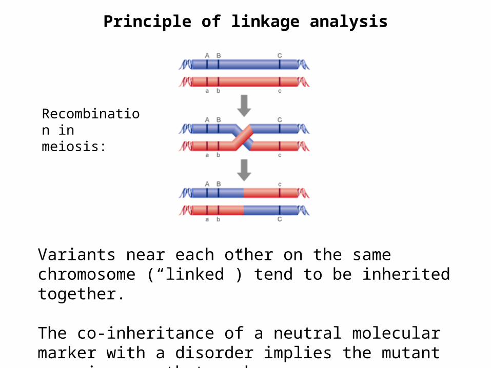
Principle of linkage analysis
Variants near each other on the same chromosome (“linked”) tend to be inherited together.
The co-inheritance of a neutral molecular marker with a disorder implies the mutant gene is near that marker.
Recombination in meiosis:

Bond et al., (2002) Nature Genet. 32: 316
MCPH5 mapped to ASPM gene
Homologous to abnormal spindle (asp) gene in Drosophila
Mutations lead to truncated protein

Expression of ASPMin developing mousebrain
Ventricular zone

Neurogenesis and migration in the cerebral cortex

A number of other genes that cause Microcephaly have also been identified:
MCPH1: Microcephalin - control of mitosis (Jackson et al., (2002) Am J Hum Genet. 71, 136-42)
MCPH3: CDK5RAP2 MCPH6: CENPJ - both involved in chromosome segregation
in mitosis (Bond et al., (2005) Nat Genet. 37, 353-5)


How do mutations in genes controlling mitosislead to microcephaly?
Aspm mRNA expressed at early stages:
- Divisions are symmetric- Progenitor pool expanding
Aspm mRNA downregluated at later stages:
- Divisions are asymmetric- Neurons being generated

Symmetric divisions at early stages generate two neuroepithelial progenitors
- expand pool of progenitors
Asymmetric divisions at later stages generate one postmitotic neuron and one progenitor
- as each progenitor can only generate a limited number of neurons this eventually depletes pool of progenitors and leads to fewer neurons

Asymmetric distribution of cytoplasmic factors coordinated with orientation of mitotic spindle

Aspm protein localises to centrosomes

Knockdown of Aspm function leads to asymmetric division

Knockdown of Aspm results in more asymmetric divisions at early stages
Effect is more progeny adopt neuronal fate and fewer retain neuroepithelial progenitor fate

Mutation of Aspm (or other genes implicated in microcephaly) causes:
1. Defect in alignment of mitotic spindle with axis of cell
2. Increase in asymmetric division at early stages
3. Failure to expand progenitor pool
4. Premature generation of neurons
5. Reduction in brain size

Conclusions:
Microcephaly caused by mutations in many genes All involved in mitosis somehow Defects in Aspm affect symmetric division Progenitor pool fails to expand - depleted too early Small brain results
ASPM, MCPH1, CDK5RAP2 all show evidence of positive selection in lineage leading to humans
Inference: Mutations in these genes that increased brain size may have been selected for in human lineage

Diversity of cell types and functions
Red blood cells
Hair cells in cochlea
Skin cells
Nervecells
Cardiac muscle cells

What makes cells different is they make different proteins
Some proteins made only in specific cell types:e.g., hemoglobin, insulin

- Express different genes related to their specific functions (neurotransmitter receptors, ion channels, etc.)
- Express specific code of transcription factors that control expression of all the other genes that make each cell unique (i.e. that specify its “identity”)
- How do they come to express that spectrum of transcription factors?
Each tissue/cell type has a different profile

Process of reiterative subdivision of embryo and progressive restriction of potential.
- specification of intermediate fates of dividing cells en route to specification of final fates of postmitotic cells
Occurs through series of cellular interactions beginningat the first cell division and continuing throughout development as morphogenetic movements shape embryo.

Gastrulation and Neural Induction
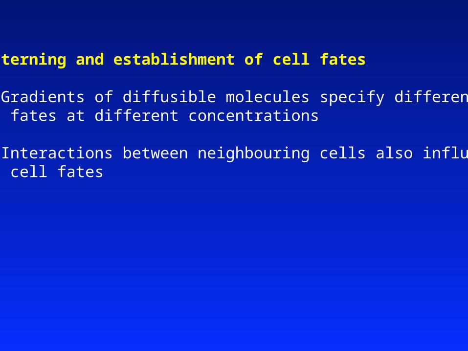
Patterning and establishment of cell fates
1. Gradients of diffusible molecules specify different fates at different concentrations
2. Interactions between neighbouring cells also influence cell fates

Different neuronal types generated from specific progenitor pools
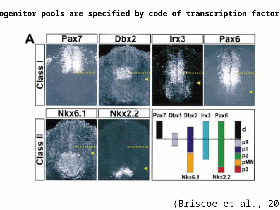
Progenitor pools are specified by code of transcription factors
(Briscoe et al., 2000)

Sharp borders between domains

Floor plate of spinal cord can induce ectopic motorneurons
Wild-type situation Floor plate ablated Floor plate grafted
motoneurons
Floor plate
(Embryological experiments in chick)
Shh conc.
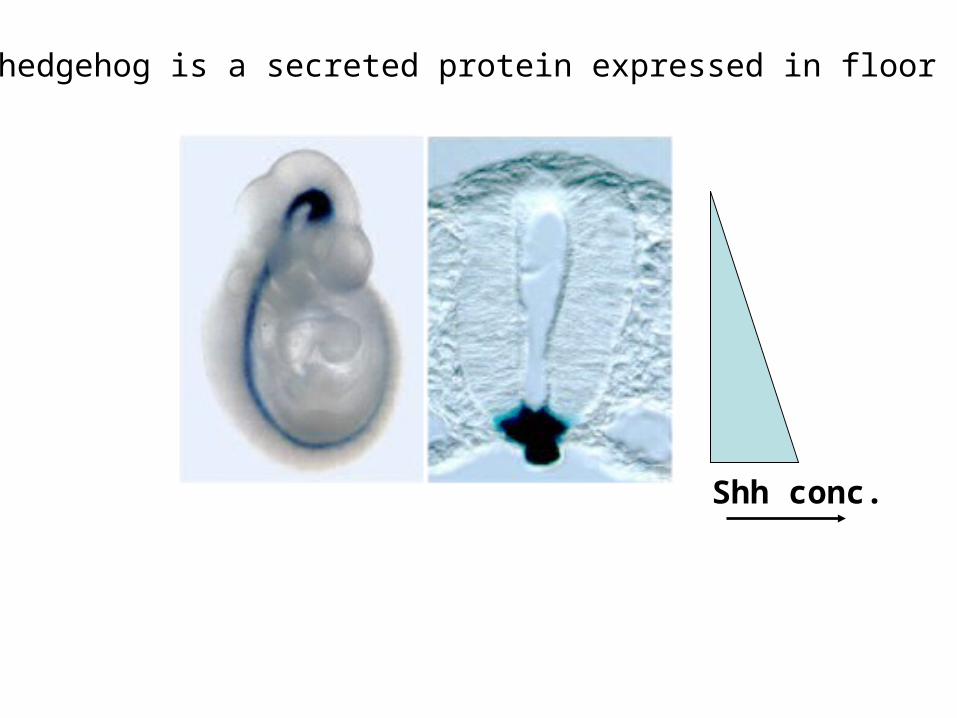
Sonic hedgehog is a secreted protein expressed in floor plate

Gradient of Shh induces different fates

Gradient of Shh induces some genes and represses others

How do you get such sharp borders?
Cross-repression between transcription factors:

Cross-repression:
Nkx2.2 activates its own transcription and represses Pax6
Pax6 activates its own transcription and represses Nkx2.2
Both genes can’t be expressed in same cell- slight imbalance amplified- graded expression becomes sharp- individual cells specified as one fate or another

Combinatorial code of transcription factors
Control expression of other genes (i.e., turn on whole “profile” of gene expression for different subtypes of neurons)
These downstream “effector” genes control various aspects of cell fate:
- Connectivity- Neurotransmitter expression- Expression of ion channels/receptors, etc.

Shh also patterns midline of brain and face

Mutations in Shh lead to Holoprosencephaly
(OMIM: 142945)

Midbrain dopaminergic neurons degenerate in Parkinson’s disease
Specification of clinically important cell types

Parkinson’s disease
Primary symptoms:
Tremor: an uncontrollable trembling or shaking
Rigidity: an abnormal stiffness of the muscles
Bradykinesia: an extreme slowness of movement and reflexes.
Caused by progressive loss of midbrain dopaminergic neurons
- can be familial (often early-onset)
Current therapies (L-dopa) only moderately effective

Midbrain dopamine neurons induced by Shh and Fgf8
Shh Fgf8

Induction of midbrain dopaminergic neurons(side view) (dorsal view)
Explants ofneural tubein vitro:
d2
v2
d3
v3
TH+ve neurons ariseonly in v3 in vivo andin explants in vitro

Add FP (source of Shh) to d3: dopaminergic neurons (TH +ve):
Add isthmus (source of Fgf8) to v2: dopaminergic neurons (TH +ve):
Block Shh function in v3 explant with antibody: no dopaminergic neurons (TH -ve):


Inducing dopaminergic neurons from stem cells in vitro


Summary
Development of the nervous system involves manydistinct processes in two main phases:
- establishment of cell identities(patterning, proliferation, neurogenesis)
- wiring (migration, axonal extension, synaptogenesis)
Defects (due to genetic or environmental causes)in any of these processes can lead to specific clinicaldisorders
Knowledge of developmental mechanisms can informefforts to promote regeneration or stem cell replacementtherapies

Transcription factors induced or repressed by Shhin concentration-dependent fashion
Explants ofmedial spinal cord plus increasing concentrations of Shh

Diffusible Shh bound by transmembrane receptor proteins that transduce a signal intracellularly, eventually leading toactivation of transcription factors.
- At different concentrations this has different effects (it is a morphogen)

High affinity and low affinity binding sites
Nkx2.2
Nkx6.1
Gli
Gli Gli Gli
Low affinity sites: Gli binds weakly, not effective at low concentrations => Nkx2.2 only expressed verynear floor plate ([Shh] high)
High affinity sites: Gli binds strongly, effective even at low concentrations => Nkx6.1 expressed further awayfrom floor plate (where [Shh] lower)