Embed Size (px)
Citation preview

University of Mississippi University of Mississippi
eGrove eGrove
Honors Theses Honors College (Sally McDonnell Barksdale Honors College)
Spring 5-9-2020
An Investigation of the Practical Challenges to Using Molecular An Investigation of the Practical Challenges to Using Molecular
Genetic Techniques to Identify the Parasite Burdens of Vertebrate Genetic Techniques to Identify the Parasite Burdens of Vertebrate
Animals from Non-Invasive Sampling Animals from Non-Invasive Sampling
Keely Ann Cox University of Mississippi Main Campus
Follow this and additional works at: https://egrove.olemiss.edu/hon_thesis
Part of the Zoology Commons
Recommended Citation Recommended Citation Cox, Keely Ann, "An Investigation of the Practical Challenges to Using Molecular Genetic Techniques to Identify the Parasite Burdens of Vertebrate Animals from Non-Invasive Sampling" (2020). Honors Theses. 1396. https://egrove.olemiss.edu/hon_thesis/1396
This Undergraduate Thesis is brought to you for free and open access by the Honors College (Sally McDonnell Barksdale Honors College) at eGrove. It has been accepted for inclusion in Honors Theses by an authorized administrator of eGrove. For more information, please contact [email protected].

An Investigation of the Practical Challenges to Using Molecular Genetic Techniques to
Identify the Parasite Burdens of Vertebrate Animals from Non-invasive Sampling
By
Keely Cox
A thesis submitted to the faculty of the University of Mississippi in partial fulfillment of
the requirements of the Sally McDonnell Barksdale Honors College
Oxford
2020
Approved by
__________________________
Advisor: Dr. Richard Buchholz
__________________________
Reader: Dr. Rebecca Symula
__________________________
Reader: Dr. Glenn Parsons

ii
© 2020
Keely Ann Cox
ALL RIGHTS RESERVED

iii
ACKNOWLEDGEMENTS
First, I would like to acknowledge my advisor, Dr. Richard Buchholz for advising me
throughout this research process and providing me with the materials and the guidance
needed to create a successful thesis project. I would also like to thank Dr. Rebecca
Symula and Dr. Glenn Parsons for their time and consideration they put forth as my
second and third readers, and Dr. Susan Balenger for helpful comments on my thesis. I
would also like to thank Victoria Monette and Alaina Skidmore for assisting me in the
lab and being constant encouragement and supporters throughout the entire process. I am
very grateful to my parents for constantly pushing me to work my hardest and try my best
in my lab work. Lastly, I would like to acknowledge the Sally McDonnell Barksdale
Honors College for giving me the opportunity to participate in the thesis project.

iv
ABSTRACT
KEELY COX: An Investigation of the Practical Challenges to Using Molecular Genetic
Techniques to Identify the Parasite Burdens of Vertebrate Animals from Non-invasive
Sampling
(Under the direction of Dr. Richard Buchholz)
Molecular genetic techniques have become popular methods in ecology and
wildlife conservation research. Advances in molecular genetic methods, particularly PCR
(polymerase chain reaction), make it possible to amplify the numbers of specific DNA
sequences from a sample with only a few original copies. Theoretically, the specificity of
this approach should make it possible for wildlife biologists to identify and quantify the
parasite and disease burden of endangered animals without being limited by the rarity of
collaborators with expertise in the taxonomy of obscure parasite taxa. Because PCR
requires just a small amount of DNA, the added benefit of a molecular genetic approach
is that non-invasive sampling methods can be used that do not harm the endangered
animals. My research involved two related projects. First I attempted to separate,
morphologically identify, sequence, and isolate DNA from nematodes present in fecal
samples collected non-invasively from the Baird’s tapir in Belize. This attempt was not
successful but I did discover several practical obstacles to this type of work that made me
wonder about the success rate of molecular parasitology in other studies of wildlife
species. The second project was a systematic review of the literature of the practical
challenges associated with using molecular genetic methods to identify parasitic
nematodes in fecal samples of domestic and wild animals, and humans. This literature
review led me to the conclusion that non-invasive sampling methods are only beneficial

v
when the sequence of the studied nematode has already been named through prior
research.

vi
TABLE OF CONTENTS
LIST OF TABLES AND FIGURES………………………………………………….....vii
Chapter One
INTRODUCTION………………………………………………………………...1
MATERIALS AND METHODS………………………………………………….5
RESULTS………………………………………………………………………..12
DISCUSSION……………………………………………………………………14
Chapter Two
INTRODUCTION……………………………………………………………….17
MATERIALS AND METHODS………………………………………………...20
RESULTS………………………………………………………………………..22
DISCUSSION……………………………………………………………………26
OVERALL CONCLUSION…..…………………………………………………30
REFERENCES…………………………………………………………..………31

vii
LIST OF TABLES AND FIGURES
Table 1 A description of the six different nematode morphotypes that were present
in formalin and ethanol preserved fecal samples from the Baird’s
tapir………………………………………………………………………..8
Table 2 A summary of the DNA extraction methods, lysis reagents, nematode
morphological types, number of nematodes used, and the outcome of each
attempt to amplify DNA…………………………………………………13
Table 3 The 18 countries covered in this review and how many studies were
conducted in each ………………………………………………………..24
Table 4 Authors’ origins compared to the location of where lab work was
completed………………………………………………………………...24
Figure 1 A scanned photo of an electrophoresis gel image with a faint band on the
positive control…………………………………………………………...13
Figure 2 A scanned photo of an electrophoresis gel image with a faint ladder, but
no positive control band………………………………………………….13
Figure 3 A flowchart showing the outcome for sample size of the decision process
for identifying peer-reviewed, empirical literature suitable for providing
data for this review……………………………………………………… 21
Figure 4 A graph that represents the fecal sample size most associated with
successful study outcomes……………………………………………….23
Figure 5 Percentage of each taxon group present in the wild animal studies……..24
Figure 6 Percentage of each taxon group present in the domestic animal studies...24
Figure 7 Percentage of each taxon group present in the captive animal studies…..24

1
CHAPTER ONE
An Attempt to Identify Nematodes in the Feces of Baird’s Tapir Using A Standard
Protocol for Lysis, Direct PCR, and Sequencing
Introduction
The Earth’s natural habitats and the wildlife in them are in decline, leading to an
extinction crisis (Pellens, 2018). As the human population continues to grow
exponentially (Hubbert, 1996) our unsustainable use of resources poses various threats to
other species. Some of these threats are obvious, such as direct harvest and persecution of
species for food, recreation and trade (Pellens, 2018). Other threats are indirect or
insidious in that they act as stressors that subtly reduce lifetime fitness of individual
animals by impairing reproduction and/or reducing survival (Balestri, 2014). These
stressors can include anthropogenic contaminants (Frena, 2016), reduction of food
availability due to habitat degradation by humans (Tylianakis, 2007), and frequent non-
consumptive disturbance by humans (Burgin, 2015) such as from ecotourists, hikers and
off-road cyclists.
Anthropogenic stressors may have cumulative effects that occur on top of
naturally occurring stressors such as intraspecific competition for food and mates
(Munns, 2006), dominance interactions (Sheldon, 1996), pursuit by predators (Munns,
2006) and infection by parasites (Sheldon, 1996). The negative health effects of
infections by parasites and other disease organisms on threatened and endangered species
are steadily becoming a focus of attention for conservation biologists (Gomez, 2013).

2
When species decline to low population size in a reduced or scattered geographic range,
the effects of parasites, when combined with the effects of other stressors, can lead to the
reduction in resistance or tolerance to certain infections, possibly contributing to the risk
of extinction. In certain areas of the Mojave desert, upper respiratory tract disease
incurred by the bacteria Mycoplasma agassizii in the desert tortoise (Gopherus agassizii)
has contributed to significant population decline in the tortoises (Hunter et al., 2008).
Another example of decline in wildlife populations due to parasites or disease is seen in
the California sea otter (Enhydra lutris nereis). Toxoplasma gondii-associated
meningoencephalitis is a disease that is responsible for 72% of otter infections and 16%
of mortality in beachcast sea otter carcasses, and is the main cause of the California sea
otter’s population decline; making them a threatened species (Miller et al., 2004).
Parasites use host resources to thrive and reproduce; lowering host fitness to
varying degrees (Sheldon 1996). Parasitic worm-like species in the phylum Nematoda are
well known for their harmful effects on domestic animals, such as heartworms and
lungworms in cats and dogs (Traversa, 2010) and habronemosis in horses (Saeed, 2019),
for example. Less commonly known is that parasitic nematodes can also harm humans
and wildlife species. Hudson (1986) found that the nematode Trichostrongylus tenuis was
associated with poor breeding success in the red grouse (Lagopus lagopus scoticus),
causing a decline in this bird species’ population. Another example is the human parasitic
threadworm, Strongyloides stercoralis, that infects around 100 million people worldwide;
causing gastrointestinal distress in healthy individuals, but can be fatal in those who are
immunocompromised (Becker, 2015).
3
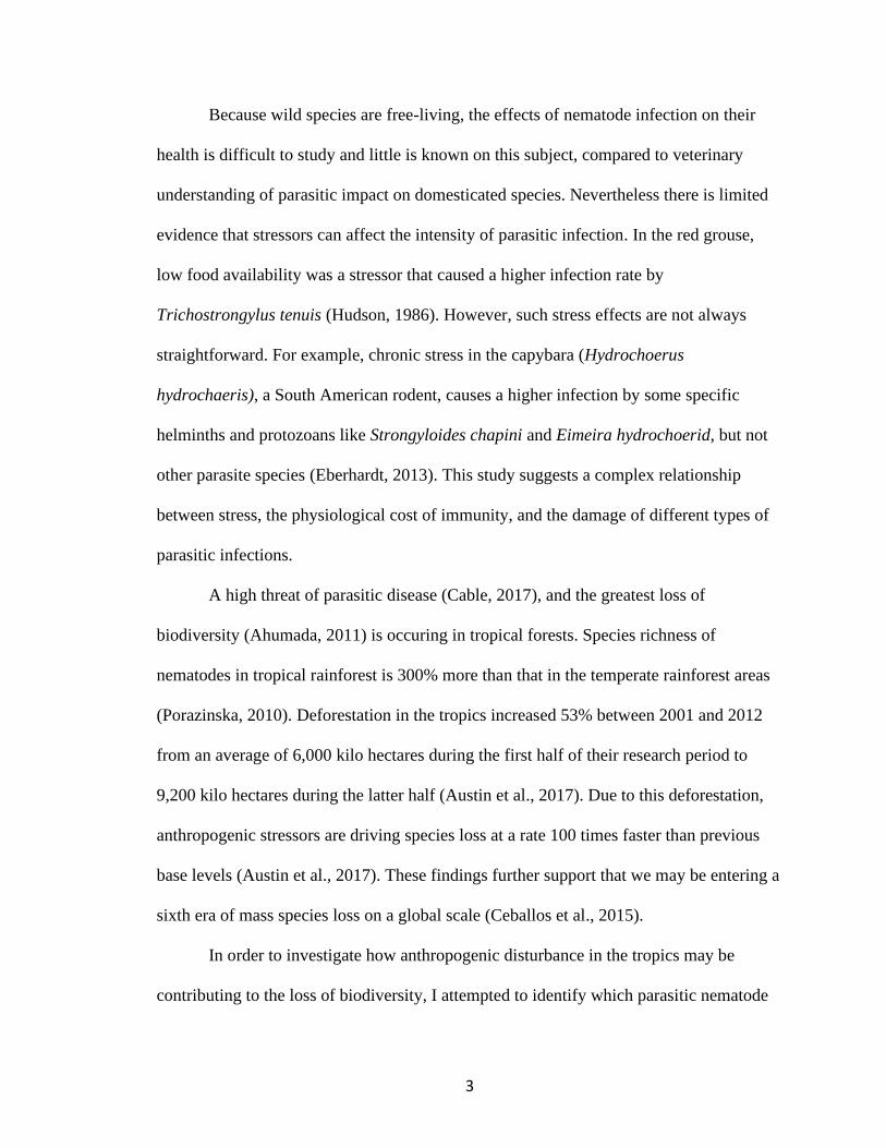
Because wild species are free-living, the effects of nematode infection on their
health is difficult to study and little is known on this subject, compared to veterinary
understanding of parasitic impact on domesticated species. Nevertheless there is limited
evidence that stressors can affect the intensity of parasitic infection. In the red grouse,
low food availability was a stressor that caused a higher infection rate by
Trichostrongylus tenuis (Hudson, 1986). However, such stress effects are not always
straightforward. For example, chronic stress in the capybara (Hydrochoerus
hydrochaeris), a South American rodent, causes a higher infection by some specific
helminths and protozoans like Strongyloides chapini and Eimeira hydrochoerid, but not
other parasite species (Eberhardt, 2013). This study suggests a complex relationship
between stress, the physiological cost of immunity, and the damage of different types of
parasitic infections.
A high threat of parasitic disease (Cable, 2017), and the greatest loss of
biodiversity (Ahumada, 2011) is occuring in tropical forests. Species richness of
nematodes in tropical rainforest is 300% more than that in the temperate rainforest areas
(Porazinska, 2010). Deforestation in the tropics increased 53% between 2001 and 2012
from an average of 6,000 kilo hectares during the first half of their research period to
9,200 kilo hectares during the latter half (Austin et al., 2017). Due to this deforestation,
anthropogenic stressors are driving species loss at a rate 100 times faster than previous
base levels (Austin et al., 2017). These findings further support that we may be entering a
sixth era of mass species loss on a global scale (Ceballos et al., 2015).
In order to investigate how anthropogenic disturbance in the tropics may be
contributing to the loss of biodiversity, I attempted to identify which parasitic nematode

4
species are found in a population of the endangered Neotropical mammal, Baird’s tapir
(Tapirus bairdii), by isolating DNA from fecal nematodes and amplifying and
sequencing their DNA. Monette (2019) used molecular methods to identify 4 individual
nematodes from one of the 6 morphotypes that she recovered from field-collected fecal
samples. This first chapter of my thesis provides an overview of my unsuccessful attempt
to identify additional nematodes morphotypes from these same fecal samples.

5
Materials and Methods
A. Study Species and Study Area
Tapirs are one of the last members of the Neotropical megafauna. They are shy
herbivores living in habitats that vary from deciduous forests to tropical rainforests in
Central and South America as well as in Southeast Asia. Tapirs can be found anywhere
from sea level to heights of at least 3,350 meters. The four species of New World tapir
are as follows: the Baird’s tapir (Tapirus bairdii), the mountain tapir (Tapirus
pinchaque), the lowland tapir (Tapirus terrestris), and the Malayan tapir (Tapirus
indicus) (The New Encyclopedia Britannica, 2010).
Tapirs are of conservation concern because they require large, inter-connected
forested areas with protection from over-hunting to persist (Naranjo 2018). The
endangered Baird’s tapir used to occur from Mexico to northern South America (Garcìa-
Marmolejo et al., 2015) but now that range has been reduced by 50% in just three
decades (Schank et al., 2015). These tapirs have critical ecological roles that help shape
the Neotropical forest community (Jorge et al., 2013). They disperse the seeds of
rainforest plants, support dung beetle diversity, and create depressions in the soil for
wallowing, thereby creating pools that can be used by other animals (Garcìa et al.,
2012, Garcìa-Marmolejo et al., 2015, O’Farrill et al., 2013).
The tapir is an ecosystem engineer that serves as a great source of seed dispersal
of trees whose fruits are in their diet. Because of this behavior, tapirs provide an ongoing
food and shelter supply for other animals in their region (O’Farrill 2011). The
populations of Baird’s tapir (Tapirus bairdii) have been reduced drastically because of

6
habitat fragmentation caused by deforestation, constraining the population of this species
to protected areas (Carillo 2019).
B. Sample Collection Methods
Baird’s tapir fecal samples were collected in North Western Belize by Monette
(2019), who provides a detailed description of the study site and sampling methodology. I
was not involved in the field collections, but provide a brief summary below, before
describing how I assisted in sorting and processing nematodes collected from the
preserved feces.
The samples were found on trails that were covered by a camera survey grid that
was used as a transect. For reach site where tapir fecal samples were found, GPS
coordinates were recorded. Each transect was hiked a second time to collect any new
feces that had been dropped. After collection of the fecal samples, they were
homogenized, strained using purified water, and allowed to settle for three hours. The
supernatant was poured out and the remaining sediment was stored in three different
ways. Monette (2019) immediately isolated fecal DNA from each sample using a fecal
DNA extraction kit. For my lab research, this DNA served as the template DNA for my
standard positives for each PCR that was run on the nematode DNA. The rest of the feces
from each field collection were divided between storage tubes for preservation in 95%
ethanol and 10% formalin.
Each of the forty-four samples preserved in the formalin and ethanol were
centrifuged in an IEC HN-SII Centrifuge at 2000 rpms for ten minutes in order to
concentrate the sediments at the bottom of the tube. After centrifugation, the sediment of

7
the sample was poured into a petri dish to be examined under a Labomed Luxeo 4Z
Stereozoom dissecting microscope (10x magnification). All of the nematodes found in
the dish were pipetted into a vial and marked with the number of nematodes found.
C. Sorting nematodes by morphology
I poured the vials containing the nematodes into a petri dish along with water
purified by reverse osmosis (RO). The dish with the RO water and ethanol was set aside
for about 15 minutes to allow the nematodes to settle to the bottom of the petri dish. Once
settled, the nematodes that were in the vial had moved to the bottom of the petri dish. I
examined the dish for nematodes at 10x magnification under a LaboMed Luxeo 4Z
Stereozoom dissecting microscope.
Monette (2019) identified six main types of nematode morphology present in the
ethanol samples. Morphology A, B, and C were crimson colored nematodes. Morphology
A contained a mouth protrusion, morphology B had no mouth protrusion, and
morphology C had a thicker body type. Morphology D, E, and F were clear and larger
nematodes. Morphology D did not have a whip-like tail, morphology E had a whip-like
tail and a smooth body type, and morphology F had a whip-like tail with ridges on the
outside of the body. These different morphology types are represented in Table 1. Using
a pipet, each nematode was extracted from the dish and placed in its own vial with
ethanol. Each vial was labelled with morphological type. After separating every
nematode, the case with the vials was properly labelled and placed into a freezer.

8
Morphology Description 100x magnified
Type photo
A Long and slender, amber-colored body
with a clear mouth protrusion; smooth
body
B
Long and slender, amber-colored body
with no mouth protrusion; smooth body
C Tear shaped, amber-colored body
with mouth parts at the wider end
of the body; body cavity near the
mouth; smooth body
D Clear body with a short tail, smooth
body
E Clear body with a long tail, smooth
body
F Clear body with long tail; ridges
on side of the body
Table 1. A description of the six different nematode morphotypes that were present in
formalin and ethanol preserved fecal samples from the Baird’s tapir. The scale lines in
each photo represent 10 μm. (Used with permission from Monette (2019)).

9
D. Parasite DNA Analysis
Lysis:
For lysis, a few individuals from each of the A-C and F morphologies were
selected to be lysed. A nematode was mouth- pipetted out of the vial and placed into a
strip tube and properly labeled. Because nematodes can be lost during the transfer
process, each tube was inspected microscopically to confirm the presence of a nematode.
The tubes were left open, or heated in an Eppendorf AG Mastercycler gradient thermal
cycler (No. 533113946) in order to evaporate any leftover ethanol that was dispensed into
the strip tubes because ethanol will inhibit cell lysis. Following the methods of Chalasani
(2016), a lysis master mix was made containing 0.3 microliters of Viagen proteinase K
(Cat# 102-T) and 19.7 microliters of Viagen DirectPCR (mouse tail) per worm. In some
lysis trials, more Proteinase K was used than recommended by Chalasani (2016), but the
total amount of lysis solution used remained at 20 microliters per worm. Twenty
microliters of lysis master mix was added to each tube of the PCR strip tube containing a
nematode, incubated at 55 degrees Celsius for 16 hours, heated at 85 degrees Celsius for
one hour, followed by refrigeration at around 4 degrees Celsius.
PCR:
A 23 µL PCR master mix solution for each nematode sample was prepared with
the following components: 12.5 microliters of Promega GoTaq green master mix, 2x, 9.5
microliters of ddH2O, 0.5 microliters of forward primer (18S), and 0.5 microliters of
backwards primer (18s). The primers target the 18S rRNA region of the nematodes and
the sequences are as follows: 5’ GGCGATCAGATA-CCGCCCTAGTT 3’ (18S 965
10
Forward) and 5’ TACAAAGGG-CAGGGACGTAAT 3’ (18S 1573R) (Powers, 2009).
Two microliters of the lysed nematode solution (containing the DNA template) were
added to each tube of PCR master mix. For the positive and negative PCR controls, two
microliters of a positive control DNA sample (fecal sample) was added to the PCR
master mix solution and two microliters of ddH2O was added to PCR master mix for the
negative control. Brief centrifugation of the PCR 0.2 milliliter strip tubes was completed
before placing them in the thermal cycler.
During the PCR process in the thermal cycler, the three main stages that occurred
were denaturing, annealing, and extending. Denaturing involved the heating of the
double-stranded template of DNA at 95 degrees Celsius for 4 minutes in order to separate
the strand into single strands. The temperature lowered to 55 degrees Celsius for 30
seconds to allow the specific primer to attach to the template DNA in the annealing
process. Finally, the temperature was raised to 72 degrees Celsius for 6 minutes and the
new strand of DNA was formed by the Taq polymerase enzyme. These three main stages
of PCR were repeated forty times, which doubled the number of DNA copies every time
the stages were repeated.
Electrophoresis:
Agarose gel electrophoresis was used to check the quantity and size of the
fragments of DNA. To make an agarose gel for electrophoresis, 1.5 grams of agarose
powder was added to a glass jar. Next, 100 milliliters of the electrophoresis gel buffer
was added to the container. This mixture was heated in one-minute intervals until it
approached boiling. After heating the mixture, two microliters of ethidium bromide were
added and swirled in the jar to mix. This mixture was poured into the electrophoresis gel
11
tray (with plastic combs) and allowed to set for 15 minutes. After the gel set, the plastic
combs were removed from the gel to reveal the wells in the gel. The gel and tray were
placed in the electrophoresis machine and the buffer solution was poured into the
machine until the gel was completely covered. Five microliters of FlashGelTM DNA
Marker 100-4000 base pair ladder was added to the first well of each row. To inspect the
quantity and quality of DNA produced by nematode lysis, a 7.5 µL total volume of 2.5
microliters of FlashGelTM loading dye (5x concentration) with 5 µL of the lysed nematode
solution were mixed together by pipetting onto a Parafilm laboratory film strip. This
mixture was then pipetted into the well in the agarose gel. The gel was electrophoresed
for 30 minutes at 110 volts. The gel was then photographed under UV transillumination
on an AlphaImager HP. The printed photographs of the gel did not result in clear bands,
so the DNA isolation process was attempted again using more than one nematode per vial
in the lysing stage. This process was repeated with two worms, three worms, and two
halved worms in each vial. The reagents were adjusted according to the amount of worms
placed in the vials; following the procedure from Chalasani (2016).

12
Results
No successful extraction of DNA from any of the studied worm morphotypes was
evident even though varying the number of individual nematodes, halving them, and
adding more proteinase K was performed (Table 2). After the lysis step, it was very
evident that the nematodes were still intact, but they were moved onto the PCR stage.
During the first few months of my lab research (March 2019-September 2019), the
positive controls amplified in each electrophoresis gel that was run, matching the
expected base pair length of 646 bps, so it was evident that PCR failure did not occur
(Fig. 1). In the latter months of my lab research (September 2019-December 2019), the
positive control bands and a strong ladder were not visible on the gel photographs, from
which I conclude that the electrophoresis gel was run for too long and the DNA fragment
ran off the gel, or that PCR failed (Fig. 2).

13
Nematode
Morphological Type
(after Monette 2019)
# of Nematodes
(per individual
PCR striptube)
Protease Volume (µL) Outcome of PCR Product
A 1 worm 0.3 Prot. K per worm Unknown
A 1 halved worm 0.3 Prot. K per worm No + control band
A 2 worms 0.3 Prot. K per worm Unknown
A 2 halved worms 0.34 Prot. K per worm No + control band
A 3 worms 0.34 Prot. K per worm Unknown
B 1 worm 0.3 Prot. K per worm Unknown
B 2 worms 0.3 Prot. K per worm Unknown
B 2 halved worms 0.34 Prot. K per worm No + control band
C 1 worm 0.3 Prot. K per worm Unknown
C 2 halved worms 0.3 Prot. K per worm No + control band
F 1 worm 0.3 Prot. K per worm Unknown
F 2 worms 0.34 Prot. K per worm Unknown
Fig. 1 A scanned photo of an
electrophoresis gel image with a faint
band on the positive control.
Fig. 2 A scanned photo of an
electrophoresis gel image with a faint
ladder, but no positive control band.
Table 2. A summary of the DNA extraction methods, lysis reagents,
nematode morphological types, number of nematodes used, and the outcome
of each attempt to amplify DNA.
+ 3 2 1 4 L
14
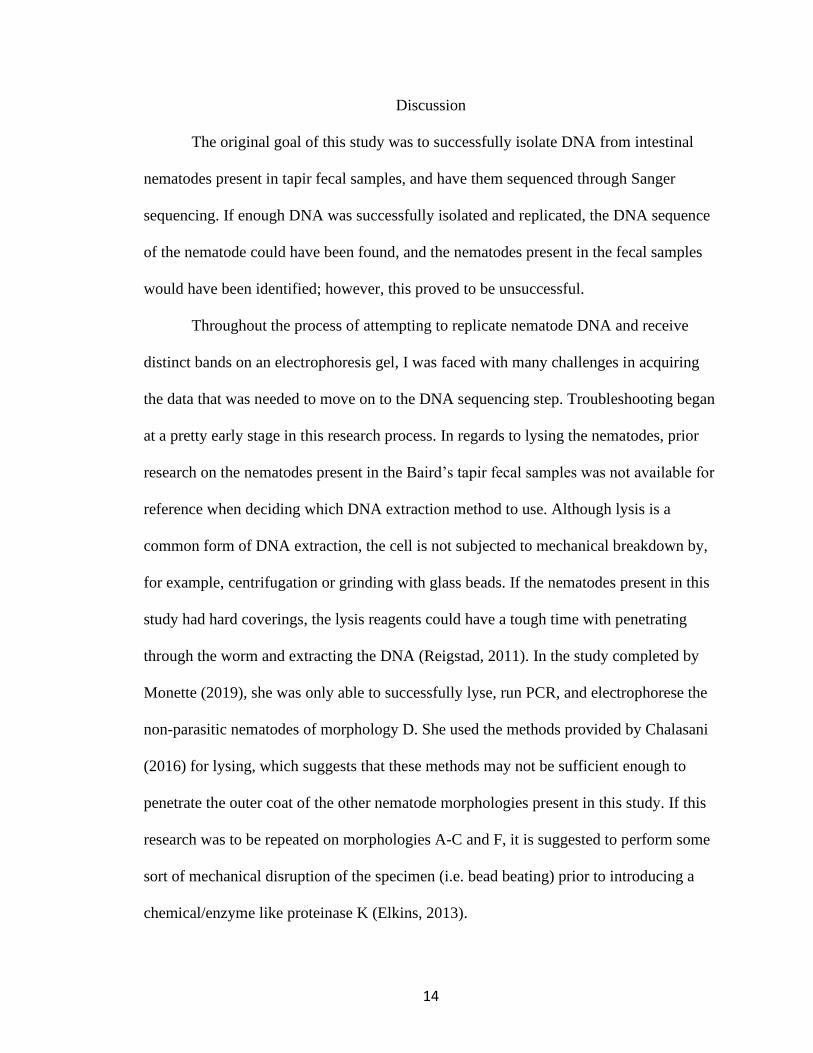
Discussion
The original goal of this study was to successfully isolate DNA from intestinal
nematodes present in tapir fecal samples, and have them sequenced through Sanger
sequencing. If enough DNA was successfully isolated and replicated, the DNA sequence
of the nematode could have been found, and the nematodes present in the fecal samples
would have been identified; however, this proved to be unsuccessful.
Throughout the process of attempting to replicate nematode DNA and receive
distinct bands on an electrophoresis gel, I was faced with many challenges in acquiring
the data that was needed to move on to the DNA sequencing step. Troubleshooting began
at a pretty early stage in this research process. In regards to lysing the nematodes, prior
research on the nematodes present in the Baird’s tapir fecal samples was not available for
reference when deciding which DNA extraction method to use. Although lysis is a
common form of DNA extraction, the cell is not subjected to mechanical breakdown by,
for example, centrifugation or grinding with glass beads. If the nematodes present in this
study had hard coverings, the lysis reagents could have a tough time with penetrating
through the worm and extracting the DNA (Reigstad, 2011). In the study completed by
Monette (2019), she was only able to successfully lyse, run PCR, and electrophorese the
non-parasitic nematodes of morphology D. She used the methods provided by Chalasani
(2016) for lysing, which suggests that these methods may not be sufficient enough to
penetrate the outer coat of the other nematode morphologies present in this study. If this
research was to be repeated on morphologies A-C and F, it is suggested to perform some
sort of mechanical disruption of the specimen (i.e. bead beating) prior to introducing a
chemical/enzyme like proteinase K (Elkins, 2013).
15
In the beginning of my lab research, it was evident by the photographs of the
electrophoresis gels completed in my study that there were no failures in PCR reagents
and the thermal cycler operation because a positive control band was evident in each gel
that was run. Some reasons behind the lack of clear bands in these gels could have been
related to the time in which the gels were run in the electrophoresis machine. If the gels
were run for too long, the bands of DNA could have run off the bottom of the gel.
Towards the end of my lab research, the photographs of the gels showed that there was
some sort of PCR failure because bands for the positive controls were no longer evident.
This shift in seeing positive control bands to no longer seeing these bands could possibly
mean I encountered malfunctions in lab equipment or spoiled PCR and lysis reagents.
In selecting the primers used for this research project, the same primers used in
the research done by Monette (2019) were also used in replicating the DNA of the
nematodes present in this study’s fecal samples. A primer that targets the 18S rRNA
region of nematodes was used because of the large number of 18S sequences that are
available on GenBank, the presence of a 18S- based phylogenetic tree, and this gene’s
nature in ensuring a complete phylogenetic coverage of the phylum Nematoda (Powers,
2009). The same vial of reverse and forward PCR primers were used for the entire
duration of this research project, so the lack of DNA amplification could have been
caused by old primers. To troubleshoot this problem, it is suggested that fresh primer
aliquots should be reconstituted or new primers should be obtained. Insufficient quantity
of the PCR primer could have also led to poor replication/ amplification. In the gels that
showed a positive control band was amplified, the absence of bands from the nematode
16
samples might be the result of insufficient template DNA for PCR to create a visible
band.
The common problems associated with the use of the polymerase chain reaction
to replicate DNA are mainly associated with reaction conditions, sequence accuracy, and
amplification yield. The lack of amplification of DNA could possibly be caused by poor
DNA integrity during DNA isolation, insufficient quantity of DNA, and complex targets
(e.g. secondary structures). To troubleshoot these problems, DNA could be stored in a TE
buffer to avoid degradation by nucleases, DNA polymerases with high sensitivity could
be chosen for amplification, and the denaturation time and temperature could be
increased to separate the double stranded DNA templates (Eggert, 2006).
The repetitive and meticulous troubleshooting of this lab research makes me
question if it is feasible to expect widespread use of molecular genetic approaches in
assessing the parasite burden of wildlife and other little known species. There were
various possible sources of error that could contribute to my lack of results, like
malfunctions in the thermal cycler and spoiled PCR primers and lysis reagents. Some
sources of failure leading to these results could be an incorrect lysis procedure for
nematode tissue and incorrect primers for this particular worm species.
If this study was to be repeated, it would require more reagents to test, updated
equipment, and adequate time to complete the troubleshooting necessary for positive
results. Successfully identifying nematodes in species like the tapir could provide further
insight into possible wildlife diseases that act as a contributing threat to wildlife and
occasionally causing population declines. However, in order to justify the use of these

17
very meticulous molecular techniques, further research is needed regarding which
methods are most effective to yield the best results.

18
CHAPTER TWO
Systematic Literature Review of the Success of Using Molecular Genetic Techniques to
Describe the Parasite Community from Fecal Samples
Introduction
An ongoing debate revolving around the conservation of endangered wildlife
species concerns the negative impact that direct, invasive handling for research purposes
has on wildlife populations (Avise et al., 1979). In order to assess genetics of wildlife
populations twenty plus years ago, it required the collection of fresh tissue in order to
complete a protein electrophoresis, which often required the animal to be killed for
scientific study (Proverbio, 2020). An alternative sample collection method that was
introduced was blood/serum sampling to study serum proteins without having to kill the
animal. When new genetic markers (restriction fragment length polymorphisms) were
introduced, the sample collection process required destruction of the study specimen in
order to extract mtDNA from fresh liver (Avise et al., 1979). The context of this
argument changed with the development of the PCR (Saiki et al., 1985).
PCR allows for the detection and reproduction of copious amounts of sequence-
specific DNA and is used by researchers and clinicians to diagnose diseases, sequence
genes, and complete quantitative and genomic studies in a rapid manner (Garibyan,
2014). PCR is an example of a molecular genetic procedure that can be completed with
samples collected non-invasively from specimens.
Non-invasive techniques do not infiltrate nor destroy healthy tissue and do not
involve tools that break skin or physically enter the body. These techniques can be seen
19
in animal conservation and medical research, but there are some cons to this type of
sample collection. Although non-invasive collection methods are painless, they suffer
from high levels of bacterial contamination, low gDNA production, or fragmented DNA
strands (Mills et al., 2000).
According to current literature, morphological and molecular identification of
nematodes are two growing methods in taxonomic and biodiversity studies. There has
been a recent movement to stray away from the traditional methods of phenotypic
identification due to the fact that these methods lack accuracy in identifying
taxonomically challenging groups. In regards to molecular techniques, the data can
violate the assumptions of phylogenetic analysis if sequences from various taxa are
changing at different rates (Abebe et al., 2011). Despite the tangible advantages of
molecular techniques, biologists and taxonomists worry that molecular techniques such
as PCR could possibly replace taxonomy; reducing the complexity of an entire biological
organism to a small fraction of that organism- the gene. The difficulty of linking DNA
sequences to the ecological functions of whole species argues against abandonment of
morphological identification all together (Tautz et al., 2002). Nevertheless the practical
value of using molecular genetic methods for parasitological identification and
quantification in wildlife is still in question because these are not model study species
about which much is known. Based on this concern, and my unsuccessful experience
using such an approach on the fecal parasites of Baird’s tapir (Chapter One), I felt that a
systematic review was needed to discover if using molecular genetic methods on fecal
samples collected non-invasively are beneficial in wildlife parasitology, and to identify
any challenges related to these methods.

20
I use a search of the scientific literature to find: a) which molecular approaches
are most commonly used to identify parasites of wild animals, b) to describe geographic
trends in samples sources, laboratory locations, and taxon inclusion, c) to evaluate
whether researchers claimed that the molecular approach to parasitology was successful,
and d) to identify the study characteristics associated with success.

21
Materials and Methods
A literature search was completed to find articles regarding the success rate of
using molecular genetic techniques to identify, replicate, and sequence parasite DNA
from fecal samples of wildlife, domestic animals, and humans.
A. Literature selection criteria
Several sources were searched in health-related search engines including
ScienceDirect, PubMedCentral, and the University of Mississippi’s One Search using the
key words: nematode, helminth, molecular, non-invasive, and feces.
The search was limited to research articles and case studies/reports published in
English from January 2008 to January 2020. First, the titles of the selected articles were
examined to identify articles that reported on non-invasive fecal collection methods,
helminths, and molecular methods such as PCR, electrophoresis, and DNA sequencing. If
these search criteria could not be found in the title, the abstract was then examined. The
data collected included the classification of the species being studied (i.e. domestic
animal, captive animal, wild animal, or human), the sample size, the molecular methods
used, and the success rate of the study completed. The successful articles were those that
identified all of their studied helminths from their non-invasively collected samples, the
partly successful articles only identified some of their helminths, and the unsuccessful
studies did not identify any of their studied helminths. I excluded articles that did not
include non-invasive sample collection methods, molecular methods, and helminth
species. The literature selection process is demonstrated in Fig 3.

22
Fig 3. A flowchart showing the outcome for sample size of the decision process for
identifying peer-reviewed, empirical literature suitable for providing data for this review.
23
Results
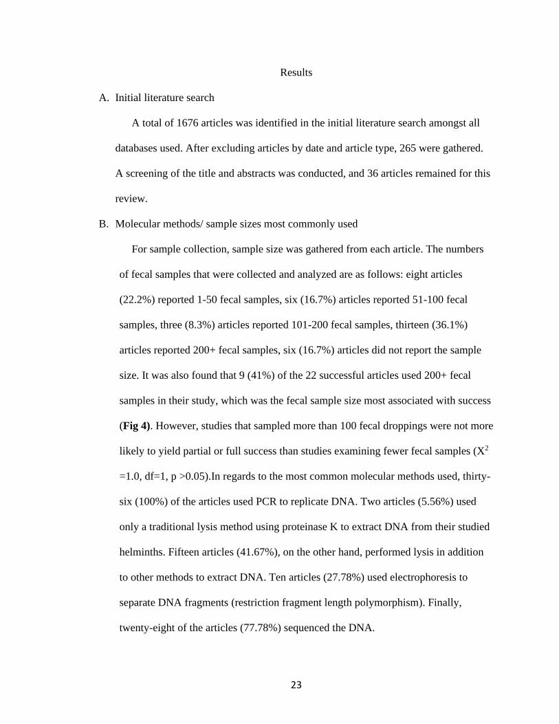
A. Initial literature search
A total of 1676 articles was identified in the initial literature search amongst all
databases used. After excluding articles by date and article type, 265 were gathered.
A screening of the title and abstracts was conducted, and 36 articles remained for this
review.
B. Molecular methods/ sample sizes most commonly used
For sample collection, sample size was gathered from each article. The numbers
of fecal samples that were collected and analyzed are as follows: eight articles
(22.2%) reported 1-50 fecal samples, six (16.7%) articles reported 51-100 fecal
samples, three (8.3%) articles reported 101-200 fecal samples, thirteen (36.1%)
articles reported 200+ fecal samples, six (16.7%) articles did not report the sample
size. It was also found that 9 (41%) of the 22 successful articles used 200+ fecal
samples in their study, which was the fecal sample size most associated with success
(Fig 4). However, studies that sampled more than 100 fecal droppings were not more
likely to yield partial or full success than studies examining fewer fecal samples (X2
=1.0, df=1, p >0.05).In regards to the most common molecular methods used, thirty-
six (100%) of the articles used PCR to replicate DNA. Two articles (5.56%) used
only a traditional lysis method using proteinase K to extract DNA from their studied
helminths. Fifteen articles (41.67%), on the other hand, performed lysis in addition
to other methods to extract DNA. Ten articles (27.78%) used electrophoresis to
separate DNA fragments (restriction fragment length polymorphism). Finally,
twenty-eight of the articles (77.78%) sequenced the DNA.

24
C. Geographic trends and taxon inclusion
The articles that were selected were done in 18 countries (Tables 3&4) between
the periods of 2008 to 2019. Three (8.33%) of the articles described human studies,
twenty-four (66.67%) wild animal studies (Fig 5), seven (19.44%) domestic animal
studies (Fig 6), and 2 (5.56%) captive animal studies (Fig 7).
0
50
100
150
200
250
0 5 10 15 20 25 30 35 40
Fec
al S
ample
Siz
e R
ange
Study Number
Success
Partly
Unsuccessful
101-200
200+
51-100
1-50
No Report
Fig 4. A graph that represents the fecal sample size most associated with successful study outcomes.

25
Country Amount of
studies
conducted in
country
Uganda 5
Canada 3
Central
African
Republic
3
USA 3
Australia 2
Colombia 2
Germany 2
France 2
Mexico 2
Argentina 1
China 1
Germany 1
Greece 1
Kenya 1
Namibia 1
Rwanda 1
Tanzania 1
Zamibia 1
Artiodactyla Perissodactyla
Carnivora Rodentia
Artiodactyla Chiroptera Carnivora
Primates Reptile Bird
Probescidea
Primate Carnivora
Fig 6. Percentage of each taxon
group present in the domestic
animal studies.
Fig 5. Percentage of each
taxon group present in the wild
animal studies.
Fig 7. Percentage of each taxon
group present in the captive
animal studies.
Table 4. Authors’ origins compared to the location of
where lab work was completed.
Table 3. The 18 countries
covered in this review and how
many studies were conducted
in each.

26
D. Rate of success
The success rate of each article is as follows: 22 were successful in the molecular
genetic identification of all of their studied nematodes, 3 were unsuccessful, 11 were
partly successful in identifying only some nematodes in the study. The unsuccessful
studies did not identify any of the nematodes in their study. Out of the successful studies,
however, 18 already had sequences for their studied helminths named prior to their
research.

27
Discussion
In this review, the most common molecular method used in wildlife parasitology
was the polymerase chain reaction to replicate certain pieces of DNA that were
successfully extracted from the nematode. Since this method was used in 100% of the
articles, it can be concluded that the polymerase chain reaction, with successful DNA
extraction, is a reliable source of replicating segments of DNA. Since the majority of the
articles relied on sequences that were already added to the genetic library, determining
the correct PCR primers and reagents was more simple compared to studies that did not
have prior research on the nematodes present in their study species. For the nine studies
that did not have a reference sequence prior to the study, more research was necessary in
order to search databases for sequences of the closest relative to their study species. Then,
primers had to be designed in order to sequence their DNA properly; proving to be a
more painstaking and tedious process compared to studies that already had their reference
sequences and recommended primers.
In regards to DNA extraction methods, only 5.56% of articles were successful in
using the traditional lysis method (with proteinase K) to extract DNA from the studied
helminths, while 41.67% of the articles used extraction methods such as phenol
chloroform extraction, sodium hydroxide extraction, and physical disruption of the tissue
followed up by PCR purification kits (i.e. SIGMA REDExtract-N-Amp Tissue PCR kit).
This suggests that the sole use of the traditional lysis method using proteinase K to
extract DNA from nematodes is not nearly as effective as using the other DNA extraction
methods in addition to PCR purification kits. The articles that used the traditional lysis

28
method with proteinase K were mostly unsuccessful. For example, Vlčková et al. (2018)
suggested that DNA degradation could have occurred due to incorrect storage, which led
to their unsuccessful study.
The fecal sample size was also taken into consideration in this review. Most of the
articles used 200+ fecal samples in their research, which was the highest number category
of fecal samples studied. Prior to conducting any sort of lab or clinical study, sample size
calculations should be considered in order to produce studies that are able to detect
clinically pertinent differences (Altman, 1991). Using unnecessarily large sample sizes
can waste resources and might pose ethical concerns if the samples are taken invasively.
However, using small sample sizes may produce unreliable and irreproducible results
(Faber, 2014). Since the 200+ sample size range was the range associated most with
successful studies, it can be concluded that larger sample sizes in molecular genetic
studies of animals would be recommended over smaller samples. Although none of the
articles in this review specified on the waste produced by their use of 200+ fecal samples,
they did specify that the techniques were all non-invasive, which takes away the ethical
concerns of their study.
Out of all the taxon groups that were present in this systematic review, 66.67%
conducted studies on wild animal species. The captive and domestic animal studies were
used to discover nematodes that were possibly causing disease and stress within related
wild animal species. For example, a study done by Lesniak (2017) focused on the
identification of parasites in domestic dogs to help identify similar parasites causing the
decline of wild grey wolves in that area. The other captive and domestic animal studies
did not mention ecological concern in the study species, but focused on discovering if the

29
parasitic nematodes could be causing disease in the human population. The wild animal
studies, on the other hand, had conservation objectives because parasitic nematodes can
cause population decline of endangered species.
Because the largest loss of biodiversity is occurring in the tropics, more studies
are necessary to identify parasites and other stressors related to this loss. Out of the 36
articles in this review, 21 of the articles were completed with studies in countries in, or on
species from, the tropics.
Trends in geographical and institutional distribution of scientific research are very
relevant in studying the ecosystem of the tropics. If research is limited to only a small
number of countries, the science community’s conclusion regarding the tropical
ecosystems could be biased since these locations may not be broadly representative
(Stocks, 2008). The majority of the articles in this review (60%) had authors that were
from, and completing their lab work in, the same country as their study species. However,
thirty-three percent of the studies collected samples from their study species and
completed their lab work in countries outside of the area where their study species was
found. Since a majority of the articles in the systematic review had authors that were
from, and completing their lab work in the country of their study species, it can be
concluded that the concern for biases is lower compared to the studies where the authors
did not complete lab work in the region of their study species.
This systematic review revealed that 61.1% of the articles studied were successful
in accomplishing their stated goal of non-invasive molecular identification of nematodes
present in wildlife species and humans. However, 82% of the successful articles stated
that they already had the sequences of their studied helminths in databases prior to their
30
study. For example, the study done by Solórzano-García (2017) focused on the non-
invasive molecular techniques to identify parasites in howler and spider monkeys.
Although the molecular methods could help with overcoming the limitations of
traditional phenotypic identification, “its utility relies on the extant genetic library and the
contributions that expand such library” (Solórzano-García, 2017).
31
Overall Conclusion
The two research projects of my thesis show that molecular genetic approaches to
identifying and quantifying the parasite burden of animals using fecal sampling will be
difficult and time-consuming but not impossible. For the foreseeable future experts on the
morphological identification of nematodes and other helminths will remain essential to
the development of reference DNA sequences for the wide variety parasitic taxa likely to
be collected by wildlife biologists. Because morphological identification of individual
parasitic worms is so time-consuming and labor intensive, however, manual counting
based on morphology is not a practical means of monitoring disease stressors in
threatened species given the immensity of the ongoing extinction crisis. New standard
methodologies for sample collection, lysis and optimization and multiplexing of PCR
must be adopted broadly in order to create reference sequence databases that can be used
easily by a variety of types of biological scientists. The results of the systematic review of
the literature suggest that progress is being made in capacity, building for molecular
genetic approaches to parasitology in tropical countries.

32
References
1. Abebe, E., et al. “A Critique of Current Methods in Nematode Taxonomy.”
African Journal of Biotechnology Vol. 10, no. 3 (2011): pp. 312–323.
2. Ahumada, J. A., et al. “Community Structure and Diversity of Tropical Forest
Mammals: Data From a Global Camera Trap Network.” Philosophical
transactions of the Royal Society of London. Series B, Biological sciences
Vol. 366, no. 1578 (2011): pp. 2703-2711.
3. Altman, D. G. “Practical Statistics for Medical Research.” London, UK:
Chapman & Hall Vol. 10, no. 10 (1991): pp. 11-19.
4. Austin, K. G., et al. “Erratum: Trends in Size of Tropical Deforestation Events
Signal Increasing Dominance of Industrial-Scale Drivers.” Environmental
Research Letters Vol. 12, no. 7 (2017). 5 054009.
5. Avise, J. C., et al. “The Use of Restriction Endonucleases to Measure
Mitochondrial DNA Sequence Relatedness in Natural Populations. I.
Population Structure and Evolution in the Genus Peromyscus.”
Genetics Vol. 92, no. 1 (1979): pp. 279-295.
6. Balestri, M., et al. “Habitat Degradation and Seasonality Affect Physiological
Stress Levels of Eulemur Collaris in Littoral Forest Fragments.” PLoS
ONE Vol. 9, no. 9 (2014): pp. 1-4.
7. Becker, S. L., et al. “Real-Time PCR for Detection of Strongyloides
Stercoralis in Human Stool Samples from Côte d’Ivoire: Diagnostic
Accuracy, Inter-Laboratory Comparison and Patterns of Hookworm Co-
Infection.” Acta Tropica Vol. 150, no.12 (2015): pp. 210–217.

33
8. Burgin, S., and N. Hardiman. “Effects of Non-Consumptive Wildlife-
Oriented Tourism on Marine Species and Prospects for Their Sustainable
Management.” Journal of Environmental Management, Vol. 151, no. 14
(2015): pp. 210–220.
9. Cable, J., et al. “Global Change, Parasite Transmission and Disease Control:
Lessons from Ecology.” Philosophical transactions of the Royal Society of
London. Series B, Biological sciences Vol. 372, no. 1719 (2017).
3721719.
10. Carrillo, N., et al. “Measuring Landscape Connectivity for Baird’s Tapir
Conservation in Fragmented Areas of Calakmul, Mexico.” Tropical
Conservation Science Vol. 12 (2019): pp. 1-15.
11. Ceballos, G., et al. “Accelerated Modern Human–Induced Species Losses:
Entering the Sixth Mass Extinction.” Science Advances Vol. 1, no. 5
(2015). 1400253.
12. Chalasani, G. C. “Characterisation and expression of the DAF-12-like nuclear
hormone receptor of the parasitic nematode Parastrongyloides
trichosuri”. Unpublished doctoral dissertation. (2016)
13. Eberhardt, A. T., et al.“Parasitism and Physiological Trade- Offs in
Stressed Capybaras.” Plos One Vol. 10, no. 1371 (2013): pp. 1-27.
14. Eggert, T. “PCR Troubleshooting: The Essential Guide. Edited by Michael
L. Altshuler.” ChemBioChem Vol. 7, no. 10 (2006): pp. 1623–1623.
15. Elkins, K. M. “DNA Extraction.” Forensic DNA Biology Vol. 10, no. 101 (2013):
pp. 39–52.

34
16. Faber J., et al. “How sample size influences research outcomes. Dental Press J
Orthod.” (2014): pp. 27-29.
17. Frena, M., et al. “Assessment of Anthropogenic Contamination with Sterol
Markers in Surface Sediments of a Tropical Estuary (Itajaí-Açu, Brazil).”
Science of The Total Environment Vol. 544 (2016): pp. 432–438.
18. Garcìa, M. J.,et al. Distribution, habitat and adaptability of the genus
Tapirus.” Integrative Zoology Vol. 7, no. 4 (2012): pp. 346-355.
19. García-Marmolejo, G., et al. “Landscape Composition Influences Abundance
Patterns and Habitat Use of Three Ungulate Species in Fragmented
Secondary Deciduous Tropical Forests, Mexico.” Global Ecology and
Conservation Vol. 3 (2015): pp. 744-755.
20. Garibyan, L., and N. Avashia. “Polymerase Chain Reaction.” The Journal of
investigative dermatology Vol. 133, no. 3 (2013): pp. 1-4.
21. Gómez, A. and E. Nichols. “Neglected wild life: Parasitic biodiversity as a
conservation target.” International journal for parasitology. Parasites and
wildlife Vol. 2 (2013): pp. 222-227.
22. Hubbert, M. K. “Exponential Growth as a Transient Phenomenon in Human
History.” Valuing the Earth: Economics, Ecology, Ethics (1996): pp. 113–
133.
23. Hudson, P. J. “The Effect of a Parasitic Nematode on the Breeding Production
of Red Grouse.” Journal of Animal Ecology Vol. 55, no. 1 (1986): pp. 85–
92.

35
24. Hunter, K. W., et al. “Western Blot Can Distinguish Natural and Acquired
Antibodies to Mycoplasma Agassizii in the Desert Tortoise (Gopherus
Agassizii).” Journal of Microbiological Methods Vol. 75, no. 3 (2008):
pp. 464–471.
25. Jorge, M. L., et al. “Mammal Defaunation as Surrogate of Trophic Cascades in a
Biodiversity Hotspot.” Biological Conservation Vol. 163 (2013): pp.
49–57.
26. Lesniak, I. et al. “Surrogate Hosts: Hunting Dogs and Recolonizing Grey
Wolves Share Their Endoparasites.” International journal for
parasitology. Parasites and wildlife Vol. 6, no. 3 (2017): pp. 278-286.
27. Miller, M. A., et al. "An Unusual Genotype of Toxoplasma Gondii is Common in
California Sea Otters (Enhydra Lutris Nereis) and is a Cause of
Mortality." International Journal for Parasitology Vol. 34, no. 3 (2004):
pp. 275.
28. Mills, L. S., et al. “Estimating Animal Abundance Using Noninvasive DNA
Sampling: Promise and Pitfalls.” Ecological Applications Vol. 10, no. 1
(2000): pp. 283–294.
29. Monette, V. “Ecological factors associated with habitat use of Baird’s
Tapirs.” University of Mississippi, Master of Science Degree dissertation.
(2019) pp. 5-14.
30. Munns, Jr. W. R. “Assessing Risks to Wildlife Populations from Multiple
Stressors: Overview of the Problem and Research Needs.” Ecology and
Society Vol. 11, no. 1 (2006). 110123.
36
31. Naranjo, E. J. “Baird’s Tapir Ecology and Conservation in Mexico Revisited.”
Tropical Conservation Science Vol. 11 (2018). 10 1177.
32. O'Farrill, G., et al. “Effective Dispersal of Large Seeds by Baird's Tapir: a Large-
Scale Field Experiment.” Journal of Tropical Ecology Vol. 28, no. 1
(2012): pp. 119–122.
33. O'Farrill, G. et al. “Frugivory and seed dispersal by tapirs: An insight on their
ecological role.” Integrative Zoology Vol. 8, no. 1 (2013): pp. 4-17.
34. Pellens, R. and P. Grandcolas, “Phylogenetics and Conservation Biology:
Drawing a Path into the Diversity of Life.” Biodiversity Conservation
and Phylogenetic Systematics Preserving Our Evolutionary
Heritage in an Extinction Crisis Vol. 14 (2018): pp. 1–11.
35. Porazinska, D. L., et al. “Ecometagenetics Confirm High Tropical Rainforest
Nematode Diversity.” Molecular Ecology Vol. 19, no. 24 (2010): pp. 1-
19.
36. Powers, N. D., et al. “Tropical Nematode Diversity: Vertical Stratification of
Nematode Communities in a Costa Rican Humid Lowland Rainforest.”
Molecular Ecology Vol. 18, no. 5 (2009): pp. 985-996.
37. Proverbio, D., et al. “Serum Protein Gel Agarose Electrophoresis in Captive
Tigers.” Animals Vol. 10, no. 4 (2020): p. 716.
38. Reigstad, L. J. and C. Schleper. “Research on Nitrification and Related
Processes, Part B.” Methods in Enzymology Vol. 496 (2011): pp. 347-350.
39. Saeed, M. A., et al. “Systematic Review of Gastrointestinal Nematodes of Horses
From Australia.” Parasites Vectors Vol. 12, no. 188 (2019): pp. 019 3445.
37
40. Saiki, R. K., et al. “Enzymatic Amplification of Beta-Globin Genomic Sequences
and Restriction Site Analysis for Diagnosis of Sickle Cell Anemia.”
Science Vol. 230, no. 4732 (1985): pp. 12350–12354.
41. Schank, C. et al. “Integrating Current Range-Wide Occurrence Data with
Species Distribution Models to Map the Potential Distribution of Baird's
Tapir.” Tapir Conservation Vol. 24, no. 15 (2015): pp. 1-7.
42. Sheldon, B. C. and S. Verhulst. “Ecological Immunology: Costly
Parasite Defenses and Trade-Offs in Evolutionary Ecology.” TREE Vol.
11, no. 8 (1996): pp. 317–321.
43. Solórzano-García, B. and G. Pérez-Ponce De León. “Helminth Parasites of
Howler and Spider Monkeys in Mexico: Insights into Molecular
Diagnostic Methods and Their Importance for Zoonotic Diseases and
Host Conservation.” International Journal for Parasitology:
Parasites and Wildlife Vol. 6, no. 2 (2017): pp. 76–84.
44. Stocks, G., et al. “The Geographical and Institutional Distribution of
Ecological Research in the Tropics.” Biotropica, Vol. 40, no. 4 (2008): pp.
397–404.
45. “Tapir.” The New Encyclopaedia Britannica, Encyclopaedia Britannica, Inc.,
(2010).
46. Tautz D., et al. “DNA Points the Way Ahead in Taxonomy.” Nature Vol. 418
(2002): p. 479.
38
47. Traversa, D., et al. “Canine and Feline Cardiopulmonary Parasitic Nematodes
in Europe: Emerging and Underestimated.” Parasites Vectors Vol. 3, no.
62 (2010): 10 1186.
48. Tylianakis, J. M., et al. “Habitat Modification Alters the Structure of Tropical
Host-Parasitoid Food Webs.” Nature Vol. 445 (2007): pp. 202–205.
49. Vlčková, K., et al. “Relationships Between Gastrointestinal Parasite Infections
and the Fecal Microbiome in Free-Ranging Western Lowland Gorillas.”
Frontiers in microbiology Vol. 9, no. 1202 (2018). 10 3389.