Embed Size (px)
Citation preview
1INTRODUCTION
1.1 The discovery
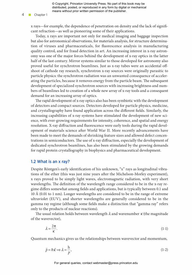
Near the end of the nineteenth century, Röntgen was experimenting with cathode ray tubes— vacuum tubes similar to old- fashioned computer monitors. After plac-ing a metal target in the electron beam, he noticed that phosphor behind a wood screen glowed and nearby photographic plates became exposed even though pro-tected from light. Röntgen realized that these effects must be due to some un-known, “x” radiation, and he quickly began to investigate, placing vari ous objects in the beam (including his wife’s hand— the fa-mous image is shown in Figure 1-1).
This commonly told story of the dis-covery of x rays is a classic tale of serendipity. Imagine it— a laboratory in Würtzburg, Germany, cluttered with all the latest scientific apparatus of 1895: vacuum tubes, photographic plates, jars and sheets of phosphors and metals, and an excited scientist randomly applying high voltages. Suddenly he notices the phosphor on the other side of the room is glowing, and William Conrad Röntgen is on his way to receiving the first Nobel Prize in Physics. Shortly into his investigations, he happens to see the out-line of the bones in his wife’s hand exposed on a photographic plate, and the field of diagnostic imaging is born.
As with most great advances, the real story is a bit more deliberate. Röntgen, along with several of his contemporaries, including Tesla and Hertz, was actively engaged in research on the emissions from cathode ray tubes. The inventor of his tube, Crookes, had previously seen shadows on photographic plates, and may have suggested that Röntgen investigate them. However, Röntgen did quite quickly realize the significance of his observations and rapidly began identifying many of the characteristics of
FIGURE 1-1. An x- ray image of Frau Röntgen’s
hand, from On a new kind of rays, Nature 53
(January 23, 1896): 274–76.
© Copyright, Princeton University Press. No part of this book may be distributed, posted, or reproduced in any form by digital or mechanical means without prior written permission of the publisher.
For general queries, contact [email protected]
■ Chapter 14
x rays— for example, the dependence of penetration on density and the lack of signifi-cant refraction—as well as pioneering some of their applications.
Today, x rays are impor tant not only for medical imaging and baggage inspection but also for astronomical observations, for materials analy sis, for structure determina-tion of viruses and phar ma ceu ti cals, for fluorescence analy sis in manufacturing quality control, and for fraud detection in art. An increasing interest in x- ray astron-omy was one of the major forces behind the development of x- ray optics in the latter half of the last century. Mirror systems similar to those developed for astronomy also proved useful for synchrotron beamlines. Just as x- ray tubes were an accidental off-shoot of cathode ray research, synchrotron x- ray sources were originally parasitic to particle physics: the synchrotron radiation was an unwanted consequence of acceler-ating the particles, because it removes energy from the particle beam. The subsequent development of specialized synchrotron sources with increasing brightness and num-bers of beamlines led to creation of a whole new array of x- ray tools and a consequent demand for an increasing array of optics.
The rapid development of x- ray optics also has been symbiotic with the development of detectors and compact sources. Detectors developed for particle physics, medicine, and crystallography have found application across the diff er ent fields. Similarly, the increasing capabilities of x- ray systems have stimulated the development of new sci-ence, with ever- growing requirements for intensity, coherence, and spatial and energy resolution. X- ray diffraction and fluorescence were early tools during the rapid devel-opment of materials science after World War II. More recently advancements have been made to meet the demands of shrinking feature sizes and allowed defect concen-trations in semiconductors. The use of x- ray diffraction, especially the development of dedicated synchrotron beamlines, has also been stimulated by the growing demands for rapid protein crystallography in biophysics and phar ma ceu ti cal development.
1.2 What is an x ray?
Despite Röntgen’s early identification of his unknown, “x” rays as longitudinal vibra-tions of the ether (this was just nine years after the Michelson- Morley experiment), x rays proved to be simply light waves, electromagnetic radiation, with very short wavelengths. The definition of the wavelength range considered to be in the x- ray re-gime differs somewhat among fields and applications, but is typically between 0.1 and 10 Å (0.01 to 1 nm). Longer wavelengths are considered to be in the range of extreme ultraviolet (EUV), and shorter wavelengths are generally considered to be in the gamma ray regime (although some fields make a distinction that “gamma ray” refers only to the products of nuclear reactions).
The usual relation holds between wavelength λ and wavenumber κ (the magnitude of the wavevector),
λ = 2πκ . (1-1)
Quantum mechanics gives us the relationships between wavevector and momentum,
!p = "
!κ ⇒ λ = hp , (1-2)
© Copyright, Princeton University Press. No part of this book may be distributed, posted, or reproduced in any form by digital or mechanical means without prior written permission of the publisher.
For general queries, contact [email protected]
Introduction ■ 5
where h is Planck’s constant and, as usual,
! = h2π
. (1-3)
Relativity gives us the relationship between momentum and energy U,
U 2 = (Mc2 )2 + p2c2 ⇒ p = U 2 − (Mc2 )2
c=U
c, (1-4)
since, as photons are massless, M = 0. Thus, wavelength and energy are related by
U = hcλ
, (1-5)
where c is the speed of light. Expressing h in units of eV s, and c in units of Å/s gives the useful result that
hc ≈ 12.4 keV Å. (1-6)
Thus, the wavelength range from 10 to 0.1 Å corresponds to 1.2 to 124 keV in photon energy. For comparison, a vis i ble light photon with a wavelength of 0.5 μm corresponds to a photon energy of 2.5 eV, or 2.5 × 10⁻³ keV. Quantum mechanics also gives us the relationship between photon energy and frequencyν,
U = hν , (1-7)
which gives us the expected relationship between wavelength and frequency,
λ = hcU = c
ν . (1-8)
EXAMPLE 1-1
a) What are the wavelength and frequency of the 8 keV x rays frequently used for pro-tein crystallography experiments?
From equation 1-5,
λ = hcU ≈ 12.4 keV iÅ
8 keV ≈1.55 Å.
From equation 1-8,
ν = cλ≈ 3×10¹⁸ Å/s
1.55 Å≈1.9×10¹⁸ Hz ≈1.9 exahertz ≈1.9 EHz.
b) For comparison, what is the wavelength of an electron with a kinetic energy of 8 keV?
The difference between the photon and electron wavelengths arises in applying equation 1-4, because the electron is not massless. The kinetic energy is the
© Copyright, Princeton University Press. No part of this book may be distributed, posted, or reproduced in any form by digital or mechanical means without prior written permission of the publisher.
For general queries, contact [email protected]
■ Chapter 16
difference between the total energy U and the rest mass energy, which for small (nonrelativistic) momentum is
Ke =U − Mec2 = Mec2( )2 + p2c2 − Mec2
= Mec2 1+ p2c2
Mec2( )2 − Mec2 ≈Mec2 1+ 12
p2c2
Mec2( )2
⎛
⎝⎜
⎞
⎠⎟ − Mec2 = p2
2Me
which is the expected, classical result. Then,
λ = hp =
h2Me(Ke )
≈ (6.6×10⁻³⁴ J i s)
2(9.1×10⁻³¹ kg)(8×10³ eV) 1.6×10⁻¹⁹ J1eV
⎛⎝⎜
⎞⎠⎟
≈1.4 ×10⁻¹¹kg m2
s2 s
kg2 m2
s2
≈1.4 ×10⁻¹¹ m ≈ 0.14 Å.
The electron wavelength is more than a factor of 10 smaller than that of the x ray with the same kinetic energy.
1.3 What makes x rays useful?
The wavelength of x rays is in the angstrom range, similar to the spacing of atoms in a crystal. Thus, the arrays of atoms in a crystal can act as a diffraction grating for x rays. The 1914 Nobel Prize in Physics was awarded to Laue for the first demonstration of diffraction of x rays by a crystal. The 1915 prize went to William Henry Bragg and Wil-liam Lawrence Bragg for the development of the theory that allows for association of crystal structure with the diffraction pattern. X- ray crystallography is routinely used today for applications such as verifying the crystal quality of films grown on silicon wafers, detecting stress in airplane engines, and determining the structure of proteins to understand their function in cancer growth. Diffraction is also used as a way to con-
trol the direction or wavelength of x rays used in a par tic u lar experiment, just as gratings are used for vis i ble light. The 1936 Nobel Prize in Chem-istry was awarded to Peter Debye for, among other things, development of the theory of dif-fraction from powders and liquids.
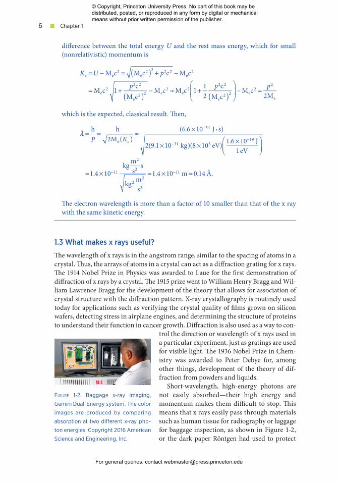
Short- wavelength, high- energy photons are not easily absorbed— their high energy and momentum makes them difficult to stop. This means that x rays easily pass through materials such as human tissue for radiography or luggage for baggage inspection, as shown in Figure 1-2, or the dark paper Röntgen had used to protect
FIGURE 1-2. Baggage x- ray imaging,
Gemini Dual- Energy system. The color
images are produced by comparing
absorption at two dif er ent x- ray pho-
ton energies. Copyright 2016 American
Science and Engineering, Inc.
© Copyright, Princeton University Press. No part of this book may be distributed, posted, or reproduced in any form by digital or mechanical means without prior written permission of the publisher.
For general queries, contact [email protected]
Introduction ■ 7
his photographic plate. Absorption increases with the electron density of the mate-rial but is lower for higher- energy photons.
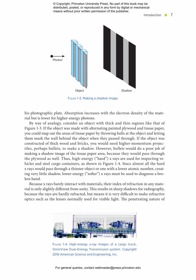
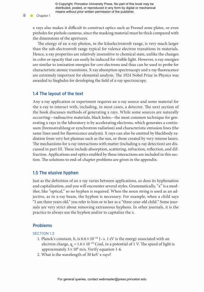
By way of analogy, consider an object with thick and thin regions like that of Figure 1-3. If the object was made with alternating painted plywood and tissue paper, you could map out the areas of tissue paper by throwing balls at the object and letting them mark the wall behind the object when they passed through. If the object was constructed of thick wood and bricks, you would need higher- momentum projec-tiles, perhaps bullets, to make a shadow. However, bullets would do a poor job of making a shadow image of the tissue paper area, because they would pass through the plywood as well. Thus, high- energy (“hard”) x rays are used for inspecting ve-hicles and steel cargo containers, as shown in Figure 1-4. Since almost all the hard x rays would pass through a thinner object or one with a lower atomic number, creat-ing very little shadow, lower- energy (“softer”) x rays must be used to diagnose a bro-ken hand.
Because x rays barely interact with materials, their index of refraction in any mate-rial is only slightly diff er ent from unity. This results in sharp shadows for radiography, because the rays are hardly refracted, but means it is very difficult to make refractive optics such as the lenses normally used for vis i ble light. The penetrating nature of
FIGURE 1-3. Making a shadow image.
Shadow
Photon
Object
FIGURE 1-4. High- energy x- ray images of a cargo truck,
OmniView Dual- Energy Transmission system. Copyright
2016 American Science and Engineering, Inc.
© Copyright, Princeton University Press. No part of this book may be distributed, posted, or reproduced in any form by digital or mechanical means without prior written permission of the publisher.
For general queries, contact [email protected]
■ Chapter 18
x rays also makes it difficult to construct optics such as Fresnel zone plates, or even pinholes for pinhole cameras, since the masking material must be thick compared with the dimensions of the apertures.
The energy of an x- ray photon, in the kiloelectronvolt range, is very much larger than the sub- electronvolt range typical for valence electron transitions in materials. Hence, x- ray properties are relatively insensitive to chemical state, unlike the changes in color or opacity that can easily be induced for vis i ble light. However, x- ray energies are similar to ionization energies for core electrons and thus can be used to probe for characteristic atomic transitions. X- ray absorption spectroscopy and x- ray fluorescence are extremely impor tant for elemental analy sis. The 1924 Nobel Prize in Physics was awarded to Siegbahn for developing the field of x- ray spectroscopy.
1.4 The layout of the text
Any x- ray application or experiment requires an x- ray source and some material for the x- ray to interact with, including, in most cases, a detector. The next section of the book discusses methods of generating x rays. While some sources are naturally occurring— radioactive materials, black holes— the most common technique for gen-erating x rays in the laboratory is by accelerating electrons, which generates a contin-uum (bremsstrahlung or synchrotron radiation) and characteristic emission lines (the same lines used for fluorescence analy sis). X rays can also be emitted by blackbody ra-diation from very hot plasmas such as the sun, or those created by very intense lasers. The mechanisms for x- ray interactions with matter (including x- ray detection) are dis-cussed in part III. These include absorption, scattering, refraction, reflection, and dif-fraction. Applications and optics enabled by these interactions are included in this sec-tion. The solutions to end- of- chapter prob lems are given in the appendix.
1.5 The elusive hyphen
Just as the definition of an x ray varies between applications, so does its hyphenation and capitalization, and you will encounter several styles. Grammatically, “x” is a mod-ifier, like “optical,” so no hyphen is required. When the noun string is used as an ad-jective, as in x- ray beam, the hyphen is necessary. For example, when a child says “I am three years old,” you refer to him or to her as a “three- year- old child.” Some jour-nals are very strict about removing extraneous hyphens. In other journals, it is the practice to always use the hyphen and/or to capitalize the x.
Prob lems
SECTION 1.21. Planck’s constant, h, is 6.6 × 10⁻³⁴ J s. 1 eV is the energy associated with an
electron charge, qe ≈ 1.6 × 10⁻¹⁹ Coul, in a potential of 1 V. The speed of light is approximately 3 × 10⁸ m/s. Verify equation 1-6.
2. What is the wavelength of 30 keV x rays?
© Copyright, Princeton University Press. No part of this book may be distributed, posted, or reproduced in any form by digital or mechanical means without prior written permission of the publisher.
For general queries, contact [email protected]
Introduction ■ 9
Further reading
General references for x- ray topicsJens Als- Nielsen and Des McMorrow, Ele ments of Modern X- ray Physics, John Wiley &
Sons, 2001.Eric Lifshin, X- ray Characterization of Materials, John Wiley & Sons, 1999.A. G. Michette and C. J. Buckley, X- ray Science and Technology, Institute of Physics
Publishing, 1993.Alan Michette and Sławka Pfauntsch, X- Rays: The First Hundred Years, John Wiley & Sons,
1996.E. Spiller, Soft X- ray Optics, SPIE Press, 1994.David Attwood and A. Sakdinawat, X- Rays and Extreme Ultraviolet Radiation: Princi ples
and Applications, Cambridge University Press, 2016.
RelativityD. Halliday, R. Res nick, and J. Walker, Fundamentals of Physics, 10th ed., John Wiley &
Sons, 2013, chapter 37.
Historical referencesArthur Stanton, Wilhelm Conrad Röntgen on a new kind of rays: Translation of a paper read
before the Würtzburg Physical and Medical Society, Nature 53 (1895): 274–76.New York Times, February 16, 1896, Nature of the X Rays.
© Copyright, Princeton University Press. No part of this book may be distributed, posted, or reproduced in any form by digital or mechanical means without prior written permission of the publisher.
For general queries, contact [email protected]


![Röntgen ’s experiment Röntgen ’s experiment · 2010-12-20 · Moseley (1887-1915) Atomic binding energies …simple laws have been found which […] make it possible to predict](https://img.pdfslide.us/doc/110x75/5e56c16f2b0e2a2b7e61d6f0/rntgen-as-experiment-rntgen-as-2010-12-20-moseley-1887-1915-atomic-binding.jpg)