Embed Size (px)
Citation preview
An Introduction To The Health An Introduction To The Health Effects of Radiation and Effects of Radiation and Radioactive MaterialsRadioactive Materials
Javier C. Waksman, MDAssociate Professor of Medicine
Director- Medical Toxicology PracticeUniversity of Colorado Hospital
• 1895 - Wilhem Conrad Roentgen discovered X-rays and in 1901 he received the first Nobel Prize for physics.
• 1903 - Marie Curie and Pierre Curie, along with Henri Becquerel were awarded the Nobel Prize in physics for their contributions to understanding radioactivity, including the properties of uranium.
• 1942 - Enrico Fermi and others started the first sustained nuclear chain reaction in a laboratory beneath the University of Chicago football stadium.
• 1945 – Nuclear bombs dropped on Japan.
Historical AwarenessHistorical Awareness
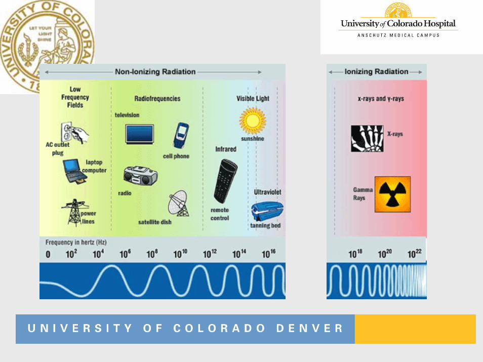
RadiationRadiation
Non-ionizing
Ionizing
NonNon--ionizing Radiationionizing Radiation
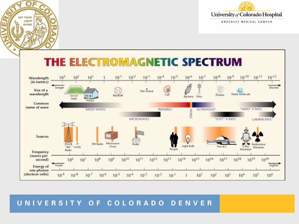
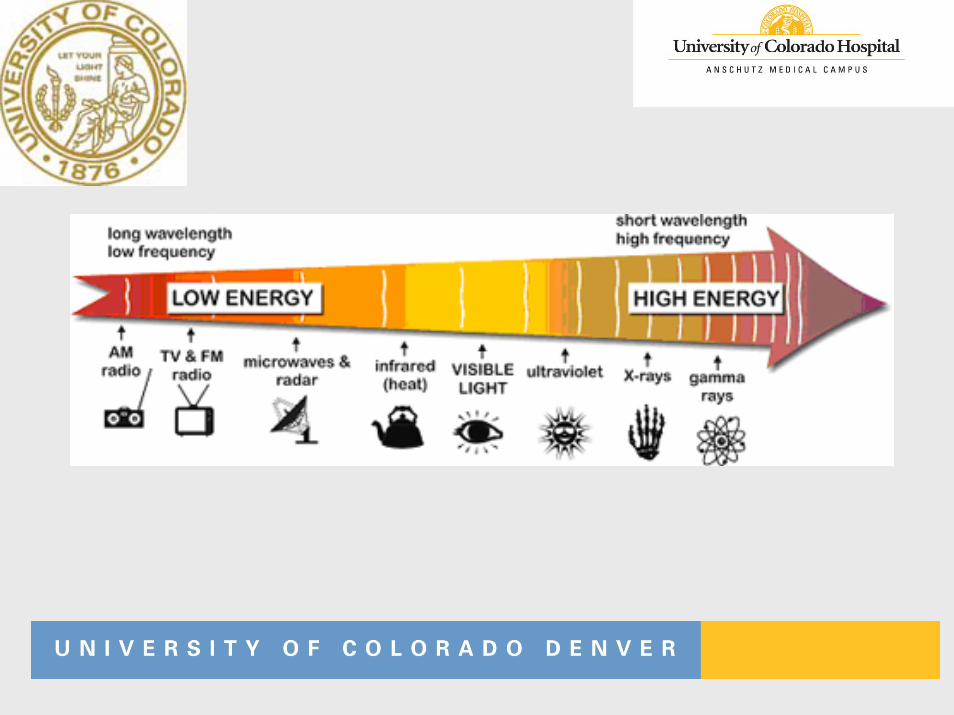
• Sources• Ultraviolet light• Visible light• Infrared radiation• Microwaves• Radio & TV• Power transmission
NonNon--ionizing Examplesionizing Examples• Ultraviolet – Black light – induce fluorescence in
some materials• Vision – very small portion that animals use to
process visual information • Heat – infrared – a little beyond the red spectrum• Radio waves – beyond infrared• Micro waves• Electrical power transmission – 60 cycles per second
with a wave length of 1 to 2 million meters.
Ultraviolet Ultraviolet -- SourcesSources
• Sun light• Most harmful UV is absorbed by the
atmosphere – depends on altitude• Fluorescent lamps• Electric arc welding
Can damage the eye (cornea)• Germicidal lamps• Eye damage from sun light• Skin cancer
Ultraviolet Ultraviolet -- EffectsEffects
• High ultraviolet – kills bacterial and other infectious agents
• High dose causes - sun burn – increased risk of skin cancer
• Pigmentation that results in suntan • Suntan lotions contain chemicals that absorb UV
radiation• Reaction in the skin to produce Vitamin D that
prevents rickets• Strongly absorbed by air – thus the danger of hole
in the atmosphere
Visible EnergyVisible Energy
• Energy between 400 and 750 nm• Standards are set for the intensity of light in the
work place (measured in candles or lumens)
Infrared RadiationInfrared Radiation
• Energy between 750 nm to 0.3 cm• The energy of heat – Heat is the transfer of
energy• Can damage – cornea, iris, retina and lens of
the eye (glass workers – “glass blower’s cataract”)
Microwaves & Radio WavesMicrowaves & Radio Waves
• Energy between 0.1 cm to 1 kilometer• Varity of industrial and home uses for heating
and information transfer (radio, TV, mobile phones)
• Produced by molecular vibration in solid bodies or crystals
• Health effects – heating, cataracts• Long-term effects being studied
Electrical PowerElectrical Power
• Standard in homes and businesses• Highest level of exposure from electric-power
generation and distribution system (high voltage power lines)
• Medical system – Magnetic imaging• Acute health effects – shock• Long-term health effects?
Ionizing RadiationIonizing Radiation
Ionizing RadiationIonizing Radiation
Ionization DefinedRadiation capable for producing ions when interacting with matter – in other words enough energy to remove an electron from an atom.
Sources – x-rays, radioactive material produce alpha, beta, and gamma radiation, cosmic rays from the sun and space.
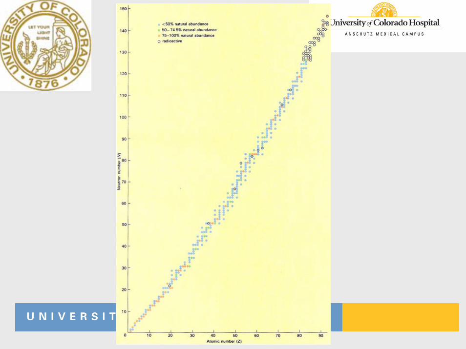
• Atoms “prefer” a particular neutron/proton ratio in the nucleus
• If “too many” neutrons or protons–Nuclei will decay–Give off ionizing radiation (particles)–Stable ratio
• In the process unstable stable–Nuclei give off particles (atom
decay)–The process liberates particles or
radiation
• Alpha α• Beta β• Gamma γ• X-ray X • Neutron η
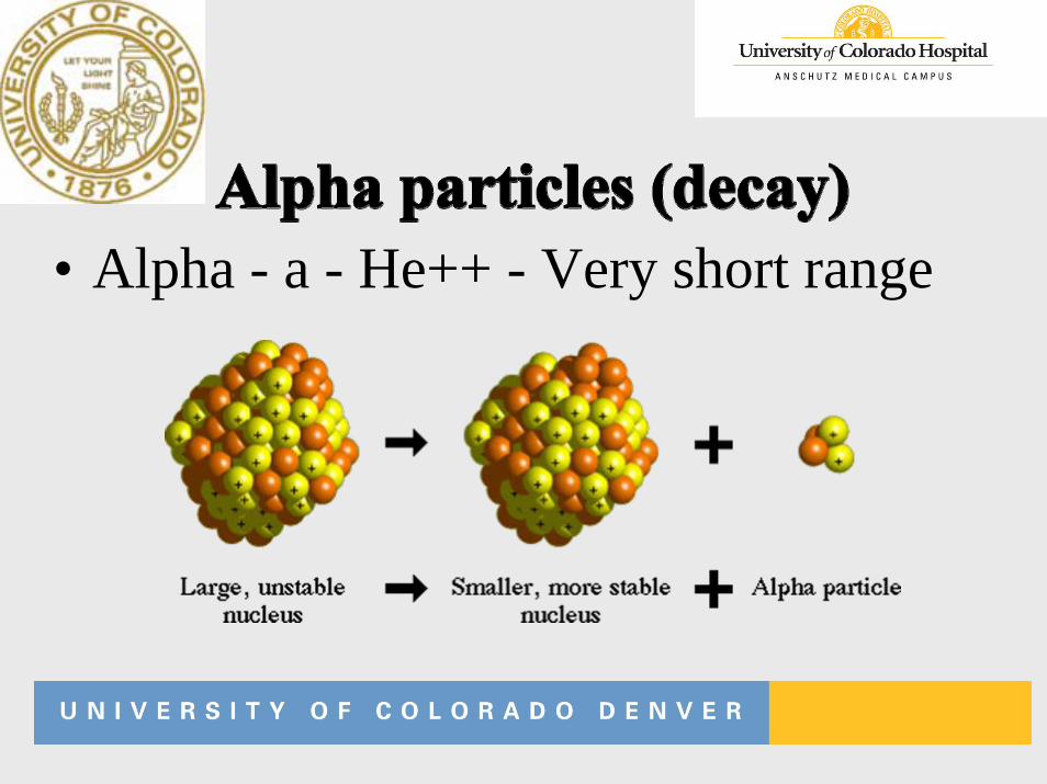
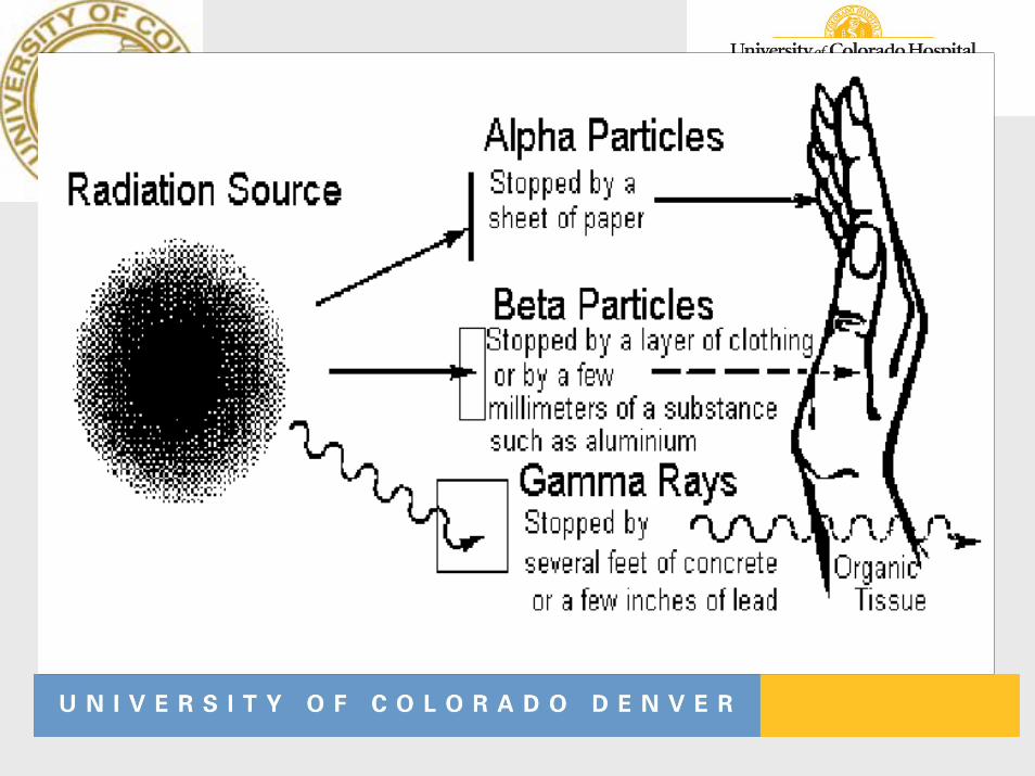
• Alpha - a - He++ - Very short range
• Primary hazard from internal exposure
• Alpha emitters can accumulate in tissue (bone, kidney, liver, lung, spleen) causing local damage
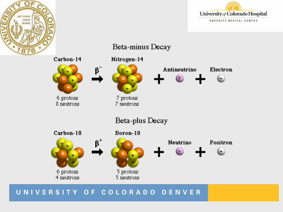
• Beta - ß- or e- - Medium Range • Positron - ß+ - Medium Range
• Can cause skin burns or be an internal hazard of ingested
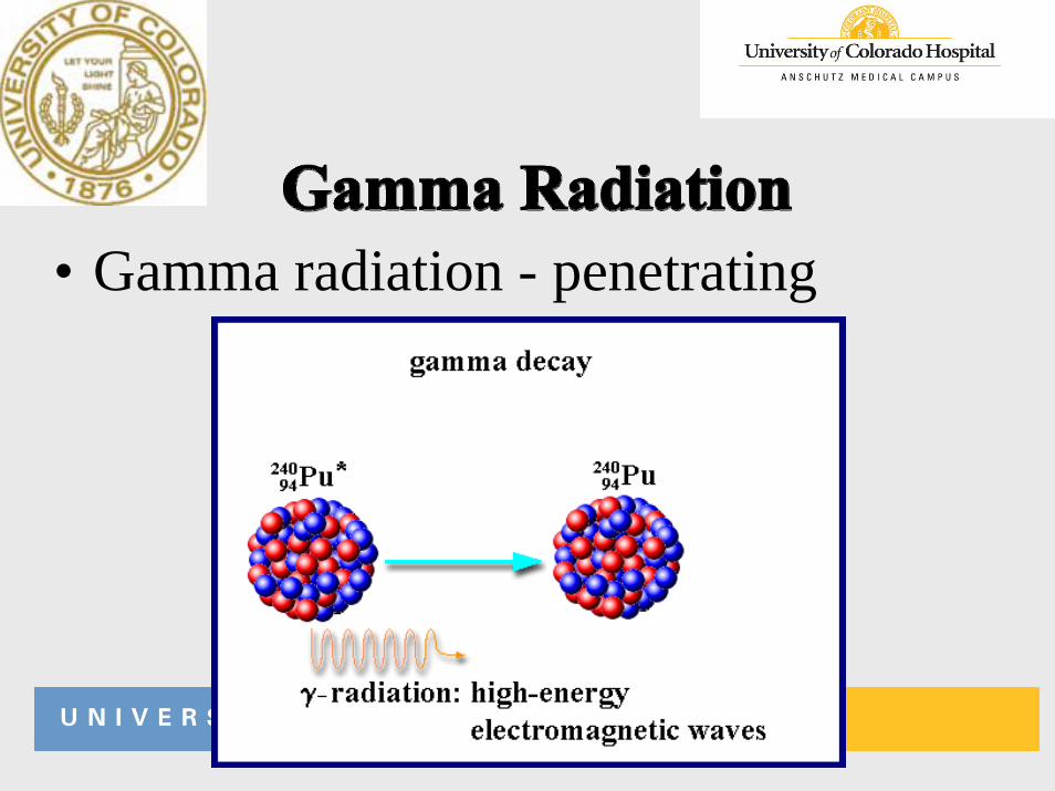
• Gamma radiation - penetrating
• Emitted from nucleus of radioactive atoms – spontaneous emission
• Emitted with kinetic energy related to radioactive source
• Highly penetrating – extensive shielding required
• Serious external radiation hazard
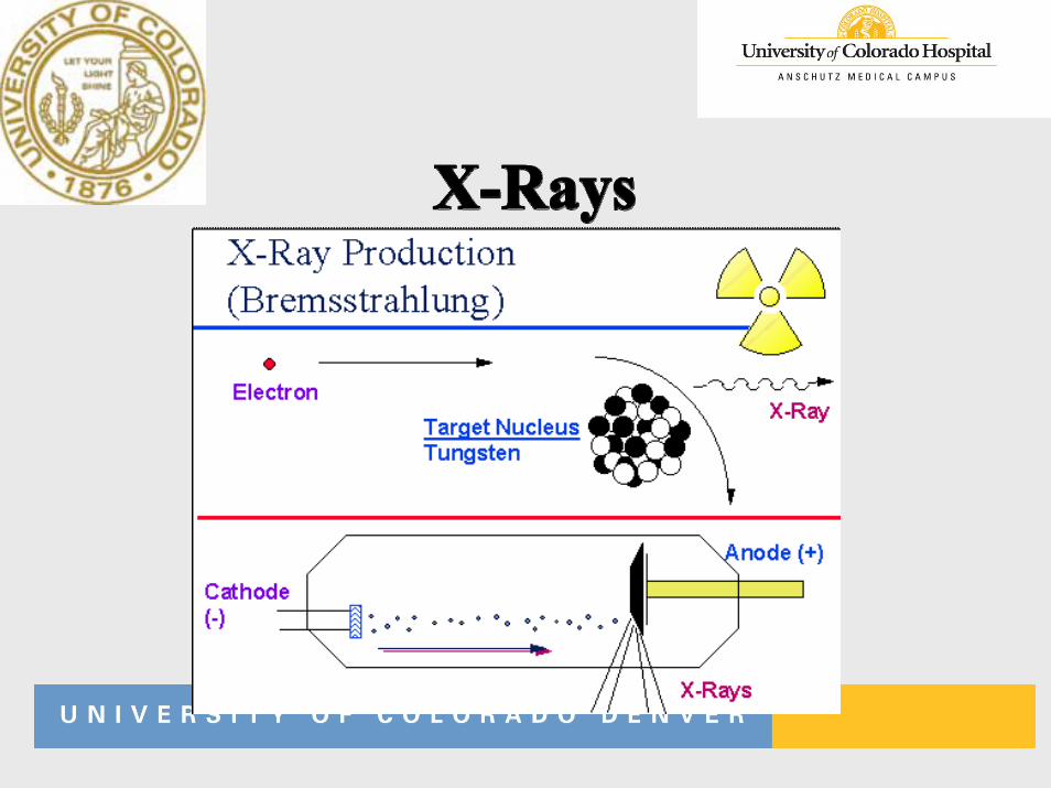
• Produced from orbiting electrons or free electrons – usually machine produced
• Produced when electrons strike a target material inside and x-ray tube
• Emitted with various energies & wavelengths• Highly penetrating – extensive shielding required• External radiation hazard• Discovered in 1895 by Roentgen
• Uncharged• Able to penetrate deeply• Hazard inside nuclear reactors
Radiation or Radioactive material?
• Radiation is energy or particles that are given off when something decays to reach a stable proton to neutron ratio.
• The radioactive material contains the unstable nuclei which are decaying.
• These often get confused.
• In order to induce radioactivity it is necessary to change the proton to neutron ratio in a nucleus
• Radiation from a radioactive material does not cause its surroundings to become radioactive
• If the radiation from a radioactive material is placed on a table or next to a person, this radiation does not cause the table or person to become radioactive.
• Only if a material is exposed to neutrons from a nuclear reactor or a high energy charged particle accelerator can a non-radioactive material become radioactive.
• Either natural or created in nuclear reactor or accelerator
• Radioactive material is unstable and emits energy in order to return to a more stable state (particles or gamma-rays)
• Half-life – time for radioactive material to decay by one-half
• Absorption of radiation energy per unit mass of absorber–Conventional unit – rad International
unit – Gray (Gy)1 Gy = 1 Joule/Kg
–1 Gy = 100 rad–1 rad = .01 Gy
• Biological damage & resulting risk from radiation dose• Dose equivalent = Dose x Quality Factor
– Quality factor: Any type of radiation compared to same absorbed dose γ or X
– QF X, γ, β = 1 – QF α = 20 (if internal) – QF η = 3-20 (depends on energy)
• Conventional unit – “rem”• SI unit – Sievert (Sv) • 1 Sv = 100 rem
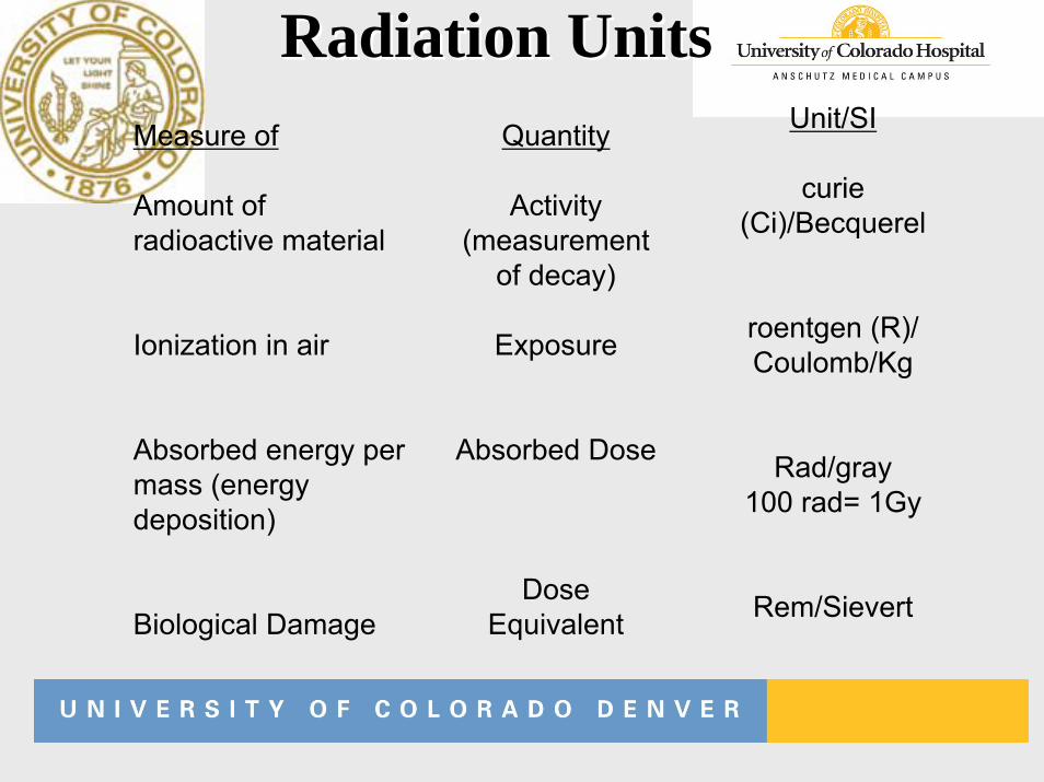
Radiation UnitsRadiation UnitsMeasure of
Amount ofradioactive material
Ionization in air
Absorbed energy per mass (energy deposition)
Biological Damage
Quantity
Activity(measurement
of decay)
Exposure
Absorbed Dose
Dose Equivalent
Unit/SI
curie (Ci)/Becquerel
roentgen (R)/ Coulomb/Kg
Rad/gray100 rad= 1Gy
Rem/Sievert
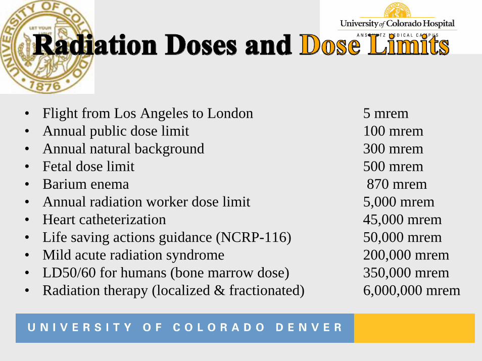
• Flight from Los Angeles to London 5 mrem• Annual public dose limit 100 mrem• Annual natural background 300 mrem• Fetal dose limit 500 mrem• Barium enema 870 mrem• Annual radiation worker dose limit 5,000 mrem• Heart catheterization 45,000 mrem• Life saving actions guidance (NCRP-116) 50,000 mrem• Mild acute radiation syndrome 200,000 mrem• LD50/60 for humans (bone marrow dose) 350,000 mrem• Radiation therapy (localized & fractionated) 6,000,000 mrem
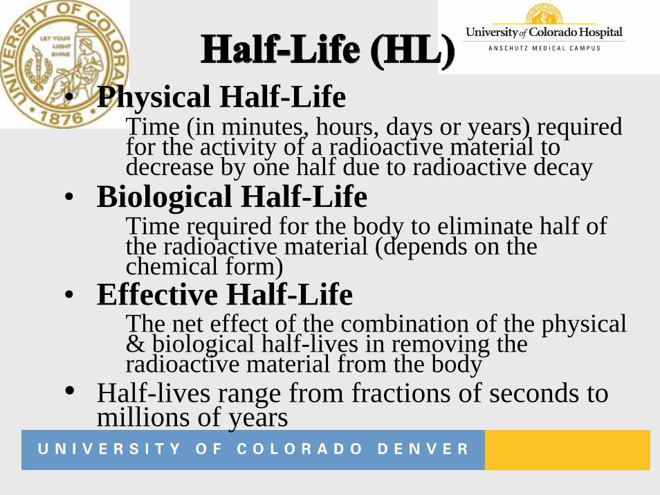
• Physical Half-LifeTime (in minutes, hours, days or years) required for the activity of a radioactive material to decrease by one half due to radioactive decay
• Biological Half-LifeTime required for the body to eliminate half of the radioactive material (depends on the chemical form)
• Effective Half-LifeThe net effect of the combination of the physical & biological half-lives in removing the radioactive material from the body
• Half-lives range from fractions of seconds to millions of years
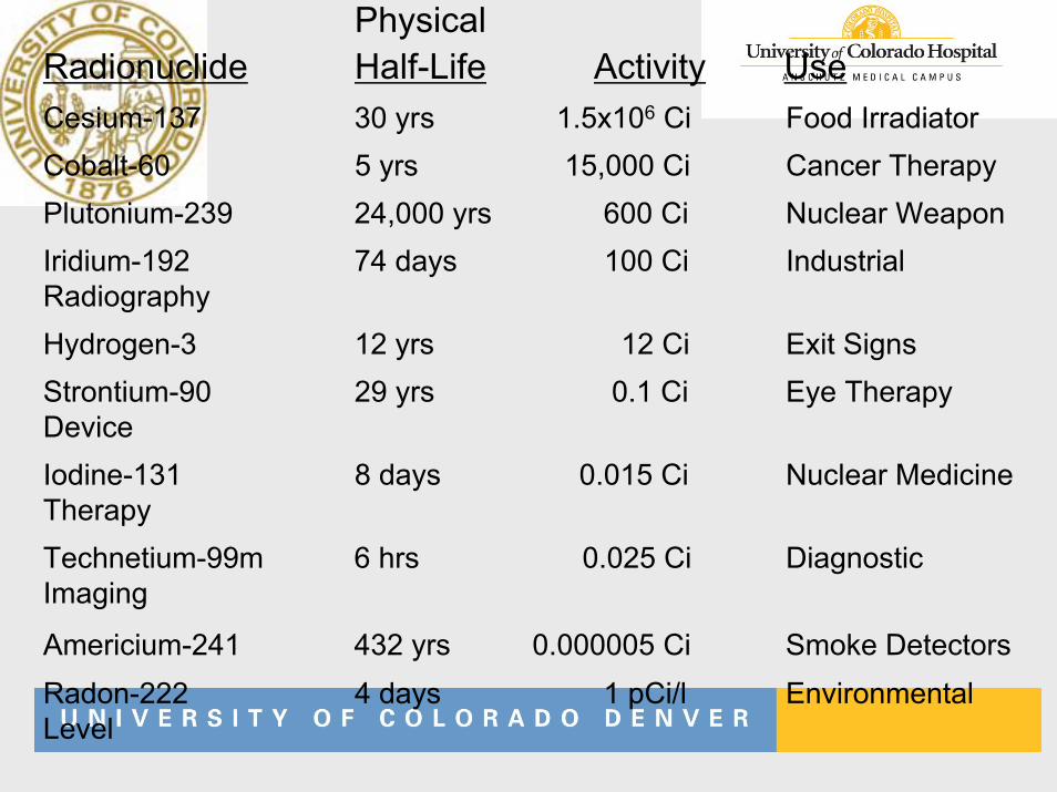
Physical Radionuclide Half-Life Activity UseCesium-137 30 yrs 1.5x10⁶ Ci Food IrradiatorCobalt-60 5 yrs 15,000 Ci Cancer TherapyPlutonium-239 24,000 yrs 600 Ci Nuclear WeaponIridium-192 74 days 100 Ci Industrial RadiographyHydrogen-3 12 yrs 12 Ci Exit SignsStrontium-90 29 yrs 0.1 Ci Eye Therapy DeviceIodine-131 8 days 0.015 Ci Nuclear Medicine TherapyTechnetium-99m 6 hrs 0.025 Ci Diagnostic Imaging
Americium-241 432 yrs 0.000005 Ci Smoke DetectorsRadon-222 4 days 1 pCi/l Environmental Level
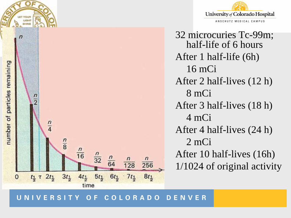
32 microcuries Tc-99m; half-life of 6 hours
After 1 half-life (6h)16 mCi
After 2 half-lives (12 h)8 mCi
After 3 half-lives (18 h)4 mCi
After 4 half-lives (24 h) 2 mCi
After 10 half-lives (16h) 1/1024 of original activity
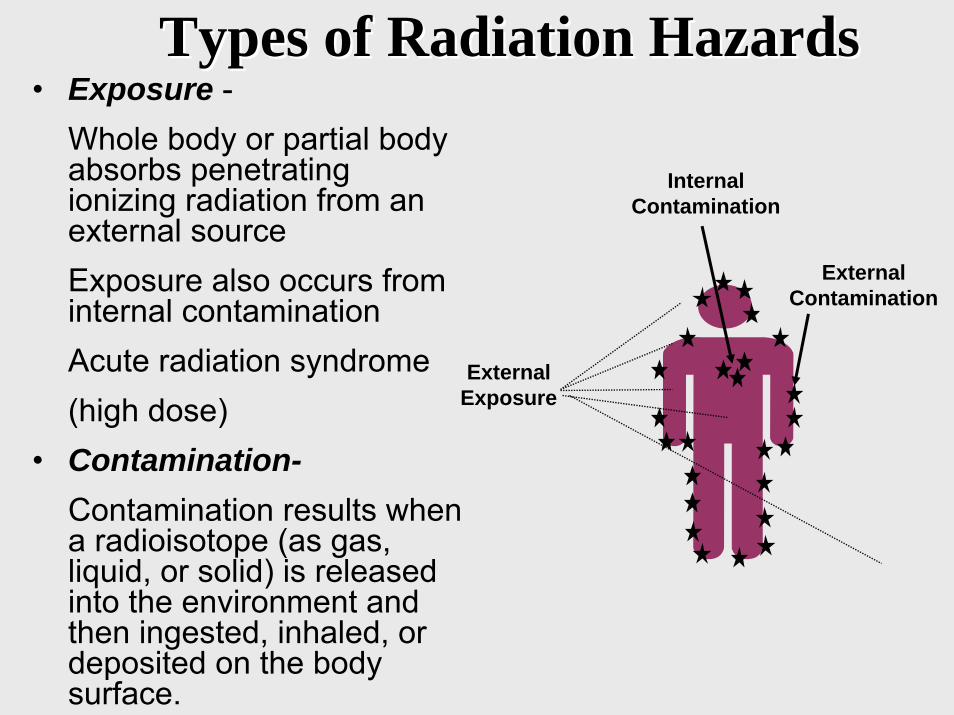
Types of Radiation HazardsTypes of Radiation Hazards• Exposure -
Whole body or partial body absorbs penetrating ionizing radiation from an external sourceExposure also occurs from internal contaminationAcute radiation syndrome(high dose)
• Contamination-Contamination results when a radioisotope (as gas, liquid, or solid) is released into the environment and then ingested, inhaled, or deposited on the body surface.
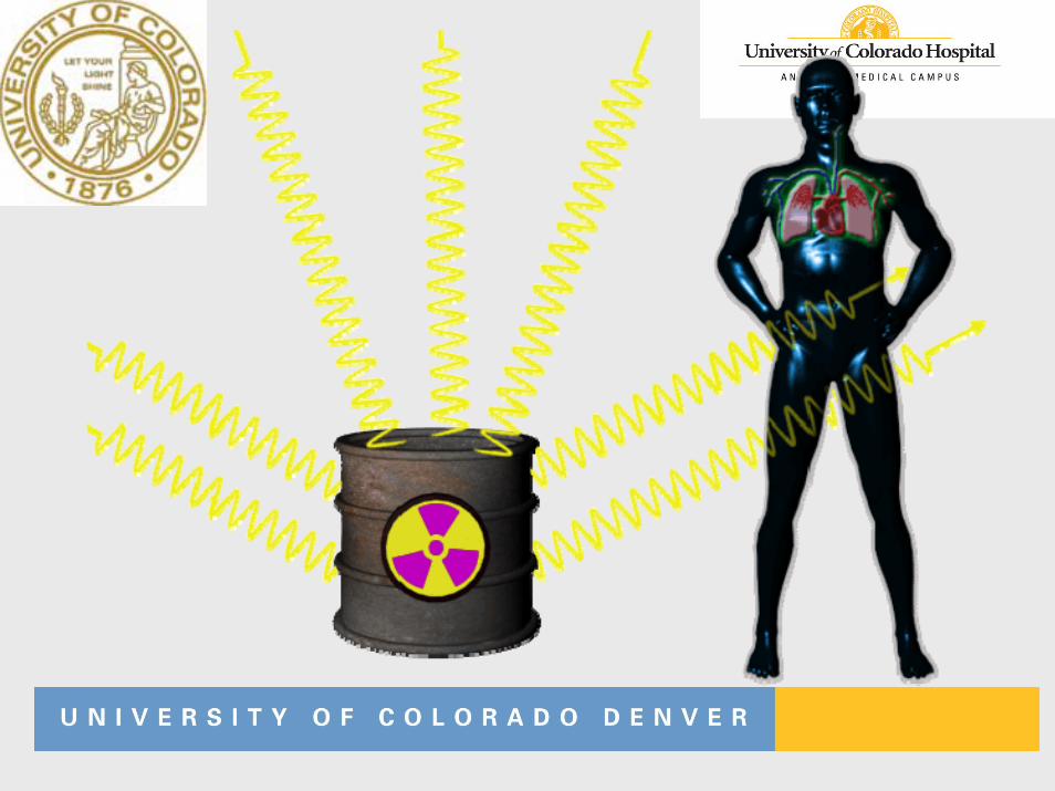
ExternalExposure
InternalContamination
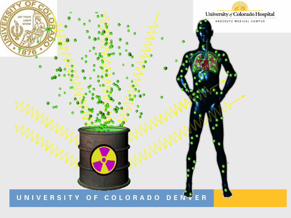
ExternalContamination
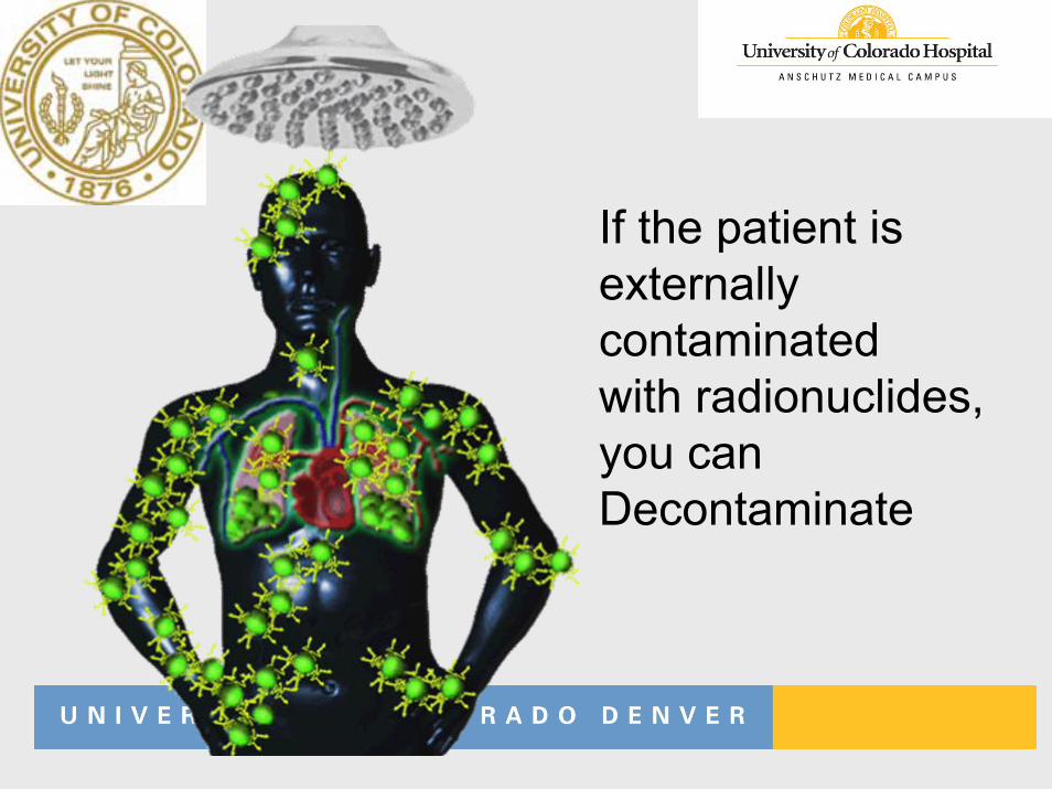
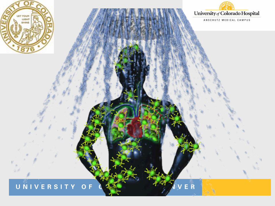
If the patient isexternally contaminatedwith radionuclides,you canDecontaminate
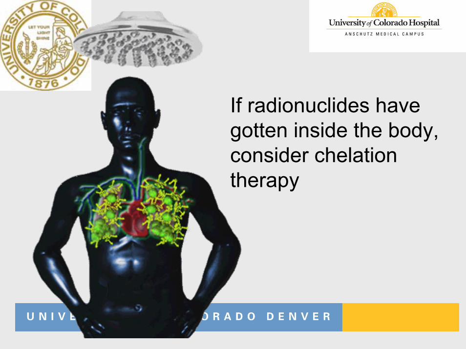
If radionuclides havegotten inside the body, consider chelationtherapy
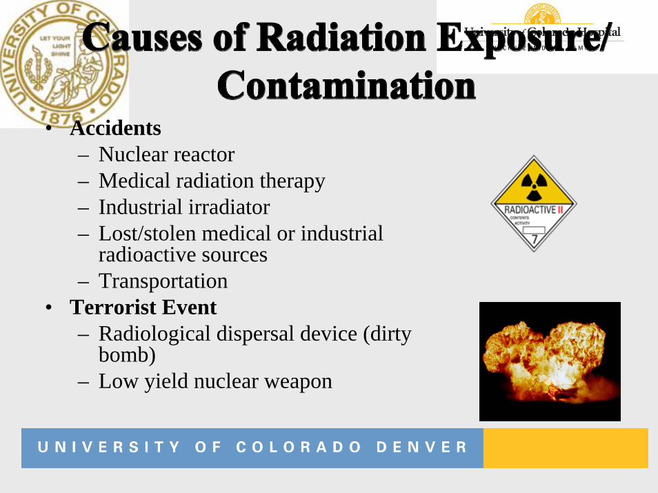
• Accidents– Nuclear reactor– Medical radiation therapy– Industrial irradiator– Lost/stolen medical or industrial
radioactive sources– Transportation
• Terrorist Event– Radiological dispersal device (dirty
bomb)– Low yield nuclear weapon
• Effects determined by: – Total dose– Dose rate – Volume of tissue irradiated – Type of radiation – Anatomical part irradiated – Individual susceptibility – Trauma / Illness
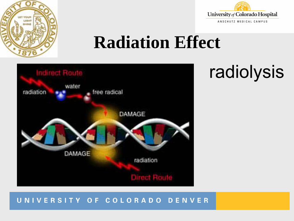
Radiation EffectRadiation Effect
radiolysis
Factors Determining Factors Determining Radiation ExposureRadiation Exposure
• Time
• Distance
• Shielding
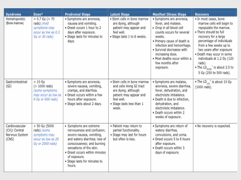
• Acute Radiation Syndrome (ARS)• Localized radiation injuries/cutaneous radiation
syndrome• Internal or external contamination• Combined radiation injuries with
- Trauma- Burns
• Fetal effects
• The required conditions for Acute Radiation Syndrome (ARS) are:– The radiation dose must be large (>0.7 Gy)– The dose usually must be external – The radiation must be penetrating– The entire body (able to reach the internal organs)– The dose must have been delivered in a short time
(usually a matter of minutes).
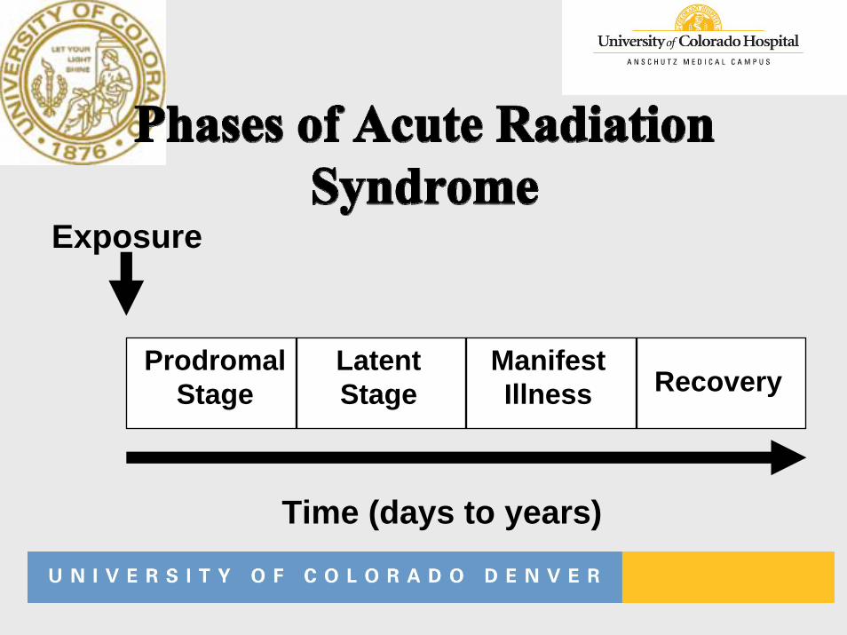
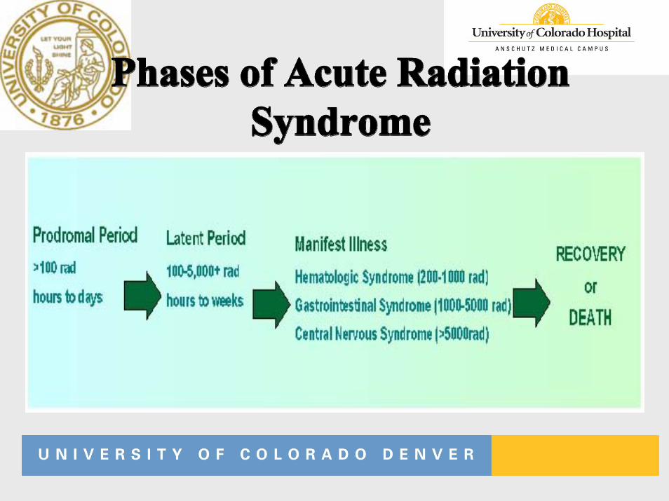
ProdromalStage
LatentStage
ManifestIllness Recovery
Time (days to years)
Exposure
• Syndrome that results from acute radiation exposure to skin
• ARS usually comes with some skin damage
• CRS without ARS (e.g X-rays, beta radiation)
• Inflammation• Erythema• Desquamation• Epilation• Blistering and ulceration
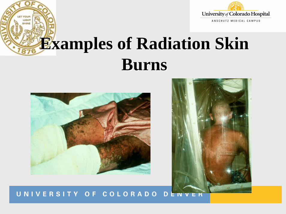
Examples of Radiation Skin Examples of Radiation Skin BurnsBurns
• Triage– ABC– Treat major trauma, burns, respiratory
injury– Blood samples: CBC– Treat contamination as needed– Assess lymphocyte depletion
• Diagnosis–History–Clinical presentation (prodromal
syndrome)–Dose reconstruction–Laboratory data
• Measure every 4 - 6 hours initial 48 hours
• Normal: approx 2500 cells/ml• > 1200: probably non-lethal• 300 to 1200 cells/ml: significant
(hospitalize)• < 300 cells/ml: critical
• Lymphocyte depletion kinetics• Chromosome analysis (dicentrics)
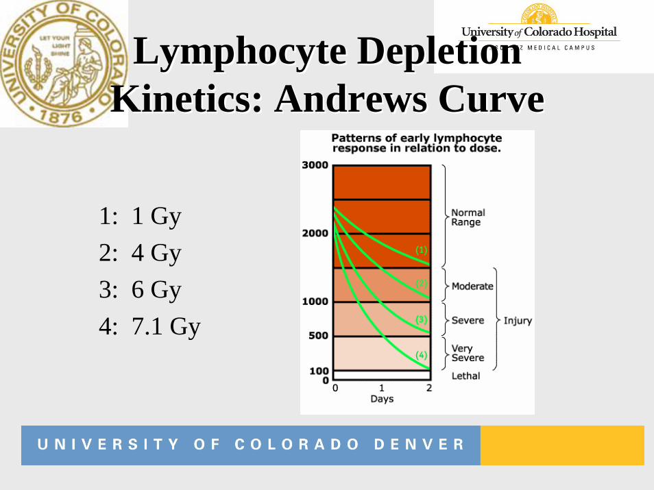
Lymphocyte Depletion Lymphocyte Depletion Kinetics: Andrews CurveKinetics: Andrews Curve
1: 1 Gy2: 4 Gy3: 6 Gy4: 7.1 Gy
• Chromosome with 2 centromeres• Dicentrics are considered the most
sensitive and most specific for assessing radiation dose.
• Treatment– Supportive care– Growth factors (for WBC, platelets, RBC)– Chelators/ antidotes– Stem cell transplants– Psychological support
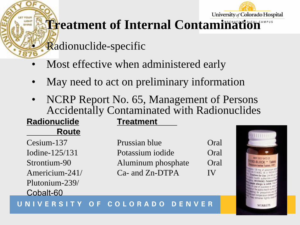
• Radionuclide-specific• Most effective when administered early• May need to act on preliminary information• NCRP Report No. 65, Management of Persons
Accidentally Contaminated with Radionuclides
Treatment of Internal ContaminationTreatment of Internal Contamination
Radionuclide TreatmentRoute
Cesium-137 Prussian blue OralIodine-125/131 Potassium iodide OralStrontium-90 Aluminum phosphate OralAmericium-241/ Ca- and Zn-DTPA IVPlutonium-239/Cobalt-60
• Ionizing radiation in high doses is a proven carcinogen (atomic bomb survivors, Chernobyl)
• Unclear whether it is a carcinogen at low doses (< 20 rem)
• Natural incidence of cancer ~ 40%; mortality ~ 25%• Risk of fatal cancer is estimated as ~ 4% per 100 rem• A dose of 5 rem increases the risk of fatal cancer
by ~ 0.2%• A dose of 25 rem increases the risk of fatal cancer
by ~ 1%
• Magnitude of hereditary risk per rem is 10% that of fatal cancer risk
• Risk to caregivers who would likely receive low doses is very small - 5 rem increases the risk of severe hereditary effects by ~ 0.02%
• Risk of severe hereditary effects to a patient population receiving high doses is estimated as ~ 0.4% per 100 rem
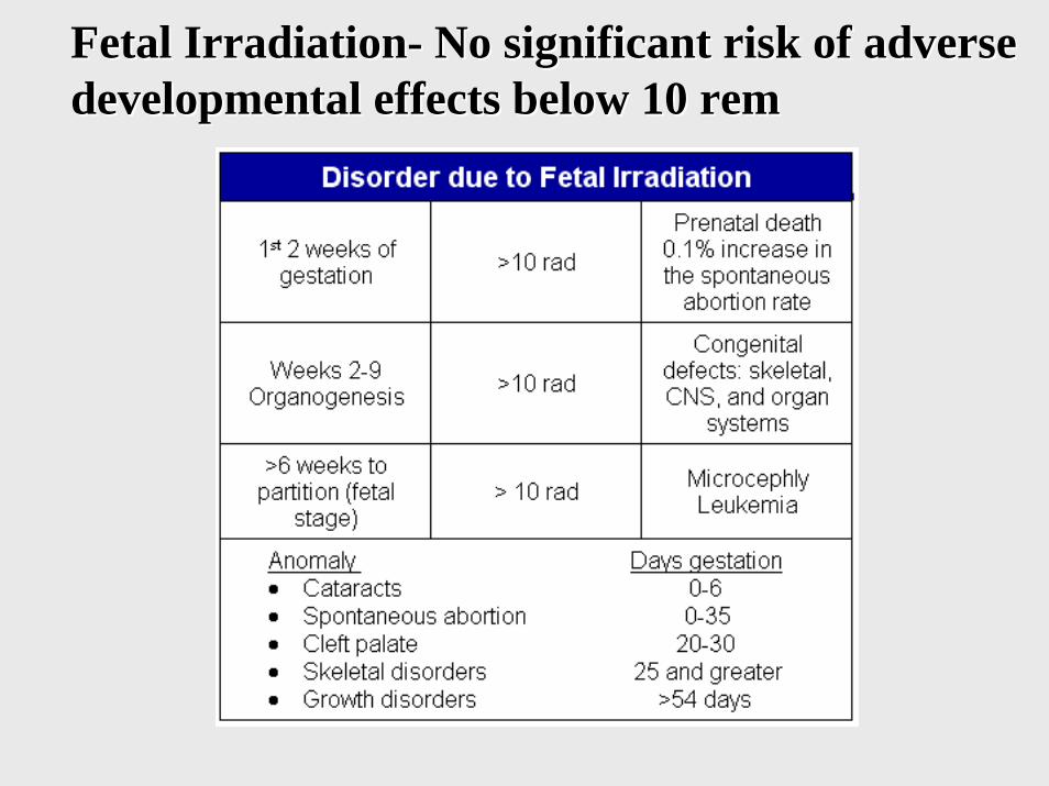
Fetal IrradiationFetal Irradiation-- No significant risk of adverse No significant risk of adverse developmental effects below 10 developmental effects below 10 remrem
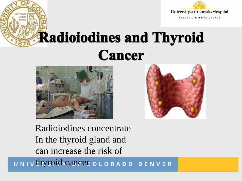
Radioiodines concentrateIn the thyroid gland and can increase the risk ofthyroid cancer
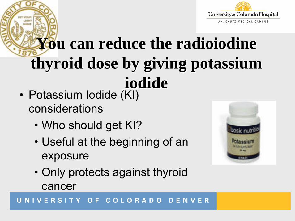
You can reduce the radioiodine You can reduce the radioiodine thyroid dose by giving potassium thyroid dose by giving potassium
iodideiodide• Potassium Iodide (KI)
considerations• Who should get KI?• Useful at the beginning of an
exposure• Only protects against thyroid
cancer
• Ionizing radiation includes:–Electromagnetic radiation: X and
gamma–Particulate radiation: alpha, beta,
neutrons
• Patient can be:– Irradiated externally– Contaminated with radionuclides
• Which patients are radioactive?– Those contaminated with radionuclides– These patients need to be decontaminated– Some internally deposited radionuclides
can be removed with chelation therapy
• Protect yourself from radiation:–Reduce the time of exposure–Increase the distance from the
radiation source–Apply shielding between
yourself and the radiation source
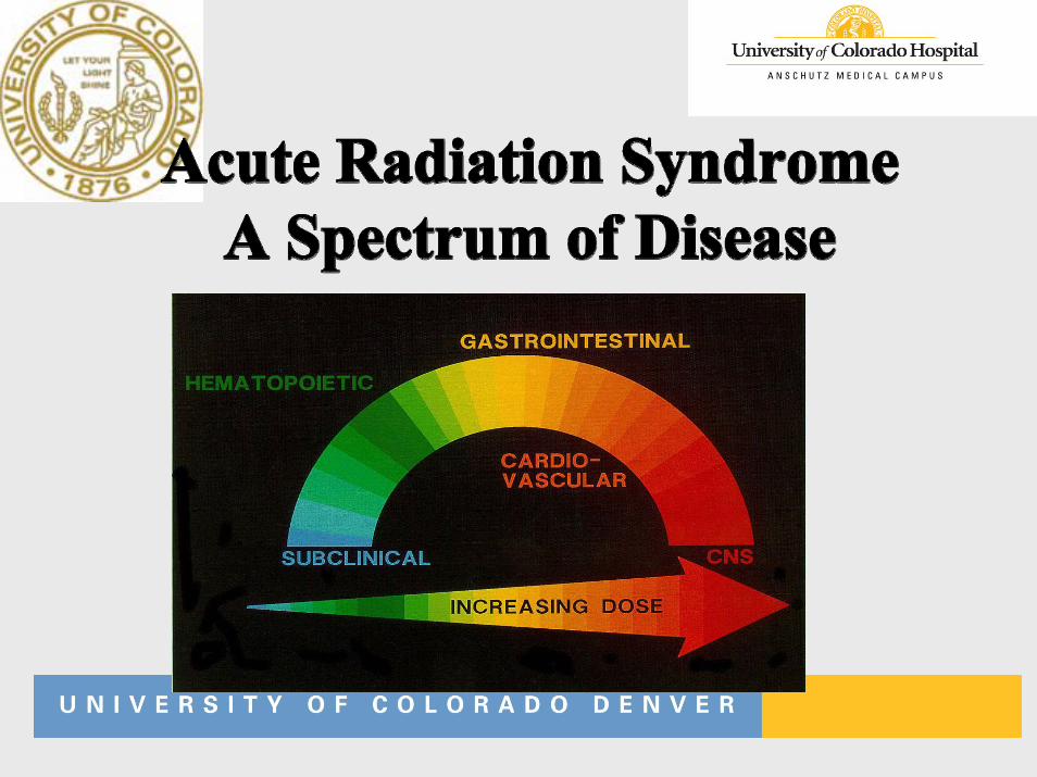
• Acute Radiation Syndrome:– Stages progress from hematopoetic to gastrointestinal to
central nervous system with increasing dose– The absolute lymphocyte count is the best predictor of dose
• Long-term consequences– Increase in cancer, especially thyroid cancer– With radioiodine exposure, thyroid dose can be reduced by
using KI

Thank you!