Embed Size (px)
Citation preview
Serology: Enzyme-linked immunosorbent assay (ELISA)
1 | P a g e
Serology: Enzyme-linked immunosorbent assay (ELISA)
Authors: Dr. RW Worthington (Retired)
Licensed under a Creative Commons Attribution license.
TABLE OF CONTENTS
Introduction ................................................................................................................................................. 2
Indirect ELISA .............................................................................................................................................. 2
Variations of the indirect ELISA ............................................................................................................... 4
ELISA antibody response curves ............................................................................................................. 7
Competitive and blocking ELISAs ........................................................................................................... 17
Variations of competitive ELISAs ........................................................................................................... 19
A competitive ELISA .............................................................................................................................. 20
Antigen capture ELISAs ........................................................................................................................... 22
Serology: Enzyme-linked immunosorbent assay (ELISA)
2 | P a g e
INTRODUCTION
Enzyme-linked immunosorbent assays (ELISAs) are the most commonly used serological diagnostic
tests. The OIE recommends ELISA as the prescribed or alternative test for international trade for most
diseases. ELISAs are also available for a wide variety of other diseases and analytical assays. The
three most commonly used variations of the ELISA test are the indirect ELISA; competitive and
blocking ELISAs and antigen capture ELISAs.
INDIRECT ELISA
In the indirect ELISA antigen is immobilised by coating it onto an ELISA microtitre plate. Serum
suspected of containing antibody is then added and antibody, if present, attaches to the immobilised
antigen and a second antibody (Ab2) that has been conjugated to an enzyme is used to identify the
attached antibody.
Antigens may be suspensions of bacteria, tissue culture preparations, pelleted virus particles, extracts
of infectious tissues, concentrated supernatants from cultures, proteins produced from cloned cells by
recombinant DNA technology, highly purified antigens etc. Protein and lipopolysaccharide antigens
usually attach readily to polystyrene ELISA plates by non-covalent bonding. They are dissolved or
suspended in pH 9.6 bicarbonate buffer or in phosphate buffered saline and simply pipetted into the
wells of the plate. Methods are available for covalently binding antigens to plates but are generally
unnecessary for tests for infectious diseases. The plates are then washed to remove any unbound
antigen. The wells are then filled with a solution containing either bovine serum albumen or dried milk
powder or another suitable protein to block unoccupied binding sites on the polystyrene. After this
they are washed, dried or filled with buffer and stored until used. Before use plates are washed
repeatedly with buffer solution containing 0.05%-0.1% Tween 20. Washing is usually done with a
plate washer that repeatedly fills wells with buffer and then sucks it out again. Where only a few tests
are done or a laboratory cannot afford a plate washer, plates can be washed in a bucket containing
buffer or water with 0.05% Tween 20. Plates are shaken to remover the blocking solution, tapped on a
towel or blotting paper to make sure all the liquid is removed and then dipped into the washing
solution, swirled gently, shaken to remove the fluid and again tapped dry on a towel or blotting paper.
This sequence is repeated as many times as required.
The use of Tween 20 in buffers is essential as it prevents the non-specific attachment of protein to the
polystyrene plates but does not interfere with antibody antigen reactions. It is used at a concentration
of 0.05 - 0.1% in washing solutions and conjugate and test serum diluent buffers.
Test sera are suitably diluted in PBS/Tween 20 (PBST) and dispensed into wells on the plate and the
plate incubated for the required length of time before washing to remove surplus reagents.
At this stage the Ab2 or detector antibody is added to identify any antibodies that have been trapped
by the antigen on the plate. The Ab2 is an antibody raised in a different species of animal (antiserum
donor animal) to the animals for which test is used. According to the requirements of the test the
Serology: Enzyme-linked immunosorbent assay (ELISA)
3 | P a g e
donor animal may be immunised with IgG, IgM or pooled immunoglobulins. For use in a test for cattle
sera, anti-bovine sera are often raised in rabbits or goats. Rabbit antiserum raised against bovine
immunoglobulins will cross react substantially with immunoglobulins from related ruminant species.
However, these cross reactions are of no consequence for many tests as all that is required is that
Ab2 identifies the antibody captured on the plate and this antibody can only have come from the
serum being tested. Once a suitable serum has been produced, an immunoglobulin preparation is
prepared from it by ammonium sulphate precipitation, ion exchange chromatography or another
suitable method.
Figure 1: Principle of indirect ELISA
The Ab2 used is covalently bound (conjugated) to a suitable enzyme; the conjugated Ab2/enzyme is
usually referred to as the conjugate. The most commonly used enzyme is horseradish peroxidase but
other enzymes have also been used (Table 1). There are several methods of conjugating enzyme
and antibody and for optimal results the number of enzyme molecules bound to each antibody
molecule should be optimised. However, most conjugates are now commercially available and few
diagnostic laboratories prepare their own.
After incubation to allow the conjugate immunoglobulins to attach to the antibodies that have already
attached to the antigen, the unbound conjugate is removed by washing. It is assumed that the amount
of antibody bound to the antigen is proportional to the enzyme activity on the plate. A substrate is
added to the plate and the enzyme catalyses a reaction on the substrate that results in the
development of a coloured product. Horseradish peroxidase catalyses a reaction in which a
commonly used substrate orthophenylene diamine (OPD) is oxidised by hydrogen peroxide to a
yellow/brown oxidation product. The amount of coloured product that develops is measured
spectrophotometrically at a wavelength of 490 nm on an ELISA plate reader. If no plate reader is
available a reasonable estimate of the amount of reaction can be made by visual comparison of test
sera and standards. Provided there is an excess of substrate and the reaction is stopped before
substrate is used up or accumulated waste products inhibit the reaction, the colour that develops is
proportional to the amount of enzyme, and therefore to the amount of antibody, on the plate. Methods
Serology: Enzyme-linked immunosorbent assay (ELISA)
4 | P a g e
of calculating and expressing the data are discussed in Section 1.3. Some examples of enzymes and
substrates used in ELISAs are given in Table 1.
Table 1: Some enzymes and enzyme substrates used in ELISAs.
Enzyme Substrate
Horseradish peroxidase Orthophenylene diamine (OPD) and hydrogen peroxide
Horseradish peroxidase 2,2azino-bis-[3-ethylbenzothiazoline-5-sulphonic acid} (ABTS) and hydrogen peroxide
Horseradish peroxidase Dimethyl aminobenzaldehyde (DMAB) , 3-methyl-2-benzothazolinone hydrochloride (MBTH) and hydrogen peroxide
Horseradish peroxidase Tetramethyl benzidine and hydrogen peroxide
Alkaline phosphatase Nitroblue tetrazolium/5-bromo-chloro-indoxylphosphate
Urease Urea and an agent for the colorimetric detection of ammonium ions
On each plate there must be appropriate controls. A number of negative sera are included on each
plate to give the baseline for a negative reaction. Positive controls vary with the test method. There
may be a single positive control, low, medium and strong positive controls or a series of dilutions of a
positive control depending on how the results are to be calculated. There should also be controls to
show that substrate on its own is stable, and that it reacts with conjugate but not with antigen. The
antigen control contains antigen (attached to the plate) and substrate. The conjugate control contains
antigen, conjugate and substrate.
Variations of the indirect ELISA
There are many minor and major variations to the way ELISA tests are carried out. A few are
discussed below:
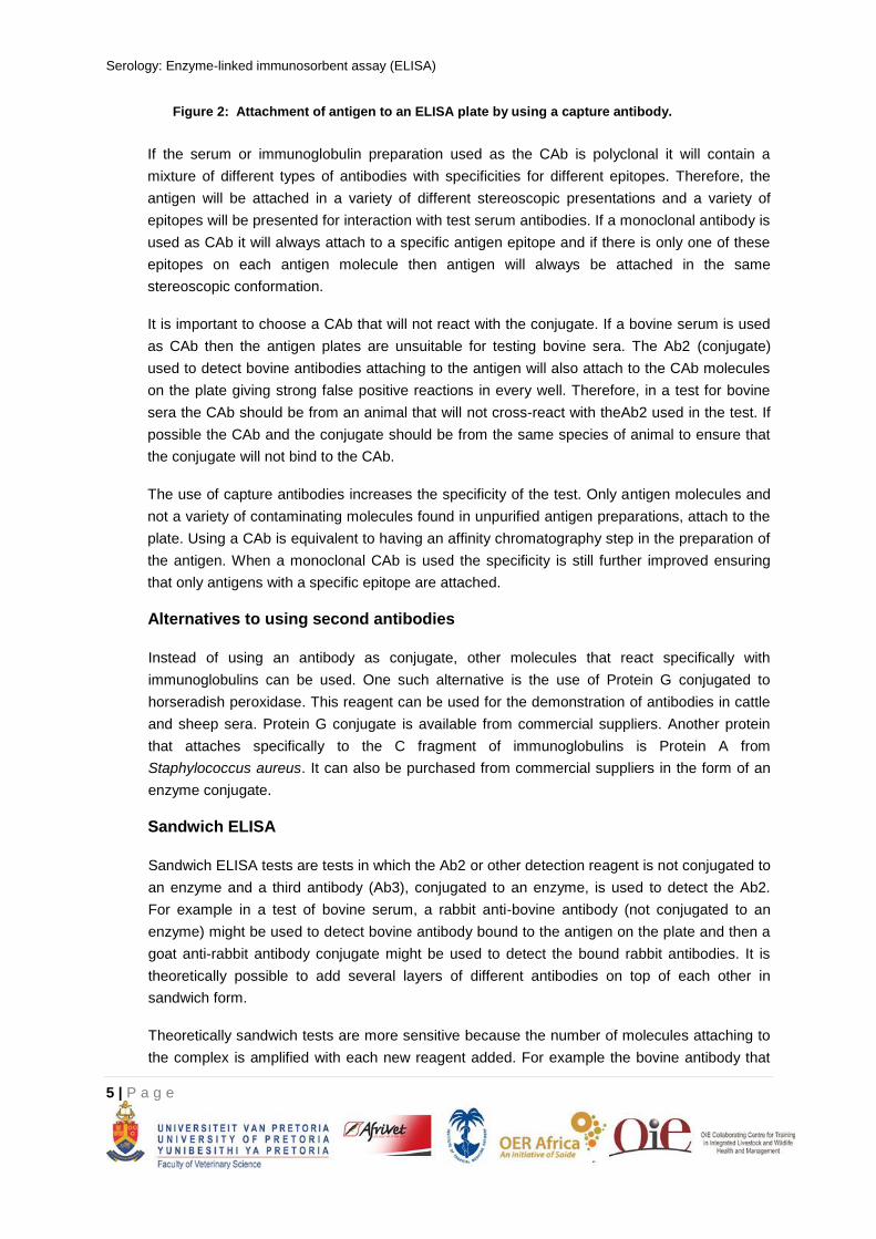
Use of capture antibody for attaching antigen to plate
In some tests, plates are coated with an antibody to the antigen. After the antibody has
attached to the plate, antigen is added and it attaches to the antibody that has been coated
onto the plate (
Figure 2). The antibody used for this purpose is called a capture antibody (CAb).
Serology: Enzyme-linked immunosorbent assay (ELISA)
5 | P a g e
Figure 2: Attachment of antigen to an ELISA plate by using a capture antibody.
If the serum or immunoglobulin preparation used as the CAb is polyclonal it will contain a
mixture of different types of antibodies with specificities for different epitopes. Therefore, the
antigen will be attached in a variety of different stereoscopic presentations and a variety of
epitopes will be presented for interaction with test serum antibodies. If a monoclonal antibody is
used as CAb it will always attach to a specific antigen epitope and if there is only one of these
epitopes on each antigen molecule then antigen will always be attached in the same
stereoscopic conformation.
It is important to choose a CAb that will not react with the conjugate. If a bovine serum is used
as CAb then the antigen plates are unsuitable for testing bovine sera. The Ab2 (conjugate)
used to detect bovine antibodies attaching to the antigen will also attach to the CAb molecules
on the plate giving strong false positive reactions in every well. Therefore, in a test for bovine
sera the CAb should be from an animal that will not cross-react with theAb2 used in the test. If
possible the CAb and the conjugate should be from the same species of animal to ensure that
the conjugate will not bind to the CAb.
The use of capture antibodies increases the specificity of the test. Only antigen molecules and
not a variety of contaminating molecules found in unpurified antigen preparations, attach to the
plate. Using a CAb is equivalent to having an affinity chromatography step in the preparation of
the antigen. When a monoclonal CAb is used the specificity is still further improved ensuring
that only antigens with a specific epitope are attached.
Alternatives to using second antibodies
Instead of using an antibody as conjugate, other molecules that react specifically with
immunoglobulins can be used. One such alternative is the use of Protein G conjugated to
horseradish peroxidase. This reagent can be used for the demonstration of antibodies in cattle
and sheep sera. Protein G conjugate is available from commercial suppliers. Another protein
that attaches specifically to the C fragment of immunoglobulins is Protein A from
Staphylococcus aureus. It can also be purchased from commercial suppliers in the form of an
enzyme conjugate.
Sandwich ELISA
Sandwich ELISA tests are tests in which the Ab2 or other detection reagent is not conjugated to
an enzyme and a third antibody (Ab3), conjugated to an enzyme, is used to detect the Ab2.
For example in a test of bovine serum, a rabbit anti-bovine antibody (not conjugated to an
enzyme) might be used to detect bovine antibody bound to the antigen on the plate and then a
goat anti-rabbit antibody conjugate might be used to detect the bound rabbit antibodies. It is
theoretically possible to add several layers of different antibodies on top of each other in
sandwich form.
Theoretically sandwich tests are more sensitive because the number of molecules attaching to
the complex is amplified with each new reagent added. For example the bovine antibody that
Serology: Enzyme-linked immunosorbent assay (ELISA)
6 | P a g e
attaches to the antigen has several epitopes on its surface and can bind to a number of
molecules of rabbit anti-bovine antibody. Each of these can in turn bind several molecules of
the goat anti-bovine conjugate. Therefore at each addition of another detection reagent the
number of molecules that binds to the complex is multiplied (
Figure 3).
Figure 3: Amplification of bound enzyme molecules in a two-step sandwich ELISA.
In practice analytical sensitivity is seldom a limiting factor in the use of ELISAs for the
diagnosis of infectious diseases and sandwich ELISAs are more likely to be used for the other
reasons. For example where the Ab2 is a MAb it is convenient and economical to use it in a
non-conjugated form. The bound MAb can then be detected with an anti-mouse conjugate.
This same anti-mouse conjugate can be used in all tests in which Ab2 is a mouse monoclonal
antibody. Anti-mouse conjugates are readily available from commercial sources.
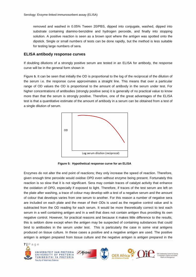
Another variation of the sandwich technique makes use of the high affinity and specificity of
the binding of biotin by the protein avidin. Instead of conjugating theAb2 (or detection reagent)
to an enzyme it is conjugated to biotin. Avidin conjugated to an enzyme is then used to detect
the antibody biotin complexes (Figure 4). A biotin/anti-mouse antibody in conjunction with an
avidin enzyme conjugate can be used to detect all MAb reagents originating from mouse
cells.
Figure 4: Use of the biotin/avidin system for the detection of antibody in an indirect ELIS.
Dot ELISAs
Antigen can be absorbed onto nitrocellulose e.g. spotted onto a nitrocellulose dipstick.
Unbound binding sites on the nitrocellulose are then blocked with milk powder or another
suitable blocking agent. The dipstick can then be dipped into serum for an appropriate time,
Serology: Enzyme-linked immunosorbent assay (ELISA)
7 | P a g e
removed and washed in 0.05% Tween 20/PBS, dipped into conjugate, washed, dipped into
substrate containing diamino-benzidine and hydrogen peroxide, and finally into stopping
solution. A positive reaction is seen as a brown spot where the antigen was spotted onto the
dipstick. Single or small numbers of tests can be done rapidly, but the method is less suitable
for testing large numbers of sera.
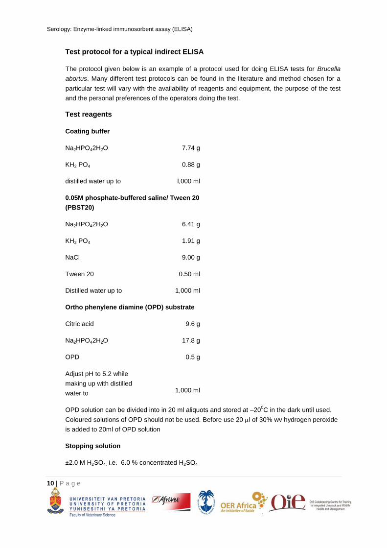
ELISA antibody response curves
If doubling dilutions of a strongly positive serum are tested in an ELISA for antibody, the response
curve will be in the general form shown in
Figure 5. It can be seen that initially the OD is proportional to the log of the reciprocal of the dilution of
the serum i.e. the response curve approximates a straight line. This means that over a particular
range of OD values the OD is proportional to the amount of antibody in the serum under test. For
higher concentrations of antibodies (strongly positive sera) it is generally of no practical value to know
more than that the serum is strongly positive. Therefore, one of the great advantages of the ELISA
test is that a quantitative estimate of the amount of antibody in a serum can be obtained from a test of
a single dilution of serum.
Figure 5: Hypothetical response curve for an ELISA
Enzymes do not alter the end point of reactions; they only increase the speed of reaction. Therefore,
given enough time peroxide would oxidise OPD even without enzyme being present. Fortunately this
reaction is so slow that it is not significant. Sera may contain traces of catalyst activity that enhance
the oxidation of OPD, especially if exposed to light. Therefore, if traces of the test serum are left on
the plate after washing, a trace of colour may develop with a test of a negative serum and the amount
of colour that develops varies from one serum to another. For this reason a number of negative sera
are included on each plate and the mean of their ODs is used as the negative control value and is
subtracted from the OD reading for each serum. It would be more theoretically correct to test each
serum in a well containing antigen and in a well that does not contain antigen thus providing its own
negative control. However, for practical reasons and because it makes little difference to the results,
this is seldom done except when the antigen may be suspected of containing substances that could
bind to antibodies in the serum under test. This is particularly the case in some viral antigens
produced on tissue culture. In these cases a positive and a negative antigen are used. The positive
antigen is antigen prepared from tissue culture and the negative antigen is antigen prepared in the

Serology: Enzyme-linked immunosorbent assay (ELISA)
8 | P a g e
same way from tissue culture cells that have not been inoculated with virus. The OD of the test using
“negative antigen” is subtracted from the OD of the test with normal antigen.
Assuming that 20 wells are used for various controls on a plate it is still possible to do 76 tests on a
single plate compared to the 11 tests that can be done with the CFT. This also makes it cost efficient
to do all test in duplicate on each plate. Despite this some workers still prefer to test doubling dilutions
of each serum in the traditional manner used in other serological tests.
The analytical sensitivity of ELISA tests is very high and all sera must be suitably diluted before
testing. The most suitable dilution at which to test sera must be determined for each test. In the
Brucella abortus ELISA test sera are diluted 1/200 for testing.
Calculation of results
It is not possible under normal laboratory testing conditions to get identical results in terms of OD
when testing a serum repeatedly on different plates or days. For this reason the results are expressed
as being relative to the result for a control serum that has been standardised against an international
or national control serum, and is tested on the same plate as the serum being tested. Results can be
calculated and expressed in a number of ways:
A single dilution of strongly positive serum may be used as control. In this case the control serum
is diluted to a dilution that gives a response on the linear portion of the dose response curve for
that serum. A dilution is often chosen where the response is slightly below the plateau phase of
the curve. However, any dilution that gives a result falling on the linear part of the response curve
can be used, provided the same dilution is always used. In this case a result for a serum is
expressed as a percentage of the reaction for the control serum. The result then becomes:
100
seracontrolnegativeofODMeanserumcontrolofODMean
seracontrolnegativeofODMeantestedserumofaliquotsforODMean
The threshold for a suspicious or positive result is given as a percentage of the positive control
serum.
The antibody content of the control serum may be expressed in units. A number of dilutions of
serum, which when tested give results that fall on the linear portion of the response curve, are
tested on each test plate. A best-fit response function of OD against the log of the units of
antibody is calculated (assuming it is a straight line fitting the function y = ax + b, where a = the
slope of the line and b = the intercept on the y axis). The number of units of antibody in each test
sample is then calculated from the mathematical function. Alternatively a graph of the response
(OD) against units of antibody for the control serum can be plotted and the results for each
serum read from the graph. However, in practice graphing of results is clumsy and impractical for
testing large numbers of sera and generally the data is fed directly from the ELISA plate reader
into a computer. The computer is programmed to calculate the regression function for the control
serum dilutions and then calculates the number of units of antibody in each test serum (see
Section 1.4.4).

Serology: Enzyme-linked immunosorbent assay (ELISA)
9 | P a g e
In some cases a weak positive serum is included as a control and sera giving reactions equal or
greater than the weak positive serum are classed as positive. In this case strong and medium
level positives may also be included on the plate as further reference points.
Serology: Enzyme-linked immunosorbent assay (ELISA)
10 | P a g e
Test protocol for a typical indirect ELISA
The protocol given below is an example of a protocol used for doing ELISA tests for Brucella
abortus. Many different test protocols can be found in the literature and method chosen for a
particular test will vary with the availability of reagents and equipment, the purpose of the test
and the personal preferences of the operators doing the test.
Test reagents
Coating buffer
Na2HPO42H2O 7.74 g
KH2 PO4 0.88 g
distilled water up to l,000 ml
0.05M phosphate-buffered saline/ Tween 20
(PBST20)
Na2HPO42H2O 6.41 g
KH2 PO4 1.91 g
NaCl 9.00 g
Tween 20 0.50 ml
Distilled water up to 1,000 ml
Ortho phenylene diamine (OPD) substrate
Citric acid 9.6 g
Na2HPO42H2O 17.8 g
OPD 0.5 g
Adjust pH to 5.2 while
making up with distilled
water to
1,000 ml
OPD solution can be divided into in 20 ml aliquots and stored at –200C in the dark until used.
Coloured solutions of OPD should not be used. Before use 20 l of 30% wv hydrogen peroxide
is added to 20ml of OPD solution
Stopping solution
±2.0 M H2SO4, i.e. 6.0 % concentrated H2SO4
Serology: Enzyme-linked immunosorbent assay (ELISA)
11 | P a g e
Conjugate
Protein G conjugated to horseradish peroxidase, from a commercial supplier.
Antigen
A suspension of Brucella abortus cells as used in the CFT and agglutination tests.
Control sera
The national or laboratory standard, positive control serum and six negative control sera are
used.
Titration of antigen when optimal conjugate dilution is known
Suitable dilutions of a commercially available Brucella abortus agglutination antigen are
made up in coating buffer e.g. doubling dilutions from 1/50 to 1/1,600.
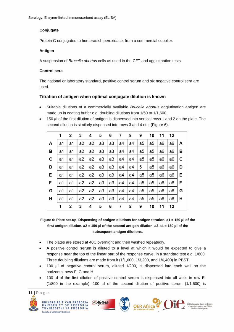
150 l of the first dilution of antigen is dispensed into vertical rows 1 and 2 on the plate. The
second dilution is similarly dispensed into rows 3 and 4 etc. (Figure 6).
Figure 6: Plate set-up. Dispensing of antigen dilutions for antigen titration. a1 = 150 l of the
first antigen dilution. a2 = 150 l of the second antigen dilution. a3-a4 = 150 l of the
subsequent antigen dilutions.
The plates are stored at 40C overnight and then washed repeatedly.
A positive control serum is diluted to a level at which it would be expected to give a
response near the top of the linear part of the response curve, in a standard test e.g. 1/800.
Three doubling dilutions are made from it (1/1,600, 1/3,200, and 1/6,400) in PBST.
100 l of negative control serum, diluted 1/200, is dispensed into each well on the
horizontal rows F, G and H.
100 l of the first dilution of positive control serum is dispensed into all wells in row E.
(1/800 in the example). 100 l of the second dilution of positive serum (1/1,600) is
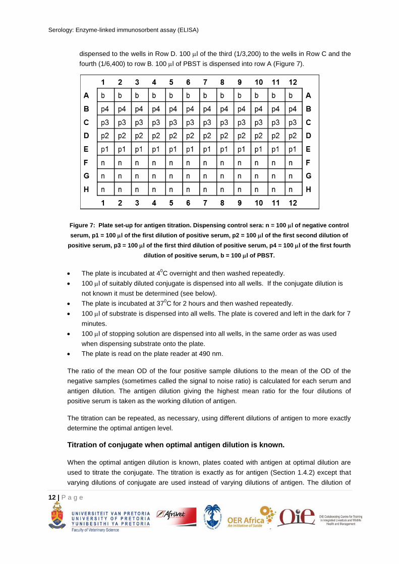
Serology: Enzyme-linked immunosorbent assay (ELISA)
12 | P a g e
dispensed to the wells in Row D. 100 l of the third (1/3,200) to the wells in Row C and the
fourth (1/6,400) to row B. 100 l of PBST is dispensed into row A (Figure 7).
Figure 7: Plate set-up for antigen titration. Dispensing control sera: n = 100 l of negative control
serum, p1 = 100 l of the first dilution of positive serum, p2 = 100 l of the first second dilution of
positive serum, p3 = 100 l of the first third dilution of positive serum, p4 = 100 l of the first fourth
dilution of positive serum, b = 100 l of PBST.
The plate is incubated at 40C overnight and then washed repeatedly.
100 l of suitably diluted conjugate is dispensed into all wells. If the conjugate dilution is
not known it must be determined (see below).
The plate is incubated at 370C for 2 hours and then washed repeatedly.
100 l of substrate is dispensed into all wells. The plate is covered and left in the dark for 7
minutes.
100 l of stopping solution are dispensed into all wells, in the same order as was used
when dispensing substrate onto the plate.
The plate is read on the plate reader at 490 nm.
The ratio of the mean OD of the four positive sample dilutions to the mean of the OD of the
negative samples (sometimes called the signal to noise ratio) is calculated for each serum and
antigen dilution. The antigen dilution giving the highest mean ratio for the four dilutions of
positive serum is taken as the working dilution of antigen.
The titration can be repeated, as necessary, using different dilutions of antigen to more exactly
determine the optimal antigen level.
Titration of conjugate when optimal antigen dilution is known.
When the optimal antigen dilution is known, plates coated with antigen at optimal dilution are
used to titrate the conjugate. The titration is exactly as for antigen (Section 1.4.2) except that
varying dilutions of conjugate are used instead of varying dilutions of antigen. The dilution of
Serology: Enzyme-linked immunosorbent assay (ELISA)
13 | P a g e
conjugate used should be that giving the best mean ratio of OD positive serum to OD negative
serum for the four dilutions of positive serum.
Antigen/conjugate checkerboard titration
When a new test is being set up and neither antigen nor conjugate dilution is known two
checkerboard titrations are done, one using positive control serum and one using negative
control serum. Dilutions of antigen are varied in the vertical rows of the plate and the dilutions
of conjugate are varied in the horizontal rows of the plate. The positive and negative serum
checkerboards are done in alternate rows on the same plate as described below. The dilutions
of antigen and conjugate selected for further standardisation are those giving the highest ratio
of OD positive serum to OD negative serum.
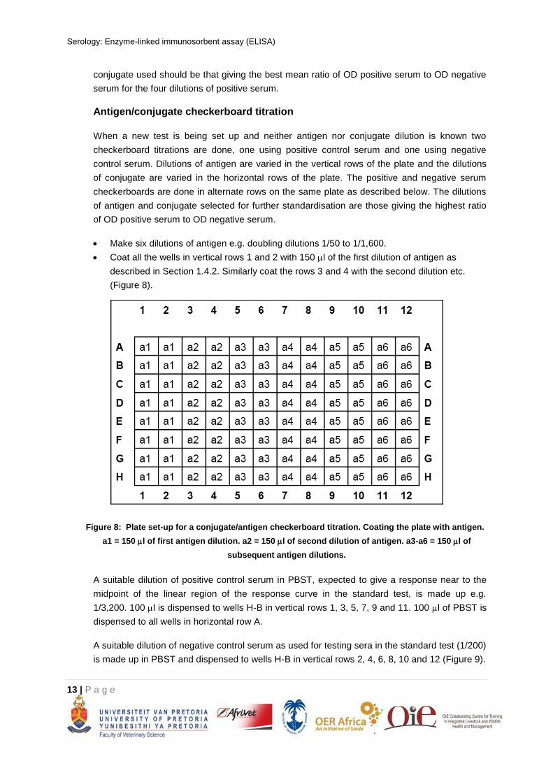
Make six dilutions of antigen e.g. doubling dilutions 1/50 to 1/1,600.
Coat all the wells in vertical rows 1 and 2 with 150 l of the first dilution of antigen as
described in Section 1.4.2. Similarly coat the rows 3 and 4 with the second dilution etc.
(Figure 8).
Figure 8: Plate set-up for a conjugate/antigen checkerboard titration. Coating the plate with antigen.
a1 = 150 l of first antigen dilution. a2 = 150 l of second dilution of antigen. a3-a6 = 150 l of
subsequent antigen dilutions.
A suitable dilution of positive control serum in PBST, expected to give a response near to the
midpoint of the linear region of the response curve in the standard test, is made up e.g.
1/3,200. 100 l is dispensed to wells H-B in vertical rows 1, 3, 5, 7, 9 and 11. 100 l of PBST is
dispensed to all wells in horizontal row A.
A suitable dilution of negative control serum as used for testing sera in the standard test (1/200)
is made up in PBST and dispensed to wells H-B in vertical rows 2, 4, 6, 8, 10 and 12 (Figure 9).
Serology: Enzyme-linked immunosorbent assay (ELISA)
14 | P a g e
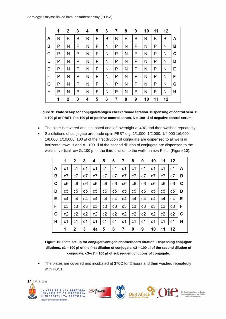
Figure 9: Plate set-up for conjugate/antigen checkerboard titration. Dispensing of control sera. B
= 100 l of PBST. P = 100 l of positive control serum. N = 100 l of negative control serum.
The plate is covered and incubated and left overnight at 40C and then washed repeatedly.
Six dilutions of conjugate are made up in PBST e.g. 1/1,000, 1/2,000, 1/4,000 1/6,000,
1/8,000, 1/10,000. 100 l of the first dilution of conjugate are dispensed to all wells in
horizontal rows H and A. 100 l of the second dilution of conjugate are dispensed to the
wells of vertical row G, 100 l of the third dilution to the wells on row F etc. (Figure 10).
Figure 10: Plate set-up for conjugate/antigen checkerboard titration. Dispensing conjugate
dilutions. c1 = 100 l of the first dilution of conjugate. c2 = 100 l of the second dilution of
conjugate. c3–c7 = 100 l of subsequent dilutions of conjugate.
The plates are covered and incubated at 370C for 2 hours and then washed repeatedly
with PBST.
Serology: Enzyme-linked immunosorbent assay (ELISA)
15 | P a g e
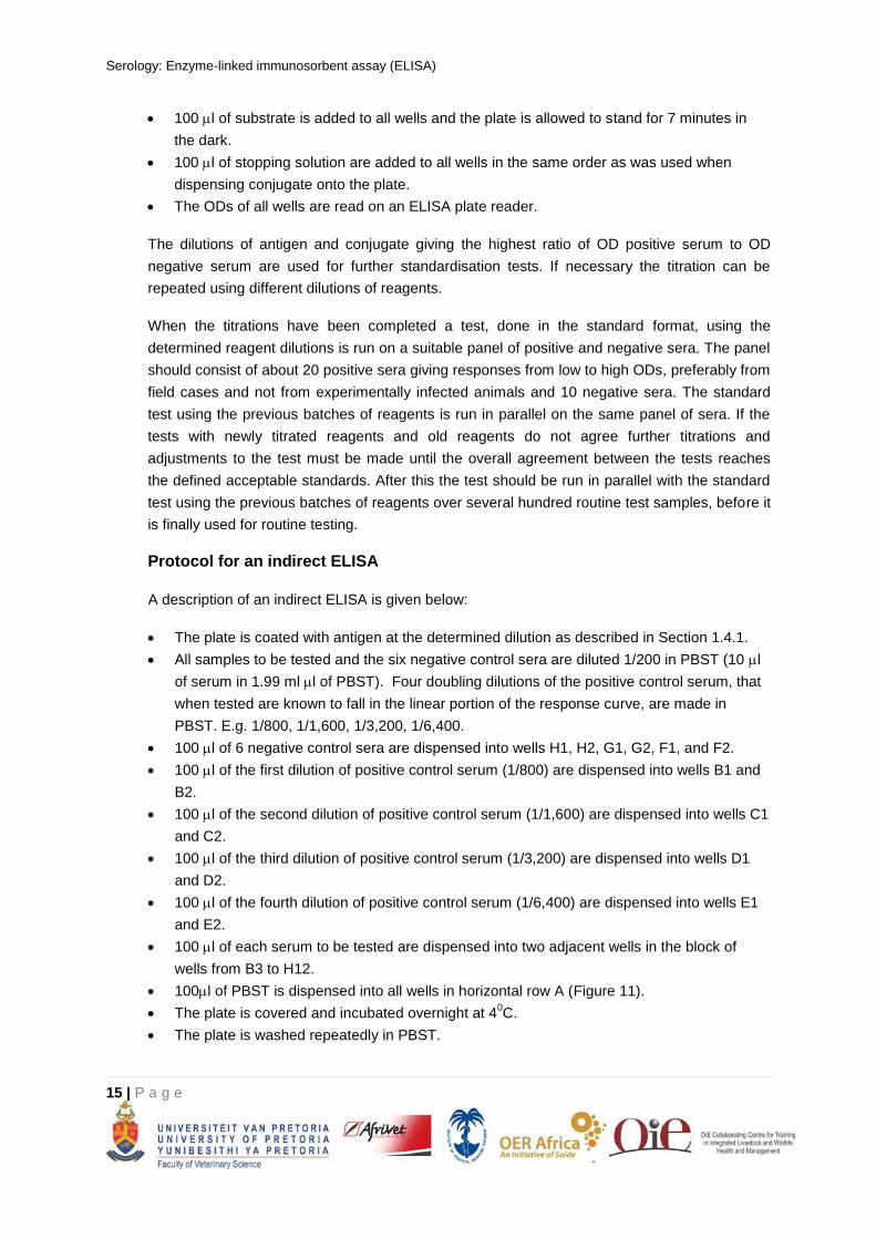
100 l of substrate is added to all wells and the plate is allowed to stand for 7 minutes in
the dark.
100 l of stopping solution are added to all wells in the same order as was used when
dispensing conjugate onto the plate.
The ODs of all wells are read on an ELISA plate reader.
The dilutions of antigen and conjugate giving the highest ratio of OD positive serum to OD
negative serum are used for further standardisation tests. If necessary the titration can be
repeated using different dilutions of reagents.
When the titrations have been completed a test, done in the standard format, using the
determined reagent dilutions is run on a suitable panel of positive and negative sera. The panel
should consist of about 20 positive sera giving responses from low to high ODs, preferably from
field cases and not from experimentally infected animals and 10 negative sera. The standard
test using the previous batches of reagents is run in parallel on the same panel of sera. If the
tests with newly titrated reagents and old reagents do not agree further titrations and
adjustments to the test must be made until the overall agreement between the tests reaches
the defined acceptable standards. After this the test should be run in parallel with the standard
test using the previous batches of reagents over several hundred routine test samples, before it
is finally used for routine testing.
Protocol for an indirect ELISA
A description of an indirect ELISA is given below:
The plate is coated with antigen at the determined dilution as described in Section 1.4.1.
All samples to be tested and the six negative control sera are diluted 1/200 in PBST (10 l
of serum in 1.99 ml l of PBST). Four doubling dilutions of the positive control serum, that
when tested are known to fall in the linear portion of the response curve, are made in
PBST. E.g. 1/800, 1/1,600, 1/3,200, 1/6,400.
100 l of 6 negative control sera are dispensed into wells H1, H2, G1, G2, F1, and F2.
100 l of the first dilution of positive control serum (1/800) are dispensed into wells B1 and
B2.
100 l of the second dilution of positive control serum (1/1,600) are dispensed into wells C1
and C2.
100 l of the third dilution of positive control serum (1/3,200) are dispensed into wells D1
and D2.
100 l of the fourth dilution of positive control serum (1/6,400) are dispensed into wells E1
and E2.
100 l of each serum to be tested are dispensed into two adjacent wells in the block of
wells from B3 to H12.
100l of PBST is dispensed into all wells in horizontal row A (Figure 11).
The plate is covered and incubated overnight at 40C.
The plate is washed repeatedly in PBST.
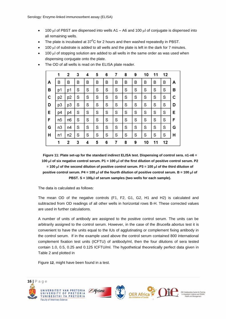
Serology: Enzyme-linked immunosorbent assay (ELISA)
16 | P a g e
100 l of PBST are dispensed into wells A1 – A6 and 100 l of conjugate is dispensed into
all remaining wells.
The plate is incubated at 370C for 2 hours and then washed repeatedly in PBST.
100 l of substrate is added to all wells and the plate is left in the dark for 7 minutes.
100 l of stopping solution are added to all wells in the same order as was used when
dispensing conjugate onto the plate.
The OD of all wells is read on the ELISA plate reader.
Figure 11: Plate set-up for the standard indirect ELISA test. Dispensing of control sera. n1-n6 =
100 l of six negative control serum. P1 = 100 l of the first dilution of positive control serum. P2
= 100 l of the second dilution of positive control serum. P3 = 100 l of the third dilution of
positive control serum. P4 = 100 l of the fourth dilution of positive control serum. B = 100 l of
PBST. S = 100l of serum samples (two wells for each sample).
The data is calculated as follows:
The mean OD of the negative controls (F1, F2, G1, G2, H1 and H2) is calculated and
subtracted from OD readings of all other wells in horizontal rows B-H. These corrected values
are used in further calculations.
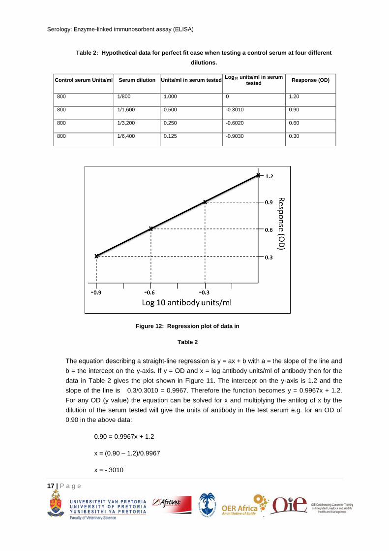
A number of units of antibody are assigned to the positive control serum. The units can be
arbitrarily assigned to the control serum. However, in the case of the Brucella abortus test it is
convenient to have the units equal to the IUs of agglutinating or complement fixing antibody in
the control serum. If in the example used above the control serum contained 800 international
complement fixation test units (ICFTU) of antibody/ml, then the four dilutions of sera tested
contain 1.0, 0.5, 0.25 and 0.125 ICFTU/ml. The hypothetical theoretically perfect data given in
Table 2 and plotted in
Figure 12, might have been found in a test.
Serology: Enzyme-linked immunosorbent assay (ELISA)
17 | P a g e
Table 2: Hypothetical data for perfect fit case when testing a control serum at four different
dilutions.
Control serum Units/ml Serum dilution Units/ml in serum tested Log10 units/ml in serum
tested Response (OD)
800 1/800 1.000 0 1.20
800 1/1,600 0.500 -0.3010 0.90
800 1/3,200 0.250 -0.6020 0.60
800 1/6,400 0.125 -0.9030 0.30
Figure 12: Regression plot of data in
Table 2
The equation describing a straight-line regression is y = ax + b with a = the slope of the line and
b = the intercept on the y-axis. If y = OD and x = log antibody units/ml of antibody then for the
data in Table 2 gives the plot shown in Figure 11. The intercept on the y-axis is 1.2 and the
slope of the line is 0.3/0.3010 = 0.9967. Therefore the function becomes y = 0.9967x + 1.2.
For any OD (y value) the equation can be solved for x and multiplying the antilog of x by the
dilution of the serum tested will give the units of antibody in the test serum e.g. for an OD of
0.90 in the above data:
0.90 = 0.9967x + 1.2
x = (0.90 – 1.2)/0.9967
x = -.3010
Serology: Enzyme-linked immunosorbent assay (ELISA)
18 | P a g e
and 1,600 (antilog -.3010) = 800 units of antibody which is the correct answer for this control
serum.
Therefore, the units of antibody for any serum tested will be: antilog ((OD – b)/a) multiplied by
the dilution of the serum tested.
This may seem unnecessarily complicated but once the computer is programmed the results
are generated automatically and no calculation is involved.
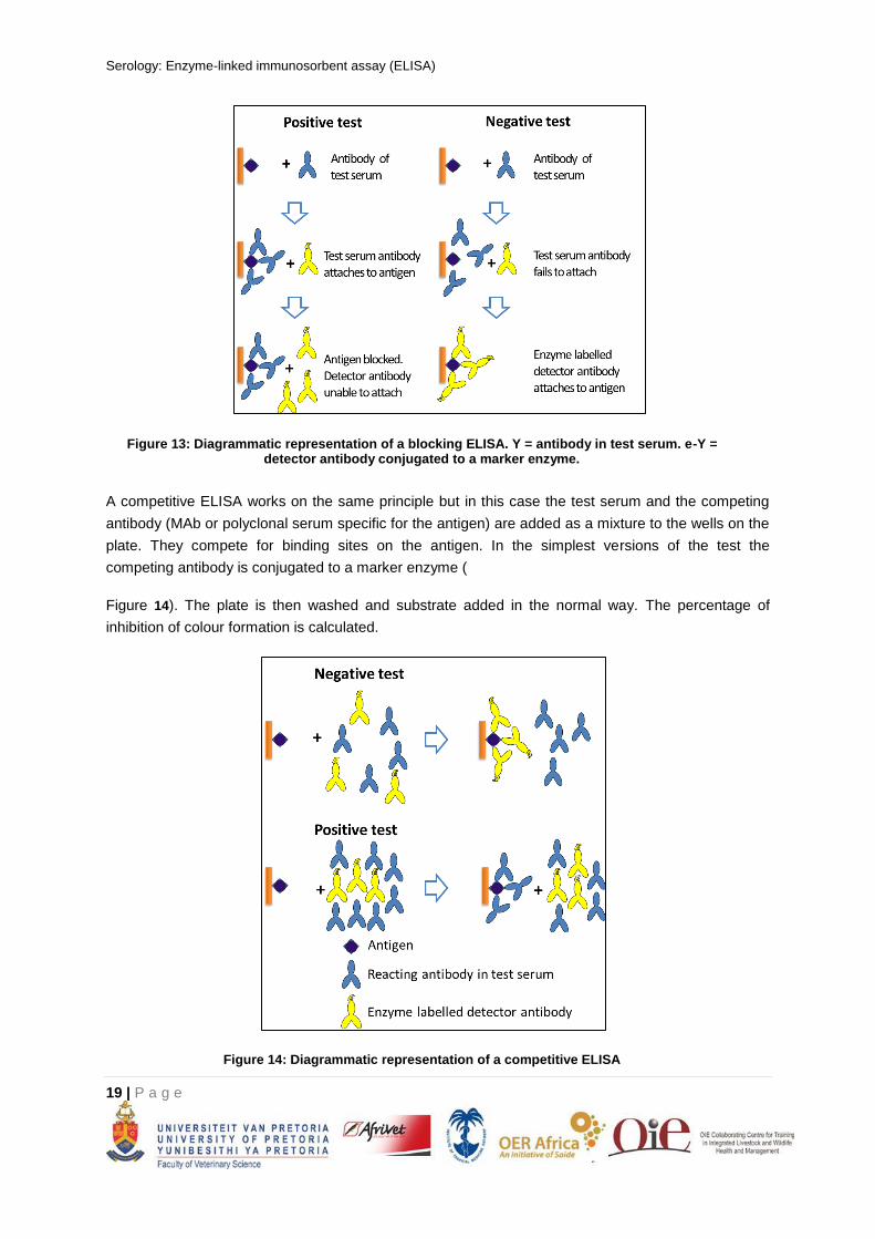
COMPETITIVE AND BLOCKING ELISAS
Competitive and blocking ELISAs work on the principle that two antibodies compete for a binding site
on an epitope. Competitive and blocking ELISAs are very similar. Tests in which the serum being
tested and a competing antiserum (or antibody preparation) are reacted simultaneously with the
antigen are called competitive ELISAs. Tests in which the serum being tested and the competing
antiserum (or antibody preparation) are reacted sequentially with the antigen are called blocking
ELISAs (see below).
Competitive and blocking ELISAs have distinct advantages over indirect ELISAs and are very
commonly used. They are done using either monoclonal antibodies (MAbs) or polyclonal sera as the
competing antibody. However, they are best utilised when the test is designed to take advantage of
the high specificity of MAbs. A MAb that recognises an immunodominant epitope can be used for
providing a test of high sensitivity and a MAb against a highly specific epitope can be used in a test
designed to have great specificity. Furthermore, the specificity provided by the MAb allows tests with
high specificity to be done with crude antigens.
In the simplest configuration of a typical blocking test a MAb preparation with specificity for an
immunodominant epitope on the antigen could be used as an enzyme labelled competing antibody.
The antigen would be attached to a plate in the normal manner. The serum to be tested would be
dispensed into the relevant wells on the plate and antibody in the serum allowed to attach to available
matching epitopes, thus blocking these epitopes so that they are not available to interact with other
antibodies. In the next stage of the test the MAb coupled to an enzyme would be dispensed into the
relevant wells. If all the relevant epitopes are already blocked by antibody from the test serum the
MAb will not attach and after washing the wells and adding substrate no colour will develop (see
Figure 13). In practice only some of the relevant epitopes may be blocked if the test serum contains a
low level of antibody. Therefore, results are expressed as the percentage reduction of colour
development compared to a test with a negative control serum in which no blocking occurs. Blocking
ELISAs may be done at a single test serum dilution, or in some tests doubling dilutions of sera are
tested. A cut-off point is established as a percentage of inhibition of colour development. A blocking
ELISA can also be done with a polyclonal serum used as the competing antibody preparation.
Serology: Enzyme-linked immunosorbent assay (ELISA)
19 | P a g e
Figure 13: Diagrammatic representation of a blocking ELISA. Y = antibody in test serum. e-Y = detector antibody conjugated to a marker enzyme.
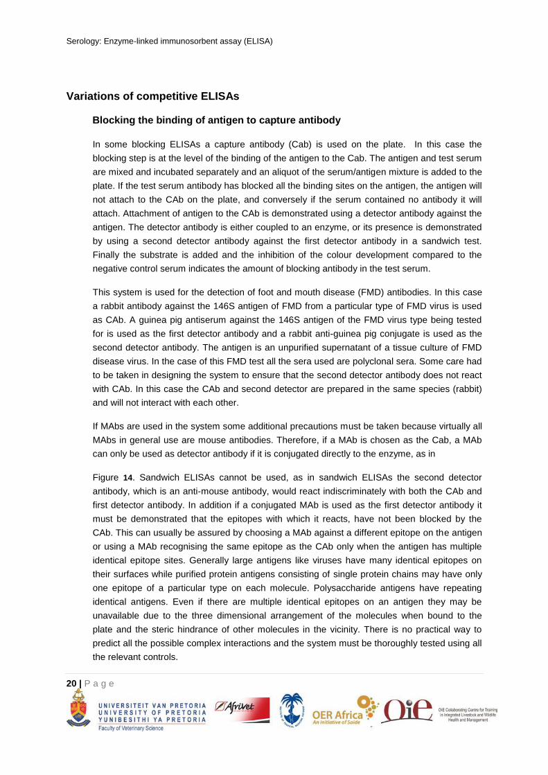
A competitive ELISA works on the same principle but in this case the test serum and the competing
antibody (MAb or polyclonal serum specific for the antigen) are added as a mixture to the wells on the
plate. They compete for binding sites on the antigen. In the simplest versions of the test the
competing antibody is conjugated to a marker enzyme (
Figure 14). The plate is then washed and substrate added in the normal way. The percentage of
inhibition of colour formation is calculated.
Figure 14: Diagrammatic representation of a competitive ELISA
Serology: Enzyme-linked immunosorbent assay (ELISA)
20 | P a g e
Variations of competitive ELISAs
Blocking the binding of antigen to capture antibody
In some blocking ELISAs a capture antibody (Cab) is used on the plate. In this case the
blocking step is at the level of the binding of the antigen to the Cab. The antigen and test serum
are mixed and incubated separately and an aliquot of the serum/antigen mixture is added to the
plate. If the test serum antibody has blocked all the binding sites on the antigen, the antigen will
not attach to the CAb on the plate, and conversely if the serum contained no antibody it will
attach. Attachment of antigen to the CAb is demonstrated using a detector antibody against the
antigen. The detector antibody is either coupled to an enzyme, or its presence is demonstrated
by using a second detector antibody against the first detector antibody in a sandwich test.
Finally the substrate is added and the inhibition of the colour development compared to the
negative control serum indicates the amount of blocking antibody in the test serum.
This system is used for the detection of foot and mouth disease (FMD) antibodies. In this case
a rabbit antibody against the 146S antigen of FMD from a particular type of FMD virus is used
as CAb. A guinea pig antiserum against the 146S antigen of the FMD virus type being tested
for is used as the first detector antibody and a rabbit anti-guinea pig conjugate is used as the
second detector antibody. The antigen is an unpurified supernatant of a tissue culture of FMD
disease virus. In the case of this FMD test all the sera used are polyclonal sera. Some care had
to be taken in designing the system to ensure that the second detector antibody does not react
with CAb. In this case the CAb and second detector are prepared in the same species (rabbit)
and will not interact with each other.
If MAbs are used in the system some additional precautions must be taken because virtually all
MAbs in general use are mouse antibodies. Therefore, if a MAb is chosen as the Cab, a MAb
can only be used as detector antibody if it is conjugated directly to the enzyme, as in
Figure 14. Sandwich ELISAs cannot be used, as in sandwich ELISAs the second detector
antibody, which is an anti-mouse antibody, would react indiscriminately with both the CAb and
first detector antibody. In addition if a conjugated MAb is used as the first detector antibody it
must be demonstrated that the epitopes with which it reacts, have not been blocked by the
CAb. This can usually be assured by choosing a MAb against a different epitope on the antigen
or using a MAb recognising the same epitope as the CAb only when the antigen has multiple
identical epitope sites. Generally large antigens like viruses have many identical epitopes on
their surfaces while purified protein antigens consisting of single protein chains may have only
one epitope of a particular type on each molecule. Polysaccharide antigens have repeating
identical antigens. Even if there are multiple identical epitopes on an antigen they may be
unavailable due to the three dimensional arrangement of the molecules when bound to the
plate and the steric hindrance of other molecules in the vicinity. There is no practical way to
predict all the possible complex interactions and the system must be thoroughly tested using all
the relevant controls.
Serology: Enzyme-linked immunosorbent assay (ELISA)
21 | P a g e
In some systems the CAb is a MAb thus providing good specificity and the detector is a
polyclonal antibody that can react with a variety of epitopes on the antigen thereby increasing
the sensitivity.
Blocking the binding of antibody to antigen coated onto the plate.
Antigen can be attached directly to the plate or a CAb can be used. If the CAb is a MAb, it will
provide for a high degree of specificity of binding of antigen to the plate. Test serum is then
added to the plate and incubated followed by the addition of the detector antibody. The
detector antibody may be conjugated to an enzyme or be unconjugated. If the detector antibody
is unconjugated, it will have to be detected by the addition of a conjugated second detector
antibody to detect the presence of first detector antibody on the plate. This type of test differs
from that described in 2.1.1 in that the blocking step by test sera takes place after antigen is
already attached to the plate and is aimed at blocking the attachment of the detector antibody
to the antigen.
A typical example of this type of test is the blocking ELISA for the detection of swine vesicular
disease virus antibodies in serum. In this test a MAb is used to fix antigen to the plate, followed
by addition of test serum, which if it contains antibody will block the relevant epitope. The
detector antibody is the same MAb that was used as CAb but conjugated to an enzyme. Finally
substrate is added and colour allowed developing. The reduction in the amount of colour
development compared to the negative control provides an indication of the amount of blocking
antibody by the test serum. In this case there are multiple identical epitope sites that can react
with the MAb, on the antigen particles. This allows the same MAb to be used as CAb and
detector antibody.
The usual variations involving the use, avidin/biotin systems, sandwich ELISAs, monoclonal or
polyclonal sera etc. can be used as appropriate.
A competitive ELISA
An example is given below of a competitive ELISA for rinderpest antibody. The protocol has been
adapted from the description of this test given in the OIE Manual of Standards for Diagnostic tests
and Vaccines. In this example all critical reagents can be purchased from the OIE reference
laboratory for Rinderpest.
Reagents
Phosphate buffered saline (PBS) is 0.01M phosphate buffered saline pH 7.4 unless
otherwise specified.
Blocking buffer is 0.01M PBS with 0.1% Tween 20 and 0.3% normal bovine serum.
Antigen is freeze-dried Kabete O strain, an attenuated strain of rinderpest virus. It is
reconstituted in distilled water and diluted according to the supplier’s instructions in PBS.
The competing antibody is a MAb specific for the H antigen of the rinderpest virus (MAb
C1).
Serology: Enzyme-linked immunosorbent assay (ELISA)
22 | P a g e
A rabbit anti-mouse antibody conjugated to horseradish peroxidase used to detect MAb
C1.
Substrate is OPD/H2O2 solution as described in Section 1.4.1.
Negative control sera consist of six sera from animals from a disease free and
unvaccinated population of animals.
The positive control serum is a positive control obtained from the reference laboratory or a
positive control serum that has been standardised against a reference serum. The positive
control serum should be tested in suitable dilutions expected to give a strong, medium and
weak positive result.
Assay protocol
The antigen is reconstituted in 0.01M PBS according to the supplier’s instructions and 50 l
dispensed into all wells of a microtitre plate. The plate is sealed and incubated for 1 hour at
370C on a shaker and washed repeatedly in 0.002M PBS pH 7.4.
40 l of blocking buffer is added to all wells in the horizontal rows B – H and 50 l of
blocking buffer is added to the wells in horizontal row A (Figure 15).
10 l of each test serum is added to two wells on the plate horizontal rows B – H. The six
negative control sera and the positive control sera are also tested.
50 l of MAb C1, diluted according to the supplier’s instructions in blocking buffer (or
previous titration), is added to all wells except wells A1-A6, which receive 50l of PBS.
The plates are sealed and incubated for 1 hour at 370C on a shaker and then washed
repeatedly in 0.002 M phosphate buffer.
50l of rabbit anti-mouse conjugate suitably diluted in blocking buffer, is added to each well
except wells A1-A3 and A10-A12. 50 l of blocking buffer is added to wells A1-A3 and A10-
A12. The plates are sealed and incubated for 1 hour at 370C on a shaker and washed
repeatedly in 0.002M PBS pH 7.4.
50l of OPD/H2O2 solution is added to each plate and the plates are incubated at room
temperature for 7minutes.
50l of 1M sulphuric acid is added to each plate and the OD read at 490nm on an ELISA
plate reader.
Serology: Enzyme-linked immunosorbent assay (ELISA)
23 | P a g e
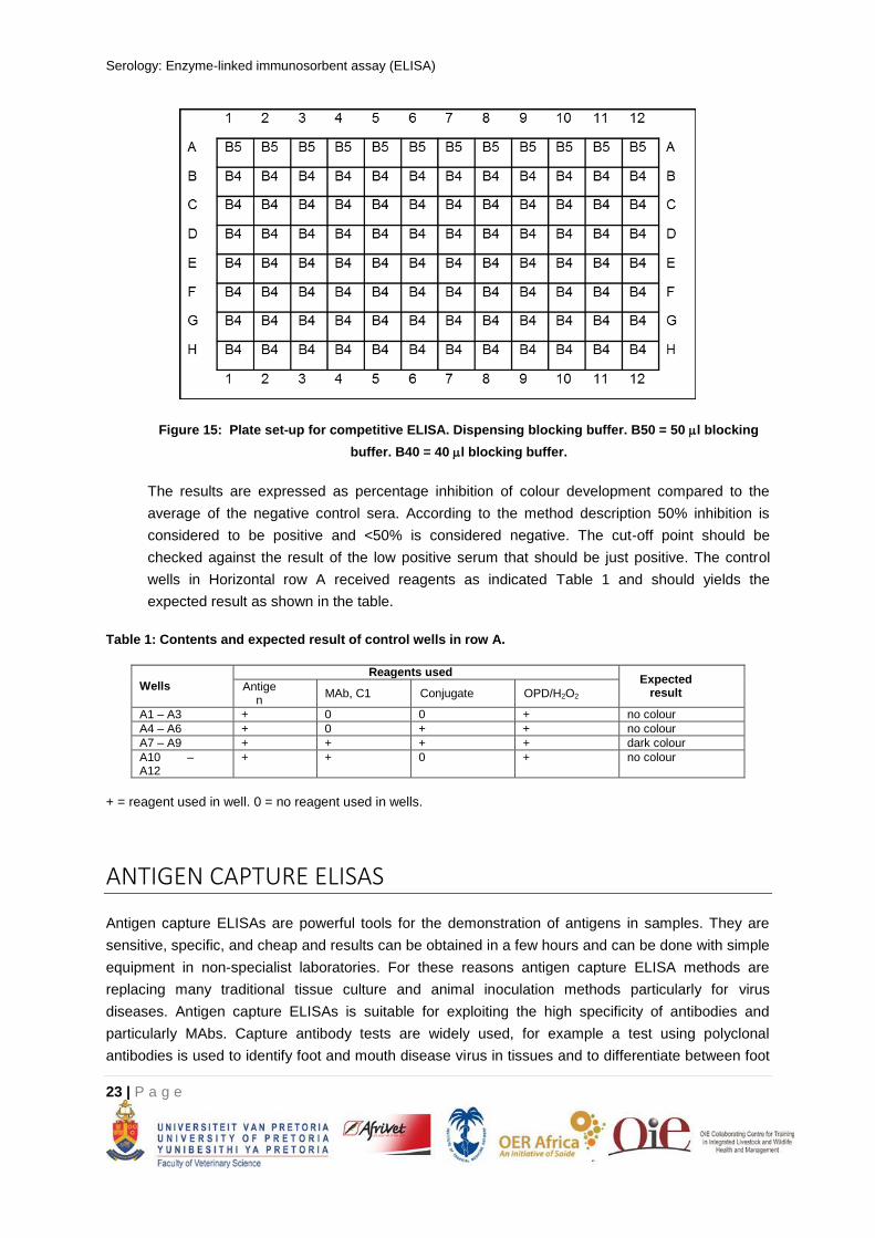
Figure 15: Plate set-up for competitive ELISA. Dispensing blocking buffer. B50 = 50 l blocking
buffer. B40 = 40 l blocking buffer.
The results are expressed as percentage inhibition of colour development compared to the
average of the negative control sera. According to the method description 50% inhibition is
considered to be positive and <50% is considered negative. The cut-off point should be
checked against the result of the low positive serum that should be just positive. The control
wells in Horizontal row A received reagents as indicated Table 1 and should yields the
expected result as shown in the table.
Table 1: Contents and expected result of control wells in row A.
Wells
Reagents used Expected
result Antige
n MAb, C1 Conjugate OPD/H2O2
A1 – A3 + 0 0 + no colour
A4 – A6 + 0 + + no colour
A7 – A9 + + + + dark colour
A10 – A12
+ + 0 + no colour
+ = reagent used in well. 0 = no reagent used in wells.
ANTIGEN CAPTURE ELISAS
Antigen capture ELISAs are powerful tools for the demonstration of antigens in samples. They are
sensitive, specific, and cheap and results can be obtained in a few hours and can be done with simple
equipment in non-specialist laboratories. For these reasons antigen capture ELISA methods are
replacing many traditional tissue culture and animal inoculation methods particularly for virus
diseases. Antigen capture ELISAs is suitable for exploiting the high specificity of antibodies and
particularly MAbs. Capture antibody tests are widely used, for example a test using polyclonal
antibodies is used to identify foot and mouth disease virus in tissues and to differentiate between foot
Serology: Enzyme-linked immunosorbent assay (ELISA)
24 | P a g e
and mouth disease virus types. Similarly a test using MAbs is able to distinguish between the closely
related rinderpest and peste des petits ruminants (PPR) viruses.
In the case of foot and mouth disease rabbit anti-sera to each of the seven types of foot and mouth
disease virus are coated onto rows of wells on a plate. These act as the capture antibody and virus
particles bind to them when tissue samples are dispensed into the wells. Guinea pig sera against the
seven types of virus are added and finally a rabbit anti-guinea pig conjugate is used to identify the
rows in which virus and guinea pig antibody have bound. After each step the plates are thoroughly
washed and finally substrate is added and colour allowed developing in the usual way. The test can
also be used to identify the virus type in tissue culture amplified virus.
In the case of Rinderpest and PPR a single MAb against an epitope on the N protein, which is
common to both viruses, is used as a capture antibody. Virus of either disease will then bind to the
plate when infected tissue samples are applied to it. Biotinylated MAbs with specificity for either
rinderpest or PPR are then used to identify the virus that has bound to the plate and an avidin enzyme
conjugate is added. Plates are thoroughly washed between each step and finally substrate is added
and the colour allowed developing in the normal manner.
Both the above tests could therefore be described as antigen capture, sandwich ELISAs. Many other
antigen capture ELISAs are commonly used to identify virus in tissue samples.