Embed Size (px)
Citation preview
Received 08/12/2020 Review began 09/12/2020 Review ended 09/13/2020 Published 09/16/2020
© Copyright 2020Patterson et al. This is an open accessarticle distributed under the terms of theCreative Commons Attribution LicenseCC-BY 4.0., which permits unrestricteduse, distribution, and reproduction in anymedium, provided the original author andsource are credited.
An Interesting Civilian Case of ComplexMaxillofacial Trauma Due to TargetFragmentation Following Bullet Impact andReview of the Branches of the Maxillary ArteryBrian Patterson , Sophia Sangar , Raja Gnanadev , George Makkar , Michael Neeki
1. Surgery, Arrowhead Regional Medical Center, Colton, USA 2. Head and Neck Surgery, David Geffen School ofMedicine at University of California-Los Angeles, Los Angeles, USA 3. Vascular Surgery, Arrowhead Regional MedicalCenter, Colton, USA 4. Emergency Medicine, Arrowhead Regional Medical Center, Colton, USA
Corresponding author: Raja Gnanadev, [email protected]
AbstractSerious morbidity and mortality for the operator and bystanders are associated with a lack of knowledge andfailure to utilize appropriately manufactured targets. The management of firearm-related facial trauma ischallenging and requires rapid intervention from a multidisciplinary team. We present a case of penetratingfacial trauma secondary to the fragmentation of a homemade target. We highlight how firearm operators canoptimize safety by matching ballistics with target selection and review pertinent vascular structures,including the terminal branches of the external carotid artery and branches of the maxillary artery. This casedemonstrates that trauma physicians must be well-versed with complex maxillofacial anatomy andmultimodal approaches to hemostasis.
Categories: Otolaryngology, Public Health, TraumaKeywords: trauma, external carotid artery, maxillary artery, maxillofacial, shrapnel
IntroductionShrapnel-related maxillofacial injuries are well-documented, with clear guidelines in the military literature,but the incidence and reporting of such injuries in the civilian setting is uncommon [1-4]. Militaryexperiences in the modern wars in Iraq and Afghanistan have contributed to clear guidelines for high-velocity and high-energy gunshot wounds to the face [1]. As with any trauma, following the AdvancedTrauma and Life Support protocols is critical to the management of such injuries, as is the early integrationof a multi-specialty team, including emergency department providers, critical care surgeons, vascularsurgeons, and maxillofacial surgeons [1-2].
Given regional structures, shrapnel-related maxillofacial trauma often leads to complex injury, includingbony, muscular, nervous system, and vascular injury [5]. We report a rare case of civilian shrapnel-relatedcomplex maxillofacial trauma due to target fragmentation following bullet impact. This report reviews therelevant anatomy with a focus on regional vascular structures, including the terminal branches of theexternal carotid artery and branches of the maxillary artery.
A review of the literature was conducted through Google Scholar, PubMed, and Google Books usingkeywords including, but not limited to, “maxillofacial shrapnel injury,” “civilian shrapnel injury,” “ballisticfacial injury,” and “maxillary artery trauma.”
Case PresentationA 36-year-old male was brought to the emergency department (ED) with a deep facial laceration (Figure 1)after being struck by a target fragment from a homemade target after bullet impact. The patient was firing a9-mm semi-automatic handgun. Per the patient, the target had been used frequently in the past and wasconstructed from wood and metal. After discharging his firearm multiple times, the patient reported thateverything suddenly went black and he felt his body go limp. Emergency medical services reported 1 liter ofblood loss en route.
1 2 1 3 4
Open Access CaseReport DOI: 10.7759/cureus.10484
How to cite this articlePatterson B, Sangar S, Gnanadev R, et al. (September 16, 2020) An Interesting Civilian Case of Complex Maxillofacial Trauma Due to TargetFragmentation Following Bullet Impact and Review of the Branches of the Maxillary Artery. Cureus 12(9): e10484. DOI 10.7759/cureus.10484
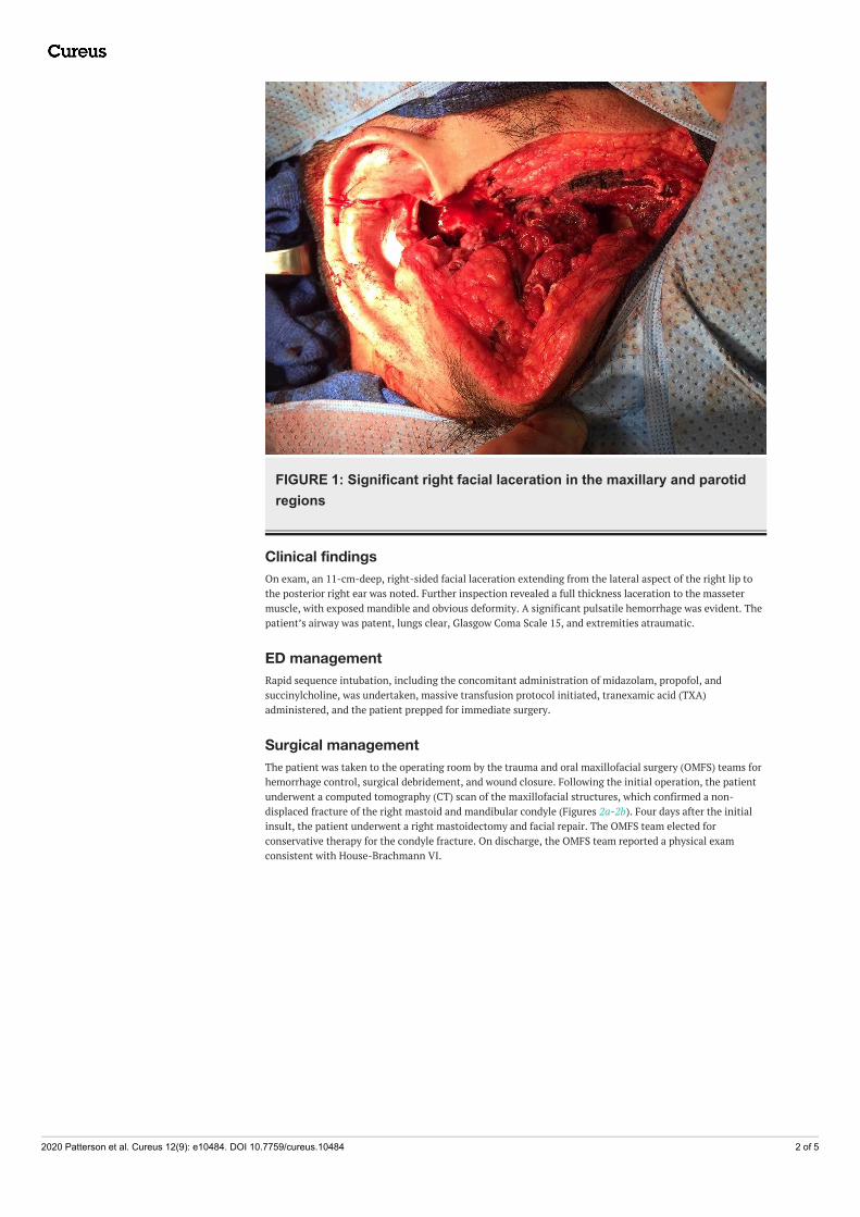
FIGURE 1: Significant right facial laceration in the maxillary and parotidregions
Clinical findingsOn exam, an 11-cm-deep, right-sided facial laceration extending from the lateral aspect of the right lip tothe posterior right ear was noted. Further inspection revealed a full thickness laceration to the massetermuscle, with exposed mandible and obvious deformity. A significant pulsatile hemorrhage was evident. Thepatient’s airway was patent, lungs clear, Glasgow Coma Scale 15, and extremities atraumatic.
ED managementRapid sequence intubation, including the concomitant administration of midazolam, propofol, andsuccinylcholine, was undertaken, massive transfusion protocol initiated, tranexamic acid (TXA)administered, and the patient prepped for immediate surgery.
Surgical managementThe patient was taken to the operating room by the trauma and oral maxillofacial surgery (OMFS) teams forhemorrhage control, surgical debridement, and wound closure. Following the initial operation, the patientunderwent a computed tomography (CT) scan of the maxillofacial structures, which confirmed a non-displaced fracture of the right mastoid and mandibular condyle (Figures 2a-2b). Four days after the initialinsult, the patient underwent a right mastoidectomy and facial repair. The OMFS team elected forconservative therapy for the condyle fracture. On discharge, the OMFS team reported a physical examconsistent with House-Brachmann VI.
2020 Patterson et al. Cureus 12(9): e10484. DOI 10.7759/cureus.10484 2 of 5
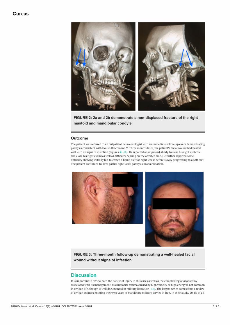
FIGURE 2: 2a and 2b demonstrate a non-displaced fracture of the rightmastoid and mandibular condyle
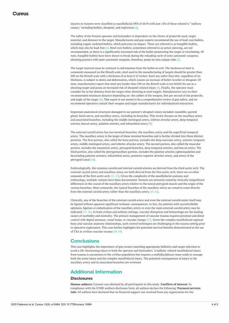
OutcomeThe patient was referred to an outpatient neuro-otologist with an immediate follow-up exam demonstratingparalysis consistent with House-Brachmann V. Three months later, the patient’s facial wound had healedwell with no signs of infection (Figures 3a-3b). He reported an improved ability to raise his right eyebrowand close his right eyelid as well as difficulty hearing on the affected side. He further reported somedifficulty chewing initially but tolerated a liquid diet for eight weeks before slowly progressing to a soft diet.The patient continued to have partial right facial paralysis on examination.
FIGURE 3: Three-month follow-up demonstrating a well-healed facialwound without signs of infection
DiscussionIt is important to review both the nature of injury in this case as well as the complex regional anatomyassociated with its management. Maxillofacial trauma caused by high velocity or high energy is not commonin civilian life, though is well documented in military literature [1,5]. The largest series comes from a reviewof civilian trainees entering their two years of mandatory military service in Iran. In their study, 20.4% of all
2020 Patterson et al. Cureus 12(9): e10484. DOI 10.7759/cureus.10484 3 of 5
injuries in trainees were classified as maxillofacial (903 of 4419) with just 14% of those related to “militarycauses,” including bullets, shrapnel, and explosions [4].
The safety of the firearm operator and bystanders is dependent on the choice of projectile used, targetmaterial, and distance to the target. Manufacturers and gun experts recommend the use of lead-core bullets,including copper-jacketed bullets, which pulverize on impact. These are referred to as frangible bullets,which may also be lead-free [6]. Steel core bullets, sometimes referred to as armor-piercing, are notrecommended, as there is a significantly increased risk of the bullet penetrating the target or ricocheting. Ofnote, frangible bullets have been shown to break during the reloading cycle of semi-automatic weapons;shooting practice with semi-automatic weapons, therefore, poses its own unique risks [7].
The target material must be resistant to deformation from the bullet as well. The hardness of steel iscommonly measured on the Brinell scale; steel used in the manufacturing of targets should be greater than500 on the Brinell scale with a thickness of at least 0.25 inches. Steel any softer than this, regardless of itsthickness, is subject to dents and deformation, which creates an increase of bullet ricochet or shrapnel. Ofnote, manufacturers report that steel any harder than 540 on the Brinell scale is too brittle for use as ashooting target and poses an increased risk of shrapnel-related injury [8]. Finally, the operator mustconsider his or her distance from the target when shooting at steel targets. Manufacturers vary on theirrecommended minimum distance depending on: the caliber of the weapon, feet per second of the projectile,and angle of the target [8]. This report is not meant to be a comprehensive review of gun safety, and werecommend operators consult their weapon and target manufacturers for individualized instruction.
Important anatomical structures damaged in our patient’s shrapnel-injury included: mandible, parotidgland, facial nerve, and maxillary artery, including its branches. This review focuses on the maxillary arteryand associated branches, including the middle meningeal artery, inferior alveolar artery, deep temporalarteries, buccal artery, palatine arteries, and infraorbital artery [9].
The external carotid artery has two terminal branches: the maxillary artery and the superficial temporalartery. The maxillary artery is the larger of these terminal branches and is further divided into three distinctportions. The first portion, also called the bony portion, includes the deep auricular artery, anterior tympanicartery, middle meningeal artery, and inferior alveolar artery. The second portion, also called the muscularportion, includes the masseteric artery, pterygoid branches, deep temporal arteries, and buccal artery. Thethird portion, also called the pterygomaxillary portion, includes the palatine arteries (sphenopalatine anddescending palatine arteries), infraorbital artery, posterior superior alveolar artery, and artery of thepterygoid canal [10].
Embryologically, the common carotid and internal carotid arteries are derived from the third aortic arch. Theexternal carotid artery and maxillary artery are both derived from the first aortic arch; there are no otherremnants of the first aortic arch [11-13]. Given the complexity of the maxillofacial anatomy andembryology, multiple variants have been documented. Variants are primarily noted by clinically insignificantdifferences in the course of the maxillary artery relative to the lateral pterygoid muscle and the origin of thevarious branches. Most commonly, the typical branches of the maxillary artery are noted to come directlyfrom the external carotid artery rather than the maxillary artery [13-14].
Clinically, any of the branches of the external carotid artery and even the external carotid artery itself maybe ligated without apparent significant ischemic consequences. In fact, for patients with uncontrollableepistaxis, ligation or embolization of the maxillary artery or even the main external carotid artery may beindicated [15-16]. In both civilian and military settings, vascular disruption and hemorrhage are the leadingcauses of morbidity and mortality. The primary management of vascular trauma requires proximal and distalcontrol with digital pressure, vessel loops, or vascular clamps [17]. Given the complex maxillofacial regionalbony and vascular anatomy relationships, such control techniques are challenging in the trauma setting priorto operative exploration. This case further highlights the potential survival benefits demonstrated in the useof TXA in civilian vascular trauma [18-19].
ConclusionsThis case highlights the importance of gun owners matching appropriate ballistics and target selection toavoid a life-threatening injury to both the operator and bystanders. A ballistic-related maxillofacial injuryfrom trauma is uncommon in the civilian population but requires a multidisciplinary team ready to manageboth the acute injury and the complex maxillofacial injury. The potential consequences of injury to themaxillary artery and its associated branches are reviewed.
Additional InformationDisclosuresHuman subjects: Consent was obtained by all participants in this study. Conflicts of interest: Incompliance with the ICMJE uniform disclosure form, all authors declare the following: Payment/servicesinfo: All authors have declared that no financial support was received from any organization for the
2020 Patterson et al. Cureus 12(9): e10484. DOI 10.7759/cureus.10484 4 of 5
submitted work. Financial relationships: All authors have declared that they have no financialrelationships at present or within the previous three years with any organizations that might have aninterest in the submitted work. Other relationships: All authors have declared that there are no otherrelationships or activities that could appear to have influenced the submitted work.
References1. Peled M, Leiser Y, Emodi O, Krausz A: Treatment protocol for high velocity/high energy gunshot injuries to
the face. Craniomaxillofac Trauma Reconstr. 2012, 5:31-40. 10.1055/s-0031-12935182. Zadik Y: The role of the military dental surgeon in treating facial injuries: a case report . Mil Med. 2007,
172:1284-1286. 10.7205/milmed.172.12.12843. Shuker ST: The immediate lifesaving management of maxillofacial, life-threatening haemorrhages due to
IED and/or shrapnel injuries: "when hazard is in hesitation, not in the action". J Craniomaxillofac Surg.2012, 40:534-540. 10.1016/j.jcms.2011.09.005
4. Kalantar MMH, Ebrahimi A, Askary A: Oral and maxillofacial injuries in civilian recruits during mandatorycombat training at military garrisons: a nationwide survey. Trauma Mon. 2012, 17:337-340.10.5812/traumamon.6982
5. Motamedi MHK: Primary management of maxillofacial hard and soft tissue gunshot and shrapnel injuries . JOral Maxillofac Surg. 2003, 61:1390-1398. 10.1016/j.joms.2003.07.001
6. US5616642A - Lead-free frangible ammunition . (1997). Accessed: April 1, 2020:https://patents.google.com/patent/US5616642A/en.
7. Frangible ammunition for training and safety: the good and the bad . (2007). Accessed: April 1, 2020:https://www.policemag.com/372947/frangible-ammunition-for-training-and-safety-the-good-and-the-bad .
8. Why you should be shooting steel targets and some guidelines . (2018). Accessed: April 1, 2020:https://www.usacarry.com/shooting-steel-targets/.
9. Netter FH: Plate 72. Atlas of Human Anatomy, Sixth Edition. Saunders (ed): Elsevier, Pennsylvania; 2014.10. Standring S: Section 4: head and neck . Gray’s Anatomy: The Anatomical Basis of Clinical Practice. Elsevier
Health Sciences, Pennsylvania; 2016. 404-677.11. Endean E, Maley B: Chapter 2, embryology. Rutherford’s Vascular Surgery and Endovascular Therapy.
Elsevier, Pennsylvania; 2018. 15-33.12. Kau T, Sinzig M, Gasser J, et al.: Aortic development and anomalies. Semin Intervent Radiol. 2007, 141-152.
10.1055/s-2007-98004013. Gofur EM, Al Khalili Y: Anatomy, Head and Neck, Internal Maxillary Arteries. StatPearls . StatPearls
[Internet], Treasure Island (FL); 2019.14. Maeda S, Aizawa Y, Kumaki K, Kageyama I: Variations in the course of the maxillary artery in Japanese
adults. Anat Sci Int. 2012, 87:187-194. 10.1007/s12565-012-0146-x15. Waldron J, Stafford N: Ligation of the external carotid artery for severe epistaxis . J Otolaryngol. 1992,
21:249-251.16. Pritikin JB, Caldarelli DD, Panje WR: Endoscopic ligation of the internal maxillary artery for treatment of
intractable posterior epistaxis. Ann Otol Rhinol Laryngol. 1998, 107:85-91. 10.1177/00034894981070020117. Laser A, Toursavadkohi S, Rasmussen T: Chapter 27, vascular trauma. Greenfield’s Surgery: Scientific
Principles & Practice. Mullholland MW, Lillemoe KD, Doherty GM, Upchurch GR, Alam H, Pawlik TM (ed):Wolters Kluwer, Pennsylvania; 2016. 699-716.
18. GnanaDev R, Dong F, Ali A, et al.: Comparing mortality and hospital length of stay in the setting of truncaland peripheral vascular trauma in patients treated with tranexamic acid on initial presentation. J Vasc Surg.2019, 69:188-189. 10.1016/j.jvs.2019.04.267
19. Neeki M, Dong F, Toy J, et al.: Tranexamic acid in civilian trauma care in the California PrehospitalAntifibrinolytic Therapy study. West J Emerg Med. 2018, 19:977-986. 10.5811/westjem.2018.8.39336
2020 Patterson et al. Cureus 12(9): e10484. DOI 10.7759/cureus.10484 5 of 5