Embed Size (px)
Citation preview

An Insight into the Molecular Basis of Salt Tolerance ofL-myo-Inositol 1-P Synthase (PcINO1) from Porteresiacoarctata (Roxb.) Tateoka, a Halophytic Wild Rice1
Krishnarup Ghosh Dastidar, Susmita Maitra, Lily Goswami, Debjani Roy, Kali Pada Das,and Arun Lahiri Majumder*
Plant Molecular and Cellular Genetics (K.G.D., S.M., L.G., A.L.M.), Bioinformatics Center (D.R.), andDepartment of Chemistry (K.P.D.), Bose Institute, Calcutta Improvement Trust Scheme-VIIM,Calcutta 700 054, India
The molecular basis of salt tolerance of L-myo-inositol 1-P synthase (MIPS; EC 5.5.1.4) from Porteresia coarctata (Roxb.) Tateoka(PcINO1, AF412340) earlier reported from this laboratory, has been analyzed by in vitro mutant and hybrid generation andsubsequent biochemical and biophysical studies of the recombinant proteins. A 37-amino acid stretch between Trp-174 and Ser-210 has been confirmed as the salt-tolerance determinant domain in PcINO1 both by loss or gain of salt tolerance by eitherdeletion or by addition to salt-sensitive MIPS(s) of Oryza (OsINO1) and Brassica juncea (BjINO1). This was further verified bygrowth analysis under salt environment of Schizosaccharomyces pombe transformed with the various gene constructs and studieson the differential behavior of mutant and wild proteins by Trp fluorescence, aggregation, and circular dichroism spectra in thepresence of salt. 4,4#-Dianilino-1,1#-binaphthyl-5,5-disulfonic acid binding experiments revealed a lower hydrophobic surfaceon PcINO1 than OsINO1, contributed by this 37-amino acid stretch explaining the differential behavior of OsINO1 and PcINO1both with respect to their enzymatic functions and thermodynamic stability in high salt environment. Detailed amino acidsequence comparison and modeling studies revealed the interposition of polar and charged residues and a well-connectedhydrogen-bonding network formed by Ser and Thr in this stretch of PcINO1. On the contrary, hydrophobic residues clusteredin two continuous stretches in the corresponding region of OsINO1 form a strong hydrophobic patch on the surface. It isconceivable that salt-tolerant MIPS proteins may be designed out of the salt-sensitive plant MIPS proteins by replacement ofthe corresponding amino acid stretch by the designated 37-amino acid stretch of PcINO1.
Metabolic engineering is an effective technology fordeveloping genetically modified stress-tolerant organ-isms through the production of stress-tolerant deter-minants like osmolytes/osmoprotectants. Specifically,engineering the structural genes coding for enzymesresponsible for biosynthesis of compatible solutes orosmolytes to develop abiotic stress-tolerant transgenicplants has become widespread in the field of stressbiology (Hasegawa et al., 2000). Apart from a numberof various compounds such as Pro, Gly betaine, tre-halose, and sugar alcohols likemannitol, etc. (Hasegawaet al., 2000), the ubiquitous inositol, a six-memberedcyclohexitol, and its other derivatives have been im-plicated to play a vital role in osmotolerance (Bohnert
et al., 1995) in addition to its known function in otherphysiological processes such as cell signaling, mem-brane biogenesis, growth regulation, phosphorousstorage, and as a high-energy phosphate donor (Biswaset al., 1984; Loewus and Murthy, 2000).
Of the eight possible geometrical isomers, myo-inositol is the most abundant in the biological systemand occupies the central position in inositol metabo-lism (Bohnert et al., 1995; Loewus and Murthy, 2000;Majumder et al., 2003). De novo biosynthesis of freemyo-inositol follows a highly conserved two-step bio-chemical pathway in all living organisms and is cat-alyzed by L-myo-inositol 1-P synthase (MIPS; EC5.5.1.4). The MIPS converts D-Glc 6-P to L-myo-inositol1-P, and is followed by its dephosphorylation by aspecific Mg21-dependent inositol monophosphataseleading to free inositol (Majumder et al., 2003). Thisfree inositol can then be channeled to the production ofdifferent methylated derivatives, which are also activeas potent osmolytes for amelioration of oxidativedamage during osmotic stress (Bohnert et al., 1995).In addition, myo-inositol is known to be utilized in theabiotic stress-induced galactinol and raffinose synthe-sis (Taji et al., 2002) apart from several other metabolicsinks of its utilization such as the alternate biosyn-thetic pathway to L-ascorbic acid (Lorence et al., 2004)or the pentosyl or uronosyl component of the plant cell
1 This work was supported by research grants to A.L.M. from theDepartment of Biotechnology, Government of India. K.G.D. and L.G.thank the Council of Scientific and Industrial Research, Governmentof India, for Senior Research Fellowships.
* Corresponding author; e-mail [email protected]; fax91–33–2334–3886.
The author responsible for distribution of materials integral to thefindings presented in this article in accordance with the policydescribed in the Instructions for Authors (www.plantphysiol.org) is:Arun Lahiri Majumder ([email protected]).
Article, publication date, and citation information can be found atwww.plantphysiol.org/cgi/doi/10.1104/pp.105.075150.
Plant Physiology, April 2006, Vol. 140, pp. 1279–1296, www.plantphysiol.org � 2006 American Society of Plant Biologists 1279

wall (Kanter et al., 2005). The MIPS has been reportedfrommore than 70 different organisms so far includinga wide variety of prokaryotic, archaeal, cyanobacterial,and eukaryotic sources. The structural gene for MIPS,termed INO1, has been cloned from a number of widelydifferent organisms that seem to share an evolution-ary conserved core catalytic domain across phyla(Majumder et al., 2003).
An increase in the production of intracellular ino-sitol and its other methylated derivatives, such asononitol and pinitol, as a result of the coordinatetranscriptional induction of the INO1 and the inositolmethyl transferase (Imt1) gene(s) for increased salttolerance have been reported in several plants includ-ing transgenic ones (Vernon and Bohnert, 1992; Ishitaniet al., 1996; Sheveleva et al., 1997; Nelson et al., 1998).Alternatively, increased production of inositol can beachieved even under salt environment by a stress-tolerant MIPS protein by virtue of its ability to remainfunctional under such condition. This has been docu-mented earlier from this laboratory through isolationand characterization of a novel salt-tolerant MIPS cod-ing gene, termed PcINO1 from the halophytic wild rice,Porteresia coarctata (Roxb.) Tateoka (Majee et al., 2004).In vitro and in vivo characterization revealed that thePcINO1 gene product is capable of retaining its enzy-matic function even in presence of 500 mM NaCl asopposed to its salt-sensitive homolog from the culti-vated rice (Oryza sativa;OsINO1; AB012107). Functionalexpression of this gene in planta led to a salt-tolerantphenotype of the transgenic tobacco (Nicotiana tabacum)with an increased intracellular inositol pool under saltstress, suggesting retention of enzymatic activity of theexpressed gene product in vivo. Although a 37-aminoacid residue stretch in PcINO1 was found to be pri-marily responsible for its salt-tolerant characteristics(Majee et al., 2004), an insight into the molecular basisof salt tolerance of this unique enzyme protein asdetermined by the 37-amino acid residue stretch, wasstill awaited.
In this study, we have established the importance ofthe 37-amino acid stretch of PcINO1 offering an insightinto the molecular basis of the salt-tolerant character-istics of this protein through biochemical and biophys-ical characterization of different in vitro generatedmutant and hybrid enzymes. This was followed by com-parison of the properties of these mutant proteinswith those of the wild as well as the salt-sensitive ricehomolog, OsINO1, and modeling studies with yeast(Saccharomyces cerevisiae) MIPS crystal structure as atemplate. Furthermore, we have also been able to de-sign a salt-tolerant MIPS out of a salt-sensitive onewith this 37-amino acid stretch pointing toward apossible biotechnological application of such studiesin the future.
For the sake of uniformity, a new gene nomenclatureof INO1 has been adopted in this manuscript, e.g.OsINO1 for the cultivated rice INO1 and PcINO1 forPorteresia coarctata INO1, in place of earlier RINO1 orPINO1 nomenclature (Majee et al., 2004). The chimeric
proteins have been designated as OsTPcINO1 orBjTPcINO1, wherein a part of PcINO1 has been in-serted onto OsINO1 or BjINO1.
RESULTS
Generation and Molecular Cloning of DPcINO1.1,DPcINO1.2, OsTPcINO1, BjINO1, and BjTPcINO1
Strategy of generating DPcINO1.1, DPcINO1.2,OsTPcINO1, and BjTPcINO1 has been schematicallydescribed in Figure 1. In the case of DPcINO1.1 (Fig.1B), a stretch of 60 nucleotides coding for 20 aminoacids from Asn-342 to Lys-361 near the C terminus ofPcINO1 has been deleted. This stretch is part of theidentified core catalytic domain of MIPS (Majumderet al., 2003) and harbors two important Lys residuesimplicated in the catalytic mechanism ofMIPS reaction(Stein andGeiger, 2002). For generatingDPcINO1.2 (Fig.1B), a 111-bp stretch corresponding to a 37-amino acidresidue stretch between Trp-174 and Ser-210 has beendeleted from near the N terminus of PcINO1 withoutaffecting the MIPS catalytic activity. In the case ofOsTPcINO1 (Fig. 1C), the advantage of having twounique restriction sites, PstI and EcoRV in both theOsINO1 and PcINO1 genes at the same position hasbeen exploited. The PstI-EcoRV fragment correspondsto a peptide larger than 37-amino acid residue stretchthat includes additionally the flanking sequences atboth sides of the Trp-174 to Ser-210 stretch that areidentical in both the OsINO1 and PcINO1 proteins.Therefore, replacing the PstI-EcoRV fragment ofOsINO1 with that of PcINO1 essentially replaced the37-amino acid stretch of OsINO1 with that of PcINO1.The corresponding 37-amino acid stretch of BjINO1,which showed approximately 98% and 55% sequenceidentity with Brassica napus (AAB06756) and S. cerevi-siae (A30902) MIPS, respectively, has been replacedwith the same of PcINO1 by PCR to generate BjTPcINO1(Fig. 1D).
Functional Complementation of DPcINO1.1, DPcINO1.2,and OsTPcINO1
Functional viability of each PcINO1 mutant andhybrid genes have been tested by the yeast functionalcomplementation assay as described earlier (Chatterjeeet al., 2004; Majee et al., 2004). Results of such experi-ments (Fig. 2) reveal that, except for the DPcINO1.1, allmutants and hybrid genes complemented the FY250strain, thereby adducing evidence that the changesmade in the constructs by in vitro mutagenesis didnot alter the MIPS catalytic activity in any of them. Wesubcloned DPcINO1.1, DPcINO1.2, and OsTPcINO1downstream of a strong constitutively active glyceral-dehyde 3-P dehydrogenase (GPD) promoter in yeastmulticopy binary vector p426GPD (Sambrook et al.,1989;Mumberg et al., 1995), followed by transformationof FY250 with p426GPD (negative control) along withthe respective constructs. All the transformants were
Dastidar et al.
1280 Plant Physiol. Vol. 140, 2006
selected on uracil dropout medium and maintained onsolid synthetic media containing no uracil and 10 mM
inositol where all grew equally well (Fig. 2A). However,when inositol concentration was gradually lowereddown to zero, only PcINO1 (Fig. 2B, 5), DPcINO1.2(Fig. 2B, 4), and OsTPcINO1 (Fig. 2B, 3) transformedcells showed growth whereas p426GPD (Fig. 2B, 1) andDPcINO1.1(Fig. 2B, 2) transformed cells showed nogrowth.
Elucidating Salt-Tolerant Phenotypeof PcINO1-Transformed
Schizosaccharomyces pombe
Functional expression of PcINO1 has earlier beenreported to confer salt-tolerant phenotype with un-abated photosynthetic functions to transformed to-bacco plants, a model photosynthetic system (Majeeet al., 2004). Following such observations, we attemptedfunctional expression of PcINO1, OsINO1, DPcINO1.2,andOsTPcINO1 hybrid genes under salt-stressed con-ditions to see their effect on the growth of a non-
photosynthetic lower eukaryote, S. pombe. Being a naturalinositol auxotroph, which requires approximately 50to 55 mM basal level of inositol for its normal growth(Ingavale and Bachhawat, 1999), S. pombe presents amodel system for in vivo complementation as well asfor studying the effect of functional expression of thegenes PcINO1, OsINO1, DPcINO1.2, and OsTPcINO1on the growth of the organism in salt environment.However in this experiment, since the cells were givensalt stress, a basal level of 2 mM inositol has beenmaintained throughout along with the increasing con-centration of NaCl. In absence of any salt or at lowersalt concentrations like 100 mM, transformed cellsshowed more or less similar growth pattern (Fig. 3,A and B). But on increasingNaCl stress beyond 100mM
NaCl, a remarkable difference in growth pattern of thetransformed cells was observed. S. pombe cells trans-formed with OsINO1 and DPcINO1.2 showed poorgrowth response when grown with further increase inNaCl concentration as opposed to PcINO1- and OsTPcINO1-transformed cells, which were found to groweven in presence of 500 mM NaCl (Fig. 3, C–F).
Figure 1. Scheme for development of different PcINO1 mutants, BjINO1, OsTPcINO1, and BjTPcINO1. A, Schematicrepresentation of PcINO1,OsINO1, andBjINO1. B, Strategy of generating deletionmutantsDPcINO1.2 andDPcINO1.1 by PCR.C,OsTPcINO1 by fragment exchange method. D, BjTPcINO1 by PCR. The major difference between OsINO1 or BjINO1 withPcINO1, as highlighted by different shading, is the 37-amino acid residue stretch (Trp-174 to Ser-210). A part of the core catalyticregion (20-amino acid residue stretch) is also highlighted (Asn-342 to Lys-361). Deletion of the 37-amino acid residue stretchresulted in DPcINO1.2 and deletion of the 20-amino acid residue stretch resulted in DPcINO1.1. Replacing the corresponding37-amino acid residue stretch from OsINO1 with that of PcINO1 generated the hybrid OsTPcINO1 or BjTPcINO1.
Molecular Insight into Salt Tolerance of MIPS from Wild Rice
Plant Physiol. Vol. 140, 2006 1281

Although extent of growth was found to be reducedeven in the case of PcINO1- and OsTPcINO1-transformed cells at 500 mM NaCl concentration, in thecase ofOsINO1 and DPcINO1.2 transformants, virtuallyno growth was observed at similar salt concentration.
Bacterial Overexpression, Purification, and in VitroMIPS Activity of Recombinant DPINO1.1, DPINO 1.2,OsTPcINO1, BjINO1, and BjTPcINO1 Proteins
DPcINO1.1, DPcINO1.2, BjINO1, OsTPcINO1, andBjTPcINO1 were overexpressed as approximately60-kD proteins, predominantly in the particulate frac-tion, as judged by the 10% SDS-PAGE and western-blotanalysis of respective supernatant and pellet of inducedcells (Fig. 4, A–D). In both SDS-PAGE and western-blotanalysis, a slight decrease in the Mr of overexpressed
protein bands were observed commensurate with thesize of DPcINO1.1 and DPcINO1.2 with the deletion of20- and 37-amino acid residue stretches, respectively.
The overexpressed DPcINO1.1, DPcINO1.2, OsTPcINO1, BjINO1, and BjTPcINO1 proteins were puri-fied to homogeneity and assayed for MIPS activityfollowing the procedure described earlier (Majee et al.,2004). When in vitro specific activity was determinedusing approximately 20 mg of each purified enzyme,both DPcINO1.2 and OsTPcINO1 were found to bealmost as active as wild PcINO1 and OsINO1 (positivecontrols), as opposed to DPcINO1.1, which turned outto be inactive (Fig. 4E). BjTPcINO1 was also found tobe equally active as BjINO1 (Fig. 4E) and very close tothose of OsINO1 and PcINO1. Both purified DPcINO1.2and OsTPcINO1 proteins were biochemically charac-terized and compared with their corresponding wildversions (Table I). Km and Vmax for D-Glc 6-P andNAD1 of purified DPcINO1.2 and OsTPcINO1 werefound to be comparable to those of the wild OsINO1and PcINO1 proteins. OsTPcINO1 was found to beoptimally active slightly in the higher pH range (8.0–8.5) in comparison to wild proteins.
In Vitro Salt Tolerance versus Sensitivity of Mutantand Hybrid Proteins
We earlier showed that with increasing concentra-tion of NaCl, the MIPS activity of PcINO1 remainedunaltered but that of OsINO1 progressively decreased(Majee et al., 2004). The 37-amino acid residue stretchdeficient mutant of PcINO1 (DPcINO1.2) or BjINO1was found to behave as OsINO1, showing a steadydecrease in MIPS activity with increased salt con-centration (Fig. 4F). On the other hand, the hybridOsTPcINO1 and BjTPcINO1, in which the 37-amino acid residue stretch of PcINO1 replaces the
Figure 2. Functional complementation of mutant genes of PcINO1 andthe hybrid derivative of OsINO1 in S. cerevisiae FY250, an inositolauxotroph. A, Growth of FY250-transformed cells with empty vector,p426GPD (1, 2control), DPcINO1.1 (2), OsTPcINO1 (3), DPcINO1.2(4), and PcINO1 (5, 1control) on complete synthetic solid media inpresence of 10 mM inositol in absence of uracil (ura). B, Growth oftransformed cells as inA above in absence of both inositol and uracil (ura).
Figure 3. Phenotypic expression of different PcI-NO1 genes in S. pombe. A to F, Growth pattern ofS. pombe PR109, transformed with differentpREP1 constructs harboring OsINO1, PcINO1,DPcINO1.2, andOsTPcINO1 genes on completesolid synthetic media containing increasingconcentration ofNaCl, as indicated. 1, Empty vec-tor, pREP1; 2, OsINO1; 3, OsTPcINO1; 4,DPcINO1.2; and 5, PcINO1.
Dastidar et al.
1282 Plant Physiol. Vol. 140, 2006

corresponding 37-amino acid residue stretch of OsINO1or BjINO1, behave like PcINO1 showing no decreasein MIPS catalytic activity on increasing salt concentra-tion up to 500 mM (Fig. 4F). These results clearlyindicate that the specific 37-amino acid residue stretchfrom Trp-174 to Ser-210 of PcINO1 provides the salt-tolerance property to OsTPcINO1 and BjTPcINO1 andits absence is responsible for the salt sensitivity ofDPcINO1.2, OsINO1, and BjINO1.
Trp Fluorescence Studies of Mutant and Hybrid Proteinsin the Presence and Absence of NaCl
We showed earlier that with increasing salt con-centration, Trp fluorescence intensity of OsINO1 de-creased significantly but that of PcINO1 decreasednegligibly (Majee et al., 2004). The Trp emission spec-tra of the mutant and hybrid proteins in presence ofincreasing concentration of NaCl have been shown in
Figure 4. Bacterial expression andwestern-blot analysis of various PcINO1mutants,Os::PcINO1, BjINO1, and Bj::PcINO1 geneproducts, their in vitro MIPS activity, and salt tolerance vis a vis salt sensitivity. A and C, Ten-percent SDS-PAGE. B and D,Correspondingwestern-blot analysis of PcINO1mutants,Os::PcINO1,BjINO1, andBj::PcINO1, respectively. A andB, Lane 1,Mr
marker; lanes 2 to 5, pellet fractions of overexpressed PcINO1,DPcINO1.1,DPcINO1.2, andOs::PcINO1, respectively; lanes 6 to9, corresponding supernatants, respectively. C and D, Lane 1, Mr marker; lane 2, OsINO1 (1 control); lanes 3 and 4, inducedBjINO1 supernatant and pellet fractions, respectively; lanes 5 and 6, induced Bj::PcINO1 supernatant and pellet fractions,respectively. E, In vitro MIPS activity of purified PcINO1 mutants, Os::PcINO1, BjINO1, and Bj::PcINO1 along with the positivecontrols PcINO1 and OsINO1. Specific activity is described in terms of mmol inositol 1-P h21 mg protein21. In each case 20 mgpurified proteinwas taken. Error bars indicate the deviation of triplicate experiments. F, Effect of increasing concentration of NaClon theMIPSactivityofDPcINO1.2 (white circle),Os::PcINO1 (black triangle), BjINO1 (white square), Bj::PcINO1 (white triangle),alongwithOsINO1 (2control, black square) and PcINO1 (1control, black circle). Approximately 20mg of each purified enzymewas assayed in presence of indicated concentration of NaCl. Error bars indicate the deviation from triplicate experiments.
Table I. Comparison of biochemical properties of DPcINO1.2 and OsTPcINO1 with their wild enzymes
SystemKm Vmax
pH OptimaTemperature
Optima �CG-6-P NAD1 G-6-P NAD1
mM mmol I 1-P/min
OsINO1a 3.0 0.188 0.072 0.068 7.5 37PcINO1a 2.5 0.166 0.095 0.087 8.0 37DPcINO1.2 2.83 0.451 0.103 0.096 8.0 37OsTPcINO1 3.19 0.293 0.115 0.081 8.0–8.5 37
aData from Majee et al. (2004).
Molecular Insight into Salt Tolerance of MIPS from Wild Rice
Plant Physiol. Vol. 140, 2006 1283
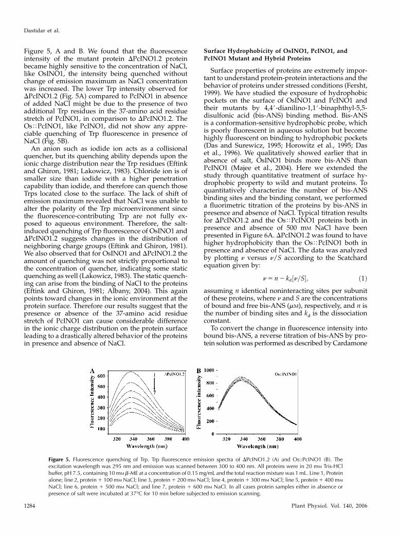
Figure 5, A and B. We found that the fluorescenceintensity of the mutant protein DPcINO1.2 proteinbecame highly sensitive to the concentration of NaCl,like OsINO1, the intensity being quenched withoutchange of emission maximum as NaCl concentrationwas increased. The lower Trp intensity observed forDPcINO1.2 (Fig. 5A) compared to PcINO1 in absenceof added NaCl might be due to the presence of twoadditional Trp residues in the 37-amino acid residuestretch of PcINO1, in comparison to DPcINO1.2. TheOsTPcINO1, like PcINO1, did not show any appre-ciable quenching of Trp fluorescence in presence ofNaCl (Fig. 5B).
An anion such as iodide ion acts as a collisionalquencher, but its quenching ability depends upon theionic charge distribution near the Trp residues (Eftinkand Ghiron, 1981; Lakowicz, 1983). Chloride ion is ofsmaller size than iodide with a higher penetrationcapability than iodide, and therefore can quench thoseTrps located close to the surface. The lack of shift ofemission maximum revealed that NaCl was unable toalter the polarity of the Trp microenvironment sincethe fluorescence-contributing Trp are not fully ex-posed to aqueous environment. Therefore, the salt-induced quenching of Trp fluorescence of OsINO1 andDPcINO1.2 suggests changes in the distribution ofneighboring charge groups (Eftink and Ghiron, 1981).We also observed that for OsINO1 and DPcINO1.2 theamount of quenching was not strictly proportional tothe concentration of quencher, indicating some staticquenching as well (Lakowicz, 1983). The static quench-ing can arise from the binding of NaCl to the proteins(Eftink and Ghiron, 1981; Albany, 2004). This againpoints toward changes in the ionic environment at theprotein surface. Therefore our results suggest that thepresence or absence of the 37-amino acid residuestretch of PcINO1 can cause considerable differencein the ionic charge distribution on the protein surfaceleading to a drastically altered behavior of the proteinsin presence and absence of NaCl.
Surface Hydrophobicity of OsINO1, PcINO1, and
PcINO1 Mutant and Hybrid Proteins
Surface properties of proteins are extremely impor-tant to understand protein-protein interactions and thebehavior of proteins under stressed conditions (Fersht,1999). We have studied the exposure of hydrophobicpockets on the surface of OsINO1 and PcINO1 andtheir mutants by 4,4#-dianilino-1,1#-binaphthyl-5,5-disulfonic acid (bis-ANS) binding method. Bis-ANSis a conformation-sensitive hydrophobic probe, whichis poorly fluorescent in aqueous solution but becomehighly fluorescent on binding to hydrophobic pockets(Das and Surewicz, 1995; Horowitz et al., 1995; Daset al., 1996). We qualitatively showed earlier that inabsence of salt, OsINO1 binds more bis-ANS thanPcINO1 (Majee et al., 2004). Here we extended thestudy through quantitative treatment of surface hy-drophobic property to wild and mutant proteins. Toquantitatively characterize the number of bis-ANSbinding sites and the binding constant, we performeda fluorimetric titration of the proteins by bis-ANS inpresence and absence of NaCl. Typical titration resultsfor DPcINO1.2 and the OsTPcINO1 proteins both inpresence and absence of 500 mM NaCl have beenpresented in Figure 6A. DPcINO1.2 was found to havehigher hydrophobicity than the OsTPcINO1 both inpresence and absence of NaCl. The data was analyzedby plotting n versus n/S according to the Scatchardequation given by:
n5 n2 kd½n=S�; ð1Þ
assuming n identical noninteracting sites per subunitof these proteins, where n and S are the concentrationsof bound and free bis-ANS (mM), respectively, and n isthe number of binding sites and kd is the dissociationconstant.
To convert the change in fluorescence intensity intobound bis-ANS, a reverse titration of bis-ANS by pro-tein solutionwas performed as described by Cardamone
Figure 5. Fluorescence quenching of Trp. Trp fluorescence emission spectra of DPcINO1.2 (A) and Os::PcINO1 (B). Theexcitation wavelength was 295 nm and emission was scanned between 300 to 400 nm. All proteins were in 20 mM Tris-HClbuffer, pH 7.5, containing 10mM b-ME at a concentration of 0.15 mg/mL and the total reaction mixture was 1 mL. Line 1, Proteinalone; line 2, protein1 100 mM NaCl; line 3, protein1 200 mM NaCl; line 4, protein1 300 mM NaCl; line 5, protein1 400 mM
NaCl; line 6, protein 1 500 mM NaCl; and line 7, protein 1 600 mM NaCl. In all cases protein samples either in absence orpresence of salt were incubated at 37�C for 10 min before subjected to emission scanning.
Dastidar et al.
1284 Plant Physiol. Vol. 140, 2006

and Puri (1992). Typical Scatchard plots for the bis-ANS-DPcINO1.2 binding in presence and absence ofsalt have been shown in Figure 6, B and C. From theslope and intercepts of this plot the number of bindingsite (n) and dissociation constant for binding (kd)was calculated. These values for OsINO1, PcINO1,DPcINO1.2, and OsTPcINO1 have been presented inTable II. It was observed that both OsINO1 andDPcINO1.2 had a huge increase in n when NaCl wasincreased from 0 to 500 mM. Relatively small changes inn were observed for PcINO1 and OsTPcINO1, decreas-ing slightly for the former and increasing a little for thelatter on moving from no salt to high salt concentra-
tion. The dissociation constant in presence of salt wasalways found to have increased somewhat than thatwithout salt. From Table II, it is seen that in absence ofsalt, removal of the 37-amino acid residue stretch ofPcINO1 from Trp-174 to Ser-210 led to a marginaldecrease of n from 1.25 to 0.94, but in the presence of500 mM NaCl, n increased approximately 10-fold from0.7 to 6.9. Therefore the absence of the 37-amino acidresidue stretch of PcINO1 in DPcINO1.2 shifts thehydrophilic-lipophilic balance of the folded protein toexpose hydrophobic sites on its surface in the presence ofNaCl. Similarly, when this 37-amino acid residue stretchof PcINO1 replaces that in OsINO1, the number of
Figure 6. Surface hydrophobicity of PcINO1 mutant proteins. A, Bis-ANS binding titration of DPcINO1.2 and OsTPcINO1 inpresence and absence of added salt. A total of 0.15 mg/mL of each protein, in 20 mM Tris-HCl (pH 7.5), and 10 mM b-ME wastitrated in total 1 mL reaction volume by aqueous solution of bis-ANS. The excitation and emission wavelengths were 390 and490 nm, respectively. The intensities at 490 nm, from the titration, were plotted as a function of bis-ANS concentration. In boththe cases of titration in no salt and salt condition, protein was incubated at 37�C either in absence or presence of salt for 10 minbefore being subjected to titration. Black square, DPcINO1.2 1 0 mM NaCl; black circle, DPcINO1.2 1 500 mM NaCl; whitetriangle, OsTPcINO11 0mMNaCl; and black triangle, OsTPcINO11 500mMNaCl. B and C, Typical representative Scatchardplot of DPcINO1.2 for the determination of stoichiometry (n) and the dissociation constant (kd) in absence (B) and presence (C) of500 mM NaCl. Respective insets show the double reciprocal plot of reverse titration data as a function of protein concentrationfor the determination of DFmax. In reverse titration also, protein samples were incubated at 37�C, either in absence or presence ofsalt for 10 min before being subjected to titration under no salt and salt conditions, respectively.
Molecular Insight into Salt Tolerance of MIPS from Wild Rice
Plant Physiol. Vol. 140, 2006 1285

hydrophobic sites decreases in absence of NaCl andmore drastically in presence of NaCl. Thus, this 37-aminoacid residue stretch is so unique that its presence inPcINO1 and OsTPcINO1 minimizes the structuralchanges of the proteins in presence of salt but its absencein DPcINO1.2 and also in OsINO1 helps change itssurface becoming highly hydrophobic.
Aggregation of OsINO1, PcINO1, and PcINO1
Mutant and Hybrid Proteins in the Presenceand Absence of NaCl
We showed earlier that in the presence of high salt,OsINO1 forms nonspecific molecular aggregates, whilePcINO1 remains unchanged (Majee et al., 2004). Tofind out the physical basis of salt sensitivity ofDPcINO1.2 vis a vis salt tolerance of OsTPcINO1, wemonitored static light scattering of purified DPcINO1.2(Fig. 7A) and OsTPcINO1 (Fig. 7B) in presence andabsence of NaCl both at 37�C and 25�C. At 37�C, alongwith increase in incubation time, DPcINO1.2 showed asteady formation of macromolecular aggregates, whichwas salt concentration dependent, like OsINO1. In thecase of DPcINO1.2 both the degree as well as the rate offormation of the aggregates is much faster than thoseof OsINO1 and increased with the increasing concen-
tration of NaCl. DPcINO1.2 reached saturation within30 min of incubation, whereas OsINO1 reached thesame at 140 min. However OsTPcINO1 in all NaClconcentrations showed no aggregation even after170 min of incubation at 37�C, like PcINO1. At 25�C,the rate as well as extent of aggregation was found tobe reduced (data not shown). Aggregation of bothOsINO1 and DPcINO1.2 was also found to be criticallydependant on the protein concentration and reducedto a great extent when protein concentration wasbelow approximately 300 mg/mL.
Circular Dichroism Spectroscopy in the Presence andAbsence of NaCl
To investigate the details of any structural changeoccurring at the level of secondary structure of thesemutants in presence of NaCl we took far-UV circulardichroism (CD) spectra of purified OsINO1, PcINO1,DPcINO1.2, and OsTPcINO1 in presence and absenceof NaCl at room temperature (Fig. 8, A–D). PcINO1and the OsTPcINO1 (Fig. 8, A and D) showed distincta-helical characteristics, which remained fairly unal-tered by the addition of salt, while the characteristicsof OsINO1 and DPcINO1.2 changed with salt (Fig. 8, Band C). In this case the spectrum changed fromw shapeto v shape but minimum shifted above 220 nm atsalt concentration of 500 mM. The spectral data werefitted to several secondary structure analysis pro-grams (Sreerama, 1993; Sreerama et al., 1999) such asSELCON3, CONTIN, CDSSTR, CDNN, etc. The gen-eral agreement between the fitted parameters fromdifferent programs were not found to be satisfactory,but in most cases an increase in b-sheet structure atthe expense of helical structure and increase in ran-dom coil contribution was found to be observed (datanot shown). The difference in salt effect on the CDcharacteristics between OsINO1 and OsTPcINO1 asalso between PcINO1 and DPcINO1.2 indicates that
Table II. Determination of stoichiometry (n) and dissociation constant(kd) of different systems in absence and presence of 500 mM NaCl
System n kd
mM
OsINO1 2 salt 2.6 6 0.047 0.684 6 0.017OsINO1 1 salt 6.32 6 0.18 1.76 6 0.063PcINO1 2 salt 1.25 6 0.0183 0.263 6 0.0064PcINO1 1 salt 0.7 6 0.021 0.32 6 0.014DPcINO1.2 2 salt 0.94 6 0.0412 0.36 6 0.024DPcINO1.2 1 salt 6.9 6 0.21 0.911 6 0.034OsTPcINO1 2 salt 0.21 6 0.004 0.16 6 0.006OsTPcINO1 1 salt 0.263 6 0.0063 0.196 6 0.008
Figure 7. Light scattering property of mutant proteins. Aggregation assay of DPcINO1.2 (A) and OsTPcINO1 (B) in differentconcentration of NaCl. Approximately 300 mg/mL of each purified protein in 20 mM Tris-HCl (pH 7.5) and 10 mM b-ME wassubjected to static light scattering at 360 nm in absence and presence of increasing concentrations of NaCl (100–500mM) at 37�Cin SHIMADZU UV-160A for up to 170 min. Absorbance values at 360 nm were plotted at 10 min intervals as a function of time.Line 1, 0 mM NaCl; line 2, 1100 mM NaCl; line 3, 1200 mM NaCl; line 4, 1300 mM NaCl; line 5, 1400 mM NaCl; and line 6,1500 mM NaCl.
Dastidar et al.
1286 Plant Physiol. Vol. 140, 2006
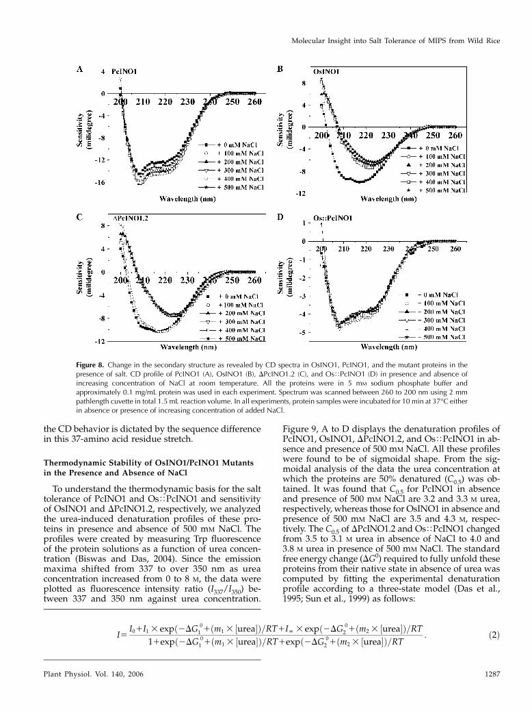
the CD behavior is dictated by the sequence differencein this 37-amino acid residue stretch.
Thermodynamic Stability of OsINO1/PcINO1 Mutantsin the Presence and Absence of NaCl
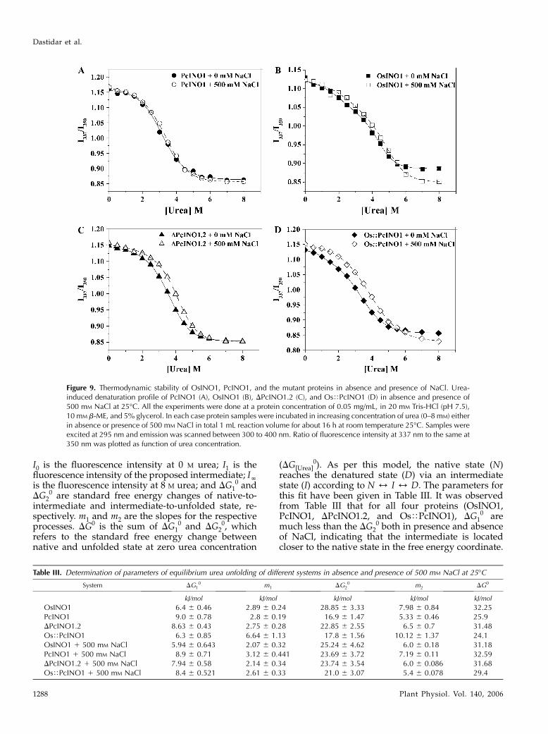
To understand the thermodynamic basis for the salttolerance of PcINO1 and OsTPcINO1 and sensitivityof OsINO1 and DPcINO1.2, respectively, we analyzedthe urea-induced denaturation profiles of these pro-teins in presence and absence of 500 mM NaCl. Theprofiles were created by measuring Trp fluorescenceof the protein solutions as a function of urea concen-tration (Biswas and Das, 2004). Since the emissionmaxima shifted from 337 to over 350 nm as ureaconcentration increased from 0 to 8 M, the data wereplotted as fluorescence intensity ratio (I337/I350) be-tween 337 and 350 nm against urea concentration.
Figure 9, A to D displays the denaturation profiles ofPcINO1, OsINO1, DPcINO1.2, and OsTPcINO1 in ab-sence and presence of 500 mM NaCl. All these profileswere found to be of sigmoidal shape. From the sig-moidal analysis of the data the urea concentration atwhich the proteins are 50% denatured (C0.5) was ob-tained. It was found that C0.5 for PcINO1 in absenceand presence of 500 mM NaCl are 3.2 and 3.3 M urea,respectively, whereas those for OsINO1 in absence andpresence of 500 mM NaCl are 3.5 and 4.3 M, respec-tively. The C0.5 of DPcINO1.2 and OsTPcINO1 changedfrom 3.5 to 3.1 M urea in absence of NaCl to 4.0 and3.8 M urea in presence of 500 mM NaCl. The standardfree energy change (DG0) required to fully unfold theseproteins from their native state in absence of urea wascomputed by fitting the experimental denaturationprofile according to a three-state model (Das et al.,1995; Sun et al., 1999) as follows:
Figure 8. Change in the secondary structure as revealed by CD spectra in OsINO1, PcINO1, and the mutant proteins in thepresence of salt. CD profile of PcINO1 (A), OsINO1 (B), DPcINO1.2 (C), and OsTPcINO1 (D) in presence and absence ofincreasing concentration of NaCl at room temperature. All the proteins were in 5 mM sodium phosphate buffer andapproximately 0.1 mg/mL protein was used in each experiment. Spectrum was scanned between 260 to 200 nm using 2 mmpathlength cuvette in total 1.5 mL reaction volume. In all experiments, protein samples were incubated for 10 min at 37�C eitherin absence or presence of increasing concentration of added NaCl.
I5I01I1 3 expð2DG
0
1 1ðm1 3 ½urea�Þ=RT1IN 3 expð2DG0
2 1ðm2 3 ½urea�Þ=RT11expð2DG
0
1 1ðm1 3 ½urea�Þ=RT1expð2DG0
2 1ðm2 3 ½urea�Þ=RT: ð2Þ
Molecular Insight into Salt Tolerance of MIPS from Wild Rice
Plant Physiol. Vol. 140, 2006 1287
I0 is the fluorescence intensity at 0 M urea; I1 is thefluorescence intensity of the proposed intermediate; INis the fluorescence intensity at 8 M urea; and DG1
0 andDG2
0 are standard free energy changes of native-to-intermediate and intermediate-to-unfolded state, re-spectively. m1 and m2 are the slopes for the respectiveprocesses. DG0 is the sum of DG1
0 and DG20, which
refers to the standard free energy change betweennative and unfolded state at zero urea concentration
(DG[Urea]0). As per this model, the native state (N)
reaches the denatured state (D) via an intermediatestate (I) according to N 4 I 4 D. The parameters forthis fit have been given in Table III. It was observedfrom Table III that for all four proteins (OsINO1,PcINO1, DPcINO1.2, and OsTPcINO1), DG1
0 aremuch less than the DG2
0 both in presence and absenceof NaCl, indicating that the intermediate is locatedcloser to the native state in the free energy coordinate.
Figure 9. Thermodynamic stability of OsINO1, PcINO1, and the mutant proteins in absence and presence of NaCl. Urea-induced denaturation profile of PcINO1 (A), OsINO1 (B), DPcINO1.2 (C), and OsTPcINO1 (D) in absence and presence of500 mM NaCl at 25�C. All the experiments were done at a protein concentration of 0.05 mg/mL, in 20 mM Tris-HCl (pH 7.5),10 mM b-ME, and 5% glycerol. In each case protein samples were incubated in increasing concentration of urea (0–8 mM) eitherin absence or presence of 500 mM NaCl in total 1 mL reaction volume for about 16 h at room temperature 25�C. Samples wereexcited at 295 nm and emission was scanned between 300 to 400 nm. Ratio of fluorescence intensity at 337 nm to the same at350 nm was plotted as function of urea concentration.
Table III. Determination of parameters of equilibrium urea unfolding of different systems in absence and presence of 500 mM NaCl at 25�C
System DG10 m1 DG2
0 m2 DG0
kJ/mol kJ/mol kJ/mol kJ/mol kJ/mol
OsINO1 6.4 6 0.46 2.89 6 0.24 28.85 6 3.33 7.98 6 0.84 32.25PcINO1 9.0 6 0.78 2.8 6 0.19 16.9 6 1.47 5.33 6 0.46 25.9DPcINO1.2 8.63 6 0.43 2.75 6 0.28 22.85 6 2.55 6.5 6 0.7 31.48OsTPcINO1 6.3 6 0.85 6.64 6 1.13 17.8 6 1.56 10.12 6 1.37 24.1OsINO1 1 500 mM NaCl 5.94 6 0.643 2.07 6 0.32 25.24 6 4.62 6.0 6 0.18 31.18PcINO1 1 500 mM NaCl 8.9 6 0.71 3.12 6 0.441 23.69 6 3.72 7.19 6 0.11 32.59DPcINO1.2 1 500 mM NaCl 7.94 6 0.58 2.14 6 0.34 23.74 6 3.54 6.0 6 0.086 31.68OsTPcINO1 1 500 mM NaCl 8.4 6 0.521 2.61 6 0.33 21.0 6 3.07 5.4 6 0.078 29.4
Dastidar et al.
1288 Plant Physiol. Vol. 140, 2006
It is therefore likely to assume that these intermediateshave very native-like structures. The stability (DG0) ofthese proteins were found to be quite different fromeach other in absence of salt but the difference dimin-ished somewhat in presence of 500 mM NaCl. It wasfound that in absence of salt, removal of 37-amino acidresidue stretch from PcINO1 changed the DG0 from25.9 kJ/mol for PcINO1 to 31.48 kJ/mol for DPcINO1.2,leading to stabilization by 5.58 kJ/mol. In other wordsthe presence of this 37-amino acid residue stretch inPcINO1 has a structure-destabilizing effect. When thesame 37-amino acid residue stretch of PcINO1 replacedthe corresponding 37-amino acid residue stretch ofOsINO1 resulting inOsTPcINO1, its stabilitywas foundto decrease from 32.25 to 24.1 kJ/mol. Thus it can besaid that arrangement of residues in this stretch is suchthat it reduces protein stability in absence of NaCl.However, both the PcINO1 and OsTPcINO1 contain-ing this 37-amino acid residue stretch when taken fromno salt to high salt situation gained extra stability of6.69 and 5.3 kJ/mol, respectively. Contrary to this, theother two proteins (DPcINO1.2 and OsINO1) that donot contain this 37-amino acid residue stretch eithershowed no extra stability or decreased stability whentaken from no salt to high salt condition.
Modeling Study
In an attempt to construct three-dimensional modelsof OsINO1 and PcINO1 proteins, sequence similaritysearch was carried out by BLAST (Altschul et al., 1997)against MIPS proteins whose crystal structures havebeen deposited in the Protein Data Bank (PDB). Figure10A shows the multiple sequence alignment done byClustalW (Thompson et al., 1994) of PcINO1, OsINO1,and the yeast MIPS (ScINO1) for which the crystalstructure (PDB ID 1P1J; Jin and Geiger, 2003) has beenobtained. Sequence similarity between OsINO1 andPcINO1 is approximately 83%, whereas the same be-tween OsINO1 and template and between PcINO1and template are approximately 60% and 50%, respec-tively. Comparison of the 37-amino acid residue stretchof OsINO1 and PcINO1 revealed that there are 21hydrophobic residues in OsINO1 compared to 15 inPcINO1. While these residues remained scattered inthe stretch of PcINO1, they were found to be distinctlyclustered in two stretches of eight and four residuesin OsINO1 (Fig. 10B). The three-dimensional modelsof monomer for both OsINO1 and PcINO1 havebeen depicted in Figure 10, C and D. Ca backbone ofhomology-modeled PcINO1 and OsINO1 were super-imposed separately on the Ca backbone of the yeastMIPS crystal structure. The pairwise root mean squarefits were 1.05 A0 and 1.1 A0, respectively, indicatingthat both the structures are essentially the same. Inhomology-modeled structures this 37-amino acid res-idue long stretch was mostly found to be in the loopregion for both the proteins. More accurately the stretchcorresponding to residues 174 to 206 is in the loopfollowed by a helix containing the rest of the stretch
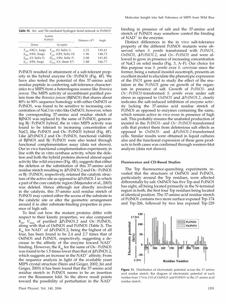
(residues 206–210; Fig. 10, C and D). Furthermore,there are a number of well-distributed Ser and Thr inthe 37-amino acid residue loop stretch of PcINO1 thanOsINO1, which are the potential candidates for well-connected hydrogen bond network. Such four distincthydrogen bonds made by Ser and Thr in PcINO1 havebeen presented in Table IV and shown in the ball-n-stick model (Fig. 10E). Moreover, when electrostaticpotential of each residue from 174 to 210 of bothPcINO1 and OsINO1 were plotted against the residuenumber, a distinct difference with respect to theirsurface electrostatic potential over this stretch wasfound (Fig. 11), where influence of charged environmentwas found to be greater over the stretch of PcINO1.
DISCUSSION
Stability of native folds of proteins and its responseto changes in their environment both under in vitroas well as in vivo conditions has been a subject ofintense study over the past several years (Jaenicke andZavodszky, 1990; Jaenicke, 1991; Taneja and Ahmad,1994; Murphy, 1995; Jaenicke and Bohm, 1998; Nelsonand Onuchic, 1998). Since it is imperative for anyprotein to retain its functional integrity under chang-ing environment, an insight into themechanism throughwhich functional proteins may achieve such potentialconstitutes an important area of investigation. An areaof considerable interest in this direction has been gen-eration of transgenic plants for acquisition of a stress-tolerant phenotype against major environmental stressby introgression of genes for such proteins, whichfunction as stress-tolerant determinants (Hasegawaet al., 2000). Our previous work on a salt-tolerant MIPS
from Porteresia coarctata (PcINO1) demonstrated thatit is a variant of the OsINO1 gene isolated from thedomesticated rice Oryza sativa, being different in a37-amino acid residue stretch (residues Trp-174 to Ser-210), and can characteristically synthesize the osmo-lyte inositol even under salt stress (Majee et al., 2004).This points toward an adaptational feature in MIPSprotein evolution across diverse taxa for a specializedfunction similar to the archaeal MIPS that is function-ally active only at high temperature (Chen et al., 2000).In this study we have established that the 37-aminoacid stretch of PcINO1 is primarily responsible forits salt-tolerance property through the biochemicaland biophysical characterization of in vitro generatedmutants.
Characteristics of the in Vitro Generated PcINO1Mutants and Hybrid Proteins vis a vis Salt Tolerance
Results presented herein clearly demonstrate that inaddition to the loss of salt tolerance by deletion of the37-amino acid residue stretch in DPcINO1.2 reportedearlier (Majee et al., 2004), replacement of the corre-sponding 37-amino acid residue stretch in OsINO1with the designated 37-amino acid residue stretch of
Molecular Insight into Salt Tolerance of MIPS from Wild Rice
Plant Physiol. Vol. 140, 2006 1289
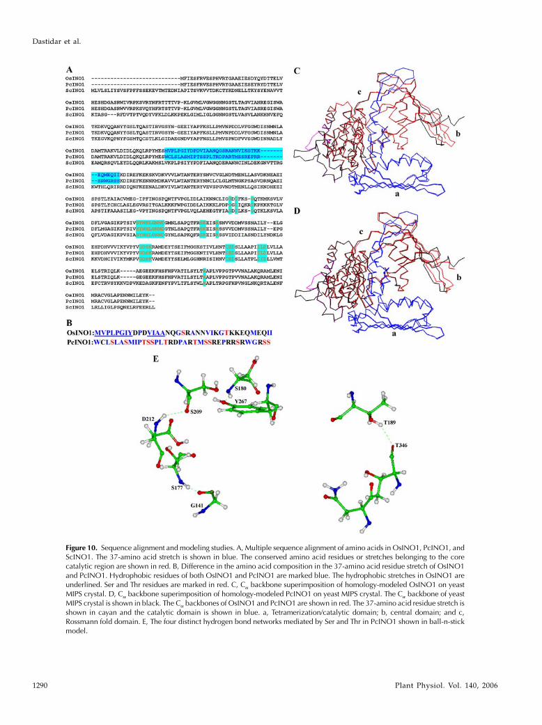
Figure 10. Sequence alignment and modeling studies. A, Multiple sequence alignment of amino acids in OsINO1, PcINO1, andScINO1. The 37-amino acid stretch is shown in blue. The conserved amino acid residues or stretches belonging to the corecatalytic region are shown in red. B, Difference in the amino acid composition in the 37-amino acid residue stretch of OsINO1and PcINO1. Hydrophobic residues of both OsINO1 and PcINO1 are marked blue. The hydrophobic stretches in OsINO1 areunderlined. Ser and Thr residues are marked in red. C, Ca backbone superimposition of homology-modeled OsINO1 on yeastMIPS crystal. D, Ca backbone superimposition of homology-modeled PcINO1 on yeast MIPS crystal. The Ca backbone of yeastMIPS crystal is shown in black. The Ca backbones of OsINO1 and PcINO1 are shown in red. The 37-amino acid residue stretch isshown in cayan and the catalytic domain is shown in blue. a, Tetramerization/catalytic domain; b, central domain; and c,Rossmann fold domain. E, The four distinct hydrogen bond networks mediated by Ser and Thr in PcINO1 shown in ball-n-stickmodel.
Dastidar et al.
1290 Plant Physiol. Vol. 140, 2006
PcINO1 resulted in attainment of a salt-tolerant prop-erty in the hybrid enzyme OsTPcINO1 (Fig. 4F). Wehave also tested the potential of this 37-amino acidresidue peptide in conferring salt-tolerance character-istics to a MIPS from a heterologous source like Brassicajuncea. The MIPS activity of recombinant purified pro-tein from the Brassica juncea (BjINO1) that shares about80% to 90% sequence homology with either OsINO1 orPcINO1, was found to be sensitive to increasing con-centration of NaCl in vitro like OsINO1; however, whenthe corresponding 37-amino acid residue stretch ofBjINO1 was replaced by the same of PcINO1, generat-ing BjTPcINO1 hybrid, the in vitro MIPS activity wasfound to be tolerant to increasing concentration ofNaCl, like PcINO1 and OsTPcINO1 hybrid (Fig. 4F).Like DPcINO1.2 and OsTPcINO1, functional viabilityof BjINO1 and BjTPcINO1 were also tested throughfunctional complementation assay (data not shown).Our in vivo functional complementation experiment, inline with the in vitro synthase activity, where the dele-tion and both the hybrid proteins showed almost equalactivity like wild enzymes (Fig. 4E), suggests that eitherthe deletion or the substitution of this 37-amino acidresidue stretch resulting inDPcINO1.2 andOsTPcINO1or BjTPcINO1, respectively, retained the catalytic struc-ture of the active site as opposed toDPcINO1.1 in whicha part of the core catalytic region (Majumder et al., 2003)was deleted. Hence although not directly involvedin the catalysis, this 37-amino acid residue stretch ofPcINO1may control either the access of the substrate tothe catalytic site or alter the geometric arrangementaround it to alter substrate-binding properties in pres-ence of high salt.To find out how the mutant proteins differ with
respect to their kinetic properties, we also comparedKm, Vmax of purified DPcINO1.2 and OsTPcINO1,along with that of OsINO1 and PcINO1 (Table I). TheKm for NAD1 of DPcINO1.2, being the highest of allfour, has been found to be 2.4 and 2.7 times that ofOsINO1 and PcINO1, respectively, suggesting a de-crease in the affinity of the enzyme toward NAD1
binding. However, the Km for the same of OsTPcINO1was found to be 1.5 times lower than that ofDPcINO1.2,which suggests an increase in the NAD1 affinity. Fromthe sequence analysis in light of the available yeastMIPS crystal structure (Stein and Geiger, 2002; Jin andGeiger, 2003) it has been found that the 37-amino acidresidue stretch in PcINO1 seems to be an insertionover the Rossmann fold. So the above result pointstoward the possibility of perturbation in the NAD1
binding in presence of salt and the 37-amino acidstretch of PcINO1 may somehow control the bindingof NAD1 to the enzyme.
Distinct differences in the in vivo salt-toleranceproperty of the different PcINO1 mutants were ob-served when S. pombe transformed with PcINO1,OsINO1, DPcINO1.2, and OsTPcINO1 and were al-lowed to grow in presence of increasing concentrationof NaCl on solid media (Fig. 3, A–F). Our choice forthis purpose was S. pombe over S. cerevisiae since theformer, being a natural inositol auxotroph, presents anexcellent model to elucidate the phenotypic expressionof the INO1 gene and to study the effect of the mu-tation in the PcINO1 gene on growth of the organ-ism in presence of salt. Growth of PcINO1- andOsTPcINO1-transformed S. pombe even under saltstress as opposed to OsINO1 and DPcINO1.2, clearlyindicates the salt-induced inhibition of enzyme activ-ity lacking the 37-amino acid residue stretch ofPcINO1 as opposed to enzymes containing the same,which remain active in vivo even in presence of highsalt. This probably ensures the unabated production ofinositol in the PcINO1- and OsTPcINO1-transformedcells that protect them from deleterious salt effects asopposed to OsINO1- and DPcINO1.2-transformedcells. Similar results were obtained in liquid culturesalso and the functional expression of these gene prod-ucts in both cases was confirmed through western-blotanalysis (data not shown).
Fluorescence and CD-Based Studies
The Trp fluorescence-quenching experiments re-vealed that the structures of OsINO1 and PcINO1,particularly around the Trp residues, were affecteddifferentially by salt. OsINO1 has five Trp and PcINO1has eight, all being located primarily in the N-terminalregion in both, the first four Trp residues being locatedat identical position. The 37-amino acid residue stretchof PcINO1 contains two more surface-exposed Trp-174and Trp-206, followed by two less exposed Trp-229
Table IV. Ser- and Thr-mediated hydrogen bond network in PcINO1
System
Distance (A0) AnglePcINO1
Donor Acceptor
T189 (HG1; loop) T346 (O; helix L) 2.25 145.61S180 (HN) (loop) Y267 (OH; helix H) 1.96 148.75S209 (O; helix F) D212 (HN; helix F) 2.08 141.85S177 (HN; loop) G141 (O; sheet S7) 1.88 166.77
Figure 11. Distribution of electrostatic potential across the 37-aminoacid residue stretch. Bar diagram of electrostatic potential of eachresidue from 174 to 210 of OsINO1 and PcINO1 in the 37-amino acidresidue stretch.
Molecular Insight into Salt Tolerance of MIPS from Wild Rice
Plant Physiol. Vol. 140, 2006 1291
and Trp-289 as indicated from modeling studies; thecorresponding region of OsINO1 having only oneburied Trp-229. Since the surface-exposed Trps donot contribute much to the Trp fluorescence intensitybecause of their low quantum yield, the intensity-contributing Trps are those away from this loop re-gion. Quenching of Trp fluorescence either of OsINO1(Majee et al., 2004) or DPcINO1.2 indicated that saltalters the conformation of both in such a way as tobring some of the buried Trps close to the surface,enhancing the collisional quenching, or by an alterna-tive mechanism of enhanced energy transfer due toproximal redistribution of Trp neighboring groups,whose polarity remained unchanged. A change insecondary structure at high salt concentration wasalso reflected in the CD data (Fig. 8, B and C). On theother hand, both PcINO1 and OsTPcINO1 are able tomaintain their structural rigidity in presence of salt,disallowing the Trp positions to be changed muchfrom those in absence of salt. Lack of change in theinternal fold of both PcINO1 and OsTPcINO1 is alsoindicated by the CD spectra, whose nature qualita-tively remained unchanged in presence of salt (Fig. 8,A and D).
Surface Hydrophobicity as an Index of Salt Sensitivityversus Salt Tolerance
The effect of this 37-amino acid residue stretch onthe salt-induced structural plasticity or rigidity isdirectly provided by the highly sensitive bis-ANSbinding results (Table II) that reflect that the presenceof this stretch in PcINO1 and OsTPcINO1 causedminimal changes in the surface exposure of the hy-drophobic pockets on increasing salt concentration butits absence in OsINO1 and DPcINO1.2 led to a largeincrease in the surface exposure of hydrophobic resi-dues (Fig. 6A). The presence of 21 hydrophobic resi-dues in clusters in OsINO1 compared to 15 scattered ofsuch in PcINO1 (Fig. 10B) generally explains the highersurface hydrophobicity of OsINO1 over PcINO1 inabsence of salt. The excess exposure of the hydropho-bic clefts in presence of salt in the cases of OsINO1 andDPcINO1.2 makes them susceptible to aggregation(Fig. 7A) as opposed to either PcINO1 or OsTPcINO1,which are resistant to such salt-induced aggregation(Fig. 7B). It may be interesting to point out here thatCD data in presence of 500 mM NaCl of OsINO1 andDPcINO1.2 points to some increase in b-sheet struc-ture at the expense of helix structure. Since salts screenthe electrostatic interactions on the protein surface, thehydrophobic interactions are screened through proteinoligomerization interaction (Biswas et al., 2002). Hence,loss of MIPS activity may be directly related to thisoligomerization. Further, the salt-induced surface hy-drophobic exposure is accompanied also by rearrange-ments of internally buried groups, the loss of enzymaticfunction may also be due to both restructuring of thecatalytic site and physical blocking of the access pathof the substrate by oligomerization.
It has also been noted that there are a good numberof well-distributed Sers and Thrs in PcINO1 thanOsINO1 particularly in the 37-amino acid residueloop stretch (Fig. 10, B and E). These residues formfour well-connected salt bridge networks with theneighboring residues, both within and outside theloop region (Table IV) that contributes to salt stabilityin PcINO1 and the OsTPcINO1. Since such an exten-sive salt bridge is not possible in OsINO1, screening ofsurface electrostatic interactions in presence of saltsomewhat destabilizes OsINO1, leading to structuralreorganization. Furthermore, as distribution of elec-trostatic potential is purely context dependent, it wasobserved that the charge distribution over this stretchof PcINO1 varies considerably from positive to nega-tive with a greater positive potential, whereas in thecase of OsINO1 almost a uniform but less charge distri-bution is observed. This possibly accounts for thehigher surface polarity over the 37-amino acid residuestretch of PcINO1, which in turn can be screened outby the salt ions and thus protects the enzyme fromthe salt-induced inactivation as opposed to OsINO1.
Assessment of Thermodynamic Stability in the
Absence and Presence of Salt
There are plenty of indications from the abovediscussion that the salt affects the stability of OsINO1and PcINO1 differently, as indicated further by theurea denaturation study (Table III). Both PcINO1 andthe OsTPcINO1 were found to be stabilized in pres-ence of salt, resulting in a DDGs
0 (5DGsalt0 2 DGno salt
0)of 6.69 and 5.3 kJ/mol. OsINO1 was destabilized asDDGs
0 and is 21.07 kJ/mol, the minus sign indicatingdestabilization. DPcINO1.2 on the other hand did notgain additional stability in presence of salt, as theDDGs
0 is nearly zero. This suggests that the existence ofthe loop region reflects upon the thermodynamicdifferences in presence of salt more prominently. In-terestingly, in absence of salt, existence of the loopsequence of PcINO1 has a destabilizing effect com-pared to DPcINO1.2, as DDG37P
0 (defined asDGPcINO10 2
DGDPcINO1.20) is25.58 kJ/mol. On the contrary, the loop
sequence of OsINO1 contributes to protein stability inabsence of salt, as DDG37R
0 (5DGOsINO10 2 DGDPcINO1.2
0)is 10.77 kJ/mol. Because of the two opposing freeenergy contributions of the two 37-amino acid residueloop stretches of OsINO1 and PcINO1 in absence ofsalt, the replacement of OsINO1 stretch by that ofPcINO1 from simple thermodynamic additivity as-sumption is predicted to result in loss of stability by6.35 kJ/mol (525.58 2 0.77 kJ/mol). Indeed theOsTPcINO1was found to have the thermodynamic sta-bility lower than that of OsINO1 by 8.15 kJ/mol from32.25 to 24.1 kJ/mol. Considering the simple assump-tion and analysis, this qualitative agreement is quitesignificant in accordance with the position of chlorideanion as a destabilizer in the Hofmeister series ofkosmotropes and chaotropes (Elcock andMcCammon,1998; Dominy et al., 2002; Apetri and Surewicz, 2003).
Dastidar et al.
1292 Plant Physiol. Vol. 140, 2006

The destabilization of OsINO1 in presence of NaClmay be considered as a typical and mildly chaotropiceffect. The stabilizing effect of NaCl on PcINO1 andthe OsTPcINO1 represents atypical salt effects (Apetriand Surewicz, 2003). This sequence-dependent differ-ence in salt effect may arise from formation anddisruption of salt bridges, which are nothing but stronghydrogen bonds (Strop and Mayo, 2000; Bosshardet al., 2004) formed between two closely spacedinteracting polar or charged residues (Table IV). En-ergy contribution toward stability of proteins for thesalt bridge formation is usually low (about 4.0 kJ/mol)and the stabilization energy is contributed mainlyfrom the increase in entropy of the solvent compen-sated by the energy required to break the hydrogenbond with water (Stigler, 1991; Strop and Mayo, 2000).The stabilization of PcINO1 and the OsTPcINO1 in thepresence of salt by 5.3 to 6.69 kJ/mol lends strongsupport in favor of the salt bridge formation.A comparable example is found in the allene oxide
cyclase gene from Bruguiera sexangula wherein a 70-amino acid stretch has been identified as the salt-tolerance determinant of the gene product (Yamadaet al., 2002). Designated as mangrin, this gene is re-ported to be capable of conferring salt tolerance whenintrogressed to a number of pro- and eukaryotic sys-tems (Yamada et al., 2002), a characteristic also sharedby PcINO1 (A. Das-Chatterjee, L. Goswami, S. Maitra,K. Ghosh Dastidar, S. Roy, B. Das, M. Majee, S.Bhattacharyya, S. Pattnaik, and A.L. Majumder, un-published data). In addition to the identification of the37-amino acid residue stretch as the salt-tolerancedeterminant in PcINO1, we have been able to demon-strate that this stretch can also attribute salt-toleranceproperty to an otherwise salt-sensitive MIPS by re-placement of the corresponding amino acid stretch.Such recombination has resulted in salt tolerance ofOsTPcINO1 or BjTPcINO1 hybrid gene products.Since the MIPS gene sequences of plants are fairlyconserved (Majumder et al., 2003), it is conceivablethat any plant MIPS may thus be made salt tolerant bysimilar protein engineering strategies.
MATERIALS AND METHODS
Reagents
D-Glc-6-P (disodium salt), nicotinamide adenine dinucleotide (disodium
salt; NAD1), phenylmethylsulfonyl fluoride, isopropyl thiogalactoside, 5#bromo 4-chloro 3-indolyl D-galactopyranoside, bis-ANS, and b-mercaptoeth-
anol (b-ME) were purchased from Sigma. All column chromatographic mate-
rials were purchased from Amersham Pharmacia and Bio-Rad. All restriction
enzymes, T4 DNA ligase, and RNase were purchased from New England
Biolabs. PCR reagents, oligonucleotides, Hifi Taq polymerase, and pGEMT-
Easy vector were purchased from GIBCO-BRL and Promega. All other chem-
icals used were of analytical reagent grade purity unless otherwise specified.
Generation and Molecular Cloning of Mutant and
Hybrid Clones of PcINO1
Two PcINO1 mutants, namely DPcINO1.1 and DPcINO1.2 were generated
by PCR as described earlier (Fig. 1B; Majee et al., 2004). Since bothOsINO1 and
PcINO1 contain one unique PstI and one unique EcoRV site at the same
position, the OsINO1/PcINO1 hybrid termed OsTPcINO1 was generated
simply by replacing a 5#-3#PstI-EcoRV fragment of OsINO1 with the same
fragment from PcINO1 through restriction digestion (Fig. 1C).The second
hybrid gene between Brassica MIPS and PcINO1 (termed as BjTPcINO1) was
generated by substituting the corresponding 37-amino acid residue stretch of
BjINO1 with that of PcINO1 by PCR using the following primers: P1, 5#-aca-tatgttcatcgagagcttccg-3#; P2, 5#-ggagctcgttctccatgtaaggcctga-3#; P3, 5#-aggtac-caaggacatgagggagttt-aag-3#; P4, 5#-gctcgagcttgttactccaggatcatgt-3#; P5, 5#-atc-gagctctggtgcctctccctggcatctatg-3#; and P6, 5#-cggggtacctgatgatctgccccatctgc-tcctt-3# (Fig. 1D). INO1 from Brassica juncea (henceforth termed BjINO1) was
PCR amplified by using primers based on the Brassica napus INO1 gene
(U66307) following the protocol for isolating OsINO1/PcINO1 (Majee et al.,
2004). All the genes (wild, mutant, and hybrids) were finally cloned into pET-
20b(1) expression vector (Novagen) at NdeI and XhoI sites and mutations
were confirmed by sequencing fromUniversity of Delhi, South campus, India.
Funtional Complementation of DPcINO1.1, DPcINO1.2,
and OsTPcINO1 in Yeast FY250 (MATæ trp1 his3 ura3leu2 Ino1THIS3) Inositol Auxotrophic Strain
A yeast (Saccharomyces cerevisiae)-based functional complementation assay
for the confirmation of the MIPS activity of the mutant and hybrid gene
products was performed as per procedure described in detail earlier (Chat-
terjee et al., 2004; Majee et al., 2004).
Expression of Wild and Mutant INO1Genes in Schizosaccharomyces pombe(h- leu1-32 ura4-D18) PR109
OsINO1, PcINO1, DPcINO1.2, and OsTPcINO1 were subcloned down-
stream of a thiamine repressible, nmt promoter in a yeast binary expression
vector pREP1 (Maundrell, 1993). pGEMT-Easy vector (Promega) harboring all
the above-mentioned genes was digested with NdeI and XhoI. The digested
products were then directionally ligated to pERP1 at NdeI and SalI sites using
T4 DNA ligase. Positive clones were checked by digestion withNdeI and SmaI.
All pREP1 constructs harboring different mutant and wild-type genes were
transformed in S. pombe cells according to the protocol described by Chaudhuri
et al. (1997).
For growth experiments, transformed S. pombe cells were allowed to grow
in complete solid synthetic Edinburgh minimal medium (EMM; Alfa et al.,
1992) supplemented with 2 mM inositol and containing different NaCl con-
centrations (0–500 mM). For this purpose first PcINO1, OsINO1, DPcINO1.2,
OsTPcINO1, and pREP1 transformed S. pombe cells were grown in EMMbroth
supplemented with 2 mM inositol at 30�C until the A600 reached between 0.6 to
0.7. Then, equal numbers of cells were plated onto solid EMM media sup-
plemented with 2 mM inositol and increasing NaCl concentration as stated
above. Plates were incubated at 30�C for 48 h.
Bacterial Overexpression and Purification of the
Recombinant Proteins
Bacterial overexpression of all mutant or hybrid genes (DPcINO1.1,
DPcINO1.2, OsTPcINO1, BjINO1, and BjTPcINO1), recovery of the expressed
recombinant proteins from the particulate fraction, and their subsequent
purification along with the wild type were carried out essentially by following
the procedure described earlier (Majee et al., 2004). After the final purification,
all proteins were extensively dialyzed against 20 mM Tris-HCl (pH 7.5), 10 mM
NH4Cl, 10 mM b-ME, and 20% glycerol concentrated and stored at 220�C for
further use.
Assay of MIPS
MIPS assay was done colorimetrically by the periodate oxidation method
of Barnett et al. (1970) and further corroborated by the inositol-1-P phospha-
tase assay described by RayChaudhuri et al. (1997). The amount of inorganic
phosphate liberated from the MIPS reaction product either by periodate
oxidation or inositol 1-P phosphatase reaction was estimated by the method of
Chen et al. (1956).
Molecular Insight into Salt Tolerance of MIPS from Wild Rice
Plant Physiol. Vol. 140, 2006 1293
Estimation of Protein
Protein was estimated according to the method of Bradford (1976) with the
Bio-Rad protein assay kit using bovine serum albumin as a standard.
Analytical SDS-PAGE and Western Blotting
Ten-percent SDS-PAGE was performed according to Laemmli (1970). Immu-
nodetection and proteins were done as described by Chatterjee et al. (2004).
Trp Fluorescence Quenching in the Presence and
Absence of NaCl
Purified DPcINO1.2, OsTPcINO1, and OsINO1 and PcINO1 (serving as
controls) were separately dialyzed against 1 L buffer (20 mM Tris-HCl, pH 7.5,
and 10 mM b-ME) with two changes overnight at 4�C. Nine-hundred micro-
liters of approximately 170 mg/mL of both the proteins were used in a total
1 mL reaction mixture in a quartz cuvette (4 mm 3 4 mm). In both cases
proteins were incubated at 37�C as well as at 25�C for 10 min both in absence
as well as in presence of different concentrations of NaCl (100–600 mM).
Excitation wavelength was selected at 295 nm and the emission was scanned
at a speed of 240 nm/min from 310 to 400 nm using excitation and emission
slit width of 5.0 nm each. Each spectrum was an average of three scans.
Appropriate control buffer spectra were subtracted from sample spectra to
generate the fluorescence spectra of the proteins. Wavelength of maximum
emission for each spectrum was determined by derivative analysis using the
instrument software.
Bis-ANS Binding Study
Each purified protein was extensively dialyzed against (20 mM Tris-HCl,
pH 7.5, and 10 mM b-ME) at 4�C. In each case 1 mL of approximately 50 mg/
mL dialyzed protein was first incubated at 37�C for 10 min in absence or
presence of 500 mM NaCl and then titrated with aqueous solution of bis-ANS
(300 mM) in total 1 mL reaction volume at room temperature. In each case bis-
ANS was added in small aliquots (2, 4, 8, and 10 mL and so on until saturation)
to the protein solution, mixed well, and incubated for 1 min. Emission was
recorded at 490 nm after exciting the samples at 390 nm, using excitation and
emission slit width of 5.0 nm each. Binding isotherms were generated by
plotting fluorescence intensity (F) at 490 nm against the wavelength values. To
determine the stoichiometry (n) and dissociation constant (kd) of these binding
phenomena, reverse bis-ANS titration was done. In this case 1 mL of 0.1 mM
bis-ANS solution was titrated by adding small aliquots (each 10 mL) of protein
(approximately 450 mg/mL), which was preincubated at 37�C for 10 min with
and without 500 mM NaCl. After each addition, the solution was mixed well
and kept at room temperature for 1 min.
Aggregation Assay in the Presence and Absence of NaCl
Purified DPcINO1.2, OsTPcINO1, and OsINO1 and PcINO1 (serving as
controls) were dialyzed separately in 1 L of buffer (20 mM Tris-HCl, pH 7.5,
and 10 mM b-ME) overnight at 4�C. Nine-hundred microliters of approxi-
mately 350 mg/mL of each protein was subjected to static light scattering at
360 nm in absence and presence of different concentrations of increasing NaCl
(100–500 mM) in total 1 mL reaction volume at 37�C in SHIMADZU UV-160A
for up to 170 min following the protocol described by Horwitz (1992). Optical
density of the samples at 360 nm was measured over a period of 170 min at
regular intervals where increased absorbance was indicative of aggregation of
the protein samples.
CD Spectroscopy in the Presence and Absence of NaCl
Protein samples were dialyzed separately against 1 L of filter-sterilized
5 mM sodium phosphate buffer, pH 7.6, with three changes overnight at 4�C.Far-UV CD spectroscopy was done with 1.350 mL of approximately 100 mg/
mL recombinant purified OsINO1, PcINO1, DPcINO1.2, and OsTPcINO1 in a
total 1.5 mL reaction volume in absence and presence of increasing concen-
tration of NaCl (100–500 mM) at room temperature. In both the cases proteins
were preincubated at 37�C for 10 min both in absence as well as in presence of
different concentrations of NaCl. An average of five scans was recorded at 0.2
step resolution, 10 nm/min scan speed, 1.0 nm band width, and 2 s time
constant in a Jasco600 spectrophotopolarimeter. Data were processed by
several secondary structure analysis programs such as SELCON3, CONTIN,
CDSSTR, and CDNN, available on the Web.
Urea Unfolding Equilibrium in the Absence and
Presence of 500 mM NaCl
Urea unfolding equilibrium was carried out spectroflurometrically in a
Hitachi-F-4500 spectrofluorimeter with all four purified enzymes under
identical conditions in absence and presence of 500 mM NaCl at room
temperature (approximately 25�C). In each case approximately 50 mg of
protein, which was extensively dialyzed against 20 mM Tris-HCl (pH 7.5),
10 mM b-ME, and 5% glycerol for approximately 16 h at 4�C, was preincubated
with 0 to 8 M (in total 13 sets) urea in total 1 mL reaction mixture in absence or
in presence of 500 mM NaCl at room temperature (approximately 25�C) foralmost 16 h. Emission spectra were recorded in a quartz cuvette (4 mm 3
4 mm) by scanning from 300 to 400 nm after excitation at 295 nm, using
excitation and emission slit width of 5.0 nm each. Each spectrum was an
average of three scans. Appropriate control buffer spectra were subtracted
from each sample spectra for each set to generate the fluorescence spectra of
the proteins at different concentrations of urea. Wavelength of maximum
emission for each spectrum was determined by derivative analysis using the
instrument software. Maximum intrinsic fluorescence intensity value for each
spectrum was normalized by taking the ratio of intensity at 337 nm and
intensity at 350 nm (I337 nm/I350 nm) using the instrument software. I337 nm/I350 nmvalues were plotted against the urea concentration in each set for each
enzyme. C0.5 value for each set was determined by fitting the plot to sigmoidal
curve using Microcal Origin 6.0.
Modeling Study of OsINO1 and PcINO1
The amino acid sequences of PcINO1 and OsINO1 were taken from
GenBank (accession nos. for PcINO1: AAP74579 and OsINO1: BAA25729).
The atomic coordinate of yeast MIPS complexed with NADH (PDB code: 1P1J;
Jin and Geiger, 2003) was obtained from the PDB (Berman et al., 2000). The
chain A of this x-ray structure was used as the template to build the three-
dimensional models of OsINO1 and PcINO1 using the program MODELLER
(Sali and Blundell, 1993). Energy minimization of both the models were
carried out by conjugate gradient method using the Consistent Valence Force
Field (Hagler et al., 1979) of the Discover program (Insight II, BIOSYM/MSI,
1995) until all the structures reached the final derivative of 0.0042 kJ/mol.
The stereochemical quality of the models was checked using the program
PROCHECK (Laskovski et al., 1993) and Ramachandran plots (Ramachandran
and Sashisekharan, 1968) were drawn for both the models, according to which
no residue was present in the disallowed region for both the models.
Programs MODELLER and PROCHECK were run on a Silicon Graphics O2
R12000 workstation. The visualization of the above studies was done using the
program InsightII (Insight II, BIOSYM/MSI, 1995).
Electrostatic potential was calculated using the program DELPHI (Insight
II, BIOSYM/MSI, 1995) implemented in InsightII solving a finite difference
solution to the nonlinear Poisson-Boltzmann equation. Dielectric constants of
80.0 and 2.0 were used for solvent and solute, respectively. The ionic strength
of the solvent was 0.145 M.
ACKNOWLEDGMENTS
Thanks are due to Prof. D. Chattopadhaya and Dr. A. Bachhawat for their
gift of S. pombe strains and plasmids.
Received December 6, 2005; revised January 5, 2006; accepted January 5, 2006;
published February 24, 2006.
LITERATURE CITED
Albany JR (2004) Structure and Dynamics of Macromolecules: Absorption
and Fluorescence Studies. Elsevier, St. Louis
Alfa C, Fantes P, Hyams J, McLeodM,Warbrick E (1992) Experiments with
Fission Yeast: A Laboratory Manual. Cold Spring Harbor Laboratory
Press, Cold Spring Harbor, NY
Dastidar et al.
1294 Plant Physiol. Vol. 140, 2006
Altschul SF, Madden TL, Schaffer AA, Zhang J, Zhang Z, Miller W,
Lipman D (1997) Gapped BLAST and PSI-BLAST: a new generation of
protein database search programs. Nucleic Acids Res 25: 3389–3402
Apetri AC, Surewicz WK (2003) Atypical effect of salts on the thermody-
namic stability of human prion protein. J Biol Chem 278: 22187–22192
Barnett JEG, Brice RE, Corina DL (1970) A colorimetric determination
of inositol monophosphatase as an assay for D glucose-6- phosphate
1-L- myo-inositol 1 phosphate cyclase. Biochem J 119: 183–186
Berman MH, Westbrook J, Feng J, Gillilang G, Bhat TN, Weissig H,
Sindyalov IN, Bourne PE (2000) The protein data bank. Nucleic Acids
Res 28: 235–242
Biswas A, Das KP (2004) Role of ATP on the interaction of alpha-crystallin
with its substrates and its implications for the molecular chaperone
function. J Biol Chem 279: 42648–42657
Biswas A, Saha S, Das KP (2002) Structural features of molecular chap-
erons: a possible micellar connection. J Surface Sci Technol 18: 1–24
Biswas BB, Ghosh B, Majumder AL (1984) myo-Inositol polyphosphates
and their role in cellular metabolism: a proposed cycle involving glucose-
6-phosphate and myo-inositol phosphates. Subcell Biochem 10: 237–280
Bohnert HJ, Nelson DE, Jensen RG (1995) Adaptations to environmental
stresses. Plant Cell 7: 1099–1111
Bosshard HR, Marti DN, Jelesarov I (2004) Protein stabilization by salt
bridges: concepts, experimental approaches and clarification of some
misunderstandings. J Mol Recognit 17: 1–16
Bradford MW (1976) Quantitation of microgram quantities of protein
utilizing the principle of protein dye binding. Anal Biochem 72: 248–254
Cardamone M, Puri BR (1992) Spectrofluorimetric assessment of the
surface hydrophobicity of proteins. Biochem J 282: 589–593
Chatterjee A, Majee M, Ghosh S, Majumder AL (2004) sll1722, an
unassigned ORF of Synechocystis PCC 6803, codes for L-myo-inositol
1-phosphate synthase. Planta 218: 989–998
Chaudhuri B, Ingavale S, Bachhawat AK (1997) apd11, a gene required
for red pigment formation in ade6 mutants of Schizosaccharomyces pombe,
encodes an enzyme required for glutathione biosynthesis: a role for
glutathione and a glutathione-conjugate pump. Genetics 145: 75–83
Chen L, Zhou C, Yang H, Roberts MF (2000) Inositol-1-phosphate synthase
from Archaeoglobus fulgidus is a class II aldolase. Biochemistry 39: 12415–
12423
Chen RS, Toribara TY, Warner H (1956) Microdetermination of phospho-
rus. Anal Chem 28: 1756–1758
Das BK, Bhattacharyya T, Roy S (1995) Characterization of a urea induced
molten globule intermediate state of glutaminyl-tRNA synthetase from
Escherichia coli. Biochemistry 34: 5242–5247
Das KP, Petrash J, Surewicz WK (1996) Conformational properties of
substrate proteins bound to a molecular chaperone alpha-crystallin.
J Biol Chem 271: 10449–10452
Das KP, Surewicz WK (1995) Temperature-induced exposure of hydro-
phobic surfaces and its effect on the chaperone activity of alpha-
crystallin. FEBS Lett 369: 321–325
Dominy BN, Perl D, Schmid FX, Brooks CL III (2002) The effects of ionic
strength on protein stability: the cold shock protein family. J Mol Biol
319: 541–554
Eftink MR, Ghiron CA (1981) Fluorescence quenching studies with pro-
teins. Anal Biochem 114: 199–227
Elcock AH, McCammon JA (1998) Electrostatic contributions to the sta-
bility of halophilic proteins. J Mol Biol 280: 731–748
Fersht AR (1999) Structure and Mechanism in Protein Science: A Guide to
Enzyme Catalysis and Protein Folding. Freeman and Company, New York
Hagler AT, Lifson S, Dauber P (1979) Consistent force field studies of inter-
molecular forces in hydrogen-bonded crystals. 2. A benchmark for the
objective comparison of alternative force. J Am Chem Soc 101: 5122–5130
Hasegawa PM, Bressan RA, Zhu JK, Bohnert HJ (2000) Plant cellular and
molecular responses to high salinity. Annu Rev Plant Physiol Plant Mol
Biol 51: 463–499
Horowitz PM, Hua S, Gibbons DL (1995) Hydrophobic surfaces that are
hidden in chaperonin Cpn60 can be exposed by formation of assembly-
competent monomers or by ionic perturbation of the oligomer. J Biol
Chem 270: 1535–1542
Horwitz J (1992) Alpha-crystallin can function as a molecular chaperone.
Proc Natl Acad Sci USA 89: 10449–10453
Ingavale SS, Bachhawat AK (1999) Restoration of inositol prototrophy in
the fission yeast Schizosaccharomyces pombe.Microbiology 145: 1903–1910
Ishitani M, Majumder AL, Bornhouser A, Michalowski CB, Jensen RG,
Bohnert HJ (1996) Coordinate transcriptional induction induction of
myo-inositol metabolism during environmental stress. Plant J 9: 537–548
Jaenicke R (1991) Protein stability and molecular adaptation to extreme
conditions. Eur J Biochem 202: 715–718
Jaenicke R, Bohm G (1998) The stability of proteins in extreme environ-
ments. Curr Opin Struct Biol 6: 738–748
Jaenicke R, Zavodszky P (1990) Protein under extreme physical conditions.
FEBS Lett 268: 344–349
Jin X, Geiger JH (2003) Structure of NAD and NADH bound 1-L-myo-
inositol 1-phosphate synthase. Acta Crystallogr D 59: 1154–1164
Kanter U, Usadel B, Guerineau F, Li Y, Pauly M, Tenhaken R (2005) The
inositol oxygenase gene family of Arabidopsis is involved in the
biosynthesis of nucleotide sugar precursors for cell-wall matrix poly-
saccharides. Planta 221: 243–254
Laemmli UK (1970) Cleavage of structural proteins during the assembly of
bacteriophage T4. Nature 227: 680–685
Lakowicz JR (1983) Principles of Fluorescence Spectroscopy. Plenum
Press, New York, pp 258–304
Laskovski RA, MacArthur MW, Moss DS, Thornton JM (1993) PRO-
CHECK: a program to check the stereochemistry of protein structure.
J Appl Crystallogr 26: 283–291
Loewus FA, Murthy PN (2000) myo-Inositol metabolism in plants. Plant Sci
150: 1–19
Lorence A, Chevone BI, Mendes P, Nessler CL (2004) myo-Inositol oxy-
genase offers a possible entry point into plant ascorbate biosynthesis.
Plant Physiol 134: 1200–1205
Majee M, Maitra S, Ghosh Dastidar K, Pattnaik S, Chatterjee A, Hait NC,
Das KP, Majumder AL (2004) A novel salt-tolerant L-myo-inositol-
1-phosphate synthase from Porteresia coarctata (Roxb.) Tateoka, a halo-
phytic wild rice. J Biol Chem 279: 28539–28552
Majumder AL, Chatterjee A, Ghosh Dastider K, Majee M (2003) Diver-
sification and evolution of L-myo-inositol 1-phosphate synthase. FEBS
Lett 533: 3–10
Maundrell K (1993) Thiamine-repressible expression vectors pREP and
pRIP for fission yeast. Gene 123: 127–130
Mumberg D, Muller R, Funk M (1995) Yeast vectors for the controlled
expression of heterologous proteins in different genetic backgrounds.
Gene 156: 119–122
Murphy KP (1995) Non covalent forces important to the conformational
stability of protein structures. In BA Shirley, ed, Protein Stability and
Folding: Theory and Practice, Vol 40. Humana Press, Totowa, NJ, pp 1–34
Nelson ED, Onuchic JN (1998) Proposed mechanism for stability of pro-
teins to evolutionary mutations. Proc Natl Acad Sci USA 95: 10682–10686
Nelson ED, Rammesmayer G, Bohnert HJ (1998) Regulation of cell specific
inositol metabolism and transport in plant salinity tolerance. Plant Cell
10: 1211–1219
Ramachandran GN, Sashisekharan V (1968) Conformation of polypep-
tides and proteins. Adv Protein Chem 23: 238–283
RayChaudhuri A, Hait NC, DasGupta S, Bhaduri TJ, Deb R, Majumder
AL (1997) L-myo-lnositol 1-phosphate synthase from plant sources (charac-
teristics of the chloroplastic and cytosolic enzymes). Plant Physiol 115: 727–736
Sali A, Blundell TL (1993) Comparative protein modeling by satisfaction
of spatial restraints. J Mol Biol 234: 779–815
Sambrook J, Fritsch EF, Maniatis T (1989) Molecular Cloning: A Labora-
tory Manual, Ed 2, Cold Spring Harbor Laboratory Press, Cold Spring
Harbor, NY
Sheveleva E, Chmara W, Bohnert HJ, Jensen RG (1997) Increased salt and
drought tolerance by D-ononitol production in transgenic Nicotiana
tabaccum L. Plant Physiol 115: 1211–1219
Sreerama N (1993) A self-consistent method for the analysis of protein
secondary structure from circular dichroism. Anal Biochem 209: 32–44
Sreerama N, Venyaminov SY, Woody RW (1999) Estimation of the number
of alpha-helical and beta-strand segments in proteins using circular
dichroism spectroscopy. Protein Sci 8: 370–380
Stein AJ, Geiger JH (2002) The crystal structure and mechanism of 1-L-
myo-inositol-1-phosphate synthase. J Biol Chem 277: 9484–9491
Stigler DS (1991) Protein stability: electrostatics and compact denatured
states. Proc Natl Acad Sci USA 88: 4176–4180
Strop P, Mayo SL (2000) Contribution of surface salt bridges to protein
stability. Biochemistry 39: 1251–1255
Sun TX, Akhtar NJ, Liang JJN (1999) Thermodynamic stability of human
lens recombinant alphaA- and alphaB-crystallins. J Biol Chem 274:
34067–34071
Molecular Insight into Salt Tolerance of MIPS from Wild Rice
Plant Physiol. Vol. 140, 2006 1295
Taji T, Ohsumi C, Iuchi S, Seki M, Kasuga M, Kobayashi M, Yamaguchi-
Shinozaki K, Shinozaki K (2002) Important roles of drought- and cold-
inducible genes for galactinol synthase in stress tolerance in Arabidopsis
thaliana. Plant J 29: 417–426
Taneja S, Ahmad F (1994) Increased thermal stability of proteins in the
presence of amino acids. Biochem J 303: 147–153
Thompson JD, Higgins DG, Gibson TJ (1994) CLUSTALW: improving the
sensitivity of progressive multiple sequence alignment through se-
quence weighing, position-specific gap penalties and weight matrix
choice. Nucleic Acids Res 22: 4673–4680
Vernon DM, Bohnert HJ (1992) A novel methyl transferase induced by
osmotic stress in the facultative halophyte Mesembryanthemum crystal-
linum. EMBO J 11: 2077–2085
Yamada A, Saitoh T, Mimura T, Ozeki Y (2002) Expression of mangrove
allene oxide cyclase enhances salt tolerance in Escherichia coli, yeast, and
tobacco cells. Plant Cell Physiol 43: 903–910
Dastidar et al.
1296 Plant Physiol. Vol. 140, 2006