Embed Size (px)
Citation preview

An Information Theoretic Framework forEukaryotic Gradient Sensing
Joseph M. Kimmel∗ and Richard M. Salter†[email protected], [email protected]
Computer Science ProgramOberlin College
Oberlin, Ohio 44074
Peter J. Thomas‡[email protected]
Departments of Mathematics, Biology and Cognitive ScienceCase Western Reserve University
Cleveland, Ohio 44106
Abstract
Chemical reaction networks by which individual cells gather and process informa-tion about their chemical environments have been dubbed “signal transduction”networks. Despite this suggestive terminology, there have been few attempts toanalyze chemical signaling systems with the quantitative tools of information the-ory. Gradient sensing in the social amoeba Dictyostelium discoideum is a wellcharacterized signal transduction system in which a cell estimates the directionof a source of diffusing chemoattractant molecules based on the spatiotemporalsequence of ligand-receptor binding events at the cell membrane. Using MonteCarlo techniques (MCell) we construct a simulation in which a collection of in-dividual ligand particles undergoing Brownian diffusion in a three-dimensionalvolume interact with receptors on the surface of a static amoeboid cell. Adaptinga method for estimation of spike train entropies described by Victor (originally dueto Kozachenko and Leonenko), we estimate lower bounds on the mutual informa-tion between the transmitted signal (direction of ligand source) and the receivedsignal (spatiotemporal pattern of receptor binding/unbinding events). Hence weprovide a quantitative framework for addressing the question: how much could thecell know, and when could it know it? We show that the time course of the mu-tual information between the cell’s surface receptors and the (unknown) gradientdirection is consistent with experimentally measured cellular response times. Wefind that the acquisition of directional information depends strongly on the timeconstant at which the intracellular response is filtered.
1 Introduction: gradient sensing in eukaryotes
Biochemical signal transduction networks provide the computational machinery by which neurons,amoebae or other single cells sense and react to their chemical environments. The precision of thischemical sensing is limited by fluctuations inherent in reaction and diffusion processes involving a
∗Current address: Computational Neuroscience Graduate Program, The University of Chicago.†Oberlin Center for Computation and Modeling, http://occam.oberlin.edu/.‡To whom correspondence should be addressed. http://www.case.edu/artsci/math/thomas/thomas.html;
Oberlin College Research Associate.
finite quantity of molecules [1, 2]. The theory of communication provides a framework that makesexplicit the noise dependence of chemical signaling. For example, in any reaction A + B → C,we may view the time varying reactant concentrations A(t) and B(t) as input signals to a noisychannel, and the product concentration C(t) as an output signal carrying information about A(t)and B(t). In the present study we show that the mutual information between the (known) state ofthe cell’s surface receptors and the (unknown) gradient direction follows a time course consistentwith experimentally measured cellular response times, reinforcing earlier claims that informationtheory can play a role in understanding biochemical cellular communication [3, 4].
Dictyostelium is a soil dwelling amoeba that aggregates into a multicellular form in order to surviveconditions of drought or starvation. During aggregation individual amoebae perform chemotaxis, orchemically guided movement, towards sources of the signaling molecule cAMP, secreted by nearbyamoebae. Quantitive studies have shown that Dictyostelium amoebae can sense shallow, static gra-dients of cAMP over long time scales (∼30 minutes), and that gradient steepness plays a crucial rolein guiding cells [5]. The chemotactic efficiency (CE), the population average of the cosine betweenthe cell displacement directions and the true gradient direction, peaks at a cAMP concentration of 25nanoMolar, similar to the equilibrium constant for the cAMP receptor (the Keq is the concentrationof cAMP at which the receptor has a 50% chance of being bound or unbound, respectively). Forsmaller or larger concentrations the CE dropped rapidly. Nevertheless over long times cells wereable (on average) to detect gradients as small as 2% change in [cAMP] per cell length. At an earlystage of development when the pattern of chemotactic centers and spirals is still forming, individ-ual amoebae presumably experience an inchoate barrage of weak, noisy and conflicting directionalsignals. When cAMP binds receptors on a cell’s surface, second messengers trigger a chain ofsubsequent intracellular events including a rapid spatial reorganization of proteins involved in cellmotility. Advances in fluorescence microscopy have revealed that the oriented subcellular responseto cAMP stimulation is already well underway within two seconds [6, 7]. In order to understand thefundamental limits to communication in this cell signaling process we abstract the problem facedby a cell to that of rapidly identifying the direction of origin of a stimulus gradient superimposed onan existing mean background concentration. We model gradient sensing as an information channelin which an input signal – the direction of a chemical source – is noisily transmitted via a gradientof diffusing signaling molecules; and the “received signal” is the spatiotemporal pattern of bindingevents between cAMP and the cAMP receptors [8]. We neglect downstream intracellular events,which cannot increase the mutual information between the state of the cell and the direction of theimposed extracellular gradient [9].
The analysis of any signal transmission system depends on precise representation of the noise cor-rupting transmitted signals. We develop a Monte Carlo simulation (MCell, [10, 11]) in which a sim-ulated cell is exposed to a cAMP distribution that evolves from a uniform background to a gradientat low (1 nMol) average concentration. The noise inherent in the communication of a diffusion-mediated signal is accurately represented by this method. Our approach bridges both the transientand the steady state regimes and allows us to estimate the amount of stimulus-related informationthat is in principle available to the cell through its receptors as a function of time after stimulusinitiation. Other efforts to address aspects of cell signaling using the conceptual tools of informa-tion theory have considered neurotransmitter release [3] and sensing temporal signals [4], but notgradient sensing in eukaryotic cells.
A typical natural habitat for social amoebae such as Dictyostelium is the complex anisotropic three-dimensional matrix of the forest floor. Under experimental conditions cells typically aggregate ona flat two-dimensional surface. We approach the problem of gradient sensing on a sphere, whichis both harder and more natural for the ameoba, while still simple enough for us analytically andnumerically. Directional data is naturally described using unit vectors in spherical coordinates, butthe ameobae receive signals as binding events involving intramembrane protein complexes, so wehave developed a method for projecting the ensemble of receptor bindings onto coordinates in R3.In loose analogy with the chemotactic efficiency [5], we compare the projected directional estimatewith the true gradient direction represented as a unit vector on S2. Consistent with observed timingof the cell’s response to cAMP stimulation, we find that the directional signal converges quicklyenough for the cell to make a decision about which direction to move within the first two secondsfollowing stimulus onset.
2 Methods
2.1 Monte Carlo simulations
Using MCell and DReAMM [10, 11] we construct a spherical cell (radius R = 7.5µm, [12]) cen-tered in a cubic volume (side length L = 30µm). N = 980 triangular tiles partition the sur-face (mesh generated by DOME1); each contained one cell surface receptor for cAMP with bind-ing rate k+ = 4.4 × 10⁷ sec−1M−1, first-order cAMP unbinding rate k− = 1.1 sec−1 [12] andKeq = k−/k+ = 25nMol cAMP.
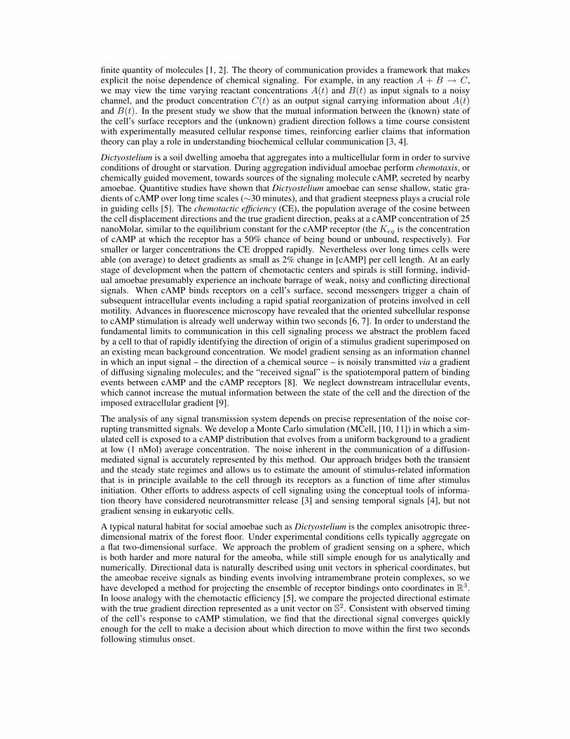
We established a baseline concentration of approximately 1nMol by releasing a cAMP bolus at time0 inside the cube with zero-flux boundary conditions imposed on each wall. At t = 2 seconds weintroduced a steady flux at the x = −L/2 wall of 1 molecule of cAMP per square micron permsec, adding signaling molecules from the left. Simultaneously, the x = +L/2 wall of the cubeassumes absorbing boundary conditions. The new boundary conditions lead (at equilibrium) to alinear gradient of 2 nMol/30µm, ranging from ≈ 2.0 nMol at the flux source wall to ≈ 0 nMol atthe absorbing wall (see Figure 1); the concentration profile approaches this new steady state withtime constant of approximately 1.25 msec. Sampling boxes centered along the planes x = ±13.5µmmeasured the local concentration, allowing us to validate the expected model behavior.
Figure 1: Gradient sensing simulations performed with MCell (a Monte Carlo simulator of cellu-lar microphysiology, http://www.mcell.cnl.salk.edu/) and rendered with DReAMM (Design, Render,and Animate MCell Models, http://www.mcell.psc.edu/). The model cell comprised a sphere tri-angulated with 980 tiles with one cAMP receptor per tile. Cell radius R = 7.5µm; cube sideL = 30µm. Left: Initial equilibrium condition, before imposition of gradient. [cAMP] ≈ 1nMol(c. 15,000 molecules in the volume outside the sphere). Right: Gradient condition after transient(c. 15,000 molecules; see Methods for details).
2.2 Analysis
2.2.1 Assumptions
We make the following assumptions to simplify the analysis of the distribution of receptor activitiesat equilibrium, whether pre- or post-stimulus onset:
1. Independence. At equilibrium, the state of each receptor (bound vs unbound) is independentof the states of the other receptors.
2. Linear Gradient. At equilibrium under the imposed gradient condition, the concentrationof ligand molecule varies linearly with position along the gradient axis.
3. Symmetry.
1http://nwg.phy.bnl.gov/∼bviren/uno/other/
(a) Rotational equivariance of receptor activities. In the absence of an applied gradientsignal, the probability distribution describing the receptor states is equivariant withrespect to arbitrary rotations of the sphere.
(b) Rotational invariance of gradient direction. The imposed gradient seen by a modelcell is equally likely to be coming from any direction; therefore the gradient directionvector is uniformly distributed over S2.
(c) Axial equivariance about the gradient direction. Once a gradient direction is imposed,the probability distribution describing receptor states is rotationally equivariant withrespect to rotations about the axis parallel with the gradient.
Berg and Purcell [1] calculate the inaccuracy in concentration estimates due to nonindependence ofadjacent receptors; for our parameters (effective receptor radius = 5nm, receptor spacing ∼ 1µm)the fractional error in estimating concentration differences due to receptor nonindependence is neg-ligible (. 10−11) [1, 2].
Because we fix receptors to be in 1:1 correspondence with surface tiles, spherical symmetry anduniform distribution of the receptors are only approximate. The gradient signal communicated viadiffusion does not involve sharp spatial changes on the scale of the distance between nearby re-ceptors, therefore spherical symmetry and uniform identical receptor distribution are good analyticapproximations of the model configuration. By rotational equivariance we mean that combiningany rotation of the sphere with a corresponding rotation of the indices labeling the N receptors,{j = 1, · · · , N}, leads to a statistically indistinguishable distribution of receptor activities. Thissame spherical symmetry is reflected in the a priori distribution of gradient directions, which isuniform over the sphere (with density 1/4π). Spherical symmetry is broken by the gradient signal,which fixes a preferred direction in space. About this axis however, we assume the system retainsthe rotational symmetry of the cylinder.
2.2.2 Mutual information of the receptors
In order to quantify the directional information available to the cell from its surface receptors weconstruct an explicit model for the receptor states and the cell’s estimated direction. We model thereceptor states via a collection of random variables {Bj} and develop an expression for the entropyof {Bj}. Then in section 2.2.3 we present a method for projecting a temporally filtered estimateddirection, g, into three (rather than N ) dimensions.
Let the random variables {Bj} Nj=1 represent the states of the N cAMP receptors on the cell surface;
Bj = 1 if the receptor is bound to a molecule of cAMP, otherwise Bj = 0. Let ~xj ∈ S2 representthe direction from the center of the center of the cell to the jth receptor. Invoking assumption 2above, we take the equilibrium concentration of cAMP at ~x to be c(~x|~g) = a+b(~x ·~g) where ~g ∈ S2
is a unit vector in the direction of the gradient. The parameter a is the mean concentration over thecell surface, and b = R|~∇c| is half the drop in concentration from one extreme on the cell surface tothe other. Before the stimulus begins, the gradient direction is undefined.
It can be shown (see Supplemental Materials) that the entropy of receptor states given a fixed gradi-ent direction ~g, H[{Bj}|~g], is given by an integral over the sphere:
H[{Bj}|~g] ∼ N
∫ π
θ=0
∫ 2π
φ=0
Φ[
a + b cos(θ)a + b cos(θ) + Keq
]sin(θ)
4πdφ dθ (as N →∞). (1)
On the other hand, if the gradient direction remains unspecified, the entropy of receptor states isgiven by
H[{Bj}] ∼ NΦ[∫ π
θ=0
∫ 2π
φ=0
(a + b cos(θ)
a + b cos(θ) + Keq
)sin(θ)
4πdφ dθ
](as N →∞), (2)
where Φ[p] ={− (p log2(p) + (1− p) log2(1− p)) , 0 < p < 1
0, p = 0 or 1
}denotes the entropy for a
binary random variable with state probabilities p and (1− p).
In both equations (1) and (2), the argument of Φ is a probability taking values 0 ≤ p ≤ 1. In (1) thevalues of Φ are averaged over the sphere; in (2) Φ is evaluated after averaging probabilities. Because

Φ[p] is convex for 0 ≤ p ≤ 1, the integral in equation 1 cannot exceed that in equation 2. Thereforethe mutual information upon receiving the signal is nonnegative (as expected):
MI[{Bj};~g] ∆= H[{Bj}]−H[{Bj}|~g] ≥ 0.
The analytic solution for equation (1) involves the polylogarithm function. For the parameters shownin the simulation (a = 1.078 nMol, b = .512 nMol, Keq = 25 nMol), the mutual information with980 receptors is 2.16 bits. As one would expect, the mutual information peaks when the meanconcentration is close to the Keq of the receptor, exceeding 16 bits when a = 25, b = 12.5 andKeq = 25 (nMol).
2.2.3 Dimension reduction
The estimate obtained above does not give tell us how quickly the directional information availableto the cell evolves over time. Direct estimate of the mutual information from stochastic simulationsis impractical because the aggregate random variables occupy a 980 dimensional space that a limitednumber of simulation runs cannot sample adequately. Instead, we construct a deterministic functionfrom the set of 980 time courses of the receptors, {Bj(t)}, to an aggregate directional estimatein R3. Because of the cylindrical symmetry inherent in the system, our directional estimator g isan unbiased estimator of the true gradient direction ~g. The estimator g(t) may be thought of asrepresenting a downstream chemical process that accumulates directional information and decayswith some time constant τ . Let {~xj}N
j=1 be the spatial locations of the N receptors on the cell’ssurface. Each vector is associated with a weight wj . Whenever the jth receptor binds a cAMPmolecule, wj is incremented by one; otherwise wj decays with time constant τ . We construct aninstantaneous estimate of the gradient direction from the linear combination of receptor positions,gτ (t) =
∑Nj=1 wj(t)~xj . This procedure reflects the accumulation and reabsorption of intracellular
second messengers released from the cell membrane upon receptor binding.
Before the stimulus is applied, the weighted directional estimates gτ are small in absolute magni-tude, with direction uniformly distributed on S2. In order to determine the information gained as theestimate vector evolves after stimulus application, we wish to determine the change in entropy in anensemble of such estimates. As the cell gains information about the direction of the gradient signalfrom its receptors, the entropy of the estimate should decrease, leading to a rise in mutual informa-tion. By repeating multiple runs (M = 600) of the simulation we obtain samples from the ensembleof direction estimates, given a particular stimulus direction, ~g. In the method of Kozachenko andLeonenko [13], adapted for the analysis of neural spike train data by Victor [14] (“KLV method”),the cumulative distribution function is approximated directly from the observed samples, and theentropy is estimated via a change of variables transformation (see below). This method may beformulated in vector spaces Rd for d > 1 ([13]), but it is not guaranteed to be unbiased in the mul-tivariate case [15] and has not been extended to curved manifolds such as the sphere. In the presentcase, however, we may exploit the symmetries inherent in the model (Assumptions 3a-3c) to reducethe empirical entropy estimation problem to one dimension.
Adapting the argument in [14] to the case of spherical data from a distribution with rotational sym-metry about a given axis, we obtain an estimate of the entropy based on a series of observations ofthe angles {θ1, · · · , θM} between the estimates gτ and the true gradient direction ~g (for details, seeSupplemental Materials):
H ∼ 1M
M∑k=1
(log2(λk) + log2(2(M − 1)) +
γ
loge(2)+ log2(2π) + log2(sin(θk))
)(3)
(as M →∞) where after sorting the θk in monotonic order, λk∆= min(|θk − θk±1|) is the distance
between each angle and its nearest neighbor in the sample, and γ is the Euler-Mascheroni constant.As shown in Figure 2, this approximation agrees with the analytic result for the uniform distribution,Hunif = log2(4π) ≈ 3.651.
3 Results
Figure 3 shows the results of M = 600 simulation runs. Panel A shows the concentration averagedacross a set of 1µm3 sample boxes, four in the x = −13.5µm plane and four in the x = +13.5µm
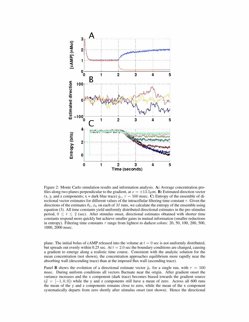
Figure 2: Monte Carlo simulation results and information analysis. A: Average concentration pro-files along two planes perpendicular to the gradient, at x = ±13.5µm. B: Estimated direction vector(x, y, and z components; x = dark blue trace) gτ , τ = 500 msec. C: Entropy of the ensemble of di-rectional vector estimates for different values of the intracellular filtering time constant τ . Given thedirections of the estimates θk, φk on each of M runs, we calculate the entropy of the ensemble usingequation (3). All time constants yield uniformly distributed directional estimates in the pre-stimulusperiod, 0 ≤ t ≤ 2 (sec). After stimulus onset, directional estimates obtained with shorter timeconstants respond more quickly but achieve smaller gains in mutual information (smaller reductionsin entropy). Filtering time constants τ range from lightest to darkest colors: 20, 50, 100, 200, 500,1000, 2000 msec.
plane. The initial bolus of cAMP released into the volume at t = 0 sec is not uniformly distributed,but spreads out evenly within 0.25 sec. At t = 2.0 sec the boundary conditions are changed, causinga gradient to emerge along a realistic time course. Consistent with the analytic solution for themean concentration (not shown), the concentration approaches equilibrium more rapidly near theabsorbing wall (descending trace) than at the imposed flux wall (ascending trace).
Panel B shows the evolution of a directional estimate vector gτ for a single run, with τ = 500msec. During uniform conditions all vectors fluctuate near the origin. After gradient onset thevariance increases and the x component (dark trace) becomes biased towards the gradient source(~g = [−1, 0, 0]) while the y and z components still have a mean of zero. Across all 600 runsthe mean of the y and z components remains close to zero, while the mean of the x componentsystematically departs from zero shortly after stimulus onset (not shown). Hence the directional
estimator is unbiased (as required by symmetry). See Supplemental Materials for the populationaverage of g.
Panel C shows the time course of the entropy of the ensemble of normalized directional estimatevectors gτ/|gτ | over M = 600 simulations, for intracellular filtering time constants ranging from 20msec to 2000 msec (light to dark shading), calculated using equation (3). Following stimulus onset,entropy decreases steadily, showing an increase in information available to the amoeba about thedirection of the stimulus; the mutual information at a given point in time is the difference betweenthe entropy at that time and before stimulus onset.
For a cell with roughly 1000 receptors the mutual information has increased at most by ∼ 2 bits ofinformation by one second (for τ = 500 msec), and at most by ∼ 3 bits of information by two sec-onds (for τ=1000 or 2000 msec), under our stimulation protocol. A one bit reduction in uncertaintyis equivalent to identifying the correct value of the x component (positive versus negative) when thestimulus direction is aligned along the x-axis. Alternatively, note that a one bit reduction results ingoing from the uniform distribution on the sphere to the uniform distribution on one hemisphere.For τ ≤ 100 msec, the weighted average with decay time τ never gains more than one bit of infor-mation about the stimulus direction, even at long times. This observation suggestions that signalingmust involve some chemical components with lifetimes longer than 100 msec. The τ = 200 msecfilter saturates after about one second, at ∼ 1 bit of information gain.
Longer lived second messengers would respond more slowly to changes from the background stim-ulus distribution, but would provide better more informative estimates over time. The τ = 500 msecestimate gains roughly two bits of information within 1.5 seconds, but not much more over time.Heuristically, we may think of a two bit gain in information as corresponding to the change from auniform distribution to one covering uniformly covering one quarter of S2, i.e. all points within π/3of the true direction. Within two seconds the τ = 1000 msec and τ = 2000 msec weighted aver-ages have each gained approximately three bits of information, equivalent to a uniform distributioncovering all points with 0.23π or 41o of the true direction.
4 Discussion & conclusions
Clearly there is an opportunity for more precise control of experimental conditions to deepen ourunderstanding of spatio-temporal information processing at the membranes of gradient-sensitivecells. Efforts in this direction are now using microfluidic technology to create carefully regulatedspatial profiles for probing cellular responses [16]. Our results suggest that molecular processesrelevant to these responses must have lasting effects ≥ 100 msec.
We use a static, immobile cell. Could cell motion relative to the medium increase sensitivity tochanges in the gradient? No: the Dictyostelium velocity required to affect concentration perceptionis on order 1cm sec−1[1], whereas reported velocities are on the order µm sec−1[5].
The chemotactic response mechanism is known to begin modifying the cell membrane on the edgefacing up the gradient within two seconds after stimulus initiation [7, 6], suggesting that the cellstrikes a balance between gathering data and deciding quickly. Indeed, our results show that thereported activation of the G-protein signaling system on the leading edge of a chemotactically re-sponsive cell [7] rises at roughly the same rate as the available chemotactic information. Resultssuch as these ([7, 6]) are obtained by introducing a pipette into the medium near the amoeba; themagnitude and time course of cAMP release are not precisely known, and when estimated the cAMPconcentration at the cell surface is over 25 nMol by a full order of magnitude.
Thomson and Kristan [17] show that for discrete probability distributions and for continuous distri-butions over linear spaces, stimulus discriminability may be better quantified using ideal observeranalysis (mean squared error, for continuous variables) than information theory. The machinery ofmean squared error (variance, expectation) do not carry over to the case of directional data withoutfundamental modifications [18]; in particular the notion of mean squared error is best representedby the mean resultant length 0 ≤ ρ ≤ 1, the expected length of the vector average of a collection ofunit vectors representing samples from directional data. A resultant with length ρ ≈ 1 correspondsto a highly focused probability density function on the sphere. In addition to measuring the mutualinformation between the gradient direction and an intracellular estimate of direction, we also cal-culated the time evolution of ρ (see Supplemental Materials.) We find that ρ rapidly approaches 1
and can exceed 0.9, depending on τ . We found that in this case at least the behavior of the meanresultant length and the mutual information are very similar; there is no evidence of discrepanciesof the sort described in [17].
We have shown that the mutual information between an arbitrarily oriented stimulus and the direc-tional signal available at the cell’s receptors evolves with a time course consistent with observedreaction times of Dictyostelium amoeba. Our results reinforce earlier claims that information theorycan play a role in understanding biochemical cellular communication.
Acknowledgments
MCell simulations were run on the Oberlin College Beowulf Cluster, supported by NSF grant CHE-0420717.
References[1] Howard C. Berg and Edward M. Purcell. Physics of chemoreception. Biophysical Journal, 20:193, 1977.[2] William Bialek and Sima Setayeshgar. Physical limits to biochemical signaling. PNAS, 102(29):10040–
10045, July 19 2005.[3] S. Qazi, A. Beltukov, and B.A. Trimmer. Simulation modeling of ligand receptor interactions at non-
equilibrium conditions: processing of noisy inputs by ionotropic receptors. Math Biosci., 187(1):93–110,Jan 2004.
[4] D. J. Spencer, S. K. Hampton, P. Park, J. P. Zurkus, and P. J. Thomas. The diffusion-limited biochemicalsignal-relay channel. In S. Thrun, L. Saul, and B. Scholkopf, editors, Advances in Neural InformationProcessing Systems 16. MIT Press, Cambridge, MA, 2004.
[5] P.R. Fisher, R. Merkl, and G. Gerisch. Quantitative analysis of cell motility and chemotaxis in Dic-tyostelium discoideum by using an image processing system and a novel chemotaxis chamber providingstationary chemical gradients. J. Cell Biology, 108:973–984, March 1989.
[6] Carole A. Parent, Brenda J. Blacklock, Wendy M. Froehlich, Douglas B. Murphy, and Peter N. Devreotes.G protein signaling events are activated at the leading edge of chemotactic cells. Cell, 95:81–91, 2 October1998.
[7] Xuehua Xu, Martin Meier-Schellersheim, Xuanmao Jiao, Lauren E. Nelson, and Tian Jin. Quantitativeimaging of single live cells reveals spatiotemporal dynamics of multistep signaling events of chemoat-tractant gradient sensing in dictyostelium. Molecular Biology of the Cell, 16:676–688, February 2005.
[8] Jan Wouter-Rappel, Peter. J Thomas, Herbert Levine, and William F. Loomis. Establishing directionduring chemotaxis in eukaryotic cells. Biophys. J., 83:1361–1367, 2002.
[9] T.M. Cover and J.A. Thomas. Elements of Information Theory. John Wiley, New York, 1990.[10] J. R. Stiles, D. Van Helden, T. M. Bartol, E.E. Salpeter, and M. M. Salpeter. Miniature endplate cur-
rent rise times less than 100 microseconds from improved dual recordings can be modeled with passiveacetylcholine diffusion from a synaptic vesicle. Proc. Natl. Acad. Sci. U.S.A., 93(12):5747–52, Jun 111996.
[11] J. R. Stiles and T. M. Bartol. Computational Neuroscience: Realistic Modeling for Experimentalists,chapter Monte Carlo methods for realistic simulation of synaptic microphysiology using MCell, pages87–127. CRC Press, Boca Raton, FL, 2001.
[12] M. Ueda, Y. Sako, T. Tanaka, P. Devreotes, and T. Yanagida. Single-molecule analysis of chemotacticsignaling in Dictyostelium cells. Science, 294:864–867, October 2001.
[13] L.F. Kozachenko and N.N. Leonenko. Probl. Peredachi Inf. [Probl. Inf. Transm.], 23(9):95, 1987.[14] Jonathan D. Victor. Binless strategies for estimation of information from neural data. Physical Review E,
66:051903, Nov 11 2002.[15] Marc M. Van Hulle. Edgeworth approximation of multivariate differential entropy. Neural Computation,
17:1903–1910, 2005.[16] Loling Song, Sharvari M. Nadkarnia, Hendrik U. Bodekera, Carsten Beta, Albert Bae, Carl Franck,
Wouter-Jan Rappel, William F. Loomis, and Eberhard Bodenschatz. Dictyostelium discoideum chemo-taxis: Threshold for directed motion. Euro. J. Cell Bio, 85(9-10):981–9, 2006.
[17] Eric E. Thomson and William B. Kristan. Quantifying stimulus discriminability: A comparison of infor-mation theory and ideal observer analysis. Neural Computation, 17:741–778, 2005.
[18] Kanti V. Mardia and Peter E. Jupp. Directional Statistics. John Wiley & Sons, West Sussex, England,2000.