Embed Size (px)
DESCRIPTION
An Incidental Finding. Patient:. Referred to Urology service following an incidental finding of a 3.7 x 3.8 cm enhancing lesion arising from the lower pole of the left kidney HxPC : Presented to A&E c/o intermittent seizures. Increasing frequency over the previous 3/52. - PowerPoint PPT Presentation
Citation preview

An Incidental Finding
Patient:
Referred to Urology service following an incidental finding of a 3.7 x 3.8 cm enhancing lesion arising from the lower pole of the left kidney
HxPC: Presented to A&E c/o intermittent seizures.
Increasing frequency over the previous 3/52.A/W generalized Left sided weakness and partial parasthesia. No cranial nerve deficitsUnremarkable blood work up
Plan: Admitted to the general internal medicine team Urgent CTB: 6 x 4.5 x 4 cm space occupying lesion in the
right parietal area. Followed by MRI brain on the same day.
Patient:
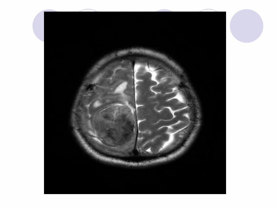
MRI showed a heterogenous 6 x 4.5 x 4 cm enhancing lesion in the right parietal area.
Extensive vasogenic odema and midline shift. Mass effect on the contralateral side. DDx: Meningioma.
A CT TAP was performed to R/O other malignant disease.
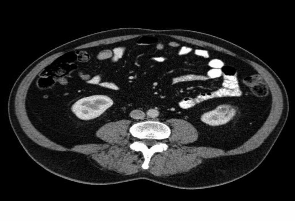

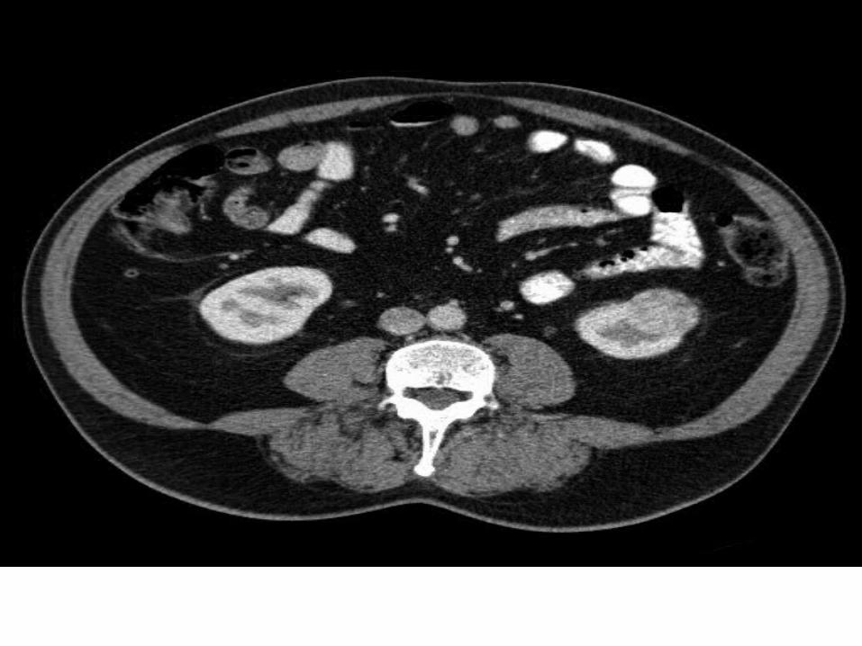
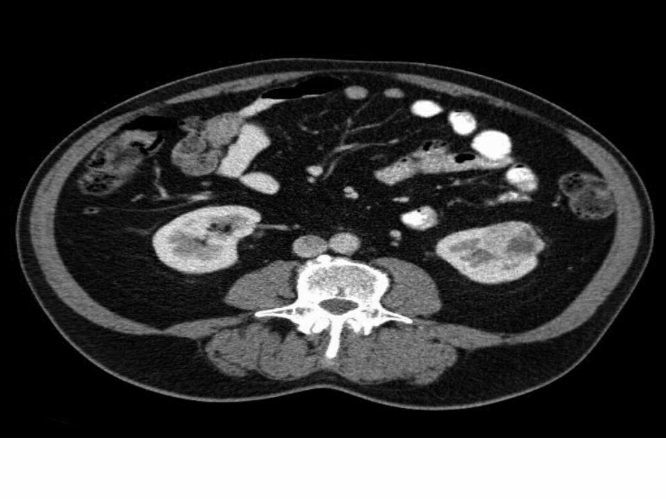
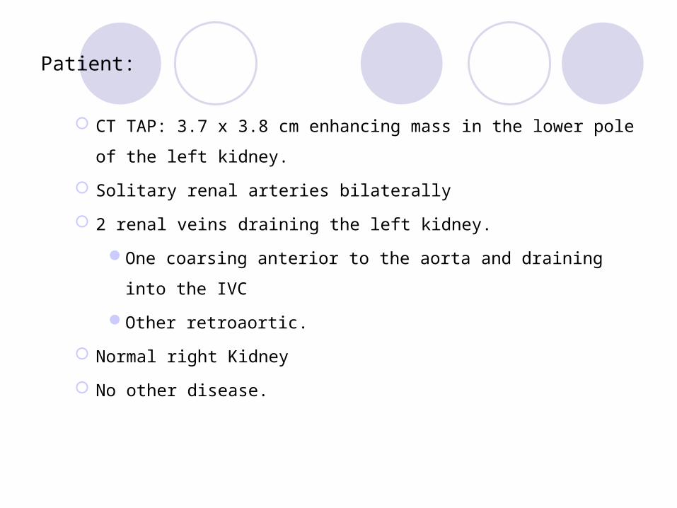
Patient:
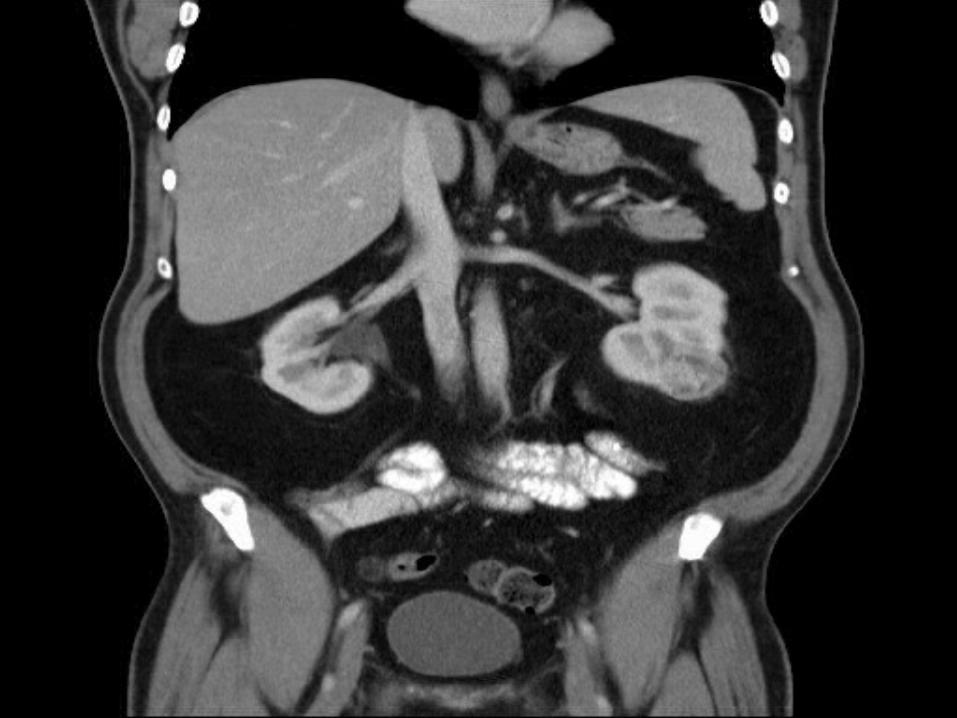
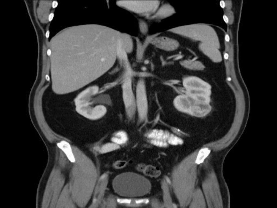
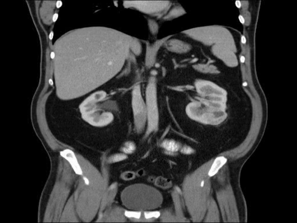
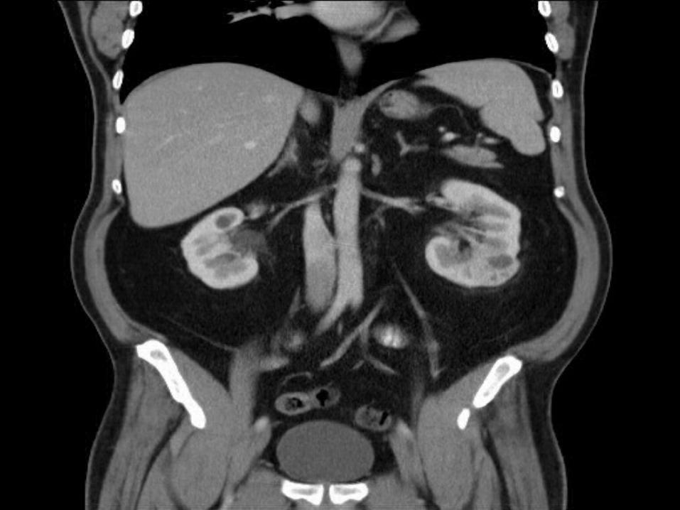
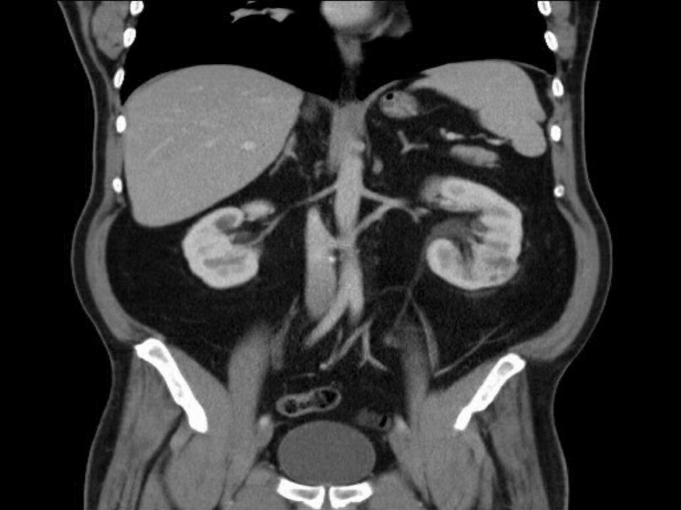
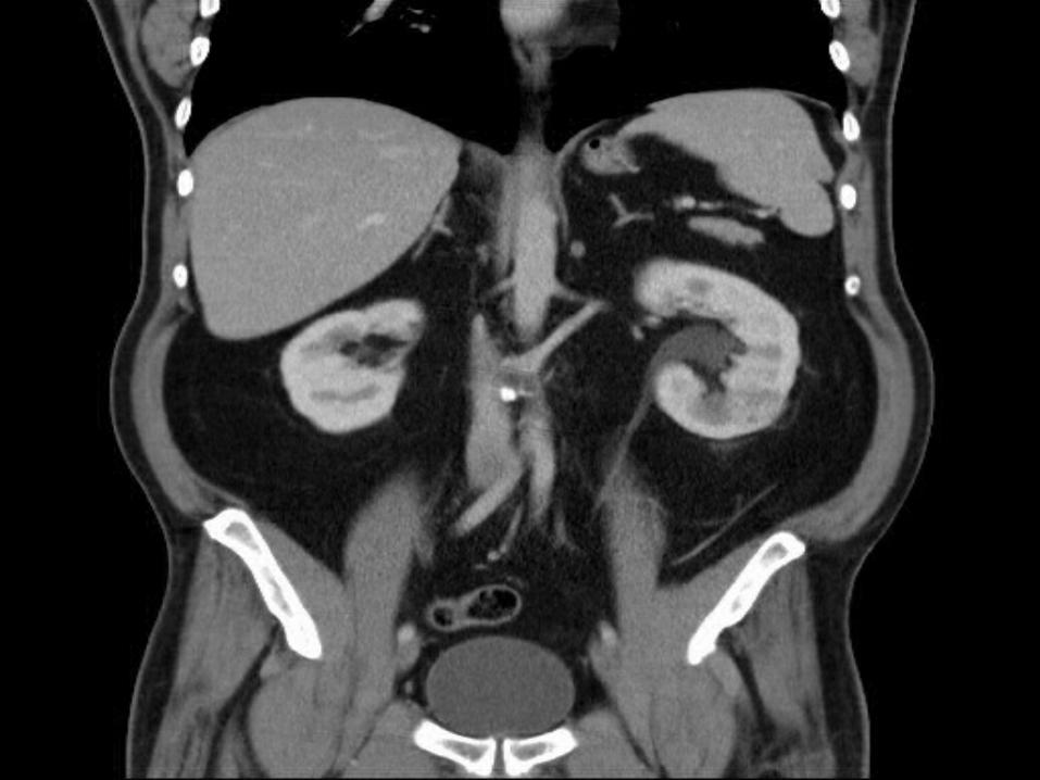
CT TAP: 3.7 x 3.8 cm enhancing mass in the lower pole of the left kidney.
Solitary renal arteries bilaterally 2 renal veins draining the left kidney.
One coarsing anterior to the aorta and draining into the IVCOther retroaortic.
Normal right Kidney No other disease.
Management:
Following resection of the Meningioma, pt was admitted from home for an elective Left partial nephrectomy
This was an open procedure using a left subcostal approach. The renal artery was isolated. The lower pole branch was selectively
isolated and this was then dissected close to the hilum. The ureter was identified and isolated, the lower pole artery was
temporarily occluded and the tumor excised. Occlusion time was 11 mins. The lower pole calyx was closed. Robinsons drain and UC were inserted.
Post-op:
Pathology : Clear cell RCC pT1a Grade 2.
Pt self removed drain day 1 post op. Spiked 38.9C temp the same evening and was commenced on
IV Tazocin Day2 CT urogram showed a no collections or leaks. Fever resolved and there was an uncomplicated course to
discharge on day 7.
Learning Points:
The incidence of RCC has been rising steadily
Highest worldwide incidences in northern Europe and NA
M:F 1.5-2:1
A/W Smoking, high BMI, and HTN
Clear cell renal cell carcinoma is the most common histologic variant (75-88%)
Particular rise in the proportion of small, asymptomatic tumors detected
incidentally via abdominal imaging.
RCC can remain occult for much of the disease course
25-32% present with metastases
2011 study looking retrospectively at 3001 Colonographic, CT abdo and CTTAP
found incidental renal masses in 443 patients (14.4%).
Smaller, pre-symptomatic lesions may be amenable to a Nephron Sparing
approach
Learning Points:
Initial management of the incidentally diagnosed or early presenting
lesion is by partial or total nephrectomy.
The aim is neprhron sparing when possible.
Particularly in peripheral tumors
Laprascopic techniques are common.
However not suited to all cases and is limited when the calyx is incised
or if inter-renal suturing is required.
Higher incidence of positive surgical margins
Longer warm ischaemic time
No significant increase in organ loss.

Thank you