Embed Size (px)
Citation preview

An embryoprotective role for glucose-6-phosphatedehydrogenase in developmental oxidative stress andchemical teratogenesis
CHRISTOPHER J. NICOL,* JULIAN ZIELENSKI,† LAP-CHEE TSUI,†,‡ ANDPETER G. WELLS*,§,1
*Department of Pharmacology, University of Toronto, Medical Sciences Building, Toronto Ontario,Canada M5S 1A8; †Department of Genetics, The Hospital for Sick Children, Toronto, Ontario,Canada M5G 1X8; ‡Department of Molecular and Medical Genetics, and Institute of MedicalSciences, University of Toronto, Toronto, Ontario, Canada M5S 1A8; and §Faculty of Pharmacy,University of Toronto, Toronto, Ontario, Canada M5S 2S2
ABSTRACT The primary recognized health riskfrom common deficiencies in glucose-6-phosphatedehydrogenase (G6PD), a cytoprotective enzyme foroxidative stress, is red blood cell hemolysis. Here weshow that litters from untreated pregnant mutantmice with a hereditary G6PD deficiency had in-creased prenatal (fetal resorptions) and postnataldeath. When treated with the anticonvulsant drugphenytoin, a human teratogen that is commonly usedin pregnant women and causes embryonic oxidativestress, G6PD-deficient dams had higher embryonicDNA oxidation and more fetal death and birthdefects. The reported G6PD gene mutation wasconfirmed and used to genotype fetal resorptions,which were primarily G6PD deficient. This is the firstevidence that G6PD is a developmentally criticalcytoprotective enzyme for both endogenous andxenobiotic-initiated embryopathic oxidative stressand DNA damage. G6PD deficiencies accordinglymay have a broader biological relevance as importantdeterminants of infertility, in utero and postnataldeath, and teratogenesis.—Nicol, C. J., Zielenski, J.,Tsui, L.-C., Wells, P. G. An embryoprotective rolefor glucose-6-phosphate dehydrogenase in develop-mental oxidative stress and chemical teratogenesis.FASEB J. 14, 111–127 (2000)
Key Words: reactive oxygen species z development z birth defectsz phenytoin z human risk
Glucose-6-phosphate 1-dehydrogenase (g6pd)(EC 1.1.1.49) is the first and rate-limiting enzymein the hexose monophosphate shunt (HMS) path-way, important for its role in the regeneration ofthe reduced form of nicotinamide adenine dinu-cleotide phosphate (NADPH) and the productionof ribose (1, 2). During cellular oxidative stress,whether endogenous in origin or initiated bydrugs or environmental chemicals, collectively re-ferred to as xenobiotics, NADPH is critical for
maintaining glutathione (GSH) in its reducedform, which is essential for detoxification of reac-tive free radicals and lipid hydroperoxides (3–5)(Fig. 1). Another important role for NADPH is themaintenance of the catalytic activity of catalase(6 – 8); hence, NADPH also is important for its rolein the detoxification of hydrogen peroxide. Theproduction of ribose by the HMS is relevant to thesynthesis of nucleotides used in RNA and DNAreplication and, hence, cell division and possiblyDNA repair (2, 9). The gene for G6PD, containing13 exons, has been localized to the X chromosome(Xq28), and the coding sequence has been re-ported for a number of species including mice,rats, Drosophila, yeast, and humans (10, 11). He-reditary deficiencies in G6PD, first identified inthe late 1950s based on studies of differentialsusceptibility to the hemolytic effects of prima-quine (12), are the most common enzymopathyknown, affecting well over 400 million peopleworldwide, and particularly those from the Medi-terranean region and selected African and Asiancountries, wherein the incidence of G6PD defi-ciency may approach 60% of some populations(2). The degree of G6PD deficiency varies fromnegligible to severe, according to both whetherone or two alleles are affected and the nature ofthe gene mutation and protein/enzyme variant(1). To date, 100 human genetic mutations involv-ing the 12 coding exons, and up to 400 enzymevariants, have been described (10, 13–15). Hema-tological problems arising in these G6PD-deficientpopulations from exposure to oxidizing xenobiot-ics have been well characterized, ranging fromhemolysis of red blood cells and hereditary non-
1 Correspondence: Faculty of Pharmacy, University of To-ronto, 19 Russell Street, Toronto, Ontario, Canada M5S 2S2.E-mail: [email protected]
1110892-6638/00/0014-0111/$02.25 © FASEB

spherocytic hemolytic anemia to sepsis and life-threatening kernicterus in the newborn (9, 16).
It currently is believed that G6PD deficienciesconstitute a problem only for mature red bloodcells, which are non-nucleated and cannot synthe-size more protective enzyme under conditions ofoxidative stress (9, 13). However, embryonic tis-sues up to and including the critical period oforganogenesis are remarkably deficient in thesynthesis of many enzymes, including most ofthose providing cytoprotection against oxidativestress, such as GSH reductase, GSH peroxidase,superoxide dismutase, and catalase (4, 17). On theother hand, elevated G6PD activity during embry-onic development corresponds to periods of bothincreased cellular proliferation and DNA synthesis(18, 19), suggesting that G6PD activity may beimportant for normal development. Accordingly,we hypothesized that G6PD-deficient embryoswould be highly susceptible to normal develop-mental oxidative stress, and even more so to thatinitiated by oxidizing xenobiotics (Fig. 1). Thishypothesis was tested in pregnant mutant C3Hmice with heterozygous (1/2) or homozygous
(2/2) deficiencies in G6PD activity, comparedwith congenic G6PD-normal controls (1/1).Dams either were allowed to deliver untreated orwere treated during organogenesis with either themost commonly used anticonvulsant drug in NorthAmerica, phenytoin (Dilantin), a human terato-gen that is representative of xenobiotics known toinitiate embryonic oxidative stress (20) or its vehi-cle. To determine the cytoprotective role of G6PDwith respect to xenobiotic-initiated embryonicDNA damage, after maternal treatment with phe-nytoin during organogenesis, individual embryoswere analyzed for both G6PD activity and DNAoxidation. The recently reported functional muta-tion in the mouse G6PD gene (21) was confirmedby a combination of direct sequencing and thedevelopment of a polymerase chain reaction(PCR)-based genotyping method, which was usedto determine the frequency of the G6PD-deficientgenotype in the remnants (resorptions) of em-bryos that died in utero. The results provide thefirst direct evidence of a critical embryoprotectiverole for G6PD in both endogenous and xenobiotic-initiated oxidative stress and DNA damage.
Figure 1. Postulated embryoprotective role of glucose-6-phosphate dehydrogenase (G6PD). Toxic reactive oxygen species (ROS)such as lipid hydroperoxides (LOOH) and hydrogen peroxide (H2O2) are formed endogenously and can be substantiallyenhanced by xenobiotic bioactivation catalyzed by the cytochromes P450 (P450s), peroxidases such as prostaglandin H synthase(PHS), and via reactions catalyzed by related enzymes such as lipoxygenases (LPOs). The detoxification of ROS by glutathione(GSH) peroxidase requires the cofactor GSH, which is oxidized to its disulfide, GSSG. The chemical reduction of GSSG andmaintenance of adequate GSH for ROS detoxification, as well as for the detoxification of free radical intermediates, ultimatelyis dependent on NADPH supplied by G6PD. In addition, G6PD-dependent production of NADPH may be important inmaintaining the activity of catalase, which also detoxifies H2O2 (37).
112 Vol. 14 January 2000 NICOL ET AL.The FASEB Journal

MATERIALS AND METHODS
Chemicals
Phenytoin (sodium salt) was purchased from Sigma Chemical(St. Louis, Mo.). Carnoy’s solution was made from 100% abso-lute ethanol, chloroform, and glacial acetic acid (6:3:1, v/v/v)(Sigma). Bio-Rad protein assay dye reagent concentrate waspurchased from Bio-Rad Laboratories (Hercules, Calif.), andAlbumin Concentrate (source: Bovine Serum) protein assaystandard was purchased from Pierce (Rockford, Ill.).
Animals
Breeding pairs of G6PD-mutant C3H mice were purchasedfrom the Medical Research Council (MRC) of England(Genetics Division, MRC Radiobiology Unit, Chilton, U.K.).Same-sex animals were housed not more than three to onemicroisolator cage containing ground corncob bedding (BetaChip, Northeastern Products, Warrensburg, N.Y.) and weremaintained in a temperature-controlled animal facility with a12 h light/dark cycle. Food (Laboratory Rodent Chow 5001;PMI Feeds, St. Louis, Mo.) and tap water were provided adlibitum. The genotype of all animals was confirmed phenotyp-ically by analysis of RBC G6PD activity. To establish a breed-ing colony, three females were housed overnight with onemale breeder starting at 5:00 p.m. Females were checked by9:00 a.m. the next morning, and the presence of a vaginalplug was designated as gestational day (GD) 1. Pregnantfemales were placed in their own microisolator cages andallowed to deliver spontaneously, with an average gestation of20.5 days. The number of pups was recorded daily, and thepups were left with their mothers until weaning, 21 days afterbirth. The number and sex of weaned pups were recorded.Pups were ear-notched for identification and phenotyped byG6PD activity using tail vein blood.
Teratogenesis
Homozygous (1/1) G6PD-normal and heterozygous (1/2)and homozygous (2/2) G6PD-deficient females were matedwith males that were hemizygous (2/y) G6PD-deficient asdescribed above. Dams were either untreated, treated intra-peritoneally at 9:00 a.m. on GDs 12 and 13 with vehicle aloneor treated with a subteratogenic (20 mg/kg) or teratogenicdose (65 mg/kg) of phenytoin in saline containing 0.002NNaOH (17) and killed by cervical dislocation on GD 19. Theuterus was exteriorized, implantations (fetuses and resorp-tions/in utero deaths) were noted, and fetuses and dissectableresorptions were removed. Fetuses were examined to deter-mine sex, weight, and external anomalies. Viable fetuses werekept warm under a heat lamp (30°C) for 2 h to assesspostpartum lethality. Fetuses subsequently were bled by de-capitation and phenotyped for RBC G6PD activity. Resorp-tions and fetal tails were stored at 280°C for future study.Pinpoint resorptions were noted and left in the uterus, whichwas stored, similar to fetal heads and bodies, in Carnoy’ssolution for future analysis. Fetuses were later examined forinternal anomalies.
G6PD gene sequencing
RNA was isolated and purified from 1/1 G6PD-normal and2/2 G6PD-deficient mouse spleen samples using a QiagenRNeasy total RNA purification kit (Qiagen, Chatsworth,Calif.) according to the manufacturer’s instructions. PurifiedRNA was subsequently converted to cDNA using a Gibco BRL
Superscript Preamplification System for First Strand cDNASynthesis (Gibco BRL [Canada], Burlington, Ontario) witholigo dT primers according to the manufacturer’s instruc-tions using a Perkin Elmer GeneAmp PCR System 2400thermal cycler (Perkin-Elmer [Canada], Mississauga, On-tario). Oligonucleotide primers for PCR reactions were syn-thesized based on previously reported C57BL/6 mouse G6PDcDNA sequences (11). cDNA coding for G6PD was subse-quently amplified using the PCR. Amplified cDNA templateswere subsequently used for direct cycle sequencing using aThermo Sequenase radiolabeled terminator cycle sequencingkit (Amersham [Canada], Oakville, Ontario) according to themanufacturer’s instructions, where sequencing primers forboth sense and antisense strands, designed to produce over-lapping sequences, were based on the published normalG6PD mouse cDNA sequence (11). Samples were run onstandard 6% polyacrylamide gels and exposed overnightprior to developing film. Data for G6PD mutant mouseintronic regions were sequenced from genomic mouse DNAsamples derived from tail snips.
G6PD genotyping
DNA was isolated from late-stage (dissectable) fetal resorp-tions by the method of Gupta (22). G6PD mouse PCR primers(sense: GGAAACTGGCTGTGCGCTAC, antisense:TCAGCTCCGGCTCTCTTCTG) were made between exon 1and intron 1, around the reported mutation site (21). PCRconditions on a Perkin Elmer 9600 thermal cycler (Perkin-Elmer [Canada]) were 94°C for 2 min, 20 s at 94°C, 20 s at58°C, and 30 s at 72°C for a total of 35 cycles, with a 5 minextension at 72°C and kept at 4°C until ready for digestion.PCR products were digested using DdeI restriction enzyme(Gibco BRL) at 37°C for 1 h and run on 3% agarose gels todetermine G6PD genotype.
G6PD phenotyping
G6PD activity was measured in RBCs, whole embryo homog-enates, and the 9,000 g supernatant from homogenizedmaternal organs using a standard reagent kit purchased fromSigma. Activities were measured over a 5 min interval at 37°Con a UV/vis spectrophotometer (model Lambda 3, Perkin-Elmer [Canada]) using a computer-assisted kinetic program.All results were standardized with respect to total proteincontent and reported in International Units per gram ofprotein (U/g). G6PD normal control standards (Sigma) wererun concurrently with samples.
DNA oxidation
Females were mated as in the teratological studies. Dams werekilled 6 h after maternal treatment with phenytoin (65 mg/kgi.p.) on GD 13. The uterus was exteriorized, and embryoswere removed and homogenized separately. Once G6PDactivity was measured, DNA was isolated from the remainderof the individual whole embryo homogenates by the methodof Gupta (22), as modified in Winn and Wells (20). Embry-onic DNA oxidation was measured by the method of Shi-genaga and Ames (23), using high-performance liquid chro-matography with electrochemical detection of 8-hydroxy-29-deoxyguanosine (8-OH-29-dG).
Protein concentration assay
Protein content was analyzed using the standard Bio-Radprotocol (Bio-Rad, Hercules, Calif.), as detected by spectro-
113G6PD PROTECTS AGAINST ROS-MEDIATED EMBRYOPATHIES

photometric absorbance at 595 nm, using bovine serumalbumin concentrate as a standard.
Statistical analysis
Binomial data were analyzed using x2 analysis or Fisher’s exacttest where appropriate. Continuous data were analyzed using atwo-way analysis of variance (ANOVA) and the Student-New-man-Keuls test. The level of significance was P , 0.05.
RESULTS
Characterization of phenotype
Figure 2 presents differences in red blood cell (RBC)G6PD activities among G6PD mutant adult progeni-tor mice (3 months of age, from our colony), withhemizygous (2/y), heterozygous (1/2), and ho-mozygous (2/2) mutants having 21, 53, and 17%,respectively, of normal RBC G6PD activity. Pheno-typing of congenic G6PD normal and mutant ani-mals was based on our progenitor RBC G6PD activ-ities (see Fig. 2) and corroborated by comparisonswith expected outcomes, from both parental matingsand neonatal sex, given that the G6PD mutation isinherited via the X chromosome.
Untreated mice
In untreated dams allowed to deliver spontaneously,compared with congenic 1/1 G6PD-normal con-trols, litter sizes for 2/2 G6PD-deficient animalswere 50% smaller at birth (Fig. 3, upper panel)(P,0.05). Subsequently, by the time of weaning,litter sizes were 90% smaller, and the incidence of
preweaning offspring death was threefold higher foruntreated 2/2G6PD-deficient dams compared with1/1 G6PD-normal controls (P,0.0001) (Fig. 3,lower panel).
Vehicle control mice
Homozygous G6PD-normal (1/1) and heterozy-gous (1/2) and homozygous (2/2) G6PD-defi-cient mice were injected intraperitoneally on GD 12and 13 with the saline/NaOH vehicle for phenytoinand killed on GD 19. Compared with 1/1 G6PD-normal dams, 1/2 and 2/2 G6PD-deficient damshad 6- and 7-fold increases, respectively, in fetalresorptions (in utero deaths) (P,0.0001), 6- and11-fold increases, respectively, in postpartum lethal-ity (P,0.0001), and 7 and 16% decreases, respec-tively, in fetal body weight (P,0.05) (Fig. 4).
Figure 2. Characterization of G6PD phenotype. Red bloodcell (RBC) G6PD activities among congenic adult progenitors(3 months of age, from our colony) for homozygous (1/1)and hemizygous G6PD-normal (1/y), and heterozygous (1/2), homozygous (2/2), and hemizygous (2/y) G6PD mu-tant mice are presented. The total number of mice from aparticular phenotype is given in parentheses. Adult G6PDactivities were used in subsequent studies to assess embryonicand maternal G6PD phenotype.
Figure 3. Spontaneous embryopathies in untreated G6PD-deficient and G6PD-normal dams. Pregnant heterozygous(1/2) and homozygous (2/2) G6PD-deficient dams, andhomozygous (1/1) G6PD-normals, were allowed to deliverspontaneously, and litter sizes were determined on the day ofbirth and 21 days later at the time of weaning. Upper panel,litter size at birth. The total number of litters from a givenmaternal phenotype is given in parentheses. The asteriskindicates a difference from 1/1 G6PD-normal controls(P,0.05). Lower panel, incidence of preweaning death of pupsborn live and dying before weaning. The total number ofviable pups for a given maternal phenotype is given inparentheses. The asterisk indicates a difference from G6PD-normal controls (P,0.0001).
114 Vol. 14 January 2000 NICOL ET AL.The FASEB Journal

With respect to embryonic phenotype, comparedwith 1/y G6PD-normal fetuses, independent of sex,1/2 and 2/2 and 2/y G6PD-deficient fetuses hadenhanced postpartum lethality, following a gene-dose pattern with 0% in 1/y G6PD-normal fetuses,10% in 1/2 G6PD-deficient fetuses, and 50% incombined 2/2 and 2/y G6PD-deficient fetuses(P,0.0001). A similar gene-dose pattern was ob-served with fetal body weight, which, compared with1/y G6PD-normal fetuses, was decreased by 6% infetuses with a mutation in one G6PD allele (1/2)and by 18% in fetuses with a mutation in all alleles(2/2 and 2/y) (P,0.05) (Fig. 5). Furthermore,the mean weight of fetuses with a mutation in all
G6PD alleles (2/2 and 2/y) was 13% lower thanthat of fetuses with a mutation in one allele (1/2)(P,0.05).
Phenytoin-treated mice
With respect to maternal phenotype, compared withvehicle-treated controls of the same phenotype, astandard teratogenic dose of phenytoin (65 mg/kg)enhanced fetal resorptions twofold in both 1/2 and2/2 G6PD-deficient dams (P,0.05), and in 1/2G6PD-deficient dams, enhanced postpartum lethal-ity threefold and decreased fetal weight by 27%
Figure 5. Embryonic DNA oxidation and embryopathies fortreated G6PD-deficient mice analyzed by embryonic pheno-type. Fetuses from dams described in Fig. 4 were phenotypedby RBC G6PD activity. Homozygous female (2/2) andhemizygous male (2/y) G6PD-deficient fetuses with all allelesmutated were identically affected, and these data were com-bined in this and all subsequent analyses. The total number ofviable fetuses for a particular phenotype is given in parenthe-ses. Upper panel, postpartum lethality was calculated as de-scribed in Fig. 4. Upper panel inset, embryonic DNA oxidationas a reflection of oxidative stress and DNA damage in GD 13embryos exposed 6 h before to phenytoin, 65 mg/kg i.p. Thetotal number of embryos from each group is given in paren-theses, and the asterisk indicates a difference from G6PD-normal littermates (P,0.03). Normal whole embryo G6PDactivity was defined as $25 U/g protein. Lower panel, meanfetal body weights. Asterisks indicate a difference from respec-tive G6PD-normal groups (P,0.05), § indicates a differencefrom vehicle controls from the same phenotype (P,0.05),and † indicates a difference from respective 1/2 G6PD-deficient fetal groups (P,0.05). a indicates that 1/1 G6PD-normal fetuses are not achievable with 2/y male breeders.
Figure 4. Embryopathies in treated normal and G6PD-defi-cient mice by maternal phenotype. Dams were treated intra-peritoneally (i.p.) on gestational days (GDs) 12 and 13 witheither phenytoin or its vehicle and killed on GD 19. Upperpanel, fetal resorptions (in utero deaths) were calculated bydividing the total number of resorptions by the total numberof implantations (resorptions plus fetuses) for a particularmaternal phenotype. The total number of implantations isgiven in parentheses. Middle panel, postpartum lethality wascalculated by dividing the total number of fetuses born liveand dying within 2 h by the total number of viable fetuses fora given maternal phenotype. The total number of viablefetuses is given in parentheses. Lower panel, for mean fetalweights, the total number of viable fetuses for a givenmaternal phenotype is given in parentheses. Asterisks indi-cate a difference from respective 1/1 G6PD-normal controls(P,0.002), § indicates a difference from vehicle controls ofthe same phenotype (P,0.05), and † indicates a differencefrom respective phenytoin (20 mg/kg) groups of the samephenotype (P,0.05).
115G6PD PROTECTS AGAINST ROS-MEDIATED EMBRYOPATHIES

(P,0.05) (Fig. 4). Among phenytoin-treated dams(65 mg/kg), compared with 1/1 G6PD-normalcontrols, in both 1/2 and 2/2 G6PD-deficientdams respectively, the incidence of fetal resorptionswas enhanced 4.8- and 5.5-fold (P,0.0003), theincidence of postpartum lethality was enhanced 16.6-and 15.4-fold (P,0.002), and fetal weight was de-creased by 18–29% (P,0.05) (Fig. 4).
With a lower dose of phenytoin (20 mg/kg) that isnonteratogenic in other strains (4, 5) as well as the wildtype of this G6PD mutant, all embryopathies in phenyt-oin-treated 1/2 and 2/2 G6PD-deficient dams wereincreased above the values observed in phenytoin-treated 1/1 G6PD-normal controls (P,0.05). How-ever, when analyzed by maternal phenotype, unlikeembryonic phenotype (see below), these values wereonly different from vehicle controls of the same mater-nal phenotype among 1/1 dams (Fig. 4).
Embryonic phenotype
With respect to embryonic phenotype, the higherdose of phenytoin (65 mg/kg) caused a 2.4-foldincrease in embryonic DNA oxidation in G6PD-deficient fetuses compared to G6PD-normal litter-mates (P,0.03) (Fig. 5, upper panel, inset), and adecrease in fetal body weight in all phenotypes, withthe weight loss being progressively worse with one ortwo mutated G6PD alleles (Fig. 5, lower panel).Thus, compared to vehicle controls of the samephenotype, the phenytoin-initiated decrease in fetalweight was 5% in 1/y G6PD-normal fetuses, 9.5%in 1/2 G6PD-deficient fetuses, and 9% in 2/2 and2/y G6PD-deficient fetuses (P,0.05). The high doseof phenytoin also appeared to enhance postpartumlethality in all G6PD-deficient fetuses, although thesedifferences were not statistically significant (Fig. 5,upper panel).
Among only those fetuses exposed to phenytoin(65 mg/kg), compared with 1/y G6PD-normal fe-tuses, which had no postpartum lethality, phenytoin-initiated postpartum lethality and decreased fetalbody weight were substantially worse in 1/2 G6PD-deficient fetuses, and even more so in 2/2 and 2/yG6PD-deficient fetuses (P,0.05) (Fig. 5). Thus, post-partum lethality initiated by phenytoin (65 mg/kg)was increased 2.9-fold in 2/2 and 2/y fetusescompared with 1/2 fetuses, which in turn weresubstantially more affected than 1/y G6PD-normalfetuses (25 vs. 0%) (P,0.05). Similarly, with pheny-toin-exposed fetuses, the mean weight of combined2/2 and 2/y fetuses was decreased by 12.9% com-pared with 1/2 fetuses, which in turn had weights10.5% lower than those in 1/y G6PD-normal fetuses(P,0.05). There were no apparent gender differ-ences in teratological susceptibility (data notshown).
Similar to the effects seen among 1/1 dams formean fetal body weight, when the data were analyzedby embryonic phenotype, there was evidence for anembryopathic effect of the lower dose of phenytoin(20 mg/kg). This pattern was observed for bothenhanced postpartum lethality and decreased fetalbody weight but was statistically significant only forthe latter in 1/2 G6PD-deficient embryos (P,0.05)(Fig. 5).
Teratological syndrome
Viable fetuses from the teratological studies wereexamined in a blinded fashion for both external andinternal anomalies (Fig. 6). These structural anom-alies, collectively referred to here as a syndrome,included cleft palate, club foot, dilated bladder,dilated cerebral ventricles, ectopic kidney, hema-toma, microcephaly, micrognathia, omphalocele,open eye, red nevus, and underdeveloped renalpapilla. A gene-dose response was observed in bothvehicle- and phenytoin-treated groups, and a drug-
Figure 6. Teratological syndrome in treated G6PD-deficientmice by embryonic and maternal phenotype. These data wereobtained from the dams described in Fig. 4 treated withphenytoin or its vehicle. Visible anomalies, collectively re-ferred to as a syndrome, included cleft palate, club foot,dilated bladder, dilated cerebral ventricle, ectopic kidney,hematoma, microcephaly, micrognathia, omphalocele, openeye, red nevus, and underdeveloped renal papilla. The sever-ity of the teratological syndrome was calculated as the totalnumber of anomalies for a given phenotype divided by thetotal number of fetuses for that phenotype. The total numberof fetuses for a particular phenotypic group is given inparentheses. Inset, teratological syndrome by maternal phe-notype. Asterisks indicate a difference from respective G6PD-normal groups (P,0.05), § indicates a difference from vehi-cle controls from the same phenotype (P,0.05), and †indicates a difference from respective 1/2 G6PD-deficientgroups (P,0.05).
116 Vol. 14 January 2000 NICOL ET AL.The FASEB Journal

dose response was observed in phenytoin-treatedanimals. This pattern was observed by both embry-onic (Fig. 6) and maternal (Fig. 6, inset) phenotype,but was most clearly defined by embryonic pheno-type. Data for 2/2(female) and 2/y (male) G6PD-deficient fetuses were analyzed independently andfound to be identical, hence these two phenotypeswith all alleles mutated were combined for all finalanalyses.
For fetuses exposed only to vehicle, anomalies in2/2 and 2/y G6PD-deficient fetuses were overtwofold higher than in either 1/2 G6PD-deficientor 1/y G6PD-normal fetuses (P,0.05) (Fig. 6). Asimilar twofold enhancement was observed by mater-nal phenotype, but was not statistically significant(Fig. 6, inset).
For fetuses exposed to the highest dose of phenyt-oin (65 mg/kg), compared with 1/y G6PD-normalfetuses, anomalies were increased 2.5-fold in 1/2G6PD-deficient fetuses and 4.5-fold (P,0.05) in2/2 and 2/y G6PD-deficient fetuses (Fig. 6). Forall G6PD-deficient fetuses, this enhancement alsowas observed relative to vehicle controls of the samephenotype (P,0.05). The higher dose of phenytoinproduced more anomalies than the lower dose (20mg/kg) in 2/2 and2/y G6PD-deficient fetuses(P,0.05). With the lower phenytoin dose, comparedwith 1/y G6PD-normal phenytoin-treated controls,anomalies were not increased in 1/2 fetuses, butwere increased almost twofold in 2/2 and 2/yfetuses (P,0.05). When compared irrespective ofembryonic phenotype, the lower dose of phenytoincaused a 1.6-fold increase in anomalies comparedwith vehicle controls (P,0.05).
By maternal phenotype, a similar pattern of phe-nytoin-enhanced anomalies was observed, exceptthat 1/2 G6PD-deficient dams were as susceptibleas 2/2 dams, and even the lower dose of phenytoinwas teratogenic. The lower phenytoin dose causedover a threefold, albeit nonsignificant, increase inanomalies in 1/2 G6PD-deficient dams comparedwith 1/1 G6PD-normal phenytoin-treated damsand a significant fourfold increase when comparedwith pooled 1/1 and 1/2 vehicle controls(P,0.05) (Fig. 6, inset). With the higher dose ofphenytoin compared with respective vehicle con-trols, there was a fivefold increase in anomaliesin 1/2 G6PD-deficient dams (P,0.05). A similarbut lower twofold enhancement was observed in2/2 6PD-deficient dams because of the 2.6-foldincrease in anomalies in the vehicle controls for thisphenotype. When combined, 1/2 and 2/2 G6PD-deficient dams treated with the higher dose ofphenytoin had over fourfold more anomalies thanrespective phenytoin-treated 1/1 G6PD-normaldams (P,0.05). Similarly, combined 1/2 and 2/2G6PD-deficient dams treated with either a high or
low dose of phenytoin had three- and twofold moreanomalies, respectively, compared with their com-bined vehicle controls (P,0.05). The higher dose ofphenytoin was more teratogenic than the lower dose,but unlike by embryonic phenotype, the differenceby maternal phenotype was statistically significantonly when the data for 1/2 and 2/2 G6PD phe-notypes were combined (P,0.05).
In G6PD-normal animals, phenytoin in either dosedid not increase fetal anomalies compared withvehicle controls in either 1/y fetuses (Fig. 6) or1/1 dams (Fig. 6, inset).
Mutational analysis and genotyping for late in uteroembryonic death
We were able to confirm the reported mutation (21)causing the heritable decrease in G6PD activity inour strain of mutant mice. This involved directsequencing of not only the full-length mutant mousecDNA, which includes the 59 and 39 untranslatedregions and the entire coding region for the G6PDprotein, but also 6 of the 12 intronic G6PD genomicDNA regions, including introns 4, 6, 7, and 10–12.Apart from the reported functional mutation in ourC3H mutant mouse strain, we found a single silent Ato C mutation, located at base 718 of the mousecDNA sequence. This silent mutation differs fromthe reported C57 mouse strain cDNA (11) andcorresponds to the human DNA sequence at base15361 (24), maintaining the amino acid sequencecode of GGC for glycine.
We also noted that the single functional pointmutation, an A to T transversion in the 59 untrans-lated region of the gene at the penultimate base ofthe 39 end of exon 1, results in the destruction of aDdeI restriction enzyme site in the mutant allele. Weused this information to develop a genotyping assayto characterize the functional G6PD mutation in theremnants of dissectable fetal resorptions from ourteratological studies (Fig. 7). In short, we designed apair of PCR primers around the mutation site, basedon the known sequence of the G6PD gene, expectedto produce a PCR fragment from mouse genomicDNA of 269 base pairs (bp) in length. For normalmice, subsequent digestion of PCR products with theDdeI restriction enzyme would produce two cleavedfragments of 214 bp and 55 bp, whereas mice withmutations in all G6PD alleles would not show achange in fragment size, and heterozygotes wereexpected to show all three fragments. All three bandswere initially observed, as expected, by polyacryl-amide gel electrophoresis (PAGE) analysis, but sub-sequent analysis of samples was performed usingagarose gel electrophoresis, in which the 55 bpfragment did not resolve from the leading xylenecyanole band of the loading buffer (Fig. 7).
117G6PD PROTECTS AGAINST ROS-MEDIATED EMBRYOPATHIES

Based on an approach previously established inour laboratory (25), these studies allowed us todetermine the potential protective role of G6PD withrespect to in utero fetal death (Fig. 8). Among thedissectable fetal resorptions reflecting in utero deathlate in gestation, compared with 1/y G6PD-normalfetuses, G6PD-deficient fetuses with a mutation ineither one (1/2) or all (2/2 and 2/y) G6PDalleles had over six- and fivefold increases, respec-tively, in in utero deaths when compared indepen-dent of treatment (P,0.0001) (Fig. 8). When ana-lyzed by treatment, compared with the 1/y G6PD-normal embryonic genotype, the incidence of latefetal resorptions was substantially increased to ap-proximately the same extent in all G6PD-deficientembryonic genotypes by both vehicle and phenytointreatments (P,0.03), although the difference wasstatistically marginal in vehicle-exposed 1/2 fetuses(P,0.06) (Fig. 8, inset).
Early in utero embryonic death
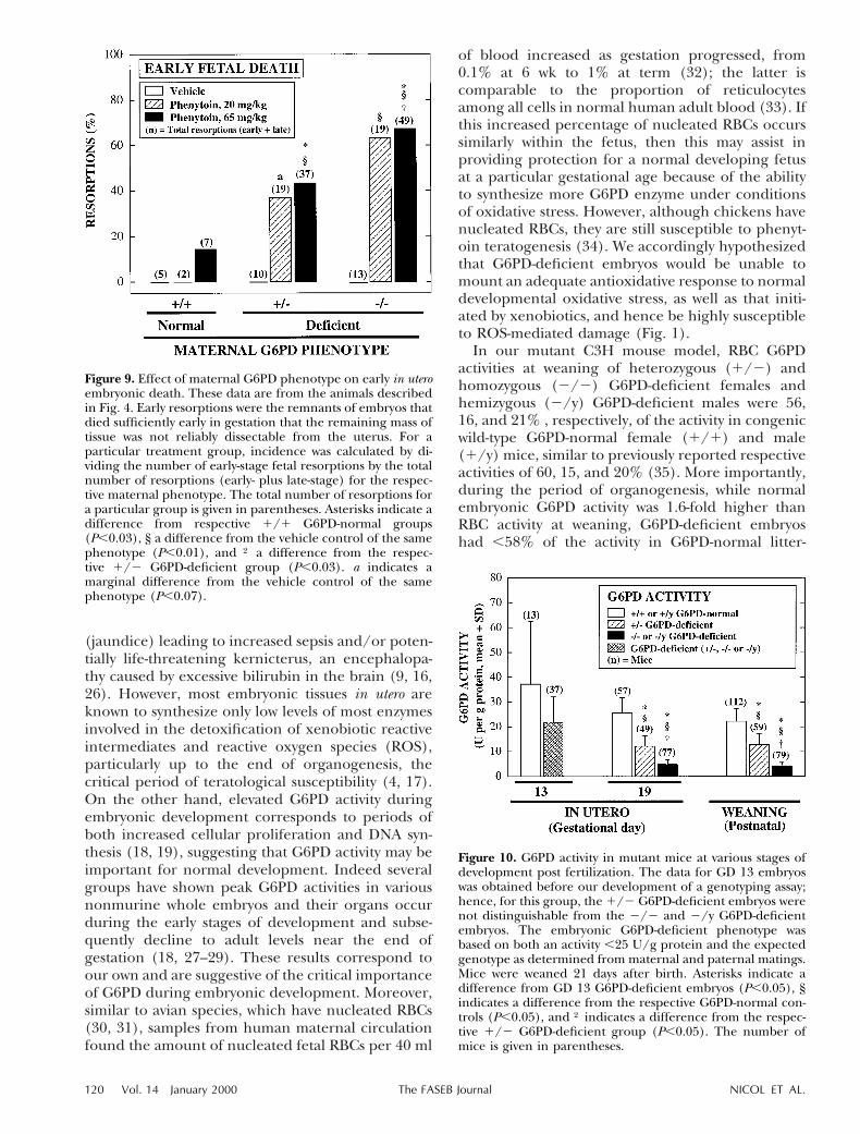
In dams treated with phenytoin, there also were asubstantial number of pinpoint resorptions thatwere too small to dissect, reflecting in utero embry-onic death early in gestation (Fig. 9). This earlyembryonic death, which was both G6PD gene dose-and phenytoin dose-related, occurred postimplan-tation, as the number of implantations did notdiffer among the maternal phenotypes. The num-ber of implantations (mean 6 sd), irrespective oftreatment, was 9.79 1/2 0.85 for 1/1 G6PD-normal dams vs. 8.13 6 3.20 for 6 and 8.82 6 3.57for 2/2 G6PD-deficient dams. In 1/2 and 2/2G6PD-deficient dams, both the low dose (20 mg/
kg) and the high dose (65 mg/kg) of phenytoinsubstantially enhanced the incidence of pinpointresorptions to a similar extent compared withrespective vehicle controls, which had no suchearly resorptions in any treatment group (P,0.01)(Fig. 9). With the high dose of phenytoin, theincidence of early embryonic death was enhancedthree- and fivefold (P,0.02), respectively, in 1/2and 2/2 G6PD-deficient dams compared with1/1 G6PD-normal dams, and the incidence was1.6-fold higher in 2/2 dams compared with 1/2dams (P,0.03). A remarkably identical pattern ofmaternal phenotypic susceptibility to, and magni-tude of, early embryonic death was observed withthe low dose of phenytoin (P,0.01), although theenhancement in 1/2 dams was statistically mar-ginal (P,0.07).
Embryonic and maternal G6PD activities
On GD 13, within the period of organogenesis,whole embryo G6PD activity was 43% lower in G6PD-deficient embryos compared with G6PD-normal lit-termates (Fig. 10). In this case, G6PD-deficient em-bryos included 1/2, 2/y, and 2/2 genotypes,because these studies preceded the establishment ofour genotyping technique. At the end of gestation,on day 19, G6PD activity in normal fetuses was lowerthan that during organogenesis and remained at thissame lower level at the time of weaning (Fig. 10) andinto adulthood (see Fig. 2). A similar pattern wasobserved in G6PD-deficient fetuses, whereby com-pared with GD 13 whole embryo activity, RBC activityon day 19 among combined fetuses with a mutationin one or all G6PD alleles had declined by 64%
Figure 7. G6PD mutant mouse genotyping assay. PCR-based, DdeI digest genotyping assay of dams and late-stage (dissectable)embryonic resorptions (in utero deaths), illustrating wild-type (1/1 or 1/y), heterozygous (1/2) and homozygous (2/2) orhemizygous (2/y) G6PD-deficient mutant mice genotypes. PCR primers were designed to generate a 269 bp product, which ondigestion with DdeI would produce fragments of 214 bp and 55 bp in 1/1 or 1/y G6PD-normal mice, no change in molecularsize in 2/2 or 2/y mutant G6PD-deficient mice, and all three fragments in 1/2 mutant G6PD-deficient mice. Lanes 1 and13 represent 123 bp ladder molecular weight controls. Lanes 2 to 7 represent PCR reaction products of phenotypicallycharacterized maternal and uncharacterized embryonic resorption DNA or a water control. Lanes 8 to 12 represent DdeI digestproducts of maternal and embryonic resorption PCR products listed above. The embryonic resorptions illustrated were obtainedfrom G6PD-dams confirmed to have a genotype different from that of the respective resorption.
118 Vol. 14 January 2000 NICOL ET AL.The FASEB Journal

(P,0.05) and remained constant thereafter. On day19, compared with 1/1 G6PD-normal littermates,RBC G6PD activity in 1/2 G6PD-deficient fetuseswas 48% of normal (P,0.05) and in 2/y and 2/2G6PD-deficient fetuses was 18% of normal (P,0.05).This pattern and the respective RBC activities re-mained similar at weaning, 58 and 19%, respectively,of normal (P,0.05) (Fig. 10).
The relative distribution of G6PD activities indifferent organs was similar for all phenotypes, withup to an 11-fold difference between the highest(spleen) and the lowest (heart) activities (Fig. 11). Inspleen, compared with 1/1 G6PD-normal dams,G6PD activities in 1/2 and 2/2 G6PD-deficientdams were 79 and 27%, respectively, of normal
(P,0.05), and similar patterns were evident forother organs and blood.
DISCUSSION
Toxicologic implications of G6PD deficiency
It is widely believed that serious health risks fromhereditary G6PD deficiencies are limited primarily tomature red blood cells, which lack a nucleus and areunable to synthesize more protective enzyme underconditions of oxidative stress. With respect to G6PDdeficiency and newborns, RBC hemolysis is known toresult in hyperferremia and hyperbilirubinemia
Figure 8. Effect of embryonic G6PD genotype on the incidence of late in utero death. These data are from the animals describedin Fig. 4. Late resorptions were the remnants of fetuses that died in utero sufficiently late in gestation that a dissectable massremained. For a particular embryonic genotype, incidence was calculated by dividing the number of typeable resorptions for agiven genotype, independent of treatment, by the total number of implantations (fetuses and resorptions) for that embryonicgenotype. The number of implantations for a particular embryonic genotype is given in parentheses. Asterisks indicate adifference from respective 1/y G6PD-normal controls (P,0.0001). Inset, the individual late-stage embryonic resorptiongenotypes for each treatment group. Incidences for a particular genotype were calculated by dividing the number of typeableresorptions for a given treatment group by the total number of dissectable resorptions for that respective treatment group. Thenumber of dissectable resorptions in each treatment group is given in parentheses. Asterisks indicate a difference fromrespective 1/y G6PD-normal controls (P,0.05). a indicates a marginal difference from respective 1/y G6PD-normal controls(P,0.06).
119G6PD PROTECTS AGAINST ROS-MEDIATED EMBRYOPATHIES
(jaundice) leading to increased sepsis and/or poten-tially life-threatening kernicterus, an encephalopa-thy caused by excessive bilirubin in the brain (9, 16,26). However, most embryonic tissues in utero areknown to synthesize only low levels of most enzymesinvolved in the detoxification of xenobiotic reactiveintermediates and reactive oxygen species (ROS),particularly up to the end of organogenesis, thecritical period of teratological susceptibility (4, 17).On the other hand, elevated G6PD activity duringembryonic development corresponds to periods ofboth increased cellular proliferation and DNA syn-thesis (18, 19), suggesting that G6PD activity may beimportant for normal development. Indeed severalgroups have shown peak G6PD activities in variousnonmurine whole embryos and their organs occurduring the early stages of development and subse-quently decline to adult levels near the end ofgestation (18, 27–29). These results correspond toour own and are suggestive of the critical importanceof G6PD during embryonic development. Moreover,similar to avian species, which have nucleated RBCs(30, 31), samples from human maternal circulationfound the amount of nucleated fetal RBCs per 40 ml
of blood increased as gestation progressed, from0.1% at 6 wk to 1% at term (32); the latter iscomparable to the proportion of reticulocytesamong all cells in normal human adult blood (33). Ifthis increased percentage of nucleated RBCs occurssimilarly within the fetus, then this may assist inproviding protection for a normal developing fetusat a particular gestational age because of the abilityto synthesize more G6PD enzyme under conditionsof oxidative stress. However, although chickens havenucleated RBCs, they are still susceptible to phenyt-oin teratogenesis (34). We accordingly hypothesizedthat G6PD-deficient embryos would be unable tomount an adequate antioxidative response to normaldevelopmental oxidative stress, as well as that initi-ated by xenobiotics, and hence be highly susceptibleto ROS-mediated damage (Fig. 1).
In our mutant C3H mouse model, RBC G6PDactivities at weaning of heterozygous (1/2) andhomozygous (2/2) G6PD-deficient females andhemizygous (2/y) G6PD-deficient males were 56,16, and 21% , respectively, of the activity in congenicwild-type G6PD-normal female (1/1) and male(1/y) mice, similar to previously reported respectiveactivities of 60, 15, and 20% (35). More importantly,during the period of organogenesis, while normalembryonic G6PD activity was 1.6-fold higher thanRBC activity at weaning, G6PD-deficient embryoshad ,58% of the activity in G6PD-normal litter-
Figure 10. G6PD activity in mutant mice at various stages ofdevelopment post fertilization. The data for GD 13 embryoswas obtained before our development of a genotyping assay;hence, for this group, the 1/2 G6PD-deficient embryos werenot distinguishable from the 2/2 and 2/y G6PD-deficientembryos. The embryonic G6PD-deficient phenotype wasbased on both an activity ,25 U/g protein and the expectedgenotype as determined from maternal and paternal matings.Mice were weaned 21 days after birth. Asterisks indicate adifference from GD 13 G6PD-deficient embryos (P,0.05), §indicates a difference from the respective G6PD-normal con-trols (P,0.05), and † indicates a difference from the respec-tive 1/2 G6PD-deficient group (P,0.05). The number ofmice is given in parentheses.
Figure 9. Effect of maternal G6PD phenotype on early in uteroembryonic death. These data are from the animals describedin Fig. 4. Early resorptions were the remnants of embryos thatdied sufficiently early in gestation that the remaining mass oftissue was not reliably dissectable from the uterus. For aparticular treatment group, incidence was calculated by di-viding the number of early-stage fetal resorptions by the totalnumber of resorptions (early- plus late-stage) for the respec-tive maternal phenotype. The total number of resorptions fora particular group is given in parentheses. Asterisks indicate adifference from respective 1/1 G6PD-normal groups(P,0.03), § a difference from the vehicle control of the samephenotype (P,0.01), and † a difference from the respec-tive 1/2 G6PD-deficient group (P,0.03). a indicates amarginal difference from the vehicle control of the samephenotype (P,0.07).
120 Vol. 14 January 2000 NICOL ET AL.The FASEB Journal

mates, potentially leaving the G6PD-deficient em-bryos more susceptible to embryopathic oxidativestress. G6PD activities likely were substantially lowerin 2/2 and 2/y than 1/2 G6PD-deficient em-bryos, but these studies were performed before thedevelopment of our genotyping assay, precludingsubclassification by embryonic genotype. However,relative differences can be inferred from fetal G6PDactivities on GD 19 when, compared with 1/y G6PD-normal littermates, 1/2 G6PD-deficient fetuses hadonly 48% of normal fetal activity, and fetuses with allalleles mutated (2/2 and 2/y) had only 18% ofnormal activity. The observed enhanced susceptibil-ity of G6PD-deficient mice to the embryopathiceffects of both normal developmental oxidativestress, and even more so to that initiated by theROS-initiating anticonvulsant drug phenytoin, indi-cates that G6PD is a critical embryoprotective en-zyme. Embryonic DNA oxidation and a broad spec-trum of apparent ROS-mediated embryopathieswere enhanced in G6PD-deficient animals, includingfetal resorptions (in utero death), postpartum andpreweaning lethality, decreased fetal body weight,
and a syndrome of structural anomalies, includingopen eye, facial, renal, bladder, and cerebral ventric-ular defects. This C3H-derived mutant strain, similarto previous findings using inbred C3H mice (36), isresistant to one of the hallmarks of phenytoin ter-atogenicity, cleft palates. Although the number offetuses affected was too small for statistical analysis, itwas interesting that cleft palates were observed onlyin G6PD-deficient fetuses treated either with vehicle(1 out of 50 1/2 fetuses and 1 out of 15 2/yfetuses) or phenytoin (65 mg/kg) (1 out of 28 1/2fetuses). The enhanced susceptibility of G6PD-defi-cient animals also corroborates other evidence indi-cating the importance of oxidative stress in themolecular mechanism of phenytoin teratogenesis(37) and further suggests that, contrary to the appar-ent safety of oxidizing drugs like phenytoin in adultG6PD-deficient patients (9), embryos in general, andG6PD-deficient embryos in particular, may be atserious risk.
Normal developmental oxidative stress andteratogenesis
In untreated mice allowed to deliver spontaneouslyin the breeding colony, the decreased litter size atbirth, and increased preweaning death in homozy-gous (2/2) G6PD-deficient dams (Fig. 3) providesthe first direct evidence that a physiological level ofendogenous oxidative stress during developmentcan be embryopathic and may contribute substan-tially to both apparent infertility and postnatal death.In addition, this is the first direct evidence that G6PDis a critical embryoprotective enzyme for normaldevelopment.
In vehicle-treated mice killed just before the timeof delivery and examined more comprehensively, astriking, broader spectrum of increased embryopa-thies, including fetal resorptions, postpartum lethal-ity, decreased fetal weight, and a syndrome of tera-tological anomalies, was observed in G6PD-deficientanimals (Figs. 4–6), suggesting an extensive embryo-pathic potential for developmental levels of ROSproduction and a broad embryoprotective role forG6PD. Given that no pinpoint resorptions reflectingearly in utero death were observed in vehicle controls,and there were no differences in the number ofimplantations among all groups, it appears that theapparent infertility in G6PD-deficient animals is be-cause of postimplantational, relatively late in uterodeath. The obligatory role of ROS in the mechanismof vehicle-initiated embryopathies is suggested by theobservation that these effects were found only inG6PD-deficient dams and fetuses. By both maternaland embryonic genotype, all developmental param-eters for vehicle-treated G6PD-normal animals werecomparable to the normal range for extensive con-
Figure 11. Maternal G6PD activity in various organs fromnormal and G6PD-deficient mice. Note the differences inscale of the y axes for each phenotype. Organs were obtainedfrom dams killed on GD 19 for the teratological studiesdescribed in Fig. 4. The number of animals is given inparentheses. Asterisks indicate a difference from the sameorgan in 1/1 G6PD-normal dams, and † indicates a differ-ence from the same organ in 1/2 G6PD-deficient dams(P,0.05).
121G6PD PROTECTS AGAINST ROS-MEDIATED EMBRYOPATHIES

trol groups from other murine strains studied in ourlaboratory (17). Previous in vivo studies in our labo-ratory also have shown that the vehicle does notmeasurably enhance oxidative stress and hydroxylradical formation (38), indicating that the observedembryopathies in vehicle-treated controls are theresult of normal developmental oxidative stress. Formany embryopathies (fetal resorptions, postpartumlethality, decreased fetal weight), increased suscepti-bility was as great in 1/2 as in 2/2 G6PD-deficientdams, indicating a potentially widespread develop-mental relevance of G6PD deficiencies. By embry-onic phenotype, the substantially increased severityof vehicle-initiated embryopathies (Fig. 5), includingteratologic anomalies (Fig. 6), in G6PD-deficientfetuses provided direct and proximal evidence thatG6PD-deficient fetuses are exquisitely susceptible todevelopmental oxidative stress. While 2/2 and 2/yG6PD-deficient fetuses were most adversely affectedfor several embryopathies, there was evidence of agene-dose effect, wherein 1/2 fetuses also hadsignificantly decreased body weight and appeared tobe at increased risk of postpartum lethality com-pared with 1/y G6PD-normal fetuses. The signifi-cant intermediary embryopathic risk for 1/2 damsand fetuses is consistent with their intermediatedeficiency in G6PD activity.
Using direct sequencing in the mutant C3Hmodel, we did not find any functional mutations ineither the entire coding region or the 39 untrans-lated region of the cDNA, nor in at least 6 of the 12intronic regions of genomic DNA. We did find astrain difference between our C3H mutant mousestrain and the reported C57 mouse cDNA (11) atbase 718; however, this sequence alteration does notchange the amino acid code for glycine at thisposition and as such is not expected to have anyeffect on G6PD activity. At that time, the functionalmutation was published, identifying a single pointmutation, an A to T transversion, in the 59 untrans-lated region of the gene at the penultimate base ofthe 39 end of exon 1 (21). We confirmed thismutation and then developed a genotyping assaythat allowed us to determine the G6PD genotype inthe dissectable remnants (resorptions) of embryosthat died in utero (Fig. 7), as we previously hadsuccessfully used in characterizing the role of p53 asa teratological suppressor gene (25). This approachis applicable to embryos that die later in gestationleaving a sufficient amount of tissue to be reliablydissectable without maternal tissue contamination.In vehicle-exposed embryos, the threefold greaterincidence of resorptions for 2/2 and 2/y G6PD-deficient fetuses compared with 1/y G6PD-normallittermates (Fig. 8, inset) shows directly that normaldevelopmental oxidative stress can play a major rolein in utero death, with G6PD serving as a critical
embryoprotective pathway. Remarkably, a similarthreefold increase in fetal resorptions was observedeven in 1/2 G6PD-deficient embryos, although thisapparent enhancement was only marginally signifi-cant (P,0.06). However, the incidence of theseresorptions appeared to be at a maximal level thatwas not further enhanced by phenytoin, and whenthe data were analyzed independent of treatment,heterozygous G6PD-deficient embryos were as sus-ceptible as embryos with mutations in all G6PDalleles, demonstrating a highly significant sixfoldincrease over G6PD-normal littermates (Fig. 8) andindicating a potentially broad population at risk.
Embryos dying early in gestation were observed asnondissectable pinpoint resorptions, however thisearlier embryopathy was not observed with vehicle-exposed embryos of any genotype, suggesting thatthe rate as well as the extent of embryolethalityinitiated by developmental oxidative stress are lessthan that initiated by xenobiotics. As well, the extentof this early embryopathy, especially in G6PD-defi-cient dams, likely contributed to the apparent lack ofa xenobiotic-initiated effect among resorptions oc-curring later in gestation. In general, the develop-mental risk in G6PD-deficient heterozygotes, in somecases equivalent to that for dams and fetuses withmutations in all G6PD alleles, reveals a potentiallysubstantial clinical risk from G6PD deficiencies un-der conditions of normal developmental oxidativestress.
Xenobiotic-initiated oxidative stress andteratogenesis
Phenytoin and related proteratogens, including thesedative drug thalidomide, are bioactivated by em-bryonic prostaglandin H synthases, lipoxygenases,and related enzymes to a free radical intermediate,which causes embryonic oxidative stress, hydroxylradical formation, and oxidative damage to DNA andother cellular macromolecules in embryonic tissues(4, 17, 37, 39, 40). Thus, the dose-dependent in-crease in embryopathies caused by phenytoin byboth maternal and embryonic analyses, togetherwith the enhanced susceptibility of G6PD-deficientdams and embryos compared with vehicle controls(Figs. 4–6, 8) and the enhanced phenytoin-initiatedDNA oxidation in G6PD-deficient embryos (Fig. 5,inset), provide the first direct evidence that G6PD isa major embryoprotective enzyme for xenobiotic-initiated oxidative stress and embryonic macromo-lecular target damage. The susceptibility of heterozy-gous (1/2) G6PD-deficient animals to phenytoinembryopathies was particularly remarkable, gener-ally exhibiting a risk intermediate, if not equal, tothat of homozygous (2/2) and hemizygous (2/y)G6PD-deficient dams and fetuses, depending on the
122 Vol. 14 January 2000 NICOL ET AL.The FASEB Journal
parameter. This pattern differed somewhat only within utero death, wherein the incidence of early but notlate fetal resorptions was enhanced over vehiclecontrols (Figs. 8, 9), although both types of in uterodeath were substantially enhanced in G6PD-deficientembryos, and the risk in heterozygotes was similar tothat for homozygous and hemizygous G6PD-defi-cient embryos. To an extent greater than that in thevehicle controls, the embryopathic susceptibility ofeven heterozygous G6PD-deficient dams and em-bryos to phenytoin suggests that G6PD deficienciesmay have broad developmental relevance for expo-sures to drugs and environmental chemicals likephenytoin that initiate oxidative stress.
With the lower, 20 mg/kg dose of phenytoin, theenhanced embryopathies (decreased fetal bodyweight, early resorptions, teratological anomalies)compared with vehicle controls was remarkable, asthis is ,40% of a threshold teratogenic dose (55mg/kg) in normal mice (17). For some embryopa-thies (e.g., early resorptions, teratological anoma-lies), a maximal response was achieved with thelower 20 mg/kg dose in both 1/2 and 2/2 and2/y G6PD-deficient littermates. These results dem-onstrate that G6PD-deficient animals are exception-ally susceptible to the embryopathic effects of xeno-biotic-initiated oxidative stress.
By both maternal and embryonic analysis, thedose-dependent increase in most embryopathies,including teratological anomalies, produced inG6PD-deficient animals by phenytoin compared withvehicle controls, corroborates the role of oxidativestress in the molecular mechanism of phenytointeratogenesis. The enhanced embryonic DNA oxida-tion observed in phenytoin-exposed G6PD-deficientembryos further suggests that oxidative damage toembryonic cellular macromolecules may play a prox-imate role in embryopathic initiation. These resultsimplicating reactive oxygen species and oxidativedamage to embryonic cellular macromolecules inthe mechanism of phenytoin teratogenicity are con-sistent with other murine studies in vivo, in embryoculture, and in vitro demonstrating phenytoin-initi-ated formation of reactive oxygen species, oxidativedamage to embryonic cellular macromolecules, anda protective role for antioxidants (vitamin E, caffeicacid, glutathione) and other antioxidative enzymessuch as glutathione reductase, glutathione peroxi-dase, superoxide dismutase, and catalase (37, 41).The embryopathic importance of enhanced DNAoxidation in G6PD-deficient embryos is consistentwith previous studies of phenytoin and benzo-[a]pyrene, another teratogen known to initiate em-bryonic oxidative stress, wherein p53-deficient micewith reduced DNA repair were shown to be moresusceptible to the embryopathic effects of thesexenobiotics (25, 42, 43).
It has been reported that under conditions ofoxidative stress, the increased expression of G6PDactivity is primarily a result of an increased rate oftranscription, with a minor contribution from post-transcriptional modifications (44), possibly due toposttranslational regulation of G6PD by small heatshock proteins (45). Given the above evidence forthe involvement of reactive oxygen species in in uterodeath and teratogenesis, it is likely that the biochem-ical mechanism underlying the embyroprotectiverole of G6PD is its production of NADPH essentialfor both glutathione reductase-dependent, and pos-sibly catalase-dependent, detoxification of lipid hy-droperoxides and hydrogen peroxide, rather thanG6PD-dependent pentose production. This is consis-tent with a recent study of diamide-initiated oxida-tive stress, which identified the essential protectiverole for G6PD as NADPH production, as distinctfrom its dispensable role in pentose synthesis, deter-mined by cloning efficiency of mouse embryonicstem cells wherein the G6PD gene was selectivelyknocked out (46). A reduction in catalase functionmay contribute to the enhanced teratologic suscep-tibility observed with G6PD deficiencies, becauseNADPH is known to maintain catalase activity notonly by preventing the formation of one of theinactive states of the enzyme (compound II), possiblythrough electron tunneling between surface-boundNADPH and the internal heme group, but also byreducing oxidized states and internal groups ofcatalase distinct from compound II, possibly includ-ing one of the active states of the enzyme (com-pound I) (6–8). This mechanism also is consistentwith other studies demonstrating a protective effectof catalase therapy against phenytoin teratogenicityin embryo culture (20) and in vivo (41). Our resultsalso show that G6PD, as distinct from isocitratedehydrogenase and malic enzyme (47), provides themajor supply of embryonic NADPH during organo-genesis, and alternative embryonic sources are inad-equate in the face of hereditary G6PD deficiencies.
Epidemiological considerations
The observed decreased litter size at birth in untreated2/2 G6PD-deficient mice and, in vehicle-treatedmice, the increased fetal resorptions in both 1/2 and2/2 G6PD-deficient dams, would present in humansas an apparent decrease in fertility. Given that implan-tations were not decreased in G6PD-deficient dams, thedecrease in viable offspring was because of postimplan-tation embryolethality rather than increased oocyte lossand/or increased preimplantation embryonic death.The gestational timing of susceptibility to oxidativestress may be because of, at least in part, the changefrom anaerobic to aerobic metabolism that begins afterimplantation. This change occurs as the mitochondrial
123G6PD PROTECTS AGAINST ROS-MEDIATED EMBRYOPATHIES

electron transport system and associated enzymes startto become functional and in rodents is complete withthe establishment of the allantoic circulation. For ratembryos, this process starts between GD 10 and 11 andends by GD 12.5, with blood circulation through theyolk sac and embryo by GD 11 (48–50). For mouseembryos, this process may run from GD 6 to 9.5, withallantoic circulation through the placenta by GD 9(51–53). Although humans do not have a yolk sacplacenta, comparisons based on stage of developmentand somite count between rat and human indicate thatGD 11 and 12.5 in the rat are equivalent to GD 28 and37 in the human, after which time the switch fromanaerobic to aerobic metabolism should be complete(48, 54).
Accordingly, it is not surprising that this postim-plantation gestational period constitutes a criticalwindow of susceptibility to ROS-mediated lethalityfor G6PD-deficient embryos. Indeed, 1/2 G6PD-deficient embryos were as susceptible as their 2/2and 2/y G6PD-deficient littermates. In humans,expected incidences for G6PD-deficient 2/y malesand 2/2 females born among all G6PD-deficientgroups in Europe were 0.7 and 5.1%, respectively,whereas the observed incidences of these groupswere only 0.3 and 2%, respectively (2). In otherwords, there were 57% fewer 2/y G6PD-deficientmales and 61% fewer 2/2 G6PD-deficient femalesborn than expected. These lower birth incidences inhuman G6PD-deficient populations imply a lowersurvival rate than expected for embryos deficient inG6PD, as was observed in our mouse model.
To date, no major deletions or frameshifts withinG6PD have been identified. Most variants result frompoint mutations or small intragenic deletions lead-ing to a decrease of only one or two residues (14).There are few published cases of heritable mildG6PD deficiencies in erythrocytes from other spe-cies, specifically in a colony of rats (55) and in onedog out of 3,300 screened (56). In both cases, themutation was not characterized, but difficulties inmaintaining the rat colony because of increasedmortality and sterility among the affected animalssuggests that a homozygous knockout of G6PDwould likely prove embryolethal. Similarly, there hasnever been a reported case of a complete humanG6PD deficiency (10).
A limited number of recent human studies provideindirect evidence of other areas where G6PD defi-ciencies may be pathologically relevant. G6PD isimportant for maintaining adequate concentrationsof reduced glutathione, which is necessary for anumber of cytoprotective antioxidative activities, in-cluding that of glutatione peroxidase (Fig. 1). Grafet al. (57) found significantly lower glutathioneperoxidase activity among 37 children with the neu-ral tube defect myelomeningocele, compared with
age-matched controls. Weber and colleagues (58)reported intractable seizures, repeated infections,and intolerance to anticonvulsants in four childrenwith glutathione peroxidase deficiencies, one ofwhom also was G6PD deficient. These adverse out-comes may have been because of granulocytopeniaand enhanced ROS-dependent signaling pathways,respectively, both of which could result from oxida-tive stress and inadequate protection from antioxi-dative enzymes. This is consistent with results from arat model of human chronic posttraumatic epilepticseizures, in which pretreatment with antioxidants(alpha-tocopherol and selenium) protected against aspectrum of iron-induced peroxidative injuries, in-cluding cavitation, neuronal loss, astrogliosis, andepileptiform discharges in rat isocortical regions,suggesting that deficiencies in cytoprotection againstperoxidative injury may increase the risk of recurrentepileptic seizures (59). Finally, a recent study foundincreased levels of G6PD activity in several relevantregions of Alzheimer’s brains compared with con-trols, possibly reflective of increased levels of cere-bral oxidative challenge (60, 61). In our mousemodel, normal adult brain G6PD activity was one ofthe lowest of all tissues examined and was furtherreduced by .21 and 72%, respectively, in 1/2 and2/2 G6PD-deficient animals (Fig. 11). If oxidativestress is involved in the mechanism of neurodegen-erative diseases (62, 63), then G6PD deficiencies maycontribute to enhanced susceptibility. These studiesare consistent with our observation of enhancedembryopathies in G6PD-deficient mice and suggestthe potential for a broader range of pathologicalsusceptibilities with G6PD deficiencies that may befurther enhanced by exposure to xenobiotics likephenytoin that initiate oxidative stress.
The commonly postulated evolutionary pressurefor the widespread prevalence of G6PD deficienciesis their advantage in providing resistance to malaria.This view is supported by the almost complete over-lap of regions of increased incidence of G6PD defi-ciency with those of elevated incidence of malarialinfections (2, 10). Recently, both heterozygous andhemizygous G6PD-deficient people were confirmedto be 46 and 58% more resistant, respectively, tosevere malarial infection (64). Malaria-infected nor-mal RBCs demonstrate increased lipid peroxidation(65) presumably because of parasite-generated H2O2
(66) and decreased antioxidant levels, with nochange in G6PD activity. The authors concluded thisto be a possible defense mechanism by which in-fected host cells seek to produce and maintainunchecked oxidative stress to their advantage in aself-sacrificing attempt to limit the spread of malarialinfection. As such, G6PD-deficient cells are moresensitive to this increased oxidative damage (66),which may help mediate their increased resistance to
124 Vol. 14 January 2000 NICOL ET AL.The FASEB Journal

such infections, either by predisposing the cell topremature destruction by the reticuloendothelialsystem (67) or macrophage-initiated lysis (68)and/or impaired growth of the parasite in G6PD-deficient erythrocytes as suggested by in vitro studies(69, 70), thus abolishing a suitable incubation envi-ronment for parasite development. However, thisapparent selective advantage must be balanced byequally disadvantageous selective pressures, not theleast of which include the developmental risks ob-served in our mouse study and suggested in humanepidemiological studies discussed above. Develop-mental risks may well be increasing as a result ofmore frequent exposure to drugs and environmentalchemicals such as phenytoin that initiate oxidativestress exquisitely toxic to G6PD-deficient embryos.Such factors may be contributing to the apparentlyrestricted increase in the prevalence of G6PD defi-ciencies in malaria-infected regions (64).
CONCLUSIONS
Hereditary deficiencies in G6PD are widely thoughtto pertain only to red blood cell hemolysis, with themost severe outcome being neonatal kernicterus (9,10, 26). Our results provide the first evidence thatG6PD is a developmentally critical enzyme, protect-ing the embryo from both endogenous and xenobi-otic-initiated oxidative stress and DNA damage thatproduce a broad range of embryopathies, includingteratologic anomalies. The developmental risk ispotentially substantial, given both the common inci-dence of hereditary G6PD deficiencies and the sus-ceptibility of even heterozygous G6PD-deficient ani-mals.
The enhanced susceptibility of G6PD-deficient em-bryos to phenytoin corroborates other studies impli-cating embryonic oxidative stress and DNA damagein the molecular mechanism of embryopathiescaused by phenytoin and related teratogens (37).G6PD deficiencies completely redefine thresholdsfor teratologic susceptibility to xenobiotic exposure,as evidenced by the enhanced susceptibility of evenheterozygous G6PD-deficient embryos to phenytoinembryopathies at a dose that is well below theminimal teratogenic dose in G6PD-normal mice.The developmental risk from xenobiotics that initi-ate oxidative stress appears to be considerablygreater than that observed in adults, because adultpatients with G6PD deficiencies are reported to benonsusceptible to RBC hemolysis initiated by drugslike phenytoin.
Preliminary reports of this work were presented at the 35th,36th, and 38th annual meetings of the Society of Toxicology(USA) (Fundam. Appl. Toxicol. Vol. 30 (Suppl. 1, Part 2), p.197, 1996; Fundam. Appl. Toxicol. Vol. 36 (Suppl. 1, Part 2), p.
98, 1997; Toxicol. Sci. 48(1-S), 17, 1999) and at the 5thInternational Meeting of the Society for the Study of Xeno-biotics (ISSX Proc. Vol. 13, p. 171, 1998). We wish to thank Dr.Michael J. Wiley (Department of Anatomy and Cell Biology,University of Toronto) for consultations pertaining to terato-logical analyses, and Danka Markiewicz and John Tzount-zouris (Department of Genetics, Hospital for Sick Children,Toronto) for their expert advice with the DNA sequencingand genotyping assays. Supported by a grant to P.G.W. fromthe Medical Research Council of Canada.
REFERENCES
1. Mehta, A. B. (1994) Glucose-6-phosphate dehydrogenase defi-ciency. Postgrad. Med. J. 70, 871–877
2. Sodeinde, O. (1992) Glucose-6-phosphate dehydrogenase defi-ciency. Baill. Clin. Hematol. 5, 367–382
3. Halliwell, B., and Gutteridge, J. M. C. (1989) Free Radicals inBiology and Medicine. Clarendon, Oxford, U. K.
4. Wells, P. G., and Winn, L. M. (1996) Biochemical toxicology ofchemical teratogenesis. Crit. Rev. Biochem. Mol. Biol. 31, 1–40
5. Wells, P. G., Kim, P. M., Nicol, C. J., Parman, T., and Winn, L. M.(1996) Reactive intermediates. In Handbook of Experimental Phar-macology: Drug Toxicity in Embryonic Development (Kavlock, R. J.,and Daston, G. P., eds) Vol. 124, Part 1, pp. 453–518, Springer-Verlag, Heidelberg, Germany
6. Kirkman, H. N., and Gaetani, G. F. (1984) Catalase: a tetramericenzyme with four tightly bound molecules of NADPH. Proc. Natl.Acad. Sci. USA 81, 4343–4347
7. Kirkman, H. N., Galiano, S., and Gaetani, G. F. (1987) Thefunction of catalase-bound NADPH. J. Biol. Chem. 262, 660–666
8. Kirkman, H. N., Rolfo, M., Ferraris, A. M., and Gaetani, G. F.(1999) Mechanisms of protection of catalase by NADPH. J. Biol.Chem. 274, 13908–13914
9. Beutler, E. (1986) Drug-induced hemolytic anemia and non-spherocytic hemolytic anemia. In Glucose-6-Phosphate Dehydroge-nase (Yoshida, A., and Beutler, E., eds) pp. 3–12, Academic,Orlando
10. Luzzatto, L., and Mehta, A. (1995) Glucose 6-phosphate dehy-drogenase deficiency. In The Metabolic and Molecular Basis ofInherited Disease (Scriver, C. R., Beaudet, A. L., Sly, W. S., andValle, D., eds) Vol. 7, pp. 3367–3398, McGraw-Hill, Toronto
11. Zollo, M., D’Urso, M., Schlessinger, D., and Chen, E. Y. (1993)Sequence of mouse glucose-6-phosphate dehydrogenase cDNA.DNA Seq.-J. DNA Seq. Map. 3, 319–322
12. Beutler, E. (1959) The hemolytic effect of primaquine andrelated compounds: a review. Blood 14, 103–109
13. Beutler, E. (1993) Study of glucose-6-phosphate dehydrogenase:history and molecular biology. Am. J. Hematol. 42, 53–58
14. Martini, G., and Ursini, M. V. (1996) A new lease of life for anold enzyme. BioEssays 18, 631–637
15. Beutler, E., Vulliamy, T., and Luzzatto, L. (1996) Hematologi-cally important mutations: glucose-6-phosphate dehydrogenase.Blood Cells Mol. Dis. 22, 49–56
16. Abu-Osba, Y. K., Mallouh, A. A., and Hann, R. W. (1989)Incidence and causes of sepsis in glucose-6-phosphate dehydro-genase-deficient newborn infants. J. Pediatr. 114, 748–752
17. Winn, L. M., and Wells, P. G. (1995) Free radical-mediatedmechanisms of anticonvulsant teratogenicity. Eur. J. Neurol. 2(Suppl. 4), 5–29
18. Newburgh, R. W., Buckingham, B., and Herrmann, H. (1962)Levels of reduced TPN generating systems in chick embryos inovo and in explants. Arch. Biochem. Biophys. 97, 94–99
19. Papaconstantinou, J. (1967) Metabolic control of growth anddifferentiation in vertebrate embryos. In The Biochemistry ofAnimal Development (Weber, R., ed) Vol. 2, pp. 57–113, Aca-demic, New York
20. Winn, L. M., and Wells, P. G. (1995) Phenytoin-initiated DNAoxidation in murine embryo culture, and embryo protection bythe antioxidative enzymes superoxide dismutase and catalase:evidence for reactive oxygen species mediated DNA oxidationin the molecular mechanism of phenytoin teratogenicity. Mol.Pharmacol. 48, 112–120
125G6PD PROTECTS AGAINST ROS-MEDIATED EMBRYOPATHIES

21. Sanders, S., Smith, D. P., Thomas, G. A., and Williams, E. D.(1997) A glucose-6-phosphate dehydrogenase (G6PD) splicesite consensus sequence mutation associated with G6PD enzymedeficiency. Mutat. Res. 374, 79–87
22. Gupta, R. C. (1984) Nonrandom binding of the carcinogenN-hydroxy-2-acetylfluorene to repetitive sequences of rat liverDNA in vivo. Proc. Natl. Acad. Sci. USA 81, 6943–6947
23. Shigenaga, M. K., and Ames, B. N. (1991) Assay for 8-hydroxy-29-deoxyguanosine: a biomarker of in vivo oxidative damage.Free Rad. Biol. Med. 10, 211–216
24. Chen, E. Y., Cheng, A., Lee, A., Kuang, W. J., Hillier, L., Green,P., Schlessinger, D., Ciccodicola, A., and D’Urso, M. (1991)Sequence of human glucose-6-phosphate dehydrogenasecloned in plasmids and a yeast artificial chromosome. Genomics10, 792–800
25. Nicol, C. J., Harrison, M. L., Laposa, R. R., Gimelshtein, I. L.,and Wells, P. G. (1995) A teratologic suppressor role for p53 inbenzo[a]pyrene-treated transgenic p53-deficient mice. Nat.Genet. 10, 181–187
26. Washington, E. C., Ector, W., Abboud, M., Ohning, B., andHolden, K. (1995) Hemolytic jaundice due to G6PD deficiencycausing kernicterus in a female newborn. South. Med. J. 88,776–779
27. Burt, A. M., and Wenger, B. S. (1961) Glucose-6-phosphatedehydrogenase activity in the brain of the developing chick. Dev.Biol. 3, 84–95
28. Jolley, R. L., Cheldelin, V. H., and Newburgh, R. W. (1958)Glucose catabolism in fetal and adult heart. J. Biol. Chem. 233,1289–1294
29. Jolley, R. L., Cheldelin, V. H., and Newburgh, R. W. (1959) Thecatabolism of glucose in soluble adult and fetal-heart prepara-tions. Biochim. Biophys. Acta 33, 64–68
30. Lucas, A. M., and Jamroz, C. (eds) (1961) Atlas of AvianHematology, pp. 17–30, United States Department of Agriculture,Washington, D.C.
31. Sinclair, G. D., and Brasch, K. (1975) The nucleated eryth-rocyte: a model of cell differentiation. Revue Can. Biol. 34,287–303
32. Ganshirt, D., Smeets, F. W., Dohr, A., Walde, C., Steen, I.,Lapucci, C., Falcinelli, C., Sant, R., Velasco, M., Garritsen, H. S.,and Holzgreve, W. (1998) Enrichment of fetal nucleated redblood cells from the maternal circulation for prenatal diagnosis:experience with triple density gradient and MACS based onmore than 600 cases. Fetal Diagnostic Ther. 13, 276–86
33. Guyton, A. C. (ed) (1987) Red blood cells, white blood cells andresistance of the body to infection. In Human Physiology andMechanisms of Disease, 4th Ed, p. 195, W. B. Saunders, Toronto
34. Singh, M., and Shah, G. L. (1989) Teratogenic effects ofphenytoin on chick embryos. Teratology 40, 453–458
35. Pretsch, W., Charles, D. J., and Merkle, S. (1988) X-linkedglucose-6-phosphate dehydrogenase (G6PD) deficiency in Musmusculus. Biochem. Genet. 26, 89–103
36. Finnell, R. H., and Chernoff, G. F. (1984) Variable patterns ofmalformation in the mouse fetal hydantoin syndrome. Am. J.Med. Genet. 19, 463–471
37. Wells, P. G., Kim, P. M., Laposa, R. R., Nicol, C. J., Parman, T.,and Winn, L. M. (1997) Oxidative damage in chemical terato-genesis. Mutat. Res. 396, 65–78
38. Kim, P. M., and Wells, P. G. (1996) Phenytoin-initiated hydroxylradical formation: characterisation by enhanced salicylate hy-droxylation. Mol. Pharmacol. 49, 172–181
39. Parman, T., Chen, G., and Wells, P. G. (1998) Free radicalintermediates of phenytoin and related teratogens: prostaglan-din H synthase-catalyzed bioactivation, electron paramagneticresonance spectrometry and photochemical product analysis.J. Biol. Chem. 273, 25079–25088
40. Parman, T., Wiley, M. J., and Wells, P. G. (1999) Free radical-mediated oxidative DNA damage in the mechanism of thalido-mide teratogenicity. Nature Med. 5, 582–585
41. Winn, L. M., and Wells, P. G. (1999) Maternal administration ofsuperoxide dismutase and catalase in phenytoin teratogenicity.Free Rad. Biol. Med. 26, 266–274
42. Laposa, R. R., and Wells, P. G. (1995) Preliminary evaluation ofphenytoin teratogenicity in transgenic mice deficient in the p53tumor suppressor gene. Fundam. Appl. Toxicol. 15, 161
43. Laposa, R. R., Chan, K. L., Wiley, M. J., and Wells, P. G. (1996)Enhanced phenytoin embryopathy in p53-deficient mice: char-
acterisation of embryonic p53 genotype and the p53, p21 andBax DNA damage response proteins. Fundam. Appl. Toxicol.30(Suppl. 1, part 2), 195
44. Ursini, M. V., Parrella, A., Rosa, G., Salzano, S., and Martini, G.(1997) Biochem. J. 323 (Pt. 3), 801–806
45. Preville, X., Salvemini, F., Giraud, S., Chaufour, S., Paul, C.,Stepien, G., Ursini, M. V., and Arrigo, A.-P. (1999) Mammaliansmall stress proteins protect against oxidative stress throughtheir ability to increase glucose-6-phosphate dehydrogenaseactivity and by maintaining optimal cellular detoxifying machin-ery. Exp. Cell Res. 247, 61–78
46. Pandolfi, P. P., Sonati, F., Rivi, R., Mason, P., Grosveld, F., andLuzzatto, L. (1995) Targeted disruption of the housekeepinggene encoding glucose-6-phosphate dehydrogenase (G6PD):G6PD is dispensable for pentose synthesis but essential fordefense against oxidative stress. EMBO J. 14, 5209–5215
47. Mayes, P.A. (1996) Biosynthesis of fatty acids. In Harper’s Bio-chemistry (Murray, R. K., Granner, D. K., Mayes, P. A., andRodwell, V. W., eds) 24th Ed, pp. 216–223, Appleton, andLange, Stamford, CA
48. Robkin, M. A. (1997) Carbon monoxide and the embryo. Int. J.Dev. Biol. 41, 283–289
49. Tanimura, T., and Shepard, T. H. (1970) Glucose metabolismby rat embryos in vitro. Proc. Soc. Exp. Biol. Med. 135, 51–53
50. Aksu, O., Mackler, B., Shepard, T. H., and Lemire, R. J.(1968) Studies of the development of congenital anomaliesin embryos of riboflavin-deficient, galactoflavin fed rats.Teratology 1, 93–102
51. Clough, J. R., and Whittingham, D. G. (1983) Metabolism of[14C]glucose by postimplantation mouse embryos in vitro. J.Embryol. Exp. Morph. 74, 133–142
52. Edwards, R. G., and Fowler, R. E. (1959) Fetal mortality in adultmice after superovulation with gonadotrophins. J. Exp. Zool. 141,299–322
53. West, J. D. (1993) A genetically defined animal model ofanembryonic pregnancy. Hum. Reprod. 8, 1316–1323
54. Altman, P. L., and Dittmer, D. S. (1972) Biology Data Book, pp.176–180, Federation of American Societies for ExperimentalBiology, Bethesda, MD
55. Werth, G., and Muller, G. (1967) Inheritable glucose-6-phos-phate dehydrogenase deficiency in erythrocytes of rats. Klin.Wochenschr. 45, 265–269
56. Smith, J. E., Ryer, K., and Wallace, L. (1976) Glucose-6-phosphate dehydrogenase deficiency in a dog. Enzyme 21,379 –382
57. Graf, W. D., Oleinik, O. E., Pippenger, C. E., Eder, D. N.,Glauser, T. A., and Shurtleff, D. B. (1995) Comparison oferythrocyte antioxidant enzyme activities and embryologic levelof neural tube defects. Eur. J. Ped. Surgery 5 (Suppl. 1), 8–11
58. Weber, G. F., Maertens, P., Meng, X., and Pippenger, C. E.(1991) Glutathione peroxidase deficiency and childhood sei-zures. Lancet 337, 1443–1444
59. Willmore, L. J., and Rubin, J. J. (1981) Antiperoxidant pretreat-ment and iron-induced epileptiform discharges in the rat: EEGand histopathologic studies. Neurology 31, 63–69
60. Balazs, L., and Leon, M. (1994) Evidence of an oxidativechallenge in the Alzheimer’s Brain. Neurochem. Res. 19, 1131–1137
61. Martins, R. N., Harper, C. G., Stokes, G. B., and Masters, C. L.(1986) Increased cerebral glucose- 6-phosphate dehydrogenaseactivity in Alzheimer’s disease may reflect oxidative stress.J. Neurochem. 46, 1042–1045
62. Cooper, A. J., and Kristal, B. S. (1997) Multiple roles ofglutathione in the central nervous system. J. Biol. Chem. 378,793–802
63. Jenner, P. (1996) Oxidative stress in Parkinson’s disease andother neurodegenerative disorders. Pathologie Biologie 44,57– 64
64. Ruwende, C., Khoo, S. C., Snow, R. W., Yates, S. N. R.,Kwiatkowski, D., Gupta, S., Warn, P., Allsopp, C. E. M., Gilbert,S. C., Peschu, N., Newbold, C. I., Greenwood, B. M., Marsh, K.,and Hill, A. V. S. (1995) Natural selection of hemi- andheterozygotes for G6PD deficiency in Africa by resistance tosevere malaria. Nature (London) 376, 246–249
65. Erel, O., Kocyigit, A., Avci, S., Aktepe, N., and Bulut, V.(1997) Oxidative stress and antioxidative status of plasma
126 Vol. 14 January 2000 NICOL ET AL.The FASEB Journal

and erythrocytes in patients with vivax malaria. Clin. Biochem.30, 631– 639
66. Friedman, M. J. (1979) Oxidant damage mediates variant redcell resistance to malaria. Nature (London) 280, 245–247
67. Eckman, J. R., and Eaton, J. W. (1979) Dependence of plasmo-dial glutathione metabolism on the host cell. Nature (London)278, 754–756
68. Yuthavong, Y., Bunyaratvej, A., and Kamchonwongpaisan, S.(1990) Increased susceptibility of malaria-infected variant eryth-rocytes to the mononuclear phagocyte system. Blood Cells 16,591–597
69. Roth, E. F., Jr., Raventos-Suarez, C., Rinaldi, A., and Nagel, R. L.(1983) Glucose-6-phosphate dehydrogenase deficiency inhibitsin vitro growth of plasmodium falciparum. Proc. Natl. Acad. Sci.USA 80, 298–299
70. Kosower, N. S., and Kosower, E. M. (1970) Molecular basisfor selective advantage of glucose-6-phosphate dehydroge-nase-deficient individuals exposed to malaria. Lancet 2, 1343–1344
Received for publication May 14, 1999.Revised for publication July 25, 1999.
127G6PD PROTECTS AGAINST ROS-MEDIATED EMBRYOPATHIES


![lezione sensibilit insulinica 091115 [modalit compatibilit ]) resistenza - definizione e... · HK-II glucose + insulin + glucose - 6 - phosphate glycogen GS glucose - 1 - phosphate](https://img.pdfslide.us/doc/110x75/5f0573df7e708231d4130924/lezione-sensibilit-insulinica-091115-modalit-compatibilit-resistenza-definizione.jpg)