Embed Size (px)
Citation preview
Contents lists available at ScienceDirect
Colloid and Interface Science Communications
journal homepage: www.elsevier.com/locate/colcom
An efficient and inexpensive method for functionalizing metallicbiomaterials used in orthopedic applicationsSoria Hamdaouia,b, Ambroise Lamberta, Hafit Khireddineb, Rémy Agniela, Annelise Cousturec,Régis Coulond, Olivier Galleta, Séverine Alfonsie, Mathilde Hindiéa,⁎
a CY Cergy Paris Université, ERRMECe, F-95000 Cergy, Franceb Laboratoire de Génie de l'Environnement, Faculté de Technologie, Université de Bejaia, 06000 Bejaia, Algeriac CY Cergy Paris Université, L2MGC, F-95000 Cergy, Franced CMS -Boilermaking and Metalwork Company, 03470 Saligny sur Roudon, Francee CY Cergy Paris Université, LPPI, F-95000 Cergy, France
A R T I C L E I N F O
Keywords:316 L stainless steelPolypyrroleElectrodepositionBiomaterialsCalcium phosphateSteam sterilizationSTRO-1+ A pre-osteoblasts
A B S T R A C T
Implantations of metallic biomaterials are being carried out more and more frequently due to accident andpopulation aging. Therefore, there is a need for new metallic implants which can combine properties such asdurability, biocompatibility and affordability. In this study, multilayer functionalized 316 L stainless steel (SS)supports resistant to steam sterilization were presented. An electropolymerization of pyrrole was performed onSS supports to obtain a protective layer. This polypyrrole coating rendered SS surface resistant to corrosion. Thenan electrodeposition of Calcium Phosphate (CaP) doped with increasing concentrations of silicon (Si) rangingfrom 0 to 2 mM was tested to improve support bone integration. The impacts of silicon addition in the CaPcoating without or after steam sterilization were analyzed by profilometry, Scanning Electron Microscopy andFourier Transform Infrared Spectroscopy. These latter revealed that CaP doped with 0.5 mM of Si constituted theoptimal support formulation, presenting sterilization resistance and good biocompatibility.
1. Introduction
The implantation of orthopedic biomaterials is widely used allaround the globe to restore physiological functions. Approximately70% of the implants used in medicine are metallic biomaterials [1] andare mainly used to repair failed hard tissue. The demand for implants isincreasing exponentially as part of the efforts to improve the life qualityof the aging population. The global market for implants is expected togrow to $115.8 billion by 2020 [2].
The three most implanted metallic biomaterials are titanium alloys,cobalt‑chromium alloys and stainless steel (SS). The 316 L SS is themost widely used alloy mainly for non-permanent implants (e.g. boneplates, screws) and dental surgery [3,4]. Despite 316 L SS having goodmechanical properties, good biocompatibility and being inexpensive,this alloy is less used for permanent implantation due to the corrosioninduced by the contact with body fluids and the release of toxic ionssuch as nickel and chromium ions which causes local inflammation[5–7]. Among all the SS implants that failed, more than 90% presentedcorrosion attack [8]. Different physical and chemical techniques havebeen developed to improve SS resistance to corrosion such as plasma
immersion ion implantation and deposition [9], surface modification ofbiomedical 316 L SS with zirconium carbonitride coatings [10], dipcoating [11], electropolishing and acid dipping [12] or sol-gel spincoating [13]. In this study, the electrodeposition of an electro-con-ductive polymer was chosen to prevent SS corrosion. In the past dec-ades steel surface passivation using conducting polymers such aspolyaniline, polypyrrole, and polythiophene has been particularly stu-died and improved [14]. A conducting polymer coating on an implantprevents the release of harmful ions into the body. Furthermore, elec-trodeposition is useful in controlling the chemical composition andthickness of the coating as demonstrated by Martins et al. [15]. De-positions are reproductive even on complex geometry or porous sup-ports and only inexpensive equipment is needed to perform electro-depositions [16,17].
The electro-conductive polymer retained for this study was poly-pyrrole (PPy) because of its high resistance to corrosion and delami-nation [18,19], easy synthesis, high conductivity, good adhesion, andgood biocompatibility [20]. PPy hydrophilicity can also be changed byelectrochemical reductions and oxidations [21] that allow to modifyPPy topography. However, the absence of PPy functional groups able to
https://doi.org/10.1016/j.colcom.2020.100282Received 14 March 2020; Received in revised form 28 May 2020; Accepted 28 May 2020
⁎ Corresponding author.E-mail address: [email protected] (M. Hindié).
Colloid and Interface Science Communications 37 (2020) 100282
2215-0382/ © 2020 Elsevier B.V. This is an open access article under the CC BY-NC-ND license (http://creativecommons.org/licenses/BY-NC-ND/4.0/).
T
interact with human body biomolecules blocks the further deposition ofexogenous biological molecules such as, for instance, extracellularmatrix glycoproteins [3]. A PPy coated implant is considered a passivebiocompatible material since no adverse tissue response is observed atthe implantation site [22]. Several approaches have recently been de-veloped to improve the biocompatibility and biointegration of PPy byincorporating other materials such as hydroxyapatite nanoparticles orzinc oxide particles [23,24]. Chakraborty et al. have also developed afast method to synthesize hydroxyapatite PPy composites by mixingcalcium phosphate (CaP) with Py solution before performing electro-deposition [25]. Another approach to functionalizing PPy coating is thedeposition of a second porous layer of strontium hydroxyapatite [16].This approach maintains hydroxyapatite porosity and the bilayerformed strongly adheres to 316 L SS. Hydroxyapatite direct coating onmetallic supports has been developed by several researchers [26] butthe corrosion resistance of hydroxyapatite could not improve in somecases [27].
Among numerous bioactive materials, osteoconductive CaP is con-ventionally employed in orthopedic surgery as a bone ceramic sub-stituting material due to its biocompatibility, bioactivity and goodadaptation under in vivo circumstances [28]. CaP is the main compo-nent of the inorganic bone matrix [29] and presents an inherent bio-compatibility when it is applied as a biomaterial in the human body[30]. CaP coatings could significantly improve the biological perfor-mance of metallic implants [31] and could promote osteointegration[32]. When seeking to improve ceramic bioactivities, silicon (Si) isoften used as a substituent or a dopant to improve the chemical struc-ture, and the mechanical strength [33] and to enhance biomimetic bonematrix [34]. Moreover, silicon (Si), which is a key element in bonegrowth, is known to enhance and stimulate osteoblast activity [35]. Sihas been found not only to promote osteoblast differentiation, but alsoto facilitate bone repair at the wounded site [36]. Previous studies havedemonstrated promising effects of Si incorporation on the bioactivity ofCaP supports [37].
Although implant strategies are largely well described, implantsterilization and resulting effects are nevertheless less studied in theliterature even though they are essential to orthopedic surgery.Biomaterial associated infections are critical complications of modernorthopedic surgery, which often lead to prolonged patient pain [38].The asepsis of functionalized biomaterial could constitute a real sci-entific and technological obstacle due to sterilization protocols thatcould damage the implant's chemical or physical properties. Differentconventional biomaterial sterilization methods such as ethylene oxide,gamma irradiation or steam sterilization could be employed. Ethyleneoxide sterilization is time consuming because the removal of all toxicresidual ethylene oxide elimination requires a long time [39], whereasgamma ray sterilization is faster but could damage medical polymerdevices [40] and imposes economic and technological constraints.Moreover, gamma ray sterilization could have deleterious effects onpatient-care equipment such as delamination in hip prosthesis andcracking in polyethylene knee bearing [41]. Compared to plasma andethylene oxide, steam sterilization could be an interesting alternativefor large-scale sterilization. Thus, steam sterilization appears as the goldstandard technique for sterilizing metal-based biomaterials and iswidely used in hospitals and medical divisions [42]. Steam sterilizationpresents several advantages as it is nontoxic, inexpensive, bactericidal,has short treatment time and good penetration [43]. However, ster-ilization techniques cannot be used on temperature- or moisture-sen-sitive materials and numerous cycles of steam sterilization may damageSS implants, inducing corrosion [44]. A new sterilization approach hasbeen recently developed based on supercritical carbon dioxide (ScCO2).This technique is efficient on temperature-sensitive materials but, todate, no specific international standard protocol has been developed forusing this procedure, whereas steam sterilization is a well-establishedmethod for sterilizing biomaterial (ISO 17665-1:2006 “Moist heat/Steam sterilization method”) [42].
The aim of this present work was to optimally functionalize 316 L SSsupports at low cost, through successive electrodepositions of a PPy filmand a coating of CaP doped with different Si concentrations. The multi-composite coatings formed onto substrates had to not be damaged bysteam sterilization. To this end, the coatings' physico-chemical prop-erties were characterized and the effects of sterilization were analyzed.
The response of the STRO-1+ A pre-osteoblasts cultured on oursupports was studied so as to investigate the effect of Si doping on cellviability and morphology. The optimal CaP+Si support synthesis pro-tocol was thus determined and its biocompatibility was tested.
2. Materials and methods
2.1. Materials
Pyrrole (Py) (98% purity) and sodium salicylate (99% purity) werepurchased from Alfa Aesar and Sigma-Aldrich respectively. The Pymonomer was distilled at 50 °C under reduced pressure to obtain col-orless purified Py solution.
The electrolytic solution for preparing the CaP+Si coating wascomposed of calcium nitrate tetrahydrate (Ca(NO3)2.4H2O, 98% purity,Sigma–Aldrich), diammoniumhydrogen phosphate ((NH4)2HPO4, 99%purity, Sigma–Aldrich) and sodium metasilicate nonahydrate(Na2SiO3.9H2O, 98% purity, Sigma–Aldrich).
2.2. Electrochemical deposition
2.2.1. Steel plates preparationMedical grade 316 L SS plates were cut by means of blue laser
technologies (CMS Saligny sur Roudon, France) using a L3030 lasercutting machine (TRUMPF, Germany) in order to obtain calibratedsupports with a size of 30 × 5 × 2 mm.
Before each electrochemical experiment, we polished the substratewith abrasive paper using different grain sizes (120, 220, 360, 400, 600,800 SieC) and a Struers LaboPol-1 polisher. The supports were thencleaned in an ultrasonic bath using permuted water, ethanol andacetone in order to remove impurities. They were then air dried.
2.2.2. PPy coating on 316 L SSThe electrochemical polymerization of Py on the 316 L SS plates was
performed using a potentiostat/galvanostat (VSP 150, Bio-Logic ScienceInstruments) combined with a three electrode system. The 316 L SSsupport was used as working electrode, a saturated calomel electrode(SCE) as reference, and a steel grid as counter-electrode. The electro-lytic solution used contained 0.1 M freshly distilled pyrrole and 0.5 Msalycilate sodium.
The applied potential was scanned between −0.5 and 1.1 V/SCE for15 cycles at a fixed scan rate of 50 mV/s, and then rinsed with per-muted water to remove unused monomer molecules prior to drying inthe air.
2.2.3. CaP coating on PPy coated 316 L SS25 mM calcium nitrate tetrahydrate Ca(NO3)2.4H2O and 15 mM
ammonium phosphate dihydrogen NH4(H2PO4) were dissolved in per-muted water to prepare the electrolyte. The electrodeposition processwas carried out at a speed of 20 mV/S, a potential applied between−1.6 and + 1.1 V/SCE, and the electrolyte temperature set at 60 °C.
2.2.4. CaP+Si coating on PPy coated 316 L SSThe CaP+Si coating was prepared with a solution of 25 mM Ca
(NO3)2.4H2O; 15 mM NH4(H2PO4) and 0.3; 0.5; 1 or 2 mMNa2SiO3.9H2O in permuted water. The same procedure used for CaPcoatings was applied.
S. Hamdaoui, et al. Colloid and Interface Science Communications 37 (2020) 100282
2
2.3. Surface characterization
2.3.1. Electrochemical corrosion testingThe corrosion behavior of coatings on 316 L SS was evaluated via
linear polarization in simulated body fluid (SBF) solution preparedaccording to the Kukobo et al. protocol [45]. The electrochemical stu-dies were performed in a conventional three-electrode cell (a 316 L SSsupport as working electrode, a reference electrode (SCE), and a steelgrid as counter-electrode). Coated and uncoated samples were im-mersed in SBF for 30 min before each experiment. The pH of theelectrolyte was maintained at 7.4. Potentiodynamic polarization studieswere carried out at a scan rate of 5 mV/s in the potential range from−1to 0.5 V. The obtained data were recorded and each experiment wasrepeated at least three times to check reproducibility.
2.3.2. Profilometric analysisThe thickness and roughness of surface deposits were determined
via profilometry (DEKTAK 150). The mean of three measurementscalculated among five different areas on each support.
2.3.3. Fourier transform infrared spectroscopy (FTIR-ATR)FTIR spectra were recorded from 4000 to 650 cm−1 on a Bruker
Tensor 27 spectrophotometer equipped with PIKE Attenuated TotalReflectance (ATR). The spectra resulted from 16 scans with a 2 cm−1
resolution. Spectrometric analyses were performed at room tempera-ture.
2.4. Biological evaluation
Samples were systematically steam sterilized for 15 min at 120 °C ina Melatronic 23 autoclave before cell culture.
2.4.1. Cell cultureSTRO-1+A (STRO) osteoblast progenitor cells [26] were kindly
provided by Dr. P. Marie (Inserm U1132, Paris Diderot University,France). They were cultured at 37 °C in Iscove's modified Dulbecco'smedium (IMDM) (Sigma-Aldrich) containing 10% (v/v) fetal bovineserum (FBS), 2 mM Glutamax® (Invitrogen), 100 units/ml penicillin(Gibco) and 0.1 mg/ml streptomycin (Gibco). The culture was in-cubated at 37 °C in a humidified atmosphere of 5% CO2 until pre-confluence.
2.4.2. Cell viabilityPre-confluent STRO cells were treated with trypsin/EDTA, har-
vested by means of centrifugation, and the cell pellet was re-suspendedin IMDM containing 10% fetal calf serum (FCS) and 1% Glutamax®. Thecell suspension was adjusted to 1 × 10⁶ cells/ml. 60 μl of cell
suspension was seeding on each support. Culture was performed for 1,3, or 8 days in a humidified atmosphere of 5% CO2, at 37 °C.
Viability tests were performed by staining cells with 4 μM calceinAM and 1 μM ethidium homodimer (LIVE/DEAD® kit for mammaliancells (Invitrogen) for 40 min at room temperature (RT). Cellularizedsupports were examined using a Leica fluorescence microscope (10×objective). Living and dead cells were stained respectively with calceinAM and ethidium bromide dimer.
Images of cells were processed using the ImageJ® software and thenanalyzed using the MATLAB® software. Measurements of cell viabilitywere done on three independent fields per sample; they were renewedin two independent experiments in duplicate.
2.4.3. Cell morphologyTo determine cell morphology, previously cellularized supports
were fixed for 15 min with 4% paraformaldehyde solution in phosphatebuffered saline (PBS). Non-specific binding sites were blocked by in-cubating the cells in PBS containing 1% bovine serum albumin (BSA)(Sigma-Aldrich) for 30 min at room temperature. Then cells were la-beled for 45 min with a solution containing 4′,6-diamidino-2-pheny-lindole dihydrochloride (DAPI) (1 μg/ml − 1) and phalloidin Alexa®532 (0.02 U/ml) (Invitrogen). Observations and image capture wereperformed using a Confocal Laser Scanning Microscope (CLSM, ZeissLSM 710) with adapted Plan-Apochromatic objectives (×63). For eachsample 3 independent fields were analyzed. Data are representative oftwo different experiments performed in duplicate.
3. Results and discussion
3.1. Support functionalization and characterization
3.1.1. Electrochemical deposition of PPy coatingsA black uniform smooth and adherent film of PPy was obtained on
316 L SS supports after applying the potential between −0.5and + 1.1 V/ECS with a scanning speed of 50 mV/s for 15 cycles. Inorder to analyze the film formed on the supports, an SEM study wasperformed.
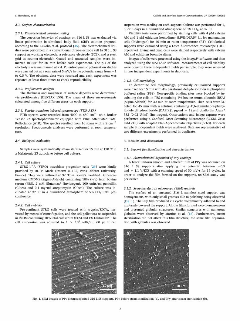
3.1.2. Scanning electron microscopy (SEM) analysisThe surface of an uncoated 316 L stainless steel support was
homogeneous, with only small grooves due to polishing being observed(Fig. 1). The PPy film produced via cyclic voltammetry adhered to anduniformly covered the support. All the films formed were homogeneousand presented globular structures. Similar structures with numerousglobules were observed by Martins et al. [15]. Furthermore, steamsterilization did not affect this film structure; the same film organiza-tion with globules was observed.
Fig. 1. SEM images of PPy electrodeposited 316 L SS supports. PPy before steam sterilization (a), and PPy after steam sterilization (b).
S. Hamdaoui, et al. Colloid and Interface Science Communications 37 (2020) 100282
3
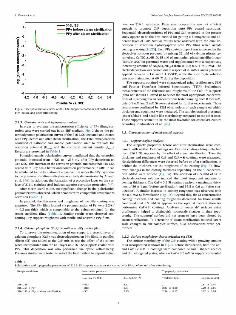
3.1.3. Corrosion tests and topography analysisIn order to evaluate the anticorrosion efficiency of PPy films, cor-
rosion tests were carried out in an SBF medium. Fig. 2 shows the po-tentiodynamic polarization curves of the 316 L SS uncoated and coatedwith PPy, before and after steam sterilization. The Tafel extrapolationsconsisted of cathodic and anodic polarization used to evaluate thecorrosion potential (Ecorr) and the corrosion current density (Jcorr).Results are presented in Table 1.
Potentiodynamic polarization curves manifested that the corrosionpotential increased from −423 to −313 mV after PPy deposition on316 L SS. This increase in the corrosion potential indicates that 316 L SScoated with PPy has a better anti-corrosion performance in SBF. It canbe attributed to the formation of a passive film under the PPy layer dueto the presence of sodium salicylate as already demonstrated by Ananthet al. [46]. In addition, the formation of a protective layer on the sur-face of 316 L stainless steel induces superior corrosion protection [47].
After steam sterilization, no significant change in the polarizationparameters was observed, which demonstrates comparable resistance tocorrosion (Table 1).
In parallel, the thickness and roughness of the PPy coating wasmeasured. The PPy films formed via polymerization of Py were 2.2+/− 0.3 μm thick which is comparable to the values obtained for thesteam sterilized films (Table 1). Similar results were observed con-cerning PPy support roughness with sterile and unsterile PPy films.
3.1.4. Calcium phosphate (CaP) deposition on PPy coated filmsTo improve the osteointegration of our support, a second layer of
calcium phosphate (CaP) was electrodeposited on PPy films. In parallel,silicon (Si) was added to the CaP mix to test the effect of the siliconwhen incorporated into the CaP layer on 316 L SS supports coated withPPy. This deposition was also performed via cyclic voltammetry.Previous studies were tested to select the best method to deposit a dual
layer on 316 L substrates. Pulse electrodeposition was not efficientenough to promote CaP deposition onto PPy coated substrates.Sequential electrodepositions of PPy and CaP proposed in the presentstudy appear to be the best method for getting a homogenous and ad-herent layer of CaP. Similar results were observed with the dual de-position of strontium hydroxyapatite onto PPy films which avoidscoating cracking [16,25]. Each PPy coated support was immersed in theelectrolyte solution prepared by mixing 25 mM of calcium nitrate tet-rahydrate Ca(NO3)2.4H2O, 15 mM of ammonium phosphate dihydrogen((NH4)H2PO4) in permuted water and supplemented with a respectivelyincreasing amount of Na2SiO3.9H2O from 0; 0.3; 0.5; 1 to 2 mM. Theelectrodeposition was carried out at a speed of 20 mV/s, and a potentialapplied between −1.6 and 1.1 V/SCE, while the electrolyte solutionwas also maintained at 60 °C during the deposition.
The supports obtained were characterized using profilometry, SEMand Fourier Transform Infrared Spectroscopy (FTIR). Preliminarymeasurements of the thickness and roughness of the CaP+Si supports(data not shown) allowed us to select the most appropriate concentra-tions of Si; among five Si concentrations tested ranging from 0 to 2 mM,only 0.5 mM and 2 mM Si were retained for further experiments. Thoseresults were confirmed by SEM observations of each sample on whichthickness and roughness were measured. The sample retained presentedless of a blade- and needle-like morphology compared to the other ones.Those supports seemed to be the most favorable for osteoblast cultureaccording to Mokabber et al. [48].
3.2. Characterization of multi-coated supports
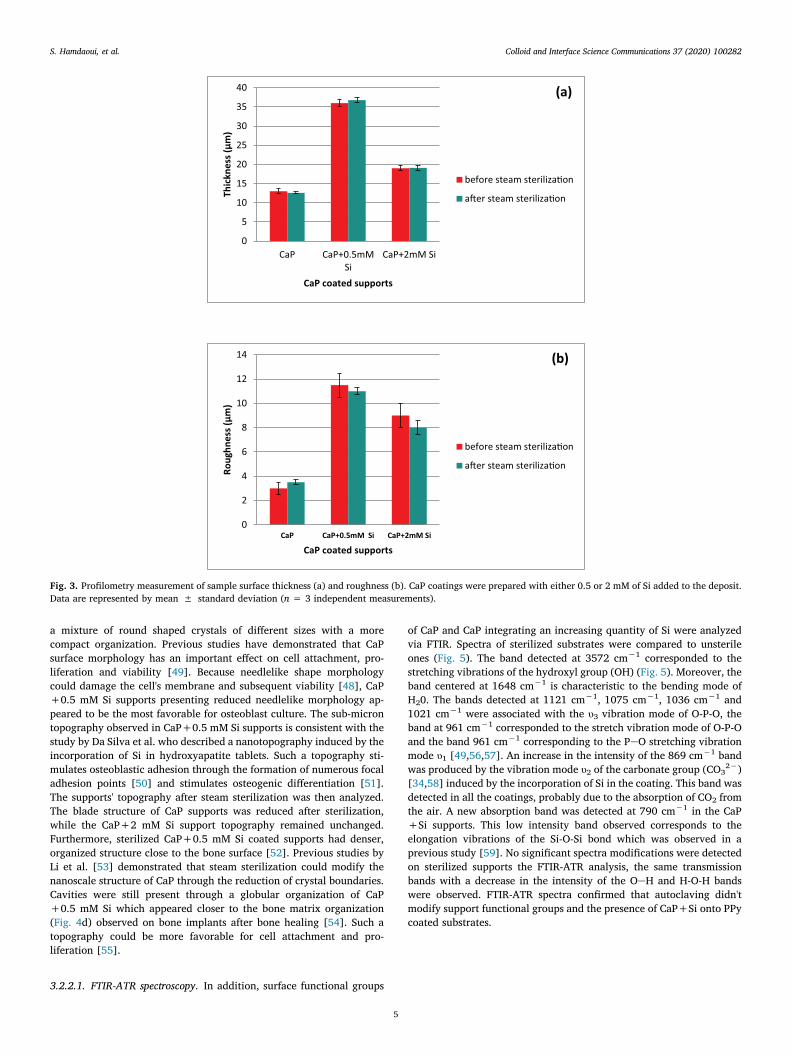
3.2.1. Support surface analysisThe supports' properties before and after sterilization were com-
pared, with neither CaP coatings nor CaP+Si coatings being detachedfrom 316 L SS supports by the effect of steam sterilization. Then thethickness and roughness of CaP and CaP+Si coatings were measured.No significant differences were observed before or after sterilization, inneither the thickness nor the roughness of the coating (Fig. 3). How-ever, changes in the coating thickness depending on the Si concentra-tion added were noticed (Fig. 3a). The addition of 0.5 mM of Si inelectrodeposition solution induced the most important increase incoating thickness. The CaP+0.5 Si coating reached a maximum thick-ness of 36±1 μm (before sterilization) and 36.8±0.6 μm (after ster-ilization). A similar increase in coating roughness was observed withthe 0.5 mM Si formulation (Fig. 3b). Beyond this, the Si concentrationcoating thickness and coating roughness decreased. So those resultsconfirmed that 0.5 mM Si appears as the optimal concentration forperforming CaP+Si coatings. Analyses of materials' surfaces usingprofilometry helped to distinguish microscale changes in their topo-graphy. The supports' surface did not seem to have been altered bysteam sterilization. To determine if steam sterilization induced lowerscale changes in our samples' surface, SEM observations were per-formed.
3.2.2. Surface morphology characterization via SEMThe surface morphology of the CaP coating with a growing amount
of Si incorporated is shown in Fig. 4. Before sterilization, both the CaPand CaP+2 mM Si coatings were composed of small shaped needlesand thin elongated plates; whereas CaP+0.5 mM Si supports presented
Fig. 2. Tafel polarization curves of 316 L SS supports coated or not coated withPPy, before and after autoclaving.
Table 1Polarization and topography parameters of 316 L SS supports coated or not coated with PPy, before and after autoclaving.
Sample conditions Polarization parameter Topography parameter
Ecorr (mV vs. SCE) Jcorr (μA.cm−2) Thickness (μm) Roughness (μm)
316 L SS −423 4.51 / 0.81 ± 0.07316 L SS + PPy −313 3.51 2.20 ± 0.30 0.20 ± 0.01316 L SS + PPy + steam sterilization −301 2.13 2.34 ± 0.17 0.22 ± 0.03
S. Hamdaoui, et al. Colloid and Interface Science Communications 37 (2020) 100282
4
a mixture of round shaped crystals of different sizes with a morecompact organization. Previous studies have demonstrated that CaPsurface morphology has an important effect on cell attachment, pro-liferation and viability [49]. Because needlelike shape morphologycould damage the cell's membrane and subsequent viability [48], CaP+0.5 mM Si supports presenting reduced needlelike morphology ap-peared to be the most favorable for osteoblast culture. The sub-microntopography observed in CaP+0.5 mM Si supports is consistent with thestudy by Da Silva et al. who described a nanotopography induced by theincorporation of Si in hydroxyapatite tablets. Such a topography sti-mulates osteoblastic adhesion through the formation of numerous focaladhesion points [50] and stimulates osteogenic differentiation [51].The supports' topography after steam sterilization was then analyzed.The blade structure of CaP supports was reduced after sterilization,while the CaP+2 mM Si support topography remained unchanged.Furthermore, sterilized CaP+0.5 mM Si coated supports had denser,organized structure close to the bone surface [52]. Previous studies byLi et al. [53] demonstrated that steam sterilization could modify thenanoscale structure of CaP through the reduction of crystal boundaries.Cavities were still present through a globular organization of CaP+0.5 mM Si which appeared closer to the bone matrix organization(Fig. 4d) observed on bone implants after bone healing [54]. Such atopography could be more favorable for cell attachment and pro-liferation [55].
3.2.2.1. FTIR-ATR spectroscopy. In addition, surface functional groups
of CaP and CaP integrating an increasing quantity of Si were analyzedvia FTIR. Spectra of sterilized substrates were compared to unsterileones (Fig. 5). The band detected at 3572 cm−1 corresponded to thestretching vibrations of the hydroxyl group (OH) (Fig. 5). Moreover, theband centered at 1648 cm−1 is characteristic to the bending mode ofH20. The bands detected at 1121 cm−1, 1075 cm−1, 1036 cm−1 and1021 cm−1 were associated with the υ3 vibration mode of O-P-O, theband at 961 cm−1 corresponded to the stretch vibration mode of O-P-Oand the band 961 cm−1 corresponding to the PeO stretching vibrationmode υ1 [49,56,57]. An increase in the intensity of the 869 cm−1 bandwas produced by the vibration mode υ2 of the carbonate group (CO32−)[34,58] induced by the incorporation of Si in the coating. This band wasdetected in all the coatings, probably due to the absorption of CO2 fromthe air. A new absorption band was detected at 790 cm−1 in the CaP+Si supports. This low intensity band observed corresponds to theelongation vibrations of the Si-O-Si bond which was observed in aprevious study [59]. No significant spectra modifications were detectedon sterilized supports the FTIR-ATR analysis, the same transmissionbands with a decrease in the intensity of the OeH and H-O-H bandswere observed. FTIR-ATR spectra confirmed that autoclaving didn'tmodify support functional groups and the presence of CaP+Si onto PPycoated substrates.
0
5
10
15
20
25
30
35
40
CaP CaP+0.5mMSi
CaP+2mM Si
Thic
knes
s (μ
m)
CaP coated supports
before steam steriliza!on
a"er steam steriliza!on
0
2
4
6
8
10
12
14
CaP CaP+0.5mM Si CaP+2mM Si
Roug
hnes
s (μ
m)
CaP coated supports
before steam steriliza!on
a"er steam steriliza!on
(a)
(b)
Fig. 3. Profilometry measurement of sample surface thickness (a) and roughness (b). CaP coatings were prepared with either 0.5 or 2 mM of Si added to the deposit.Data are represented by mean ± standard deviation (n = 3 independent measurements).
S. Hamdaoui, et al. Colloid and Interface Science Communications 37 (2020) 100282
5
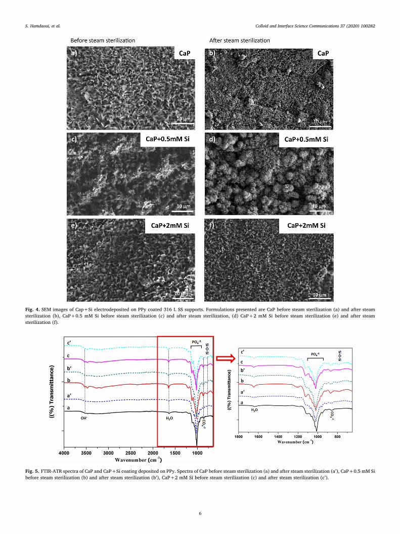
Fig. 4. SEM images of Cap+Si electrodeposited on PPy coated 316 L SS supports. Formulations presented are CaP before steam sterilization (a) and after steamsterilization (b), CaP+0.5 mM Si before steam sterilization (c) and after steam sterilization, (d) CaP+2 mM Si before steam sterilization (e) and after steamsterilization (f).
Fig. 5. FTIR-ATR spectra of CaP and CaP+Si coating deposited on PPy. Spectra of CaP before steam sterilization (a) and after steam sterilization (a’), CaP+0.5 mM Sibefore steam sterilization (b) and after steam sterilization (b’), CaP+2 mM Si before steam sterilization (c) and after steam sterilization (c’).
S. Hamdaoui, et al. Colloid and Interface Science Communications 37 (2020) 100282
6
3.3. Effects of multilayer supports on pre-osteoblast behavior
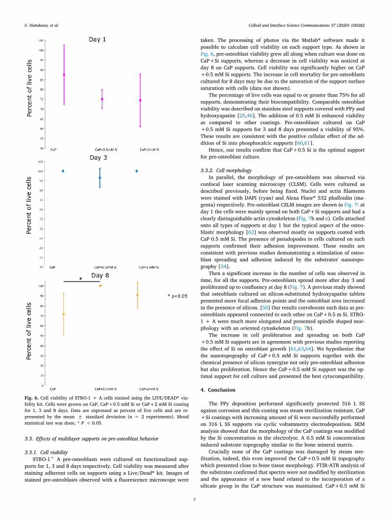
3.3.1. Cell viabilitySTRO-1+ A pre-osteoblasts were cultured on functionalized sup-
ports for 1, 3 and 8 days respectively. Cell viability was measured afterstaining adherent cells on supports using a Live/Dead® kit. Images ofstained pre-osteoblasts observed with a fluorescence microscope were
taken. The processing of photos via the Matlab® software made itpossible to calculate cell viability on each support type. As shown inFig. 6, pre-osteoblast viability grew all along when culture was done onCaP+Si supports, whereas a decrease in cell viability was noticed atday 8 on CaP supports. Cell viability was significantly higher on CaP+0.5 mM Si supports. The increase in cell mortality for pre-osteoblastscultured for 8 days may be due to the saturation of the support surfacesaturation with cells (data not shown).
The percentage of live cells was equal to or greater than 75% for allsupports, demonstrating their biocompatibility. Comparable osteoblastviability was described on stainless steel supports covered with PPy andhydroxyapatite [25,46]. The addition of 0.5 mM Si enhanced viabilityas compared to other coatings. Pre-osteoblasts cultured on CaP+0.5 mM Si supports for 3 and 8 days presented a viability of 95%.These results are consistent with the positive cellular effect of the ad-dition of Si into phosphocalcic supports [60,61].
Hence, our results confirm that CaP+0.5 Si is the optimal supportfor pre-osteoblast culture.
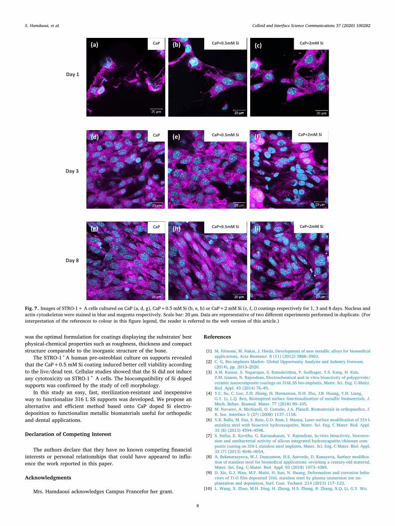
3.3.2. Cell morphologyIn parallel, the morphology of pre-osteoblasts was observed via
confocal laser scanning microscopy (CLSM). Cells were cultured asdescribed previously, before being fixed. Nuclei and actin filamentswere stained with DAPI (cyan) and Alexa Fluor® 532 phalloidin (ma-genta) respectively. Pre-osteoblast CSLM images are shown in Fig. 7: atday 1 the cells were mainly spread on both CaP+Si supports and had aclearly distinguishable actin cytoskeleton (Fig. 7b and c). Cells attachedonto all types of supports at day 1 but the typical aspect of the osteo-blasts' morphology [62] was observed mostly on supports coated withCaP 0.5 mM Si. The presence of pseudopodes in cells cultured on suchsupports confirmed their adhesion improvement. These results areconsistent with previous studies demonstrating a stimulation of osteo-blast spreading and adhesion induced by the substrates' nanotopo-graphy [34].
Then a significant increase in the number of cells was observed intime, for all the supports. Pre-osteoblasts spread more after day 3 andproliferated up to confluency at day 8 (Fig. 7). A previous study showedthat osteoblasts cultured on silicon-substituted hydroxyapatite tabletspresented more focal adhesion points and the osteoblast area increasedin the presence of silicon. [50] Our results corroborate such data as pre-osteoblasts appeared connected to each other on CaP+0.5 m Si. STRO-1 + A were much more elongated and presented spindle shaped mor-phology with an oriented cytoskeleton (Fig. 7h).
The increase in cell proliferation and spreading on both CaP+0.5 mM Si supports are in agreement with previous studies reportingthe effect of Si on osteoblast growth [61,63,64]. We hypothesize thatthe nanotopography of CaP+0.5 mM Si supports together with thechemical presence of silicon synergize not only pre-osteoblast adhesionbut also proliferation. Hence the CaP+0.5 mM Si support was the op-timal support for cell culture and presented the best cytocompatibility.
4. Conclusion
The PPy deposition performed significantly protected 316 L SSagainst corrosion and this coating was steam sterilization resistant. CaP+Si coatings with increasing amount of Si were successfully performedon 316 L SS supports via cyclic voltammetry electrodeposition. SEManalysis showed that the morphology of the CaP coatings was modifiedby the Si concentration in the electrolyte. A 0.5 mM Si concentrationinduced substrate topography similar to the bone mineral matrix.
Crucially none of the CaP coatings was damaged by steam ster-ilization, indeed, this even improved the CaP+0.5 mM Si topographywhich presented close to bone tissue morphology. FTIR-ATR analysis ofthe substrates confirmed that spectra were not modified by sterilizationand the appearance of a new band related to the incorporation of asilicate group in the CaP structure was maintained. CaP+0.5 mM Si
Fig. 6. Cell viability of STRO-1 + A cells stained using the LIVE/DEAD® via-bility kit. Cells were grown on CaP, CaP+0.5 mM Si or CaP+2 mM Si coatingfor 1, 3 and 8 days. Data are expressed as percent of live cells and are re-presented by the mean ± standard deviation (n = 2 experiments). Moodstatistical test was done, * P <0.05.
S. Hamdaoui, et al. Colloid and Interface Science Communications 37 (2020) 100282
7
was the optimal formulation for coatings displaying the substrates' bestphysical-chemical properties such as roughness, thickness and compactstructure comparable to the inorganic structure of the bone.
The STRO-1+A human pre-osteoblast culture on supports revealedthat the CaP+0.5 mM Si coating induced better cell viability accordingto the live/dead test. Cellular studies showed that the Si did not induceany cytotoxicity on STRO-1+ A cells. The biocompatibility of Si dopedsupports was confirmed by the study of cell morphology.
In this study an easy, fast, sterilization-resistant and inexpensiveway to functionalize 316 L SS supports was developed. We propose analternative and efficient method based onto CaP doped Si electro-deposition to functionalize metallic biomaterials useful for orthopedicand dental applications.
Declaration of Competing Interest
The authors declare that they have no known competing financialinterests or personal relationships that could have appeared to influ-ence the work reported in this paper.
Acknowledgments
Mrs. Hamdaoui acknowledges Campus Francefor her grant.
References
[1] M. Niinomi, M. Nakai, J. Hieda, Development of new metallic alloys for biomedicalapplications, Acta Biomater. 8 (11) (2012) 3888–3903.
[2] C. G, Bio-implants Market- Global Opportunity Analysis and Industry Forecast,(2014), pp. 2013–2020.
[3] A.M. Kumar, S. Nagarajan, S. Ramakrishna, P. Sudhagar, Y.S. Kang, H. Kim,Z.M. Gasem, N. Rajendran, Electrochemical and in vitro bioactivity of polypyrrole/ceramic nanocomposite coatings on 316L SS bio-implants, Mater. Sci. Eng. C-Mater.Biol. Appl. 43 (2014) 76–85.
[4] Y.C. Su, C. Luo, Z.H. Zhang, H. Hermawan, D.H. Zhu, J.B. Huang, Y.H. Liang,G.Y. Li, L.Q. Ren, Bioinspired surface functionalization of metallic biomaterials, J.Mech. Behav. Biomed. Mater. 77 (2018) 90–105.
[5] M. Navarro, A. Michiardi, O. Castaño, J.A. Planell, Biomaterials in orthopaedics, J.R. Soc. Interface 5 (27) (2008) 1137–1158.
[6] V.K. Balla, M. Das, S. Bose, G.D. Ram, I. Manna, Laser surface modification of 316 Lstainless steel with bioactive hydroxyapatite, Mater. Sci. Eng. C Mater. Biol. Appl.33 (8) (2013) 4594–4598.
[7] S. Sutha, K. Kavitha, G. Karunakaran, V. Rajendran, In-vitro bioactivity, biocorro-sion and antibacterial activity of silicon integrated hydroxyapatite/chitosan com-posite coating on 316 L stainless steel implants, Mater. Sci. Eng. C Mater. Biol. Appl.33 (7) (2013) 4046–4054.
[8] A. Bekmurzayeva, W.J. Duncanson, H.S. Azevedo, D. Kanayeva, Surface modifica-tion of stainless steel for biomedical applications: revisiting a century-old material,Mater. Sci. Eng. C-Mater. Biol. Appl. 93 (2018) 1073–1089.
[9] D. Xie, G.J. Wan, M.F. Maitz, H. Sun, N. Huang, Deformation and corrosion beha-viors of Ti-O film deposited 316L stainless steel by plasma immersion ion im-plantation and deposition, Surf. Coat. Technol. 214 (2013) 117–123.
[10] L. Wang, X. Zhao, M.H. Ding, H. Zheng, H.S. Zhang, B. Zhang, X.Q. Li, G.Y. Wu,
CaP CaP+2mM Si CaP+0.5mM Si
Day 1
Day 3
Day 8
CaP
CaP
CaP+2mM Si
CaP+2mM Si
)b()a( (c)
(d) (e) (f)
(g) (h) (i)
CaP+0.5mM Si
CaP+0.5mM Si
Fig. 7. Images of STRO-1+ A cells cultured on CaP (a, d, g), CaP+0.5 mM Si (b, e, h) or CaP+2 mM Si (c, f, i) coatings respectively for 1, 3 and 8 days. Nucleus andactin cytoskeleton were stained in blue and magenta respectively. Scale bar: 20 μm. Data are representative of two different experiments performed in duplicate. (Forinterpretation of the references to colour in this figure legend, the reader is referred to the web version of this article.)
S. Hamdaoui, et al. Colloid and Interface Science Communications 37 (2020) 100282
8
Surface modification of biomedical AISI 316L stainless steel with zirconium car-bonitride coatings, Appl. Surf. Sci. 340 (2015) 113–119.
[11] S. Tavakoli, S. Nemati, M. Kharaziha, S. Akbari-Alavijeh, Embedding CuO nano-particles in PDMS-SiO2 coating to improve antibacterial characteristic and corro-sion resistance, Colloid Interface Sci. Commun. 28 (2019) 20–28.
[12] A. Latifi, M. Imani, M.T. Khorasani, M.D. Joupari, Electrochemical and chemicalmethods for improving surface characteristics of 316L stainless steel for biomedicalapplications, Surf. Coat. Technol. 221 (2013) 1–12.
[13] E. Salahinejad, M.J. Hadianfard, D.D. Macdonald, M. Mozafari, D. Vashaee,L. Tayebi, A new double-layer sol-gel coating to improve the corrosion resistance ofa medical-grade stainless steel in a simulated body fluid, Mater. Lett. 97 (2013)162–165.
[14] L.L. Zhang, S.J. Liu, H.C. Han, Y. Zhou, S.C. Hu, C. He, Q.X. Yan, Studies on theformation process and anti-corrosion performance of polypyrrole film deposited onthe surface of Q235 steel by an electrochemical method, Surf. Coat. Technol. 341(2018) 95–102.
[15] N.C.T. Martins, T.M.E. Silva, M.F. Montemor, J.C.S. Fernandes, M.G.S. Ferreira,Electrodeposition and characterization of polypyrrole films on aluminium alloy6061-T6, Electrochim. Acta 53 (14) (2008) 4754–4763.
[16] D. Gopi, S. Ramya, D. Rajeswari, L. Kavitha, Corrosion protection performance ofporous strontium hydroxyapatite coating on polypyrrole coated 316L stainless steel,Colloids Surf. B-Biointerfaces 107 (2013) 130–136.
[17] D. Gopi, J. Indira, L. Kavitha, J.M.F. Ferreira, Hydroxyapatite coating on selectivelypassivated and sensitively polymer-protected surgical grade stainless steel, J. Appl.Electrochem. 43 (3) (2013) 331–345.
[18] M.B. Gonzalez, S.B. Saidman, Electrodeposition of polypyrrole on 316L stainlesssteel for corrosion prevention, Corros. Sci. 53 (1) (2011) 276–282.
[19] M. Eslami, G. Speranza, M. Fedel, N.E. Andersson, F. Deflorian, S. Omanovic,C. Zanella, Electropolymerization and possible corrosion protection effect of poly-pyrrole coatings on AA1050 (UNS A91050) in NaCl solutions, Corrosion 75 (7)(2019) 745–755.
[20] R. Balint, N.J. Cassidy, S.H. Cartmell, Conductive polymers: towards a smart bio-material for tissue engineering, Acta Biomater. 10 (6) (2014) 2341–2353.
[21] X.L. Zhao, L.L. Jin, H.F. Shi, W.J. Tong, D. Gorin, Y. Kotelevtsev, Z.W. Mao, Recentadvances of designing dynamic surfaces to regulate cell adhesion, Colloid InterfaceSci. Commun. 35 (2020).
[22] X.Y. Cui, J.F. Hetke, J.A. Wiler, D.J. Anderson, D.C. Martin, Electrochemical de-position and characterization of conducting polymer polypyrrole/PSS on multi-channel neural probes, Sensors Actuat. A-Phys. 93 (1) (2001) 8–18.
[23] R. Chakraborty, J.S. Manna, P. Saha, Development and relative comparison ofpolypyrrole-calcium phosphate composite coatings with differential concentrationof chlorophyll functionalized polymer particle achieved through pulsed electrodeposition, Surf. Coat. Technol. 363 (2019) 221–235.
[24] B. Maimaiti, N. Zhang, L. Yan, J. Luo, C. Xie, Y. Wang, C. Ma, T. Ye, Stable ZnO-doped hydroxyapatite nanocoating for anti-infection and osteogenic on titanium,Colloids Surf. B: Biointerfaces 186 (2019) 110731.
[25] R. Chakraborty, V.S. Seesala, J.S. Manna, P. Saha, S. Dhara, Synthesis, character-ization and cytocompatibility assessment of hydroxyapatite-polypyrrole compositecoating synthesized through pulsed reverse electrochemical deposition, Mater. Sci.Eng. C-Mater. Biol. Appl. 94 (2019) 597–607.
[26] R. Chakraborty, P. Saha, A comparative study on surface morphology and elec-trochemical behaviour of hydroxyapatite-calcium hydrogen phosphate compositecoating synthesized in-situ through electro chemical process under various de-position conditions, Surf. Interfaces 12 (2018) 160–167.
[27] A. Robin, G. Silva, J.L. Rosa, Corrosion behavior of HA-316L SS biocomposites inaqueous solutions, Mater. Res.-Ibero-Am. J. Mater. 16 (6) (2013) 1254–1259.
[28] J. Jeong, J.H. Kim, J.H. Shim, N.S. Hwang, C.Y. Heo, Bioactive calcium phosphatematerials and applications in bone regeneration, Biomater. Res. 23 (2019) 4.
[29] Y. Su, I. Cockerill, Y. Zheng, L. Tang, Y.X. Qin, D. Zhu, Biofunctionalization ofmetallic implants by calcium phosphate coatings, Bioact. Mater. 4 (2019) 196–206.
[30] K. Anselme, Osteoblast adhesion on biomaterials, Biomaterials 21 (7) (2000)667–681.
[31] R.A. Surmenev, M.A. Surmeneva, A.A. Ivanova, Significance of calcium phosphatecoatings for the enhancement of new bone osteogenesis–a review, Acta Biomater.10 (2) (2014) 557–579.
[32] B.W. Cunningham, N. Hu, C.M. Zorn, P.C. McAfee, Bioactive titanium calciumphosphate coating for disc arthroplasty: analysis of 58 vertebral end plates after 6-to 12-month implantation, Spine J. 9 (10) (2009) 836–845.
[33] A.F. Khan, M. Saleem, A. Afzal, A. Ali, A. Khan, A.R. Khan, Bioactive behavior ofsilicon substituted calcium phosphate based bioceramics for bone regeneration,Mater. Sci. Eng. C Mater. Biol. Appl. 35 (2014) 245–252.
[34] H.M. da Silva, M. Mateescu, A. Ponche, C. Damia, E. Champion, G. Soares,K. Anselme, Surface transformation of silicon-doped hydroxyapatite immersed inculture medium under dynamic and static conditions, Colloids Surf. B-Biointerfaces75 (1) (2010) 349–355.
[35] M. Qadir, Y.C. Li, C. Wen, Ion-substituted calcium phosphate coatings by physicalvapor deposition magnetron sputtering for biomedical applications: a review, ActaBiomater. 89 (2019) 14–32.
[36] T. Gao, H.T. Aro, H. Ylanen, E. Vuorio, Silica-based bioactive glasses modulateexpression of bone morphogenetic protein-2 mRNA in Saos-2 osteoblasts in vitro,Biomaterials 22 (12) (2001) 1475–1483.
[37] E. Boanini, M. Gazzano, A. Bigi, Ionic substitutions in calcium phosphates synthe-sized at low temperature, Acta Biomater. 6 (6) (2010) 1882–1894.
[38] C.A. Aboltins, V. Antoci, S. Bhattacharyya, M. Cross, P. Ducheyne, A.A. Freiberg,N. Hailer, P. Kay, C. Ketonis, M.R. Klement, N. Kose, M. Lee, P. Mitchell, S. Nandi,J.C. Palacio, K. Perry, H. Prieto, A. Shahi, R. Trebse, D. Turner, C.-T. Wu, H. Yazdi,Hip and knee section, prevention, prosthesis factors: proceedings of internationalconsensus on orthopedic infections, J. Arthroplast. 34 (2S) (2019) S309–S320.
[39] S. Kim, J.O. Jeong, S. Lee, J.S. Park, H.J. Gwon, S.I. Jeong, J.G. Hardy, Y.M. Lim,J.Y. Lee, Effective gamma-ray sterilization and characterization of conductivepolypyrrole biomaterials, Sci. Rep. 8 (2018).
[40] C.R. Harrell, V. Djonov, C. Fellabaum, V. Volarevic, Risks of using sterilization bygamma radiation: the other side of the coin, Int. J. Med. Sci. 15 (3) (2018) 274–279.
[41] I.R. Williams, M.B. Mayor, J.P. Collier, The impact of sterilization method on wearin knee arthroplasty, Clin. Orthop. Relat. Res. 356 (1998) 170–180.
[42] N. Ribeiro, G.C. Soares, V. Santos-Rosales, A. Concheiro, C. Alvarez-Lorenzo,C.A. Garcia-Gonzalez, A.L. Oliveira, A new era for sterilization based on super-critical CO2 technology, J. Biomed. Mater. Res. B-Appl. Biomater. 108 (2) (2020)399–428.
[43] S. Adler, M. Scherrer, F.D. Daschner, Costs of low-temperature plasma sterilizationcompared with other sterilization methods, J. Hosp. Infect. 40 (2) (1998) 125–134.
[44] C.C. Shih, Y.Y. Su, L.C. Chen, C.M. Shih, S.J. Lin, Degradation of 316L stainless steelsternal wire by steam sterilization, Acta Biomater. 6 (6) (2010) 2322–2328.
[45] T. Kokubo, H. Takadama, How useful is SBF in predicting in vivo bone bioactivity?Biomaterials 27 (15) (2006) 2907–2915.
[46] K.P. Ananth, A.J. Nathanael, S.P. Jose, T.H. Oh, D. Mangalaraj, A novel silica na-notube reinforced ionic incorporated hydroxyapatite composite coating on poly-pyrrole coated 316L SS for implant application, Mater. Sci. Eng. C-Mater. Biol. Appl.59 (2016) 1110–1124.
[47] N. Aravindan, M.V. Sangaranarayanan, Influence of solvent composition on theanti-corrosion performance of copper-polypyrrole (cu-PPy) coated 304 stainlesssteel, Prog. Org. Coat. 95 (2016) 38–45.
[48] T. Mokabber, Q. Zhou, A.I. Vakis, P. van Rijn, Y.T. Pei, Mechanical and biologicalproperties of electrodeposited calcium phosphate coatings, Mater. Sci. Eng. C-Mater. Biol. Appl. 100 (2019) 475–484.
[49] Y. Huang, M. Hao, X.F. Nian, H.X. Qiao, X.J. Zhang, X.Y. Zhang, G.Q. Song,J.C. Guo, X.F. Pang, H.L. Zhang, Strontium and copper co-substituted hydro-xyapatite-based coatings with improved antibacterial activity and cytocompatibilityfabricated by electrodeposition, Ceram. Int. 42 (10) (2016) 11876–11888.
[50] H.M. da Silva, M. Mateescu, C. Damia, E. Champion, G. Soares, K. Anselme,Importance of dynamic culture for evaluating osteoblast activity on dense silicon-substituted hydroxyapatite, Colloids Surf. B: Biointerfaces 80 (2) (2010) 138–144.
[51] J. Wang, M. Wang, F. Chen, Y. Wei, X. Chen, Y. Zhou, X. Yang, X. Zhu, C. Tu,X. Zhang, Nano-hydroxyapatite coating promotes porous calcium phosphateceramic-induced osteogenesis via BMP/Smad Signaling pathway, Int. J.Nanomedicine 14 (2019) 7987–8000.
[52] F. Mangano, M. Raspanti, H. Maghaireh, C. Mangano, Scanning Electron micro-scope (SEM) evaluation of the Interface between a nanostructured calcium-in-corporated dental implant surface and the human bone, Materials 10 (12) (2017).
[53] X. Li, B. Guo, Y. Xiao, T. Yuan, Y. Fan, X. Zhang, Influences of the steam sterilizationon the properties of calcium phosphate porous bioceramics, J. Mater. Sci. Mater.Med. 27 (1) (2016) 5.
[54] R. Depprich, H. Zipprich, M. Ommerborn, E. Mahn, L. Lammers, J. Handschel,C. Naujoks, H.P. Wiesmann, N.R. Kübler, U. Meyer, Osseointegration of zirconiaimplants: an SEM observation of the bone-implant interface, Head Face Med 4(2008) 25.
[55] F. Mastrangelo, R. Quaresima, L. Canullo, A. Scarano, L.L. Muzio, A. Piattelli,Effects of novel laser dental implant microtopography on human osteoblast pro-liferation and bone deposition, Int. J. Oral Maxillofac. Implants 35 (2) (2020)320–329.
[56] X. Li, I. Zhitomirsky, Capacitive behaviour of polypyrrole films prepared on stain-less steel substrates by electropolymerization, Mater. Lett. 76 (2012) 15–17.
[57] M. Palard, E. Champion, S. Foucaud, Synthesis of silicated hydroxyapatite Ca-10(PO4)(6-x)(SiO4)(x)(OH)(2-x), J. Solid State Chem. 181 (8) (2008) 1950–1960.
[58] Y. Huang, S.G. Han, X.F. Pang, Q.Q. Ding, Y.J. Yan, Electrodeposition of poroushydroxyapatite/calcium silicate composite coating on titanium for biomedical ap-plications, Appl. Surf. Sci. 271 (2013) 299–302.
[59] T. Jesionowski, L. Klapiszewski, G. Milczarek, Kraft lignin and silica as precursors ofadvanced composite materials and electroactive blends, J. Mater. Sci. 49 (3) (2014)1376–1385.
[60] M. Lopez-Alvarez, E.L. Solla, P. Gonzalez, J. Serra, B. Leon, A.P. Marques, R.L. Reis,Silicon-hydroxyapatite bioactive coatings (Si-HA) from diatomaceous earth andsilica. Study of adhesion and proliferation of osteoblast-like cells, J. Mater. Sci.-Mater. Med. 20 (5) (2009) 1131–1136.
[61] Y.L. Yang, K. Serpersu, W. He, S.R. Paital, N.B. Dahotre, Osteoblast interaction withlaser cladded HA and SiO2-HA coatings on ti-6Al-4V, Mater. Sci. Eng. C-Mater. Biol.Appl. 31 (8) (2011) 1643–1652.
[62] L. Casarrubios, M.C. Matesanz, S. Sanchez-Salcedo, D. Arcos, M. Vallet-Regi,M.T. Portoles, Nanocrystallinity effects on osteoblast and osteoclast response tosilicon substituted hydroxyapatite, J. Colloid Interface Sci. 482 (2016) 112–120.
[63] X. Qiu, P. Wan, L.L. Tan, X.M. Fan, K. Yang, Preliminary research on a novelbioactive silicon doped calcium phosphate coating on AZ31 magnesium alloy viaelectrodeposition, Mater. Sci. Eng. C-Mater. Biol. Appl. 36 (2014) 65–76.
[64] G. Lehmann, I. Cacciotti, P. Palmero, L. Montanaro, A. Bianco, L. Campagnolo,A. Camaioni, Differentiation of osteoblast and osteoclast precursors on pure andsilicon-substituted synthesized hydroxyapatites, Biomed. Mater. 7 (5) (2012).
S. Hamdaoui, et al. Colloid and Interface Science Communications 37 (2020) 100282
9


![Functionalizing the glycocalyx of living cells with ... · Functionalizing the glycocalyx of living cells with supramolecular guest ligands for cucurbit[8]uril-mediated assembly](https://img.pdfslide.us/doc/110x75/5ec159ef491c257e8647d3c4/functionalizing-the-glycocalyx-of-living-cells-with-functionalizing-the-glycocalyx.jpg)