Embed Size (px)
Citation preview

RESEARCH ARTICLE Open Access
An easy method to differentiate retinal arteriesfrom veins by spectral domain optical coherencetomography: retrospective, observational caseseriesYanling Ouyang, Qing Shao, Dirk Scharf, Antonia M Joussen and Florian M Heussen*
Abstract
Background: Recently it was shown that retinal vessel diameters could be measured using spectral domain opticalcoherence tomography (OCT). It has also been suggested that retinal vessels manifest different features on spectraldomain OCT (SD-OCT) depending on whether they are arteries or veins. Our study was aimed to present a reliableSD-OCT assisted method of differentiating retinal arteries from veins.
Methods: Patients who underwent circular OCT scans centred at the optic disc using a Spectralis OCT (HeidelbergEngineering, Heidelberg, Germany) were retrospectively reviewed. Individual retinal vessels were identified oninfrared reflectance (IR) images and given unique labels for subsequent grading. Vessel types (artery, vein or uncertain)assessed by IR and/or fluorescein angiography (FA) were referenced as ground truth. From OCT, presence/absence ofthe hyperreflective lower border reflectivity feature was assessed. Presence of this feature was considered indicative forretinal arteries and compared with the ground truth.
Results: A total of 452 vessels from 26 eyes of 18 patients were labelled and 398 with documented vessel type (302 byIR and 96 by FA only) were included in the study. Using SD-OCT, 338 vessels were assigned a final grade, of which,86.4% (292 vessels) were classified correctly. Forty three vessels (15 arteries and 28 veins) that IR failed to differentiate werecorrectly classified by SD-OCT. When using only IR based ground truth for vessel type the SD-OCT based classificationapproach reached a sensitivity of 0.8758/0.9297, and a specificity of 0.9297/0.8758 for arteries/veins, respectively.
Conclusion: Our method was able to classify retinal arteries and veins with a commercially available SD-OCT alone, andachieved high classification performance. Paired with OCT based vessel measurements, our study has expanded thepotential clinical implication of SD-OCT in evaluation of a variety of retinal and systemic vascular diseases.
Keywords: Optical coherence tomography, Spectral domain, Retina vessel
BackgroundRetinal blood vessels are the only visible and opticallyaccessible small blood vessels in the human body [1]. Inrecent years, researchers have found that alterations inthe arterial or venular tree of the retinal vasculature areassociated with several generalized vascular problems,such as diabetic retinopathy, cardiovascular or cerebraldisorders [2-7]. Since arteries and veins are differentlyaffected in these disease processes, retinal imaging
analyses like retinal vessel segmentation, quantitativevessel diameter measurement, or the ratio of artery tovein calibres (AVR) are of strong medical interest. A pre-requisite for assessment of the above vascular change isto separate those vessels from each other [3].Fluorescein angiography (FA) is a gold stand for classi-
fying retinal arteries and veins, but due to its invasivenature, it is not employed merely for the purpose of dif-ferentiating arteries from veins. A number of methodshave been reported in the literature for artery/vein clas-sification of retinal blood vessels, which mainly depend oncolor fundus images [3,8-13]. Using color fundus image
* Correspondence: [email protected]é-Universitätsmedizin Berlin, Department of Ophthalmology, Berlin,Germany
© 2014 Ouyang et al.; licensee BioMed Central Ltd. This is an Open Access article distributed under the terms of the CreativeCommons Attribution License (http://creativecommons.org/licenses/by/2.0), which permits unrestricted use, distribution, andreproduction in any medium, provided the original work is properly credited. The Creative Commons Public DomainDedication waiver (http://creativecommons.org/publicdomain/zero/1.0/) applies to the data made available in this article,unless otherwise stated.
Ouyang et al. BMC Ophthalmology 2014, 14:66http://www.biomedcentral.com/1471-2415/14/66

information, methods based on shape, colour and texturefeatures, [11] local contrast of the red-channel [14] or redreflex [3,9-11,13-15], have great potential to classify theretinal arteries. However, such dissimilarities affect onlythe main vessels and strongly vary depending on patients,and vessel topography [11]. Also, the problem is compli-cated by the similarity in the descriptive features of thesetwo structures and by the contrast and luminosity variabil-ity of the retina [8].Optical coherence tomography (OCT) is an imaging mo-
dality capable of providing high-resolution cross-sectionalimages of the neurosensory retina. It has been described toprovide superior morphologic information compared withcolour photography and angiography [16-18]. The evalu-ation of retinal vessels by OCT has seen raised interestlately [19-28], of which Doppler Fourier-domain (FD) OCTwas the most successful option [19]. However, Doppler FD-OCT systems are still several steps away from routine avail-ability in clinic.Recently, a spectral domain OCT (SD-OCT) based
method for measuring retinal vessel diameters was reportedusing a commercially available spectral domain OCT (SD-OCT) instrument [22,29]. It has also been suggested thatthe hyperreflective core feature in retinal vessels seen bySD-OCT manifested differently between arteries and veins
(as shown in Figure 1 in our preliminary data). Our studywas aimed to evaluate an SD-OCT based method to differ-entiate retinal vessel types, and compare these findings withthe reference from infrared images and/or FA.
MethodsData collectionAll Patients who underwent circular OCT-scans cen-tered at the optic disc using a Spectralis OCT +HRA(Heidelberg Engineering, Heidelberg, Germany) at theCharité Hospital between January 1 2013 and March 12013 were retrospectively collected. Patients were seenin our primary care unit and did not necessarily presentwith retinal problems. Thus, eyes with relatively normallooking retinal structure in the OCT covered area wereincluded. Clinical data regarding age, gender, race,history of ophthalmic diseases or surgeries, ophthalmicdiagnosis, lens status, and visual acuity were also col-lected. Approval for data collection and analysis wasobtained from the institutional review board of theCharité-Universitätsmedizin Berlin. Written consent wasobtained from all patients. The research adhered to thetenets set forth in the Declaration of Helsinki.The OCT scanning protocol consisted of a circular
scan with 3.42 - 4.04 mm diameter centered on the optic
Figure 1 One retinal artery and one vein seen on spectral domain optical coherence tomography. A. Infrared reflectance image of aretinal artery. B. The same artery shown with optical coherence line scanning. Note: the upper and lower boundary of retinal vessels presented ashyperreflective feature compared to the surrounding retinal tissue (red arrows). C. Infrared reflectance image of a retinal vein. D. The same veindemonstrated with optical coherence scan. Note: the upper and lower boundary did not present different reflectivity compared to thesurrounding retinal tissue (white arrows).
Ouyang et al. BMC Ophthalmology 2014, 14:66 Page 2 of 9http://www.biomedcentral.com/1471-2415/14/66
disc (Spectralis HRA-OCT, Heidelberg Engineering,Heidelberg, Germany). The Eyes were scanned with thehigh-resolution OCT mode with a mean image aver-aging of 30 frames per final image. Simultaneously,near-infrared reflectance (IR) pictures were capturedwith a scanning laser ophthalmoscope. Images werereviewed and analyzed using the Spectralis viewing soft-ware (Heidelberg Eye Explorer, version 1.7.1.0).
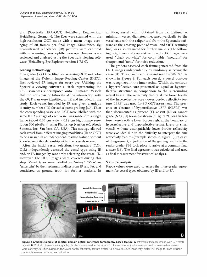
Grading methodologyOne grader (Y.O.), certified for assessing OCT and colorimages at the Doheny Image Reading Center (DIRC),first reviewed IR images for every eye. Utilizing theSpectralis viewing software a circle representing theOCT scan was superimposed onto IR images. Vesselsthat did not cross or bifurcate at the intersection withthe OCT scan were identified on IR and included in thestudy. Each vessel included by IR was given a uniqueidentity number (ID) for subsequent grading [30]. Thenthe corresponding vessels on OCT were labelled with thesame ID. An image of each vessel was made into a singleframe (about 0.05 cm wide × 0.18 cm high, image reso-lution 300 pixel/cm) using Photoshop (version 6.0, AbodeSystems, Inc, San Jose, CA, USA). This strategy allowedeach vessel from different imaging modalities (IR or OCT)to be assessed in an independent, masked fashion withoutknowledge of its relationship with other vessels or eye.After the initial vessel selection, two graders (Y.O.,
Q.S.) independently assessed the vessel type using IRand/or FA images by randomly selecting the vessel ID.However, the OCT images were covered during thisstep. Vessel types were labelled as “Artery”, “Vein” or“uncertain” by the maximum findings from IR and FA, andconsidered as ground truth for further analysis. In
addition, vessel width obtained from IR (defined asminimum vessel diameter, measured vertically to thevessel axis with the caliper tool from the Spectralis soft-ware at the crossing point of vessel and OCT scanningline) was also evaluated for further analysis. The follow-ing brightness and contrast settings for IR images wereused: “black on white” for color table, “medium” forsharpen and “none” for noise reduction.The graders assessed each frame generated from the
OCT images independently by randomly selecting thevessel ID. The structure of a vessel seen by SD-OCT isshown in Figure 2. For each vessel, a vessel contourwas recognized in the inner retina. Within the contour,a hyperreflective core presented as equal or hyperre-flective structure in comparison to the surroundingretinal tissue. The reflectivity feature at the lower borderof the hyperreflective core (lower border reflectivity fea-ture, LBRF) was used for SD-OCT assessment. The pres-ence or absence of hyperreflective LBRF (HLBRF) wasthen documented as present (Y), absent (N) or cannotgrade (NA) [31] (example shown in Figure 2). For this fea-ture, vessels with a lower border right at the boundary ofhyperreflective and hyporeflective retinal layers or smallvessels without distinguishable lower border reflectivitywere excluded due to the difficulty to interpret the truereflectivity features (example shown in Figure 3). In casesof disagreement, adjudication of the grading results by thesenior grader F.H. took place to arrive at a common finalanswer [16]. The final agreement was calculated and usedas final measurement for statistical analysis.
Statistical analysisKappa values were used to assess the inter-grader agree-ment for vessel types obtained by IR and/or FA.
Figure 2 Grading example of spectral domain optical coherence tomography based feature. A. Infrared reflectance image with 22 vesselslabeled. B. Optical coherence tomography circular scan centred at the optic disc. Retinal arteries (red arrows) and retinal veins (white arrows)were correctly classified based on the lower border reflectivity feature. Vessel No. 5 was classified incorrectly. Note: The image for each vessel ispreferably assessed without magnification.
Ouyang et al. BMC Ophthalmology 2014, 14:66 Page 3 of 9http://www.biomedcentral.com/1471-2415/14/66

OCT based features were considered corresponding tovessel types as follows: presence and absence of HLBRFwas seen indicative for arteries and veins, respectively.When using vessel type as determined by the maximumfinding from both FA and IR as ground truth, thecriteria for evaluating classification performance forarteries were assessed in two ways: 1) unclassified ves-sels included: for all 308 vessels included in the study,considering the HLBRF as an indication of arteries, andabsence of this feature (including “N” and “NA” forhyper reflectivity in the lower vessel contour) as nega-tive result; 2) unclassified vessels excluded: only caseswith gradable HLBRF were considered. Similar methodswere used for veins. Then the classification perform-ance was evaluated. For example from Figure 4, theclassification performance of arteries (unclassified ves-sels included) was calculated as below:
Sensitivity ¼ True Positive= True Positive þ False Negativeð Þ¼ a= aþ cþ eð Þ½ �;
Specificity ¼ True Negative= True Negativeþ False Positiveð Þ¼ d þ fð Þ= d þ fð Þ þ b½ �
PPV ¼ True Positive= True Positiveþ False Positiveð Þ¼ a= aþ bð Þ;
NPV ¼ True Negative= True Negativeþ False Negativeð Þ¼ d þ fð Þ= d þ fð Þ þ cþ eð Þ½ �;
Positive Likelihood Ratio ¼ Sensitivity= 1−specificityð Þ;
Negative Likelihood Ratio ¼ 1−Sensitivityð Þ=specificity;
Classification Accuracy ¼ True Positiveþ True Negativeð Þ=ðTrue Positiveþ True NegativeþFalse Positiveþ False NegativeÞ
¼ aþ d þ fð Þ½ �=½aþ d þ fð Þ þ bþ cþ eð Þ�;
False positive rate ¼ 1−Specificity
Figure 4 Example of vessel type classification used forevaluation of classification performance determined by OCTand ground truth. It demonstrates how we arrived at the numbersused to calculate sensitivity, specificity and other parametersmentioned in the methods from the original numbers (A) based oninclusion (B) or exclusion (C) of non-classified vessels.
Figure 3 Cases not applicable for grading of lower borderreflectivity feature seen on spectral domain optical coherencetomography. A-C: Red arrows: Small vessels without differentiablelower border reflectivity. D-G: White arrows: Vessels with lowerborder right at the boundary of hyperreflective and hyporeflectiveretinal layers.
Ouyang et al. BMC Ophthalmology 2014, 14:66 Page 4 of 9http://www.biomedcentral.com/1471-2415/14/66

Classification Error rate ¼ False Positiveþ False Negativeð Þ=ðTrue Positiveþ True NegativeþFalse Positiveþ False NegativeÞ
¼ bþ cþ eð Þ½ �=½aþ d þ fð Þ þ bþcþ eð Þ�
As part of a subanalysis, similar analyses were also ob-tained by using vessel types documented by IR only asground truth.To compare differences of retinal vessel diameters be-
tween groups, univariate analysis of variance (ANOVA)was used. Correction for multiple testing was performedby post hoc Bonferroni adjustment.Stata (version 10.0, College Station, TX: StataCorp LP.
United States) was used for the statistical analysis. A bilat-eral value of P < 0.05 was considered statistically significant.
ResultsCharacteristics of the study populationTwenty-six eyes of 18 patients examined with the Spec-tralis OCT during the study period with the requiredscanning protocol were included in the study. Twenty-two eyes also had FA imaging covering field 1 and/orfield 2 performed. Demographic features are summa-rized in Table 1.
Characteristics of vessel types by ground truthA total of 452 vessels from 26 eyes of 18 patients withadequate image quality and without pathology withinthe OCT scan area were labelled in the study. Amongthem, 51 vessels were documented with unknown vesseltype by using the maximum finding from IR and FA.Three more vessels were excluded due to bifurcation atthe intersection of the OCT scan. Thus, a total of 398vessels were included in the study, among which, 302
(75.9%) were classified by IR and additional 96 were onlyidentifiable by FA.
Classification of retinal vessel types by SD-OCTAmong the 398 included vessels, 27 vessels with theirlower border right at the boundary of hyperreflective
Table 1 Demographic characteristics of the study
No. patient (Women:Men) 18 (15:3)
Age
58.7 ± 17.5 (23-85)Mean ± SD (min-max),years
Systemic DiagnosisDisease (No. Patients)
Diabetic (2), Hypertension (3), Othercardiovascular diseases (3), kidney stone(1), Thalassemia (1), Sarcoidosis (1), nosystemic disease (8)
No. Eyes (Right: Left) 26 (13:13)
Snellen Visual Acuitymin - max
20/200-20/20
Choroidal/RetinalDiagnosis Disease(No. Eyes)
Uveitis (8), Myopia (3), Diabetic retinopathy(1), Age-related macular degeneration (1),Juxtafoveal Telangiectasis (1), Macular pucker(1), Central serous retinopathy (2), MultifocalChoroiditis (1), Undetermined (2), withinnormal limits (6).
Figure 5 Results of vessel type classification determined byOCT/IR and ground truth (FA + IR or IR). This figure shows thenumbers used to calculated specificity and sensitivity of OCT vs FAand IR (A), IR vs FA and IR (B), and OCT vs IR (C).
Ouyang et al. BMC Ophthalmology 2014, 14:66 Page 5 of 9http://www.biomedcentral.com/1471-2415/14/66

and hyporeflective retinal layers were documented as“NA” for the HLBRF. Additional 33 vessels also failed tohave a differentiable reflectivity feature in the lowervessel contour due to the small size of the hypercorefeature. As a result, 338 vessels were assigned a finalgrade for HLBRF in the study and differentiated intoarteries and vein, of which, 86.4% (292 vessels) wereclassified correctly. 43 vessels (15 arteries and 28 veins)that IR failed to differentiate were correctly recognizedby SD-OCT. 23 vessels crossed over one another an-other at the point of the OCT scan intersection and 21(91.3%) of them were correctly classified. Classificationperformance parameters of the current method usingFA + IR or IR only as ground truth are shown in Figure 5and Table 2 (please note, the total number of vesselsclassified varied by using different ground truth).Characteristics of vessel width for vessels that were
correctly classified by IR + FA, IR and SD-OCT aresummarized in Table 3. The mean vessel width wassignificantly different among groups with vessels deter-mined by IR, vessels recognized by IR + FA, and vesselscorrectly differentiated by SD-OCT (ANOVA, p =0.0008). The mean vessel width for vessels correctlyclassified by SD-OCT was not statistically differentfrom the mean of those determined by IR (p = 0.348);or that recognized by IR + FA (p = 0.117). However, themean vessel diameter for vessels determined by IR onlywas statistically smaller than those classified by IR + FA(P = 0.001).For cases that SD-OCT had classified incorrectly, 38
had measurable vessel width documented. Their meanvessel diameter was 100.2 ± 35.3 μm (range, 44–168).Case examples are shown in Figure 6.
Inter-grader reproducibilityAlmost perfect agreement [32] indicated by Kappavalues was achieved for inter-grader reliabilities for
vessel types by IR and/or FA (Kappa = 0.9306) andgrading for the HLBRF by SD-OCT (Kappa = 0.8872).
DiscussionRetinal arteries have clearly distinctive hyperreflectivelower borders in SD-OCT but retinal veins do not.Using this feature, our study successfully differentiatedretinal arteries from veins with a commercially availableSD-OCT instrument.OCT is commonly used in the diagnosis and mana-
gement of retinal diseases, and is already a major non-invasive imaging modality in ophthalmology [19]. Arecent report using commercially available SD-OCT hasprovided retinal vessel diameter measurements withgood reproducibility [22]. The identification of retinalarteries and veins has to be done manually by comparingthe OCT images with corresponding fundus images [22],which prevents OCT from being an independent usefultool to evaluate retinal vessels. However, retinal arteriesand veins did exhibit different reflectivity patterns atclose inspection (as shown in Figure 1 in our preliminarydata). For arteries, both borders presented as hyperre-flective in comparison to the surrounding retinal tissue;while for veins, no reflectivity difference was seen oneither border compared to the surrounding retinal tis-sue. We chose the lower border of the hyperreflectivecore to be a potential indicator because the signal of theupper border is generally difficult to differentiate fromthe surrounding nerve fiber layer, ganglion cell layer, orinner plexiform layer. The lower border also has thislimitation: when it is located near the border of hyperre-flective and hyporeflective retinal layers, the assessmentof its reflectivity is a challenge. However, this only oc-curred in 27 cases (6.8%) in our study.Our study was the first to use a commercially available
SD-OCT to classify retinal arteries and veins, and showsencouraging results. Previous literature for artery/vein
Table 2 Classification performance parameters
Performance measure
Using FA + IR as ground truth Using IR as ground truth
Unclassified vessel Unclassified vessel
Included Excluded included Excluded
Artery Vein Artery Vein Artery Vein Artery Vein
Sensitivity 0.7014 0.8000 0.8177 0.9172 0.7929 0.8947 0.8758 0.9297
Specificity 0.9278 0.8436 0.9172 0.8177 0.9323 0.8876 0.9297 0.8758
Positive Predictive Value 0.9193 0.8136 0.9193 0.8136 0.9371 0.8623 0.9371 0.8623
Negative Predictive Value 0.7261 0.8318 0.8136 0.9193 0.7799 0.9146 0.8623 0.9371
Positive Likelihood Ratio 9.7120 5.1152 9.8751 5.0307 11.7173 7.9584 12.4561 7.4864
Negative Likelihood Ratio 0.3218 0.2371 0.1988 0.1013 0.2221 0.1186 0.1336 0.0803
Classification Accuracy 0.8056 0.8235 0.8639 0.8639 0.8543 0.8907 0.9004 0.9004
Classification Error Rate 0.1944 0.1765 0.1361 0.1361 0.1457 0.1093 0.0996 0.0996
False Positive Rate 0.0722 0.1564 0.0828 0.1823 0.0677 0.1124 0.0703 0.1242
Ouyang et al. BMC Ophthalmology 2014, 14:66 Page 6 of 9http://www.biomedcentral.com/1471-2415/14/66

classification of retinal blood vessels was based mainlyon color fundus images [3,8,9,11,12,15,33,34]. Althoughother methodology, such as MRA was also used toseparate arteries from veins, [35] it was more applicablefor cerebral vessels. Our approach is not directly com-parable to these publications. One reason is that virtuallyall of the previous reports used a vessel classificationobtained by manual grading of colour images as groundtruth (including only median or large vessels that can berecognized by colour images) and compared their (semi-)automatic classification system to this ground truth. To bepossibly comparable to these reports, we utilized IR im-ages and FA images as ground truth for vessel type. Wealso included rather small vessels, which potentially re-sulted in a lower classification rate compared to other re-ports. The other difficulty for comparison is that it is notclear in these reports if unclassified vessels were includedin their sensitivity/specificity analyses. Thus, we per-formed analyses by both including and excluding the un-classified vessels in our study.Our method achieved both high sensitivity and speci-
ficity for detection of retinal arteries: a sensitivity of0.7929 (unclassified vessels included) or 0.8758 (unclas-sified vessels excluded) comparable with the methodsfrom other groups, and a specificity of 0.93, higher thanthat reported by Saez et al. and Relan et al. [12,33]. The
positive predictive value was 0.9371 for arteries. A falsepositive rate of 0.0677 or 0.0703 for arteries was alsolower than in previous reports [12]. In addition, our sys-tem results in a higher positive likelihood ratio of11.7173 or 12.4561 and equivalent negative likelihoodratio of 0.2221 or 0.1336 for arteries as compared toSaez et al. and Relan et al. [12,33] which confirms thehigh reliability of our proposed classification technique.Furthermore, the percentage of correct classification forarteries by our system was similar to those reported[10,33,36]. This good performance also held true for ret-inal veins. Altogether, our approach has shown highclassification accuracy and specificity for both arteriesand veins, which is at least analogous to the certifiedclassification systems reported in the literature.The current method has several potential advantages.
Diametric to the classification method based on IR, whichis vessel width dependent (larger vessels are most likelycorrectly classified), the current SD-OCT based techniquecould correctly classify both large and small vessels. Thesame point could be made from the descriptive data wherethe OCT based classification failed: vessels as small as44 μm and as large as 168 μm were interpreted witha wrong vessel type. Thus, the current SD-OCT basedmethod is more scanning angle or feature dependent. Oneadditional advantage of the current method is that theclassification of vessel types is not compromised at retinalvessel crossings. Generally, to correctly classify the retinalartery/vein at a crossing point is still a challenging task[9-11,13]. Our method was able to achieve 91.3% correctclassification rate for the crossing vessels. Nevertheless,this new method has considerable limitations, of which wewant to name the most important ones:
1. The system is sensitive to the quality of the images.If the image quality is not good enough, which isespecially the case in the outer regions of the image -the reflectivity feature could not be assessed.
2. Vessels with a lower border right at the boundary ofhyperreflective and hyporeflective retinal layers are
Table 3 Characteristics of vessel width for vessels withcorrect classification
No. vesselswith measureable
vessel width
Vessel width (μm)
Mean SD (min-max)
All vessels by IR + FA 377 112.8 39.1 33-226
All vessels by IR 294 125.0 34.7 53-226
All Vessels by OCT 322 120.9 38.5 33-226
Arteries by IR + FA 190 105.0 28.0 35-180
Arteries by OCT 133 109.7 25.9 35-180
Veins by IR + FA 162 123.9 47.0 33-220
Veins by OCT 140 129.1 46.5 33-220
Figure 6 False negative cases for spectral domain optical coherence tomography based vessel type. A-G: Red arrows: Retinal vessel typedetermined by infrared reflectance and/or fluorescein angiography as arteries, but were incorrectly classified as veins by SD-OCT. White arrow: Retinalvessel type determined by infrared reflectance and/or fluorescein angiography as a vein, but were incorrectly classified as an artery by SD-OCT.
Ouyang et al. BMC Ophthalmology 2014, 14:66 Page 7 of 9http://www.biomedcentral.com/1471-2415/14/66

limited by this method, because the true reflectivityfeatures are difficult to interpret (example in Figure 3).
3. Some very small vessels without differentiable lowerborder reflectivity cannot be assessed (example inFigure 3).
The nature of the reflectivity difference between arter-ies and veins seen by SD-OCT is not known for certain.One explanation may be that the ultrastructural differ-ences in the vascular wall of arteries and veins cause thisdifference in reflectivity. Interestingly though, one ofthe most important features for the discrimination ofarteries and veins by colour fundus images is the centralreflex in the red color channel [3,9,10]. It is caused bylight reflection at the back of the vessel. The hypothesisis that because arteries carry blood rich in oxygen theirinner part is brighter than their walls compared to veins,which makes the central reflex feature more obvious inarteries [3]. The same phenomenon may possibly explainour findings through altered reflectivity features. Still,this hypothesis needs to be verified in the future.Our study has a number of limitations. Firstly, the
differentiation of retinal vessels would strongly dependon image quality. However, this also holds true for theobservation of other anatomical structures (especially forsubtle changes), e.g. outer plexiform outer boundary,[37] or external limiting membrane. Secondly, due to theretrospective design, the reference vessel types werelimited through the use of fundus images. Although FAwas added in the assessment, it was done not specificallyfocusing on Field 1, so it only helped to differentiate 96more vessels, leaving 51 with still unknown vessel type,which had to be excluded as ground truth. Using thesame scanning protocol, prospective studies could bedesigned to include FA centered on the optic disc toenable better recognition of vessel types. In addition,further studies could aim to assess the performance ofusing SD-OCT based features to classify retinal vesseltypes based on different scanning patterns. Last andmost importantly, some cases were classified incorrectlyby the current method, as shown in Figure 6, the major-ity of which were arteries that were interpreted as veinsdue to the lack of the HLBRF. When the difference ofthe reflectivity feature is not obvious by manual grading,the approach failed. However, based on the currentconcept, a software-based grading system, which maybetter recognize the reflectivity difference, could poten-tially be designed by extracting the lower border reflect-ivity feature from targeted vessels.
ConclusionIn summary, we were able to use an SD-OCT instru-ment to differentiate retinal vessel types with goodclassification performance. The findings from our study
suggest that retinal vessel type assessment and diam-eter measurements can be achieved with the currentgeneration of SD-OCTs and be routinely implementedin the clinic. Further studies could be designed to usethe current easy and non-invasive method to evaluate avariety of retinal vascular diseases, or possibly systemicvascular disorders.
AbbreviationsOCT: Optical coherence tomography; SD-OCT: Spectral domain OCT;IR: Infrared reflectance; FA: Fluorescein angiography; DIRC: Doheny imagereading center; LBRF: Lower border reflectivity feature; HLBRF: HyperreflectiveLBRF.
Competing interestsDr. Florian Heussen receives travel grants from the Allergan European RetinaPanel. Dr. Antonia Joussen receives financial compensation for lectures fromNovartis, Allergan Inc. and Bayer AG. The authors declare that they have nocompeting interests.
Authors’ contributionsYO carried out the collection, analysis and interpretation of data; participatedin drafting the manuscript. QS involved in data collection, analysis and revisionof the draft. DS took part in data collection. AJ participated in its design andcoordination and helped to revise the draft. FH was responsible for thepreliminary thoughts and analyses, original study design, coordination andrevision of the manuscript. All authors read and approved the final manuscript.
AcknowledgementsWe do not acknowledge to anyone in particular.
Received: 30 December 2013 Accepted: 28 April 2014Published: 15 May 2014
References1. Suzuki Y: Direct measurement of retinal vessel diameter: comparison
with microdensitometric methods based on fundus photographs.Surv Ophthalmol 1995, 39:S57–565.
2. Ritt M, Schmieder RE: Wall-to-lumen ratio of retinal arterioles as a tool toassess vascular changes. Hypertension 2009, 54:384–387.
3. Mirsharif Q, Tajeripour F, Pourreza H: Automated characterization of bloodvessels as arteries and veins in retinal images. Comput Med ImagingGraph 2013, 37:607–617.
4. Ikram MK, De Jong FJ, Van Dijk EJ, Prins ND, Hofman A, Breteler MMB, DeJong PTVM: Retinal vessel diameters and cerebral small vessel disease:the rotterdam scan study. Brain 2006, 129:182–188.
5. Ahmadabadi MN, Rooholamini F, Esfahani M, Karkhaneh R: Association ofretinal vascular diameter and vascular branching angle with diabeticretinopathy stage, a cross-sectional study. Iran J Ophthalmol 2011,23:21–26.
6. Ikram MK, De Jong FJ, Bos MJ, Vingerling JR, Hofman A, Koudstaal PJ, DeJong PTVM, Breteler MMB: Retinal vessel diameters and risk of stroke: therotterdam study. Neurology 2006, 66:1339–1343.
7. Wong TY, Klein R, Sharrett AR, Manolio TA, Hubbard LD, Marino EK, Kuller L,Burke G, Tracy RP, Polak JF, Gottdiener JS, Siscovick DS: The prevalence andrisk factors of retinal microvascular abnormalities in older persons: thecardiovascular health study. Ophthalmology 2003, 110:658–666.
8. Grisan E, Ruggeri A: A divide et impera strategy for automaticclassification of retinal vessels into arteries and veins. In IEEE Int Conf EngMed Biol Soc. 2003:890–893.
9. Kondermann C, Kondermann D, Yan M: Blood vessel classification intoarteries and veins in retinal images. Med Imag 2007 Image Process 2007,6512:651247–651249.
10. Muramatsu C, Hatanaka Y, Iwase T, Hara T, Fujita H: Computerized medicalimaging and graphics automated selection of major arteries and veinsfor measurement of arteriolar-to-venular diameter ratio on retinal fundusimages. Comput Med Imaging Graph 2011, 35:472–480.
Ouyang et al. BMC Ophthalmology 2014, 14:66 Page 8 of 9http://www.biomedcentral.com/1471-2415/14/66
11. Rothaus K, Jiang X, Rhiem P: Separation of the retinal vascular graph inarteries and veins based upon structural knowledge. Image Vis Comput2009, 27:864–875.
12. Saez M, González-Vázquez S, González-Penedo M, Barceló MA, Pena-Seijo M,Col-lde-Tuero G, Pose-Reino A: Development of an automated system toclassify retinal vessels into arteries and veins. In Comput Methods ProgramsBiomed. 108th edition. Elsevier Ireland Ltd; 2012:367–376.
13. Li H, Hsu W, Lee ML, Wang H: A piecewise gaussian model for profiling anddifferentiating retinal vessels. In IEEE Int Conf Image Process. 2003:1069–1072.
14. Chrbstek R, Wolf M, Donath K, Niemann H, Michelsont G: Automatedcalculation of retinal arteriovenous ratio for detection and monitoring ofcerebrovascular disease based on assessment of morphological changesof retinal vascular system. IAPR Work Mach Vis Appl 2002, 31:240–243.
15. Jelinek HF, Depardieu C, Lucas C, Cornforth DJ, Huang W, Cree MJ: TowardsVessel Characterisation in the Vicinity of the Optic Disc in Digital RetinalImages. In Image Vis Comput Conf. 2005:2–7.
16. Ouyang Y, Keane PA, Sadda SR, Walsh AC: Detection of cystoid macularedema with three-dimensional optical coherence tomography versusfluorescein angiography. Invest Ophthalmol Vis Sci 2010, 51:5213–5218.
17. Ouyang Y, Heussen FM, Keane PA, Sadda SR, Walsh AC: The retinal diseasescreening study: prospective comparison of nonmydriatic fundusphotography and optical coherence tomography for detection of retinalirregularities. Invest Ophthalmol Vis Sci 2013, 54:1460–1468.
18. Ouyang Y, Heussen FM, Keane PA, Sadda SR, Walsh AC: The retinal diseasescreening study: retrospective comparison of nonmydriatic fundusphotography and three-dimensional optical coherence tomography fordetection of retinal irregularities. Invest Ophthalmol Vis Sci 2013,54:5694–5700.
19. Wang Y, Fawzi AA, Varma R, Sadun AA, Zhang X, Tan O, Izatt JA, Huang D:Pilot study of optical coherence tomography measurement of retinalblood flow in retinal and optic nerve diseases. Invest Ophthalmol Vis Sci2011, 52:840–845.
20. Fischer MD, Huber G, Feng Y, Tanimoto N, Mühlfriedel R, Beck SC, Tröger E,Kernstock C, Preising MN, Lorenz B, Hammes HP, Seeliger MW: In vivoassessment of retinal vascular wall dimensions. Invest Ophthalmol Vis Sci2010, 51:5254–5259.
21. Pilch M, Wenner Y, Strohmayr E, Preising M, Friedburg C, Meyer Zu BextenE, Lorenz B, Stieger K: Automated segmentation of retinal blood vesselsin spectral domain optical coherence tomography scans. Biomed OptExpress 2012, 3:1478–14791.
22. Goldenberg D, Shahar J, Loewenstein A, Goldstein M: Diameters of retinalblood vessels in a healthy cohort as measured by spectral domainoptical coherence tomography. Retina 2013, 33:1888–1894.
23. Mookhtiar M, Downey L: Combined OCT and colour fundus photographyin virtual clinic assessments of wet AMD patients. Eye (Lond) 2012, 26:619.author reply 620.
24. Tan O, Wang Y, Konduru RK, Zhang X, Sadda SR, Huang D: Doppler opticalcoherence tomography of retinal circulation. J Vis Exp 2012, 67:e3524.
25. Golzan SM, Avolio A, Graham SL: Minimising retinal vessel artefacts inoptical coherence tomography images. Comput Methods Programs Biomed2011, 104:206–211.
26. Raff U, Harazny JM, Titze SI, Schmidt BM, Michelson G, Schmieder RE: Saltintake determines retinal arteriolar structure in treatment resistanthypertension independent of blood pressure. Atherosclerosis 2012,222:235–240.
27. Koprowski R, Teper SJ, Węglarz B, Wylęgała E, Krejca M, Wróbel Z: Fullyautomatic algorithm for the analysis of vessels in the angiographicimage of the eye fundus. Biomed Eng Online 2012, 11:35.
28. Konduru RK, Tan O, Nittala MG, Huang D, Sadda SR: Reproducibility ofretinal blood flow measurements derived from semi-automated DopplerOCT analysis. Ophthalmic Surg Lasers Imaging 2012, 43:25–31.
29. Muraoka Y, Tsujikawa A, Kumagai K, Akiba M, Ogino K, Murakami T,Akagi-Kurashige Y, Miyamoto K, Yoshimura N: Age- and hypertension-dependent changes in retinal vessel diameter and wall thickness: anoptical coherence tomography study. Am J Ophthalmol 2013,156:706–714. e2.
30. Ouyang Y, Heussen FM, Hariri A, Keane PA, Sadda SR: Optical coherencetomography-based observation of the natural history of drusenoid lesionin eyes with dry age-related macular degeneration. Ophthalmology 2013,120:2656–2665.
31. Ouyang Y, Heussen FM, Keane PA, Pappuru RKR, Sadda SR, Walsh AC:Juxtapapillary pigment epithelium detachment observed in asymptomaticparticipants using optical coherence tomography. Invest Ophthalmol Vis Sci2013, 54:1144–1449.
32. Landis JR, Koch GG: The measurement of observer agreement forcategorical data. Biometrics 1977, 33:159–174.
33. Relan D, Macgillivray T, Ballerini L, Trucco E: Retinal vessel classification:sorting arteries and veins. In EMBC. 2013:7369–7399. In Press.
34. Hyun K, Hyun-Sung N, Jong-Mo S, Hae-Young Lee H-SK: Differentiation ofartery and vein in digital fundus photograph. In IFMBE Proc. 2012:575–578.
35. Lei T, Udupa JK, Saha PK, Odhner D: Artery-vein separation via MRA–animage processing approach. IEEE Trans Med Imaging 2001, 20:689–703.
36. Mendrik A, Vonken E, Van Ginneken B, Smit E, Waaijer A, Bertolini G,Viergever MA, Prokop M: Automatic segmentation of intracranial arteriesand veins in four-dimensional cerebral CT perfusion scans. Med Phys2010, 37:2956.
37. Ouyang Y, Walsh AC, Keane PA, Heussen FM, Pappuru RKR, Sadda SR:Different phenotypes of the appearance of the outer plexiform layer onoptical coherence tomography. Graefes Arch Clin Exp Ophthalmol 2013,251:2311–2317.
doi:10.1186/1471-2415-14-66Cite this article as: Ouyang et al.: An easy method to differentiate retinalarteries from veins by spectral domain optical coherence tomography:retrospective, observational case series. BMC Ophthalmology 2014 14:66.
Submit your next manuscript to BioMed Centraland take full advantage of:
• Convenient online submission
• Thorough peer review
• No space constraints or color figure charges
• Immediate publication on acceptance
• Inclusion in PubMed, CAS, Scopus and Google Scholar
• Research which is freely available for redistribution
Submit your manuscript at www.biomedcentral.com/submit
Ouyang et al. BMC Ophthalmology 2014, 14:66 Page 9 of 9http://www.biomedcentral.com/1471-2415/14/66