Embed Size (px)
Citation preview
HAL Id: hal-01074821https://hal.inria.fr/hal-01074821
Submitted on 7 Jan 2015
HAL is a multi-disciplinary open accessarchive for the deposit and dissemination of sci-entific research documents, whether they are pub-lished or not. The documents may come fromteaching and research institutions in France orabroad, or from public or private research centers.
L’archive ouverte pluridisciplinaire HAL, estdestinée au dépôt et à la diffusion de documentsscientifiques de niveau recherche, publiés ou non,émanant des établissements d’enseignement et derecherche français ou étrangers, des laboratoirespublics ou privés.
Copyright
An Auxin-Mediated Shift toward Growth IsotropyPromotes Organ Formation at the Shoot Meristem in
ArabidopsisMassimiliano Sassi, Olivier Ali, Frédéric Boudon, Gladys Cloarec, Ursula
Abad, Coralie Cellier, Xu Chen, Benjamin Gilles, Pascale Milani, Jìrí Friml,et al.
To cite this version:Massimiliano Sassi, Olivier Ali, Frédéric Boudon, Gladys Cloarec, Ursula Abad, et al.. An Auxin-Mediated Shift toward Growth Isotropy Promotes Organ Formation at the Shoot Meristem in Ara-bidopsis. Current Biology - CB, Elsevier, 2014, 24 (19), pp.2335-2342. �10.1016/j.cub.2014.08.036�.�hal-01074821�
Please cite this article in press as: Sassi et al., An Auxin-Mediated Shift toward Growth Isotropy Promotes Organ Formation at theShoot Meristem in Arabidopsis, Current Biology (2014), http://dx.doi.org/10.1016/j.cub.2014.08.036
An Auxin-Mediated Shift tow
Current Biology 24, 1–8, October 6, 2014 ª2014 Elsevier Ltd All rights reserved http://dx.doi.org/10.1016/j.cub.2014.08.036
Reportard Growth
Isotropy Promotes Organ Formationat the Shoot Meristem in Arabidopsis
Massimiliano Sassi,1,* Olivier Ali,1,2 Frederic Boudon,2
Gladys Cloarec,1 Ursula Abad,1 Coralie Cellier,1 Xu Chen,3,4
Benjamin Gilles,5 Pascale Milani,1,6 Ji�rı Friml,3,4
Teva Vernoux,1 Christophe Godin,2 Olivier Hamant,1,6
and Jan Traas1,*1Laboratoire de Reproduction et Developpement des Plantes,INRA, CNRS, ENS, UCB Lyon 1, 46 Allee d’Italie, 69364 LyonCedex 07, France2CIRAD, INRIA, INRA, Virtual Plants INRIA Team, UMR AGAP,Avenue Agropolis, TA 108/02, 34398 Montpellier Cedex 5,France3Department of Plant Systems Biology, Flanders Institute forBiotechnology (VIB), and Department of Plant Biotechnologyand Bioinformatics, Ghent University, Technologiepark 927,9052 Gent, Belgium4Institute of Science and Technology Austria (IST Austria),Am Campus 1, 3400 Klosterneuburg, Austria5Laboratoire d’Informatique, de Robotique et deMicrolectronique de Montpellier, CNRS, UniversiteMontpellier 2, 34090 Montpellier, France6Laboratoire Joliot Curie, CNRS, ENS Lyon, Universite deLyon, 46 Allee d’Italie, 69364 Lyon Cedex 07, France
Summary
To control morphogenesis, molecular regulatory networks
have to interfere with the mechanical properties of the indi-
vidual cells of developing organs and tissues, but how thisis achieved is not well known. We study this issue here in
the shoot meristem of higher plants, a group of undifferenti-ated cells where complex changes in growth rates and direc-
tions lead to the continuous formation of new organs [1, 2].Here, we show that the plant hormone auxin plays an impor-
tant role in this process via a dual, local effect on theextracellular matrix, the cell wall, which determines cell
shape. Our study reveals that auxin not only causes a limitedreduction in wall stiffness but also directly interferes with
wall anisotropy via the regulation of cortical microtubule dy-namics. We further show that to induce growth isotropy and
organ outgrowth, auxin somehow interferes with the corticalmicrotubule-ordering activity of a network of proteins, in-
cluding AUXIN BINDING PROTEIN 1 and KATANIN 1. Numer-ical simulations further indicate that the induced isotropy is
sufficient to amplify the effects of the relatively minorchanges in wall stiffness to promote organogenesis and
the establishment of new growth axes in a robust manner.
Results and Discussion
How shape is regulated in multicellular organisms is akey question in developmental biology. Plants provide a sys-tem of choice to explore this issue because of their contin-uous, life-spanning organogenesis. By forming a system of
*Correspondence: [email protected] (M.S.), jan.traas@ens-
lyon.fr (J.T.)
branched axes, plants explore and occupy space in an effi-cient manner. This mode of growth largely depends on shootapical meristems (SAMs), groups of undifferentiated cellsthat initiate all the aerial organs of the plant [1, 2]. To generatea branched structure, the SAMs have to form new axes in newdirections. The maintenance of existing growth directions andthe definition of new ones are, therefore, fundamental to theestablishment of plant architecture.The generation of lateral organs at the SAM depends on the
hormone auxin (IAA). At the SAM epidermis, auxin is activelytransported via the PIN-FORMED1 (PIN1) efflux carrier,concentrating the hormone at precise locations, where it trig-gers the formation of new primordia [3–6]. It is, however,unclear how auxin triggers the changes in the initiation ofnew growth axes.In plants, the directions and rates of growth largely depend
on the mechanical properties of the polysaccharidic cell wallthat glues cells together, preventing cell sliding and migration.It is widely accepted that the irreversible, plastic yielding of thewall to the internal turgor pressure, through wall synthesis andremodeling, controls cell expansion. Previous studies havepointed to the importance of wall remodeling and elasticityat the SAM, via wall-loosening proteins [7, 8]. Auxin is thoughtto promote primordia formation, at least in part, through thisprocess [1, 2]. However, this represents a rather one-sidedview of the contribution of wall mechanics to organogenesis.Here, we focus our attention on another essential aspect ofthe wall, i.e., its mechanical anisotropy. The anisotropic prop-erties of the cell wall are mainly determined by the orientationof the rigid cellulose microfibrils [9, 10]. This orientation iscontrolled by cortical microtubules (CMTs), which guide thecellulose synthase complexes [11–13]. As a consequence,the mechanical anisotropy of the wall can be inferred fromCMT organization. When auxin transport is genetically orchemically inhibited, as in pin1 mutants or after treatmentwith Naphtylphtalamic acid (NPA), plants are unable to formorgans, resulting in pin-shaped apices that only grow alongthe vertical axis [3, 14]. In these apices, ordered circumferen-tial CMT patterns at the periphery of the SAM force the tissueto grow in one main direction [15]. To induce a lateral organ,auxin must somehow break the anisotropic growth of thestem, but how this occurs is not yet understood.To investigate this issue, we set up an experimental system
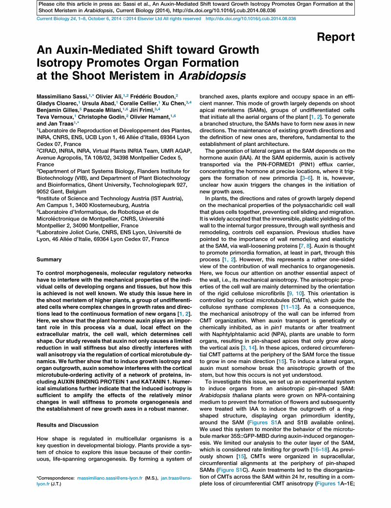
to induce organs from an anisotropic pin-shaped SAM:Arabidopsis thaliana plants were grown on NPA-containingmedium to prevent the formation of flowers and subsequentlywere treated with IAA to induce the outgrowth of a ring-shaped structure, displaying organ primordium identity,around the SAM (Figures S1A and S1B available online).We used this system to monitor the behavior of the microtu-bule marker 35S::GFP-MBD during auxin-induced organogen-esis. We limited our analysis to the outer layer of the SAM,which is considered rate limiting for growth [16–18]. As previ-ously shown [15], CMTs were organized in supracellular,circumferential alignments at the periphery of pin-shapedSAMs (Figure S1C). Auxin treatments led to the disorganiza-tion of CMTs across the SAM within 24 hr, resulting in a com-plete loss of circumferential CMT anisotropy (Figures 1A–1E;
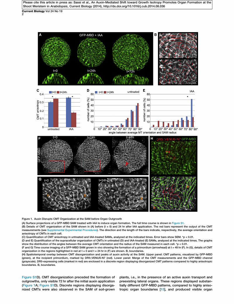
Figure 1. Auxin Disrupts CMT Organization at the SAM before Organ Outgrowth
(A) Surface projections of a GFP-MBD SAM treated with IAA to induce organ formation. The full time course is shown in Figure S1.
(B) Details of CMT organization of the SAM shown in (A) before (t = 0) and 24 hr after IAA application. The red bars represent the output of the CMT
measurements (see Supplemental Experimental Procedures). The direction and the length of the bars indicate, respectively, the average orientation and
anisotropy of CMTs in each cell.
(C) Quantification of CMT anisotropy in untreated and IAA-treated SAMs, analyzed at the indicated times. Error bars show SEM. *p < 0.01.
(D and E) Quantification of the supracellular organization of CMTs in untreated (D) and IAA-treated (E) SAMs, analyzed at the indicated times. The graphs
show the distribution of the angles between the average CMT orientation and the radius of the SAM measured in each cell. *p < 0.01.
(F and G) Time course imaging of a GFP-MBD SAM grown in vivo showing the formation of a primordium (arrowhead) at t = 48 hr (F). In (G), details of CMT
organization in the regions highlighted in red at t = 0 and t = 24 hr in (F) are shown. B, boundaries.
(H) Spatiotemporal overlap between CMT disorganization and peaks of auxin activity at the SAM. Upper panel: CMT patterns, visualized by GFP-MBD
(green), at the incipient primordium, marked by DR5::VENUS-N7 (red). Lower panel: Merge of the CMT measurements and the GFP-MBD channel
(grayscale). DR5-expressing cells (marked in red) are enclosed in a discrete region displaying disorganized CMT patterns compared to highly anisotropic
boundaries. B, boundaries.
Current Biology Vol 24 No 192
Please cite this article in press as: Sassi et al., An Auxin-Mediated Shift toward Growth Isotropy Promotes Organ Formation at theShoot Meristem in Arabidopsis, Current Biology (2014), http://dx.doi.org/10.1016/j.cub.2014.08.036
Figure S1D). CMT disorganization preceded the formation ofoutgrowths, only visible 72 hr after the initial auxin application(Figure 1A; Figure S1D). Discrete regions displaying disorga-nized CMTs were also observed in the SAM of soil-grown
plants, i.e., in the presence of an active auxin transport andpreexisting lateral organs. These regions displayed substan-tially different GFP-MBD patterns, compared to highly aniso-tropic organ boundaries [15], and produced visible organ
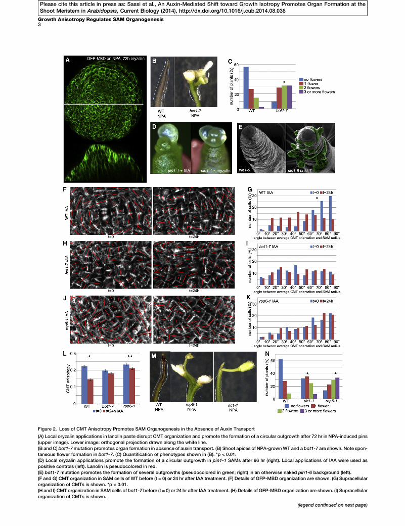
Figure 2. Loss of CMT Anisotropy Promotes SAM Organogenesis in the Absence of Auxin Transport
(A) Local oryzalin applications in lanolin paste disrupt CMT organization and promote the formation of a circular outgrowth after 72 hr in NPA-induced pins
(upper image). Lower image: orthogonal projection drawn along the white line.
(B and C) bot1-7mutation promotes organ formation in absence of auxin transport. (B) Shoot apices of NPA-grownWT and a bot1-7 are shown. Note spon-
taneous flower formation in bot1-7. (C) Quantification of phenotypes shown in (B). *p < 0.01.
(D) Local oryzalin applications promote the formation of a circular outgrowth in pin1-1 SAMs after 96 hr (right). Local applications of IAA were used as
positive controls (left). Lanolin is pseudocolored in red.
(E) bot1-7 mutation promotes the formation of several outgrowths (pseudocolored in green; right) in an otherwise naked pin1-6 background (left).
(F and G) CMT organization in SAM cells of WT before (t = 0) or 24 hr after IAA treatment. (F) Details of GFP-MBD organization are shown. (G) Supracellular
organization of CMTs is shown. *p < 0.01.
(H and I) CMT organization in SAM cells of bot1-7 before (t = 0) or 24 hr after IAA treatment. (H) Details of GFP-MBD organization are shown. (I) Supracellular
organization of CMTs is shown.
(legend continued on next page)
Growth Anisotropy Regulates SAM Organogenesis3
Please cite this article in press as: Sassi et al., An Auxin-Mediated Shift toward Growth Isotropy Promotes Organ Formation at theShoot Meristem in Arabidopsis, Current Biology (2014), http://dx.doi.org/10.1016/j.cub.2014.08.036
Current Biology Vol 24 No 194
Please cite this article in press as: Sassi et al., An Auxin-Mediated Shift toward Growth Isotropy Promotes Organ Formation at theShoot Meristem in Arabidopsis, Current Biology (2014), http://dx.doi.org/10.1016/j.cub.2014.08.036
primordia 24 hr later (Figures 1F and 1G). Disorganized GFP-MBD patterns at the SAM correlated in time and space withpeaks of high auxin activity inferred from the auxin-signalingreporter DR5::VENUS-N7, the earliest marker of incipientprimordia identified to date [5] (Figure 1H). These results sug-gest that, in vivo, changes in CMT anisotropy coincide withauxin maxima and precede organ outgrowth at the SAM.
We next investigated whether CMT disorganization couldbe instrumental in the formation of lateral outgrowths. To thisend, we used the microtubule-depolymerizing drug oryzalinto alter CMT dynamics at the SAM of NPA-induced pins. Localapplications of oryzalin in lanolin paste on naked meristemscaused CMT disorganization, leading to ring-like outgrowths(Figure 2A). We next altered CMT dynamics by using thebotero1-7 (bot1-7) allele of KATANIN1 (KTN1). KTN1 encodesa microtubule-severing protein that promotes bundling andordered CMT patterns [19–21]. KTN1 is active in the SAM,where it contributes to the formation of anisotropic CMT arrays[22]. bot1-7 mutants grown on NPA displayed spontaneousoutgrowths that were preceded by localized CMT disorganiza-tion (Figure S2A). Indeed, more than 90% of the bot1-7 popu-lation (n = 183) produced flowers (compared to w40% inthe wild-type [WT] population, n = 143) (Figures 2B and 2C)on NPA.
Moreover, we used the pin1 mutant to perturb auxin distri-bution, and we tested whether oryzalin and the bot1-7 muta-tion could rescue its defective organogenesis. We found that63.4% (n = 41) of pin1-1 SAMs treated locally with oryzalindeveloped outgrowths after 4 days (Figure 2D). Similar fre-quencies were obtained when pin1-1 SAMs were locallytreated with IAA (66.6%; n = 33) (Figure 2D). Likewise, themajority of pin1-6 bot1-7 double mutants (77.8%; n = 14) dis-played a high number of lateral outgrowths around the SAM,whereas high numbers are only rarely (10.0%; n = 10) observedin pin1-6 single mutants (Figure 2E). Together, these resultspoint to a causal link between loss of CMT anisotropy andorganogenesis.
In auxin transport-depleted backgrounds, the bot1-7 muta-tion replicates the effect of auxin treatments on CMT organiza-tion and organ initiation. Relevantly, auxin treatments were notable to further affect CMTs in bot1-7 SAMs (Figures 2F, 2G,and 2L; Figure S2B). This suggests that (1) at low auxin con-centrations, KTN1 leads to the formation of a pin, and (2)KTN1 is no longer able to maintain a pin in the presence ofhigh auxin levels. It was recently proposed that auxin regulatesKTN1 function through RHO-LIKE GTPASE FROM PLANTS 6(ROP6) and its effector ROP-INTERACTIVE CRIB MOTIF-CONTAINING PROTEIN 1 (RIC1), which binds to and activatesKTN1 [23–26]. Both ROP6 and RIC1 were expressed at theshoot apex, albeit at different levels (Figure S2C), and their cor-responding mutants displayed a significant increase in organformation on NPA (rop6-1: 86%, n = 146; ric1-1: 68%, n =210) compared to the WT (36%, n = 174) (Figures 2M and2N). CMT responses to auxin treatments were impaired inrop6-1 because no changes in CMT organization at the tissuelevel and only a minor decrease of cellular CMT anisotropywere observed (Figures 2J, 2K, and 2L; Figure S2B).
(J and K) CMT organization in SAM cells of rop6-1 before (t = 0) or 24 hr after IAA
organization of CMTs is shown.
(L) Quantification of CMT anisotropy in the SAM cells of WT, bot1-7, and rop6-
show SEM.
(M andN) rop6-1 and ric1-1promote organ formation in absence of auxin transp
(P) Quantification of phenotypes shown in (N). *p < 0.01.
The ROP6-dependent regulation of CMT organization hasbeen associated with the extracellular auxin receptor AUXINBINDING PROTEIN 1 (ABP1) signaling [25–27]. ABP1 is ex-pressed at the SAM (Figure S2C) [28]; therefore, we investi-gated whether it could be involved in the regulation of CMTorganization at the SAM. Because ABP1 knockout mutantsare embryo lethal [29, 30], we first employed the viableabp1-5 allele, containing a point mutation in the auxin-bindingpocket that reduces the affinity to auxin [25, 31]. Compared tothe WT, abp1-5 mutants displayed relatively subtle promotionof organ formation on NPA (Figures 3A and 3B). Indeed,abp1-5 SAMs displayed only minor alterations in the circum-ferential CMT organization (Figures 3E and 3F, t = 0; Fig-ure S3A, t = 0) and were still able to respond to auxintreatments, albeit to a lesser extent compared to the WT (Fig-ures 3C–3F and 3I; Figure S3A). Consequently, auxin-inducedorgan initiation was severely delayed in abp1-5 SAMs com-pared to the WT (Figures S3B and S3C). To gain insights intoABP1 function in the regulation of CMT organization andSAM organogenesis, we generated a transheterozygousmutant bearing one copy of the auxin-insensitive abp1-5 andone copy of the embryo-lethal null abp1-1s allele [30]. The re-sulting abp1-1s/abp1-5 was viable and displayed enhancedorgan formation and more-random CMT organization com-pared to the parental abp1-5 on NPA (Figures 3A and 3B; Fig-ure S3D). CMTs in abp1-1s/abp1-5 SAMs were completelyinsensitive to auxin treatments for both cell and tissue re-sponses (Figures 3G, 3H, and 3I; Figure S3A), similar tobot1-7 mutants. This suggests that ABP1 and KTN1 act in asimilar manner to regulate CMT organization and SAM organ-ogenesis. Coherently, bot1-7 abp1-5mutants displayed organinitiation phenotypes on NPA, similar to those of the parentalbot1-7 (Figure S3E), and no novel additive phenotypes wereobserved. This is in line with the hypothesis that ABP1 andKTN1 act in the same pathway, as previously reported in othersystems [25–27], although further experiments are needed toclarify this issue.Together, our results suggest that in the absence of auxin
accumulation, a network of interacting proteins, includingKTN1, ROP6/RIC1, and ABP1, keep microtubular arrays atthe SAM in an anisotropic state. This is sufficient to inhibitspontaneous lateral outgrowth, leading to the formation of apin-shaped stem. In the presence of high auxin levels, thesemolecules are no longer able to maintain CMT anisotropy,which induces a shift toward isotropic cell walls. As a result,organ outgrowth is promoted.As discussed above, previous studies have suggested a role
of cell wall remodeling and extensibility in SAM organogenesis[7, 8]. In addition, there are indications that inner layers of themeristem loosen their walls just before organ outgrowth, leav-ing the outer layer as the main growth-limiting factor [32, 33].This is consistent with the observation that the outer layerhas thicker cell walls than the inner layers [18]. Altogether,this implies that organogenesis results from the modulationof both cell wall stiffness and anisotropy. Why would twodifferent layers of regulation be required? To explore thisquestion, we used an in silico model in the form of a virtual
treatment. (J) Details of GFP-MBD organization are shown. (K) Supracellular
1 before (t = 0) and 24 hr after IAA treatment. *p < 0.01; **p < 0.05. Error bars
ort. (M) Shoot apices of NPA-grownWT, rop6-1, and ric1-1plants are shown.
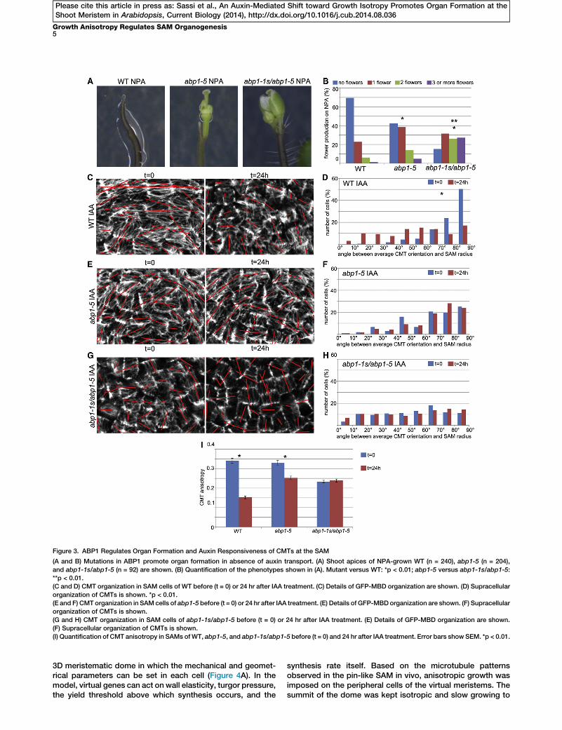
Figure 3. ABP1 Regulates Organ Formation and Auxin Responsiveness of CMTs at the SAM
(A and B) Mutations in ABP1 promote organ formation in absence of auxin transport. (A) Shoot apices of NPA-grown WT (n = 240), abp1-5 (n = 204),
and abp1-1s/abp1-5 (n = 92) are shown. (B) Quantification of the phenotypes shown in (A). Mutant versus WT: *p < 0.01; abp1-5 versus abp1-1s/abp1-5:
**p < 0.01.
(C and D) CMT organization in SAM cells of WT before (t = 0) or 24 hr after IAA treatment. (C) Details of GFP-MBD organization are shown. (D) Supracellular
organization of CMTs is shown. *p < 0.01.
(E and F) CMT organization in SAM cells of abp1-5 before (t = 0) or 24 hr after IAA treatment. (E) Details of GFP-MBDorganization are shown. (F) Supracellular
organization of CMTs is shown.
(G and H) CMT organization in SAM cells of abp1-1s/abp1-5 before (t = 0) or 24 hr after IAA treatment. (E) Details of GFP-MBD organization are shown.
(F) Supracellular organization of CMTs is shown.
(I) Quantification of CMT anisotropy in SAMs ofWT, abp1-5, and abp1-1s/abp1-5 before (t = 0) and 24 hr after IAA treatment. Error bars show SEM. *p < 0.01.
Growth Anisotropy Regulates SAM Organogenesis5
Please cite this article in press as: Sassi et al., An Auxin-Mediated Shift toward Growth Isotropy Promotes Organ Formation at theShoot Meristem in Arabidopsis, Current Biology (2014), http://dx.doi.org/10.1016/j.cub.2014.08.036
3D meristematic dome in which the mechanical and geomet-rical parameters can be set in each cell (Figure 4A). In themodel, virtual genes can act on wall elasticity, turgor pressure,the yield threshold above which synthesis occurs, and the
synthesis rate itself. Based on the microtubule patternsobserved in the pin-like SAM in vivo, anisotropic growth wasimposed on the peripheral cells of the virtual meristems. Thesummit of the dome was kept isotropic and slow growing to
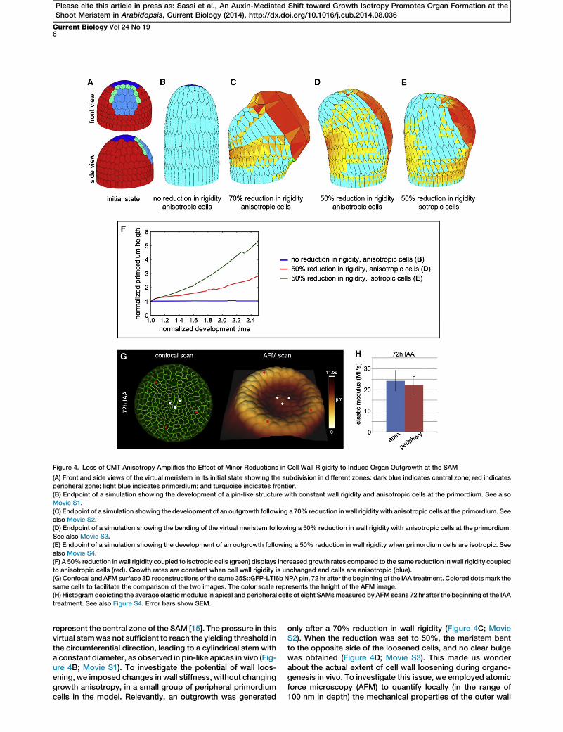
Figure 4. Loss of CMT Anisotropy Amplifies the Effect of Minor Reductions in Cell Wall Rigidity to Induce Organ Outgrowth at the SAM
(A) Front and side views of the virtual meristem in its initial state showing the subdivision in different zones: dark blue indicates central zone; red indicates
peripheral zone; light blue indicates primordium; and turquoise indicates frontier.
(B) Endpoint of a simulation showing the development of a pin-like structure with constant wall rigidity and anisotropic cells at the primordium. See also
Movie S1.
(C) Endpoint of a simulation showing the development of an outgrowth following a 70% reduction in wall rigidity with anisotropic cells at the primordium. See
also Movie S2.
(D) Endpoint of a simulation showing the bending of the virtual meristem following a 50% reduction in wall rigidity with anisotropic cells at the primordium.
See also Movie S3.
(E) Endpoint of a simulation showing the development of an outgrowth following a 50% reduction in wall rigidity when primordium cells are isotropic. See
also Movie S4.
(F) A 50% reduction in wall rigidity coupled to isotropic cells (green) displays increased growth rates compared to the same reduction in wall rigidity coupled
to anisotropic cells (red). Growth rates are constant when cell wall rigidity is unchanged and cells are anisotropic (blue).
(G) Confocal and AFMsurface 3D reconstructions of the same 35S::GFP-LTI6bNPApin, 72 hr after the beginning of the IAA treatment. Colored dotsmark the
same cells to facilitate the comparison of the two images. The color scale represents the height of the AFM image.
(H) Histogram depicting the average elastic modulus in apical and peripheral cells of eight SAMsmeasured by AFM scans 72 hr after the beginning of the IAA
treatment. See also Figure S4. Error bars show SEM.
Current Biology Vol 24 No 196
Please cite this article in press as: Sassi et al., An Auxin-Mediated Shift toward Growth Isotropy Promotes Organ Formation at theShoot Meristem in Arabidopsis, Current Biology (2014), http://dx.doi.org/10.1016/j.cub.2014.08.036
represent the central zone of the SAM [15]. The pressure in thisvirtual stemwas not sufficient to reach the yielding threshold inthe circumferential direction, leading to a cylindrical stem witha constant diameter, as observed in pin-like apices in vivo (Fig-ure 4B; Movie S1). To investigate the potential of wall loos-ening, we imposed changes in wall stiffness, without changinggrowth anisotropy, in a small group of peripheral primordiumcells in the model. Relevantly, an outgrowth was generated
only after a 70% reduction in wall rigidity (Figure 4C; MovieS2). When the reduction was set to 50%, the meristem bentto the opposite side of the loosened cells, and no clear bulgewas obtained (Figure 4D; Movie S3). This made us wonderabout the actual extent of cell wall loosening during organo-genesis in vivo. To investigate this issue, we employed atomicforce microscopy (AFM) to quantify locally (in the range of100 nm in depth) the mechanical properties of the outer wall
Growth Anisotropy Regulates SAM Organogenesis7
Please cite this article in press as: Sassi et al., An Auxin-Mediated Shift toward Growth Isotropy Promotes Organ Formation at theShoot Meristem in Arabidopsis, Current Biology (2014), http://dx.doi.org/10.1016/j.cub.2014.08.036
[34]. Using this protocol on auxin-treated SAMs, we observedvariable changes in the elastic modulus, with a tendency of theoutgrowth to become slightly softer than the apex (Figures 4Gand 4H). Such changes did not exceed 30% (Figure S4), indi-cating that major reductions in the outer wall rigidity do notoccur during organ initiation.
We therefore performed a series of additional simulations,this time combining a limited reduction in wall stiffness witha shift to isotropy in the primordium. This led to the formationof an outgrowth at the flank of the virtual meristem (Figure 4E;Movie S4). Relevantly, the combination of limited looseningand isotropy led to increased primordium growth ratescompared to the simulations using limited loosening alone(Figure 4F). Our theoretical analysis therefore suggests thatthe anisotropy-to-isotropy shift could promote organ forma-tion by amplifying the effect of relatively minor reductions inthe outer wall rigidity at the SAM.
In conclusion, we show that at the SAM periphery, auxin in-terferes with CMT anisotropy, which normally forces the apicaltissues to develop into a single cylindrical axis. By locallycounteracting the polarizing effect of these circumferentialCMT arrays, auxin generates new axes for lateral organoutgrowth without perturbing the vertical growth of the stem,allowing the plant to explore efficiently new directions inspace.
Supplemental Information
Supplemental Information includes Supplemental Experimental Proce-
dures, four figures, and four movies and can be found with this article online
at http://dx.doi.org/10.1016/j.cub.2014.08.036.
Author Contributions
M.S. and J.T. designed the study.M.S., C.C., and U.A. performed the biolog-
ical experiments. G.C. and O.A. carried out the AFM analysis. O.A., F.B.,
B.G., and C.G. carried out numerical simulations. X.C., J.F., T.V., and O.H.
provided materials/reagents. M.S. and J.T. analyzed the data and wrote
the manuscript, with contributions from all the authors.
Acknowledgments
This work was funded by grants from EraSysBio+ (iSAM) and ERC (Morpho-
dynamics). We thank Dolf Weijers (Wageningen University) and Zhenbiao
Yang (Riverside University) for generously providing seeds. We thank all
the member of the J.T. laboratory for helpful discussions and Arezki Bou-
daoud and Roberta Galletti for helpful discussions and critical reading of
the manuscript.
Received: January 6, 2014
Revised: July 3, 2014
Accepted: August 15, 2014
Published: September 25, 2014
References
1. Murray, J.A.H., Jones, A., Godin, C., and Traas, J. (2012). Systems anal-
ysis of shoot apical meristem growth and development: integrating hor-
monal and mechanical signaling. Plant Cell 24, 3907–3919.
2. Sassi, M., and Vernoux, T. (2013). Auxin and self-organization at the
shoot apical meristem. J. Exp. Bot. 64, 2579–2592.
3. Reinhardt, D., Mandel, T., and Kuhlemeier, C. (2000). Auxin regulates
the initiation and radial position of plant lateral organs. Plant Cell 12,
507–518.
4. Reinhardt, D., Pesce, E.-R., Stieger, P., Mandel, T., Baltensperger, K.,
Bennett, M., Traas, J., Friml, J., and Kuhlemeier, C. (2003). Regulation
of phyllotaxis by polar auxin transport. Nature 426, 255–260.
5. Heisler, M.G., Ohno, C., Das, P., Sieber, P., Reddy, G.V., Long, J.A., and
Meyerowitz, E.M. (2005). Patterns of auxin transport and gene
expression during primordium development revealed by live imaging
of the Arabidopsis inflorescence meristem. Curr. Biol. 15, 1899–1911.
6. Benkova, E., Michniewicz, M., Sauer, M., Teichmann, T., Seifertova, D.,
Jurgens, G., and Friml, J. (2003). Local, efflux-dependent auxin gradi-
ents as a common module for plant organ formation. Cell 115, 591–602.
7. Fleming, A.J., McQueen-Mason, S., Mandel, T., and Kuhlemeier, C.
(1997). Induction of leaf primordia by the cell wall protein expansin.
Science 276, 1415–1418.
8. Peaucelle, A., Louvet, R., Johansen, J.N., Hofte, H., Laufs, P., Pelloux,
J., and Mouille, G. (2008). Arabidopsis phyllotaxis is controlled by the
methyl-esterification status of cell-wall pectins. Curr. Biol. 18, 1943–
1948.
9. Somerville, C., Bauer, S., Brininstool, G., Facette, M., Hamann, T., Milne,
J., Osborne, E., Paredez, A., Persson, S., Raab, T., et al. (2004). Toward a
systems approach to understanding plant cell walls. Science 306, 2206–
2211.
10. Cosgrove, D.J. (2005). Growth of the plant cell wall. Nat. Rev. Mol. Cell
Biol. 6, 850–861.
11. Paredez, A.R., Somerville, C.R., and Ehrhardt, D.W. (2006). Visualization
of cellulose synthase demonstrates functional associationwithmicrotu-
bules. Science 312, 1491–1495.
12. Gutierrez, R., Lindeboom, J.J., Paredez, A.R., Emons, A.M.C., and
Ehrhardt, D.W. (2009). Arabidopsis cortical microtubules position cellu-
lose synthase delivery to the plasma membrane and interact with cellu-
lose synthase trafficking compartments. Nat. Cell Biol. 11, 797–806.
13. Bringmann, M., Li, E., Sampathkumar, A., Kocabek, T., Hauser, M.-T.,
and Persson, S. (2012). POM-POM2/cellulose synthase interacting1 is
essential for the functional association of cellulose synthase and micro-
tubules in Arabidopsis. Plant Cell 24, 163–177.
14. Okada, K., Ueda, J., Komaki, M.K., Bell, C.J., and Shimura, Y. (1991).
Requirement of the auxin polar transport system in early stages of
Arabidopsis floral bud formation. Plant Cell 3, 677–684.
15. Hamant, O., Heisler, M.G., Jonsson, H., Krupinski, P., Uyttewaal, M.,
Bokov, P., Corson, F., Sahlin, P., Boudaoud, A., Meyerowitz, E.M.,
et al. (2008). Developmental patterning by mechanical signals in
Arabidopsis. Science 322, 1650–1655.
16. Savaldi-Goldstein, S., Peto, C., and Chory, J. (2007). The epidermis both
drives and restricts plant shoot growth. Nature 446, 199–202.
17. Reinhardt, D., Frenz, M., Mandel, T., and Kuhlemeier, C. (2003).
Microsurgical and laser ablation analysis of interactions between the
zones and layers of the tomato shoot apical meristem. Development
130, 4073–4083.
18. Kierzkowski, D., Nakayama, N., Routier-Kierzkowska, A.-L., Weber, A.,
Bayer, E., Schorderet, M., Reinhardt, D., Kuhlemeier, C., and Smith,
R.S. (2012). Elastic domains regulate growth and organogenesis in the
plant shoot apical meristem. Science 335, 1096–1099.
19. Bichet, A., Desnos, T., Turner, S., Grandjean, O., and Hofte, H. (2001).
BOTERO1 is required for normal orientation of cortical microtubules
and anisotropic cell expansion in Arabidopsis. Plant J. 25, 137–148.
20. Burk, D.H., and Ye, Z.H. (2002). Alteration of oriented deposition of cel-
lulose microfibrils by mutation of a katanin-like microtubule-severing
protein. Plant Cell 14, 2145–2160.
21. Stoppin-Mellet, V., Gaillard, J., and Vantard, M. (2006). Katanin’s
severing activity favors bundling of cortical microtubules in plants.
Plant J. 46, 1009–1017.
22. Uyttewaal, M., Burian, A., Alim, K., Landrein, B., Borowska-Wykret, D.,
Dedieu, A., Peaucelle, A., Ludynia, M., Traas, J., Boudaoud, A., et al.
(2012). Mechanical stress acts via katanin to amplify differences in
growth rate between adjacent cells in Arabidopsis. Cell 149, 439–451.
23. Fu, Y., Gu, Y., Zheng, Z., Wasteneys, G., and Yang, Z. (2005).
Arabidopsis interdigitating cell growth requires two antagonistic
pathways with opposing action on cell morphogenesis. Cell 120,
687–700.
24. Fu, Y., Xu, T., Zhu, L., Wen, M., and Yang, Z. (2009). A ROP GTPase
signaling pathway controls cortical microtubule ordering and cell
expansion in Arabidopsis. Curr. Biol. 19, 1827–1832.
25. Xu, T., Wen, M., Nagawa, S., Fu, Y., Chen, J.-G., Wu, M.-J., Perrot-
Rechenmann, C., Friml, J., Jones, A.M., and Yang, Z. (2010). Cell
surface- and rho GTPase-based auxin signaling controls cellular inter-
digitation in Arabidopsis. Cell 143, 99–110.
26. Lin, D., Cao, L., Zhou, Z., Zhu, L., Ehrhardt, D., Yang, Z., and Fu, Y.
(2013). Rho GTPase signaling activates microtubule severing to pro-
mote microtubule ordering in Arabidopsis. Curr. Biol. 23, 290–297.
Current Biology Vol 24 No 198
Please cite this article in press as: Sassi et al., An Auxin-Mediated Shift toward Growth Isotropy Promotes Organ Formation at theShoot Meristem in Arabidopsis, Current Biology (2014), http://dx.doi.org/10.1016/j.cub.2014.08.036
27. Xu, T., Dai, N., Chen, J., Nagawa, S., Cao, M., Li, H., Zhou, Z., Chen, X.,
De Rycke, R., Rakusova, H., et al. (2014). Cell surface ABP1-TMK auxin-
sensing complex activates ROP GTPase signaling. Science 343, 1025–
1028.
28. Braun, N., Wyrzykowska, J., Muller, P., David, K., Couch, D., Perrot-
Rechenmann, C., and Fleming, A.J. (2008). Conditional repression of
AUXIN BINDING PROTEIN1 reveals that it coordinates cell division
and cell expansion during postembryonic shoot development in
Arabidopsis and tobacco. Plant Cell 20, 2746–2762.
29. Chen, J.G., Ullah, H., Young, J.C., Sussman, M.R., and Jones, A.M.
(2001). ABP1 is required for organized cell elongation and division in
Arabidopsis embryogenesis. Genes Dev. 15, 902–911.
30. Tzafrir, I., Pena-Muralla, R., Dickerman, A., Berg, M., Rogers, R.,
Hutchens, S., Sweeney, T.C., McElver, J., Aux, G., Patton, D., and
Meinke, D. (2004). Identification of genes required for embryo develop-
ment in Arabidopsis. Plant Physiol. 135, 1206–1220.
31. Robert, S., Kleine-Vehn, J., Barbez, E., Sauer, M., Paciorek, T., Baster,
P., Vanneste, S., Zhang, J., Simon, S., �Covanova, M., et al. (2010).
ABP1 mediates auxin inhibition of clathrin-dependent endocytosis in
Arabidopsis. Cell 143, 111–121.
32. Peaucelle, A., Braybrook, S.A., Le Guillou, L., Bron, E., Kuhlemeier, C.,
and Hofte, H. (2011). Pectin-induced changes in cell wall mechanics
underlie organ initiation in Arabidopsis. Curr. Biol. 21, 1720–1726.
33. Braybrook, S.A., and Peaucelle, A. (2013). Mechano-chemical aspects
of organ formation in Arabidopsis thaliana: the relationship between
auxin and pectin. PLoS ONE 8, e57813.
34. Milani, P., Gholamirad, M., Traas, J., Arneodo, A., Boudaoud, A., Argoul,
F., and Hamant, O. (2011). In vivo analysis of local wall stiffness at the
shoot apical meristem in Arabidopsis using atomic force microscopy.
Plant J. 67, 1116–1123.