Embed Size (px)
Citation preview
An autopsy study of a fouled reverse osmosis membrane element used in a brackish water treatment plant
This is the Published version of the following publication
Gray, Stephen R, Tran, Thuy, Bolto, Brian, Hoang, Manh and Ostarcevic, Eddy(2007) An autopsy study of a fouled reverse osmosis membrane element usedin a brackish water treatment plant. Water Research, 41 (17). pp. 3915-3923. ISSN 00431354
The publisher’s official version can be found at
Note that access to this version may require subscription.
Downloaded from VU Research Repository https://vuir.vu.edu.au/2035/
AN AUTOPSY STUDY OF A FOULED REVERSE OSMOSIS MEMBRANE
ELEMENT USED IN A BRACKISH WATER TREATMENT PLANT
1
2
3
4
5
6
7
8
9
10
11
12
13
14
15
16
17
18
19
20
21
22
23
24
Thuy Tran*, Brian Bolto*, Stephen Gray1*, Manh Hoang and Eddy Ostarcevic2
CSIRO Manufacturing & Materials Technology, Private Bag 33, Clayton South,Vic. 3169
1 Victoria University, Werribee Campus, PO Box 14428, Melbourne, Vic. 8001
2 GWMWater, PO Box 481, Horsham, Vic. 3402
* Authors to whom correspondence should be addressed. Tel: +61-3- 9545 2046; Fax: +61-3-
9544 1128; E-mail: [email protected]; [email protected]; [email protected]
Abstract
The fouling of a spiral wound RO membrane after nearly one year of service in a brackish
water treatment plant was investigated using optical and electron microscopic methods, FTIR
and ICP-AES. Both the top surface and the cross-section of the fouled membrane were
analysed to monitor the development of the fouling layer. It has been found that the extent of
fouling was uneven across the membrane surface with regions underneath or in the vicinity of
the strands of the feed spacer being more severely affected. The fouling appeared to have
developed through different stages. In particular, it consisted of an initial thin fouling layer of
an amorphous matrix with embedded particulate matter. The amorphous matrix comprised
organic–Al–P complexes and the particulate matter was mostly aluminium silicates.
Subsequently, as the fouling layer reached a thickness of about 5 to 7 μm, further amorphous
material, which is suggested to include extracellular polymeric substances such as
polysaccharides, started to deposit on top of the existing fouling layer. This secondary
amorphous material did not seem to contain any particulate matter nor any inorganic elements
1
25
26
27
28
29
within it, but acted as a substrate upon which aluminium silicate crystals grew exclusively in
the absence of other foulants, including natural organic matter (NOM).
Key words: Reverse osmosis, fouling, water treatment, desalination, fouling mechanisms
2
1. Introduction 30
31
32
33
34
35
36
37
38
39
40
41
42
43
44
45
46
47
48
49
50
51
52
53
Reverse osmosis (RO) is a commonly used process in desalination and advanced wastewater
treatment. However, like other membrane filtration processes, fouling is a major obstacle in
the efficient operation of RO systems. Membrane fouling causes deterioration of both the
quantity and quality of treated water, and consequently results in higher treatment costs.
Foulants may be classed into one of four major categories: sparingly soluble inorganic
compounds, colloidal or particulate matter, dissolved organic substances, and microorganisms
(Speth et al., 2000). Fouling by sparingly soluble inorganic compounds is governed by
concentration polarization and scale layer formation when the product of the concentration of
the soluble components exceeds the solubility limit (Boerlage et al., 1999). Particulate and
colloidal matter rejected by the membrane may form compact cakes, which introduce an
additional resistance barrier to filtration (Gabelich et al., 2002). Organic fouling is governed in
part by interactions between the membrane surface and the organic foulants, as well as
between the organic foulants themselves (Dalvi et al., 2000). Microbial attachment and growth
on the membrane surface leads to the formation of biofilms, which consist of microbial cells
embedded in an extracellular polymeric substances matrix produced by the microbes
(Ivnitskya et al., 2005). Despite various research efforts, to date the characterization of sea
water fouling of RO membranes has not progressed significantly, compared to low-pressure
membrane fouling by surface and ground waters (Kumar et al., 2006).
Although membrane fouling is traditionally measured by flux decline with time, this method is
inadequate for characterizing fouling development in a RO process. It has been shown that
when the permeate flux is noticeably affected, the membrane is so severely fouled that
3
54
55
56
57
58
59
60
61
62
63
64
65
66
67
68
69
70
71
72
73
74
75
76
77
78
restoration to its original permeability may become impossible (Tay and Song, 2006).
Autopsies of fouled membranes have also been carried out in order to better understand the
physico-chemical processes governing the fouling (see, for example, Butt et al., 1997; Speth et
al., 1998; Sahachaiyunta et al., 2002; Vrouwenvelder and van der Kooij, 2002; Gwona et al.,
2003). The methods of chemical and structural analyses used in these studies including
inductively coupled plasma mass spectrometry (ICP-MS), gas chromatography / mass
spectrometry (GC-MS), Fourier transform infra-red spectroscopy (FTIR) and X-ray diffraction
(XRD) provide only the average composition of the surface deposits. Because these deposits
are complex and heterogeneous, information on average composition is of limited value in
elucidating the fouling mechanisms. Direct observations using optical and electron
microscopic methods, including scanning electron microscope (SEM) and associated energy-
dispersive X-ray spectroscopy (EDS), often focus on the top surface deposits, but not on the
underlying deposit layers. This leads to an incomplete understanding of the deposition kinetics
of various foulants, and therefore of the fouling mechanisms, particularly where thicker
deposits have been developed.
Another issue is that whilst the distinction between inorganic, colloidal, organic and biological
fouling is useful, RO membranes in a typical operation are likely to be exposed to all
categories of foulants. Because of the complex nature of fouling, many mechanistic studies on
RO membrane fouling have focused on one foulant type for the purpose of simplicity.
However, it is very important to understand the effects of interactions between various foulant
types on the fouling mechanisms. For instance, it has recently been reported that the enhanced
concentration polarization of salt ions within the colloidal cake layer may result in an increase
in osmotic pressure and rapid flux decline during cake layer development (Hoek and
Elimelech, 2003; Lee et al., 2005). As well, the interactions between colloidal and organic
4
79
80
81
82
83
84
85
86
87
88
89
90
91
92
93
94
95
96
97
98
99
100
101
102
foulants has been found to give rise to considerable synergistic effects, as manifested by a
significantly higher flux decline compared to the additive effects of colloidal fouling and
organic fouling alone (Li and Elimelech, 2006).
This paper presents the autopsy results of a spiral wound RO membrane after nearly one year
of service in a water treatment facility. Analytical techniques used in the investigation of the
surface deposits include inductively coupled plasma atomic emission spectrometer (ICP-AES),
FTIR, optical and electron microscopic methods. Both the top surface and the cross-section of
the fouled membrane were analyzed to provide further insights into the development of the
fouling layer.
2. Materials and Methods
The fouled spiral wound RO membrane element
The fouled RO membrane element (FILMTECH, BW30LE-440DRY) selected for the autopsy
study had been in service for nearly one year in a water treatment facility operated by
GWMWater in Hopetoun, western Victoria, Australia. The RO desalination plant was
integrated into the water treatment facility in response to the increased salinity of surface
water in the region due to the extended drought in recent years. The plant was capable of
producing 250KL/d of permeate and included a concentrate recycle stream to improve
recovery to 80%. A phosphonate-based antiscalant was used in the RO operation and pre-
chlorination was not carried out in the treatment process because of the high levels of
disinfection by-product precursors.
5
103
104
105
106
107
108
109
110
111
112
113
114
115
116
117
118
119
120
121
122
123
124
125
126
127
Prior to the RO treatment, the raw water from catchments in the Grampians Ranges and stored
in open reservoirs had undergone a pre-treatment process including coagulation (aluminium
sulphate), flocculation, dissolved air flotation and filtration (DAFF), pH correction and
cartridge filtration using 5 and 1 µm pore size filters. The filtered water had pH of 9.1, total
dissolved solids of 900 mg/L, total organic carbon (TOC) of 12 mg/L and turbidity of 0.5
NTU. Chemical analysis of the filtered water was carried out and the results are presented in
Table 1.
The extended drought created conditions that promoted algal growth in the storage reservoirs
and this change had a detrimental effect on the performance of the DAFF process as well as
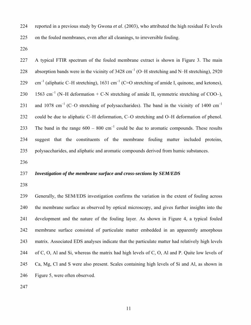
the desalination plant, resulting in a significant decline in production. The algal outbreak
required clean-in-place (CIP) events to be scheduled every month, but the flux decline was
significant and the original aim of operating at 80% recovery was not possible. Even after the
DAFF process was optimised to remove the algal cells and better water was secured from
another reservoir, the RO desalination plant could only operate at 75% recovery at best.
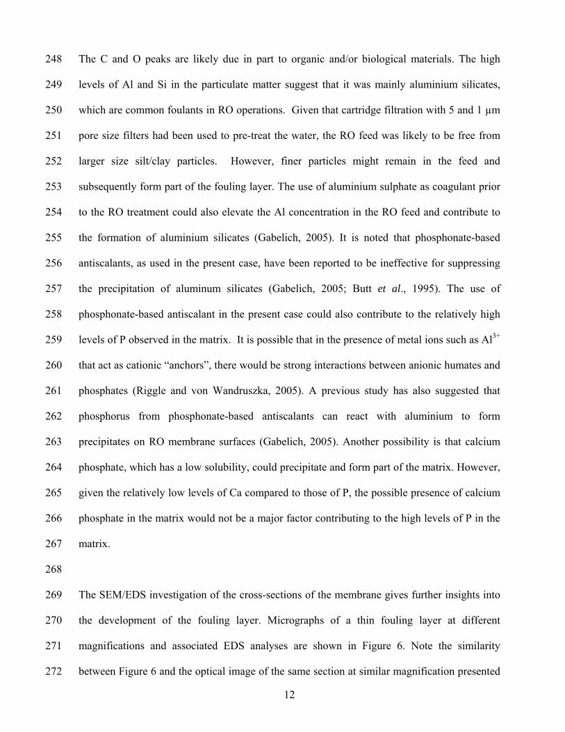
Following the algal outbreak, a fouled RO membrane element was selected for the autopsy
study. Surface deposits were scraped from the fouled membrane surface and analysed by ICP-
AES and FTIR. The middle section between the feed end and the concentrate end of the fouled
membrane was also cut into various coupons and prepared for optical and electron
microscopic studies.
ICP-AES analysis
6
128
129
130
131
132
133
134
135
136
137
138
139
140
141
142
143
144
145
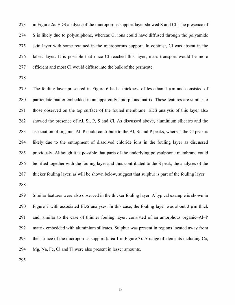
146
147
148
149
150
151
The surface deposits were digested in duplicate with 1:1 HNO3 on a hotplate prior to analysis
by using a Varian Vista ICP-AES. A general scan including Al, As, Au, Ba, Be, Bi, Ca, Cd,
Ce, Co, Cr, Cu, Fe, Ga, Ge, Hf, Hg, In, K, La, Mg, Mn, Mo, Na, Nb, Ni, P, Pb, Pd, Pt, S, Sb,
Se, Si, Sn, Sr, Ta, Th, Ti, U, V, W, Y, Zn and Zr elements were carried out. Only the elements
detected in trace levels and above are reported in the results. Chloride concentrations were
determined by analysing the sample in duplicate by potentiometric titration with silver nitrate.
FTIR analysis
Approximately 1.5 mg of dried sample was ground and mixed with approximately 50 mg of
anhydrous KBr and subsequently pressed into a disc. FTIR spectra (500–4000 cm−1) of the
discs were obtained using a Perkin Elmer 2000 FT-IR spectrophotometer in transmission
mode with KBr as the background reference.
Optical and electron microscopic analyses
An Olympus BHSM Metallographic Optical Microscope was used for general observation of
the fouled membrane sections. The microstructures of the surface deposits were analyzed
using a Philips XL30 field emission SEM operating at 5-15kV in conjunction with EDS to
obtain chemical information. EDS spot analysis using a spot diameter of about 3 nm at
selected areas on the samples was carried out. Since the X-ray sampling volume is close to the
electron-sample interaction volume, the spot analysis data typically included X-ray signals
generated from a sampling volume of about 1 μm3 (Goodhew and Humphreys, 1992).
7
Both the top surface and the cross-section of the fouled membrane coupons were analysed.
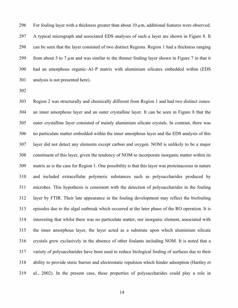
For the top surface analyses, the membrane coupons were mounted on a holder using double-
sided carbon tape. For the cross-section analyses, the coupons were embedded in a polymeric
resin in such a way that their cross-section was oriented perpendicular to the incoming light /
electron beam. The samples were then polished with various grades of diamond paste using
oil-based lubricant before analyses.
152
153
154
155
156
157
158
159
160
161
162
163
164
165
166
167
168
169
170
171
172
173
174
175
3. Results and Discussion
General observations by optical microscopy
Generally, the deposits were distributed unevenly across the membrane surface. Optical
images of the membrane surface before and after the feed spacer was removed are shown in
Figures 1a and b, respectively. It can be seen that regions underneath or in the vicinity of the
spacer strands were covered by brown stains, whereas the extent of staining in regions located
further away was generally less severe and varied considerably. Examination of regions near
the strands at higher magnifications also revealed the occasional presence of microorganisms,
as shown in the inset of Figure 1b.
The uneven fouling is also evident from the investigation of the cross-sections which showed a
considerable variation in the thickness of the surface deposits. In particular, many deposits in
regions underneath or close to the spacer strands had a thickness of about 90 μm or more, as
illustrated in Figure 2(a), whereas those located further away were thinner and had a thickness
ranging from less than 1 to about 25 μm, as shown in Figures 2(b) and (c).
8
176
177
178
179
180
181
182
183
184
185
186
187
188
189
190
191
192
193
194
195
196
197
198
199
A major objective of using the feed spacer is to promote eddy mixing which increases mass
transfer and reduces concentration polarization (Belfort and Guter, 1972). Whilst turbulence is
created between the spacer strands, it is also known that the spacer may promote excessive
particle precipitation in regions close to the strands (Gimmelshtein and Semiat, 2005). This
undesirable effect is evident in the present study from the observation of the thick deposits in
these regions. The presence of the thick deposits could lead to detrimental consequences. In
particular, they could act as an effective barrier to prevent water in the local environment from
penetrating through the underlying membrane, and therefore could greatly diminish the local
water flux. They could also have adverse effects on the feed flow properties, for instance, by
distorting the flow path and lowering the cross-flow velocity in the feed channel, which could
in turn contribute to the uneven and enhanced fouling across the membrane surface.
The difficulty in characterizing fouling is often attributed to the complexity of feed water
composition and to the different fouling mechanisms of different foulant types. Feed water is
usually characterized using common water analysis parameters such as the concentration of
each foulant present in the water. The flow properties and rate of fouling are often assumed to
be uniform throughout the membrane surface. The observations in the present study highlight
the importance of local variations in the hydrodynamic conditions in that they may lead to
considerable uneven fouling, and therefore should be featured more prominently in the
characterization of RO membrane fouling.
Analyses of surface deposits by ICP-AES and FTIR
9
200
201
202
203
204
205
206
207
208
209
210
211
212
213
214
215
216
217
218
219
220
221
222
223
The results from ICP-AES analysis are shown in Table 2. The major elements detected
included Al (2570 ppm), Ca (2760 ppm) and P (1225 ppm). Lesser amounts of Fe (590 ppm),
S (865 ppm), Si (410 ppm), Mg (320 ppm), K (110 ppm) and Na (190 ppm) were also present.
A relatively high level of Cl was detected (1430 ppm). Low levels of Ba, Cr, Cu, Ni, Sr, Ti, Zn
and Zr were also identified in the deposits.
Generally, the presence of negative ions, including bicarbonate, silicate and sulphate, in the
RO feed is important for the precipitation of various compounds. Common deposits found on
fouled RO membranes include aluminium silicates, carbonate compounds of Ca and Mg, and
sulphate compounds of Ca, Sr and Ba (see, for example, Yiantsios et al., 2005; Butt et al.,
1997). Metal ions, most notably Ca2+, may also form complexes with natural organic matter
(NOM), giving rise to subsequent formation of intermolecular bridges amongst organic foulant
molecules and enhanced membrane fouling (Li and Elimelech, 2004). As well, where a
fouling layer has developed on the membrane surface, the layer may entrap and hinder back-
diffusion of dissolved salt ions, resulting in an increase in concentrations of the salt ions near
the membrane surface (Herzberg and Elimelech, 2007; Hoek and Elimelech, 2003).
These deposition mechanisms could operate during the development of the fouling layer in the
current case, resulting in various types of deposits detected on the fouled membrane surface. It
is noted that the use of aluminium sulphate coagulant and phosphonate-based antiscalant prior
to the RO treatment could also raise the levels of Al, S and P in the feed and contribute to the
relatively high levels of these elements in the deposits. This issue will be discussed further in a
later section. As well, while only trace amount of Fe was detected in the RO feed, a relatively
high level of Fe was present on the fouled membrane deposits. A similar finding was also
10
reported in a previous study by Gwona et al. (2003), who attributed the high residual Fe levels
on the fouled membranes, even after all cleanings, to irreversible fouling.
224
225
226
227
228
229
230
231
232
233
234
235
236
237
238
239
240
241
242
243
244
245
246
247
A typical FTIR spectrum of the fouled membrane extract is shown in Figure 3. The main
absorption bands were in the vicinity of 3428 cm–1 (O–H stretching and N–H stretching), 2920
cm–1 (aliphatic C–H stretching), 1631 cm–1 (C=O stretching of amide I, quinone, and ketones),
1563 cm–1 (N–H deformation + C-N stretching of amide II, symmetric stretching of COO–),
and 1078 cm–1 (C–O stretching of polysaccharides). The band in the vicinity of 1400 cm–1
could be due to aliphatic C–H deformation, C–O stretching and O–H deformation of phenol.
The band in the range 600 – 800 cm–1 could be due to aromatic compounds. These results
suggest that the constituents of the membrane fouling matter included proteins,
polysaccharides, and aliphatic and aromatic compounds derived from humic substances.
Investigation of the membrane surface and cross-sections by SEM/EDS
Generally, the SEM/EDS investigation confirms the variation in the extent of fouling across
the membrane surface as observed by optical microscopy, and gives further insights into the
development and the nature of the fouling layer. As shown in Figure 4, a typical fouled
membrane surface consisted of particulate matter embedded in an apparently amorphous
matrix. Associated EDS analyses indicate that the particulate matter had relatively high levels
of C, O, Al and Si, whereas the matrix had high levels of C, O, Al and P. Quite low levels of
Ca, Mg, Cl and S were also present. Scales containing high levels of Si and Al, as shown in
Figure 5, were often observed.
11
248
249
250
251
252
253
254
255
256
257
258
259
260
261
262
263
264
265
266
267
268
269
270
271
272
The C and O peaks are likely due in part to organic and/or biological materials. The high
levels of Al and Si in the particulate matter suggest that it was mainly aluminium silicates,
which are common foulants in RO operations. Given that cartridge filtration with 5 and 1 µm
pore size filters had been used to pre-treat the water, the RO feed was likely to be free from
larger size silt/clay particles. However, finer particles might remain in the feed and
subsequently form part of the fouling layer. The use of aluminium sulphate as coagulant prior
to the RO treatment could also elevate the Al concentration in the RO feed and contribute to
the formation of aluminium silicates (Gabelich, 2005). It is noted that phosphonate-based
antiscalants, as used in the present case, have been reported to be ineffective for suppressing
the precipitation of aluminum silicates (Gabelich, 2005; Butt et al., 1995). The use of
phosphonate-based antiscalant in the present case could also contribute to the relatively high
levels of P observed in the matrix. It is possible that in the presence of metal ions such as Al3+
that act as cationic “anchors”, there would be strong interactions between anionic humates and
phosphates (Riggle and von Wandruszka, 2005). A previous study has also suggested that
phosphorus from phosphonate-based antiscalants can react with aluminium to form
precipitates on RO membrane surfaces (Gabelich, 2005). Another possibility is that calcium
phosphate, which has a low solubility, could precipitate and form part of the matrix. However,
given the relatively low levels of Ca compared to those of P, the possible presence of calcium
phosphate in the matrix would not be a major factor contributing to the high levels of P in the
matrix.
The SEM/EDS investigation of the cross-sections of the membrane gives further insights into
the development of the fouling layer. Micrographs of a thin fouling layer at different
magnifications and associated EDS analyses are shown in Figure 6. Note the similarity
between Figure 6 and the optical image of the same section at similar magnification presented
12
273
274
275
276
277
278
279
280
281
282
283
284
285
286
287
288
289
290
291
292
293
294
295
in Figure 2c. EDS analysis of the microporous support layer showed S and Cl. The presence of
S is likely due to polysulphone, whereas Cl ions could have diffused through the polyamide
skin layer with some retained in the microporous support. In contrast, Cl was absent in the
fabric layer. It is possible that once Cl reached this layer, mass transport would be more
efficient and most Cl would diffuse into the bulk of the permeate.
The fouling layer presented in Figure 6 had a thickness of less than 1 μm and consisted of
particulate matter embedded in an apparently amorphous matrix. These features are similar to
those observed on the top surface of the fouled membrane. EDS analysis of this layer also
showed the presence of Al, Si, P, S and Cl. As discussed above, aluminium silicates and the
association of organic–Al–P could contribute to the Al, Si and P peaks, whereas the Cl peak is
likely due to the entrapment of dissolved chloride ions in the fouling layer as discussed
previously. Although it is possible that parts of the underlying polysulphone membrane could
be lifted together with the fouling layer and thus contributed to the S peak, the analyses of the
thicker fouling layer, as will be shown below, suggest that sulphur is part of the fouling layer.
Similar features were also observed in the thicker fouling layer. A typical example is shown in
Figure 7 with associated EDS analyses. In this case, the fouling layer was about 3 μm thick
and, similar to the case of thinner fouling layer, consisted of an amorphous organic–Al–P
matrix embedded with aluminium silicates. Sulphur was present in regions located away from
the surface of the microporous support (area 1 in Figure 7). A range of elements including Ca,
Mg, Na, Fe, Cl and Ti were also present in lesser amounts.
13
296
297
298
299
300
301
302
303
304
305
306
307
308
309
310
311
312
313
314
315
316
317
318
319
For fouling layer with a thickness greater than about 10 μm, additional features were observed.
A typical micrograph and associated EDS analyses of such a layer are shown in Figure 8. It
can be seen that the layer consisted of two distinct Regions. Region 1 had a thickness ranging
from about 5 to 7 μm and was similar to the thinner fouling layer shown in Figure 7 in that it
had an amorphous organic–Al–P matrix with aluminium silicates embedded within (EDS
analysis is not presented here).
Region 2 was structurally and chemically different from Region 1 and had two distinct zones:
an inner amorphous layer and an outer crystalline layer. It can be seen in Figure 8 that the
outer crystalline layer consisted of mainly aluminium silicate crystals. In contrast, there was
no particulate matter embedded within the inner amorphous layer and the EDS analysis of this
layer did not detect any elements except carbon and oxygen. NOM is unlikely to be a major
constituent of this layer, given the tendency of NOM to incorporate inorganic matter within its
matrix as is the case for Region 1. One possibility is that this layer was proteinaceous in nature
and included extracellular polymeric substances such as polysaccharides produced by
microbes. This hypothesis is consistent with the detection of polysaccharides in the fouling
layer by FTIR. Their late appearance in the fouling development may reflect the biofouling
episodes due to the algal outbreak which occurred at the later phase of the RO operation. It is
interesting that whilst there was no particulate matter, nor inorganic element, associated with
the inner amorphous layer, the layer acted as a substrate upon which aluminium silicate
crystals grew exclusively in the absence of other foulants including NOM. It is noted that a
variety of polysaccharides have been used to reduce biological fouling of surfaces due to their
ability to provide steric barrier and electrostatic repulsion which hinder adsorption (Hartley et
al., 2002). In the present case, these properties of polysaccharides could play a role in
14
320
321
322
323
324
325
326
327
328
329
330
331
332
333
334
335
336
337
338
339
340
341
342
343
facilitating the crystal growth, but had the effect of preventing the deposition of larger
foulants.
4. Conclusions
This paper presents the autopsy results of a spiral wound RO membrane after nearly one year
of service in a brackish water treatment plant using optical and electron microscopic methods,
FTIR and ICP-AES. Both the top surface and the cross-section of the fouled membrane were
analysed to provide further insights into the development of the fouling layer. The results
obtained from different techniques are consistent and complementary to each other. A number
of conclusions are made:
1. The extent of fouling was uneven across the membrane surface with regions underneath or
in the vicinity of the feed spacer strands being most affected. The fouling in regions located
further away from the strands was generally less severe, but varied considerably. These results
highlight the importance of local variations in the hydrodynamic conditions in characterizing
RO fouling.
2. The major inorganic elements in the fouling layer included Al, Ca and P. The use of
aluminium sulphate as coagulant and phosphonate-based as antiscalant could contribute to the
high levels of Al and P. Lesser amounts of Fe, S, Si, Mg, K and Na were also present. Other
constituents of the fouling layer included proteins, polysaccharides, and aliphatic and aromatic
compounds derived from humic substances.
15
344
345
346
347
348
349
350
351
352
353
354
355
356
357
358
359
360
361
362
363
364
365
366
367
3. The fouling appeared to have developed through different stages as reflected in the
differences in composition and structure of the fouling layer depending on its thickness. In
particular, it consisted of an initial thin fouling layer of an amorphous matrix with embedded
particulate matter. The amorphous matrix comprised organic–Al–P complexes and the
particulate matter was mostly aluminium silicates. Subsequently, as the fouling layer reached a
thickness of about 5 to 7 μm, a secondary amorphous material, which is suggested to be
proteinaceous in nature and could include extracellular polymeric substances such as
polysaccharides, started to deposit on top of the existing fouling layer. This secondary
amorphous material did not seem to contain any particulate matter nor any inorganic elements
within it, but acted as a substrate upon which aluminium silicate crystals grew exclusively in
the absence of other foulants including NOM.
A key difference between the approach adopted in the current study and those applied in
previous autopsy studies is that the current study investigates not only the top surface, but also
the cross-section of the fouled membrane. As can be seen in this study, the information
obtained from the cross-section investigation provides insights into deposition kinetics which
are important for the development of a more complete understanding of the fouling
mechanisms. Such information would not be readily available from the traditional approach of
analysing the top surface. In this study, the absence of NOM and inorganic particulate matter
in the secondary fouling layer and the exclusive growth of aluminium silicates on top of this
layer are particularly interesting. Work is already underway to identify the nature of this layer,
which, as suggested, could include extracellular polymeric substances. This information,
together with the identification and isolation of bacterial strains responsible for the production
of these extracellular polymeric substances, may have implications in the development of anti-
16
368
369
370
371
372
373
374
375
376
377
378
379
380
381
382
383
384
385
386
387
388
389
390
391
fouling strategies aimed at preventing the deposition of NOM and particulate matter on RO
membranes.
Acknowledgment
This work was funded in part by a grant from CSIRO National Research Flagships Program.
The authors would like to thank Anita Hill for helpful discussions, Buu Dao and James Mardel
for the FTIR analyses, and Yesim Gozukara for the ICP-AES work.
References
Belfort, G. and Guter, G. A. (1972) An experimental study of electrodialysis hydrodynamics.
Desalination 10, 221-262.
Boerlage S. F. E., Kennedy, M. D., Witkamp, G. J., van der Hoek, J. P. and Schippers, J. C.
(1999) BaSO4 solubility prediction in reverse osmosis systems. J. Membr. Sci. 159, 47-
59.
Butt, F. H, Rahman, F. and Baduruthamal U. (1995) Identification of scale deposits through
membrane autopsy. Desalination 101, 219-230.
Butt, F. H., Rahman, F. and Baduruthamal, U. (1997) Characterization of foulants by autopsy
of RO desalination membranes. Desalination 114, 51-64.
Dalvi, A. G. I., Al-Rasheed, R. and Javeed, M. A. (2000) Studies on organic foulants in the
seawater feed of reverse osmosis plants of SWCC. In Proceedings of the Conference on
Membranes in Drinking and Industrial Water Production, Paris, France; vol. 2;
Desalination Publications: L’Aquila, Italy, pp 459-474.
17
392
393
394
395
396
397
398
399
400
401
402
403
404
405
406
407
408
409
410
411
412
413
414
415
416
Gabelich, C. J., Yun, T. I., Coffey, B. M. and Suffet, I. H. (2002) Effects of aluminium sulfate
and ferric chloride coagulant residuals on polyamide membrane performance.
Desalination 150, 15-30.
Gabelich, C. J., Chen, W. R., Yun, T. I., Coffey, B. M. and Suffet, I. H. (2005) The role of
dissolved aluminum in silica chemistry for membrane processes. Desalination 180,
307–319.
Gimmelshtein, M. and Semiat, R. (2005) Investigation of flow next to membrane walls. J.
Membr. Sci. 264, 137–150.
Goodhew P. J. and Humphreys F. J. (1992), Electron Microscopy and Analysis, Taylor &
Francis Ltd, 4 John Street, London WCIN 2ET, 2nd edition.
Gwona, E. M., Yu, M. J., Oh, H. K. and Ylee, Y. H. (2003) Fouling characteristics of NF and
RO operated for removal of dissolved matter from groundwater. Water Research 37,
2989–2997.
Hartley, P. G., McArthur, S. L., McLean, K. M. and Griesser, H. J. (2002) Physicochemical
properties of polysaccharide coatings based on grafted multilayer assemblies. Langmuir
18, 2483-2494.
Herzberg, M. and Elimelech, M. (2007) Biofouling of reverse osmosis membranes: Role of
biofilm-enhanced osmotic pressure. J. Membr. Sci. 295, 11–20.
Hoek, E. M. V. and Elimelech, M. (2003) Cake-enhanced concentration polarization: a new
fouling mechanism for salt-rejecting membranes. Environ. Sci. Technol. 37, 5581–5588.
Ivnitskya, H., Katza, I., Minzc, D., Shimonid, E., Chene, Y., Tarchitzkye, J., Semiatb R. and
Dosoretza, C. G. (2005) Characterization of membrane biofouling in nanofiltration
processes of wastewater treatment. Desalination 185, 255–268.
Kumar, M., Adham, S. and Pearce, W. R. (2006). Investigation of seawater reverse osmosis
fouling and its relationship to pre-treatment type. Environ. Sci. Technol. 40, 2037-2044.
18
19
417
418
419
420
421
422
423
424
425
426
427
428
429
430
431
432
433
434
435
436
437
438
439
Lee, S., Cho, J. and Elimelech, M. (2005) Combined influence of natural organic matter
(NOM) and colloidal particles on nanofiltration membrane fouling. J. Membr. Sci. 262,
27–41.
Li, Q. and Elimelech, M. (2004) Organic fouling and chemical cleaning of nanofiltration
membranes: measurements and mechanisms. Environ. Sci. Technol. 38, 4683-4693.
Li, Q. and Elimelech, M. (2006) Synergistic effects in combined fouling of a loose
nanofiltration membrane by colloidal materials and natural organic matter. J. Membr.
Sci. 278, 72–82.
Riggle J. and von Wandruszka, R. (2005) Binding of inorganic phosphate to dissolved metal
humates. Talanta 66, 372-375.
Speth T. F., Gusses A. M. and Summers, R. S. (2000) Evaluation of nanofiltration
pretreatments for flux loss control. Desalination 130, 31-44.
Speth, T. F., Summers, R. S. and Gusses, A. M. (1998) Nanofiltration foulants from a treated
surface water. Environ. Sci. Technol. 32, 3612-3617.
Sahachaiyunta, P., Koo, T. and Sheikholeslami, R. (2002) Effect of several inorganic species
on silica fouling in RO membranes. Desalination 144, 373-378.
Tay, K. G. and Song, L. (2005) A more effective method for fouling characterization in a full-
scale reverse osmosis process. Desalination 177, 95-107.
Vrouwenvelder, J. S. and van der Kooij, D. (2002) Diagnosis of fouling problems of NF and
RO membrane installations by a quick scan. Desalination 153, 121-124.
Yiantsios, S. G., Sioutopoulos, D. and Karabelas, A. J. (2005) Colloidal fouling of RO
membranes: an overview of key issues and efforts to develop improved prediction
techniques. Desalination 183, 257–272.