Embed Size (px)
Citation preview
Journal of Neurology, Neurosurgery, and Psychiatry, 1981, 44, 202-208
An anatomical basis for the Neck-Tongue SyndromeNIKOLAI BOGDUK
From the Division of Neurology, The Prince Henry Hospital, Little Bay, and the School of Medicine,University of New South Wales, Sydney, Australia
SUMMARY The C2 nerve roots and rami were dissected in five cadavers to explore the patho-genesis of Neck-Tongue Syndrome. The most likely cause of the simultaneous occurrence ofsuboccipital pain and ipsilateral numbness of the tongue is an abnormal subluxation of one
lateral atlanto-axial joint with impaction of the C2 ventral ramus against the subluxatedarticular processes.
Recently, Lance and Anthony' described a "Neck-Tongue Syndrome," affecting four adolescentpatients, in whom sudden rotatory movements ofthe head precipitated unilateral suboccipital painand ipsilateral numbness of the tongue. Theauthors argued that the symptoms were due tocompression of the second cervical roots in theatlanto-axial space; the numbness of the tonguewas caused by compression of proprioceptivefibres coursing from the tongue through the ansahypoglossi, the cervical plexus, and finally thesecond cervical dorsal root. To explore the patho-genesis of this syndrome an anatomical study ofthe C2 spinal nerve, roots and rami was under-taken. This paper reports the results of this studyand a more detailed interpretation of themechanism of Neck-Tongue Syndrome isadvanced.
Methods
With the aid of a X40 dissecting microscope, the C2spinal nerves, nerve roots and rami were dissectedin five embalmed human adult cadavers. To assess theeffect on the C2 nerve roots and rami of rotation ofthe atlas, all muscles attaching to the axis, atlas andskull were resected leaving the nerves in situ. Suchpreparations allowed the head and atlas to be rotatedon the axis through a range of up to 30° to eitherside and a degree of extension which was limited bycontact of the posterior arch of the atlas with thelamina of the axis. The relationship of the nerves inquestion to the bony elements during these movementscould then be studied by direct observation.
Address for reprint requests: Dr N Bogduk, Division of Neurology,Prince Henry Hospital, Little Bay, Sydney, NSW 2036, Australia.
Accepted 30 July 1980
Resecting the dorsal neck muscles does not affectthe position and skeletal relations of the extra-dural C2 roots, spinal nerve and rami. In all tennerves studied the relations of these nerves werethe same.The C2 ventral and dorsal roots unite to form
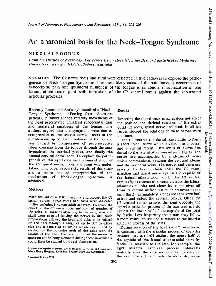
a short spinal nerve which divides into a dorsaland a ventral ramus. This series of nerves liesdorsal to the lateral atlanto-axial joint (fig 1). Thenerves are accompanied by a plexus of veinswhich communicate between the epidural plexusand the vertebral veins. The nerves and veins areinvested by fascia which holds the C2 roots,ganglion and spinal nerve against the capsule ofthe lateral atlanto-axial joint. The C2 ventralramus (fig 1) courses transversely across the lateralatlanto-axial joint and along its course gives offfrom its ventral surface, articular branches to thejoint (fig 2). Ultimately it arches over the vertebralartery and enters the cervical plexus. Often theC2 ventral ramus crosses the joint opposite thesuperior articular process of the axis and is heldagainst the lower half of the capsule of the jointby fascia. Less frequently the ramus may followa more rostral course and is related to the inferiorarticular process of the atlas.During rotation of the head the C2 roots move
in company with the articular process of the atlasbecause they are held against the upper half ofthe capsule of the lateral atlanto-axial joint byfascia. In rotation to the left, for example, theright atlanteal articular process sublaxatesventrally over the superior articular process ofthe axis. The right C2 roots therefore also move
202
by copyright. on 5 July 2019 by guest. P
rotectedhttp://jnnp.bm
j.com/
J Neurol N
eurosurg Psychiatry: first published as 10.1136/jnnp.44.3.202 on 1 M
arch 1981. Dow
nloaded from
An anatomical basis for the Neck-Tongue Syndrome
ventrally in company with the atlas. If the C2ventral ramus is related to the atlanteal articularprocess it too displaces ventrally with the atlas.However, when the ventral ramus crosses the axialprocess the ventral displacement of the atlascauses the ventral ramus to be drawn over themargin of the lateral atlanto-axial joint sincedistally and laterally the ventral ramus is relativelyfixed by fascia to the lower half of the jointcapsule.During normal rotation of the atlas there is a
considerable degree of subluxation of the lateralatlanto-axial joint and so, although covered bythe joint capsule, the margins of the articularprocesses forming the joint overlap and form
projecting edges. In rotation to the left, forexample, the right atlanteal process displacesventrally exposing the dorsal margin of the rightsuperior articular process of the axis. In thosecases where the right C2 ventral ramus crossesthe joint margin (fig 3A), during rotation it is ineffect drawn across this exposed dorsal margin,separated from it only by the capsule of the joint(fig 3B). In rotation to the right a similar butinverted effect occurs. The right inferior articularprocess of the atlas subluxates dorsally, and theright C2 spinal nerve and ventral ramus are drawnacross the projecting dorsal edge of the inferiorarticular process of the atlas (fig 3C).The C2 roots lie mainly within the vertebral
cp
Fig 1 The relationship between
)FX)the C2 dorsal root ganglion (g),spinal nerve (sn), and ventralramus (vr) and the lateral
\2 atlanto-axial joint (j), posteriorarch of the atlas (paa), thelamina of the axis (la), and thevertebral artery (va).A: Schematic illustrations of adorsal view. B: Dissection of aright dorsolateral view. dr=C2dorsal ramuv (cut at its point ofbranching). cp=cervical plexus.
A, ton=third occipital nerve. c=communicating branch C2 toC3. paam=posterior atlanto-axial membrane.
203
by copyright. on 5 July 2019 by guest. P
rotectedhttp://jnnp.bm
j.com/
J Neurol N
eurosurg Psychiatry: first published as 10.1136/jnnp.44.3.202 on 1 M
arch 1981. Dow
nloaded from

Nikolai Bogduk
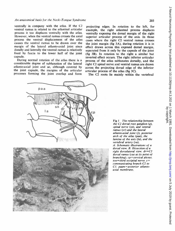
Fig 2 Close up view of an articular branch (arrow)to the left lateral atlanto-axial joint (j) from the C2ventral ramus (vr). The ramus, spinal nerve (sn) andganglion (g) have been retracted to expose thearticular nerve. dr=C2 dorsal ramus.
canal deep to the atlas. Therefore, they themselvesare not susceptible to bony impingement duringany motion of the head. However, the C2 dorsalroot ganglion lies dorsal to the lateral atlanto-axial joint. During most movements of the axisthis ganglion is free from bony impingement(fig 4A and B) but during rotation combinedwith forced extension it may be compressed be-tween the posterior arch of the atlas and thesuperior articular process of the axis (fig 4C).This phenomenon has been discussed previously2and it should be noted that, in a normal joint, inorder to compress the ganglion the extension mustbe forced. One the other hand if the joint sub-luxates abnormally it is conceivable that theganglion could be compressed.
Discussion
Textbooks of anatomyl 4 do not provide detaileddescriptions of the relations of the C2 roots, spinal
nerve, and rami. Previous authors3-8 have rectifiedcertain misconceptions and deficiencies relating tothe anatomy of these nerves and the descriptiongiven in the present study agrees with and incor-porates these changes. The most notable featureshighlighted in this and previous studies5-8 are thatthe C2 ganglion and spinal nerve lie dorsal to thelateral atlanto-axial joint and that this joint isinnervated by the C2 ventral ramus. These factsunderlie the following interpretation of themechanism of Neck-Tongue Syndrome.
In the Neck-Tongue Syndrome, numbness ofthe tongue and suboccipital pain are triggeredby rotation of the head. The site of pain and theprecipitating manoeuvre clearly implicate anabnormality at upper cervical levels. The tonguesymptoms have been discussed by Lance andAnthony' who reviewed the available data on thecourse of afferent fibres from the tongue; and itseems well established that proprioceptive fibresfrom the tongue do pass via the ansa hypoglossito the C2 dorsal root.Lance and Anthony' suggested that all the
symptoms of Neck-Tongue Syndrome were dueto compression of the C2 nerve roots in theatlanto-axial space. However, as described above,the C2 roots lie deep within the vertebral canaland are not susceptible to bony compression. Theonly elements of the C2 nerves which aresusceptible to bony impingement are the C2 dorsalroot ganglion and the C2 ventral ramus. Theganglion may be compressed between the posteriorarch of the atlas and the superior articular pro-cess of the axis, during combined extension androtation (fig 4C). C2 ventral rami which cross themargin of the lateral atlanto-axial joint may bestretched over the edge of the atlanteal or axialarticular process, during rotation of the atlas(fig 3).Although the other symptoms of Neck-Tongue
Syndrome may be explicable in terms of nervecompression, the pain is not, for it has beendemonstrated that experimental compression ofspinal nerves9 or peripheral nerves'0 producesparaesthesiae but not pain. A more satisfyingexplanation is that patients with Neck-TongueSyndrome suffer a temporary abnormal sub-luxation of their lateral atlanto-axial joint whichstrains the joint capsule, thus causing pain. Sub-luxations have not been documented in Neck-Tongue Syndrome since, because of the inter-mittent nature of the condition, patients have notbeen examined during an attack. However, thatsome such instability occurs is suggested by thehistory of one of Lance and Anthony's patients
204
by copyright. on 5 July 2019 by guest. P
rotectedhttp://jnnp.bm
j.com/
J Neurol N
eurosurg Psychiatry: first published as 10.1136/jnnp.44.3.202 on 1 M
arch 1981. Dow
nloaded from
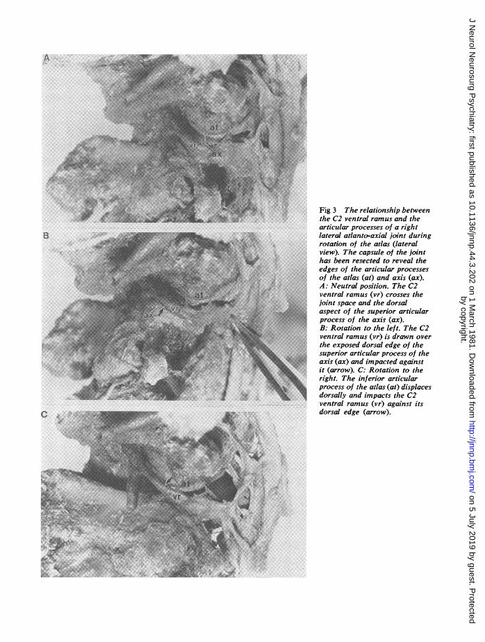
C . #~~~~~~~~~~~~~~~~~~~~~~~~~~~~~~~~~~~~~~~~~~~~~~~~~~~~~~~~~~~~~~~~~~~~~Z;:;+ ~~~~~~~~~~~~Fig3 The relationship betweent{@^ : _ t s g ~~~~~~~~~theC2 ventral ramus and the
articular processes of a rightn... sS- _DYb ....'> :-: j- "-'\ ................lateral atlanto-axial joint during3? l: X w e ~~~~~~~~~~~~view). The capsule of the joint
iX J j jL, r j 8' ~~~~~~~~edgesof the articular processes
.:s.5.......:....A: Neutra position. The C2:: Z . i | * | / ;- :B- S -} ~~ventral ramus (vr) crosses the.. . f_ _, ll l |s:ot- 2 ............................. {joint space and the dorsalAa5F~~~~~~ tN. asp i+ect of the sueiraticular)
s ^ t ^ - ^- ti; ~~~~~~~~prces of th axis (ax).adipctdaasBg ot(atio toRationhe t thetC2
th 2ventral ramus(risdawnd over
sueirarticular processs of theghlaxisrax)atlndoaimacoiteduaginst
,40~~~~ ~ ~ ~ ~ ~ ~~~~~~t(rrw.CRotation totheals(aea;.'. edrigh t.T heinferiorarticularprocess of theatlass(at)adisplacesd orsallyand impacts the C2ventral ramus (vr) against its
C .. adtdorsal edge (arow).
.:;: ._................X ec.o th spror riua
by copyright. on 5 July 2019 by guest. P
rotectedhttp://jnnp.bm
j.com/
J Neurol N
eurosurg Psychiatry: first published as 10.1136/jnnp.44.3.202 on 1 M
arch 1981. Dow
nloaded from
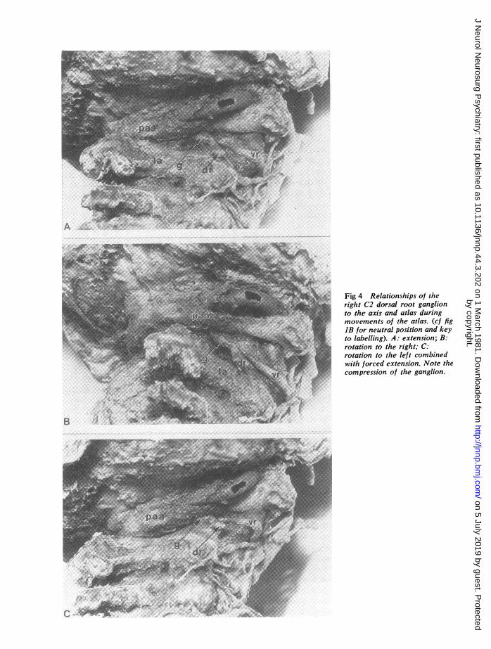
Fig 4 Relationships of theright C2 dorsal root ganglionto the axis and atlas duringmovements of the atlas. (cf figIB for neutral position and keyto labelling). A: extension; B:rotation to the right; C:rotation to the left combinedwith forced extension. Note thecompression of the ganglion.
by copyright. on 5 July 2019 by guest. P
rotectedhttp://jnnp.bm
j.com/
J Neurol N
eurosurg Psychiatry: first published as 10.1136/jnnp.44.3.202 on 1 M
arch 1981. Dow
nloaded from

An anatomical basis for the Neck-Tongue Syndrome
who reported recurrent episodes in which his head"fell back" forcing him to look upwards and tothe right. Normal posture was restored by flexionand side to side movement of the head.Apart from numbness of the tongue, another
symptom of Neck-Tongue Syndrome is numbnessof the skin behind the ear, reported by two
patients.' The retro-auricular skin is innervatedby the lesser occipital nerve and the posteriorbranch of the greater auricular nerve both ofwhich are derived from the C2 ventral ramus.'3Proprioceptive fibres from the tongue also returnvia the cervical plexus to the C2 ventral ramus.1Thus all the "numbness" sensations of Neck-Tongue Syndrome are consistent with compression
of the C2 ventral ramus. As shown in the presentstudy, during normal rotation of the atlas, theC2 ventral ramus may be impacted against theedge of an articular process of the lateral atlanto-axial joint. If patients with Neck-Tongue Syn-drome were indeed suffering in addition an
abnormal subluxation of that joint then the likeli-hood of inpaction would be greater.
It may be questioned whether compressionor irritation of proprioceptive fibres from thetongue could cause numbness. However, it iswell known that patients with Bell's palsy fre-quently complain of "numbness" althoughtrigeminal sensation is intact.1" 12 This "numb-ness" is explicable in terms of compression of theproprioceptive fibres which are known to run inthe facial nerve'3 Thus the sensation of "numb-ness" in both Bell's palsy and Neck-TongueSyndrome does not imply a loss of touch and painsensation but rather reflects the impaired functionof deep proprioceptive afferents.
It is notable that none of the patients describedby Lance and Anthony' complained of numbnessin the paramedian skin of the occiput and vertex.
This is the area supplied by the greater occipitalnerve which is derived from the C2 dorsal ramus.
This sparing of the dorsal ramus fibres indicatesthat the ventral ramus alone was compressed, andnot the dorsal root ganglion, for otherwisesymptoms should have occurred in both ventraland dorsal ramus territories. Moreover, the pre-
cipitating factor in Neck-Tongue Syndrome isrotation of the head. This manoeuvre may com-
promise the C2 ventral ramus but not the C2dorsal root ganglion, which further indicates theexclusive role of the C2 ventral ramus in Neck-Tongue Syndrome.
Notwithstanding this, the vulnerability of theC2 dorsal root ganglion to compression duringrotation combined with extension does theoreti-
cally make it a potential source of symptoms, butsuch a Neck-Tongue Syndrome would then in-clude numbness in the greater occipital nerveterritory.While the present study postulates a plausible
mechanism for the principal features of Neck-Tongue Syndrome, it does not explain the numb-ness of the fingers reported by two patients inLance and Anthony's series.1 This symptomcannot be related to C2 ventral ramus com-pression, and the present data do not providean explanation. Lance and Anthony' felt that itmay have been due to traction on the lower nerveroots, exerted through the dura mater, duringsubluxation of the atlas, but there is no ana-tomical data to support this hypothesis. Reid'4studied the extent of movement of the dura andspinal cord during flexion/extension movementsof the vertebral column and head, but comparabledata with respect to rotation are lacking. Thus an
explanation of the upper limb features of Neck-Tongue Syndrome awaits further investigation.
The author thanks Professor J W Lance for hiscomments during the preparation of thismanuscript.
R-ferences
1 Lance JW, Anthony WI. Neck-tongue syndromeon sudden turning of the head. J Neurol Neuro-surg Psychiatry 1980; 43:97-101.
2 Bogduk N. The anatomy of occipital neuralgia.Clin Exp Neurol 19880 (in press).
3 Gray's Anatomy. 35th ed. Warwick R, WilliamsPL, ed. London: Longmans 1973.
4 Cunningham DJ. Textbook of Anatomy. 11th ed.Romanes GJ ed. London: Oxford UniversityPress, 1972.
5 Guerrier Y, Colin R. Le deuxieme nerf cervicalCA Ass Anat 41e Congres 19,54; 813-6.
6 Lazorthes G, Gaubert J. L'innervation desarticulations interapophysa res vertebrales. CRAss Anat 43e Reunion 1956; 488-94.
7 Juskiewvenski S, Lazorthes F, Boulard PY,Lazorthes G. Le deuxieme ,zerf cervical. CR AssAnat 53e Congres 1968; 1044-7.
8 Lazorthes G. Les branches posterieures des nerfsrachidiens et le p!an articulaire vertebral pos-terieur. Ann Med Phys 1972; 15:192-202.
9 MacNab I. The mechaniEm cf spondylogenic pain.In: Hirsch C, Zotterman Y, eds. Cervical Pain.Oxford: Pergamon 1972; 89-95.
10 MacKenzie RA, Burke D, Skuse NF, LethleanAK. Fibre function and perception during cutane-ou:, nerve block. J Neurol Neurosurg Psychiatry1975; 38:865-73.
207
by copyright. on 5 July 2019 by guest. P
rotectedhttp://jnnp.bm
j.com/
J Neurol N
eurosurg Psychiatry: first published as 10.1136/jnnp.44.3.202 on 1 M
arch 1981. Dow
nloaded from
208
11 Davidson's Principles and Practice of Medicinel1th ed. MacLeod J, ed. Edinburgh: ChurchillLivingstone 1974; 821.
12 Victor M, Adams RD. Diseases of cranial nerves.In Harrison's Principles of Internal Medicine 8thed. Isselbacher KJ, Adams RD, Braumvald E,Petersdorf RG, Wilson JD, eds. New York:
Nikolai Bogduk
McGraw-Hill, 1980; Ch 376; 202-3.13 Fay T. Atypical neuralgia. Arch Neurol Psychiat
1927; 18:309-15.14 Reid JD. Effects of flexion-extension movements
of the head and spine upon the spinal cord andnerve roots. J Neurol Neurosurg Psychiat 1960;23:214-21.
by copyright. on 5 July 2019 by guest. P
rotectedhttp://jnnp.bm
j.com/
J Neurol N
eurosurg Psychiatry: first published as 10.1136/jnnp.44.3.202 on 1 M
arch 1981. Dow
nloaded from