Embed Size (px)
Citation preview
Amplified DNA Product Separation for Forensic Analysts
This course is provided free of charge and is part of a series designed to teach about DNA and forensic DNA use and analysis. Find this course live, online at: http://dna.gov/training/separation Updated: October 8, 2008
www.DNA.gov
About this Course This PDF file has been created from the free, self-paced online course “Crime Scene and DNA Basics for Forensic Analysts.” To learn more and take this and other courses online, go to http://www.dna.gov/training/online-training/. Most courses are free but you must first register at http://register.dna.gov. If you already are registered for any course on DNA.gov, you may login directly at the course URL, e.g., http://letraining.dna.gov or you can reach the courses by using the URL http://www.dna.gov/training and selecting the “link. Questions? If you have any questions about this file or any of the courses or content on DNA.gov, visit us online at http://www.dna.gov/more/contactus/.
Links in this File Most courses from DNA.Gov contain animations, videos, downloadable documents and/or links to other useful Web sites. If you are using a printed, paper version of this course, you will not have access to those features. If you are viewing the course as a PDF file online, you may be able to use some of these features if you are connected to the Internet. Animations, Audio and Video. Throughout this course, there may be links to animation, audio or video files. To listen to or view these files, you need to be connected to the Internet and have the requisite plug-in applications installed on your computer. Links to other Web Sites. To listen to or view any animation, audio or video files, you need to be connected to the Internet and have the requisite plug-in applications installed on your computer. Legal Policies and Disclaimers See Legal Policies and Disclaimers for information on Links to Other Web Sites, Copyright Status and Citation and Disclaimer of Liability and Endorsement.
Overview
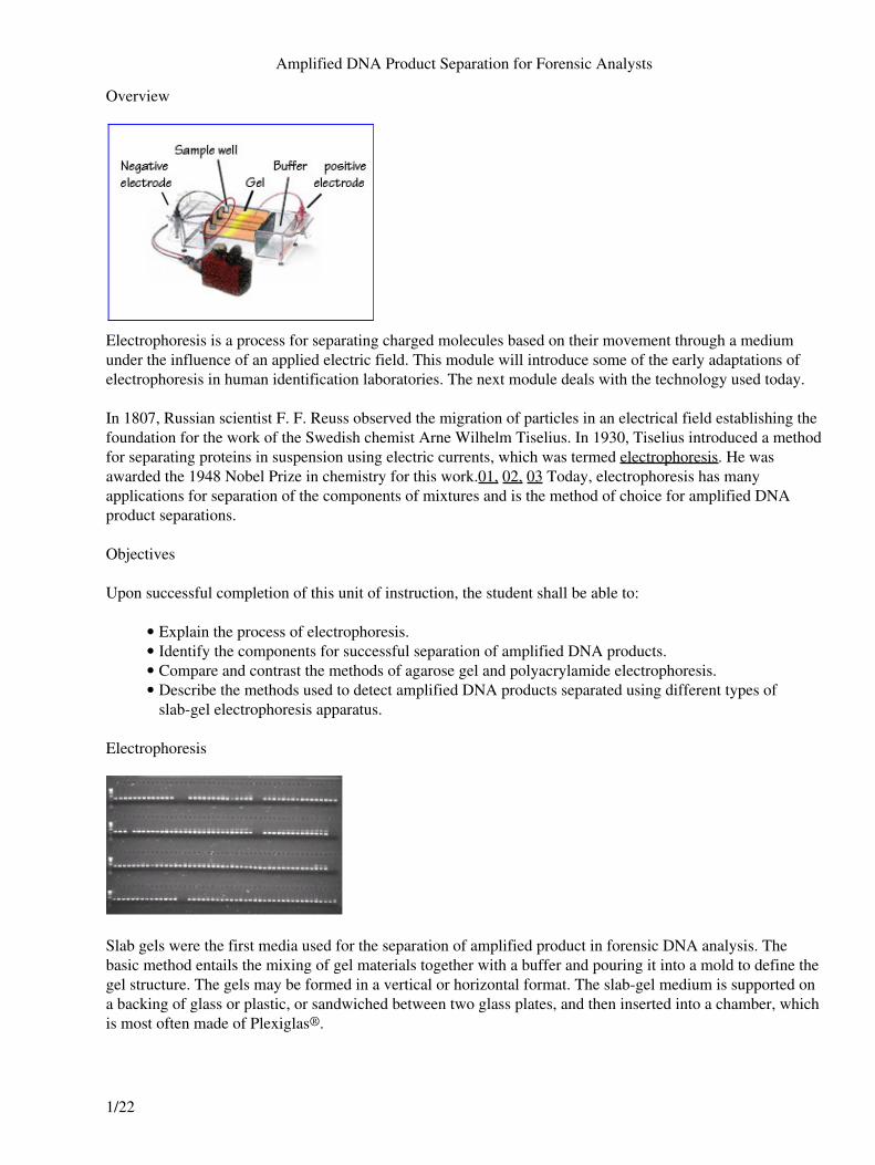
Electrophoresis is a process for separating charged molecules based on their movement through a mediumunder the influence of an applied electric field. This module will introduce some of the early adaptations ofelectrophoresis in human identification laboratories. The next module deals with the technology used today.
In 1807, Russian scientist F. F. Reuss observed the migration of particles in an electrical field establishing thefoundation for the work of the Swedish chemist Arne Wilhelm Tiselius. In 1930, Tiselius introduced a methodfor separating proteins in suspension using electric currents, which was termed electrophoresis. He wasawarded the 1948 Nobel Prize in chemistry for this work.01, 02, 03 Today, electrophoresis has manyapplications for separation of the components of mixtures and is the method of choice for amplified DNAproduct separations.
Objectives
Upon successful completion of this unit of instruction, the student shall be able to:
Explain the process of electrophoresis.• Identify the components for successful separation of amplified DNA products.• Compare and contrast the methods of agarose gel and polyacrylamide electrophoresis.• Describe the methods used to detect amplified DNA products separated using different types ofslab-gel electrophoresis apparatus.
•
Electrophoresis
Slab gels were the first media used for the separation of amplified product in forensic DNA analysis. Thebasic method entails the mixing of gel materials together with a buffer and pouring it into a mold to define thegel structure. The gels may be formed in a vertical or horizontal format. The slab-gel medium is supported ona backing of glass or plastic, or sandwiched between two glass plates, and then inserted into a chamber, whichis most often made of Plexiglas®.
Amplified DNA Product Separation for Forensic Analysts
1/22
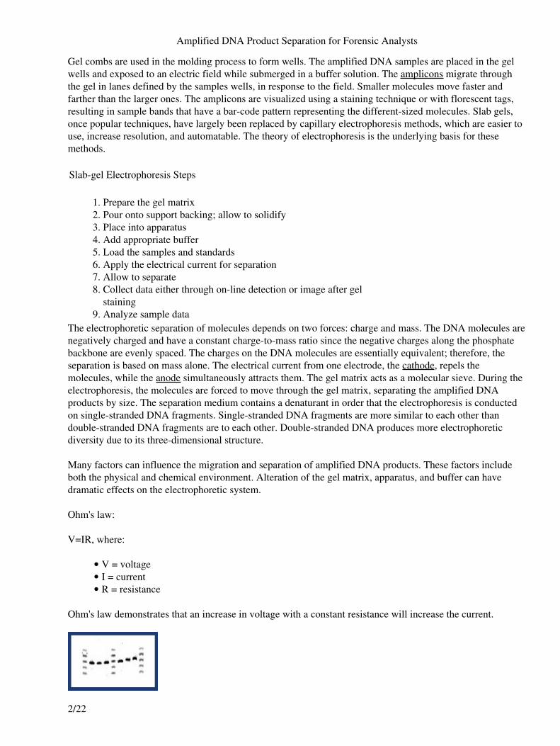
Gel combs are used in the molding process to form wells. The amplified DNA samples are placed in the gelwells and exposed to an electric field while submerged in a buffer solution. The amplicons migrate throughthe gel in lanes defined by the samples wells, in response to the field. Smaller molecules move faster andfarther than the larger ones. The amplicons are visualized using a staining technique or with florescent tags,resulting in sample bands that have a bar-code pattern representing the different-sized molecules. Slab gels,once popular techniques, have largely been replaced by capillary electrophoresis methods, which are easier touse, increase resolution, and automatable. The theory of electrophoresis is the underlying basis for thesemethods.
Slab-gel Electrophoresis Steps
Prepare the gel matrix1. Pour onto support backing; allow to solidify2. Place into apparatus3. Add appropriate buffer4. Load the samples and standards5. Apply the electrical current for separation6. Allow to separate7. Collect data either through on-line detection or image after gelstaining
8.
Analyze sample data9. The electrophoretic separation of molecules depends on two forces: charge and mass. The DNA molecules arenegatively charged and have a constant charge-to-mass ratio since the negative charges along the phosphatebackbone are evenly spaced. The charges on the DNA molecules are essentially equivalent; therefore, theseparation is based on mass alone. The electrical current from one electrode, the cathode, repels themolecules, while the anode simultaneously attracts them. The gel matrix acts as a molecular sieve. During theelectrophoresis, the molecules are forced to move through the gel matrix, separating the amplified DNAproducts by size. The separation medium contains a denaturant in order that the electrophoresis is conductedon single-stranded DNA fragments. Single-stranded DNA fragments are more similar to each other thandouble-stranded DNA fragments are to each other. Double-stranded DNA produces more electrophoreticdiversity due to its three-dimensional structure.
Many factors can influence the migration and separation of amplified DNA products. These factors includeboth the physical and chemical environment. Alteration of the gel matrix, apparatus, and buffer can havedramatic effects on the electrophoretic system.
Ohm's law:
V=IR, where:
V = voltage• I = current• R = resistance•
Ohm's law demonstrates that an increase in voltage with a constant resistance will increase the current.
Amplified DNA Product Separation for Forensic Analysts
2/22
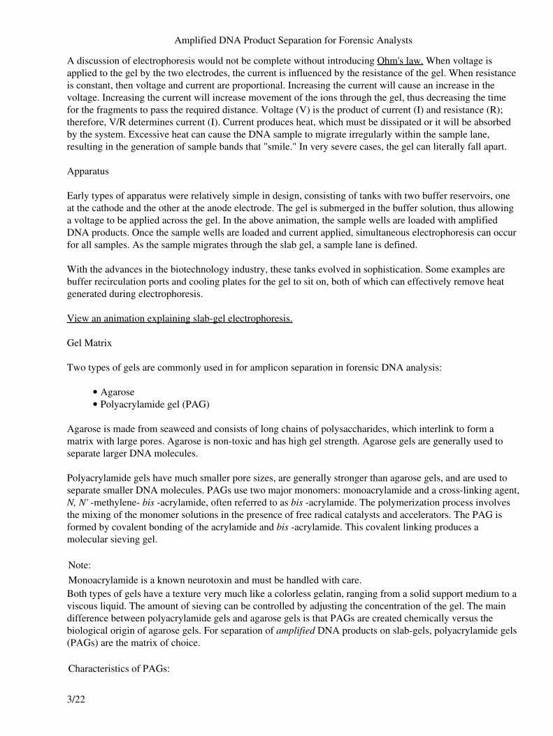
A discussion of electrophoresis would not be complete without introducing Ohm's law. When voltage isapplied to the gel by the two electrodes, the current is influenced by the resistance of the gel. When resistanceis constant, then voltage and current are proportional. Increasing the current will cause an increase in thevoltage. Increasing the current will increase movement of the ions through the gel, thus decreasing the timefor the fragments to pass the required distance. Voltage (V) is the product of current (I) and resistance (R);therefore, V/R determines current (I). Current produces heat, which must be dissipated or it will be absorbedby the system. Excessive heat can cause the DNA sample to migrate irregularly within the sample lane,resulting in the generation of sample bands that "smile." In very severe cases, the gel can literally fall apart.
Apparatus
Early types of apparatus were relatively simple in design, consisting of tanks with two buffer reservoirs, oneat the cathode and the other at the anode electrode. The gel is submerged in the buffer solution, thus allowinga voltage to be applied across the gel. In the above animation, the sample wells are loaded with amplifiedDNA products. Once the sample wells are loaded and current applied, simultaneous electrophoresis can occurfor all samples. As the sample migrates through the slab gel, a sample lane is defined.
With the advances in the biotechnology industry, these tanks evolved in sophistication. Some examples arebuffer recirculation ports and cooling plates for the gel to sit on, both of which can effectively remove heatgenerated during electrophoresis.
View an animation explaining slab-gel electrophoresis.
Gel Matrix
Two types of gels are commonly used in for amplicon separation in forensic DNA analysis:
Agarose• Polyacrylamide gel (PAG)•
Agarose is made from seaweed and consists of long chains of polysaccharides, which interlink to form amatrix with large pores. Agarose is non-toxic and has high gel strength. Agarose gels are generally used toseparate larger DNA molecules.
Polyacrylamide gels have much smaller pore sizes, are generally stronger than agarose gels, and are used toseparate smaller DNA molecules. PAGs use two major monomers: monoacrylamide and a cross-linking agent,N, N' -methylene- bis -acrylamide, often referred to as bis -acrylamide. The polymerization process involvesthe mixing of the monomer solutions in the presence of free radical catalysts and accelerators. The PAG isformed by covalent bonding of the acrylamide and bis -acrylamide. This covalent linking produces amolecular sieving gel.
Note:Monoacrylamide is a known neurotoxin and must be handled with care.Both types of gels have a texture very much like a colorless gelatin, ranging from a solid support medium to aviscous liquid. The amount of sieving can be controlled by adjusting the concentration of the gel. The maindifference between polyacrylamide gels and agarose gels is that PAGs are created chemically versus thebiological origin of agarose gels. For separation of amplified DNA products on slab-gels, polyacrylamide gels(PAGs) are the matrix of choice.
Characteristics of PAGs:
Amplified DNA Product Separation for Forensic Analysts
3/22
Transparent in the visible and portions of the UV spectrum• Reproducible, completely chemically synthetic compared to biological agarose gels• Strength of gel and easy to handle• Non-reactive with samples since there is no charge associated with it; therefore, the DNA fragmentsare not subjected to electroendosmotic effects
•
Stable• Thin gels can be cast which can promote better separations at higher field strengths due to efficientheat dissipation
•
Permanent record of separation• Pore size can be manipulated to increase molecular sieving• Different buffers can be used such that resolution and run times can be manipulated• Toxic• Need to formulate gel mixture and pour the gel• Need to degas the gel mixture under a vacuum to reduce bubbles• Possibility of bubble formation, even if degassed• Wait for polymerization – time-consuming•
View an animation about polyacrylamide gels.
Buffer System
Electrophoresis requires the use of a buffer system. A buffer is a chemical system that maintains a relativelyconstant pH even when strong acids or bases are added. Buffer solutions contain either a weak acid or a weakbase and one of their salts. Buffers not only establish a pH, but provide ions to support conductivity. Duringelectrophoresis, the electric field electrolyses the water molecules into H+ and OH- ions that migrate to therespective migrate to the respective electrodes.
The increase in H+ and OH- alters the pH at each electrode. A buffer can effectively neutralize the ions so thatthe pH of the system is maintained. The makeup of the buffer system is critical in the separation of theamplified DNA products by ensuring that the sample molecules are ionized and controlled. Changes to thebuffer system can lead to poor separation and lack of reproducibility. Small changes in pH can changemolecular charge; whereas, large changes in pH can cause serious, irreversible changes in molecular structure.Nucleic acids are relatively unaffected by pH changes due to their negative charge.04
The choice of buffer system will affect the resolution of the components in the mixture. Both continuous anddiscontinuous buffer systems can be used.
A continuous system uses the same buffer for the tank and gel. In continuous systems, molecular charge andgel pore size are the only factors that have any effect on the separation and stacking or concentration of asample into a band. A concentrated band of sample forms where the molecules are slowed down at theinterface of the buffer and the gel. Since smaller molecules have less of a difference between their freesolution mobility and their mobility in a gel, the stacking of smaller fragments is not as favorable as it will bein a discontinuous buffer system.
In a discontinuous system, the tank and gel buffers are different from each other. Samples are loaded onto alarge-pore gel, called a stacking gel, which overlays a smaller pore resolving gel. The stacking gel serves toconcentrate the DNA molecules on top of the resolving gel. After entering the resolving gel, the DNAmolecules are separated according to molecular size. The major advantage of a discontinuous buffer system isthe increased resolution and concentration of the sample band.
Amplified DNA Product Separation for Forensic Analysts
4/22
Historical Methods
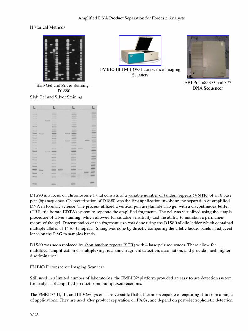
Slab Gel and Silver Staining -D1S80
FMBIO III FMBIO® fluorescence ImagingScanners
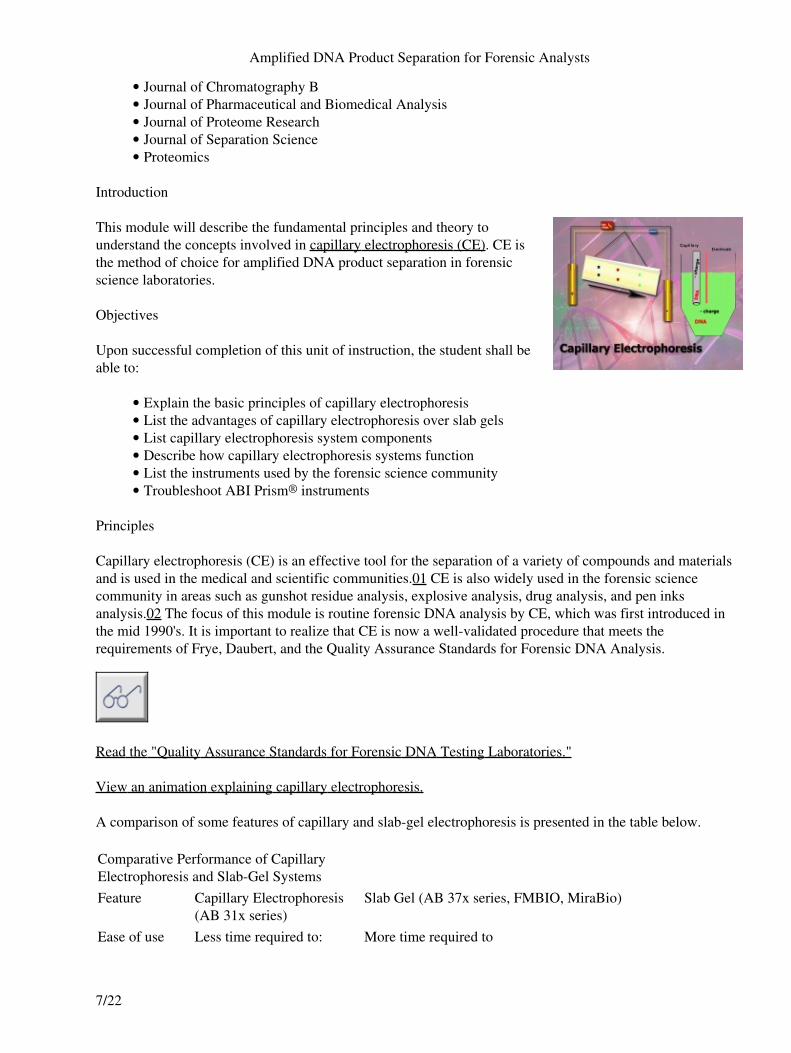
ABI Prism® 373 and 377DNA Sequencer
Slab Gel and Silver Staining
D1S80 is a locus on chromosome 1 that consists of a variable number of tandem repeats (VNTR) of a 16 basepair (bp) sequence. Characterization of D1S80 was the first application involving the separation of amplifiedDNA in forensic science. The process utilized a vertical polyacrylamide slab gel with a discontinuous buffer(TBE, tris-borate-EDTA) system to separate the amplified fragments. The gel was visualized using the simpleprocedure of silver staining, which allowed for suitable sensitivity and the ability to maintain a permanentrecord of the gel. Determination of the fragment size was done using the D1S80 allelic ladder which containedmultiple alleles of 14 to 41 repeats. Sizing was done by directly comparing the allelic ladder bands in adjacentlanes on the PAG to samples bands.
D1S80 was soon replaced by short tandem repeats (STR) with 4 base pair sequences. These allow formultilocus amplification or multiplexing, real-time fragment detection, automation, and provide much higherdiscrimination.
FMBIO Fluorescence Imaging Scanners
Still used in a limited number of laboratories, the FMBIO® platform provided an easy to use detection systemfor analysis of amplified product from multiplexed reactions.
The FMBIO® II, III, and III Plus systems are versatile flatbed scanners capable of capturing data from a rangeof applications. They are used after product separation on PAGs, and depend on post-electrophoretic detection
Amplified DNA Product Separation for Forensic Analysts
5/22
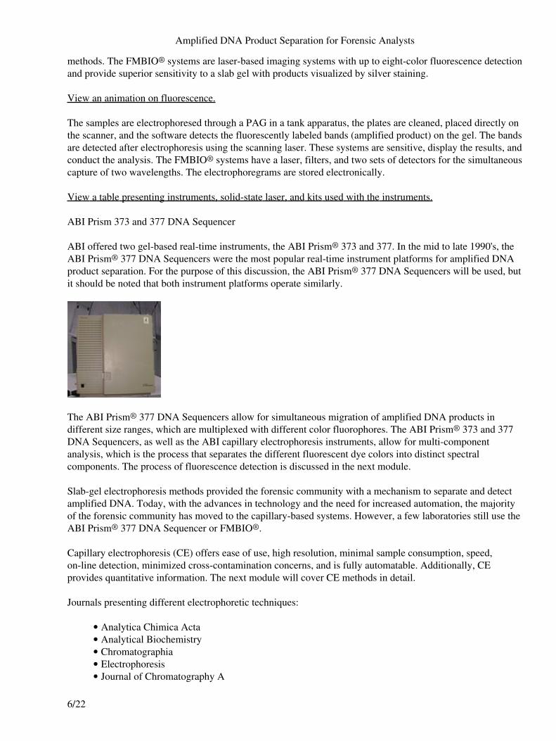
methods. The FMBIO® systems are laser-based imaging systems with up to eight-color fluorescence detectionand provide superior sensitivity to a slab gel with products visualized by silver staining.
View an animation on fluorescence.
The samples are electrophoresed through a PAG in a tank apparatus, the plates are cleaned, placed directly onthe scanner, and the software detects the fluorescently labeled bands (amplified product) on the gel. The bandsare detected after electrophoresis using the scanning laser. These systems are sensitive, display the results, andconduct the analysis. The FMBIO® systems have a laser, filters, and two sets of detectors for the simultaneouscapture of two wavelengths. The electrophoregrams are stored electronically.
View a table presenting instruments, solid-state laser, and kits used with the instruments.
ABI Prism 373 and 377 DNA Sequencer
ABI offered two gel-based real-time instruments, the ABI Prism® 373 and 377. In the mid to late 1990's, theABI Prism® 377 DNA Sequencers were the most popular real-time instrument platforms for amplified DNAproduct separation. For the purpose of this discussion, the ABI Prism® 377 DNA Sequencers will be used, butit should be noted that both instrument platforms operate similarly.
The ABI Prism® 377 DNA Sequencers allow for simultaneous migration of amplified DNA products indifferent size ranges, which are multiplexed with different color fluorophores. The ABI Prism® 373 and 377DNA Sequencers, as well as the ABI capillary electrophoresis instruments, allow for multi-componentanalysis, which is the process that separates the different fluorescent dye colors into distinct spectralcomponents. The process of fluorescence detection is discussed in the next module.
Slab-gel electrophoresis methods provided the forensic community with a mechanism to separate and detectamplified DNA. Today, with the advances in technology and the need for increased automation, the majorityof the forensic community has moved to the capillary-based systems. However, a few laboratories still use theABI Prism® 377 DNA Sequencer or FMBIO®.
Capillary electrophoresis (CE) offers ease of use, high resolution, minimal sample consumption, speed,on-line detection, minimized cross-contamination concerns, and is fully automatable. Additionally, CEprovides quantitative information. The next module will cover CE methods in detail.
Journals presenting different electrophoretic techniques:
Analytica Chimica Acta• Analytical Biochemistry• Chromatographia• Electrophoresis• Journal of Chromatography A•
Amplified DNA Product Separation for Forensic Analysts
6/22
Journal of Chromatography B• Journal of Pharmaceutical and Biomedical Analysis• Journal of Proteome Research• Journal of Separation Science• Proteomics•
Introduction
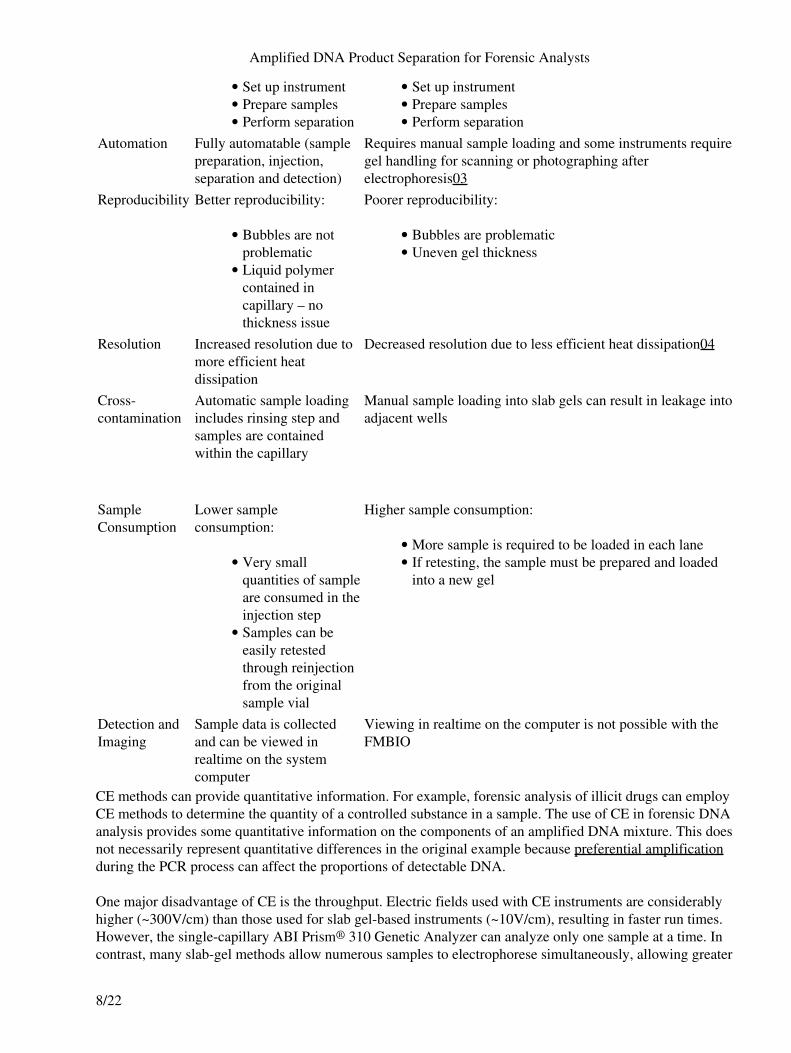
This module will describe the fundamental principles and theory tounderstand the concepts involved in capillary electrophoresis (CE). CE isthe method of choice for amplified DNA product separation in forensicscience laboratories.
Objectives
Upon successful completion of this unit of instruction, the student shall beable to:
Explain the basic principles of capillary electrophoresis• List the advantages of capillary electrophoresis over slab gels• List capillary electrophoresis system components• Describe how capillary electrophoresis systems function• List the instruments used by the forensic science community• Troubleshoot ABI Prism® instruments•
Principles
Capillary electrophoresis (CE) is an effective tool for the separation of a variety of compounds and materialsand is used in the medical and scientific communities.01 CE is also widely used in the forensic sciencecommunity in areas such as gunshot residue analysis, explosive analysis, drug analysis, and pen inksanalysis.02 The focus of this module is routine forensic DNA analysis by CE, which was first introduced inthe mid 1990's. It is important to realize that CE is now a well-validated procedure that meets therequirements of Frye, Daubert, and the Quality Assurance Standards for Forensic DNA Analysis.
Read the "Quality Assurance Standards for Forensic DNA Testing Laboratories."
View an animation explaining capillary electrophoresis.
A comparison of some features of capillary and slab-gel electrophoresis is presented in the table below.
Comparative Performance of CapillaryElectrophoresis and Slab-Gel SystemsFeature Capillary Electrophoresis
(AB 31x series)Slab Gel (AB 37x series, FMBIO, MiraBio)
Ease of use Less time required to: More time required to
Amplified DNA Product Separation for Forensic Analysts
7/22
Set up instrument• Prepare samples• Perform separation•
Set up instrument• Prepare samples• Perform separation•
Automation Fully automatable (samplepreparation, injection,separation and detection)
Requires manual sample loading and some instruments requiregel handling for scanning or photographing afterelectrophoresis03
Reproducibility Better reproducibility:
Bubbles are notproblematic
•
Liquid polymercontained incapillary – nothickness issue
•
Poorer reproducibility:
Bubbles are problematic• Uneven gel thickness•
Resolution Increased resolution due tomore efficient heatdissipation
Decreased resolution due to less efficient heat dissipation04
Cross-contamination
Automatic sample loadingincludes rinsing step andsamples are containedwithin the capillary
Manual sample loading into slab gels can result in leakage intoadjacent wells
SampleConsumption
Lower sampleconsumption:
Very smallquantities of sampleare consumed in theinjection step
•
Samples can beeasily retestedthrough reinjectionfrom the originalsample vial
•
Higher sample consumption:
More sample is required to be loaded in each lane• If retesting, the sample must be prepared and loadedinto a new gel
•
Detection andImaging
Sample data is collectedand can be viewed inrealtime on the systemcomputer
Viewing in realtime on the computer is not possible with theFMBIO
CE methods can provide quantitative information. For example, forensic analysis of illicit drugs can employCE methods to determine the quantity of a controlled substance in a sample. The use of CE in forensic DNAanalysis provides some quantitative information on the components of an amplified DNA mixture. This doesnot necessarily represent quantitative differences in the original example because preferential amplificationduring the PCR process can affect the proportions of detectable DNA.
One major disadvantage of CE is the throughput. Electric fields used with CE instruments are considerablyhigher (~300V/cm) than those used for slab gel-based instruments (~10V/cm), resulting in faster run times.However, the single-capillary ABI Prism® 310 Genetic Analyzer can analyze only one sample at a time. Incontrast, many slab-gel methods allow numerous samples to electrophorese simultaneously, allowing greater
Amplified DNA Product Separation for Forensic Analysts
8/22
throughput even though the electrophoresis time is approximately twice as long as that in CE.
The development of capillary array electrophoresis (CAE) instruments, which allow for multiple samples tobe run in parallel, resulted in throughput capabilities equal to or surpassing that of slab-gel methods.
CE Systems Components
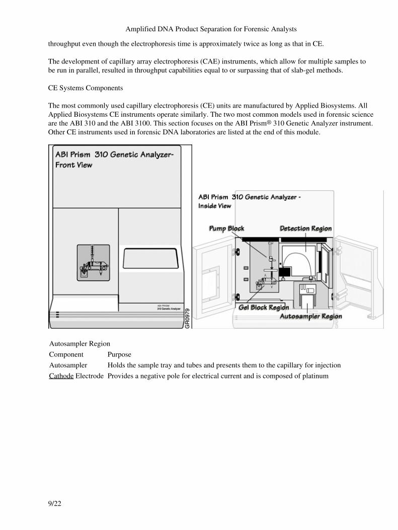
The most commonly used capillary electrophoresis (CE) units are manufactured by Applied Biosystems. AllApplied Biosystems CE instruments operate similarly. The two most common models used in forensic scienceare the ABI 310 and the ABI 3100. This section focuses on the ABI Prism® 310 Genetic Analyzer instrument.Other CE instruments used in forensic DNA laboratories are listed at the end of this module.
Autosampler RegionComponent PurposeAutosampler Holds the sample tray and tubes and presents them to the capillary for injectionCathode Electrode Provides a negative pole for electrical current and is composed of platinum
Amplified DNA Product Separation for Forensic Analysts
9/22
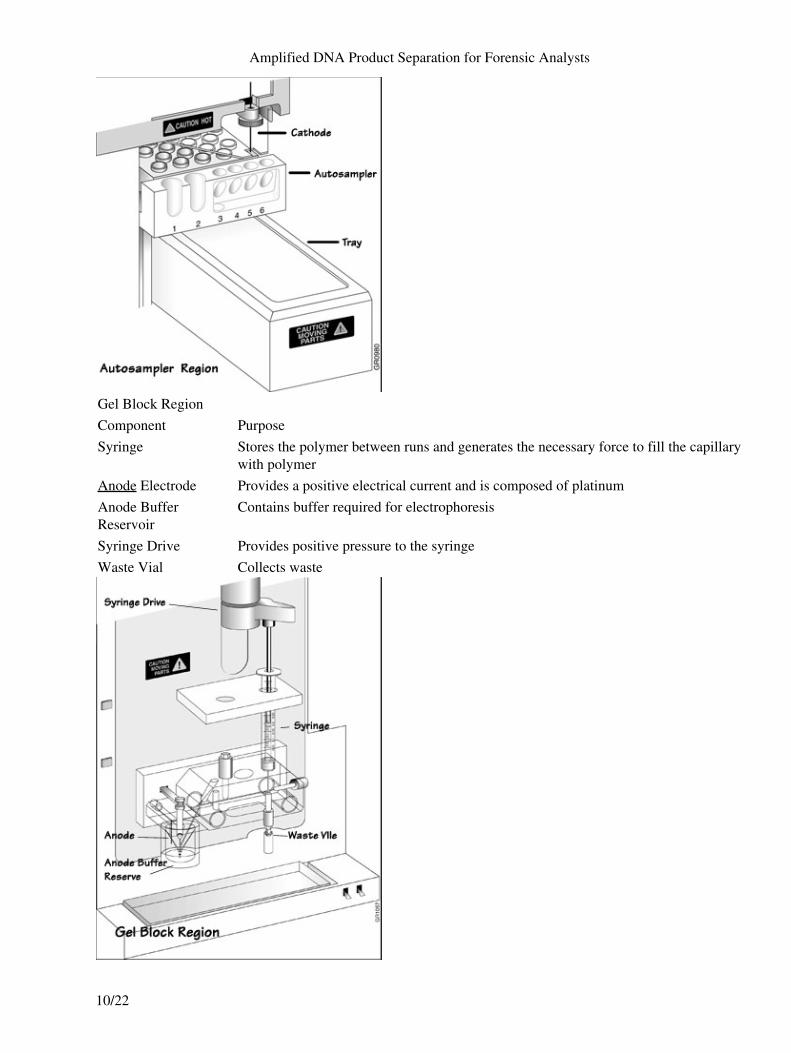
Gel Block RegionComponent PurposeSyringe Stores the polymer between runs and generates the necessary force to fill the capillary
with polymerAnode Electrode Provides a positive electrical current and is composed of platinumAnode BufferReservoir
Contains buffer required for electrophoresis
Syringe Drive Provides positive pressure to the syringeWaste Vial Collects waste
Amplified DNA Product Separation for Forensic Analysts
10/22
Ions migrate through the capillary during electrophoresis. Positive ions will gather at the anode and negativelycharged ions will gather at the cathode. The movement of ions creates an imbalance called buffer depletion.Buffer depletion can impair separation of DNA fragments due to a reduction in current. It is important toreplenish or replace the buffer regularly to compensate for buffer depletion.
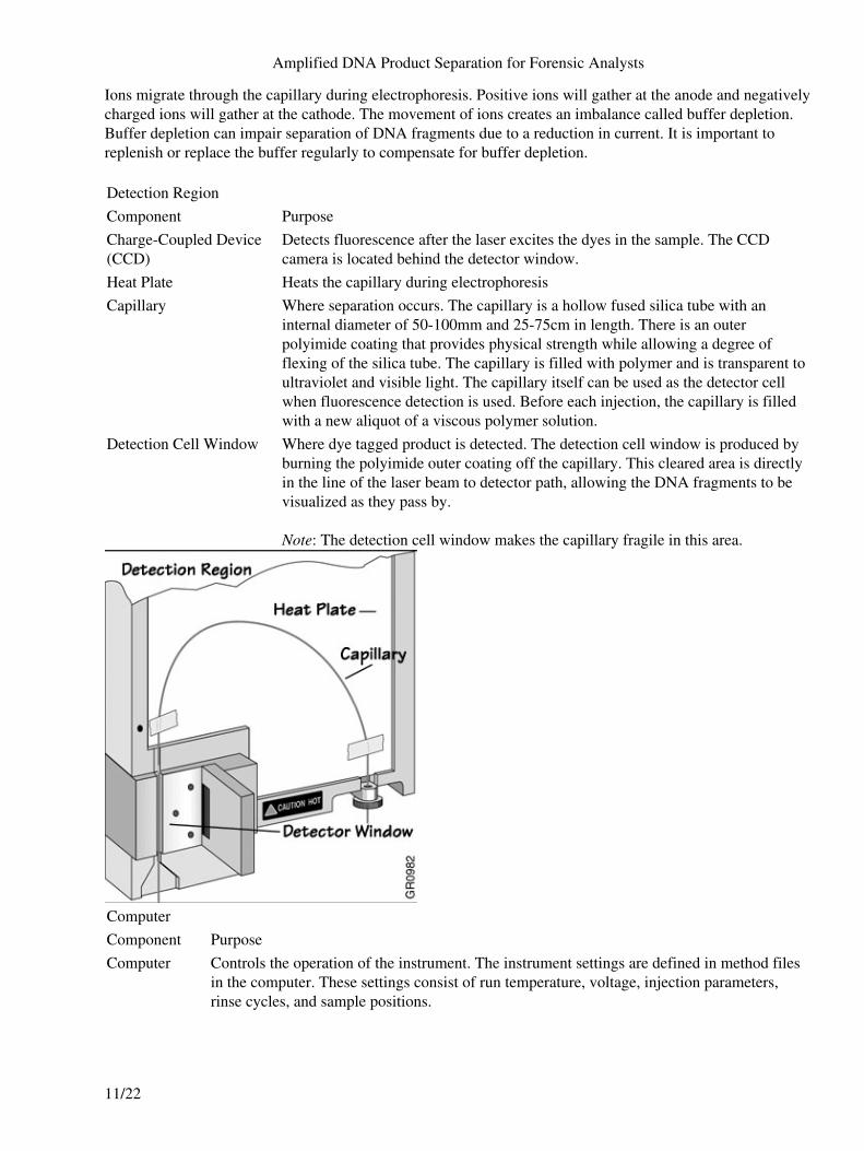
Detection RegionComponent PurposeCharge-Coupled Device(CCD)
Detects fluorescence after the laser excites the dyes in the sample. The CCDcamera is located behind the detector window.
Heat Plate Heats the capillary during electrophoresisCapillary Where separation occurs. The capillary is a hollow fused silica tube with an
internal diameter of 50-100mm and 25-75cm in length. There is an outerpolyimide coating that provides physical strength while allowing a degree offlexing of the silica tube. The capillary is filled with polymer and is transparent toultraviolet and visible light. The capillary itself can be used as the detector cellwhen fluorescence detection is used. Before each injection, the capillary is filledwith a new aliquot of a viscous polymer solution.
Detection Cell Window Where dye tagged product is detected. The detection cell window is produced byburning the polyimide outer coating off the capillary. This cleared area is directlyin the line of the laser beam to detector path, allowing the DNA fragments to bevisualized as they pass by.
Note: The detection cell window makes the capillary fragile in this area.
ComputerComponent PurposeComputer Controls the operation of the instrument. The instrument settings are defined in method files
in the computer. These settings consist of run temperature, voltage, injection parameters,rinse cycles, and sample positions.
Amplified DNA Product Separation for Forensic Analysts
11/22
Capillary Electrophoresis Systems Function
Sample Preparation
Samples run on Capillary electrophoresis (CE) instruments are amplified as described in course: DNAAmplification, the Multiplexing Module before placing them into the autosampler tray.
Electrokinetic Injection
There are three methods used for sample introduction in CE systems:
Hydrodynamic or pressure injection• Siphoning• Electrokinetic injection05•
All of these methods require the immersion of the capillary end into the sample. Electrokinetic injection is theonly method used in forensic DNA analysis.
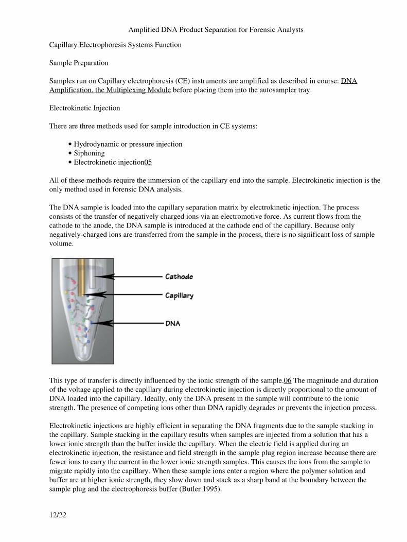
The DNA sample is loaded into the capillary separation matrix by electrokinetic injection. The processconsists of the transfer of negatively charged ions via an electromotive force. As current flows from thecathode to the anode, the DNA sample is introduced at the cathode end of the capillary. Because onlynegatively-charged ions are transferred from the sample in the process, there is no significant loss of samplevolume.
This type of transfer is directly influenced by the ionic strength of the sample.06 The magnitude and durationof the voltage applied to the capillary during electrokinetic injection is directly proportional to the amount ofDNA loaded into the capillary. Ideally, only the DNA present in the sample will contribute to the ionicstrength. The presence of competing ions other than DNA rapidly degrades or prevents the injection process.
Electrokinetic injections are highly efficient in separating the DNA fragments due to the sample stacking inthe capillary. Sample stacking in the capillary results when samples are injected from a solution that has alower ionic strength than the buffer inside the capillary. When the electric field is applied during anelectrokinetic injection, the resistance and field strength in the sample plug region increase because there arefewer ions to carry the current in the lower ionic strength samples. This causes the ions from the sample tomigrate rapidly into the capillary. When these sample ions enter a region where the polymer solution andbuffer are at higher ionic strength, they slow down and stack as a sharp band at the boundary between thesample plug and the electrophoresis buffer (Butler 1995).
Amplified DNA Product Separation for Forensic Analysts
12/22
Separation
The cathode buffer reservoir, a water reservoir, and a waste vial are located in the autosampler. A short periodof electrophoresis injects the sample into the capillary. Next, the autosampler moves the cathode bufferreservoir to the capillary and cathode electrode to continue electrophoresis. The buffer used in the capillaryelectrophoresis (CE) system is viscous and adheres to the exterior of the capillary during the buffer fill step. Inthe subsequent injection step this material could contaminate the sample, change its ionic strength, and reducethe quantity of material injected. The water reservoir is used to wash the cathode and capillary tip betweensample injections.
The buffer solution moves through the capillary under the influence of an electric field. This phenomenon istermed electroosmotic or electroendosmotic flow. The direction of the electroosmotic flow is toward thepositively charged anode, which means that the buffer flows from the source vial, through the capillary,through the detector, to the destination vial. The DNA fragments migrate towards the anode reservoir.
Performance Optimized Polymers™ dynamically coat the capillary wall to control electroosmotic flow duringelectrophoresis. The most common polymer used is ABI's Performance Optimized Polymer™-4(POP-4™).07 This polymer was developed to meet the following specifications:
detect alleles differing in size by a single base (up to 250 base pairs in length)• size alleles of the same length with a precision of less than 0.15 nucleotide standard deviation• require less than 30 minutes analysis time per sample• provide capillary life of at least 100 injections• provide a highly denaturing environment for the DNA samples07•
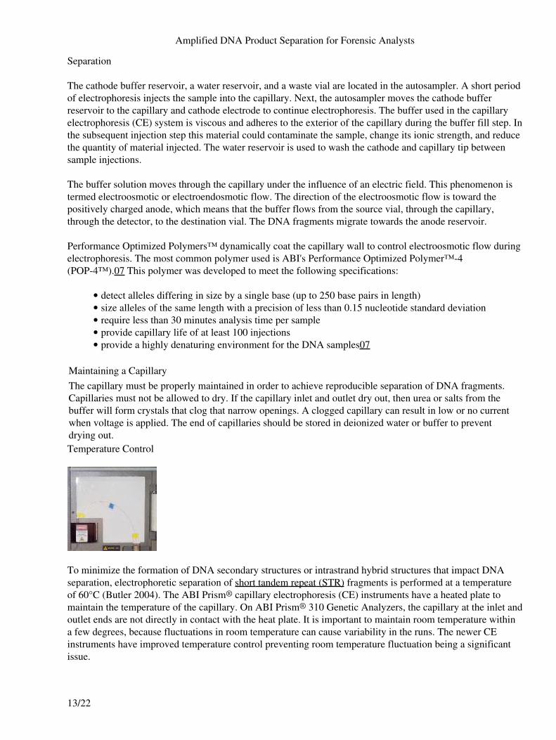
Maintaining a CapillaryThe capillary must be properly maintained in order to achieve reproducible separation of DNA fragments.Capillaries must not be allowed to dry. If the capillary inlet and outlet dry out, then urea or salts from thebuffer will form crystals that clog that narrow openings. A clogged capillary can result in low or no currentwhen voltage is applied. The end of capillaries should be stored in deionized water or buffer to preventdrying out.Temperature Control
To minimize the formation of DNA secondary structures or intrastrand hybrid structures that impact DNAseparation, electrophoretic separation of short tandem repeat (STR) fragments is performed at a temperatureof 60°C (Butler 2004). The ABI Prism® capillary electrophoresis (CE) instruments have a heated plate tomaintain the temperature of the capillary. On ABI Prism® 310 Genetic Analyzers, the capillary at the inlet andoutlet ends are not directly in contact with the heat plate. It is important to maintain room temperature withina few degrees, because fluctuations in room temperature can cause variability in the runs. The newer CEinstruments have improved temperature control preventing room temperature fluctuation being a significantissue.
Amplified DNA Product Separation for Forensic Analysts
13/22
Fluorescent Detection
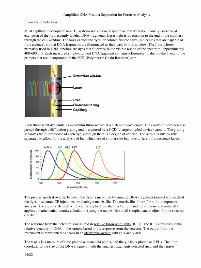
Most capillary electrophoresis (CE) systems use a form of spectroscopic detection, namely laser-basedexcitation of the fluorescently labeled DNA fragments. Laser light is directed on to the end of the capillarythrough the cell window. The laser excites the dyes, or colored fluorophores (molecules that are capable offluorescence), so that DNA fragments are illuminated as they pass by this window. The fluorophoresprimarily used in DNA labeling are dyes that fluoresce in the visible region of the spectrum (approximately400-600nm). Each measured single-stranded DNA fragment contains a fluorescent label on the 5' end of theprimers that are incorporated in the PCR (Polymerase Chain Reaction) step.
Each fluorescent dye emits its maximum fluorescence at a different wavelength. The emitted fluorescence ispassed through a diffraction grating and is captured by a CCD (charge-coupled device) camera. The gratingseparates the fluorescence of each dye, although there is a degree of overlap. The output is sufficientlyseparated to allow for the analysis of loci which are of similar size but have different fluorescence labels.
The precise spectral overlap between the dyes is measured by running DNA fragments labeled with each ofthe dyes in separate CE injections, producing a matrix file. The matrix file allows for multi-componentanalysis. The appropriate matrix file can be applied to data on a CE run, and the software automaticallyapplies a mathematical matrix calculation (using the matrix file) to all sample data to adjust for the spectraloverlap.
The response from the detector is measured in relative fluorescent units (RFU). The RFU correlates to therelative quantity of DNA in the sample based on its response from the detector. The output from theinstrument is represented as peaks in an electropherogram with an x and y axis.
The x-axis is a measure of time plotted as scan data points, and the y-axis is plotted as RFUs. The timecorrelates to the size of the DNA fragment, with the smallest fragments detected first, and the largest
Amplified DNA Product Separation for Forensic Analysts
14/22
fragments detected last.
Data Analysis
The steps for assigning an allele call to each peak are:
Data collection1. Peak recognition2. Color separation3. Peak sizing4. Allelic ladder comparison5. Allele assignment (i.e., genotype)6.
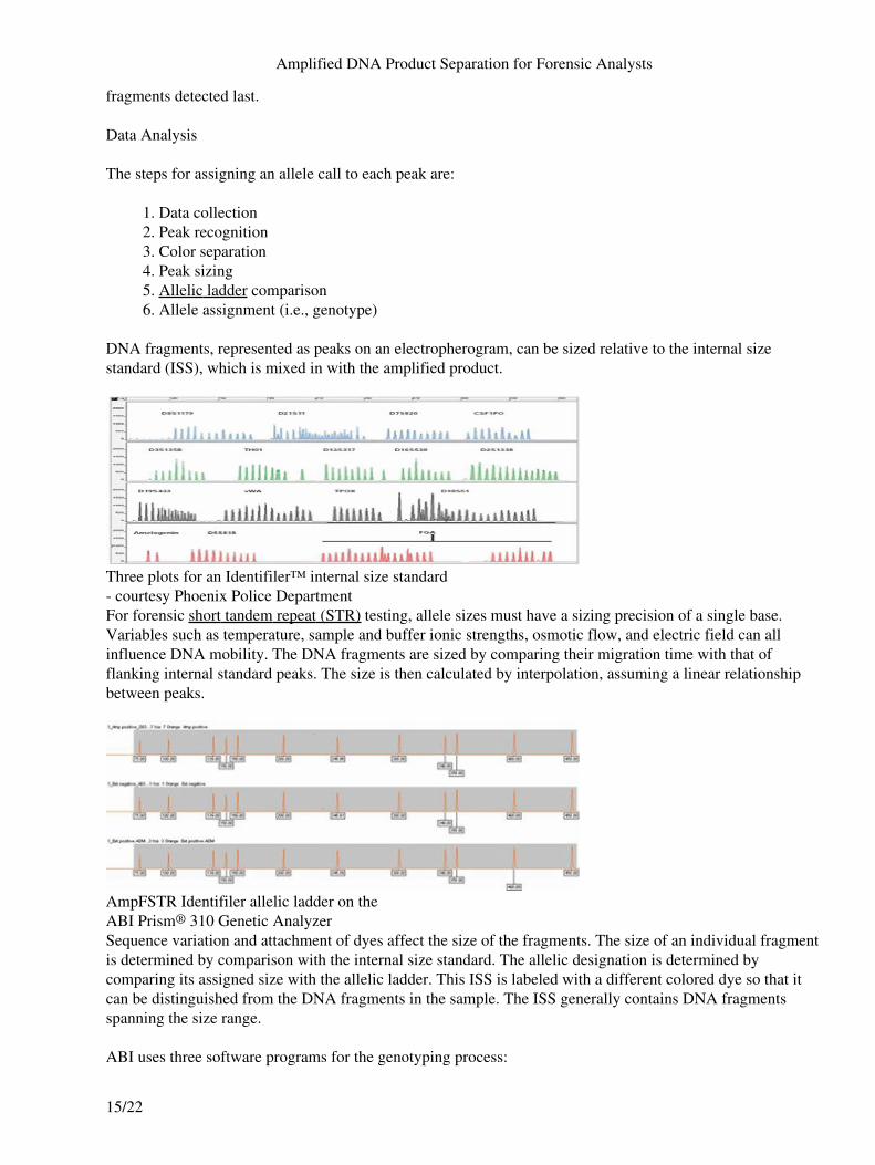
DNA fragments, represented as peaks on an electropherogram, can be sized relative to the internal sizestandard (ISS), which is mixed in with the amplified product.
Three plots for an Identifiler™ internal size standard- courtesy Phoenix Police DepartmentFor forensic short tandem repeat (STR) testing, allele sizes must have a sizing precision of a single base.Variables such as temperature, sample and buffer ionic strengths, osmotic flow, and electric field can allinfluence DNA mobility. The DNA fragments are sized by comparing their migration time with that offlanking internal standard peaks. The size is then calculated by interpolation, assuming a linear relationshipbetween peaks.
AmpFSTR Identifiler allelic ladder on theABI Prism® 310 Genetic AnalyzerSequence variation and attachment of dyes affect the size of the fragments. The size of an individual fragmentis determined by comparison with the internal size standard. The allelic designation is determined bycomparing its assigned size with the allelic ladder. This ISS is labeled with a different colored dye so that itcan be distinguished from the DNA fragments in the sample. The ISS generally contains DNA fragmentsspanning the size range.
ABI uses three software programs for the genotyping process:
Amplified DNA Product Separation for Forensic Analysts
15/22
GeneScan®• Genotyper®• GeneMapper® ID•
GeneScan® software spectrally resolves the dye colors for each peak and is used to size the DNA fragments ineach sample. These data are then imported in the Genotyper® program, which compares the sizes of alleles inthe allelic ladder to those obtained for each sample. GeneMapper® ID is a program that was released byApplied Biosystems (AB) in 2003, which combines the functions of GeneScan® and Genotyper®.
Instrumentation
ABI Prism® 310 Genetic Analyzer is a single capillary instrument with multiple color fluorescence detection.The instrument allows for unattended operation. An analyst loads the samples into the autosampler, places acapillary and syringe full of polymer solution in the instrument, and initiates the run. Computer programsprocess the data and genotype information.
Capillary Array Electrophoresis (CAE) Instruments
Capillary array electrophoresis (CAE) instruments have multiple capillaries in parallel with multicolorfluorescence detection. Advantages of these instruments are:
Simultaneous injection of multiple samples• Unattended operation• High throughput processing•
Array Comparison of InstrumentsInstrument # of Capillaries
per ArrayABI Prism® 3100-Avant Genetic Analyzer / 3130Genetic Analyzer
4
ABI Prism® 3100 Genetic Analyzer / 3130 xlGenetic Analyzer
16
ABI Prism® 3700 Genetic Analyzer 96AB (Applied Biosystems) manufactures a group of instruments that have similar features except for the typesof pump and the number of capillaries in the array. These instruments are referred to as the ABI Prism® 31xx.
Troubleshooting
Troubleshooting GuideIssue Possible Cause ActionPoor mobility andreproducibility
Temperature fluctuationCheck instrument oven• Check environment temperature•
Blocked, dirty capillary Change capillarySample-wall interaction Rinse capillary with polymerIon depletion of buffers Replace buffers
Buffer siphoning Buffer reservoir levels not equal Equalize buffer levelsPoor sensitivity Reamplify or add more amplified product
Amplified DNA Product Separation for Forensic Analysts
16/22
Not enough amplified product insampleBubble in sample tube, capillaryexposed to bubble
Reinject
Poor laser power Service call, possible laser replacementIon competition during electrokineticinjection Check quality of formamide•
Compare results of sample topositive control included in kit - ifokay, then evaluate extraction andsample preparation
•
Poor matrix Rerun matrixGradual change incurrent
Different cathode/anode buffers and/orconcentration
Confirm buffers
Current fluctuations Bubbles in the system Conduct a visual check for bubbles and clearby flushing with fresh polymer.
No current Plugged capillaryFlush with polymer• Replace capillary•
Wrong buffer Confirm buffersNoisy baseline Poor matrix Rerun matrixFlat baseline Capillary not aligned in detector
windowRealign capillary
No voltageConfirm voltage settings• Ensure capillary ends are immersedin buffer
•
Sample not injected Confirm the following:
Sample in vial• Sample is thoroughly mixed withformamide and is not on the side ofthe tube
•
Capillary end in sample solution• Injection time•
Laser not functioning Service call, possible laser replacementClogged capillary
Replenish with fresh polymer• Replace capillary•
Electrical arcing Buffer spill in high voltage areas Clean autosampler tray with distilled waterand wipe dry
Poor peak shape Incorrect current causes:
peak distortion
tailing [low current]
Replace buffer
Capillary failure
Amplified DNA Product Separation for Forensic Analysts
17/22
Replenish capillary with freshpolymer
•
Replace capillary• Introduction
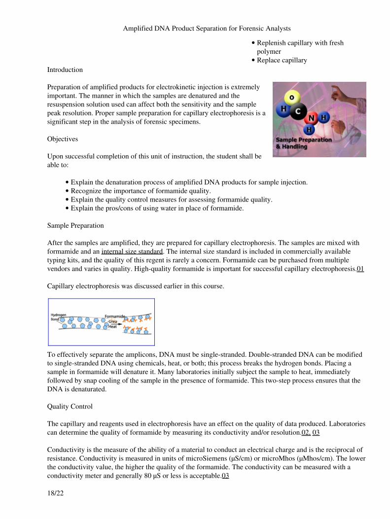
Preparation of amplified products for electrokinetic injection is extremelyimportant. The manner in which the samples are denatured and theresuspension solution used can affect both the sensitivity and the samplepeak resolution. Proper sample preparation for capillary electrophoresis is asignificant step in the analysis of forensic specimens.
Objectives
Upon successful completion of this unit of instruction, the student shall beable to:
Explain the denaturation process of amplified DNA products for sample injection.• Recognize the importance of formamide quality.• Explain the quality control measures for assessing formamide quality.• Explain the pros/cons of using water in place of formamide.•
Sample Preparation
After the samples are amplified, they are prepared for capillary electrophoresis. The samples are mixed withformamide and an internal size standard. The internal size standard is included in commercially availabletyping kits, and the quality of this regent is rarely a concern. Formamide can be purchased from multiplevendors and varies in quality. High-quality formamide is important for successful capillary electrophoresis.01
Capillary electrophoresis was discussed earlier in this course.
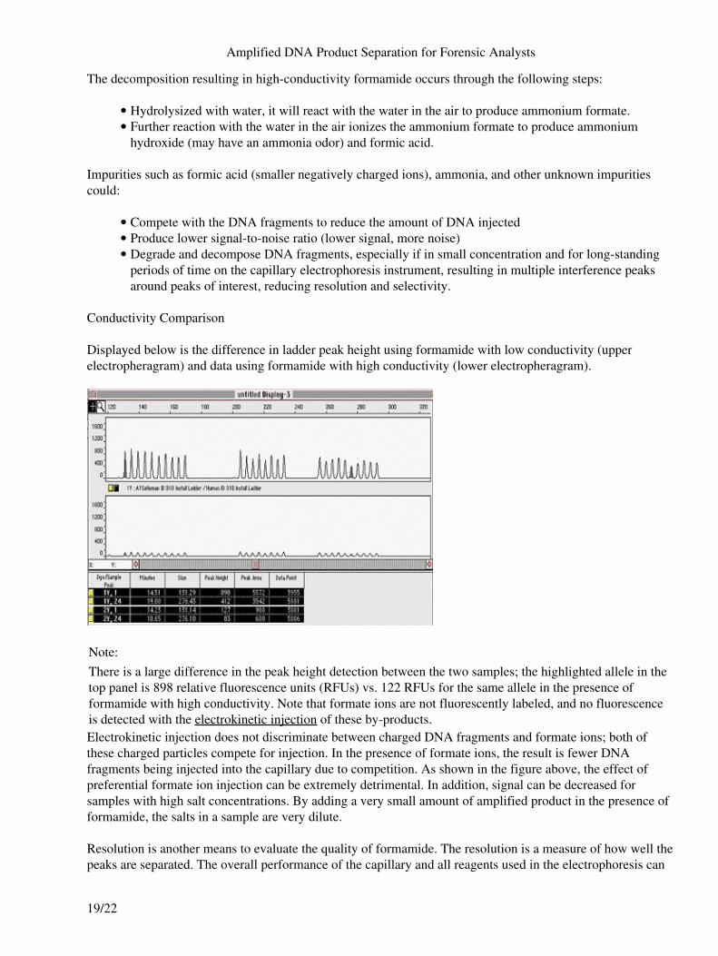
To effectively separate the amplicons, DNA must be single-stranded. Double-stranded DNA can be modifiedto single-stranded DNA using chemicals, heat, or both; this process breaks the hydrogen bonds. Placing asample in formamide will denature it. Many laboratories initially subject the sample to heat, immediatelyfollowed by snap cooling of the sample in the presence of formamide. This two-step process ensures that theDNA is denaturated.
Quality Control
The capillary and reagents used in electrophoresis have an effect on the quality of data produced. Laboratoriescan determine the quality of formamide by measuring its conductivity and/or resolution.02, 03
Conductivity is the measure of the ability of a material to conduct an electrical charge and is the reciprocal ofresistance. Conductivity is measured in units of microSiemens (µS/cm) or microMhos (µMhos/cm). The lowerthe conductivity value, the higher the quality of the formamide. The conductivity can be measured with aconductivity meter and generally 80 µS or less is acceptable.03
Amplified DNA Product Separation for Forensic Analysts
18/22
The decomposition resulting in high-conductivity formamide occurs through the following steps:
Hydrolysized with water, it will react with the water in the air to produce ammonium formate.• Further reaction with the water in the air ionizes the ammonium formate to produce ammoniumhydroxide (may have an ammonia odor) and formic acid.
•
Impurities such as formic acid (smaller negatively charged ions), ammonia, and other unknown impuritiescould:
Compete with the DNA fragments to reduce the amount of DNA injected• Produce lower signal-to-noise ratio (lower signal, more noise)• Degrade and decompose DNA fragments, especially if in small concentration and for long-standingperiods of time on the capillary electrophoresis instrument, resulting in multiple interference peaksaround peaks of interest, reducing resolution and selectivity.
•
Conductivity Comparison
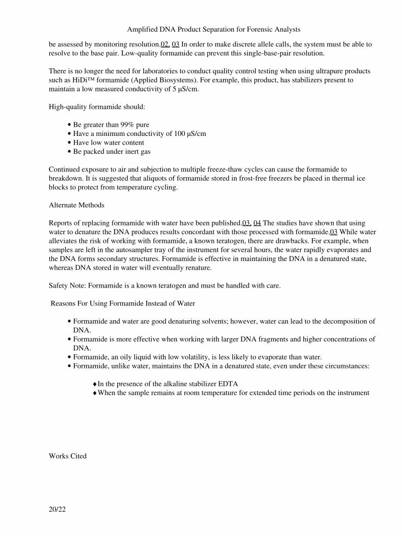
Displayed below is the difference in ladder peak height using formamide with low conductivity (upperelectropheragram) and data using formamide with high conductivity (lower electropheragram).
Note:There is a large difference in the peak height detection between the two samples; the highlighted allele in thetop panel is 898 relative fluorescence units (RFUs) vs. 122 RFUs for the same allele in the presence offormamide with high conductivity. Note that formate ions are not fluorescently labeled, and no fluorescenceis detected with the electrokinetic injection of these by-products.Electrokinetic injection does not discriminate between charged DNA fragments and formate ions; both ofthese charged particles compete for injection. In the presence of formate ions, the result is fewer DNAfragments being injected into the capillary due to competition. As shown in the figure above, the effect ofpreferential formate ion injection can be extremely detrimental. In addition, signal can be decreased forsamples with high salt concentrations. By adding a very small amount of amplified product in the presence offormamide, the salts in a sample are very dilute.
Resolution is another means to evaluate the quality of formamide. The resolution is a measure of how well thepeaks are separated. The overall performance of the capillary and all reagents used in the electrophoresis can
Amplified DNA Product Separation for Forensic Analysts
19/22
be assessed by monitoring resolution.02, 03 In order to make discrete allele calls, the system must be able toresolve to the base pair. Low-quality formamide can prevent this single-base-pair resolution.
There is no longer the need for laboratories to conduct quality control testing when using ultrapure productssuch as HiDi™ formamide (Applied Biosystems). For example, this product, has stabilizers present tomaintain a low measured conductivity of 5 µS/cm.
High-quality formamide should:
Be greater than 99% pure• Have a minimum conductivity of 100 µS/cm• Have low water content• Be packed under inert gas•
Continued exposure to air and subjection to multiple freeze-thaw cycles can cause the formamide tobreakdown. It is suggested that aliquots of formamide stored in frost-free freezers be placed in thermal iceblocks to protect from temperature cycling.
Alternate Methods
Reports of replacing formamide with water have been published.03, 04 The studies have shown that usingwater to denature the DNA produces results concordant with those processed with formamide.03 While wateralleviates the risk of working with formamide, a known teratogen, there are drawbacks. For example, whensamples are left in the autosampler tray of the instrument for several hours, the water rapidly evaporates andthe DNA forms secondary structures. Formamide is effective in maintaining the DNA in a denatured state,whereas DNA stored in water will eventually renature.
Safety Note: Formamide is a known teratogen and must be handled with care.
Reasons For Using Formamide Instead of Water
Formamide and water are good denaturing solvents; however, water can lead to the decomposition ofDNA.
•
Formamide is more effective when working with larger DNA fragments and higher concentrations ofDNA.
•
Formamide, an oily liquid with low volatility, is less likely to evaporate than water.• Formamide, unlike water, maintains the DNA in a denatured state, even under these circumstances:
In the presence of the alkaline stabilizer EDTA! When the sample remains at room temperature for extended time periods on the instrument!
•
Works Cited
Amplified DNA Product Separation for Forensic Analysts
20/22
Applied Biosystems. 2000.AmpFSTR® Profiler Plus™ PCR Amplification Kit User's Manual.1. Buel, E., M. LaFountain, M. Schwartz, M. Walkinshaw. 2001. Evaluation of capillary electrophoresisthrough resolution measurements. J Forensic Sci 46 (2): 341–5.
2.
Butler, J. M., E. Buel, F. Crivellente, and B. R. McCord. 2004. Forensic DNA typing by capillaryelectrophoresis using the ABI Prism 310 and 3100 genetic analyzers for STR analysis.Electrophoresis 25 (10–11): 1397–412.
3.
Biega, L. A., and B. W. Duceman. 1999. Substitution of H2O for formamide in the samplepreparation protocol for STR analysis using the capillary electrophoresis system: The effects onprecision, resolution, and capillary life. J Forensic Sci 44 (5): 1029–31.
4.
Author: Rhonda RobyRhonda K. Roby, MPH, has 17 years experience in the applications of DNA technology for forensicand human identification DNA testing. She is currently pursuing her doctoral degree in ForensicGenetics and Evolution at the University of Granada in Spain and is conducting research in support ofthe NIJ's Missing Persons Program at the University of North Texas Health Science Center. Her mostrecent study, Forensic DNA Databasing: Expert Systems for High-Throughput Analysis of SingleSource Samples, is forthcoming from the National Institute of Justice publications department.Author: Debbie FigarelliDebbie Figarelli serves as DNA Technical Leader at the National Forensic Science Technology Center.Debbie assists with the development of DNA training programs and participates in compliance auditsof DNA laboratories.Author: Rhonda RobyRhonda K. Roby, MPH, has 17 years experience in the applications of DNA technology for forensicand human identification DNA testing. She is currently pursuing her doctoral degree in ForensicGenetics and Evolution at the University of Granada in Spain and is conducting research in support ofthe NIJ's Missing Persons Program at the University of North Texas Health Science Center. Her mostrecent study, Forensic DNA Databasing: Expert Systems for High-Throughput Analysis of SingleSource Samples, is forthcoming from the National Institute of Justice publications department.Author: Debbie FigarelliDebbie Figarelli serves as DNA Technical Leader at the National Forensic Science Technology Center.Debbie assists with the development of DNA training programs and participates in compliance auditsof DNA laboratories.Author: Rhonda RobyRhonda K. Roby, MPH, has 17 years experience in the applications of DNA technology for forensicand human identification DNA testing. She is currently pursuing her doctoral degree in ForensicGenetics and Evolution at the University of Granada in Spain and is conducting research in support ofthe NIJ's Missing Persons Program at the University of North Texas Health Science Center. Her mostrecent study, Forensic DNA Databasing: Expert Systems for High-Throughput Analysis of SingleSource Samples, is forthcoming from the National Institute of Justice publications department.Author: Debbie Figarelli
Amplified DNA Product Separation for Forensic Analysts
21/22

Debbie Figarelli serves as DNA Technical Leader at the National Forensic Science Technology Center.Debbie assists with the development of DNA training programs and participates in compliance auditsof DNA laboratories.
Amplified DNA Product Separation for Forensic Analysts
22/22


![Crime Scene and DNA Basics for Forensic Analysts[1]](https://img.pdfslide.us/doc/110x75/5469ec6bb4af9fba2b8b4984/crime-scene-and-dna-basics-for-forensic-analysts1.jpg)