Embed Size (px)
Citation preview
Amplification of P. falciparum Cytoadherence throughInduction of a Pro-Adhesive State in Host EndotheliumYang Wu1, Tadge Szestak1, Monique Stins2, Alister G. Craig1*
1 Liverpool School of Tropical Medicine, Liverpool, United Kingdom, 2 RT Johnson Division of NeuroImmunology, Johns Hopkins School of Medicine, Baltimore, Maryland,
United States of America
Abstract
This study examined the ability of P.falciparum-infected erythrocytes (IE) to induce a pro-adhesive environment in the hostendothelium during malaria infection, prior to the systemic cytokine activation seen in the later phase of disease. Previouswork had shown increases in receptor levels but had not measured to actual impact on IE binding. Using a co-culturesystem with a range of endothelial cells (EC) and IE with different cytoadherent properties, we have characterised thespecific expression of adhesion receptors and subsequent IE binding by FACS and adhesion assays. We have also examinedthe specific signalling pathways induced during co-culture that are potentially involved in the induction of receptorexpression. The results confirmed that ICAM-1 is up-regulated, albeit at much lower levels than seen with TNF activation, inresponse to co-culture with infected erythrocytes in all three tissue endothelial cell types tested but that up-regulation ofVCAM-1 is tissue-dependent. This small increase in the levels of EC receptors correlated with large changes in IE adhesionability. Co-culture with either RBC or IE increased the potential of subsequent adhesion indicating priming/modulationeffects on EC which make them more susceptible to adhesion and thereby the recruitment of IE. Trypsin surface digestion ofIE and the use of a Pfsbp1-knockout (ko) parasite line abrogated the up-regulation of ICAM-1 and reduced IE binding to ECsuggesting that PfEMP-1 and other molecules exported to the IE surface via the PfSBP1 pathway are major mediators of thisphenotype. This was also supported by the higher induction of EC adhesion receptors by adherent IE compared to isogenic,non-adherent lines.
Citation: Wu Y, Szestak T, Stins M, Craig AG (2011) Amplification of P. falciparum Cytoadherence through Induction of a Pro-Adhesive State in HostEndothelium. PLoS ONE 6(10): e24784. doi:10.1371/journal.pone.0024784
Editor: Georges Snounou, Universite Pierre et Marie Curie, France
Received May 25, 2011; Accepted August 17, 2011; Published October 17, 2011
Copyright: � 2011 Wu et al. This is an open-access article distributed under the terms of the Creative Commons Attribution License, which permits unrestricteduse, distribution, and reproduction in any medium, provided the original author and source are credited.
Funding: This work was funded by the Wellcome Trust (grant number 085391). The funders had no role in study design, data collection and analysis, decision topublish, or preparation of the manuscript.
Competing Interests: The authors have declared that no competing interests exist.
* E-mail: [email protected]
Introduction
Plasmodium falciparum infection is a major cause of severe disease
associated with a range of clinical syndromes including cerebral
malaria (CM). The exact nature of the pathology underlying
severe malaria is not fully understood but two main mechanisms
have been considered, namely cytoadherence and inflammation.
Organ specific pathogenesis such as that seen in CM is believed to
be mediated by sequestering infected erythrocytes (IE) on
endothelial cells (EC), and/or by rosetting between infected and
uninfected erythrocytes to form clumps in the microvasculature of
major organs [1–4]. Several immune and inflammatory mediators
are likely to be heavily implicated in the process leading to this
[5,6]. Factors such as high levels of plasma pro-inflammatory
cytokines and circulating immune-complexes might play a role in
activating/damaging EC in the course of infection [7].
The primary mechanism of cytoadherence of the asexual-stage
P. falciparum IE is complex involving a range of host receptors (e.g.
intercellular adhesion molecule-1 (ICAM-1), CD36, CD31 and P-
selectin) interacting with a family of parasite-encoded proteins
(mainly the variable and diverse var gene products, encoding
PfEMP-1 (Plasmodium falciparum erythrocyte membrane protein-1)),
that are displayed on the surface of IE [8]. This is further
complicated by the ability of these receptors to cooperate in
achieving efficient IE binding to EC [9–11]. Additionally, the
production of high levels of TNF and other pro-inflammatory
mediators is a key feature of malaria infection which contributes to
the systemic and organ-related malaria syndromes [12]. These
pro-inflammatory cytokines can produce endothelial activation by
up-regulating or de novo synthesis of adhesion receptors and
cytokines to increase sequestration of PRBCs within the
microvasculature and, in some cases, contribute to the develop-
ment of chronic inflammation by recruiting leukocytes into tissues
[13,14]. For example, ICAM-1 is markedly up-regulated in severe
malaria and has been implicated as being involved in progression
to cerebral disease [15–17].
Correlation between disease and cytoadherence phenotype is
not simple but has been demonstrated in a number of cases
[3,18,19]. Using var gene upstream regions as molecular tags,
additional associations between PfEMP1 variant type and severe
malaria have been revealed [20,21]. Many studies have suggested
that adhesion molecules play a role in malaria pathogenesis and
patient survival [22,23]. However, in the early stages of infection
although some indication of inflammation has been noted in
human experimental infections [24], elevated adhesion molecule
expression such as ICAM-1 has not been observed. Thus the
parasite faces a challenge to modify the host environment to
support IE adhesion during the early stages of infection in the
PLoS ONE | www.plosone.org 1 October 2011 | Volume 6 | Issue 10 | e24784
erythrocytic cycle; the ability to modulate the host environment to
produce efficient cytoadherence would be of great benefit to
parasite survival and transmission.
A number of groups have examined the effect of IE on EC
responses in co-culture systems with respect to functional
outcomes on EC such as cell adhesion, cell apoptosis and/or
survival, inflammatory responses and signal transduction. The
first report was delivered by Udeinya and Akogyeram in 1993,
who showed induction of adhesiveness in HUVEC by P.falci-
parum-infected erythrocytes up to 250% and that this induction
was related to direct contact via EC adhesive ligands [25,26]. In
2005, Viebig et al showed direct induction of ICAM-1 on
HUVEC by IE [27], while other work by our group showed the
induction of ICAM-1 required low concentrations of TNF [28].
A fourth study has demonstrated that IE alone do not have the
ability to induce ICAM-1 in HUVEC but could do so in brain-
derived EC [29]. In these experiments, IE induced dose- and
exposure-dependent ICAM-1 up-regulation in HBMEC was
seen, with both membrane-associated IE and parasite-derived
soluble factors involved in this process. Co-culture of IE also
induced nuclear translocation of NF-kB in HBMEC, which is
linked to the ICAM-1 expression [7,29]. All of these studies have
confirmed that cell-cell contact is a critical step for the activation
of endothelial cells and the required direct physical contact can
be as little as 30 to 60 minutes [29]. In all cases there was no
requirement of high doses of TNF for adhesion molecule
induction. These responses would be expected to produce a
pro-adhesive state increasing subsequent IE cytoadherence, as
also demonstrated for IE-induced ectophosphorylation of CD36
[30], but this has not been performed. Therefore there is a need
to extend this work using multiple endothelial cell types and
parasite lines to verify the hypothesis that increased cytoadher-
ence can be mediated by previous IE exposure and to understand
the mechanisms underpinning this.
Most investigations studying the role of IE on endothelial
activation use HUVEC [31] and human dermal microvascular
endothelial cells (HDMEC) [32], both of which are primary cells
and approximate the physiological features of in vivo cells more
closely than cloned or transformed cell lines but are not derived
from one of the key tissues involved in cerebral malaria, namely
the brain. There are some examples of the use of primary brain-
derived EC, mainly from the Stins group [33]. These cells, which
possess the ability to form tight junctions, are critical to the
integrity of the blood-brain barrier and serve as the best in vitro
model for understanding the impact of parasite-EC interactions in
cerebral malaria (CM) [34,35]. However they are difficult to
obtain and are routinely replaced by immortalised lines that
resemble the behaviour of primary cells. This is important as
different EC subtypes vary widely in their expression profiles; for
example, HUVEC do not express CD36 nor the chemokine
receptor CXCR4 [36]; HDMEC express CD36; human brain
microvascular endothelial cells (HBMEC) express CXCR4 [28].
ItG, A4 and C24 [37,38] are isogenic parasite lines that have
distinctive cytoadherence properties; ItG binds strongly to ICAM-
1 and to CD36, A4 binds strongly to CD36 and to ICAM-1
moderately, and C24 binds strongly to CD36 but binding to
ICAM-1 is minimal. These strain-specific properties, based on
variable expression of PfEMP1, enable different IE-EC interac-
tions [39]. In this study we have used an in vitro co-culture model
where intact IE are placed in direct contact with different human
EC monolayers (different in the adhesion receptors displayed and
their responsiveness to inflammatory cytokines) to evaluate
whether co-culture with normal or parasitized erythrocytes (with
adhesive and non-adhesive lines) activate endothelial cells in vitro
and particularly whether they up-regulate adhesion molecule
expression that translates into a pro-adhesive effect.
Results
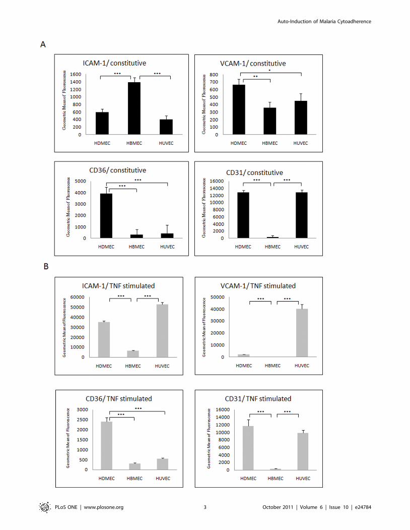
Expression levels of endothelial cell adhesion moleculesAdhesion molecules were expressed at different levels in the
three un-stimulated endothelial cell lines. As seen in Figure 1A,
HBMEC had the highest constitutive expression level of ICAM-1,
significantly higher than HDMEC and HUVEC (p,0.001).
HDMEC had the highest constitutive expression level of CD36,
significantly higher than HBMEC and HUVEC (p,0.001). The
constitutive expression level of CD31 in HBMEC was significantly
lower than both HDMEC and HUVEC (p,0.001). The levels of
VCAM-1 were slightly higher in HDMEC compared to HBMEC
and HUVEC. In response to TNF stimulation all three cell types
increased ICAM-1 expression significantly, although HUVEC had
the highest levels while HBMEC had relatively much lower
expression levels. VCAM-1 expression was significantly increased
only in HUVEC. CD36 was significantly decreased after TNF
stimulation in HDMEC but was still relatively high compared to
the levels seen for HUVEC and HBMEC, which remained
unaffected (and at low levels) by TNF. CD31 levels showed, as
expected, no significant changes on FACS after TNF stimulation
on all three EC types (Figure 1B).
Kinetics of ICAM-1expression in the three endothelial celllines
We used ICAM-1 as an example to measure the dynamic
changes in receptor expression after co-culture with IE (ItG in this
experiment) with the three endothelial cell types. Co-culture of ItG
with HBMEC or HUVEC induced a significant increase in
ICAM-1 expression starting from 3 hours co-culture, while in
HDMEC it took 8 hours to produce a significant increase.
However, the maximum ICAM-1 induction in all three endothe-
lial cell lines was overnight co-culture (16 hours). The rate of
increase in HBMEC was greater than that in HDMEC and
HUVEC (data not shown).
Expression patterns of EC adhesion molecules in co-culture models
Variable levels of receptor expression on EC in response to IE
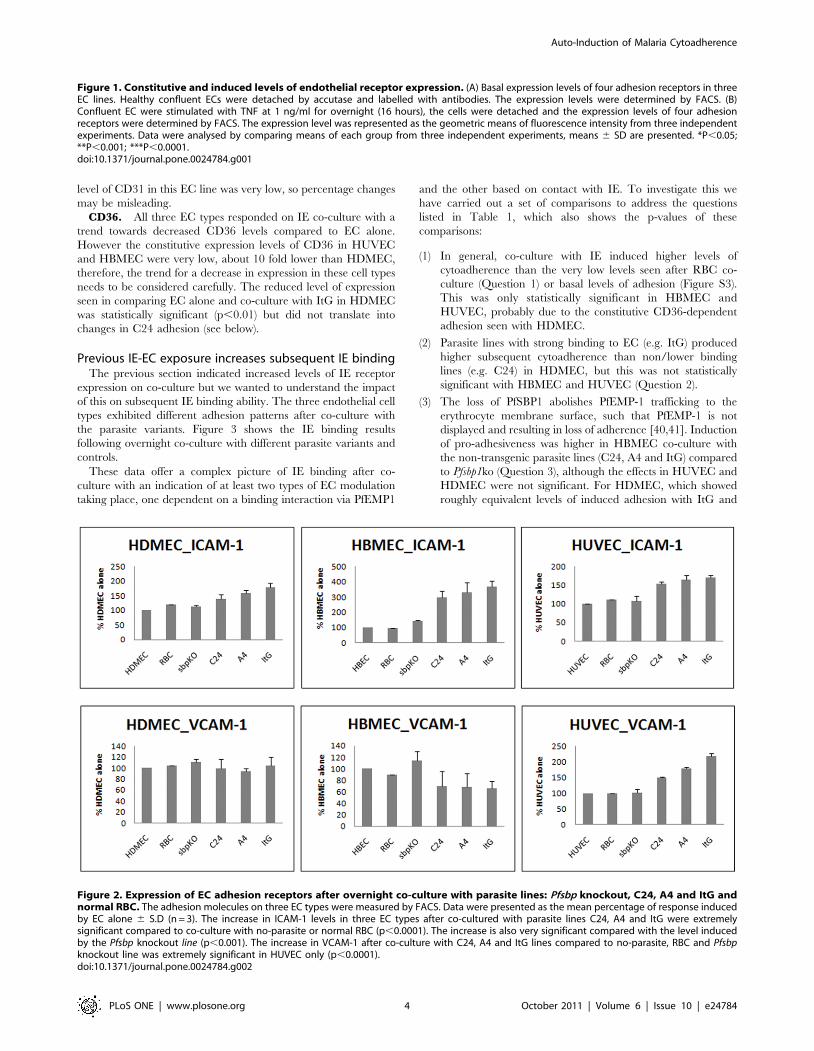
co-culture were observed (Figure 2 and Figure S1).ICAM-1. The expression of ICAM-1 increased significantly
compared to EC alone after co-culturing with IE for the three
parasite lines with all three endothelial cell types regardless of
ICAM-1-binding (A4 and ItG) or non-binding (C24) parasite
strains, with a gradation in response such that ItG.A4.C24.
However the levels of ICAM-1 in all three EC lines on co-culture
were significantly higher than co-culture with the Pfsbp1 knockout
parasite line only when the ICAM-1-binding lines A4 and ItG
were used.
VCAM-1. The expression of VCAM-1 increased significantly
after co-culture with IE for all three parasite lines in HUVEC only
and these effects were significantly higher than co-culturing with
the Pfsbp1 knockout strain. There was no significant induction or
inhibition of VCAM-1 in HDMEC or HBMEC.CD31. Co-culture with IE with all three PfEMP1-expressing
parasite lines decreased CD31 expression in the three EC types,
although the percentage changes were different. There appeared
to be a small but consistent reduction in CD31 levels on HUVEC
and HDMEC when cultured with IE (regardless of the expression
of PfEMP1). The consistent reduction seen with HBMEC is
interesting but it should be noted that the constitutive expression
Auto-Induction of Malaria Cytoadherence
PLoS ONE | www.plosone.org 2 October 2011 | Volume 6 | Issue 10 | e24784
Auto-Induction of Malaria Cytoadherence
PLoS ONE | www.plosone.org 3 October 2011 | Volume 6 | Issue 10 | e24784
level of CD31 in this EC line was very low, so percentage changes
may be misleading.
CD36. All three EC types responded on IE co-culture with a
trend towards decreased CD36 levels compared to EC alone.
However the constitutive expression levels of CD36 in HUVEC
and HBMEC were very low, about 10 fold lower than HDMEC,
therefore, the trend for a decrease in expression in these cell types
needs to be considered carefully. The reduced level of expression
seen in comparing EC alone and co-culture with ItG in HDMEC
was statistically significant (p,0.01) but did not translate into
changes in C24 adhesion (see below).
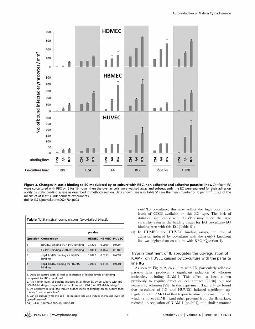
Previous IE-EC exposure increases subsequent IE bindingThe previous section indicated increased levels of IE receptor
expression on co-culture but we wanted to understand the impact
of this on subsequent IE binding ability. The three endothelial cell
types exhibited different adhesion patterns after co-culture with
the parasite variants. Figure 3 shows the IE binding results
following overnight co-culture with different parasite variants and
controls.
These data offer a complex picture of IE binding after co-
culture with an indication of at least two types of EC modulation
taking place, one dependent on a binding interaction via PfEMP1
and the other based on contact with IE. To investigate this we
have carried out a set of comparisons to address the questions
listed in Table 1, which also shows the p-values of these
comparisons:
(1) In general, co-culture with IE induced higher levels of
cytoadherence than the very low levels seen after RBC co-
culture (Question 1) or basal levels of adhesion (Figure S3).
This was only statistically significant in HBMEC and
HUVEC, probably due to the constitutive CD36-dependent
adhesion seen with HDMEC.
(2) Parasite lines with strong binding to EC (e.g. ItG) produced
higher subsequent cytoadherence than non/lower binding
lines (e.g. C24) in HDMEC, but this was not statistically
significant with HBMEC and HUVEC (Question 2).
(3) The loss of PfSBP1 abolishes PfEMP-1 trafficking to the
erythrocyte membrane surface, such that PfEMP-1 is not
displayed and resulting in loss of adherence [40,41]. Induction
of pro-adhesiveness was higher in HBMEC co-culture with
the non-transgenic parasite lines (C24, A4 and ItG) compared
to Pfsbp1ko (Question 3), although the effects in HUVEC and
HDMEC were not significant. For HDMEC, which showed
roughly equivalent levels of induced adhesion with ItG and
Figure 2. Expression of EC adhesion receptors after overnight co-culture with parasite lines: Pfsbp knockout, C24, A4 and ItG andnormal RBC. The adhesion molecules on three EC types were measured by FACS. Data were presented as the mean percentage of response inducedby EC alone 6 S.D (n = 3). The increase in ICAM-1 levels in three EC types after co-cultured with parasite lines C24, A4 and ItG were extremelysignificant compared to co-culture with no-parasite or normal RBC (p,0.0001). The increase is also very significant compared with the level inducedby the Pfsbp knockout line (p,0.001). The increase in VCAM-1 after co-culture with C24, A4 and ItG lines compared to no-parasite, RBC and Pfsbpknockout line was extremely significant in HUVEC only (p,0.0001).doi:10.1371/journal.pone.0024784.g002
Figure 1. Constitutive and induced levels of endothelial receptor expression. (A) Basal expression levels of four adhesion receptors in threeEC lines. Healthy confluent ECs were detached by accutase and labelled with antibodies. The expression levels were determined by FACS. (B)Confluent EC were stimulated with TNF at 1 ng/ml for overnight (16 hours), the cells were detached and the expression levels of four adhesionreceptors were determined by FACS. The expression level was represented as the geometric means of fluorescence intensity from three independentexperiments. Data were analysed by comparing means of each group from three independent experiments, means 6 SD are presented. *P,0.05;**P,0.001; ***P,0.0001.doi:10.1371/journal.pone.0024784.g001
Auto-Induction of Malaria Cytoadherence
PLoS ONE | www.plosone.org 4 October 2011 | Volume 6 | Issue 10 | e24784
Pfsbp1ko co-culture, this may reflect the high constitutive
levels of CD36 available on this EC type. The lack of
statistical significance with HUVEC may reflect the large
variability seen in the binding assays for ItG co-culture/ItG
binding seen with this EC (Table S1).
(4) In HDMEC and HUVEC binding assays, the level of
adhesion induced by co-culture with the Pfsbp-1 knockout
line was higher than co-culture with RBC (Question 4).
Trypsin treatment of IE abrogates the up-regulation ofICAM-1 on HUVEC caused by co-culture with the parasiteline ItG
As seen in Figure 2, co-culture with IE, particularly adhesive
parasite lines, produces a significant induction of adhesion
molecules, including ICAM-1. This effect has been shown
previously to require direct cell-cell contact [29,30] but not
necessarily adhesion [29]. In this experiment (Figure 4) we found
that co-culture of ItG and HUVEC induced significant up-
regulation of ICAM-1 but that trypsin treatment of co-cultured IE,
which removes PfEMP1 (and other proteins) from the IE surface,
reduced up-regulation of ICAM-1 (p,0.01), in a similar manner
Figure 3. Changes in static binding to EC modulated by co-culture with RBC, non-adhesive and adhesive parasite lines. Confluent ECwere co-cultured with RBC or IE for 16 hours, then the overlay cells were washed away and subsequently the EC were analysed for their adhesionability by static binding assays as described in methods section. Data shown (see also Table S1) are the mean number of IE per mm2 6 S.E of themeans of at least 4 independent experiments.doi:10.1371/journal.pone.0024784.g003
Table 1. Statistical comparisons (two-tailed t-test).
p-value
Question Comparison HDMEC HBMEC HUVEC
1 RBC/ItG binding vs A4/ItG binding 0.1200 0.0029 0.0007
2 C24/ItG binding vs ItG/ItG binding 0.0004 0.1652 0.1185
3 sbp1 ko/ItG binding vs ItG/ItGbinding
0.2072 0.0253 0.4092
4 sbp1 ko/ItG binding vs RBC/ItGbinding
0.0030 0.3125 0.0001
1. Does co-culture with IE lead to induction of higher levels of bindingcompared to RBC co-culture?2. Are higher levels of binding induced in all three EC by co-culture with ItG(ICAM-1-binding) compared to co-culture with C24 (non ICAM-1-binding)?3. Do adherent IE (e.g. ItG) induce higher levels of binding on co-culture thanthe sbp1 ko parasite line?4. Can co-culture with the sbp1 ko parasite line also induce increased levels ofcytoadherence?doi:10.1371/journal.pone.0024784.t001
Auto-Induction of Malaria Cytoadherence
PLoS ONE | www.plosone.org 5 October 2011 | Volume 6 | Issue 10 | e24784
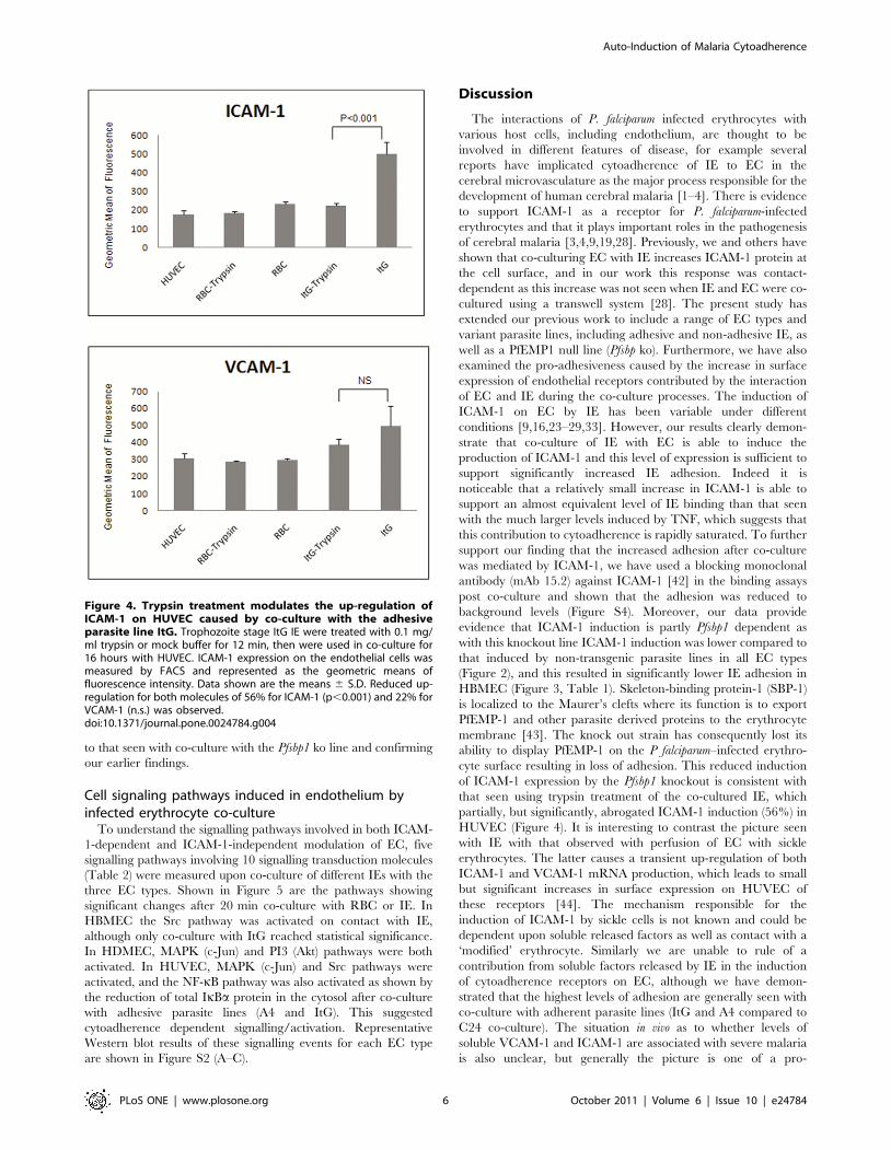
to that seen with co-culture with the Pfsbp1 ko line and confirming
our earlier findings.
Cell signaling pathways induced in endothelium byinfected erythrocyte co-culture
To understand the signalling pathways involved in both ICAM-
1-dependent and ICAM-1-independent modulation of EC, five
signalling pathways involving 10 signalling transduction molecules
(Table 2) were measured upon co-culture of different IEs with the
three EC types. Shown in Figure 5 are the pathways showing
significant changes after 20 min co-culture with RBC or IE. In
HBMEC the Src pathway was activated on contact with IE,
although only co-culture with ItG reached statistical significance.
In HDMEC, MAPK (c-Jun) and PI3 (Akt) pathways were both
activated. In HUVEC, MAPK (c-Jun) and Src pathways were
activated, and the NF-kB pathway was also activated as shown by
the reduction of total IkBa protein in the cytosol after co-culture
with adhesive parasite lines (A4 and ItG). This suggested
cytoadherence dependent signalling/activation. Representative
Western blot results of these signalling events for each EC type
are shown in Figure S2 (A–C).
Discussion
The interactions of P. falciparum infected erythrocytes with
various host cells, including endothelium, are thought to be
involved in different features of disease, for example several
reports have implicated cytoadherence of IE to EC in the
cerebral microvasculature as the major process responsible for the
development of human cerebral malaria [1–4]. There is evidence
to support ICAM-1 as a receptor for P. falciparum-infected
erythrocytes and that it plays important roles in the pathogenesis
of cerebral malaria [3,4,9,19,28]. Previously, we and others have
shown that co-culturing EC with IE increases ICAM-1 protein at
the cell surface, and in our work this response was contact-
dependent as this increase was not seen when IE and EC were co-
cultured using a transwell system [28]. The present study has
extended our previous work to include a range of EC types and
variant parasite lines, including adhesive and non-adhesive IE, as
well as a PfEMP1 null line (Pfsbp ko). Furthermore, we have also
examined the pro-adhesiveness caused by the increase in surface
expression of endothelial receptors contributed by the interaction
of EC and IE during the co-culture processes. The induction of
ICAM-1 on EC by IE has been variable under different
conditions [9,16,23–29,33]. However, our results clearly demon-
strate that co-culture of IE with EC is able to induce the
production of ICAM-1 and this level of expression is sufficient to
support significantly increased IE adhesion. Indeed it is
noticeable that a relatively small increase in ICAM-1 is able to
support an almost equivalent level of IE binding than that seen
with the much larger levels induced by TNF, which suggests that
this contribution to cytoadherence is rapidly saturated. To further
support our finding that the increased adhesion after co-culture
was mediated by ICAM-1, we have used a blocking monoclonal
antibody (mAb 15.2) against ICAM-1 [42] in the binding assays
post co-culture and shown that the adhesion was reduced to
background levels (Figure S4). Moreover, our data provide
evidence that ICAM-1 induction is partly Pfsbp1 dependent as
with this knockout line ICAM-1 induction was lower compared to
that induced by non-transgenic parasite lines in all EC types
(Figure 2), and this resulted in significantly lower IE adhesion in
HBMEC (Figure 3, Table 1). Skeleton-binding protein-1 (SBP-1)
is localized to the Maurer’s clefts where its function is to export
PfEMP-1 and other parasite derived proteins to the erythrocyte
membrane [43]. The knock out strain has consequently lost its
ability to display PfEMP-1 on the P falciparum–infected erythro-
cyte surface resulting in loss of adhesion. This reduced induction
of ICAM-1 expression by the Pfsbp1 knockout is consistent with
that seen using trypsin treatment of the co-cultured IE, which
partially, but significantly, abrogated ICAM-1 induction (56%) in
HUVEC (Figure 4). It is interesting to contrast the picture seen
with IE with that observed with perfusion of EC with sickle
erythrocytes. The latter causes a transient up-regulation of both
ICAM-1 and VCAM-1 mRNA production, which leads to small
but significant increases in surface expression on HUVEC of
these receptors [44]. The mechanism responsible for the
induction of ICAM-1 by sickle cells is not known and could be
dependent upon soluble released factors as well as contact with a
‘modified’ erythrocyte. Similarly we are unable to rule of a
contribution from soluble factors released by IE in the induction
of cytoadherence receptors on EC, although we have demon-
strated that the highest levels of adhesion are generally seen with
co-culture with adherent parasite lines (ItG and A4 compared to
C24 co-culture). The situation in vivo as to whether levels of
soluble VCAM-1 and ICAM-1 are associated with severe malaria
is also unclear, but generally the picture is one of a pro-
Figure 4. Trypsin treatment modulates the up-regulation ofICAM-1 on HUVEC caused by co-culture with the adhesiveparasite line ItG. Trophozoite stage ItG IE were treated with 0.1 mg/ml trypsin or mock buffer for 12 min, then were used in co-culture for16 hours with HUVEC. ICAM-1 expression on the endothelial cells wasmeasured by FACS and represented as the geometric means offluorescence intensity. Data shown are the means 6 S.D. Reduced up-regulation for both molecules of 56% for ICAM-1 (p,0.001) and 22% forVCAM-1 (n.s.) was observed.doi:10.1371/journal.pone.0024784.g004
Auto-Induction of Malaria Cytoadherence
PLoS ONE | www.plosone.org 6 October 2011 | Volume 6 | Issue 10 | e24784
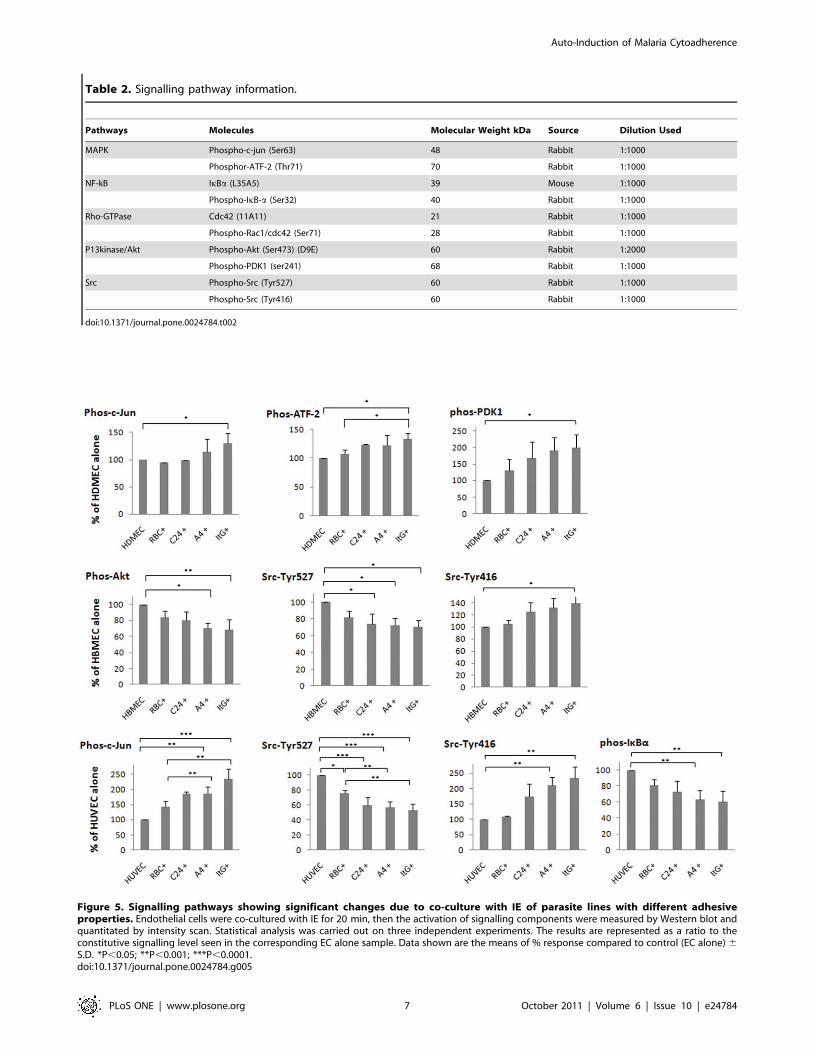
Figure 5. Signalling pathways showing significant changes due to co-culture with IE of parasite lines with different adhesiveproperties. Endothelial cells were co-cultured with IE for 20 min, then the activation of signalling components were measured by Western blot andquantitated by intensity scan. Statistical analysis was carried out on three independent experiments. The results are represented as a ratio to theconstitutive signalling level seen in the corresponding EC alone sample. Data shown are the means of % response compared to control (EC alone) 6
S.D. *P,0.05; **P,0.001; ***P,0.0001.doi:10.1371/journal.pone.0024784.g005
Table 2. Signalling pathway information.
Pathways Molecules Molecular Weight kDa Source Dilution Used
MAPK Phospho-c-jun (Ser63) 48 Rabbit 1:1000
Phosphor-ATF-2 (Thr71) 70 Rabbit 1:1000
NF-kB IkBa (L35A5) 39 Mouse 1:1000
Phospho-IkB-a (Ser32) 40 Rabbit 1:1000
Rho-GTPase Cdc42 (11A11) 21 Rabbit 1:1000
Phospho-Rac1/cdc42 (Ser71) 28 Rabbit 1:1000
P13kinase/Akt Phospho-Akt (Ser473) (D9E) 60 Rabbit 1:2000
Phospho-PDK1 (ser241) 68 Rabbit 1:1000
Src Phospho-Src (Tyr527) 60 Rabbit 1:1000
Phospho-Src (Tyr416) 60 Rabbit 1:1000
doi:10.1371/journal.pone.0024784.t002
Auto-Induction of Malaria Cytoadherence
PLoS ONE | www.plosone.org 7 October 2011 | Volume 6 | Issue 10 | e24784
inflammatory disease with concomitant increases in endothelial
biomarkers in patients with severe disease [45–47].
The binding patterns seen with HDMEC after co-culture are
complicated, for example, Pfsbp-1 knockout IE does not increase
ICAM-1 surface expression on EC but induced a similar IE
binding profile to that seen with ItG co-incubation. This is
probably mainly due to the presence of CD36 on HDMEC, which
is constitutively expressed on this type of endothelium, but this
does not fully explain the binding profiles produced by co-culture
with C24 and A4 that result in lower subsequent binding than co-
culture with the Pfsbp1 knockout line. The role of CD36 in this
effect is supported by the absence of this pattern in HBMEC and
HUVEC, which do not express significant amounts of CD36 on
their surface. Further work will be required to dissect the specific
receptor contributions to adhesion in the HDMEC system.
The co-culture of IE with endothelial cells results in not only
significant changes in the levels of adhesion molecules, but these
translate into enhanced binding of IE to EC. Phenotypically, the
effects caused by IE co-culture, particularly ItG, resemble those
induced by TNF stimulation but vary according to EC type
(Figure 3 & data not shown). The response of EC to several
cytokines, especially TNF, has been considered as one of the
important determinants of pathology during infection with a
number of pathogens [48–50]. However, in vitro and in vivo studies
do not always find a consistent correlation between TNF level and
disease [51,52], and TNF is implicated in mediating both
protection and pathogenicity during malaria infection [53].
Specific MAPK pathways have been linked to severe malaria
through work on the response of macrophages to P. falciparum GPI,
normally resulting in TNF secretion. Despite this it has been
difficult to find a direct link between high levels of TNF and severe
malaria disease, particularly in children [54], and this could be, in
part, due to the low threshold seen in our work for maximal IE
adhesion at levels of receptor expression, much lower than that
induced by high levels of TNF. Thus TNF may play some role,
such as ensuring sufficient receptor expression for efficient
cytoadherence, but that progression to severe disease such as
cerebral malaria requires other factors for its aetiology. EC do not
secrete appreciable amounts of TNF [55], however the interaction
between ICAM-1 and IE may not only be acting as an adhesion
molecule for IE, targeting them to tissues such as the brain, but
also directly contributing to the inflammatory response of
endothelium by altering intracellular signals [48] resulting in
rearrangements of the actin cytoskeleton and activation of local
inflammatory cascades (5).
In our studies on var gene expression by IE in different tissues
from the Malawi PM study [56] we have seen tissue-specific
distribution of different variants, which supports IE accumulation
based on receptor usage and the potential for differential
pathology in different organs due to non-random distribution of
IE during sequestration. For example, cerebral endothelium
performs a critical function with the blood-brain barrier (BBB)
forming a tight barrier to maintain homeostasis for adjacent
neuronal cells. Several lines of evidence suggest that BBB function
is impaired during cerebral malaria [57]. One mechanism for this
may be the intracellular signals produced by specific IE
cytoadherence to EC receptors. Our previous studies have shown
that JNK, ERK-1/2 and p38 MAP kinases can be activated in
TNF-stimulated EC (expressing high levels of ICAM-1), which is
dependent on ICAM-1 mediated IE adhesion [58]. This is
consistent with the finding that cross-linking of ICAM-1 with
monoclonal antibodies activates the MAPK kinases ERK-1/2
and/or JNK in EC [59,60]. In brain microvascular EC, ICAM-1
can trigger Src tyrosine kinase activity and tyrosine phosphory-
lation on cortactin [61]. In the present study, we found that
MAPK pathways were activated in HUVEC and HDMEC, but
not in HBMEC. PI3 modulation was observed in HDMEC and
HUVEC, but with HDMEC showing a significant activation in
ItG co-culture, whereas PI3 was deactivated in HBMEC after co-
culture with A4 and ItG. In general the intensity of signalling
activation was correlated with the adhesion avidity of the parasite
line, along similar lines to the behaviour observed previously [58].
Other signalling pathways are also affected by IE co-culture (Src
and NFkB), again demonstrating IE and endothelial specificity for
this effect, although it is possible that we have missed some events
through the use of standard timings to measure a spectrum of
signalling activities. Nevertheless the picture produced is a
complex one, suggesting modulation of specific signalling path-
ways in different tissue EC in response to differential stimulation
by parasite variants. Further work will be needed to dissect the
critical signalling pathways contributing to the variety of EC
phenotypes demonstrated on IE co-culture.
Taken together, the present study has systematically investigat-
ed the expression of adhesion receptors on EC in response to IE
co-culture. It appears that increased expression of ICAM-l is
responsible for at least some of the observed enhancement in IE
adhesiveness. These events are mediated, in part, by surface
molecules on IE, the most likely candidate being PfEMP-1,
although we cannot discount a role for other surface proteins. Our
findings are in line with previous data [7,24–28,62], suggesting
that certain parasite variants, e.g. high avidity binders, have the
ability to modulate endothelium to produce a pro-adhesive state
preceding a systemic pro-inflammatory host response, and tri-
ggering specific signalling pathways that could lead to localised EC
dysfunction. The induction of adhesion molecules is likely to
create a vascular environment that favours adhesion events,
lengthens microcirculation transient times and thus supports the
concomitant recruitment of IE, platelets and (potentially) other
blood cells to bind to the endothelium of microvessels, leading to
the blockage of blood vessels as seen in the retina of CM patients
[4,63,64].
In conclusion, our observations suggest mechanisms for the
efficient sequestration of IE seen early in an infection in the
absence of profound systemic host activation and the pathophys-
iology of severe malaria based on the targeted recruitment of
parasite variants to specific tissues. A better understanding of these
mechanisms will help us to design effective therapies aimed at
alleviating the morbidity and mortality associated with severe
disease.
Materials and Methods
Parasite culturePlasmodium falciparum isolates used in this study were: ItG [37],
A4 and C24 [38] and Pfsbp-1 knockout strain (Pfsbp1-ko). The
Pfsbp-1 knockout line was derived from CS2, in which PfEMP-1
transport (along with other proteins) to the RBC surface is
disrupted [40,41]. Parasites were cultured in vitro in group O+
human erythrocytes in RPMI 1640 medium (supplemented with
37.5 mM HEPES, 7 mM D-glucose, 6 mM NaOH, 25 mg ml21
gentamicin sulphate, 2 mM L-glutamine and 10% human serum)
at a pH of 7.2 in a gas mixture of 96% nitrogen, 3% carbon
dioxide and 1% oxygen. To minimize the effect of antigenic
switching in culture, a batch of stabilates was prepared from post-
selection cultures and used for no more than three weeks. For co-
culture experiments, mature trophozoites (20–29 hours after
invasion) were enriched by plasmagel flotation. For adhesion
assays, trophozoites of a later stage (30–34 hours after invasion)
Auto-Induction of Malaria Cytoadherence
PLoS ONE | www.plosone.org 8 October 2011 | Volume 6 | Issue 10 | e24784
were used. All parasite and EC cultures were regularly monitored
for mycoplasma using Takara PCR mycoplasma detection kit
(Cambrex Biosciences, UK).
Trypsin treatmentSurface-exposed PfEMP1 was removed by proteolytic digestion
of parasitized RBCs as described previously [28]. Briefly, IE (ItG
line) were enriched by Plasmagel flotation, and washed three times
with medium without sera and twice with PBS. IE were then
divided into two sets, one set was kept in PBS and another set was
incubated with 0.1 mg/ml TPCK trypsin (Thermo Scientific,
Northumberland, UK) in PBS for 12 min at room temperature
with gentle mixing several times by hand. The reaction was
stopped by adding foetal calf serum (FCS) to a final concentration
of 10%. Both sets were washed three times with PBS and two times
with corresponding EC medium. Treated and untreated IE at
40% parasitaemia and 1% haematocrit (hct) were used in co-
culture for 16 hours, after which ICAM-1 expression on the EC
was measured. The use of high parasitaemia (40%) in co-culture
was to mimic the in vivo situation in which foci of packed vessels are
observed rather than uniform distribution of adherent IE within
the microvasculature.
Endothelial cells and co-culture conditionsPooled primary human umbilical vein endothelial cells
(HUVEC) and pooled human dermal microvascular endothelial
cells (HDMEC) were obtained from Promocell (Heidelberg,
Germany). Cells were maintained in complete growth medium
supplied by Promocell according to the company’s instructions. To
maintain EC features and reduce inter-experiment variation, cells
at passages five to six were used. The human brain microvascular
endothelial cells (HBMEC) cell line was obtained through
collaboration with Monique Stins (Johns Hopkins, USA) and
was grown in MCDB 131 medium (Invitrogen, UK) supplemented
with 10% FCS, 2 mM L-glutamine (Sigma UK), 10 ng/ml
epidermal growth factor (Becton Dickinson UK) and 1 mg/ml
hydrocortisone (Invitrogen UK). These cells were used up to
passage 20. ECs were co-cultured with IE and uninfected RBCs as
previously described [28] with modifications. Briefly, ECs were
grown on 1% gelatin (Sigma, UK) coated in 25 cm2 flasks or 24
well plates with appropriate growth medium until confluent. Then
the culture medium was replaced with either fresh growth medium
(without hydrocortisone) alone or with normal erythrocytes, or
with mature-trophozoite IE (C24, A4, ItG). IE were enriched by
Plasmagel flotation (40% parasitaemia) and were adjusted to 1%
haematocrit (hct). Fresh medium with low TNF (5 pg/ml) or high
TNF (2 ng/ml) (Invitrogen, UK) were also included as controls.
Co-cultures were carried out at 37uC in 5% CO2 incubator for
16 hours.
Cytoadherence AssayThe experimental procedures for cytoadherence assays
[9,28,65] were adapted to study the effects of co-culture on
subsequent cytoadherence. ECs were seeded onto 13 mm
thermanox coverslips (Nunc) coated with 1% gelatin in 24-well
plates. The confluent ECs were co-cultured with normal RBC or
IE of different parasite lines in complete EC medium. EC culture
medium alone and TNF stimulated EC were also included as
controls. After 16 hours co-culture, RBC or IE or TNF were
removed and the EC washed with fresh medium once. The
coverslips with co-cultured EC were picked up and dipped briefly
in water to lyse adherent IEs followed by immediate neutralisation
with binding buffer (RPMI 1640 medium, supplemented with
HEPES and 6 mM glucose, pH 7.2) with gentle shaking to remove
excess red blood cells. The washed coverslips were placed in a new
24 well plate and washed again until there was no RBC or IE on
the EC visible under the microscope. IE used for adhesion assay
were prepared at 1% hct and 3% parasitaemia in binding buffer.
Uninfected RBCs were also cultured for at least 24 hours and used
at 1% hct. 0.5 ml of IE suspension was applied to each well
containing EC and incubated at 37uC for 1 h with gentle
resuspension by rotation every 10 min. Unbound cells were
removed by two washes in binding buffer followed by 2630 min
gravity washes. Cells were fixed with 1% glutaraldehyde/PBS for
1 h. After staining with 5% Geimsa for 30 min, coverslips were
washed in water, air dried and mounted using DPX hard set
mounting medium. 6–10 fields of each cover slip were counted at
3006magnification for the number of bound IE. Each experiment
was performed in duplicate and on three independent occasions.
Results were converted to bound IE/mm2.
Flow cytometryThe expression of endothelial cell markers was measured by
fluorescence-activated cell sorting (FACS) (FACScan; Becton
Dickinson, San Jose, CA). Specific fluorescence-conjugated anti-
bodies were used in this study, including: APC Mouse anti-human
CD54 (ICAM-1), FITC Mouse anti-human CD36, PE Mouse
anti-human CD106 (VCAM-1) and FITC Mouse anti-human
CD31 (PECAM) (Becton Dickinson). Nonspecific fluorescence was
assessed using corresponding isotype control antibodies. After
16 hours of co-culture, the EC culture medium including co-
cultured RBC or IE was gently removed and EC washed once
with fresh medium, followed by three to four further washes with
phosphate-buffered saline (PBS) to remove any adherent IEs. ECs
then were detached by gentle accutase (Sigma UK) treatment,
washed with PBS containing 1% BSA and stained with antibodies
for 60 min on ice in 100 ml of the recommended dilution. Cells
were washed twice in PBS with 1% BSA and a FACS analysis was
carried out using a FACS Calibur with the CellQuest Pro software
(BD Biosciences). The expression of surface molecules was
indicated by geometric mean of the fluorescence intensity.
Statistical analysisValues are reported as means 6 SD for each set of experiments
except for those reported for Figure 3, which are shown as means 6
SEM, as the experiments were conducted independently at least 4
times. The results were evaluated with a two-tailed Student t test using
GraphPad InStat. Statistical significance was defined as p,0.05.
Supporting Information
Figure S1 The expression of EC adhesion receptors CD36 and
CD31 after overnight co-culture with parasite lines: Pfsbp knock-
out, C24, A4 and ItG and normal RBC. The adhesion molecules
on three EC types were measured by FACS. The expression level
was represented as the geometric means of fluorescence intensity.
Data were analysed by comparing means of each group from three
independent experiments.
(TIF)
Figure S2 Endothelial cells were co-cultured with IEs for
20 min, the activation of signalling components were measured
by Western blot. Shown are representatives of Western blots from
the three EC types: (A) HBMEC; (B) HDMEC; and (C) HUVEC.
(TIF)
Figure S3 Eendothelial cells were grown on coverslips in
corresponding growth medium without cytokine stimulation until
confluent. Standard binding assays were conducted using either
Auto-Induction of Malaria Cytoadherence
PLoS ONE | www.plosone.org 9 October 2011 | Volume 6 | Issue 10 | e24784
normal RBC or IE lines as described in the Methods section. Data
shown are the mean number of IE per mm2 6 S.D. (n = 2).
(TIF)
Figure S4 ICAM-1 blocking antibody (15.2) inhibits binding
induced by co-culture. Confluent HUVEC cells were grown on
coverslips and co-cultured with ItG for 16 hours. Then the overlay
cells were washed away (as described in the Methods section) and
subsequently the HUVEC were analysed for their adhesion ability
by static binding assays using the ItG line with or without ICAM-1
blocking antibody (mAb 15.2) or an unrelated control antibody.
Data shown are the mean number of IE per mm2 6 S.D. (n = 2).
(TIF)
Table S1 Data (mean 6 S.E.) for the co-culture IE adhesion assays
described in Figure 3. The data are presented as IE bound/mm2.
(DOCX)
Acknowledgments
The authors wish to thank Profs. Alex Maier and Alan Cowman for
supplying Pfsbp knockout parasite line; Dr. Adam Wright for help with flow
cytometry techniques; the staff and patients of Ward 7Y and the
Gastroenterology Unit, Royal Liverpool Hospital, for their generous
donation of serum for parasite culture.
Author Contributions
Conceived and designed the experiments: YW AGC. Performed the
experiments: YW TS. Analyzed the data: YW AGC. Contributed
reagents/materials/analysis tools: MS. Wrote the paper: YW AGC.
References
1. Berendt AR, Tumer GD, Newbold CI (1994) Cerebral malaria: thesequestration hypothesis. Parasitol Today 10: 412–414.
2. MacPherson GG, Warrell MJ, White NJ, Looareesuwan S, Warrell DA (1985)
Human cerebral malaria, A quantitative ultrastructural analysis of parasitizederythrocyte sequestration. Am J Pathol 119: 385–401.
3. Newbold C, Warn P, Black G, Berendt A, Craig A, et al. (1997) Receptor-
specific adhesion and clinical disease in Plasmodium falciparum. Am J Trop
Med Hyg 57(4): 389–398.
4. Turner GD, Morrison H, Jones M, Davis TM, Looareesuwan S, et al. (1994) Animmunohistochemical study of the pathology of fatal malaria. Evidence for
widespread endothelial activation and a potential role for intercellular adhesionmolecule-1 in cerebral sequestration. Am J Pathol 145(5): 1057–1069.
5. Schofield L, Grau GE (2005) Immunological processes in malaria pathogenesis.
Nat Rev Immuno 5: 722–735.
6. Marsh K, Kinyanjui S (2006) Immune effector mechanisms in malaria. Parasite
Immunol 28: 51–60.
7. Tripathi AK, Sha W, Shulaev V, Stins MF, Sullivan DJ (2009) Plasmodiumfalciparum-infected erythrocytes induce NF-kappaB regulated inflammatory
pathways in human cerebral endothelium. Blood 114(19): 4243–4252.
8. Cooke B, Coppel R, Wahlgren M (2000) Falciparum malaria: sticking up,standing out and out-standing. Parasitol Today 16: 416–420.
9. Gray C, McCormick C, Turner G, Craig AG (2003) ICAM-1 can play a major
role in mediating P. falciparum adhesion to endothelium under flow. Mol
Biochem Parasitol 128: 187–193.
10. McCormick CJ, Craig A, Roberts D, Newbold CI, Berendt AR (1997)Intercellular adhesion molecule-1 and CD36 synergize to mediate adherence of
Plasmodium falciparum-infected erythrocytes to cultured human microvascularendothelial cells. J Clin Invest 100: 2521–2529.
11. Yipp BG, Anand S, Schollaardt T, Patel KD, Looareesuwan S, et al. (2000)
Synergism of multiple adhesion molecules in mediating cytoadherence of
Plasmodium falciparum-infected erythrocytes to microvascular endothelial cellsunder flow. Blood 96: 2292–2298.
12. Brown H, Turner G, Rogerson S, Tembo M, Mwenechanya J, et al. (1999)
Cytokine expression in the brain in human cerebral malaria. J Infect Dis 180:1742–1746.
13. Pober JS, Cotran RS (1990) Cytokines and endothelial cell biology. Physiol Rev
70: 427–451.
14. Nitcheu J, Bonduelle O, Combadiere C, Tefit M, Seilhean D, et al. (2003)
Perforin-dependent brain-infiltrating cytotoxic CD8+ T Lymphocytes mediateexperimental cerebral malaria pathogenesis. J Immunol 170: 2221–2228.
15. Graninger W, Prada J, Neifer S, Zotter G, Thalhammer F, et al. (1994)
Upregulation of ICAM-I by Plasmodium falciparum: in vitro and in vivostudies.J Clin Pathol 47: 653–656.
16. Berendt AR, Simmons DL, Tansey J, C.I. Newbold CI, Marsh K (1989)
Intercellular adhesion molecule-1 is an endothelial cell adhesion receptor forPlasmodium falciparum. Nature 341: 57–59.
17. Turner GD, Morrison H, Jones M, Davis TM, Looareesuwan S, et al. (1994) An
immunohistochemical study of the pathology of fatal malaria. Evidence for
widespread endothelial activation and a potential role for intercellular adhesionmolecule-1 in cerebral sequestration. Am J Pathol 145(5): 1057–1069.
18. Heddini A, Pettersson F, Kai O, Shafi J, Obiero J, et al. (2001) Fresh isolates
from children with severe Plasmodium falciparum malaria bind to multiplereceptors. Infect Immun 69: 5849–5856.
19. Ochola LB, Siddondo BR, Ocholla H, Nkya S, Kimani EN, et al. (2011) Specific
receptor usage in Plasmodium falciparum cytoadherence is associated with
disease outcome. PLoS One 6(3): e14741.
20. Kyriacou HM, GStone GN, Challis RJ, Raza A, Lyke KE, et al. (2006)Differential var gene transcription in Plasmodium falciparum isolates from
patients with cerebral malaria compared to hyperparasitaemia. Mol BiochemParasitol 150: 211–218.
21. Jensen AT, Magistrado P, Sharp S, Joergensen L, Lavstsen T, et al. (2004)
Plasmodium falciparum associated with severe childhood malaria preferentially
expresses PfEMP1 encoded by group A var genes. J Exp Med 199: 1179–1190.
22. Craig A, Scherf A (2001) Molecules on the surface of the Plasmodium falciparum
infected erythrocyte and their role in malaria pathogenesis and immune evasion.
Mol Biochem.Parasitol 115: 129–143.
23. Rowe JA, Claessens A, Corrigan RA, Arman M (2009) Review. Adhesion of
Plasmodium falciparum-infected erythrocytes to human cells: molecular
mechanisms and therapeutic implications. Expert Rev Mol Med 11: e16.
24. Ockenhouse CF, Hu WC, Kester KE, Cummings JF, Stewart A, et al. (2006)
Common and divergent immune response signaling pathways discovered in
peripheral blood mononuclear cell gene expression patterns in presymptomatic
and clinically apparent malaria. Infect Immun 74(10): 5561–5573.
25. Udeinya IJ, Schmidt JA, Aikawa M, Miller LH, Green I (1981) Falciparum
malaria-infected erythrocytes specifically bind to cultured human endothelial
cells. Science 213: 555–557.
26. Udeinya IJ, Akogyeram CO (1993) Induction of adhesiveness in human
endothelial cells by Plasmodium falciparum-infected erythrocytes. Am J Trop Med
Hyg 48: 488–495.
27. Viebig NK, Wulbrand U, Forster R, Andrews KT, Lanzer M, et al. (2005)
Direct activation of human endothelial cells by Plasmodium falciparum-infected
erythrocytes. Infect Immun 73: 3271–3277.
28. Chakravorty SJ, Carret C, Nash GB, Ivens A, Szestak T, et al. (2007) Altered
phenotype and gene transcription in endothelial cells induced by Plasmodium
falciparum - infected red blood cells: pathogenic or protective? Int J Parasitol 37:
975–987.
29. Tripathi AK, Sullivan DJ, Stins MF (2006) Plasmodium falciparum-infected
erythrocytes increase intercellular adhesion molecule 1 expressin on brain
endothelium through NF-kappaB. Infect Immun 74: 3262–3270.
30. Ho M, Hoang HL, Lee KM, Liu N, MacRae T, et al. (2005) Ectopho-
sphorylation of CD36 regulates cytoadherence of Plasmodium falciparum to
microvascular endothelium under flow conditions. Infect Immun 73(12):
8179–8187.
31. Beekhuizen H, van Furth R (1994) Growth Characteristics of Cultured Human
Macrovascular Venous and Arterial and Microvascular Endothelial Cells. J Vasc
Res 31: 230–239.
32. Petzelbauer P, Bender J, Wilson J, Pober JS (1993) Heterogeneity of dermal
microvascular endothelial cell antigen expression and cytokine responsiveness in
situ and in cell culture. J Immunol 151: 5062–5072.
33. Stins MF, Gilles F, Kim KS (1997) Selective expression of adhesion molecules on
human brain microvascular endothelial cells. J Neuroimmunol 76(1–2): 81–90.
34. Stins MF, Nemani PV, Wass C, Kim KS (1999) Escherichia coli binding to and
invasion of brain microvascular endothelial cells derived from humans and rats
of different ages. Infect Immun 67(10): 5522–5525.
35. Stins MF, Badger J, Kim KS (2001) Bacterial invasion and transcytosis in
transfected human brain microvascular endothelial cells. Microb Pathog 30(1):
19–28.
36. Swerlick RA, Lee KH, Wick TM, Lawley TJ (1992) Human dermal
microvascular endothelial but not human umbilical vein endothelial cells
express CD36 in vivo and in vitro. Journal of Immunology 148(1): 78–83.
37. Ockenhouse CF, Tandon NN, Magowan C, Jamieson GA, Chulay JD (1989)
Identification of a platelet membrane glycoprotein as a falciparum malaria
sequestration receptor. Science 243: 1469–1471.
38. Roberts DJ, Craig AG, Berendt AR, Pinches R, Nash G, et al. (1992) Rapid
switching to multiple antigenic and adhesive phenotypes in malaria. Nature 357:
689–692.
39. Wu Y, Craig A (2006) Comparative proteomic analysis of metabolically labelled
proteins from Plasmodium falciparum isolates with different adhesion properties.
Malar J 5: 67–73.
Auto-Induction of Malaria Cytoadherence
PLoS ONE | www.plosone.org 10 October 2011 | Volume 6 | Issue 10 | e24784
40. Maier AG, Rug M, O’Neill MT, Beeson JG, Marti M, et al. (2007) Skeleton-
binding protein 1 functions at the parasitophorous vacuole membrane to trafficPfEMP1 to the Plasmodium falciparum-infected erythrocyte surface. Blood
109(3): 1289–1297.
41. Maier AG, Rug M, O’Neill MT, Brown M, Chakravorty S, et al. (2008)Exported proteins required for virulence and rigidity of Plasmodium falciparum-
infected human erythrocytes. Cell 134(1): 48–61.42. Berendt AR, McDowall A, Craig AG, Bates PA, Sternberg MJ, et al. (1992) The
binding site on ICAM-1 for Plasmodium falciparum-infected erythrocytes
overlaps, but is distinct from, the LFA-1-binding site. Cell 68(1): 71–81.43. Cooke BM, Buckingham DW, Glenister FK, Fernandez KM, Bannister LH,
et al. (2006) A Maurer’s cleft-associated protein is essential for expression of themajor malaria virulence antigen on the surface of infected red blood cells. J Cell
Biol 172(6): 899–908.44. Shiu YT, Udden MM, McIntire LV (2000) Perfusion sickle erythrocytes up-
regulates ICAM-1 and VCAM-1 gene expression in cultured human endothelial
cells. Blood 95: 3232–3241.45. Tchinda VH, Tadem AD, Tako EA, Tene G, Fogako J, et al. (2007) Severe
malaria in Cameroonian children: correlation between plasma levels of threesoluble inducible adhesion molecules and TNF-alpha. Acta Trop 102(1): 20–28.
46. Conroy AL, Phiri H, Hawkes M, Glover S, Mallewa M, et al. (2010)
Endothelium-based biomarkers are associated with cerebral malaria inMalawian children: aretrospective case-control study. PLoS One 5(12): e15291.
47. Kawai S, Matsumoto J, Aikawa M, Matsuda H (2003) Increased plasma levels ofsoluble intercellular adhesion molecule-1 (sICAM-1) and soluble vascular cell
molecule-1 (sVCAM-1) associated with disease severity in a primate model forsevere human malaria: Plasmodium coatneyi-Infected Japanese macaques
(Macaca fuscata). J Vet Med Sci 65(5): 629–631.
48. Lawson C, Wolf S (2009) ICAM-1 signalling in endothelial cells. Pharmacol Rep61(1): 22–32.
49. Kita E, Yunou Y, Kurioka T, Harada H, Yoshikawa S, et al. (2000) Pathogenicmechanism of mouse brain damage caused by oral infection with Shiga toxin-
producing Escherichia coli O157:H7. Infect Immun 68(3): 1207–1214.
50. Qureshi MH, Cook-Mills J, Doherty DE, Garvy BA (2003) TNF-alpha-dependent ICAM-1- and VCAM-1-mediated inflammatory responses are
delayed in neonatal mice infected with Pneumocystis carinii. J Immunol171(9): 4700–4707.
51. Clark IA, MacMicking JD, Gray KM, Rockett KA, Cowden WB (1992) Malariamimicry with tumor necrosis factor. Contrasts between species of murine
malaria and Plasmodium falciparum Am J Pathol 140(2): 325–336.
52. Kwiatkowski D, Hill AVS, Sambou I, Twumasi P, Castracane J, et al. (1990)TNF concentration in fatal cerebral, non-fatal cerebral, and uncomplicated
Plasmodium falciparum malaria. Lancet 336: 1201–1204.
53. Richards AL (1997) Tumour necrosis factor and associated cytokines in the
host’s response to malaria. Int J Parasitol 27(10): 1251–1263.
54. Lovegrove FE, Tangpukdee N, Opoka RO, Lafferty EI, Rajwans N (2009)
Serum angiopoietin-1 and -2 levels discriminate cerebral malaria from
uncomplicated malaria and predict clinical outcome in African children. PLoS
One 4(3): e4912.
55. Lu Z, Serghides L, Patel SN, Degousee N, Rubin BB, et al. (2006) Disruption of
JNK2 decreases the cytokine response to Plasmodium falciparum glycosylpho-
sphatidylinositol in vitro and confers protection in a cerebral malaria model.
J Immunol 177: 6344–6352.
56. Montgomery J, Mphande FA, Berriman M, Pain A, Rogerson SJ, et al. (2007)
Differential var gene expression in the organs of patients dying of falciparum
malaria. Mol Microbiol 65(4): 959–967.
57. Medana IM, Turner GD (2006) Human cerebral malaria and the blood-brain
barrier. Int J Parasitol 36: 555–568.
58. Jenkins N, Wu Y, Chakravorty S, Kai O, Marsh K, et al. (2007) Plasmodium
falciparum intercellular adhesion molecule-1-based cytoadherence-related
signalling in human endothelial cells. J Infect Dis 196(2): 321–327.
59. Etienne-Manneville S, Manneville JB, Adamson P, Wilbourn B, Greenwood J,
et al. (2000) ICAM-1-coupled cytoskeletal rearrangements and transendothelial
lymphocyte migration involve intracellular calcium signalling in brain
endothelial cell lines. J Immunol 165(6): 3375–3383.
60. Rothlein R, Kishimoto TK, Mainolfi E (1994) Cross-linking of ICAM-1 induces
co-signaling of an oxidative burst from mononuclear leukocytes. J Immunol 152:
2488–2495.
61. Durieu-Trautmann O, Chaverot N, Cazaubon S, Strosberg AD, Couraud PO
(1994) Intercellular adhesion molecule 1 activation induces tyrosine phosphor-
ylation of the cytoskeleton-associated protein cortactin in brain microvessel
endothelial cells. J Biol Chem 269: 12536–12540.
62. Pino P, Vouldoukis I, Kolb JP, Mahmoudi N, Desportes-Livage I, et al. (2003)
Plasmodium falciparum—infected erythrocyte adhesion induces caspase activa-
tionand apoptosis in human endothelial cells. J Infect Dis 187: 1283–1290.
63. White VA, Lewallen S, Beare NA, Molyneux ME, Taylor TE (2009) Retinal
pathology of pediatric cerebral malaria in Malawi. PLoS One 4(1): e4317.
64. Maude RJ, Beare NA, Abu Sayeed A, Chang CC, Charunwatthana Faiz P, et al.
(2009) The spectrum of retinopathy in adults with Plasmodium falciparum
malaria. Trans R Soc Trop Med Hyg 103(7): 665–671.
65. Hughes KR, Biagini GA, Craig AG (2010) Continued cytoadherence of
Plasmodium falciparum infected red blood cells after antimalarial treatment.
Mol Biochem Parasitol 169(2): 71–78.
Auto-Induction of Malaria Cytoadherence
PLoS ONE | www.plosone.org 11 October 2011 | Volume 6 | Issue 10 | e24784