Embed Size (px)
Citation preview
Amphiregulin is an essential mediator of estrogenreceptor � function in mammary gland developmentLaura Ciarloni*, Sonia Mallepell*, and Cathrin Brisken*†‡
*Swiss Institute for Experimental Cancer Research, National Center of Competence in Research, Molecular Oncology, Chemin des Boveresses 155, CH-1066Epalinges, Switzerland; and †Ecole Polytechnique Federale de Lausanne, CH-1015 Lausanne, Switzerland
Communicated by Pierre Chambon, Mouse Clinical Institute and Institut de Genetique et de Biologie Moleculaire et Cellulaire, Strasbourg, France,December 28, 2006 (received for review December 15, 2006)
Most mammary gland development occurs after birth under thecontrol of systemic hormones. Estrogens induce mammary epithe-lial cell proliferation during puberty via epithelial estrogen recep-tor � (ER�) by a paracrine mechanism. Epidermal growth factorreceptor (EGFR) signaling has long been implicated downstream ofER� signaling, and several EGFR ligands have been described asestrogen-target genes in tumor cell lines. Here, we show thatamphiregulin is the unique EGF family member to be transcrip-tionally induced by estrogen in the mammary glands of puberalmice at a time of exponential expansion of the ductal system. Infact, we find that estrogens induce amphiregulin through the ER�and require amphiregulin to induce proliferation of the mammaryepithelium. Like ER�, amphiregulin is required in the epithelium ofpuberal mice for epithelial proliferation, terminal end buds forma-tion, and ductal elongation. Subsequent stages, such as side-branching and alveologenesis, are not affected. When amphiregu-lin�/� mammary epithelial cells are in close vicinity to wild-typecells, they proliferate and contribute to all cell compartments of theductal outgrowth. Thus, amphiregulin is an important paracrinemediator of estrogen function specifically required for puberty-induced ductal elongation, but not for any earlier or later devel-opmental stages.
ductal morphogenesis � epithelial-stromal cross-talk � paracrine
The mammary gland is the only organ that undergoes most of itsdevelopment after birth, with the female reproductive hor-
mones estrogen, progesterone, and prolactin acting as masterregulators (1, 2). During embryogenesis, a rudimentary ductalsystem develops that grows isometrically with the rest of the bodyduring the first weeks of life. At the onset of puberty, when theovaries start to secrete estrogens, the ducts extend from the nipplearea into a pad of fatty connective tissue that lies under the skin. Thetips of the ducts enlarge to form club-shaped structures calledterminal end buds (TEBs), which contain highly proliferative cells(3). The ducts penetrate the fat pad by branching dichotomously.Subsequently, the complexity of the milk duct system increases withrepeated estrous cycles through the growth of lateral branches.Side-branching is controlled by progesterone and intensifies duringpregnancy (4). Subsequently, alveoli bud off the ducts and differ-entiate to become sites of milk production, a process controlled byprolactin receptor signaling (5).
The epidermal growth factor receptor (EGFR) signaling path-way has long been implicated in mammary gland development andhuman breast cancer (6). EGFR, a member of the ErbB receptortyrosine kinase family (7), is activated by members of the EGF-likefamily of ligands, including EGF, transforming growth factor �(TGF-�), amphiregulin, heparin binding-EGF (HB-EGF), beta-cellulin (BTC), and epiregulin (EPR). These ligands are producedas transmembrane precursors that are proteolytically cleaved andshed from the cell surface (8).
A model was long held whereby estrogens acting on ER� in thestroma induce EGF ligands, which in turn stimulate proliferation ofneighboring epithelial cells during puberty (9, 10). This model wasinspired by early observations that arrest of ductal outgrowth and
disappearance of TEBs seen in mice ovariectomized during pubertywere rescued when 17-�-estradiol was administered locally bymeans of slow-release pellets (11, 12). Similarly, pellets releasingEGF, TGF-�, or amphiregulin were able to induce cell prolifera-tion, TEB formation, and ductal elongation (13–15). However,tissue recombination experiments with EGFR-deficient mammaryglands revealed that EGFR is required in the mammary stroma forductal morphogenesis rather than in the epithelium (16–18). Fur-thermore, we recently demonstrated that estrogens drive ductalelongation via the epithelial estrogen receptor � (ER�) and thatthey act by paracrine mechanism mediated by an unknown factor(19). Here, we identify amphiregulin as the key mediator of ER�signaling essential for the massive epithelial cell proliferation char-acteristic of pubertal ductal elongation.
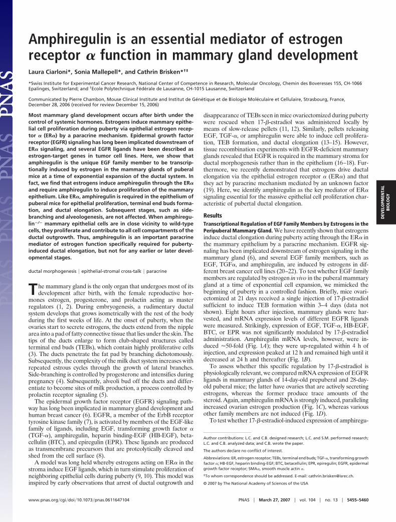
ResultsTranscriptional Regulation of EGF Family Members by Estrogens in thePeripuberal Mammary Gland. We have recently shown that estrogensinduce ductal elongation during puberty acting through the ER� inthe mammary epithelium by a paracrine mechanism. EGFR sig-naling has been implicated downstream of estrogen signaling in themammary gland (6), and several EGF family members, such asEGF, TGF�, and amphiregulin, are induced by estrogens in dif-ferent breast cancer cell lines (20–22). To test whether EGF familymembers are regulated by estrogen in vivo in the puberal mammarygland at a time of exponential cell expansion, we mimicked thebeginning of puberty in a controlled fashion. Briefly, mice ovari-ectomized at 21 days received a single injection of 17-�-estradiolsufficient to induce TEB formation within 3–4 days (data notshown). Eight hours after injection, mammary glands were har-vested, and mRNA expression levels of different EGFR ligandswere measured. Strikingly, expression of EGF, TGF-�, HB-EGF,BTC, or EPR was not significantly modulated by 17-�-estradioladministration. Amphiregulin mRNA levels, however, were in-duced �50-fold (Fig. 1A); they were up-regulated within 4 h ofinjection, and expression peaked at 12 h and remained high until itdecreased at 24 h and thereafter (Fig. 1B).
To assess whether this specific regulation by 17-�-estradiol isphysiologically relevant, we compared mRNA expression of EGFRligands in mammary glands of 14-day-old prepuberal and 28-day-old puberal mice; the latter have ovaries that are actively secretingestrogens, whereas the former produce trace amounts of thesteroid. Again, amphiregulin mRNA is strongly induced, parallelingincreased ovarian estrogen production (Fig. 1C), whereas variousother family members are not induced (Fig. 1D).
To test whether 17-�-estradiol-induced expression of amphiregu-
Author contributions: L.C. and C.B. designed research; L.C. and S.M. performed research;L.C. and C.B. analyzed data; and C.B. wrote the paper.
The authors declare no conflict of interest.
Abbreviations: ER, estrogen receptor; TEBs, terminal end buds; TGF-�, transforming growthfactor �; HB-EGF, heparin binding-EGF; BTC, betacellulin; EPR, epiregulin; EGFR, epidermalgrowth factor receptor; SMA�, smooth muscle actin �.
‡To whom correspondence should be addressed. E-mail: [email protected].
© 2007 by The National Academy of Sciences of the USA
www.pnas.org�cgi�doi�10.1073�pnas.0611647104 PNAS � March 27, 2007 � vol. 104 � no. 13 � 5455–5460
DEV
ELO
PMEN
TAL
BIO
LOG
Y

lin is mediated by ER�, we stimulated prepuberal ER��/� mice byusing the same protocol as above. At this stage, ER��/� andwild-type (WT) glands are phenotypically undistinguishable (19).In the absence of ER�, amphiregulin mRNA did not increase (Fig.1E). Thus, amphiregulin expression is strongly regulated by 17-�-estradiol in the puberal mammary gland by ER�-dependent tran-scriptional activation.
Mammary Gland Development in Amphiregulin-Deficient Mice. Ourfinding that estrogens specifically control expression of amphiregu-lin and not of other EGF family members could provide anexplanation as to why deletion of amphiregulin, but not of TGF�or EGF, impairs mammary gland development (23). To assesswhether the phenotype is specifically linked to estrogen action, weanalyzed mammary glands of amphiregulin�/� and their WTlittermates at critical developmental stages by whole-mountmicroscopy.
At birth, mutant and WT mammary glands were indistinguish-able (data not shown). Similarly, in both genotypes, the rudimen-tary ductal systems grew isometrically until puberty (Fig. 2 A andB) with comparable numbers of branching points (Fig. 2G). TEBsdeveloped in glands of 6-week-old WT females, whereas the ductaltips did not enlarge in amphiregulin�/� glands (Fig. 2 C and D). At3 months of age, the WT glands were fully arborized and side-branching occurred, whereas amphiregulin�/� glands still displayeda rudimental ductal tree (Fig. 2 E and F). These observations wereconsistent with amphiregulin acting downstream of estrogen sig-naling during puberal development. However, it was conceivablethat the inability to grow at puberty was secondary to a structuraldefect acquired before this stage. To address this concern, we
analyzed prepuberal glands histologically and performed immu-nostainings for smooth muscle actin � (SMA�), a marker ofmyoepithelial cells. At 24 days, in both WT and amphiregulin�/�
mice, the luminal and myoepithelial layers were intact (Fig. 2 H andI). Thus, at least histologically, hormone-independent mammarydevelopment is normal. Histological analysis of mammary glandsfrom 26-day-old mice revealed that in amphiregulin�/� females,ductal tips failed to enlarge, whereas TEBs formed in the WTlittermates (Fig. 2 J and K).
During pregnancy, the rudimentary ductal system of the amphi-regulin�/� mice, notwithstanding its limited expansion, still under-went side-branching and alveologenesis to an extent comparable toWT littermates (Fig. 2 L and M). Histological analysis revealeddistended alveoli containing lipid droplets as seen in WT glands atthis stage (Fig. 2 N and O). Strikingly, we never observed acompletely filled fat pad, even after 12 pregnancies (Fig. 2P). Thus,amphiregulin is specifically required for estrogen-induced ductalelongation during puberty, but not for the preceding or later stagesof mammary gland development, including side-branching, alve-ologenesis, or milk production.
The Role of Amphiregulin in Mediating Estrogen-Induced Proliferationand TEB Formation. The observation that puberal outgrowth iscompletely blocked in the absence of amphiregulin, together withthe finding that this growth factor is transcriptionally regulated byestrogen in the mammary gland, suggested that amphiregulinmediates estrogen function during puberty. To test this directly, westimulated ovariectomized amphiregulin�/� and WT females with17-�-estradiol and monitored proliferation and TEB formationover 4 days. In WT glands, proliferation assessed by BrdU incor-poration was detected 24 h after 17-�-estradiol stimulation (Table1) and peaked at 48 h, with 31 � 6.7% of the epithelial cellsincorporating BrdU (Fig. 3B). Amphiregulin�/� glands analyzed at48 h did not show any increase in BrdU incorporation more thanunstimulated glands (Fig. 3A, Table 1), but cells in the inguinallymph nodes showed BrdU incorporation (Fig. 3A Inset), validatingthe BrdU administration. Similarly, 26-day-old amphiregulin�/�
mice did not display more proliferation than 14-day-old mice (Fig.3C), whereas WT glands displayed 18.78 � 4.6% of BrdU-positivecells (Fig. 3D). Four days after 17-�-estradiol injection, numerousTEBs developed in the WT glands but not in amphiregulin�/�
glands (data not shown). Thus, amphiregulin is required for estro-gen-induced epithelial proliferation in peripuberal mice, and in theabsence of amphiregulin estrogen-induced proliferation and TEBformation are completely abolished.
To exclude the possibility that estrogen-dependent proliferationis impaired in amphiregulin�/� glands because of altered ER�expression and/or downstream signaling, we determined the recep-tor status in the mutant glands by immunohistochemistry. Amphi-regulin�/� and WT glands displayed comparable ER� expressionin 24-day-old mice (data not shown). Moreover, glands of bothgenotypes showed similar inductions of the estrogen-target proges-terone receptor in response to 17-�-estradiol stimulation (data notshown), indicating that ER� signaling remains intact in amphiregu-lin�/� mammary glands.
The Role of Amphiregulin in the Mammary Epithelium. To determineto what extent the mammary phenotype of the amphiregulin�/�
mice is attributable to mammary epithelial amphiregulin, we per-formed mammary gland recombination experiments by using tissuefrom amphiregulin�/� and WT littermates. In 3-week-old mice, theinguinal glands can be cleared of endogenous epithelium by surgi-cally removing the nipple-near half that contains the rudimentaryductal system. Mammary epithelial cells that are introduced intothe remaining ‘‘cleared’’ fat pad will give rise to a new ductal system.They can grow out from a piece of breast tissue that is implanted(24) or from single-cell suspensions injected into the fat pad (25).A caveat of this experimental approach lies in the possibility that
Fig. 1. Regulation of EGFR ligands’ expression by estrogens. QuantitativeRT-PCR analysis of mammary gland mRNA for amphiregulin and other EGFRligands normalized to keratin 18. (A and B) Mice ovariectomized beforepuberty were injected with either vehicle (open bars) or 17-�-estradiol (filledbars) and analyzed either for EGFR ligand expression 8 h later (A) or foramphiregulin over 48 h (B). (C and D) Mammary glands of 14- to 28-day-oldmice were analyzed for amphiregulin expression (C) or for EGFR ligandexpression (14- and 21-day-old females, open and filled bars, respectively) (D).Bars report the mean values obtained from three different mice. Error barsindicate standard deviation. Relative increase refers to control treated (A andB) or to the 14-day-old mice (C and D). (E) RT-PCR analysis of amphiregulin andkeratin 18 expression in glands from ovariectomized WT and ER��/� mice 6 hafter administration of 17-�-estradiol (�) or vehicle (�).
5456 � www.pnas.org�cgi�doi�10.1073�pnas.0611647104 Ciarloni et al.

endogenous epithelium can be inadvertently left behind and com-pete with the grafted epithelium for fat pad reconstitution. Tocircumvent this problem, we crossed the mutant amphiregulin alleleinto a transgenic strain that ubiquitously expresses GFP. By graftingGFP-positive donor tissue into GFP-negative hosts, we can readilydistinguish the grafted from endogenous epithelium. Furthermore,this approach ensured that we engrafted comparable amounts ofepithelium on both sides; we prepared pieces of donor tissue underUV illumination before implanting. The engrafted glands wereanalyzed 2–4 months later by whole-mount microscopy. Amphi-regulin�/� epithelial grafts exhibited no outgrowth (Fig. 4A),whereas the WT counterpart filled the fat pad (Fig. 4B). Impor-
tantly, in contrast to ER��/� epithelial grafts that remained rudi-mentary during pregnancy, amphiregulin�/� epithelia underwentside-branching and alveologenesis (data not shown), resulting inincreased fat pad filling (Table 2). Thus, amphiregulin, like ER�, isrequired for ductal outgrowth during puberty in the mammaryepithelium. However, amphiregulin�/� epithelial cells can stillproliferate and differentiate in response to progesterone andprolactin.
Generation of Amphiregulin�/� and WT Chimeric Epithelia. To assesswhether amphiregulin mediates estrogen-induced proliferation inan autocrine/cell autonomous or paracrine fashion, we createdmosaic mammary epithelia containing both WT and amphiregu-
Fig. 2. Developmental analysis of amphiregulin�/� mammary glands. (A–F) Whole-mount micrographs of inguinal glands from amphiregulin�/� (A, C, and E)and WT (B, D, and F) females were analyzed at the following developmental stages: day 24 (A and B), 6 weeks (C and D), 3 months (E and F). (Scale bars: A–F,5 mm; A�–F�, 1 mm.) Micrographs are representative of glands from 12 mice analyzed per time point. (G) Number of branching points in inguinal mammary glandsof 24-day-old amphiregulin�/� (filled bar, n � 6) and WT (open bar, n � 7) females. (H and I) Histological sections of mammary glands from 24-day-oldamphiregulin�/� and WT mice stained by immunohistochemistry with an anti-SMA� antibody to highlight the myoepithelial cells (brown) counterstained withhematoxylin. H&E (J and K) staining of paraffin sections from 6-week-old amphiregulin�/� (J) and WT (K) glands. (Scale bar: 50 �m.) (L and M) Whole-mountmicrographs of amphiregulin�/� (L) and WT (M) glands at 16.5 days of pregnancy. (N and O) Histological sections of amphiregulin�/� (N) and WT (O) mammaryglands at 18.5 days of pregnancy. (P) Whole-mount micrograph of gland from an amphiregulin�/� female after 12 pregnancies.
Table 1. Epithelial cell proliferation in amphiregulin�/� mammary glands
Control, %
E
14 d, % 26 d, %24 h, % 48 h, %
Amphiregulin�/� 0.4 � 0.12 / 0.2 � 0.14 0.2 � 0.15 0.4 � 0.13WT 0.36 � 0.17 3.5 � 1.7 31 � 6.7 0.3 � 0.12 18.78 � 4.60
The percentage of cells staining positive for BrdU in mammary glands from amphiregulin�/� and WT mice thatwere ovariectomized at puberty and treated with 17-�-estradiol (E) or vehicle (control) for 24 or 48 h, and intactmice analyzed when they were 14 or 26 days old. More than 1,000 cells were counted on at least one section fromthree different mice. /, not analyzed.
Ciarloni et al. PNAS � March 27, 2007 � vol. 104 � no. 13 � 5457
DEV
ELO
PMEN
TAL
BIO
LOG
Y

lin�/� cells. The cells of the two distinct genotypes were derivedfrom mice carrying additionally either a GFP or LacZ transgene,allowing us to discriminate between mutant and WT cells. Of 68successfully engrafted glands with a 1:1 mixture, 31 were composedof both cell populations (Fig. 5 A and B), whereas 37 werecomposed of WT cells only. As expected, no outgrowth was foundto contain only amphiregulin�/� cells. Histological analysis ofglands engrafted with amphiregulin�/� LacZ� and WT cells re-vealed that amphiregulin�/� cells, identified by X-gal staining, arepresent among both cap and body cells of the TEBs (Fig. 5 C andD, arrows). Immunostainings for GFP on glands reconstituted withamphiregulin�/� GFP� and WT cells showed that amphiregulin�/�
cells, visualized by immunohistochemistry with an antibody againstGFP, are found in both luminal and myoepithelial compartments inthe mature ducts (Fig. 5 E and F, arrows).
The extensive contribution of amphiregulin�/� cells to the re-constitution suggested to us that the mutant cells actively prolifer-ate. To assess whether this is indeed the case, we performed doubleimmunofluorescence for GFP, thereby labeling amphiregulin�/�
cells and BrdU to mark the proliferating cells. GFP-positive cellswere found to incorporate BrdU (Fig. 5G Right, arrows), indicatingthat, indeed, amphiregulin�/� mammary epithelial cells proliferateactively. Interestingly, when chimeric epithelia are generated fromdifferentially marked WT cells, two distinct types of chimerism areobserved. Either entire ductal segments are constituted by one typeof cell or the ducts show a patchy distribution of the two groups ofcells (L.C. and C.B., unpublished observations). Glands containingamphiregulin�/� cells showed only the latter type of chimerism (Fig.5 E and F), indicating that close proximity to nearby WT cells isrequired to receive, directly or indirectly, the proliferative signal.We conclude that amphiregulin is an important paracrine mediatorof estrogen-induced proliferation during ductal morphogenesis.
DiscussionMost mammary gland development occurs after birth under thecontrol of female reproductive hormones. The advent of gene-targeting combined with powerful tissue recombination techniquesallows dissecting the mechanisms by which systemic hormones elicitproliferation and morphogenesis. Immunohistochemical studies onhuman breast tissue (26) and rodent mammary glands (27, 28)revealed that in normal tissue steroid receptor expression andproliferation are dissociated. We recently provided genetic evi-dence that ERa�/� mammary epithelial cells completely fail toproliferate in vivo (19). However, when the mutant cells weregrafted together with WT cells, they proliferated and contributedto all aspects of ductal morphogenesis, indicating that estrogens actby a paracrine mechanism in vivo (19). The nature of the down-stream signals that ER�-positive cells send to ERa�/� cells inresponse to estrogens remained elusive. Here, we identify amphi-regulin as a key mediator of paracrine estrogen action required forthe massive mammary epithelial cell proliferation that results inductal outgrowth during puberty.
Our findings support a model in which hormones acting on themammary epithelium recruit a series of local factors that act byparacrine mechanisms to trigger proliferation of nearby cells (29,30). More specifically, amphiregulin emerges as a central mediatorof estrogen function, while we have previously shown that Wnt-4 isan important mediator of paracrine progesterone-induced side-branching (31), and that prolactin requires IGF-2 to induce alveolarproliferation (32). These indirect signaling mechanisms ensure thatthe systemic stimulus is amplified within the target organ overseveral cell diameters and over time, and that the behavior ofdifferent cells and cell types participating in the morphogenic eventare coordinated and fine-tuned with local requirements.
The downstream events of amphiregulin action remain to beexplored. The only known receptor for amphiregulin, EGFR, isexpressed in both stromal and epithelial compartments (23, 33), butis required for ductal elongation in the stroma and not in theepithelium (16–18). Although this does not rule out that EGFRsignaling also has a role in the epithelium, the prime targets ofamphiregulin are stromal cells. In the simplest scenario, the stromalfibroblasts stimulated by amphiregulin could send back a mitogenicsignal to the neighboring epithelial cells. Several growth factors,such as HGF, IGF1, and FGF10, are good candidates because theyare expressed in the mammary stroma at the time of ductal
Fig. 3. Estrogen-induced proliferation in amphiregulin�/� and WT mam-mary glands. Immunohistochemistry was performed on sections of amphi-regulin�/� (A and C) or WT (B and D) glands either from 3-week-old ovariec-tomized females 48 h after 17-�-estradiol stimulation (A and B) or 26-day-oldmice (C and D) with an anti-BrdU antibody. Positively stained lymph nodesfrom the same glands are shown as positive control for BrdU incorporation (Aand C Insets). (Scale bars: 50 �m.)
Fig. 4. Amphiregulin�/� epithelial transplants. Representative outgrowth ofamphiregulin�/� (A) or WT (B) GFP� mammary epithelium transplanted intoWT cleared fat pads. The glands were derived from virgin recipients andobserved directly under the fluorescence stereomicroscope (B). (Scale bar:5 mm.)
Table 2. Development of amphiregulin�/� mammary epithelium
MiceNo. with nooutgrowth
No. with �20%outgrowth
No. with 20–50%outgrowth
Total no.of mice
Virgin 11 2 0 13Pregnant and postpartum 3 2 4 9
Results of mammary gland reconstitution experiments with amphiregulin�/� mammary epithelium. Contralat-eral control grafts showed complete outgrowth. Different extent of outgrowth was observed depending on thedevelopmental stage of the host.
5458 � www.pnas.org�cgi�doi�10.1073�pnas.0611647104 Ciarloni et al.

elongation, whereas the respective receptors are found in theepithelium (34). Alternatively, down-modulation of inhibitory path-ways, such as TGF� signaling, may be involved (35). Yet morecomplex interactions might be required; thus, macrophages andeosinophils present in the mammary stroma have a role in ductalelongation (36) and could be attracted and activated by signalsdownstream of EGFR activation.
It is also unclear how amphiregulin, secreted by epithelial cellsmost likely of the ER�-positive luminal subtype, can reach thestromal target cells through a layer of myoepithelial cells and thebasal lamina. Deletion of the disintegrin and metalloproteinase(ADAM) 17 in the mammary epithelium blocks ductal outgrowthsimilarly to amphiregulin (18). Analysis of tissue recombinantsshowed that ADAM17 is also required for EGFR activation,although to what extent this normally occurs in the stroma versus
the epithelium is unclear (18). Whether the cleavage suffices toactivate amphiregulin or whether the protein needs to be furthermodified and/or actively transported to the stromal target cellsremains to be addressed. ADAM17 may also be required moreindirectly for processing extracellular matrix and/or the basal laminato enable amphiregulin to reach the stroma.
We found that the deletion of amphiregulin strictly and specif-ically affects ductal elongation, not preceding or subsequent devel-opmental stages. At variance with our results, previous reportssuggested that some TEBs can develop and ductal outgrowth canoccur in the absence of amphiregulin (18, 23), whereas alveologen-esis was impaired (23). The discrepancies may be because of themixed genetic background used in previous studies, as opposed tothe pure C57BL/6 used here. Moreover, some of the conclusionswere reached based on recombinant glands grafted under thekidney capsule, an environment that is less physiological than themammary fat pad (18).
Our finding that the ductal outgrowth defect in amphiregulin�/�
mice is not overcome by multiple pregnancies is surprising giventhat other EGFR ligands, in particular EGF, are highly expressedin the glands during pregnancy (37). This suggests that eitheramphiregulin activates a unique signaling response downstream ofEGFR or that EGF and/or other ligands are not expressed in theright cell types and/or not processed in the same ways as amphi-regulin. We further speculate that the ability to bind heparin (38)that distinguishes amphiregulin from other family members maybestow this protein with unique functions in the reciprocal inter-actions with extracellular matrix and/or stromal components.
How does estrogen-induced ductal elongation relate to humanbreast cancer? When human breast tissue transplanted in mice wasstimulated with estradiol under conditions that induce proliferation(39), amphiregulin was among the most highly induced genes (40),strongly suggesting that amphiregulin may similarly be involved inmediating estrogen-induced proliferation in the human breastepithelium. Estrogens and ER� signaling have an important role inthe pathogenesis of breast cancer, with �60% of carcinomasexpressing ER� and relying on estrogens for growth (41). In normalbreast tissue, both human and rodent, steroid receptor expressionand proliferation are mostly dissociated (26–28), whereas in tumorsamples, most proliferative cells express ER� (26). Our experi-ments do not address whether amphiregulin can act in an autocrineand/or paracrine fashion on ER�-positive cells. Interestingly,TGF-� actively represses proliferation of ER�-positive cells (42),which are probably exposed to the same growth factors as theirER�-negative neighbors. This suggests that ER�-positive cellsmight escape the inhibitory influence of TGF-� during tumorigen-esis and thereby become responsive to the auto/paracrine effects ofamphiregulin resulting in proliferation. To what extent direct versusindirect stimuli account for the growth of ER�-positive breastcarcinomas remains to be addressed.
Intriguingly, expression profiles of mammary glands from virginand parous mice as well as distantly related rat strains revealed thatamphiregulin is one of the genes most strongly down-modulatedafter the first pregnancy (43, 44). This down-modulation correlateswith a decrease in susceptibility to tumorigenesis, suggesting thisfactor is linked to breast carcinogenesis.
MethodsMice. ROSA26 mice (45) were purchased from The JacksonLaboratory (Bar Harbor, ME). GFP transgenic, amphiregulin, andER�-deficient mice were described (see refs. 23, 46, and 47).ROSA26 mice were bred in 129SV/C57BL/6, and ER�, amphi-regulin, and GFP mice were bred in C57BL/6 genetic background.The presence of the �-galactosidase transgene was assessed bysubjecting a piece of tail to the X-Gal staining procedure describedbelow.
Fig. 5. Amphiregulin�/� and WT chimeric mammary epithelia. Whole-mountpreparations of a cleared fat pad showing representative mammary reconstitu-tion on injection with a mixture of amphiregulin�/� GFP� (green), and WTROSA26� (blue) epithelial cells in a 1:1 ratio. (A and B) GFP was visualized directlyunder the fluorescence stereomicroscope (A); �-galactosidase activity revealedafter X-Gal staining (B). (C and D) Histological sections of glands reconstitutedwith amphiregulin�/� LacZ� and WT cells. Amphiregulin�/� LacZ� cells (blue)were found both among the body (C, arrow) and the cap cells (D, arrow). (E andF) Glands reconstituted with amphiregulin�/� GFP� and WT cells were sectionedand stained with anti-GFP antibody. Both luminal (F, arrow) and myoepithelialcells (E, arrow) show GFP staining (brown). (G) Glands reconstituted with amphi-regulin�/� GFP�andWTcellsweresectionedandcostainedwithanti-BrdU(Left),and anti-GFP antibodies (Center) merge (Right) reveals double-positive cells.(Scale bars: A and B, 500 �m; C–G, 50 �m.)
Ciarloni et al. PNAS � March 27, 2007 � vol. 104 � no. 13 � 5459
DEV
ELO
PMEN
TAL
BIO
LOG
Y

Transplantation of Mammary Epithelium. The fat pads of 3-week-oldC57BL/6 or 129SV/C57BL/6 females were cleared. Pieces ofmammary tissue of 1 mm in diameter were removed from thenipple region (24). Alternatively, the cleared fat pads wereinjected with mixtures of primary mammary epithelial cells asdescribed (see ref. 25).
Hormone Treatment. 17-�-Estradiol (Sigma–Aldrich, St. Louis,MO) 5 mg/ml 100% ethanol stock was diluted in PBS. Three-week-old female mice were ovariectomized and 10 days later wereinjected s.c. with 17-�-estradiol (5 ng/g of body weight) or vehicle.
Mammary Gland Whole Mounts. Mammary glands were dissected,spread onto a glass slide, fixed in a 1:3 mixture of glacial aceticacid/100% ethanol, hydrated, stained overnight in 0.2% carmine(Sigma) and 0.5% AlK(SO4)2, dehydrated in graded solutions ofethanol, and cleared in 1:2 benzyl alcohol/benzyl benzoate (Sigma)as described (see ref. 48). Digital pictures were taken on a Leica(Wetzlar, Germany) MZFLIII stereoscope or Leica DM2000 mi-croscope with Leica DC300F and Pixelink (Ottawa, ON, Canada)PL-A622C, respectively.
X-Gal Staining. The transplanted mammary glands were dissected,fixed for 1 h in 1.5% formaldehyde in PBS, washed three times over3 h with rinse buffer (2 mM MgCl2/0.1% sodium deoxycholate/0.2%Nonidet P-40 in PBS) and rotated in X-Gal staining solution (1mg/ml X-Gal, 5 mM potassium ferricyanide, and 5 mM potassiumferrocyanide in rinse buffer) at 37°C for 18 h, washed in PBS, andprocessed for whole mounting as described above.
Histological Examination and Immunohistochemistry. For histologicalexamination, whole-mounted mammary glands were washed in100% ethanol before paraffin embeddment or freshly isolatedglands were fixed with 4% paraformaldehyde. Sections were cut at4 �m. Mice were injected with BrdU (Sigma) 2 h before death. Thefollowing antibodies were used: rat monoclonal anti-BrdU (1:300;OBT0030; Oxford Biotechnology, Oxfordshire, U.K.), mousemonoclonal anti-�-SMA (1:400; Ab-1; NeoMarkers, Fremont,CA), rabbit polyclonal anti-GFP (1:5,000; A6455; MolecularProbes, Eugene, OR), rabbit monoclonal anti-PR (1:500; SP2;
NeoMarkers), and rabbit polyclonal anti-ER� (1:10,000; sc-542;Santa Cruz Biotechnology, Santa Cruz, CA). The antibodies wereapplied overnight at 4°C after antigen retrieval in citrate buffer.Biotinylated secondary antibodies were detected with VectastainElite kit (Vector Laboratories, Burlingame, CA). For immunoflu-orescence, the anti-rat Alexa Fluor 568- and anti-rabbit Alexa Fluor488-conjugated secondary antibodies were used. Pictures wereacquired with a Leica DM2000 microscope and Pixelink PL-A622Ccamera and Zeiss Axioplan 2-imaging fluorescence microscopewith Axiocam MRm camera (Zeiss, Thornwood, NY).
Quantitative Real-Time PCR (QRT-PCR). Glands were homogenized inTRIzol (Invitrogen, Carlsbad, CA), and RNA was extracted withchloroform and processed with the RNeasy Kit (Qiagen, Valencia,CA). Total RNA was reverse transcribed by using reverse tran-scriptase (GIBCO BRL/ Invitrogen) and random hexamers (RocheDiagnostics, Indianapolis, IN). The resulting cDNAs were used forQRT-PCR analysis by using the iCycler apparatus (Bio-Rad, Her-cules, CA) and SYBR Green PCR Core Reagents system (Qiagen).Results were evaluated with iCycler iQ real-time detection systemsoftware (Bio-Rad). The following forward and reverse primerswere used: amphiregulin, GCCATTATGCAGCTGCTTTG-GAGC and TGTTTTTCTTGGGCTTAATCACCT; EGF,GCAACTCCGTCCGGGCGAGGA and GAAGATGACTGT-GGTCCCGGG; TGF-�, GTGGCTGCAGCACCCTGCGCTand GATCAGCACACAGGTGATAATGAGG; HB-EGF,CTCCCACTGGATCCACAAAC and GGCATGGGTCT-CTCTTCTTC; EPR, CACCGAGAAAGAAGGATGGA andGGGATCGTCTTCCATCTGAA; BTC, CCCCAAGCAGTA-CAAGCATT and TGAACACCACCATGACCACT; and ker-atin 18 (36).
We thank G. P. Dotto for carefully reading the manuscript; N. Muellerand C. Morel for technical assistance; and Drs. D. Lee (University ofGeorgia, Athens, GA), P. Chambon (Institut de Genetique et de BiologieMoleculaire et Cellulaire and College de France, Illkirch Cedex, France),and M. Okabe (Osaka University, Suita, Japan) for generously providingthe amphiregulin, ER� mutant, and the C57BL/6-Tg(Act-EGFP) strain,respectively. This work was supported by National Center of Compe-tence in Research, Molecular Oncology, Grant NRP50/SNF.
1. Lyons WR, Li CH, Johnson RE (1958) Recent Prog Horm Res 14:219–248.2. Nandi S (1958) J Natl Cancer Inst 21:1039–1063.3. Hinck L, Silberstein GB (2005) Breast Cancer Res 7:245–251.4. Shyamala G (1999) J Mammary Gland Biol Neoplasia 4:89–104.5. Brisken C (2002) J Mammary Gland Biol Neoplasia 7:39–48.6. Troyer KL, Lee DC (2001) J Mammary Gland Biol Neoplasia 6:7–21.7. Stern DF (2003) Exp Cell Res 284:89–98.8. Harris RC, Chung E, Coffey RJ (2003) Exp Cell Res 284:2–13.9. Cunha GR, Young P, Hom YK, Cooke PS, Taylor JA, Lubahn DB (1997) J Mammary
Gland Biol Neoplasia 2:393–402.10. Woodward TL, Xie JW, Haslam SZ (1998) J Mammary Gland Biol Neoplasia 3:117–131.11. Daniel CW, Silberstein GB, Strickland P (1987) Cancer Res 47:6052–6057.12. Silberstein GB, Van Horn K, Shyamala G, Daniel CW (1994) Endocrinology 134:84–90.13. Coleman S, Silberstein GB, Daniel CW (1988) Dev Biol 127:304–315.14. Snedeker SM, Brown CF, DiAugustine RP (1991) Proc Natl Acad Sci USA 88:276–280.15. Kenney NJ, Smith GH, Rosenberg K, Cutler ML, Dickson RB (1996) Cell Growth
Differ 7:1769–1781.16. Sebastian J, Richards RG, Walker MP, Wiesen JF, Werb Z, Derynck R, Hom YK,
Cunha GR, DiAugustine RP (1998) Cell Growth Differ 9:777–785.17. Wiesen JF, Young P, Werb Z, Cunha GR (1999) Development (Cambridge, UK)
126:335–344.18. Sternlicht MD, Sunnarborg SW, Kouros-Mehr H, Yu Y, Lee DC, Werb Z (2005)
Development (Cambridge, UK) 132:3923–3933.19. Mallepell S, Krust A, Chambon P, Brisken C (2006) Proc Natl Acad Sci USA
103:2196–2201.20. Bates SE, Davidson NE, Valverius EM, Freter CE, Dickson RB, Tam JP, Kudlow JE,
Lippman ME, Salomon DS (1988) Mol Endocrinol 2:543–555.21. Dickson RB, Huff KK, Spencer EM, Lippman ME (1986) Endocrinology 118:138–142.22. Kenney NJ, Saeki T, Gottardis M, Kim N, Garcia-Morales P, Martin MB, Normanno N,
Ciardiello F, Day A, Cutler ML (1993) J Cell Physiol 156:497–514.23. Luetteke NC, Qiu TH, Fenton SE, Troyer KL, Riedel RF, Chang A, Lee DC (1999)
Development (Cambridge, UK) 126:2739–2750.24. Deome KB, Faulkin LJ, Jr, Bern HA, Blair PB (1959) Cancer Res 19:515–520.25. Daniel CW, Deome KB (1965) Science 149:634–636.26. Clarke RB, Howell A, Potten CS, Anderson E (1997) Cancer Res 57:4987–4991.
27. Seagroves TN, Lydon JP, Hovey RC, Vonderhaar BK, Rosen JM (2000) MolEndocrinol 14:359–368.
28. Russo J, Ao X, Grill C, Russo IH (1999) Breast Cancer Res Treat 53:217–227.29. Silberstein GB (2001) Microsc Res Tech 52:155–162.30. Clarke RB (2003) Steroids 68:789–794.31. Brisken C, Heineman A, Chavarria T, Elenbaas B, Tan J, Dey SK, McMahon JA,
McMahon AP, Weinberg RA (2000) Genes Dev 14:650–654.32. Brisken C, Ayyannan A, Nguyen C, Heineman A, Reinhardt F, Tan J, Dey SK, Dotto
GP, Weinberg RA (2002) Dev Cell 3:877–887.33. DiAugustine RP, Richards RG, Sebastian J (1997) J Mammary Gland Biol Neoplasia
2:109–117.34. Imagawa W, Pedchenko VK, Helber J, Zhang H (2002) J Steroid Biochem Mol Biol
80:213–230.35. Silberstein GB, Daniel CW (1987) Science 237:291–293.36. Gouon-Evans V, Lin EY, Pollard JW (2002) Breast Cancer Res 4:155–164.37. Schroeder JA, Lee DC (1998) Cell Growth Differ 9:451–464.38. Schuger L, Johnson GR, Gilbride K, Plowman GD, Mandel R (1996) Development
(Cambridge, UK) 122:1759–1767.39. Clarke RB, Howell A, Anderson E (1997) Breast Cancer Res Treat 45:121–133.40. Wilson CL, Sims AH, Howell A, Miller CJ, Clarke RB (2006) Endocr Relat Cancer
13:617–628.41. Platet N, Cathiard AM, Gleizes M, Garcia M (2004) Crit Rev Oncol Hematol 51:55–67.42. Ewan KB, Oketch-Rabah HA, Ravani SA, Shyamala G, Moses HL, Barcellos-Hoff
MH (2005) Am J Pathol 167:409–417.43. D’Cruz CM, Moody SE, Master SR, Hartman JL, Keiper EA, Imielinski MB, Cox JD,
Wang JY, Ha SI, Keister BA, Chodosh LA (2002) Mol Endocrinol 16:2034–2051.44. Blakely CM, Stoddard AJ, Belka GK, Dugan KD, Notarfrancesco KL, Moody SE,
D’Cruz CM, Chodosh LA (2006) Cancer Res 66:6421–6431.45. Friedrich G, Soriano P (1991) Genes Dev 5:1513–1523.46. Okabe M, Ikawa M, Kominami K, Nakanishi T, Nishimune Y (1997) FEBS Lett
407:313–319.47. Dupont S, Krust A, Gansmuller A, Dierich A, Chambon P, Mark M (2000)
Development (Cambridge UK) 127:4277–4291.48. Mueller SO, Clark JA, Myers PH, Korach KS (2002) Endocrinology 143:2357–2365.
5460 � www.pnas.org�cgi�doi�10.1073�pnas.0611647104 Ciarloni et al.