Embed Size (px)
Citation preview
This journal is © The Royal Society of Chemistry 2021 Chem. Commun., 2021, 57, 12725–12740 | 12725
Cite this: Chem. Commun., 2021,
57, 12725
Amphiphilic DNA nanostructures for bottom-upsynthetic biology
Roger Rubio-Sanchez, †ab Giacomo Fabrini, †cb Pietro Cicuta a andLorenzo Di Michele *cab
DNA nanotechnology enables the construction of sophisticated biomimetic nanomachines that are
increasingly central to the growing efforts of creating complex cell-like entities from the bottom-up.
DNA nanostructures have been proposed as both structural and functional elements of these artificial
cells, and in many instances are decorated with hydrophobic moieties to enable interfacing with
synthetic lipid bilayers or regulating bulk self-organisation. In this feature article we review recent efforts
to design biomimetic membrane-anchored DNA nanostructures capable of imparting complex
functionalities to cell-like objects, such as regulated adhesion, tissue formation, communication and
transport. We then discuss the ability of hydrophobic modifications to enable the self-assembly of DNA-
based nanostructured frameworks with prescribed morphology and functionality, and explore the
relevance of these novel materials for artificial cell science and beyond. Finally, we comment on the yet
mostly unexpressed potential of amphiphilic DNA-nanotechnology as a complete toolbox for bottom-
up synthetic biology – a figurative and literal scaffold upon which the next generation of synthetic cells
could be built.
1 Introduction
Since its advent in the eighties,1 DNA nanotechnology hasevolved from an inspiring concept to a practical toolkit drivingadvances in several areas of fundamental and applied science.2
Owing to the programmability and selectivity of the Watson–Crick base-pairing, DNA motifs have been widely employed to
a Biological and Soft Systems, Cavendish Laboratory, University of Cambridge,
JJ Thomson Avenue, Cambridge CB3 0HE, UK. E-mail: [email protected] fabriCELL, Molecular Sciences Research Hub, Imperial College London,
London W12 0BZ, UKc Department of Chemistry, Molecular Sciences Research Hub,
Imperial College London, London W12 0BZ, UK
Roger Rubio-Sanchez
Roger Rubio-Sanchez is a final-year PhD candidate at theCavendish Laboratory, University ofCambridge. In 2015 he completed aBSc in Biotechnology Engineeringat the Monterrey Institute ofTechnology and Higher Educationin Mexico City. He later joined theEPSRC Doctoral Training Centre inNanoscience and Nanotechnology atCambridge, and obtained a MRes inNanoscience and Nanotechnology in2018. Under the supervision of DrLorenzo Di Michele, his PhD
research focuses on developing DNA-based synthetic replicas of cell-surface receptors for artificial cells. He is interested in artificial cellbioengineering, DNA nanotechnology, membrane biophysics, and softcondensed matter.
Giacomo Fabrini
Giacomo Fabrini obtained hisBSc (Electronic Engineering) andMSc (Nanotechnology Engineering)degrees from Universita degli Studidi Roma La Sapienza in 2017 and2019, respectively, and thencontinued his postgraduate studiesunder the supervision of Dr LorenzoDi Michele in the MembraneBiophysics Group at the Depart-ment of Chemistry of ImperialCollege London. His main researchtopic is the self-assembly of stimuli-responsive DNA phases.
† These authors contributed equally to this work.
Received 6th August 2021,Accepted 28th October 2021
DOI: 10.1039/d1cc04311k
rsc.li/chemcomm
ChemComm
FEATURE ARTICLE
Ope
n A
cces
s A
rtic
le. P
ublis
hed
on 2
8 O
ctob
er 2
021.
Dow
nloa
ded
on 4
/3/2
022
1:44
:12
AM
. T
his
artic
le is
lice
nsed
und
er a
Cre
ativ
e C
omm
ons
Attr
ibut
ion
3.0
Unp
orte
d L
icen
ce.
View Article OnlineView Journal | View Issue
12726 | Chem. Commun., 2021, 57, 12725–12740 This journal is © The Royal Society of Chemistry 2021
experimentally explore the self-assembly phenomenology ofmacromolecular,3 colloidal4–7 and nanoparticle systems.8–10
By affording unprecedented control over the shape,11
interactions,12,13 and mechanical properties of nano-structures,14 the DNA origami technology has opened furthernew avenues for the field, with concrete applications to biomo-lecular scaffolding,15–17 single-molecule analysis,18,19
biosensing,20 nanomedicine,21 imaging,22 and the constructionof advanced materials.23,24 In parallel to structural control, ourgrowing understanding of nucleic acid kinetics and thermo-dynamics has resulted in the ability to program dynamicresponses,25 marking the advent of DNA-based molecularcomputing26–28 and the development of proof-of-conceptactuable nanodevices,29,30 biosensors,31,32 and technologiesfor optical imaging.33–37
Uniquely among artificial molecular-scale devices, the sheervariety of architectures and responses demonstrated withnucleic-acid nanostructures makes them comparable withbiologically occurring machinery in terms of complexity andfunctionality.38–41 While early incarnations of DNA nano-technology drew inspiration from the simplest biochemistryand biological nanomachines, efforts in the field now aim tomatch and often surpass what biological evolution hasproduced.42,43
It has therefore become apparent that DNA nanotechnologyholds great potential as a toolkit for engineering all aspects ofman-made mimics of biological cells – the central objective ofthe rapidly expanding discipline of bottom-up synthetic biology.The biomimetic microrobots, often referred to as artificial cells,are constructed from inanimate molecular components toreplicate emergent responses typically observed in biologicalcells, including energy conversion, communication, motion,adaptation and, although yet unachieved, replication andevolution.44–46 Besides their use as minimalistic models forunravelling the principles of life and probing pathways for its
emergence,47,48 artificial cells are expected to revolutionisenumerous emerging technologies, from biosynthesis to materialsand healthcare. In the latter context, notably, artificial cells areenvisaged as the basis of next-generation personalised therapeuticsolutions where, operating in vivo, the microrobots could sensedisease-related biomarkers, record their presence, and respondwith the in situ synthesis and delivery of therapeutic agents toreduce toxicity and boost the efficacy of treatments.49–51 Whilestill far from the self-sustaining complexity of their biologicalcounterparts, artificial cell technologies carry several potentialadvantages compared to mainstream platforms based onengineered biological cells, including a greater freedom of designand choice of materials, compatibility with non-biologicalcomponents, efficient use of resources and possible avoidanceof redundancies, limited biological risks, and less stringentregulatory constraints.
The construction of cell-like objects requires control over(at least) two distinct, yet intertwined aspects: compartmentalisationand information processing. The former is needed to separatethe synthetic cell from its surroundings and establish theinternal chemical heterogeneity required to sustain syntheticmolecular pathways. Several materials, from colloids52,53 topolymers54 to proteins27,55 have been adopted to build semi-permeable compartments for artificial cells, but the mostcommon implementations rely on lipid bilayer membranesowing to their facile production, bio-compatibility and generalsimilarity with cell membranes.56 Membrane-less compartmentsin the form of hydrogels57–59 and polymer coacervates60–63 repre-sent a robust and increasingly popular alternative, mimickingsimilar environments found in biological cells.64–67 Informationprocessing, broadly including environmental/molecular sensing,signal transduction, communication, and information storage/replication/propagation, is in most cases sustained by reconsti-tuted biological machinery such as receptors, enzymes, ion-gatingand pumping channels, and cell-free transcription/translation
Pietro Cicuta
Pietro Cicuta studied Physics inMilan (Laurea, 1999) andCambridge (PhD, 2003), workingon various topics of soft matter(liquid interfaces, membranes,microfluidics) and biologicalphysics (physical aspects andimaging of blood stage malaria,bacteria, algae and motile cilia).His lab develops a range ofinstrumentation and softwareespecially for automated opticalimaging. Lorenzo Di Michele
Lorenzo Di Michele studiedcondensed matter physics for hisundergraduates and master at theUniversity of L’Aquila (Abruzzo,Italy), before obtaining a PhD inphysics at the Cavendish Labora-tory, Unviersity of Cambridge, in2013. He then held in the sameinstitution a Oppenheimer EarlyCareer Research Fellowship (2013–2016), a Leverhulme Early CareerReseach Fellowship (2016–2018)and a Royal Society UniversityResearch Fellowship (URF, 2018–
2019). In 2019 Lorenzo joined the Department of Chemistry at ImperialCollege London as a URF/Lecturer, and then URF/Senior Lecturer (2021).His research interests include soft condensed matter, DNA nanotechnol-ogy, and synthetic biology.
Feature Article ChemComm
Ope
n A
cces
s A
rtic
le. P
ublis
hed
on 2
8 O
ctob
er 2
021.
Dow
nloa
ded
on 4
/3/2
022
1:44
:12
AM
. T
his
artic
le is
lice
nsed
und
er a
Cre
ativ
e C
omm
ons
Attr
ibut
ion
3.0
Unp
orte
d L
icen
ce.
View Article Online
This journal is © The Royal Society of Chemistry 2021 Chem. Commun., 2021, 57, 12725–12740 | 12727
systems.44,46,68,69 In view of the ubiquitous presence of lipidinterfaces, membrane-bound protein machinery is often featuredin artificial cellular systems, regulating transport, adhesion andmorphological responses.69–73
In this feature article we delve into several examples ofsynthetic DNA nanostructures designed to replicate the struc-ture and functionality of biological molecular machinery in thecontext of cell mimicry, focusing in particular on bilayer-anchored devices acting as synthetic receptors mediatingadhesion,74–80 tissue formation,74,81 communication82 andmembrane sculpting.83–87 Attachment of the DNA nanostructuresto lipid bilayers often requires their modification with hydro-phobic moieties, such as cholesterol, tocopherol or lipids, givingthe nanostructures an amphiphilic character.88 Besides enablingmembrane anchoring, the latter can also be exploited to enhancethe self-assembly capabilities of DNA nanostructures in the bulk.In the second part of this feature article we review recent findingson the construction of nanoporous networks from amphiphilicDNA motifs with well controlled structure and functionality,which could form the basis of membrane-less compartments forartificial cells and, more broadly, impact biotechnology andtherapeutics.89–91 Finally, we discuss the broader implicationsof (amphiphilic) DNA nanotechnology as a versatile andintegrated toolkit to simultaneously prescribe structural andinformation processing aspects in synthetic cells, identify keyoutstanding challenges, and express our view on those function-alities that could have the greatest fundamental and technologicalimpact.
2 Programming biomimetic responseswith membrane-associated DNAnanodevices
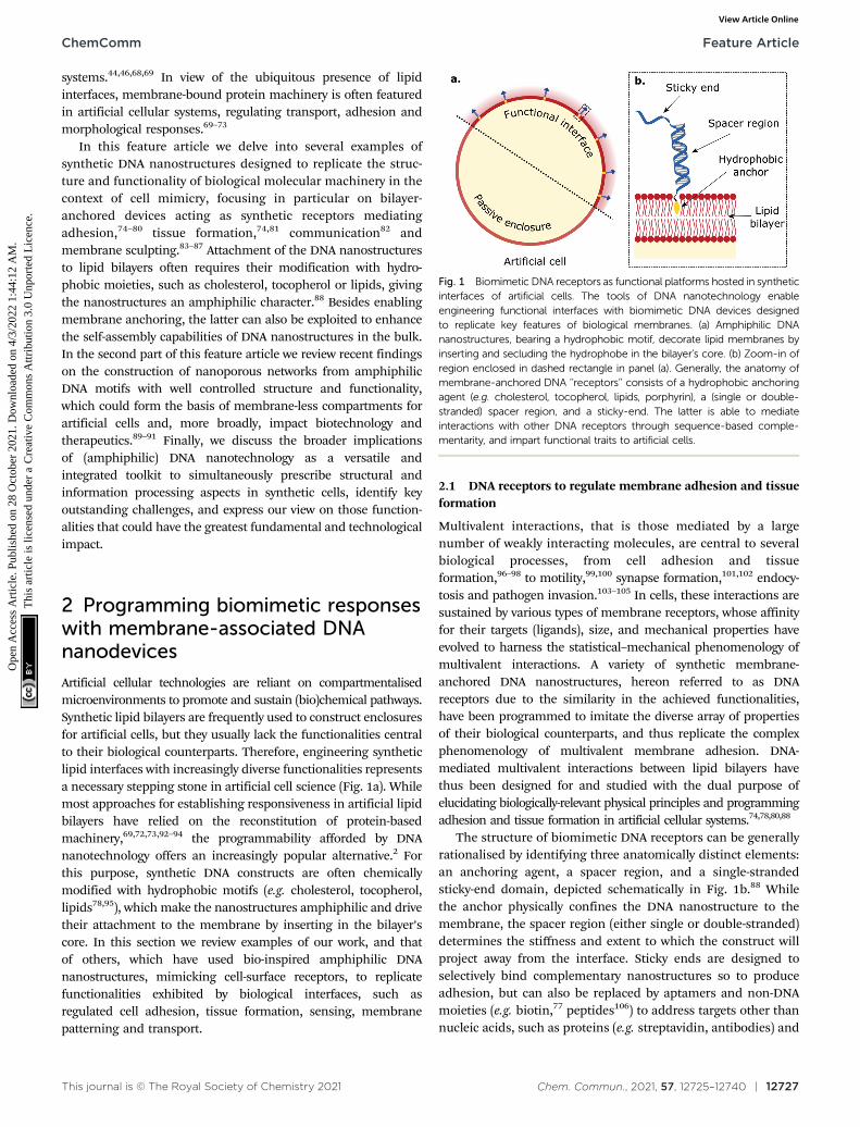
Artificial cellular technologies are reliant on compartmentalisedmicroenvironments to promote and sustain (bio)chemical pathways.Synthetic lipid bilayers are frequently used to construct enclosuresfor artificial cells, but they usually lack the functionalities centralto their biological counterparts. Therefore, engineering syntheticlipid interfaces with increasingly diverse functionalities representsa necessary stepping stone in artificial cell science (Fig. 1a). Whilemost approaches for establishing responsiveness in artificial lipidbilayers have relied on the reconstitution of protein-basedmachinery,69,72,73,92–94 the programmability afforded by DNAnanotechnology offers an increasingly popular alternative.2 Forthis purpose, synthetic DNA constructs are often chemicallymodified with hydrophobic motifs (e.g. cholesterol, tocopherol,lipids78,95), which make the nanostructures amphiphilic and drivetheir attachment to the membrane by inserting in the bilayer’score. In this section we review examples of our work, and thatof others, which have used bio-inspired amphiphilic DNAnanostructures, mimicking cell-surface receptors, to replicatefunctionalities exhibited by biological interfaces, such asregulated cell adhesion, tissue formation, sensing, membranepatterning and transport.
2.1 DNA receptors to regulate membrane adhesion and tissueformation
Multivalent interactions, that is those mediated by a largenumber of weakly interacting molecules, are central to severalbiological processes, from cell adhesion and tissueformation,96–98 to motility,99,100 synapse formation,101,102 endocy-tosis and pathogen invasion.103–105 In cells, these interactions aresustained by various types of membrane receptors, whose affinityfor their targets (ligands), size, and mechanical properties haveevolved to harness the statistical–mechanical phenomenology ofmultivalent interactions. A variety of synthetic membrane-anchored DNA nanostructures, hereon referred to as DNAreceptors due to the similarity in the achieved functionalities,have been programmed to imitate the diverse array of propertiesof their biological counterparts, and thus replicate the complexphenomenology of multivalent membrane adhesion. DNA-mediated multivalent interactions between lipid bilayers havethus been designed for and studied with the dual purpose ofelucidating biologically-relevant physical principles and programmingadhesion and tissue formation in artificial cellular systems.74,78,80,88
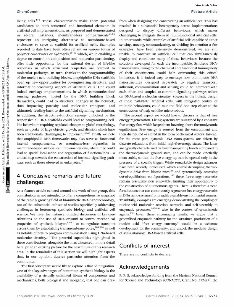
The structure of biomimetic DNA receptors can be generallyrationalised by identifying three anatomically distinct elements:an anchoring agent, a spacer region, and a single-strandedsticky-end domain, depicted schematically in Fig. 1b.88 Whilethe anchor physically confines the DNA nanostructure to themembrane, the spacer region (either single or double-stranded)determines the stiffness and extent to which the construct willproject away from the interface. Sticky ends are designed toselectively bind complementary nanostructures so to produceadhesion, but can also be replaced by aptamers and non-DNAmoieties (e.g. biotin,77 peptides106) to address targets other thannucleic acids, such as proteins (e.g. streptavidin, antibodies) and
Fig. 1 Biomimetic DNA receptors as functional platforms hosted in syntheticinterfaces of artificial cells. The tools of DNA nanotechnology enableengineering functional interfaces with biomimetic DNA devices designedto replicate key features of biological membranes. (a) Amphiphilic DNAnanostructures, bearing a hydrophobic motif, decorate lipid membranes byinserting and secluding the hydrophobe in the bilayer’s core. (b) Zoom-in ofregion enclosed in dashed rectangle in panel (a). Generally, the anatomy ofmembrane-anchored DNA ‘‘receptors’’ consists of a hydrophobic anchoringagent (e.g. cholesterol, tocopherol, lipids, porphyrin), a (single or double-stranded) spacer region, and a sticky-end. The latter is able to mediateinteractions with other DNA receptors through sequence-based comple-mentarity, and impart functional traits to artificial cells.
ChemComm Feature Article
Ope
n A
cces
s A
rtic
le. P
ublis
hed
on 2
8 O
ctob
er 2
021.
Dow
nloa
ded
on 4
/3/2
022
1:44
:12
AM
. T
his
artic
le is
lice
nsed
und
er a
Cre
ativ
e C
omm
ons
Attr
ibut
ion
3.0
Unp
orte
d L
icen
ce.
View Article Online
12728 | Chem. Commun., 2021, 57, 12725–12740 This journal is © The Royal Society of Chemistry 2021
metabolites. These membrane-bound DNA nanostructures dis-play lateral diffusivity at rates similar to the surroundinglipids,107–109 which thus enables their lateral re-distribution asdriven by free-energy minimisation.88 In what follows, we willrefer to these DNA constructs either using the bio-inspired term‘‘receptors’’, or simply as ‘‘linkers’’.
The emergence of bioconjugation chemistries has allowed tolink DNA molecules to a variety of functional moieties,106,110
such as hydrophobic and amphiphilic molecules,111–114 thusexpanding the power of DNA nanotechnology beyond the realmof base-pairing. Such strategies enabled to interface DNAconstructs with lipid-based supramolecular structures to exertcontrol over their self-assembling properties. Indeed, eventhough DNA-mediated interactions have been applied toprogram the self-assembly of Brownian objects since the1990s,4,5,8–10 the extension of this concept to synthetic lipidbilayers was popularised only in the mid 2000s thanks to thecontributions of Boxer, Hook and co-workers.115–119 In earlyexperiments, DNA-decorated Small Unilamellar Vesicles (SUVs)were tethered to Supported Lipid Bilayers (SLBs) via linkerswith complementary sticky-ends. This setup was harnessed toelucidate the lateral diffusivity of the DNA-tethered SUVs115 andthe kinetics of their docking onto the SLB.116 Replacing SUVswith cell-size Giant Unilamellar Vesicles (GUVs), Boxer and co-workers demonstrated the generation of (multi-storey) bilayerpatches, formed through the controllable adhesion and ruptureof GUVs onto SLBs.117,118 With the same system, the authorsdemonstrated the occurrence of lateral phase separationbetween DNA linkers of different lengths, arising from theminimisation of membrane bending energy.119 The latterphenomenon is reminiscent of the size-induced phase separationobserved between receptors involved in immune synapses.96,101,102,120
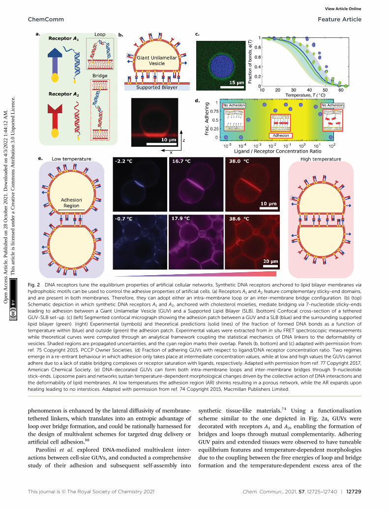
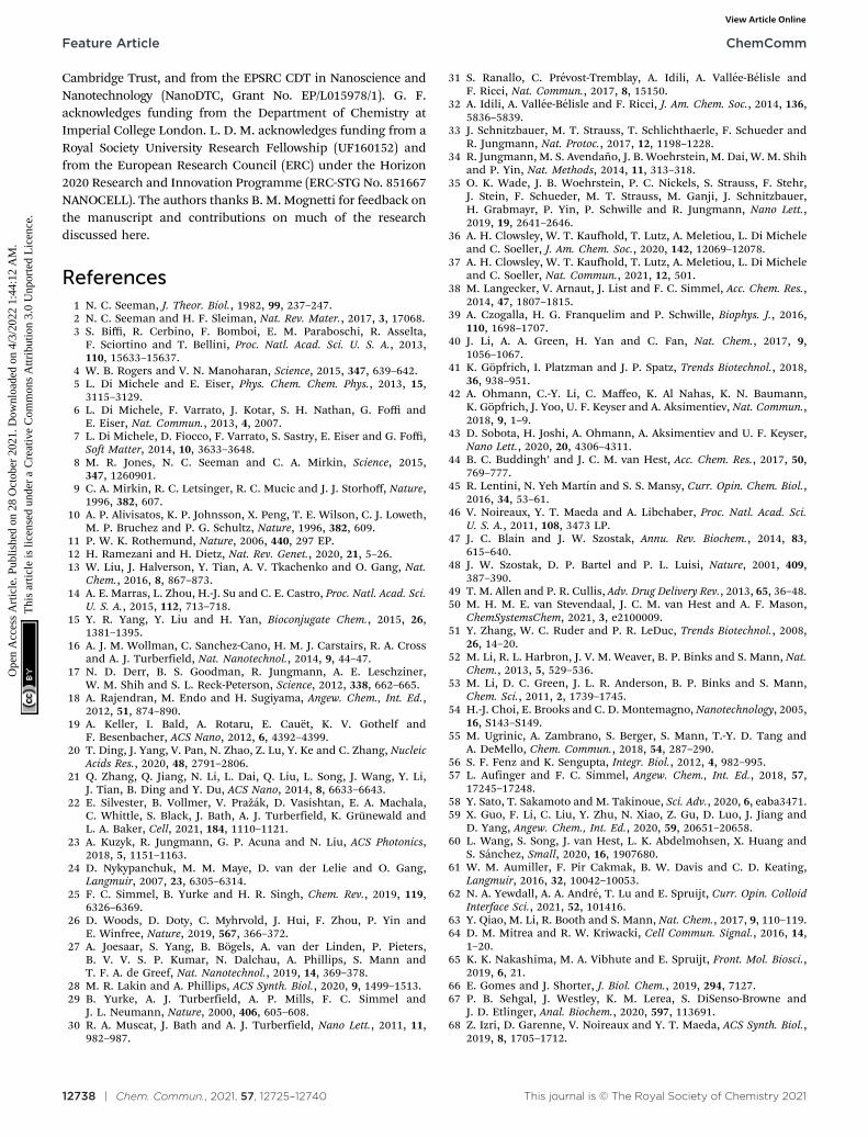
Systems of interacting GUVs and SLBs have been sub-sequently adopted to shed light on the statistical mechanicalphenomenology of multivalent interactions between fluidmembranes, thus paving the route to the rational design ofartificial-cell adhesion and deepening our understanding ofanalogous biological processes. For their contribution, Shimo-bayashi et al. decorated both GUVs and SLB with two types ofDNA receptors (A1 and A2) able to interact with each other viacomplementary sticky-ends, as shown in Fig. 2a.75 Thus, bothintra-membrane loops and inter-membrane bridges could form,but only the latter would contribute to GUV–SLB adhesion. Theauthors proposed a statistical–mechanical model which couldpredict the probability of loop and bridge formation, alongsidethe resulting GUV–SLB adhesion free energy. The adoptedexperimental configuration (Fig. 2b), coupled to strategically-positioned cyanine fluorphores, allowed the authors to measurethe temperature-dependent probability of bond formationthorough FRET, both inside and outside the GUV–SLB adhesionarea (Fig. 2c). The experimental observation of a lower meltingtemperature for loop-dimers (present outside of the adhesionpatch) matched the model predictions (Fig. 2c). The GUV–SLBconfigurations also allowed the authors to extract the membranetension of the GUV through flickering spectroscopy.121 Also inthis instance, comparison between experiments and the
statistical–mechanical model revealed good agreement, bothfor the absolute value of the tension and its weak dependenceon temperature.
Amjad et al. adopted a similar GUV–SLB system, butreplaced the sticky ends on the DNA nanostructures with biotinmodifications, so that adhesion would only be mediated bystreptavidin. Indeed, in their implementation, membrane-bound DNA constructs featuring biotin moieties behaved as‘‘receptors’’, while streptavidin molecules assumed the role of‘‘ligands’’.77 Given the tetravalent nature of the latter, thisdesign choice allowed them to explore the coupled role of ligandvalency and ligand concentration on multivalent interactions.Liposome adhesion emerged only in a specific range of ligand/receptor concentration ratios, as shown in Fig. 2d, that is thoseratios allowing for at least one receptor per bilayer to bind thesame streptavidin moiety. This results in the formation of di- ormulti-valent DNA–streptavidin complexes (i.e. two to four DNAreceptors bound to the same streptavidin). At low ratios, strep-tavidin molecules were not numerous enough, resulting in toofew DNA–streptavidin linkages to stabilise adhesion. Similarly, atvery high concentration ratios, most of the streptavidin ligandswould be bound to a single DNA receptor, thus also suppressingthe formation of complexes featuring at least one receptor perbilayer. Only intermediate ligand/receptor ratios promoted theformation of complexes able to bridge the membranes and,therefore, sustain GUV–SLB adhesion. The work of Amjad et al.also showcased the promise that DNA–membrane systems holdfor bioanalytical devices, where strategic design updates to targetanalytes of interest could pave the way for next-generationbiosensing platforms. Indeed, one can envisage using the sharponset of adhesion, observed upon reaching the analyte criticalconcentration, as a readout mechanism, for instance, bycoupling it to optical or electrochemical measurements.
Beales and Vanderlick extended the application of multi-valent DNA-mediated interactions from vesicle–SLB systems to theself-assembly of free-standing Large Unilamellar Vesicles (LUVs),exploring the effect of design and environmental parametersincluding surface coverage, ionic strength, temperature andmembrane charge.79,122 With the aim of further enriching theavailable range of responses, Hernandez-Ainsa et al. demon-strated the light-controlled aggregation of LUVs, by utilisingazobenzene motifs to tether the DNA linkers to themembranes.123 These photo-active moieties undergo conformationalchanges upon exposure to UV light, switching between cis and transconfigurations characterised by different degrees of hydrophobicity.The transition thus results in a change in the affinity of theanchors for the bilayer, leading to light-triggered LUV aggrega-tion and disassembly. Bachmann et al. combined experiments,coarse-grained computer simulations, and the aforementionedanalytical framework for multivalent interactions to providequantitative insight into the phase behaviour of DNA-functionalised LUVs.76 The authors focused on the effect ofDNA-linker density on the melting temperature of the self-assembled phases and observed, intriguingly, that no aggregationoccurs below a critical coverage owing to the competition betweenintra-LUV loops and inter-LUV bridges.76 This emergent
Feature Article ChemComm
Ope
n A
cces
s A
rtic
le. P
ublis
hed
on 2
8 O
ctob
er 2
021.
Dow
nloa
ded
on 4
/3/2
022
1:44
:12
AM
. T
his
artic
le is
lice
nsed
und
er a
Cre
ativ
e C
omm
ons
Attr
ibut
ion
3.0
Unp
orte
d L
icen
ce.
View Article Online
This journal is © The Royal Society of Chemistry 2021 Chem. Commun., 2021, 57, 12725–12740 | 12729
phenomenon is enhanced by the lateral diffusivity of membrane-tethered linkers, which translates into an entropic advantage ofloop over bridge formation, and could be rationally harnessed forthe design of multivalent schemes for targeted drug delivery orartificial cell adhesion.88
Parolini et al. explored DNA-mediated multivalent inter-actions between cell-size GUVs, and conducted a comprehensivestudy of their adhesion and subsequent self-assembly into
synthetic tissue-like materials.74 Using a functionalisationscheme similar to the one depicted in Fig. 2a, GUVs weredecorated with receptors A1 and A2, enabling the formation ofbridges and loops through mutual complementarity. AdheringGUV pairs and extended tissues were observed to have tuneableequilibrium features and temperature-dependent morphologiesdue to the coupling between the free energies of loop and bridgeformation and the temperature-dependent excess area of the
Fig. 2 DNA receptors tune the equilibrium properties of artificial cellular networks. Synthetic DNA receptors anchored to lipid bilayer membranes viahydrophobic motifs can be used to control the adhesive properties of artificial cells. (a) Receptors A1 and A2 feature complementary sticky-end domains,and are present in both membranes. Therefore, they can adopt either an intra-membrane loop or an inter-membrane bridge configuration. (b) (top)Schematic depiction in which synthetic DNA receptors A1 and A2, anchored with cholesterol moieties, mediate bridging via 7-nucleotide sticky-endsleading to adhesion between a Giant Unilamellar Vesicle (GUV) and a Supported Lipid Bilayer (SLB). (bottom) Confocal cross-section of a tetheredGUV-SLB set-up. (c) (left) Segmented confocal micrograph showing the adhesion patch between a GUV and a SLB (blue) and the surrounding supportedlipid bilayer (green). (right) Experimental (symbols) and theoretical predictions (solid lines) of the fraction of formed DNA bonds as a function oftemperature within (blue) and outside (green) the adhesion patch. Experimental values were extracted from in situ FRET spectroscopic measurementswhile theoretical curves were computed through an analytical framework coupling the statistical mechanics of DNA linkers to the deformability ofvesicles. Shaded regions are propagated uncertainties, and the cyan region marks their overlap. Panels (b, bottom) and (c) adapted with permission fromref. 75 Copyright 2015, PCCP Owner Societies. (d) Fraction of adhering GUVs with respect to ligand/DNA receptor concentration ratio. Two regimesemerge in a re-entrant behaviour in which adhesion only takes place at intermediate concentration values, while at low and high values the GUVs cannotadhere due to a lack of stable bridging complexes or receptor saturation with ligands, respectively. Adapted with permission from ref. 77 Copyright 2017,American Chemical Society. (e) DNA-decorated GUVs can form both intra-membrane loops and inter-membrane bridges through 9-nucleotidestick-ends. Liposome pairs and networks sustain temperature-dependent morphological changes driven by the collective action of DNA interactions andthe deformability of lipid membranes. At low temperatures the adhesion region (AR) shrinks resulting in a porous network, while the AR expands uponheating leading to no interstices. Adapted with permission from ref. 74 Copyright 2015, Macmillan Publishers Limited.
ChemComm Feature Article
Ope
n A
cces
s A
rtic
le. P
ublis
hed
on 2
8 O
ctob
er 2
021.
Dow
nloa
ded
on 4
/3/2
022
1:44
:12
AM
. T
his
artic
le is
lice
nsed
und
er a
Cre
ativ
e C
omm
ons
Attr
ibut
ion
3.0
Unp
orte
d L
icen
ce.
View Article Online
12730 | Chem. Commun., 2021, 57, 12725–12740 This journal is © The Royal Society of Chemistry 2021
bilayer. In particular, the artificial tissues displayed an unusualthermal response, contracting upon heating and swellingon cooling (negative thermal expansion coefficient), as aconsequence of changes in the extension of the GUV–GUVcontact regions, and thus of the interstices present within thetissue (Fig. 2e). The observed behaviour is fully supported by themultivalent analytical framework mentioned above, wherethe temperature-dependent GUV area is accounted for. Suchsynthetic tissues pave the way for the construction of bio-compatible filtering devices and scaffolding platforms applicablein regenerative medicine.
Besides providing fine control over the static properties ofself-assembling synthetic cellular networks, multivalent DNA-mediated interactions also enable programming of adhesionkinetics. While the diffusion of liposomes and bilayer-anchoredspecies are prescribed by the size, phase, and composition ofthe vesicles, the kinetics of DNA–DNA interactions can beengineered using well-established strand displacementmechanisms.124,125 This concept has been exploited by Parolini
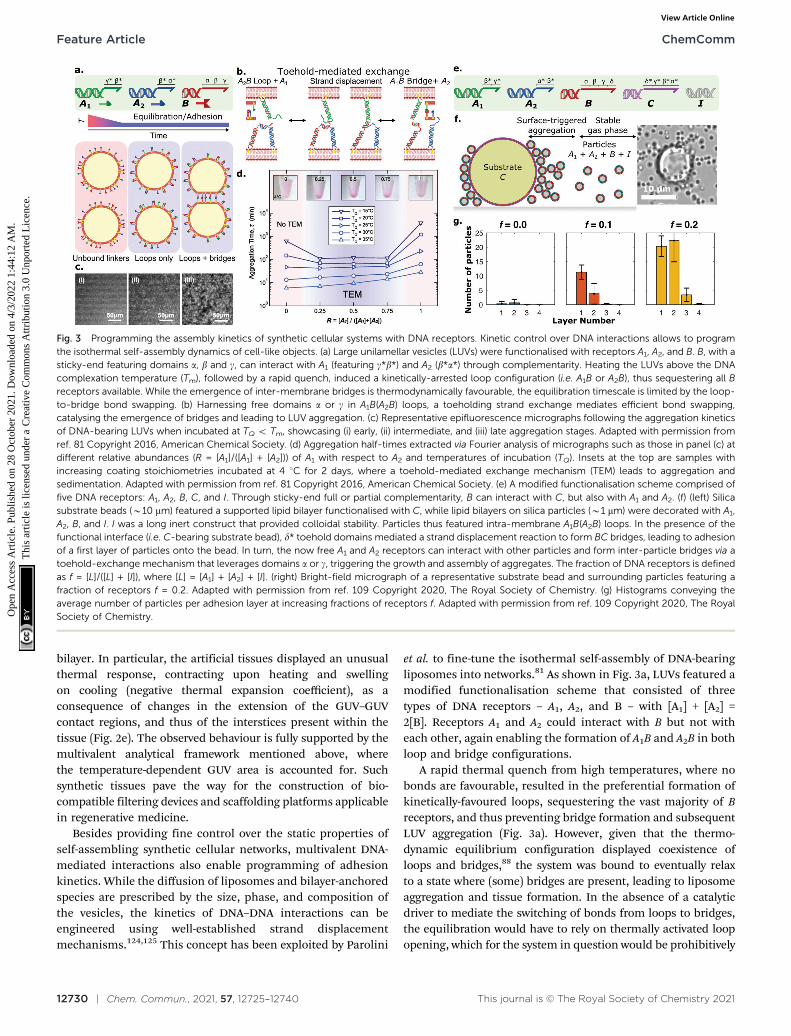
et al. to fine-tune the isothermal self-assembly of DNA-bearingliposomes into networks.81 As shown in Fig. 3a, LUVs featured amodified functionalisation scheme that consisted of threetypes of DNA receptors – A1, A2, and B – with [A1] + [A2] =2[B]. Receptors A1 and A2 could interact with B but not witheach other, again enabling the formation of A1B and A2B in bothloop and bridge configurations.
A rapid thermal quench from high temperatures, where nobonds are favourable, resulted in the preferential formation ofkinetically-favoured loops, sequestering the vast majority of Breceptors, and thus preventing bridge formation and subsequentLUV aggregation (Fig. 3a). However, given that the thermo-dynamic equilibrium configuration displayed coexistence ofloops and bridges,88 the system was bound to eventually relaxto a state where (some) bridges are present, leading to liposomeaggregation and tissue formation. In the absence of a catalyticdriver to mediate the switching of bonds from loops to bridges,the equilibration would have to rely on thermally activated loopopening, which for the system in question would be prohibitively
Fig. 3 Programming the assembly kinetics of synthetic cellular systems with DNA receptors. Kinetic control over DNA interactions allows to programthe isothermal self-assembly dynamics of cell-like objects. (a) Large unilamellar vesicles (LUVs) were functionalised with receptors A1, A2, and B. B, with asticky-end featuring domains a, b and g, can interact with A1 (featuring g*b*) and A2 (b*a*) through complementarity. Heating the LUVs above the DNAcomplexation temperature (Tm), followed by a rapid quench, induced a kinetically-arrested loop configuration (i.e. A1B or A2B), thus sequestering all Breceptors available. While the emergence of inter-membrane bridges is thermodynamically favourable, the equilibration timescale is limited by the loop-to-bridge bond swapping. (b) Harnessing free domains a or g in A1B(A2B) loops, a toeholding strand exchange mediates efficient bond swapping,catalysing the emergence of bridges and leading to LUV aggregation. (c) Representative epifluorescence micrographs following the aggregation kineticsof DNA-bearing LUVs when incubated at TQ o Tm, showcasing (i) early, (ii) intermediate, and (iii) late aggregation stages. Adapted with permission fromref. 81 Copyright 2016, American Chemical Society. (d) Aggregation half-times extracted via Fourier analysis of micrographs such as those in panel (c) atdifferent relative abundances (R = [A1]/([A1] + [A2])) of A1 with respect to A2 and temperatures of incubation (TQ). Insets at the top are samples withincreasing coating stoichiometries incubated at 4 1C for 2 days, where a toehold-mediated exchange mechanism (TEM) leads to aggregation andsedimentation. Adapted with permission from ref. 81 Copyright 2016, American Chemical Society. (e) A modified functionalisation scheme comprised offive DNA receptors: A1, A2, B, C, and I. Through sticky-end full or partial complementarity, B can interact with C, but also with A1 and A2. (f) (left) Silicasubstrate beads (B10 mm) featured a supported lipid bilayer functionalised with C, while lipid bilayers on silica particles (B1 mm) were decorated with A1,A2, B, and I. I was a long inert construct that provided colloidal stability. Particles thus featured intra-membrane A1B(A2B) loops. In the presence of thefunctional interface (i.e. C-bearing substrate bead), d* toehold domains mediated a strand displacement reaction to form BC bridges, leading to adhesionof a first layer of particles onto the bead. In turn, the now free A1 and A2 receptors can interact with other particles and form inter-particle bridges via atoehold-exchange mechanism that leverages domains a or g, triggering the growth and assembly of aggregates. The fraction of DNA receptors is definedas f = [L]/([L] + [I]), where [L] = [A1] + [A2] + [I]. (right) Bright-field micrograph of a representative substrate bead and surrounding particles featuring afraction of receptors f = 0.2. Adapted with permission from ref. 109 Copyright 2020, The Royal Society of Chemistry. (g) Histograms conveying theaverage number of particles per adhesion layer at increasing fractions of receptors f. Adapted with permission from ref. 109 Copyright 2020, The RoyalSociety of Chemistry.
Feature Article ChemComm
Ope
n A
cces
s A
rtic
le. P
ublis
hed
on 2
8 O
ctob
er 2
021.
Dow
nloa
ded
on 4
/3/2
022
1:44
:12
AM
. T
his
artic
le is
lice
nsed
und
er a
Cre
ativ
e C
omm
ons
Attr
ibut
ion
3.0
Unp
orte
d L
icen
ce.
View Article Online
This journal is © The Royal Society of Chemistry 2021 Chem. Commun., 2021, 57, 12725–12740 | 12731
slow at room temperature owing to the relative stability of A1Band A2B interactions. The authors demonstrated how the bond-swapping kinetics, the emergence of bridges, and thesubsequent formation of aggregates can be accelerated by atoehold-mediated exchange mechanism (TEM),124 as shown inFig. 3b. Specifically, an unbound ssDNA domain available on Bwhen forming a loop with A1(A2), offered a toehold for unpairedA2(A1) linkers on a nearby partner liposome, leading to bridgeformation. The isothermal aggregation time of LUVs, extractedvia Fourier analysis of fluorescence micrographs such as thoseshown in Fig. 3c, could be tuned over orders of magnitude bycontrolling the relative abundances of A1 and A2, which in turnimpacted the availability of bond-swapping partners (Fig. 3d).Experimental trends were qualitatively replicated by a kineticmodel which accounted for the (toeholding-modulated) rates offormation and breakup of two and three-linker DNAcomplexes.126 The ability of prescribing timescales of vesicle–vesicle adhesion onset could be invaluable for applicationswhere precise control is required over the time-dependentmechanical properties of artificial tissues. For instance, onecould envisage the design of synthetic cell formulations thatcan be injected in vivo while in liquid form, and programmed toreadily set shortly afterwards to form a tissue implant.
Besides temporal control, multivalent DNA-mediatedinteractions can also be harnessed to program spatially hetero-geneous self-assembly phenomena, as demonstrated byLanfranco et al.109 Here, the authors exploited a toehold-mediated exchange interaction scheme similar to the onediscussed above,81 and combined it with rationally-designedbond competition to demonstrate self-limiting colloidal self-assembly triggered by a functional interface. This concept, firstdemonstrated computationally by Jana and Mognetti,127 isreminiscent of several instances of biological self-assembly,where the occurrence and features of macro-molecular aggre-gates are finely regulated by their interactions with nearbyinterfaces. The experimental implementation of Lanfrancoet al. consisted of bilayer coated silica substrates (large beads)and micron-size colloidal particles, both decorated by DNAlinkers. The functionalisation scheme included five types ofDNA nanostructures, as depicted in Fig. 3e: A1, A2, B, C, andI. Silica substrate beads featured receptors C, while particleswere functionalised with constructs A1, A2, B and I. Receptors A1
and A2 could bind to B, but not each other, while B could alsobind to C. Construct I was a long inert double-strandednanostructure introduced to provide particle–particle stericrepulsion. The latter prevented the formation of A1B(A2B)inter-particle bridges which would lead to spontaneousaggregation, resulting in turn in a stable colloidal gas phase.Here, most A1, A2 and B constructs were engaged in intra-particle loops, given that [A1] + [A2] = [B]. In the presence ofthe functional substrate, toehold domains in receptors C couldcatalyse the break-up of A1B or A2B loops, leading to particleadhesion onto the substrate via irreversible BC bridges.The sequestration of B linkers freed up initially saturated A1
and A2 constructs, shifting the thermodynamic equilibrium infavour of inter-particle bridges, whose formation kinetics was
facilitated by toehold-mediated exchange as discussed above.This process led to the formation of a second particle layer ontothe substrate, and could propagate to multiple layers in a self-limiting chain reaction, as depicted schematically and shownwith a representative micrograph in Fig. 3f. Such a strategyshowcased that the self-assembly and growth of the colloidalaggregate could be regulated by the presence of the functionalsubstrate, while the final size of the aggregates could becontrolled by fine-tuning the stoichiometries of receptors andinert constructs (Fig. 3g). Besides enabling spatially-coordinated colloidal self-assembly, this approach alsoprovides a means of signal amplification in which theformation of large colloidal aggregates could report on thepresence of biological analytes and biomarkers.
2.2 Beyond adhesion: biosensing platforms and membranesculpting with DNA nanostructures
The functionalities of biological membrane machinery go wellbeyond controlling adhesion, as proteolipid interfaces are knownto sustain and mediate critical responses such as sensing,communication, transport and morphological adaptation. Buildingonto the solutions developed for the ‘‘simple’’ sticky constructsdiscussed in the previous sections, several groups haveexploited the functional versatility of amphiphilic DNA nano-technology to replicate some of these capabilities in syntheticcellular systems. In this section we review some notable examplesfrom our work and that of others, with particular emphasis onbiosensing, membrane patterning and transport.
Cell membranes host receptors dedicated to transducingchemical signals and linking them to downstream signallingpathways. In some instances, such as for immunity-relevanttoll-like receptors,128 signalling is triggered by analyte-mediated dimerisation of receptors. Kaufhold et al. tookinspiration from this mechanism to implement a membrane-hosted biosensing platform reliant on target-induced DNAstrand displacement (TIDSD).82,129,130 Here, the target analyteco-localises an invader DNA construct with a substrate-incumbent dimer, catalysing a strand displacement reactionsimilar to what happens for conventional toehold-mediated stranddisplacement.129 The authors demonstrated that compared toanalogous circuitry freely diffusing in bulk, membrane hostedTIDSD showed up to a 2-fold increase in response rate, asconfirmed with experiments and coarse-grained computersimulations.131 Moreover, they showed that the membrane scaffoldhelps reducing false positive signals, or leakage, a highly-covetedfeature that could unlock their applicability in biosensingtechnologies.
Many of the functionalities mediated by cell-membranereceptors rely on a tight regulation of their lateral distributionon the plasma and internal membranes of the cells.While receptor complexation is to some extent responsible forlateral organisation, it is believed that preferential affinityfor specific lipid micro-environments may also be critical.The notable example is that of lipid rafts, hypothesised torecruit membrane proteins and underpin processes like signaltransduction, membrane trafficking, and lipid sorting.132
ChemComm Feature Article
Ope
n A
cces
s A
rtic
le. P
ublis
hed
on 2
8 O
ctob
er 2
021.
Dow
nloa
ded
on 4
/3/2
022
1:44
:12
AM
. T
his
artic
le is
lice
nsed
und
er a
Cre
ativ
e C
omm
ons
Attr
ibut
ion
3.0
Unp
orte
d L
icen
ce.
View Article Online
12732 | Chem. Commun., 2021, 57, 12725–12740 This journal is © The Royal Society of Chemistry 2021
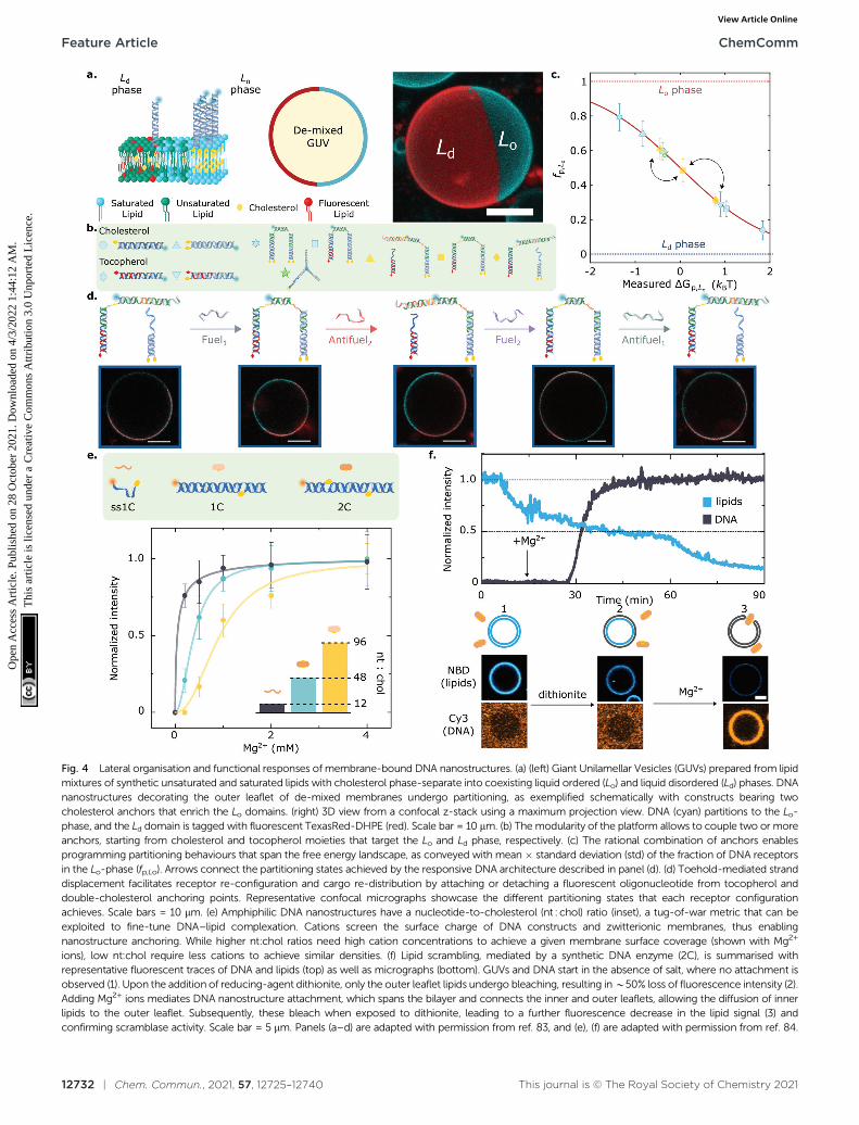
Fig. 4 Lateral organisation and functional responses of membrane-bound DNA nanostructures. (a) (left) Giant Unilamellar Vesicles (GUVs) prepared from lipidmixtures of synthetic unsaturated and saturated lipids with cholesterol phase-separate into coexisting liquid ordered (Lo) and liquid disordered (Ld) phases. DNAnanostructures decorating the outer leaflet of de-mixed membranes undergo partitioning, as exemplified schematically with constructs bearing twocholesterol anchors that enrich the Lo domains. (right) 3D view from a confocal z-stack using a maximum projection view. DNA (cyan) partitions to the Lo-phase, and the Ld domain is tagged with fluorescent TexasRed-DHPE (red). Scale bar = 10 mm. (b) The modularity of the platform allows to couple two or moreanchors, starting from cholesterol and tocopherol moieties that target the Lo and Ld phase, respectively. (c) The rational combination of anchors enablesprogramming partitioning behaviours that span the free energy landscape, as conveyed with mean � standard deviation (std) of the fraction of DNA receptorsin the Lo-phase (fp,Lo). Arrows connect the partitioning states achieved by the responsive DNA architecture described in panel (d). (d) Toehold-mediated stranddisplacement facilitates receptor re-configuration and cargo re-distribution by attaching or detaching a fluorescent oligonucleotide from tocopherol anddouble-cholesterol anchoring points. Representative confocal micrographs showcase the different partitioning states that each receptor configurationachieves. Scale bars = 10 mm. (e) Amphiphilic DNA nanostructures have a nucleotide-to-cholesterol (nt : chol) ratio (inset), a tug-of-war metric that can beexploited to fine-tune DNA–lipid complexation. Cations screen the surface charge of DNA constructs and zwitterionic membranes, thus enablingnanostructure anchoring. While higher nt:chol ratios need high cation concentrations to achieve a given membrane surface coverage (shown with Mg2+
ions), low nt:chol require less cations to achieve similar densities. (f) Lipid scrambling, mediated by a synthetic DNA enzyme (2C), is summarised withrepresentative fluorescent traces of DNA and lipids (top) as well as micrographs (bottom). GUVs and DNA start in the absence of salt, where no attachment isobserved (1). Upon the addition of reducing-agent dithionite, only the outer leaflet lipids undergo bleaching, resulting in B50% loss of fluorescence intensity (2).Adding Mg2+ ions mediates DNA nanostructure attachment, which spans the bilayer and connects the inner and outer leaflets, allowing the diffusion of innerlipids to the outer leaflet. Subsequently, these bleach when exposed to dithionite, leading to a further fluorescence decrease in the lipid signal (3) andconfirming scramblase activity. Scale bar = 5 mm. Panels (a–d) are adapted with permission from ref. 83, and (e), (f) are adapted with permission from ref. 84.
Feature Article ChemComm
Ope
n A
cces
s A
rtic
le. P
ublis
hed
on 2
8 O
ctob
er 2
021.
Dow
nloa
ded
on 4
/3/2
022
1:44
:12
AM
. T
his
artic
le is
lice
nsed
und
er a
Cre
ativ
e C
omm
ons
Attr
ibut
ion
3.0
Unp
orte
d L
icen
ce.
View Article Online
This journal is © The Royal Society of Chemistry 2021 Chem. Commun., 2021, 57, 12725–12740 | 12733
Combining amphiphilic DNA nanostructures with multi-component synthetic membranes displaying a rich phasebehaviour133 enables biomimetic regulation of the lateral dis-tribution, and consequently the functionality, of membraneinclusions.
Key for achieving this objective is the ability of amphiphilicDNA nanostructures to selectively enrich different (coexisting)lipid phases, depending on the chemical identity of the anchoringmotifs,134,135 the lipid composition of the membrane,80 and thesize of the nanostructures.83,107 Indeed, harnessing the preferentialaffinity that cholesterol and tocopherol moieties have for liquid-ordered (Lo) and liquid-disordered (Ld) lipid phases, respectively,Rubio-Sanchez et al. showed that the lateral distribution ofmembrane-tethered DNA nanostructures can be statically anddynamically programmed.83 In this contribution, DNA receptorswere interfaced with GUVs displaying coexistence of Lo and Ld
phases, each occupying a hemispherical domain and resultingin a Janus-like geometry, as shown in Fig. 4a. The syntheticDNA nanostructures were anchored to the bilayer via cholesterol,tocopherol, or combinations thereof (Fig. 4b), and were demon-strated to display a programmable tendency to distribute across thetwo domains, dependent on the number and chemical identity ofthe anchors. In particular, the authors demonstrated that the freeenergy change driving preferential partitioning is approximatelyadditive in the contributions from each individual anchor featuredin the construct. Non-additive effects were also observed for specificanchor combinations, membrane compositions and nanostructuredesign, notably including those induced by steric interactionsbetween bulkier nanostructures. Thus, by prescribing anchorcombination as well as changes to nanostructure size and topology,the partitioning of the DNA devices could be programmed toachieve several states that fully spanned the partitioning landscape,as shown in Fig. 4c.
Furthermore, the functionality of the platform was illustratedwith a proof-of-concept biomimetic DNA architecture responsive tomolecular cues. Exploiting toehold-mediated strand displacement,Fuel/Antifuel strands, capable of inducing re-configuration,enabled a fluorescent model cargo to attach or detach fromanchoring points associated to distinct partitioning behaviours(Fig. 4d). The latter enabled the DNA receptors to reversiblytransport the fluorescent cargoes across the surface of the vesiclesby attaining programmed partitioned states. Importantly, there-shuffling action of the DNA receptors, which evokes therecruitment of cell-surface entities, was observed to have character-istic re-distribution times of B5 min,83 which are comparable tothat of biological machinery involved in T-cell activation136 andclathrin-mediated endocytosis,103,137 with equilibration timescalesin the order of tens to hundreds of seconds.
Further adding to the arsenal of effects that one can exploitto program the response of DNA-decorated membranes,Morzy et al. unveiled the key role of cations in regulating theinteractions between functional nucleic acid nanostructuresand model bilayers.84 Combining experiments and atomisticsimulations, they demonstrated that gel-phase zwitterionicmembranes can bind unmodified DNA nanostructures thanksto the action of divalent cations bridging the anionic groups on
the DNA and lipid head-groups. Interestingly, the attractiveinteractions did not occur for liquid-phase membranes, unlockingpathways to modulate membrane-DNA complexation through anyof the external stimuli or design parameters that can influencelipid phase, including temperature and sterol content.84
Furthermore, Morzy et al. showed that cation concentration andchemical nature offer a handle to fine tune the affinity betweenhydrophobe-modified DNA constructs and liquid-phasemembranes, modulating the competition between hydrophobicity-mediated attraction and Coulomb repulsion through chargescreening. The authors found that the degree of screeningrequired to trigger membrane attachment is dependent on a‘‘tug-of-war’’ ratio between the number of negatively chargednucleotides and that of hydrophobic anchors in the constructs,as shown in Fig. 4e for a library of cholesterol-modifiednanostructures. Morzy et al. then exploited this effect toreversibly trigger membrane attachment and activation of aDNA-based synthetic enzyme via the addition of magnesium.The synthetic ‘‘scramblase’’ enzyme, a device previously intro-duced by Ohmann et al.42 and later simplified by Sobota et al.,43
has the ability to catalyse exchange of lipids between the twomembrane leaflets (scrambling) once bound to, and theninserted across, the bilayer. To demonstrate the cation-mediated activation of the enzyme, a fluorophore reductionassay was used in which the membranes were doped with lipidstagged with NBD, a molecule that is fluorescent in its oxidisedstate and undergoes bleaching upon reduction. The addition ofmembrane-impermeable reducing agent dithionite bleachedNBD on the outher leaflet resulting in a B50% loss in fluores-cence emission from the membrane (Fig. 4f). The addition ofMg2+ caused attachment and activation of the synthetic DNAenzyme. This in turn triggered transport of un-bleached NBD-tagged lipids to the outer leaflet, their exposure to dithionite,and a further decrease in fluorescence, thus demonstratingscramblase activity.
Indeed, as discussed in this section, amphiphilic DNAnanostructures show great promise to readily design andconstruct a vast array of devices capable of imparting andreplicating functionalities associated to biological interfacesin artificial cellular membranes. Combining hydrophobesof different chemical identities and properties with thetuneable size, topology, and responsiveness of DNA architec-tures is key for engineering ever-more sophisticated andprogrammable biomimetic responses. We envisage thatcoupling (amphiphilic) DNA nanotechnology with modelmembranes will open up a breadth of avenues for artificialcell science, and revolutionise the state-of-the-art in bottom-upsynthetic biology.
3 Bulk self-assembly of amphiphilicDNA nanostructures
Besides enabling prescribable interactions with lipid membranes,the functionalisation of DNA nanostructures with hydrophobicmoieties, ranging from small molecules to dendrons and
ChemComm Feature Article
Ope
n A
cces
s A
rtic
le. P
ublis
hed
on 2
8 O
ctob
er 2
021.
Dow
nloa
ded
on 4
/3/2
022
1:44
:12
AM
. T
his
artic
le is
lice
nsed
und
er a
Cre
ativ
e C
omm
ons
Attr
ibut
ion
3.0
Unp
orte
d L
icen
ce.
View Article Online
12734 | Chem. Commun., 2021, 57, 12725–12740 This journal is © The Royal Society of Chemistry 2021
polymers,138 has been exploited to enhance their self-assemblycapabilities beyond what is achievable by base-pairing.
While base-pairing offers exquisite control over interactionstrength and selectivity, its rigid ‘‘lock-and-key’’ nature means thatprogramming higher-order self-assembly of the nanostructuresrequires very precise geometrical and thermodynamicoptimisation.139–142 The attachment of hydrophobic tags providesDNA nanostructures with an amphiphilic character, grantingaccess to self-assembly pathways where the thermodynamicground state can be determined through the size, shape andtopology of the DNA amphiphilies – all features that can be easilyand robustly prescribed.143–147 While amphiphilic self-assemblyprinciples do not allow for the molecular-scale localisation
precision of base-paring, they are agnostic to the fine detailsof the nanostructures, which makes them robust againstpolydispersity and small design variations. In addition, theamphiphilic character of the resulting hydrophobised-DNAphases makes them ideal for programming interactions withother biological macromolecules and lipophilic small molecules,a useful characteristic for several applications including drugdelivery and – relevant to the present discussion – the constructionof biomimetic systems. In this section we review instances in whichthe amphiphilic self-assembly principle has been applied toprogramming higher-order self-assembly of DNA nanostructures.We largely focus on multi-functional nano-porous phasesdeveloped by our group in recent years, and comment on their
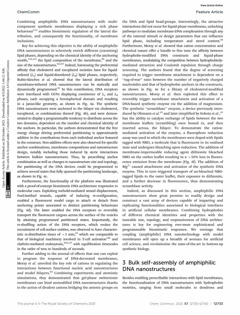
Fig. 5 Self-assembly of amphiphilic DNA C-Stars into frameworks with tunable porosity. (a) C-Stars are comprised of four different core strands (blue)dictating the nanostar shape, and four cholesterolised strands (orange) granting amphiphilicity. (b) Schematic of self-assembly mechanism for C-Stars.A stoichiometric mixture of cholesterolised and core strands is heated and then slowly annealed. Above the melting temperature of the duplex arms (left)the mixture comprises both cholesterol-induced micelles and single-stranded core-forming oligonucleotides. Upon cooling below TAgg = 77.1 � 0.2 1C(centre), the core nanostar motifs start forming via hybridisation, thus bridging the micelles. DLS measurements (inset) demonstrate the nucleation andgrowth of aggregates over time, as exemplified by the increase in hydrodynamic diameter Dh, eventually leading to extended crystalline frameworks(right). (c) Controlling arm length allows for lattice parameter tunability. Bright-field micrographs (left) depict rhombic-dodecahedral crystallites forC-Stars with l = 21–42 bp, while l = 51 bp produces amorphous spherical aggregates. SAXS powder diffraction patterns and the derived radially averagedprofiles (centre) confirm BCC crystalline phase of C-Star aggregates for l = 21–42 bp and amorphous nature for l = 51 bp. Red vertical lines illustrate bestfit to Bragg peaks for BCC symmetry. (right) Schematic illustrating the mechanism behind arm length control. Lattice parameter tunability ensurescontrollable porosity, thus enabling the use of C-Star frameworks as macromolecular sieves. (d) Porosity was assessed via permeation assays with a rangeof fluorescent probes: sodium fluorescein (FAM), fluorescein-labelled 3 kDa dextran (3 kDa DXT-FAM), fluorescein-labelled 10 kDa dextran (10 kDa DXT-FAM), recombinant GFP (rGFP) and Alexa647-labelled streptavidin (SAv-A647). The ratio x between average fluorescence intensity (from confocalmicrographs) inside single crystals and in the surrounding probe-rich solution was used as proxy for probe permeation and partitioning. Gray band at thebottom depicts background fluorescence intensity. (e) Representative confocal micrographs corresponding to the data in (d). All scale bars = 10 mm.Panels (a–e) are adapted with permission from ref. 90. Copyright 2018, American Chemical Society. Panel (b) is adapted with permission from ref. 89.Copyright 2017, American Chemical Society.
Feature Article ChemComm
Ope
n A
cces
s A
rtic
le. P
ublis
hed
on 2
8 O
ctob
er 2
021.
Dow
nloa
ded
on 4
/3/2
022
1:44
:12
AM
. T
his
artic
le is
lice
nsed
und
er a
Cre
ativ
e C
omm
ons
Attr
ibut
ion
3.0
Unp
orte
d L
icen
ce.
View Article Online
This journal is © The Royal Society of Chemistry 2021 Chem. Commun., 2021, 57, 12725–12740 | 12735
potential application in bottom-up synthetic biology as well asother contexts.
Among the first examples of complex objects self-assembledfrom amphipilic DNA nanostructures are the DNA-somes,developed by Luo and coworkers.148 These particles, formedfrom lipid-modified DNA junctions could be tuned in size andhave been shown to aid intra-cellular delivery of miRNA. Albertset al. have later developed their version of DNA-somes, whichthanks to pH-responsive i-motifs were able to switch betweennetworks and vesicle-like objects, and may represent interestingand highly-programmable alternatives to lipid or polymer-basedmembranes for artificial cell implementations.149 Anothernotable example is the one demonstrated by Sleiman andcoworkers, who explored the use of hydrophobic polymer150,151
or dendrite152 moieties to guide the self-assembly of nanocagesin unique architectures, including intra or inter-molecularmicellar assemblies capable of loading a hydrophobic cargo.
Brady et al. introduced a versatile approach to the self-assembly of nano-porous, functional DNA phases reliant oncholesterolised nanostructures dubbed C-Stars.89–91 As shownin Fig. 5, these are simple DNA junctions featuring (typically)four dsDNA arms. However, rather than terminating in ssDNAsticky ends, as initially proposed by Seeman1,153 and laterimplemented by multiple groups,3,154–163 the arms are tippedby a cholesterol moiety, which confers an amphiphiliccharacter to the nanostructures and drives their self-assemblyinto extended frameworks. Cholesterolised and non-cholesterolised ssDNA components were stoichiometricallymixed, and then slowly cooled from a high temperature – abovethe melting temperature of any duplex present. Upon cooling,the designed dsDNA motifs formed and started cross-linkingexisting DNA–cholesterol micelles, until a phase transition wasencountered. At this stage aggregates nucleated in the bulk andstarted to coalesce and grow, ultimately forming an extendednetwork (Fig. 5b). The resulting aggregates were shown to havean amorphous character if quenching was rapid, or formcrystalline phases if annealed at a slow rate, as determinedthrough SAXS and microscopy (Fig. 5c). C-Star single crystalswere observed to exceed 40 mm in size, and the latticeparameter of the cubic unit cell could be finely programmedby changing arm length, spanning a range between B18 andB34 nm.90 Such a difference in lattice parameter directlytranslated into a difference in the network pore size, thusallowing C-Star aggregates to behave as controllable molecularsieves, where certain macromolecules can permeate and otherscannot due to their bulky size. The authors performed permeationassays on various probes to confirm this behaviour, assummarised in Fig. 5d and e. Small molecules such as sodiumfuorescein easily permeated even the lowest-porosity frameworks,while bulkier dextrans displayed a marked permeation increaseupon increasing arm length. Notably, the emptiest crystallineframeworks achieved a free volume fraction in excess of 85%,comparable with ultra-high porosity metall–organicframeworks,90 thus confirming the potential of C-Star frameworksfor cargo-loading applications, as required, for instance, in drugdelivery applications.
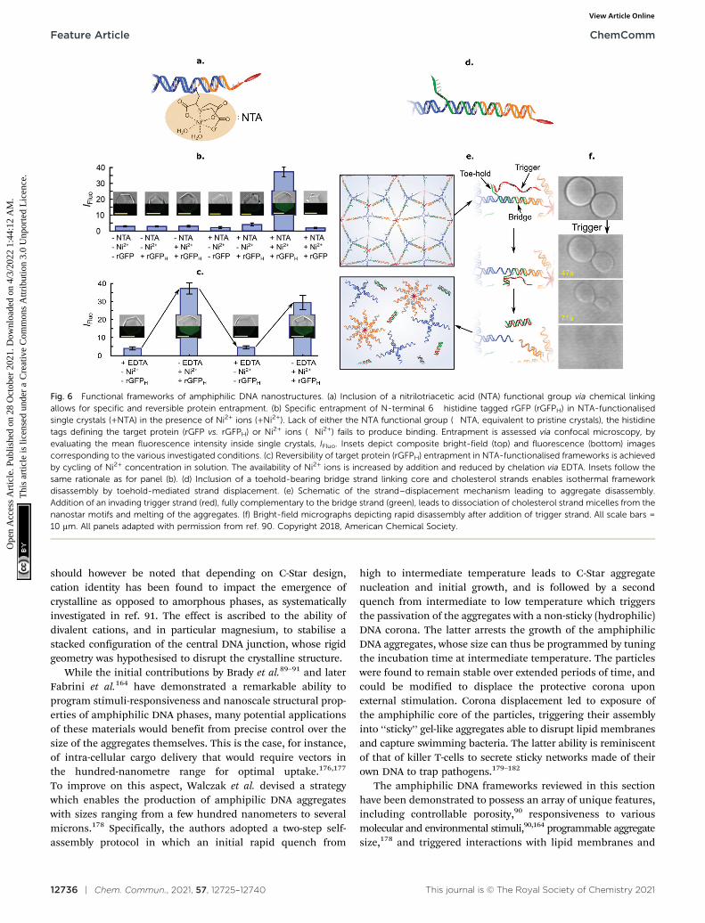
The facile functionalisation of DNA nanotructures furtherallowed Brady et al. to make the cargo-loading ability of C-Starframeworks selective and stimuli-responsive.90 To this end, theauthors functionalised individual C-Stars with a nitrilotriaceticacid (NTA) group, as shown in Fig. 6a, which is capable ofselectively binding His-tagged proteins in the presence of Ni2+
ions. The authors applied this system to demonstrate selectivetrapping of His-tagged recombinant Green Fluorescent Protein(rGFP) (Fig. 6b), and its reversible release upon Ni2+ chelationvia EDTA (Fig. 6c).
Besides releasing pre-loaded cargoes upon exposure toexternal cues, frameworks of amphiphilic DNA junctionswere also shown to structurally respond to various stimuli. Forinstance, Brady et al. implemented a design change to the basicC-Star architectures, shown in Fig. 6d, to make the crystallinephases able to disassemble in the presence of a trigger ssDNAstrand. The scheme relied on a toeholding reaction, throughwhich the trigger sequestered a bridge strand initially linking thecholesterolised moiety to the nanostar core, thus leading toisothermal melting of the aggregates (Fig. 6e and f).
To further expand the range of stimuli to which amphiphilicDNA frameworks are able to respond, Fabrini et al. recentlydeveloped a strategy to render the materials structurally suscep-tible to changes in the identity and concentration of cations insolution.164 To achieve such functionality, the authors replacedthe central junction in C-Stars with a tetramolecular DNAG-quadruplex (G4), a non-canonical motif formed in guanine-rich nucleic acids and stabilised by alkali metal ions.165,166 Byexploiting the different degrees of G4 stabilisation offered bystrongly-promoting K+ ions and other metal ions (Li+, Mg2+), themodified amphiphilic nanostars, dubbed Quad-Stars, displayedthe sought sensitivity towards cation identity and concentration.The authors demonstrated that self-assembly rates dependsteeply on K+ concentrations and the length of the G4-formingguanine runs. The frameworks could be quickly disassembled bychelating K+, and thus release previously loaded cargoes. Finally,the inclusion of a photosensitising G4-binding porphyrinenabled the light-induced (irreversible) disassembly of thehydrogel aggregates, adding yet another route to triggerstructural responses in the amphiphilic DNA frameworks.
While such ad hoc design modifications can lead to de-stabilisation of the amphiphilic DNA frameworks in responseto changes in the ionic conditions, ‘‘conventional’’ C-Stardesigns have been shown to remain stable under a broad rangeof ionic strength and, importantly, at physiological values(1� phosphate-buffered saline, PBS).91 This characteristic iskey for foreseen biomimetic applications, and gives amphiphilicDNA building blocks an advantage over other network-formingDNA motifs (including small nanostructures139 andorigami167–169), whose stability has been reported to rely on theavailability of divalent cations.170 While, indeed, some DNAorigami have been shown to remain stable even in low-Mg2+
buffers,171,172 structural integrity heavily depends on topologicalcomplexity,171,173,174 with more intricate 3D architectures ornetworks175 requiring either divalent cations or extremely high(non-physiological) concentrations of monovalent salts.173 It
ChemComm Feature Article
Ope
n A
cces
s A
rtic
le. P
ublis
hed
on 2
8 O
ctob
er 2
021.
Dow
nloa
ded
on 4
/3/2
022
1:44
:12
AM
. T
his
artic
le is
lice
nsed
und
er a
Cre
ativ
e C
omm
ons
Attr
ibut
ion
3.0
Unp
orte
d L
icen
ce.
View Article Online
12736 | Chem. Commun., 2021, 57, 12725–12740 This journal is © The Royal Society of Chemistry 2021
should however be noted that depending on C-Star design,cation identity has been found to impact the emergence ofcrystalline as opposed to amorphous phases, as systematicallyinvestigated in ref. 91. The effect is ascribed to the ability ofdivalent cations, and in particular magnesium, to stabilise astacked configuration of the central DNA junction, whose rigidgeometry was hypothesised to disrupt the crystalline structure.
While the initial contributions by Brady et al.89–91 and laterFabrini et al.164 have demonstrated a remarkable ability toprogram stimuli-responsiveness and nanoscale structural prop-erties of amphiphilic DNA phases, many potential applicationsof these materials would benefit from precise control over thesize of the aggregates themselves. This is the case, for instance,of intra-cellular cargo delivery that would require vectors inthe hundred-nanometre range for optimal uptake.176,177
To improve on this aspect, Walczak et al. devised a strategywhich enables the production of amphipilic DNA aggregateswith sizes ranging from a few hundred nanometers to severalmicrons.178 Specifically, the authors adopted a two-step self-assembly protocol in which an initial rapid quench from
high to intermediate temperature leads to C-Star aggregatenucleation and initial growth, and is followed by a secondquench from intermediate to low temperature which triggersthe passivation of the aggregates with a non-sticky (hydrophilic)DNA corona. The latter arrests the growth of the amphiphilicDNA aggregates, whose size can thus be programmed by tuningthe incubation time at intermediate temperature. The particleswere found to remain stable over extended periods of time, andcould be modified to displace the protective corona uponexternal stimulation. Corona displacement led to exposure ofthe amphiphilic core of the particles, triggering their assemblyinto ‘‘sticky’’ gel-like aggregates able to disrupt lipid membranesand capture swimming bacteria. The latter ability is reminiscentof that of killer T-cells to secrete sticky networks made of theirown DNA to trap pathogens.179–182
The amphiphilic DNA frameworks reviewed in this sectionhave been demonstrated to possess an array of unique features,including controllable porosity,90 responsiveness to variousmolecular and environmental stimuli,90,164 programmable aggregatesize,178 and triggered interactions with lipid membranes and
Fig. 6 Functional frameworks of amphiphilic DNA nanostructures. (a) Inclusion of a nitrilotriacetic acid (NTA) functional group via chemical linkingallows for specific and reversible protein entrapment. (b) Specific entrapment of N-terminal 6� histidine tagged rGFP (rGFPH) in NTA-functionalisedsingle crystals (+NTA) in the presence of Ni2+ ions (+Ni2+). Lack of either the NTA functional group (�NTA, equivalent to pristine crystals), the histidinetags defining the target protein (rGFP vs. rGFPH) or Ni2+ ions (�Ni2+) fails to produce binding. Entrapment is assessed via confocal microscopy, byevaluating the mean fluorescence intensity inside single crystals, IFluo. Insets depict composite bright-field (top) and fluorescence (bottom) imagescorresponding to the various investigated conditions. (c) Reversibility of target protein (rGFPH) entrapment in NTA-functionalised frameworks is achievedby cycling of Ni2+ concentration in solution. The availability of Ni2+ ions is increased by addition and reduced by chelation via EDTA. Insets follow thesame rationale as for panel (b). (d) Inclusion of a toehold-bearing bridge strand linking core and cholesterol strands enables isothermal frameworkdisassembly by toehold-mediated strand displacement. (e) Schematic of the strand–displacement mechanism leading to aggregate disassembly.Addition of an invading trigger strand (red), fully complementary to the bridge strand (green), leads to dissociation of cholesterol strand micelles from thenanostar motifs and melting of the aggregates. (f) Bright-field micrographs depicting rapid disassembly after addition of trigger strand. All scale bars =10 mm. All panels adapted with permission from ref. 90. Copyright 2018, American Chemical Society.
Feature Article ChemComm
Ope
n A
cces
s A
rtic
le. P
ublis
hed
on 2
8 O
ctob
er 2
021.
Dow
nloa
ded
on 4
/3/2
022
1:44
:12
AM
. T
his
artic
le is
lice
nsed
und
er a
Cre
ativ
e C
omm
ons
Attr
ibut
ion
3.0
Unp
orte
d L
icen
ce.
View Article Online
This journal is © The Royal Society of Chemistry 2021 Chem. Commun., 2021, 57, 12725–12740 | 12737
living cells.178 These characteristics make them potentialcandidates as both structural and functional elements inartificial cell implementations. As proposed and demonstratedin several instances, membrane-less compartments64,65
represent an intriguing alternative to membrane-basedenclosures to serve as scaffold for artificial cells. Examplesreported to date have been often reliant on various forms ofpolymer coacervates or hydrogels,59–63 which, while enabling adegree on control on composition and molecular partitioning,offer little opportunity for the rational design of life-likeresponses in which structural properties are coupled tomolecular pathways. In turn, thanks to the programmabilityof the nucleic acid building blocks, amphiphilic DNA scaffoldsopen up new opportunities for co-engineering structural andinformation-processing aspects of artificial cells. One couldindeed envisage implementations in which communication/sensing pathways, hosted by the DNA building blockthemselves, could lead to structural changes in the network,thus impacting porosity and molecular transport, andultimately feeding-back to the artificial signalling mechanism.In addition, the structure–function synergy unlocked by theresponsive all-DNA scaffolds could lead to programming cell-like behaviours requiring regulated changes to global morphology,such as uptake of large objects, growth, and division which havebeen traditionally challenging to implement.58,63 Finally we notethat amphiphilic DNA frameworks may also serve as functionalinternal compartments, or membrane-less organelles inmembrane-based artificial cell implementations, where they couldlead to spatial localisation and segregation of functionalities – acritical step towards the construction of intricate signalling path-ways such as those observed in eukaryotes.57
4 Conclusive remarks and futurechallenges
As a feature article centred around the work of our group, thiscontribution is not intended to offer a comprehensive snapshotof the rapidly growing field of biomimetic DNA nanotechnology,nor of the substantial sub-set of studies specifically addressingchallenges in bottom-up synthetic biology and artificial cellscience. We have, for instance, omitted discussion of key con-tributions on the use of DNA origami to control mechanicalproperties of synthetic bilayers85–87 or to regulate transportacross them by establishing transmembrane pores,183,184 as wellas notable efforts to program communication using DNA-basedmolecular circuitry.27 The powerful capabilities highlighted inthese contributions, alongside the ones discussed in more detailhere, print an exciting picture for the near future of this researcharea. In the remainder of this section we will highlight aspectsthat, in our opinion, deserve particular attention from thecommunity.
The first concept we would like to explore is that of integration.One of the key advantages of bottom-up synthetic biology is theavailability of a virtually unlimited library of components andmechanisms, both biological and inorganic, that one can draw
from when designing and constructing an artificial cell. This hasresulted in a substantial heterogeneity across implementationsdesigned to display different behaviours, which makeschallenging to integrate them in multi-functional artificial cells.In other words, while examples of artificial cells capable of eithersensing, moving, communicating, or dividing (to mention a fewexamples) have been extensively demonstrated, we are stillunable to construct an artificial cell that can simultaneouslydisplay and coordinate many of those behaviours because thesolutions developed for each are incompatible. Synthetic DNA-nanosystems, owing to the chemical and functional homogeneityof their constituents, could help overcoming this criticallimitation. It is indeed easy to envisage how biomimetic DNAnanostructures designed separately to regulate transport,adhesion, communication and sensing could be interfaced witheach other, and coupled to common signalling pathways relianton DNA-based molecular circuitry. Success in the holistic designof these ‘‘all-DNA’’ artificial cells, with integrated control ofmultiple behaviours, could take the field one step closer to theconstruction of truly cell-like micro-robots.
The second aspect we would like to discuss is that of freeenergy regeneration. Living systems are sustained by a constantfree energy flux, which keeps them hovering above thermodynamicequilibrium. Free energy is sourced from the environment andthen distributed or stored in the form of chemical vectors. Instead,for the most part, dynamic DNA nano-systems are driven bydiscrete relaxations from initial high-free-energy states. The latterare typically characterised by fewer base-pairing bonds compared tothe thermodynamic ground state, and can be made kineticallymeta-stable, so that the free energy tap can be opened only in thepresence of a specific trigger. While remarkable design advanceshave been recently introduced, which enable decoupling thermo-dynamic drive from kinetic rates185 and systematically accessingout-of-equilibrium configurations,186 these free-energy reservoirsremain essentially non renewable, limiting their applicability tothe construction of autonomous agents. There is therefore a needfor solutions that can continuously regenerate free energy reservoirsin DNA nano-systems from readily available environmental sources.Thankfully, examples are emerging demonstrating the coupling ofnucleic-acid molecular reaction networks and self-assembly toenzymatic processes,187–191 also in the context of cytomimeticagents.192 Given these encouraging results, we argue that ageneralised enzymatic pathway for the sustained production of anucleic acid ‘‘free energy currency’’ would be a welcomedevelopment for the community, and unlock the modular designof self-sustaining, DNA-based artificial cells.
Conflicts of interest
There are no conflicts to declare.
Acknowledgements
R. R. S. acknowledges funding from the Mexican National Councilfor Science and Technology (CONACYT, Grant No. 472427), the
ChemComm Feature Article
Ope
n A
cces
s A
rtic
le. P
ublis
hed
on 2
8 O
ctob
er 2
021.
Dow
nloa
ded
on 4
/3/2
022
1:44
:12
AM
. T
his
artic
le is
lice
nsed
und
er a
Cre
ativ
e C
omm
ons
Attr
ibut
ion
3.0
Unp
orte
d L
icen
ce.
View Article Online
12738 | Chem. Commun., 2021, 57, 12725–12740 This journal is © The Royal Society of Chemistry 2021
Cambridge Trust, and from the EPSRC CDT in Nanoscience andNanotechnology (NanoDTC, Grant No. EP/L015978/1). G. F.acknowledges funding from the Department of Chemistry atImperial College London. L. D. M. acknowledges funding from aRoyal Society University Research Fellowship (UF160152) andfrom the European Research Council (ERC) under the Horizon2020 Research and Innovation Programme (ERC-STG No. 851667NANOCELL). The authors thanks B. M. Mognetti for feedback onthe manuscript and contributions on much of the researchdiscussed here.
References1 N. C. Seeman, J. Theor. Biol., 1982, 99, 237–247.2 N. C. Seeman and H. F. Sleiman, Nat. Rev. Mater., 2017, 3, 17068.3 S. Biffi, R. Cerbino, F. Bomboi, E. M. Paraboschi, R. Asselta,
F. Sciortino and T. Bellini, Proc. Natl. Acad. Sci. U. S. A., 2013,110, 15633–15637.
4 W. B. Rogers and V. N. Manoharan, Science, 2015, 347, 639–642.5 L. Di Michele and E. Eiser, Phys. Chem. Chem. Phys., 2013, 15,
3115–3129.6 L. Di Michele, F. Varrato, J. Kotar, S. H. Nathan, G. Foffi and
E. Eiser, Nat. Commun., 2013, 4, 2007.7 L. Di Michele, D. Fiocco, F. Varrato, S. Sastry, E. Eiser and G. Foffi,
Soft Matter, 2014, 10, 3633–3648.8 M. R. Jones, N. C. Seeman and C. A. Mirkin, Science, 2015,
347, 1260901.9 C. A. Mirkin, R. C. Letsinger, R. C. Mucic and J. J. Storhoff, Nature,
1996, 382, 607.10 A. P. Alivisatos, K. P. Johnsson, X. Peng, T. E. Wilson, C. J. Loweth,
M. P. Bruchez and P. G. Schultz, Nature, 1996, 382, 609.11 P. W. K. Rothemund, Nature, 2006, 440, 297 EP.12 H. Ramezani and H. Dietz, Nat. Rev. Genet., 2020, 21, 5–26.13 W. Liu, J. Halverson, Y. Tian, A. V. Tkachenko and O. Gang, Nat.
Chem., 2016, 8, 867–873.14 A. E. Marras, L. Zhou, H.-J. Su and C. E. Castro, Proc. Natl. Acad. Sci.
U. S. A., 2015, 112, 713–718.15 Y. R. Yang, Y. Liu and H. Yan, Bioconjugate Chem., 2015, 26,
1381–1395.16 A. J. M. Wollman, C. Sanchez-Cano, H. M. J. Carstairs, R. A. Cross
and A. J. Turberfield, Nat. Nanotechnol., 2014, 9, 44–47.17 N. D. Derr, B. S. Goodman, R. Jungmann, A. E. Leschziner,
W. M. Shih and S. L. Reck-Peterson, Science, 2012, 338, 662–665.18 A. Rajendran, M. Endo and H. Sugiyama, Angew. Chem., Int. Ed.,
2012, 51, 874–890.19 A. Keller, I. Bald, A. Rotaru, E. Cauet, K. V. Gothelf and
F. Besenbacher, ACS Nano, 2012, 6, 4392–4399.20 T. Ding, J. Yang, V. Pan, N. Zhao, Z. Lu, Y. Ke and C. Zhang, Nucleic
Acids Res., 2020, 48, 2791–2806.21 Q. Zhang, Q. Jiang, N. Li, L. Dai, Q. Liu, L. Song, J. Wang, Y. Li,
J. Tian, B. Ding and Y. Du, ACS Nano, 2014, 8, 6633–6643.22 E. Silvester, B. Vollmer, V. Prazak, D. Vasishtan, E. A. Machala,
C. Whittle, S. Black, J. Bath, A. J. Turberfield, K. Grunewald andL. A. Baker, Cell, 2021, 184, 1110–1121.
23 A. Kuzyk, R. Jungmann, G. P. Acuna and N. Liu, ACS Photonics,2018, 5, 1151–1163.
24 D. Nykypanchuk, M. M. Maye, D. van der Lelie and O. Gang,Langmuir, 2007, 23, 6305–6314.
25 F. C. Simmel, B. Yurke and H. R. Singh, Chem. Rev., 2019, 119,6326–6369.
26 D. Woods, D. Doty, C. Myhrvold, J. Hui, F. Zhou, P. Yin andE. Winfree, Nature, 2019, 567, 366–372.
27 A. Joesaar, S. Yang, B. Bogels, A. van der Linden, P. Pieters,B. V. V. S. P. Kumar, N. Dalchau, A. Phillips, S. Mann andT. F. A. de Greef, Nat. Nanotechnol., 2019, 14, 369–378.
28 M. R. Lakin and A. Phillips, ACS Synth. Biol., 2020, 9, 1499–1513.29 B. Yurke, A. J. Turberfield, A. P. Mills, F. C. Simmel and
J. L. Neumann, Nature, 2000, 406, 605–608.30 R. A. Muscat, J. Bath and A. J. Turberfield, Nano Lett., 2011, 11,
982–987.
31 S. Ranallo, C. Prevost-Tremblay, A. Idili, A. Vallee-Belisle andF. Ricci, Nat. Commun., 2017, 8, 15150.
32 A. Idili, A. Vallee-Belisle and F. Ricci, J. Am. Chem. Soc., 2014, 136,5836–5839.
33 J. Schnitzbauer, M. T. Strauss, T. Schlichthaerle, F. Schueder andR. Jungmann, Nat. Protoc., 2017, 12, 1198–1228.
34 R. Jungmann, M. S. Avendano, J. B. Woehrstein, M. Dai, W. M. Shihand P. Yin, Nat. Methods, 2014, 11, 313–318.
35 O. K. Wade, J. B. Woehrstein, P. C. Nickels, S. Strauss, F. Stehr,J. Stein, F. Schueder, M. T. Strauss, M. Ganji, J. Schnitzbauer,H. Grabmayr, P. Yin, P. Schwille and R. Jungmann, Nano Lett.,2019, 19, 2641–2646.
36 A. H. Clowsley, W. T. Kaufhold, T. Lutz, A. Meletiou, L. Di Micheleand C. Soeller, J. Am. Chem. Soc., 2020, 142, 12069–12078.
37 A. H. Clowsley, W. T. Kaufhold, T. Lutz, A. Meletiou, L. Di Micheleand C. Soeller, Nat. Commun., 2021, 12, 501.
38 M. Langecker, V. Arnaut, J. List and F. C. Simmel, Acc. Chem. Res.,2014, 47, 1807–1815.
39 A. Czogalla, H. G. Franquelim and P. Schwille, Biophys. J., 2016,110, 1698–1707.
40 J. Li, A. A. Green, H. Yan and C. Fan, Nat. Chem., 2017, 9,1056–1067.
41 K. Gopfrich, I. Platzman and J. P. Spatz, Trends Biotechnol., 2018,36, 938–951.
42 A. Ohmann, C.-Y. Li, C. Maffeo, K. Al Nahas, K. N. Baumann,K. Gopfrich, J. Yoo, U. F. Keyser and A. Aksimentiev, Nat. Commun.,2018, 9, 1–9.
43 D. Sobota, H. Joshi, A. Ohmann, A. Aksimentiev and U. F. Keyser,Nano Lett., 2020, 20, 4306–4311.
44 B. C. Buddingh’ and J. C. M. van Hest, Acc. Chem. Res., 2017, 50,769–777.
45 R. Lentini, N. Yeh Martın and S. S. Mansy, Curr. Opin. Chem. Biol.,2016, 34, 53–61.
46 V. Noireaux, Y. T. Maeda and A. Libchaber, Proc. Natl. Acad. Sci.U. S. A., 2011, 108, 3473 LP.
47 J. C. Blain and J. W. Szostak, Annu. Rev. Biochem., 2014, 83,615–640.
48 J. W. Szostak, D. P. Bartel and P. L. Luisi, Nature, 2001, 409,387–390.
49 T. M. Allen and P. R. Cullis, Adv. Drug Delivery Rev., 2013, 65, 36–48.50 M. H. M. E. van Stevendaal, J. C. M. van Hest and A. F. Mason,
ChemSystemsChem, 2021, 3, e2100009.51 Y. Zhang, W. C. Ruder and P. R. LeDuc, Trends Biotechnol., 2008,
26, 14–20.52 M. Li, R. L. Harbron, J. V. M. Weaver, B. P. Binks and S. Mann, Nat.
Chem., 2013, 5, 529–536.53 M. Li, D. C. Green, J. L. R. Anderson, B. P. Binks and S. Mann,
Chem. Sci., 2011, 2, 1739–1745.54 H.-J. Choi, E. Brooks and C. D. Montemagno, Nanotechnology, 2005,
16, S143–S149.55 M. Ugrinic, A. Zambrano, S. Berger, S. Mann, T.-Y. D. Tang and
A. DeMello, Chem. Commun., 2018, 54, 287–290.56 S. F. Fenz and K. Sengupta, Integr. Biol., 2012, 4, 982–995.57 L. Aufinger and F. C. Simmel, Angew. Chem., Int. Ed., 2018, 57,
17245–17248.58 Y. Sato, T. Sakamoto and M. Takinoue, Sci. Adv., 2020, 6, eaba3471.59 X. Guo, F. Li, C. Liu, Y. Zhu, N. Xiao, Z. Gu, D. Luo, J. Jiang and
D. Yang, Angew. Chem., Int. Ed., 2020, 59, 20651–20658.60 L. Wang, S. Song, J. van Hest, L. K. Abdelmohsen, X. Huang and
S. Sanchez, Small, 2020, 16, 1907680.61 W. M. Aumiller, F. Pir Cakmak, B. W. Davis and C. D. Keating,
Langmuir, 2016, 32, 10042–10053.62 N. A. Yewdall, A. A. Andre, T. Lu and E. Spruijt, Curr. Opin. Colloid
Interface Sci., 2021, 52, 101416.63 Y. Qiao, M. Li, R. Booth and S. Mann, Nat. Chem., 2017, 9, 110–119.64 D. M. Mitrea and R. W. Kriwacki, Cell Commun. Signal., 2016, 14,
1–20.65 K. K. Nakashima, M. A. Vibhute and E. Spruijt, Front. Mol. Biosci.,
2019, 6, 21.66 E. Gomes and J. Shorter, J. Biol. Chem., 2019, 294, 7127.67 P. B. Sehgal, J. Westley, K. M. Lerea, S. DiSenso-Browne and
J. D. Etlinger, Anal. Biochem., 2020, 597, 113691.68 Z. Izri, D. Garenne, V. Noireaux and Y. T. Maeda, ACS Synth. Biol.,
2019, 8, 1705–1712.
Feature Article ChemComm
Ope
n A
cces
s A
rtic
le. P
ublis
hed
on 2
8 O
ctob
er 2
021.
Dow
nloa
ded
on 4
/3/2
022
1:44
:12
AM
. T
his
artic
le is
lice
nsed
und
er a
Cre
ativ
e C
omm
ons
Attr
ibut
ion
3.0
Unp
orte
d L
icen
ce.
View Article Online
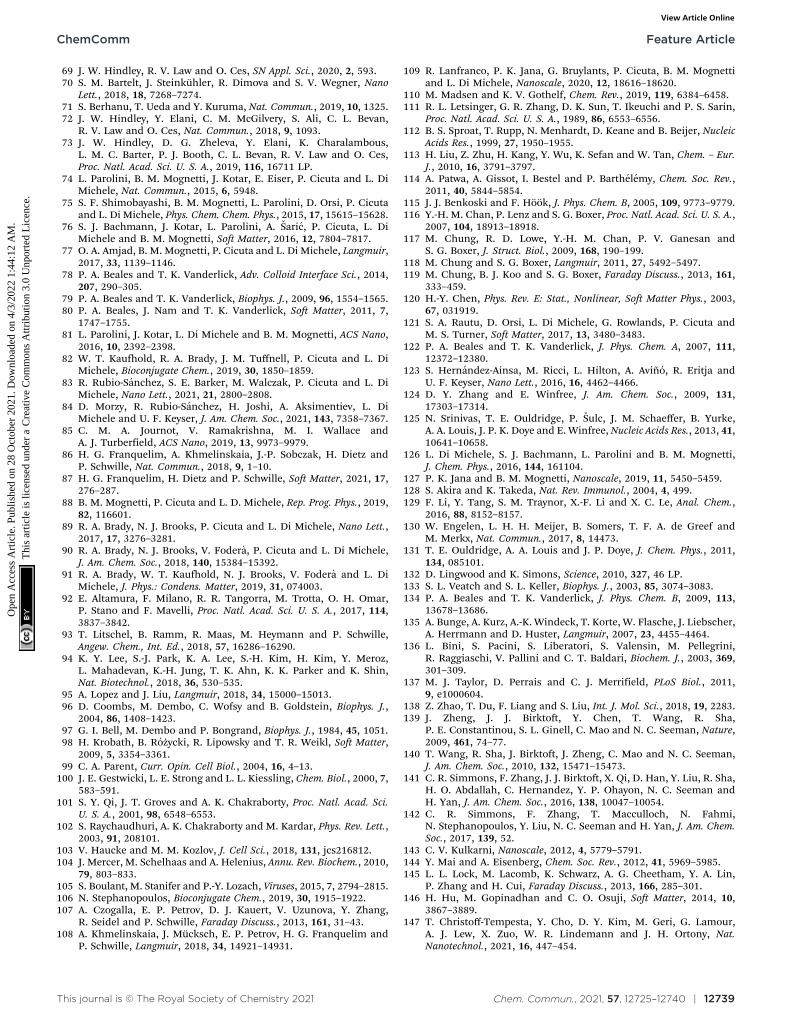
This journal is © The Royal Society of Chemistry 2021 Chem. Commun., 2021, 57, 12725–12740 | 12739
69 J. W. Hindley, R. V. Law and O. Ces, SN Appl. Sci., 2020, 2, 593.70 S. M. Bartelt, J. Steinkuhler, R. Dimova and S. V. Wegner, Nano
Lett., 2018, 18, 7268–7274.71 S. Berhanu, T. Ueda and Y. Kuruma, Nat. Commun., 2019, 10, 1325.72 J. W. Hindley, Y. Elani, C. M. McGilvery, S. Ali, C. L. Bevan,
R. V. Law and O. Ces, Nat. Commun., 2018, 9, 1093.73 J. W. Hindley, D. G. Zheleva, Y. Elani, K. Charalambous,
L. M. C. Barter, P. J. Booth, C. L. Bevan, R. V. Law and O. Ces,Proc. Natl. Acad. Sci. U. S. A., 2019, 116, 16711 LP.
74 L. Parolini, B. M. Mognetti, J. Kotar, E. Eiser, P. Cicuta and L. DiMichele, Nat. Commun., 2015, 6, 5948.
75 S. F. Shimobayashi, B. M. Mognetti, L. Parolini, D. Orsi, P. Cicutaand L. Di Michele, Phys. Chem. Chem. Phys., 2015, 17, 15615–15628.
76 S. J. Bachmann, J. Kotar, L. Parolini, A. Saric, P. Cicuta, L. DiMichele and B. M. Mognetti, Soft Matter, 2016, 12, 7804–7817.
77 O. A. Amjad, B. M. Mognetti, P. Cicuta and L. Di Michele, Langmuir,2017, 33, 1139–1146.
78 P. A. Beales and T. K. Vanderlick, Adv. Colloid Interface Sci., 2014,207, 290–305.
79 P. A. Beales and T. K. Vanderlick, Biophys. J., 2009, 96, 1554–1565.80 P. A. Beales, J. Nam and T. K. Vanderlick, Soft Matter, 2011, 7,
1747–1755.81 L. Parolini, J. Kotar, L. Di Michele and B. M. Mognetti, ACS Nano,
2016, 10, 2392–2398.82 W. T. Kaufhold, R. A. Brady, J. M. Tuffnell, P. Cicuta and L. Di
Michele, Bioconjugate Chem., 2019, 30, 1850–1859.83 R. Rubio-Sanchez, S. E. Barker, M. Walczak, P. Cicuta and L. Di
Michele, Nano Lett., 2021, 21, 2800–2808.84 D. Morzy, R. Rubio-Sanchez, H. Joshi, A. Aksimentiev, L. Di
Michele and U. F. Keyser, J. Am. Chem. Soc., 2021, 143, 7358–7367.85 C. M. A. Journot, V. Ramakrishna, M. I. Wallace and
A. J. Turberfield, ACS Nano, 2019, 13, 9973–9979.86 H. G. Franquelim, A. Khmelinskaia, J.-P. Sobczak, H. Dietz and
P. Schwille, Nat. Commun., 2018, 9, 1–10.87 H. G. Franquelim, H. Dietz and P. Schwille, Soft Matter, 2021, 17,
276–287.88 B. M. Mognetti, P. Cicuta and L. D. Michele, Rep. Prog. Phys., 2019,
82, 116601.89 R. A. Brady, N. J. Brooks, P. Cicuta and L. Di Michele, Nano Lett.,
2017, 17, 3276–3281.90 R. A. Brady, N. J. Brooks, V. Fodera, P. Cicuta and L. Di Michele,
J. Am. Chem. Soc., 2018, 140, 15384–15392.91 R. A. Brady, W. T. Kaufhold, N. J. Brooks, V. Fodera and L. Di
Michele, J. Phys.: Condens. Matter, 2019, 31, 074003.92 E. Altamura, F. Milano, R. R. Tangorra, M. Trotta, O. H. Omar,
P. Stano and F. Mavelli, Proc. Natl. Acad. Sci. U. S. A., 2017, 114,3837–3842.
93 T. Litschel, B. Ramm, R. Maas, M. Heymann and P. Schwille,Angew. Chem., Int. Ed., 2018, 57, 16286–16290.
94 K. Y. Lee, S.-J. Park, K. A. Lee, S.-H. Kim, H. Kim, Y. Meroz,L. Mahadevan, K.-H. Jung, T. K. Ahn, K. K. Parker and K. Shin,Nat. Biotechnol., 2018, 36, 530–535.
95 A. Lopez and J. Liu, Langmuir, 2018, 34, 15000–15013.96 D. Coombs, M. Dembo, C. Wofsy and B. Goldstein, Biophys. J.,
2004, 86, 1408–1423.97 G. I. Bell, M. Dembo and P. Bongrand, Biophys. J., 1984, 45, 1051.98 H. Krobath, B. Rozycki, R. Lipowsky and T. R. Weikl, Soft Matter,
2009, 5, 3354–3361.99 C. A. Parent, Curr. Opin. Cell Biol., 2004, 16, 4–13.
100 J. E. Gestwicki, L. E. Strong and L. L. Kiessling, Chem. Biol., 2000, 7,583–591.
101 S. Y. Qi, J. T. Groves and A. K. Chakraborty, Proc. Natl. Acad. Sci.U. S. A., 2001, 98, 6548–6553.
102 S. Raychaudhuri, A. K. Chakraborty and M. Kardar, Phys. Rev. Lett.,2003, 91, 208101.
103 V. Haucke and M. M. Kozlov, J. Cell Sci., 2018, 131, jcs216812.104 J. Mercer, M. Schelhaas and A. Helenius, Annu. Rev. Biochem., 2010,
79, 803–833.105 S. Boulant, M. Stanifer and P.-Y. Lozach, Viruses, 2015, 7, 2794–2815.106 N. Stephanopoulos, Bioconjugate Chem., 2019, 30, 1915–1922.107 A. Czogalla, E. P. Petrov, D. J. Kauert, V. Uzunova, Y. Zhang,
R. Seidel and P. Schwille, Faraday Discuss., 2013, 161, 31–43.108 A. Khmelinskaia, J. Mucksch, E. P. Petrov, H. G. Franquelim and
P. Schwille, Langmuir, 2018, 34, 14921–14931.
109 R. Lanfranco, P. K. Jana, G. Bruylants, P. Cicuta, B. M. Mognettiand L. Di Michele, Nanoscale, 2020, 12, 18616–18620.
110 M. Madsen and K. V. Gothelf, Chem. Rev., 2019, 119, 6384–6458.111 R. L. Letsinger, G. R. Zhang, D. K. Sun, T. Ikeuchi and P. S. Sarin,
Proc. Natl. Acad. Sci. U. S. A., 1989, 86, 6553–6556.112 B. S. Sproat, T. Rupp, N. Menhardt, D. Keane and B. Beijer, Nucleic
Acids Res., 1999, 27, 1950–1955.113 H. Liu, Z. Zhu, H. Kang, Y. Wu, K. Sefan and W. Tan, Chem. – Eur.
J., 2010, 16, 3791–3797.114 A. Patwa, A. Gissot, I. Bestel and P. Barthelemy, Chem. Soc. Rev.,
2011, 40, 5844–5854.115 J. J. Benkoski and F. Hook, J. Phys. Chem. B, 2005, 109, 9773–9779.116 Y.-H. M. Chan, P. Lenz and S. G. Boxer, Proc. Natl. Acad. Sci. U. S. A.,
2007, 104, 18913–18918.117 M. Chung, R. D. Lowe, Y.-H. M. Chan, P. V. Ganesan and
S. G. Boxer, J. Struct. Biol., 2009, 168, 190–199.118 M. Chung and S. G. Boxer, Langmuir, 2011, 27, 5492–5497.119 M. Chung, B. J. Koo and S. G. Boxer, Faraday Discuss., 2013, 161,
333–459.120 H.-Y. Chen, Phys. Rev. E: Stat., Nonlinear, Soft Matter Phys., 2003,
67, 031919.121 S. A. Rautu, D. Orsi, L. Di Michele, G. Rowlands, P. Cicuta and
M. S. Turner, Soft Matter, 2017, 13, 3480–3483.122 P. A. Beales and T. K. Vanderlick, J. Phys. Chem. A, 2007, 111,
12372–12380.123 S. Hernandez-Ainsa, M. Ricci, L. Hilton, A. Avino, R. Eritja and
U. F. Keyser, Nano Lett., 2016, 16, 4462–4466.124 D. Y. Zhang and E. Winfree, J. Am. Chem. Soc., 2009, 131,
17303–17314.125 N. Srinivas, T. E. Ouldridge, P. Sulc, J. M. Schaeffer, B. Yurke,
A. A. Louis, J. P. K. Doye and E. Winfree, Nucleic Acids Res., 2013, 41,10641–10658.
126 L. Di Michele, S. J. Bachmann, L. Parolini and B. M. Mognetti,J. Chem. Phys., 2016, 144, 161104.
127 P. K. Jana and B. M. Mognetti, Nanoscale, 2019, 11, 5450–5459.128 S. Akira and K. Takeda, Nat. Rev. Immunol., 2004, 4, 499.129 F. Li, Y. Tang, S. M. Traynor, X.-F. Li and X. C. Le, Anal. Chem.,
2016, 88, 8152–8157.130 W. Engelen, L. H. H. Meijer, B. Somers, T. F. A. de Greef and
M. Merkx, Nat. Commun., 2017, 8, 14473.131 T. E. Ouldridge, A. A. Louis and J. P. Doye, J. Chem. Phys., 2011,
134, 085101.132 D. Lingwood and K. Simons, Science, 2010, 327, 46 LP.133 S. L. Veatch and S. L. Keller, Biophys. J., 2003, 85, 3074–3083.134 P. A. Beales and T. K. Vanderlick, J. Phys. Chem. B, 2009, 113,
13678–13686.135 A. Bunge, A. Kurz, A.-K. Windeck, T. Korte, W. Flasche, J. Liebscher,
A. Herrmann and D. Huster, Langmuir, 2007, 23, 4455–4464.136 L. Bini, S. Pacini, S. Liberatori, S. Valensin, M. Pellegrini,
R. Raggiaschi, V. Pallini and C. T. Baldari, Biochem. J., 2003, 369,301–309.
137 M. J. Taylor, D. Perrais and C. J. Merrifield, PLoS Biol., 2011,9, e1000604.
138 Z. Zhao, T. Du, F. Liang and S. Liu, Int. J. Mol. Sci., 2018, 19, 2283.139 J. Zheng, J. J. Birktoft, Y. Chen, T. Wang, R. Sha,
P. E. Constantinou, S. L. Ginell, C. Mao and N. C. Seeman, Nature,2009, 461, 74–77.
140 T. Wang, R. Sha, J. Birktoft, J. Zheng, C. Mao and N. C. Seeman,J. Am. Chem. Soc., 2010, 132, 15471–15473.
141 C. R. Simmons, F. Zhang, J. J. Birktoft, X. Qi, D. Han, Y. Liu, R. Sha,H. O. Abdallah, C. Hernandez, Y. P. Ohayon, N. C. Seeman andH. Yan, J. Am. Chem. Soc., 2016, 138, 10047–10054.
142 C. R. Simmons, F. Zhang, T. Macculloch, N. Fahmi,N. Stephanopoulos, Y. Liu, N. C. Seeman and H. Yan, J. Am. Chem.Soc., 2017, 139, 52.
143 C. V. Kulkarni, Nanoscale, 2012, 4, 5779–5791.144 Y. Mai and A. Eisenberg, Chem. Soc. Rev., 2012, 41, 5969–5985.145 L. L. Lock, M. Lacomb, K. Schwarz, A. G. Cheetham, Y. A. Lin,
P. Zhang and H. Cui, Faraday Discuss., 2013, 166, 285–301.146 H. Hu, M. Gopinadhan and C. O. Osuji, Soft Matter, 2014, 10,
3867–3889.147 T. Christoff-Tempesta, Y. Cho, D. Y. Kim, M. Geri, G. Lamour,
A. J. Lew, X. Zuo, W. R. Lindemann and J. H. Ortony, Nat.Nanotechnol., 2021, 16, 447–454.
ChemComm Feature Article
Ope
n A
cces
s A
rtic
le. P
ublis
hed
on 2
8 O
ctob
er 2
021.
Dow
nloa
ded
on 4
/3/2
022
1:44
:12
AM
. T
his
artic
le is
lice
nsed
und
er a
Cre
ativ
e C
omm
ons
Attr
ibut
ion
3.0
Unp
orte
d L
icen
ce.
View Article Online

12740 | Chem. Commun., 2021, 57, 12725–12740 This journal is © The Royal Society of Chemistry 2021
148 Y. H. Roh, J. B. Lee, P. Kiatwuthinon, M. R. Hartman, J. J. Cha,S. H. Um, D. A. Muller and D. Luo, Small, 2011, 7, 74–78.
149 S. K. Albert, M. Golla, H. V. P. Thelu, N. Krishnan and R. Varghese,Chem. – Eur. J., 2017, 23, 8348–8352.
150 C. J. Serpell, T. G. W. Edwardson, P. Chidchob, K. M. M. Carneiroand H. F. Sleiman, J. Am. Chem. Soc., 2014, 136, 15767–15774.
151 P. Chidchob, T. G. W. Edwardson, C. J. Serpell and H. F. Sleiman,J. Am. Chem. Soc., 2016, 138, 4416–4425.
152 T. G. W. Edwardson, K. M. M. Carneiro, C. K. Mclaughlin,C. J. Serpell and H. F. Sleiman, Nat. Chem., 2013, 5, 868–875.
153 N. C. Seeman and N. C. Seeman, J. Biomol. Struct. Dyn., 1985, 3,11–34.
154 L. Rovigatti, F. Smallenburg, F. Romano and F. Sciortino, ACSNano, 2014, 8, 3567–3574.
155 L. Rovigatti, F. Bomboi and F. Sciortino, J. Chem. Phys., 2014,140, 154903.
156 F. Bomboi, S. Biffi, R. Cerbino, T. Bellini, F. Bordi and F. Sciortino,Eur. Phys. J. E: Soft Matter Biol. Phys., 2015, 38, 64.
157 S. Biffi, R. Cerbino, G. Nava, F. Bomboi, F. Sciortino and T. Bellini,Soft Matter, 2015, 11, 3132–3138.
158 E. Lattuada, D. Caprara, V. Lamberti and F. Sciortino, Nanoscale,2020, 12, 23012.
159 C. Kurokawa, K. Fujiwara, M. Morita, I. Kawamata, Y. Kawagishi,A. Sakai, Y. Murayama, S. I. M. Nomura, S. Murata, M. Takinoueand M. Yanagisawa, Proc. Natl. Acad. Sci. U. S. A., 2017, 114,7228–7233.
160 D. T. Nguyen and O. A. Saleh, Soft Matter, 2017, 13, 5421–5427.161 Y. Li, Y. D. Tseng, S. Y. Kwon, L. D’Espaux, J. S. Bunch,
P. L. Mceuen and D. Luo, Nat. Mater., 2004, 3, 38–42.162 S. H. Um, J. B. Lee, N. Park, S. Y. Kwon, C. C. Umbach and D. Luo,
Nat. Mater., 2006, 5, 797–801.163 N. Park, S. H. Um, H. Funabashi, J. Xu and D. Luo, Nat. Mater.,
2009, 8, 432–437.164 G. Fabrini, A. Minard, R. A. Brady, M. Di Antonio and L. Di
Michele, ChemRxiv [Preprint], 2021, available from: 10.33774/chemrxiv-2021-kxwg0.
165 S. Burge, G. N. Parkinson, P. Hazel, A. K. Todd and S. Neidle,Nucleic Acids Res., 2006, 34, 5402–5415.
166 D. Bhattacharyya, G. M. Arachchilage and S. Basu, Front. Chem.,2016, 4, 1–14.
167 T. Zhang, C. Hartl, K. Frank, A. Heuer-Jungemann, S. Fischer,P. C. Nickels, B. Nickel and T. Liedl, Adv. Mater., 2018, 30, 1800273.
168 Z. Lin, H. Emamy, B. Minevich, Y. Xiong, S. Xiang, S. Kumar, Y. Keand O. Gang, J. Am. Chem. Soc., 2020, 142, 17531–17542.
169 Y. Tian, J. R. Lhermitte, L. Bai, T. Vo, H. L. Xin, H. Li, R. Li,M. Fukuto, K. G. Yager, J. S. Kahn, Y. Xiong, B. Minevich,S. K. Kumar and O. Gang, Nat. Mater., 2020, 19, 789–796.
170 S. Dey, C. Fan, K. V. Gothelf, J. Li, C. Lin, L. Liu, N. Liu,M. A. D. Nijenhuis, B. Sacca, F. C. Simmel, H. Yan and P. Zhan,Nat. Rev. Methods Primers, 2021, 1, 13.
171 C. Kielar, Y. Xin, B. Shen, M. A. Kostiainen, G. Grundmeier,V. Linko and A. Keller, Angew. Chem., Int. Ed., 2018, 57, 9470–9474.
172 Y. Xin, A. A. Zargariantabrizi, G. Grundmeier and A. Keller, Molecules,2021, 26, 4798.
173 T. G. Martin and H. Dietz, Nat. Commun., 2012, 3, 1103.174 J. Hahn, S. F. J. Wickham, W. M. Shih and S. D. Perrault, ACS Nano,
2014, 8, 8765–8775.175 S. Loescher and A. Walther, Angew. Chem., Int. Ed., 2020, 59,
5515–5520.176 P. Foroozandeh and A. A. Aziz, Nanoscale Res. Lett., 2018, 13, 1–12.177 M. S. D. Almeida, E. Susnik, B. Drasler, P. Taladriz-Blanco, A. Petri-
Fink and B. Rothen-Rutishauser, Chem. Soc. Rev., 2021, 50,5397–5434.
178 M. Walczak, R. A. Brady, L. Mancini, C. Contini, R. Rubio-Sanchez,W. T. Kaufhold, P. Cicuta and L. Di Michele, Nat. Commun., 2021,12, 4743.
179 M. J. Kaplan and M. Radic, J. Immunol., 2012, 189, 2689–2695.180 G. S. Selders, A. E. Fetz, M. Z. Radic and G. L. Bowlin, Regen.
Biomater., 2017, 4, 55–68.181 C. M. de Bont, W. C. Boelens and G. J. M. Pruijn, Cell. Mol.
Immunol., 2018, 16, 19–27.182 V. Papayannopoulos, Nat. Rev. Immunol., 2017, 18, 134–147.183 N. A. Bell and U. F. Keyser, FEBS Lett., 2014, 588, 3564–3570.184 S. Howorka, Nat. Nanotechnol., 2017, 12, 619–630.185 N. E. C. Haley, T. E. Ouldridge, I. M. Ruiz, A. Geraldini, A. A. Louis,
J. Bath and A. J. Turberfield, Nat. Commun., 2020, 11, 1–11.186 J. Cabello-Garcia, W. Bae, G.-B. V. Stan and T. E. Ouldridge, ACS
Nano, 2021, 15, 3272–3283.187 S. Schaffter, K.-L. Chen, J. O’Brien, M. Noble, A. Murugan and
R. Schulman, Preprint in Research Square.188 S. W. Schaffter and R. Schulman, Nat. Chem., 2019, 11, 829–838.189 E. Franco, E. Friedrichs, J. Kim, R. Jungmann, R. Murray,
E. Winfree and F. C. Simmel, Proc. Natl. Acad. Sci. U. S. A., 2011,108, E784–E793.
190 L. N. Green, H. K. K. Subramanian, V. Mardanlou, J. Kim,R. F. Hariadi and E. Franco, Nat. Chem., 2019, 11, 510–520.
191 S. Agarwal and E. Franco, J. Am. Chem. Soc., 2019, 141, 7831–7841.192 S. Agarwal, M. A. Klocke, P. E. Pungchai and E. Franco, Nat.
Commun., 2021, 12, 3557.
Feature Article ChemComm
Ope
n A
cces
s A
rtic
le. P
ublis
hed
on 2
8 O
ctob
er 2
021.
Dow
nloa
ded
on 4
/3/2
022
1:44
:12
AM
. T
his
artic
le is
lice
nsed
und
er a
Cre
ativ
e C
omm
ons
Attr
ibut
ion
3.0
Unp
orte
d L
icen
ce.
View Article Online