Embed Size (px)
Citation preview
ANNUAL CONGRESS OF THE ROMANIAN CONTACT LENS ASSOCIATION
AMT, vol II, nr. 1, 2010, pag. 45
AMNIOTIC MEMBRANE – A TEMPORARY SOLUTION
IN CASE OF PERFORATED CORNEAL ULCER
CRISTINA NICULA1, D. NICULA2, M. BLIDARU3, LAURA STĂNILĂ4,
SIMINA MUREŞAN5
1,2,4 Optilens Clinic Cluj-Napoca, 3,5 UMF Cluj-Napoca
Abstract: We present the case of a female patient, 30
years old, who developed a perforated corneal ulcer at
which we made an amniotic membrane graft as a
temporary solution in order to save the eye
Keywords: amniotic membrane, corneal ulcer
Rezumat: Se prezintă cazul unei paciente cu ulcer
cornean perforat, la care s-a efectuat un transplant de
membrană amniotică, ca soluţie temporară pentru
salvarea globului ocular.
Cuvinte cheie: membrana amniotica, ulcer cornean
INTRODUCTION
In 1940, Roth [7] was the first who used
amniotic membrane for ophthalmic purpose.
Tseng and col. [9,10] have done recent
transplantation of amniotic membrane for the
management of patients with corneal defects.
The indications for amniotic membrane graft are:
persistent epithelial defects, progressive corneal ulcer,
painful pseudophakic keratopathy, corneal neurotrophic
alteration, simblepharon, conjunctival melanoma, bolouse
epidermolisis (children) [3,4,5,6,8]. The amniotic
membrane can also be used, but with minimal efficiency
in: stem cell deficiency, Steven-Johnson syndrome,
trachoma, ocular pemfigoid [1]. In this cases it is
associated with limbal stem cells graft.
The clinical properties of amniotic membrane
are: facilitates corneal epitelisation (migration and
adhesion of epithelial cells), conservation of functional
limbal cell stem, decrease of inflamation (citokinases,
proteases inhibitors), decrease of scarring process,
diminish neovascularisation [2].
PURPOSE
The purpose of the paper is to established the
efficiency of amniotic membrane in a case of perforated
corneal ulcer.
History
The paper presents the case of a 30 years old
female, who had a surgical intervention for a right
acustico-vestibular neurinoma, two years ago. After the
surgery she remained with a peripheral facial paralyses
and lagophtalmy on the right eye. In time she developed a
neuroparalitical keratites, having local treatment with
nonsteroidical drugs (Indocolir 3x/zi), artificial tears
(Systane 2x/zi) and epitelisation drugs (Corneregel 2x/zi).
The patient refuses the blepharoraphy and according with
her mother’s indication she instilated in this eye a drog
called Tataneasa tincture. Immediately after the
instillation she accuses intensive ocular pain, epiphora
and blepharospasm. At the slit lamp examination we
discovered a central corneal ulceration, very large, which
retained the dye. We recommended continuing the local
treatment with epitelisation drugs, artificial tears,
nonsteroidical drugs and ocular patch. Unfortunately the
evolution was to descemetocel and corneal perforation.
Reasons for ophthalmologic consultation
These reasons consists of: worm liquid on the
face, ocular pain and blepharospasm, symptoms and signs
present at right eye.
Pathological personal data
We retain the operation for the acustico-
vestibular neurinoma, followed by facial paralyses.
Ocular exam revealed
RVA = PSL
LVA = 1
Ocular refraction: RE – cannot be done, LE - + 0,5
Slit lamp examination
o RE:
- mixed conjuntival congestion;
- the cornea is hypotransparent with a
central corneal perforation, about 2mm,
with inclavated iris at this level,
surrounded with corneal edema;
- anterior chamber is absent, the iris
being pushed to the corneal
endothelium
o LE: normal anterior segment;
Ocular fundus:
o RE – cannot be seen;
o LE – normal.
Positive diagnosis
On the base of the history and ocular exam we
put the diagnosis of: RE: Lagophtalmy. Perforated corneal
ulcer. Exogenous acute uveitis.
Evolution
In the absence of a correct and urgent treatment
the evolution of the case is to endophtalmitis,
panophtalmitis and functional and anatomical loss of the
eye.

ANNUAL CONGRESS OF THE ROMANIAN CONTACT LENS ASSOCIATION
AMT, vol II, nr. 1, 2010, pag. 46
Treatment
In this case the treatment has to be done urgent
and follows the obturation of the corneal perforation.
Because of the lack of the possibilities to make the
corneal graft the single modality to maintain the eye is the
amniotic membrane graft.
We made the prelevation of amniotic membrane
from a female placenta operated by caesarian delivery
with HIV, HVB, HVC, syphilis tests negative. We also
made the separation of amniotic membrane by dissection
after washing with cephalosporin and stored in sterile
conditions.
The surgery technique consists of:
- topic anesthesia with Benoxinat;
- introduction of viscoelastic substance at the level of
perforation, in order to push backward the iris (fig.
1);
- perilimbal conjunctive incision (fig. 2);
- placement of amniotic membrane with the epithelial
face upward on the cornea (fig. 3);
- inseration of amniotic membrane under the bulbar
conjunctiva (fig. 4);
- surget suture of the amniotic membrane to the
conjunctiva with Vycril 7.0 (fig. 5);
- placement of a therapeutically contact lens;
- ocular bandage till the following day.
Figure no. 1. Viscoelastic substance injection in the
corneal perforation
Figure no. 2. Perilimbal conjunctival incision
Figure no. 3. Placement of the amniotic membrane
Figure no. 4. Insertion of amniotic membrane under
the conjunctiva
Figure no. 5. Suture of the amniotic membrane to the
conjunctiva
Postoperative treatment consist of instillation of
Floxal 0,3% - 3x/day, in association with Flumetol –
3x/day and Systane – 2x/day.
The evolution was favorable. In the first day after
the surgery the patient didn’t have any ocular pain, the
amniotic membrane was on the place and the anterior
chamber was small but present.
The follow up was made at 48-72h and than at
every 3 days for a period of 2 month. After 3 weeks we
saw the complete resorbtion of the amniotic membrane,
with the total epitelisation of the corneal ulcer and the
stabilization of the cornea, without inflammation. After 6
weeks, appeared a central corneal scar with a normal
anterior chamber.
After one year the patient had a corneal graft in
Budapest and a implantation of a golden platelet in the
superior eyelid in order to diminish the lagophthalmy.
Prognosis
The visual prognosis of the patient is strictly
dependent on the presence of lagophthalmy which was
not solved completely and can influence the corneal graft
statement.
Case particularities
The unfavorable evolution of the case to corneal
ulcer and perforation was determined by the lack of
blepharophy at the certain moment and the association of
local medication of the patient, which represented the
trigger.
CONCLUSIONS Amniotic membrane transplantation is a
temporary solution in tectonic correction of corneal or
conjunctival defects
ANNUAL CONGRESS OF THE ROMANIAN CONTACT LENS ASSOCIATION
AMT, vol II, nr. 1, 2010, pag. 47
BIBLIOGRAPHY 1. Di Pascuale MA, Espana EM, Liu DT, Kawakita T,
Li W et all. Correlation of corneal complications
with eyelid cicatricial pathologies in patients with
Steven-Johnson syndrome and toxic epidermal
necrolysis syndrome. Ophthalmology 2005;
112:904-912.
2. Gabric N, Mravicic I, Dekaris I, Karaman Z,
Mitrovic S. Human amniotic membrane in the
reconstruction of the ocular surface. Doc
Ophthalmol. 1999; 98:273-283.
3. John T, Foulks GN, John ME, Cheng K, Hu D.
Amniotic membrane in the surgical management of
acute toxic epidermal necrolysis. Ophthalmology
2002; 109:351-360.
4. Pires RTF, Tseng SCG, Prabhasawat P,
Puangsricharern V, Maskin SL, Kim JC, Tan DTH.
Amniotic membrane transplantation for
symptomatic bullous keratopathy. Arch.
Ophthalmol., 1999; 117:1291-1297.
5. Prabhasawat P, Tesavibul N et all. Efficacy of
amniotic membrame patching for acute chemical
and thermal ocular burns. J.Med.Assoc.Thai. 2007;
90:319-326.
6. Prabhasawat P, Tesavibul N, Komolsuradej W.
Single and multilayer amniotic membrane
transplantation for persistent corneal epithelial
defect with and without stromal thinning and
perforation. Br.J.Ophthalmol., 2001; 85:1455-1463.
7. Roth A. Plastic repair of conjunctival defects with
fetal membrane. Arch. Ophthalmol., 1940, 23:522-
525.
8. Tejwani S, Kolari RS, Sangwan VS, Rao GN. Role
of amniotic membrane graft for ocular chemical and
thermal injuries. Cornea 2007; 26:21-26.
9. Tseng SCG, Espana EM, Kawakita T, Di Pascuale
MA, et. all. How does amniotic membrane work?
The ocular surface 2004; 2:177-187.
10. Tseng SCG, Prabhasawat P, Barton K, Gray T,
Meller D. Amniotic membrane transplantation with
or without limbal allografts for corneal surface
reconstruction in patients with limbal stem cell
deficiency. Arch. Ophthalmol., 1998; 116:431-441.

ANNUAL CONGRESS OF THE ROMANIAN CONTACT LENS ASSOCIATION
AMT, vol II, nr. 1, 2010, pag. 48
CONTROVERSIES BETWEEN TORIC AND RIGID
CONTACT LENSES FOR HIGH ASTIGMATISM
CORRECTION
CATALINA CORBU1 MIHAELA CONSTANTIN2
1 Oftaclinic Bucharest, 2 Emergency Hospital of Ophthalmology, Bucharest
Abstract: The paper presents theoretical aspects
concerning the astigmatism like cause, classification,
correction by toric and rigid contact lenses. Advantages,
disadvantage and practical aspects of these lenses use for
correction of high astigmatism are discussed.
Keywords: toric contact lenses, rigid contact lenses,
astigmatism
Rezumat: Lucrarea prezinta aspecte teoretice privind
astigmatismul – cauze, clasificare, corectie prin lentile de
contact torice si dure. Sint discutate avantajele,
dezavantajele si aspecte practice ale folosirii acestor
tipuri de lentile in corectia astigmatismelor mari.
Cuvinte cheie: astigmatism, lentile de contact moi, lentile
rigide
INTRODUCERE
The astigmatism represents a refraction disorder
in which the light comes parallel from the infinite focus in
many regular or irregular points after passing ocular
diopter. Regular astigmatism presents an orderly
geometrical deviation, has the principal meridians 90
degrees apart and is corrected with spectacles. Irregular
astigmatism presents an disorderly deviation, the principal
meridians are not 90 degrees apart and this astigmatism
cannot be completely corrected with soft toric lenses.
Isac Newton described the astigmatism in 1670
for first date.
Total astigmatism represents the sum of anterior
corneal astigmatism and intern astigmatism. Intern
astigmatism is the sum of posterior corneal astigmatism,
lens astigmatism, retinal astigmatism and optical
aberrations. High astigmatism has a value higher to 2.50D
and can corrected with toric soft contact lenses or rigid
contact lenses. The table shows the patients distribution
with astigmatism which want wearing contact lenses. We
observe that almost 10% from patients have astigmatism
higher 2.50D.
Holden study shows that 25% patients have an
astigmatism <0.25D and 10% patients which need
correction have an astigmatism > 2.75D. The same study
showed that the astigmatism evolutes during life:
- the percent of with – the - rule astigmatism decrease
from 75% in the first decade to 20% at fifth decade.
- the percent of against the rule astigmatism increase
from 20% in the first years to 65% at 50 years old.
- the percent of oblique astigmatism represent 20% -
25% and is invariably during life.
Astigmatism value Percentage
> 0.25 D 76.5%
> 0.50 D 61.5%
> 0.75 D 45.4%
> 1 D 34.8%
> 1.25 D 24.8%
> 1.50 D 19.2%
> 1.75 D 15.8%
> 2.25 D 10%
> 2.75 D 6%
> 3 D 3.4%
These changes are determinate by: decrease
corneal base curvature on main meridians, the horizontal
base curvature is almost equal with the vertical base
curvature; the physiological astigmatism disappear during
life; the palpebral pressure decreases on vertical meridian
– with age the collagen structure changes the rigidity and
favors change with – the- rule astigmatism in against the
rule astigmatism.
Correction of irregular astigmatism is different in
function of Amsler classification:
- Stadium I is characterized by oblique astigmatism
and is corrected with spectacles or soft contact lenses.
- Stadium II is characterized by high irregular
astigmatism and is corrected with rigid contact
lenses.
- Stadium III is characterized by almost impossible
recorded astigmatism and sometimes is corrected
with rigid contact lenses.
- Stadium IV the astigmatism is impossible recorded,
corneal opacities need correction with rigid contact
lenses and/or piggyback, or are not corrected.
Correction of astigmatism with soft and rigid
contact lenses founds two new interfaces one between the
air and the anterior surface of lens and other between the
posterior surface lens and the anterior surface of tear film.
The correction of total astigmatism with contact lens
ANNUAL CONGRESS OF THE ROMANIAN CONTACT LENS ASSOCIATION
AMT, vol II, nr. 1, 2010, pag. 49
needs a good static and dynamic centration of contact lens
without rotation on the cornea.
Correction of cornean astigmatism or mixt
astigmatism is perform with soft posterior toric lens.
Intern astigmatism is correct with anterior or posterior
toric lens.
Soft toric contact lens is indicating for:
- astigmatism higher 1D
- intolerance at rigid contact lens
- improper correction of visual acuity with soft
spherical contact lens
- raport sphere/cylinder <4/1.
The using toric contact lens for the correction of
astigmatism has the following advantages: comfort –
frequently is the first option for patient (studies and
practices show that rigid contact lenses have the similar
comfort too, after the patients were accommodated); the
stability is influenced by hydrostatic, gravitational and
palpebral forces, the superior side is cover by superior lid
and with blinking the lid slips on contact lens without its
dislocation; visual acuity is high; is possible the wearing
for long time with maintain optical stability at blinking
time; these contact lenses have a less influence for corneal
metabolism with better tolerance; ocular injuries is rarely.
Disadvantages are represented by: fragility, short
time for use, difficulties in correction of high
astigmatism; hardly cleaning and desinfection with
destroyer of lens; deposits on the lens (mucus, proteins);
infection risk (fungi’s, bacterial contamination) anything
lesion at level of contact lens creates adherents situs for
potential microorganism; “perfidies” lenses with a high
rate ischemic or infection complications in defections
using (wearing use, hygiene, ophthalmologic control);
they can determine visual acuity disorder even in
perfectly fitting, due to dehydratation of lens, the
astigmatism or the lens deposits.
Rigid contact lens gas permeable (RGPCL)
apparition marked an important step in evolution of
contact lenses by satisfactory contribution for corneal
oxygen permeability permitting a normal corneal
metabolism and decrease intolerance injures with corneal
edema. There are spherical, toric and bitoric RGP.
Anterior toric RGP contact lens corrects intern or mixt
astigmatism. For residual astigmatism can be use bitoric
RGP contact lens. Corneal astigmatism is corrected with
spherical RGP contact lens and residual astigmatim is
corrected by anterior toric RGP lens.
Rigid contact lens is use for:
- correction almost 90% anterior corneal astigmatism
- correction of astigmatism higher 3 D, uncorrected
with soft toric lenses
- correction of irregular astigmatism
- patients with giant-papillary conjunctivitis,
precarious hygiene.
Advantages for using RGP contact lenses are
represented by:
- better correction for astigmatism so we have a better
optic quality than soft toric lenses (patients with
astigmatism higher 1D have a less visual acuity by
correction with soft lenses because these lenses
adhere on corneal toric surface)
- higher and stable visual performance
- the flux of tears is permit under contact lenses and at
patients with sicca syndrome maintain hydrofoils
state
- durability
- easy maintenance.
Disadvantages are representing by: long time for
adaptation, shorter time of tolerance, optical instability in
blinking time, easy dislocation, higher risk for loosing,
frequently blinking, palpebral ptosis, ocular irritation. For
irregular astigmatism, sometimes unrecorded, with K
medium >60D is indicating to use piggyback technique
which has the following advantages:
- better comfort
- good centration
- lower loosing risk
- protection for the apex.
Disadvantages of this technique are representing
by manipulation and maintenance for two lenses and
higher price.
We present six cases with high astigmatism
corrected with contact lens.
Case I:
Male patient, 25 years old with refraction RE : -
3.25 sph <> -3.75 cyl ax 42. UCVA for RE is 0.3. BCVA
for RE with toric lens is 0.7 with -2.25 cyl ax 40 and by
RGP contact lens (7.5/-2.25/9.85) is 10/10.
Case 2: Male patient, 17 years old, with refraction at RE:
-2.25 sph <> -3.50 cyl ax 15. UCVA RE is 0.05. BCVA
RE is 1.0 with toric lens -2.50 sf <> -2.25 cyl ax 10
Case 3:
Male patient with keratoconus, 21 years old with
refraction at LE: -1.50 sph <> -8.50 cyl ax 158. UCVA
LE is 0.1. BCVA LE is 0.7 with RGP contact lens 7.6/-
3/10.0
Case 4:
Male patient with keratoconus has refraction at
RE: -4.50sph <> -9.50 cyl ax 14 and LE: -2.25 spf <> -
8.50 cyl ax 165 UCVA RE is 0.05. BCVA RE is 0.8 with
RGP 7.1/-6.50/10.0. UCVA LE is 0.1 BCVA LE is 1 with
RGP 7.4/-3/10.0
Case 5:
Male patient 17 years old with refraction at RE:
+3.25 sph <> -5 cyl ax 23.
UCVA RE is 0.05.
BCVA RE is 0.5 with toric lens -2.25 cyl ax 20;
uncorrected with RGP contact lens.
Case 6:
Female patients, 16 years old has refraction at
RE: +4 sph <> -4 cyl ax 18. UCVA RE is 0.1 BCVA with
toric lens +1 sph<> -1.75 cyl ax 20 is 0.6; uncorrected
with RGP contact lens.
CONCLUSIONS
Soft toric and rigid contact lenses are a solution
for correction high astigmatism, the option of patient has
ANNUAL CONGRESS OF THE ROMANIAN CONTACT LENS ASSOCIATION
AMT, vol II, nr. 1, 2010, pag. 50
been major importance in choosing.
Rigid gas permeable contact lenses are preferred
physiologic for long wearing fitting by high
transmissibility of oxygen. Improvement of visual acuity
with this kind of lens for patient with keratoconus is an
evidence of diagnosis confirmation.
Correction of astigmatism with soft toric lens is
easier, but these must have higher oxygen permeability
and frequently there change. In cases with hyperopia
refraction, visual acuity is correct with toric contact lens.
BIBLIOGRAPHY 1. Chaudhry M., Contact Lens Primer 2007
2. Pavan Langston D., Manual of ocular Diagnosis and
Therapy 2002, 398-430.
3. Malet F., Les Lentilles de contact 2009, 373 – 461.

ANNUAL CONGRESS OF THE ROMANIAN CONTACT LENS ASSOCIATION
AMT, vol II, nr. 1, 2010, pag. 51
THE CROSS – LINKING TECHNIQUE IN CORNEAL
PATHOLOGY
CRISTINA NICULA
Optilens Clinic Cluj-Napoca
Abstract: Cross – Linking (CXL) is a photopolimerization
of the stromal fibrillas, in order to increase their stiffness
and resistance to the keratectasia, through the combined
action of a photosensitizing substance (riboflavin and
Vitamin B2) with the irradiation of ultraviolet light
performed with an illuminator in a solid state of UVA
kind (18). The main objective is to slow down or arrest
the progression of keratoconus, avoiding, or at least
delaying the necessity of keratoplasty.
Keywords: cross-linking, keratoconus
Rezumat: Cross – Linking (CXL) este un proces de
fotopolimerizare al fibrelor stromale corneene, în vederea
creşterii rezistenţei şi rigidităţii corneene, prin acţiunea
combinată a substanţei fotopolimerizante – riboflavina -
şi a radiaţiei ultraviolete eliberată de un aparat special -
CMB X LINKER (18). Obiectivul tehnicii de cross-linking
a fost de a încetini sau stopa progresia keratoconusului,
astfel evitând sau cel puţin întârziind necesitatea
transplantului corneean.
Cuvinte cheie: cross-linking, keratoconus
Definition
Cross – Linking (CXL) is a photopolimerization
of the stromal fibrillas, in order to increase their stiffness
and resistance to the keratectasia, through the combined
action of a photosensitizing substance (riboflavin and
Vitamin B2) with the irradiation of ultraviolet light
performed with an illuminator in a solid state of UVA
kind (18).
History of Cross – Linking
CXL was developped from 1993 till 1997 by
Gregor Wollensak, Theo Seiler and Eberhard Spoerl
(University of Dresden, Germany) (19). First patients
treated in 1998.
Objective of Cross – Linking The main objective is to slow down or arrest the
progression of keratoconus, avoiding, or at least delaying
the necessity of keratoplasty.
The rationale of the procedure is supported by
the fact that very few young diabetic patients are affected
by keratoconus (17).
In the rarest of occasions, pre-existing
development of keratoconus before the onset of diabetes
does not show any progression due to the natural cross-
linking effect of glucose (chemical cross-linking) (21).
The principle of Cross – Linking Photopolymerization using the UV-light was
found to be the most promising technique to achieve
cross-links in connective tissue.
Photopolymerization is activated by means of a
non-toxic and soluble photomediator and a wavelenght
which is absorbed strongly enough to protect deeper
layers of the eye (riboflavin) (3).
Mechanism of Cross – Linking
UV-A radiation with administration of riboflavin
– dextran solution as a photosensitizer, represent the
trigger of the mechanism. After the combining action
singlet oxygen and superoxide free radicals are liberated
and the physical CXL of the corneal collagen fibers is
done.
Intrahelical and interhelical cross-links can be
formed within or between the tropocollagen units that
comprise the individual collagen fibrils.
Intermicrofibrillar cross-links can form between adjacent
collagen microfibrils that comprise the collagen lamellae.
This goes to slow down of thinning and increase strenght
of cornea (25).
Biomechanical changes in the cornea after Cross –
Linking
Andreasson and col. (2) showed in their studies
that the biomechanical strenght of the cornea is
abnormally low in keratoconus. Elsheikh and Hadley and
Krueger and Seiler (7,8,12,17) have demonstrated that
keratoconus rarely occurs in instances where corneal
stiffening is increased by enhanced collagen CXL
(elderly, diabetes).
The basis of enhanced biomechanical strength in
cornea after CXL consist of the formation of covalent
cross-links and interaction of free radicals with
aminoacids in neighbouring collagen molecules after the
combined action of the photosensitizer : riboflavin
applied on the desepithelized surface of the cornea and
exposure to UV-A light (25).
It was not yet demonstrated if the cross-links are
confined to collagen or they occur nonspecifically in
tissue.
Theoretically the collagen fibers surfaces are too
widely separated to allow direct interfibrillar linkages.
Corneal collagen associates with other bridging collagens

ANNUAL CONGRESS OF THE ROMANIAN CONTACT LENS ASSOCIATION
AMT, vol II, nr. 1, 2010, pag. 52
and proteoglycan molecules which determine indirect
interfibrillar cross-linking via these molecules (26).
Müller and col. (15) showed in a study made on
sheep cornea that cross-linked corneas swell less than
normal. Their results were that riboflavin – dextran
reduce the sheep corneal thickness up to 17% in 35
minutes of exposure. They couldn’t explain if the
resistance to stromal swelling in CXL – treated corneas is
due to presence of dextran or indirect interfibrillar CXL.
Wollensak and col. (25) demonstrated that CXL
halts the progression of keratoconus and even a slight
regression. Braun and col. (3) studied the corneal rigidity
after CXL, showing that there is a decrease in corneal
elasticity after riboflavin / UVA CXL (contact ultrasonic
device). Luce and col. (13) established that corneal
hysteresis is an indicator of corneal viscoelasticity
(Ocular Response Analyzer). Albe (1) and Zadok (26)
demonstrated in their studies in ovine cornea that CXL
increases corneal hysteresis. In the same time it was
shown that biomechanical measurements may be
influenced by changes in parameters during CXL
(thickness + hydration).
Spoerl and col. (19) measured the biomechanical
stress – strain from cross-linked corneas. Other authors
(22,23) showed stiffening following treatment and
increase in corneal rigidity with 320% in humans.
In long term follow up the stiffening effect is
maintained over 8 months (22,24). In time long term
durability of the strengthening effect appeared (24).
Advantages of Cross – Linking
The advantages of Cross – Linking are: easy to
perform, noticeably reduced time of procedure, localized-
selective treatment, lack of scarring, availability of
riboflavin, non-toxic and hydrosoluble, good stromal
penetration. Cross – Linking technique is safe. Riboflavin
can decrease of about 95% the UVA light intensity,
allowing to remain below the endothelium citotoxic
threshold for corneal thickness > 440 micron (<0.36
mW/cm2).The riboflavin role consist of: absorption and
concentration of the UV radiation, is the
photosensibilizing agent for the production of a kind of
reactive oxygen and gives endothelial protection. The
riboflavin 0.1% solution contains dextran T500 20%
which maintains the osmolarity, avoiding corneal soaking
and swelling during the treatment.
Known risks of Cross – Linking The Cross – Linking technique gave no side
effects for the corneal endothelium, lens and retina. Post
surgery the patient complained of pain and feeling of
foreign body for 24 – 48 hours, till reepitelization is
complete and hyper-tearing for 24-72 hours. Transitory
corneal edema with visual haze for 30 – 60 days was seen.
The treatment does not exclude the possibility of
keratoplasty.
Does Cross – Linking promote artificial aging ?
CXL increases corneal stiffness and resistance to
enzymatic digestion (18,20). In the same time CXL alters
cornea’s behavior under thermal, hydrostatic and
electrophoretic stress (20), but the exact magnitude of its
clinical effects cannot be quantified.
Marshall and col. (14) studied 9 corneas who
underwent epithelial debridement alone. Other corneas
had epithelial debridement and CXL and others epithelial
debridement and CXL with glutaraldehide 10%. The
results showed the increase in corneal stiffness linear with
age. The corneal young’s modules increased 4,3 times so
that CXL ages the cornea 600 years in aprox. 600
seconds.
Types of Cross-Linking There are 4 types of Cross-Linking:
1. Enzymatic Cross-Linking (the natural collagen
Cross-Linking)
2. Chemical Cross-Linking (glutaraldehyde,
formaldehyde)
3. Photochemical Cross-Linking (UVA rays, ionizing
radiations)
4. Photooxidative Cross-Linking (riboflavin-UVA)
Indications of Cross-Linking:
1. keratoconus:
- progressive keratoconus in younger patients,
patients with early diagnosis or slightly older
patients showing refractive instability (increasing
astigmatism or myopia – topographical analises);
- established keratoconus and intolerance of hard
contact lenses (CXL improve the shape of cornea
for better contact fitting)(4);
- recurrent keratoconus after penetrating keratoplasty
( 24);
2. pellucid marginal degeneration;
3. post LASIK ectasia (4) ;
4. bullous keratoplasty (16);
5. corneal melting and ulcers (5);
6. pathologic myopia (10,11).
Contraindications of Cross-Linking:
1. corneal pachymetry under 400 µm;
2. central corneal scars;
3. Vogt striae;
4. epithelial healing disorders such as map dot
dystrophy and rheumatism disorders;
5. refractive radial keratotomy;
6. previous herpes simplex virus keratitis (UV-A may
induce herpes reactivation);
7. pregnancy.
Ocular examination before Cross-Linking:
The ocular examination before Cross-Linking
starts with the discussion with the patient about CXL
(Preoperative Councelling). It has to give the following
information to the patient :
- the goal of CXL is not a refractive endpoint;
- additional chair time and patience;
- explain the technique, its effects (limits further
progression);
- spectacles or CL still be required;
- can continue their lifestyle + jobs;
- postoperative course (haze, pain, delay in
epithelisation, infection, sterile infiltrates, tear
dysfunction);
- RGP – removed 2 weeks before CXL;

ANNUAL CONGRESS OF THE ROMANIAN CONTACT LENS ASSOCIATION
AMT, vol II, nr. 1, 2010, pag. 53
- vit.C (antioxidant) – discontinued 1 week before
CXL in patients with atopic eye disorders or poor tear
function;
- the effect of CXL – may take 3 - 6 months;
- note a slightly loss of BCVA.
The ocular examination consists of:
- UCVA, BCVA;
- ocular refraction;
- keratometry (must be less than 60D);
- corneal topography (Pentacam);
- pachymetry (at least 400 µ);
- counting of endothelial cells
Technique of Cross-Linking:
a. The standard protocol:
- sterile opening in the surgery room of the ophthalmic
solution of riboflavin 0,1% - dextran 20%;
- verification of the power of the illuminator UVA
array in a solid state CBMX linker with a UVA
power meter;
- topical anesthesia (alcaine) – 3-4 drops, 15-20min
before CCL;
- removal of the corneal epithelium 9mm of diameter;
- instillation of a drop of alcaine;
- instillation of riboflavine 0,1% every 3 min for 30min
before the irradiation;
- corneal irradiation of central 9mm through the
CMBX linker + instillation of riboflavine 0,1% every
3min – 30min;
- instillation of ofloxacin + indocolyre;
- TCL for 3-4 days
b. The modified protocol:
b1. In thin corneas (advanced progressive keratectasia)
In this protocol is used the hypoosmolar
riboflavine solution 0,1% (9) (does not contain dextran).
After the removal of the epithelium it is instillated this
solution till the width of cornea is 400 µm. After that the
technique is similar as in the standard protocol.
b2. Transepithelial tensioactive mediated Cross-Linking
(Protocol of Roberto Pinelli).
In this protocol is used the riboflavine that
contains benzalkonium chloride (BAK). The solution is
applied directly onto the intact epithelium. The
mechanism of BAK is to change the surface tension in
order to allow riboflavine to penetrate the cornea. The
advantages are: less invasive, fewer complications,
increased patient comfort (6).
Postoperative management:
The therapeutic contact lenses are applied for 4-5
days. The patient will use local antibiotic, steroids and
lubricants for 4-6 weeks. The check up is at 28, 48 hours
and at day 4 or 5 to remove the contact lens. The follow
up is at 6 weeks, 3, 6, 9, 12 months after CXL.
Results of Cross-Linking in Keratoconus
Concerning the topography, the frontal apical
elevation showed a significant reduction starting from the
2nd month after surgery (25). The spherical aberration
and high orders doesn’t show statistically significant
variations. The coma aberration is highly reduced from
the first month after CXL.
Regarding the keratometric values there is a
regression of aprox. 2D in 70% of patients (18,24) after 3-
6 months from CXL. Other authors (21) revealed a mean
K reduction of 2.1D.
About the refractive errors they were reduced
with 1,14D (20). Spoerl and col. (18) showed a reduction
of spherical equivalent of 2,5D.
The pachymetry (ultrasonic pachymetry) showed
a little increase in the first month (because of the
postoperative corneal edema) and after 3 months no
significant changes were observed (23).
The visual acuity with and without correction
improved in 68% (21), with one or two Snellen lines at 3
months, even if the procedure is not a refractive one.
Cross-Linking offers the stability of the cornea.
For visual rehabilitation (after 6 months) we can associate
a therapeutic partial topography-guided PRK on cross-
linked cornea (3) or intracorneal rings. Both this methods
normalizes the corneal surface by reducing irregular
Astigmatism, and potentially the refractive error as well.
Cross-Linking in bullous keratoplasty (BK)
For a normal activity, cornea needs a normal
endothelial pump function. No endothelial pump function
gives swelling of the cornea and fluids accumulates in
extracellular spaces between the collagen fibers and
lamellae (16).
The principle of CXL in BK is the concept of
stromal compaction with enhanced resistance to osmotic
and hydrostatic fluid accumulation (12).
Cross-Linking in corneal ulceration and melting The principle of Cross-Linking in this severe
keratites is to increase resistance to collagénase, pepsin,
trypsin digestion, especially in the anterior half of the
cornea (the biochemical effect of CXL).
Spoerl and col. (19) showed in Cross-Linked
pigs eyes the doubling of the digestion time of enzyme.
Cross-Linking in pathologic Myopia
The principle of CXL in high Myopia is to
change in the biomechanical weakened sclera.
Wollansek (22) and Iseli (10) studied the CXL
made on the sclera in living rabbit eyes. The results
showed an increase in stiffening.
Take home messages
1. The biomechanical strength of the cornea is
abnormally low in keratoconus patients, which may
play a role in the progression of the disease.
2. CXL may hold the progression of keratoconus, by
strengthening individual corneal collagen fibers.
3. CXL ages the cornea 600 years in approximately 600
seconds.
4. Uses of CXL are multiplying: keratoconus, pellucid
marginal degeneration, postlasik ectasia, bullous
keratopathy and pathologic myopia.
5. Modified treatment parameters for CXL may be used
in patients with advanced keratectasia who can still
achieve satisfying visual acuity with contact lenses.
6. Hipoosmolar riboflavin solution can be used to
increase corneal depth in corneas too thin for
standard protocol.
ANNUAL CONGRESS OF THE ROMANIAN CONTACT LENS ASSOCIATION
AMT, vol II, nr. 1, 2010, pag. 54
7. The treatment must prevent damage to the corneal
endothelium, iris, lens and retina.
8. Simultaneous surface ablation with CXL produces
topographic improvements and visual reabilitation.
9. ICRS regularize the front surface of the cornea by
building tissue in the mid-periphery.
REFERENCES 1. Albe E. Measuring corneal biomechanical properties
in keratoconic eyes undergoing crosslinking.Cataract
and Refractive Surgery Today Europe 2008;5;33-34
2. Andreasson TT, Simonsen AH, Oxlund
H.Biomechanical properties of keratoconus and
normal cornea.Exp Eye Res 1980; 312:435-444
3. Braun E, Kanellopoulos J, Pe L Jankov
M.Ribovlavin/Ultraviolet-A-induced collagen cross-
linking in the Management of Keratoconus.Invest
Ophthalmol Vis Sci.2005; 46;4964
4. Charters L, Numerous risk factors found for corneal
ectasia after Lasik.Ophthalmology Times .Aug
1,2007
5. Corkin R., CXL Indications and Patient Selection.J
Cataract Refract Surg.2009;04;32-33
6. Elshawaf HI. TCXL Trasepithelial corneal cross-
linking. Paper presented at the IV International
Congres of CXL December 5-6 2008 Dresden
Germany
7. Elsheikh A, Brown M, Alhasso D, Rama P,
Campanelli M, Garway-Heath D.Experimental
assessment of corneal anisotrophy.J Refract
Surg.2008; 24;178-187
8. Hadley JC, Meek KM, Malik NS.The effect of
glycation on charge distribution and swelling
behaviour of corneal and scleral collagen.Invest
Ophthalmol Vis Sci. 1996;37; S1010.
9. Hafezi F, Mrochen M, Iseli Hp, Seiler T, Collagen
Cross Linking with UVA and
hypoosmolarRiboflavin solution in thin corneas.
Cataract Refract Surg.2009;04;32-33
10. Iseli PH, Spoerl E, Wiedermann P, Krueger RR,
Seiler T. Efficacy and safty of blue light scleral
cross-linking. J Refract Surg 2008;24:752-755
11. Krachmer JH, Feder RS, Berlin MW.Keratoconus
and related noninflammatory corneal thinning
disorders.Surv Ophthalmol1984;28:293-322
12. Krueger RR, Ramos-Esteban JC, How might corneal
elasticity help us understand diabetes and intraocular
pressure?J Refract Surg. 2007; 23;85-88
13. Luce DA.Determining in vivo biomechanical
properties of the cornea with an ocular response
analyser.J Cataract Refract Surg.2005;31;156-162
14. Marshall J, Knox-Cartwright N., Does CXL Promote
Artificial Aging. J. Cataract Refract
Surg.2009;04;22-23
15. Muller L, Pels E, Vrensen G.The specific architecture
of the anterior stroma accounts for maintenance of
corneal curvature.Br J Ophthalmol.2001; 85; 437-443
16. Rao GN, Aquavela JV, Goldberg SH, Berg SL
Pseudofakic bullos kerathopaty. Relation ship to
preoperative corneal endothelial status.
Ophtalmology 1984; 91:1135-1140
17. Seiler T, Huhle S, Spoerl E, Kunath H.Manifest
diabetes and keratoconus: A retrospective case-
control study.Graefes Arch Clin Exp Ophthalmol.
2000; 238:822-825
18. Spoerl E , Huhle M, Seiler T. Induction of crosslinks
in corneal tissue. Exp Eye Res. 1998;66;97-103
19. Spoerl E, Wollensak G, Seiler T.Increased resistance
of crosslinked cornea against enzymatic
digestion.Current Eye Research 2004;24:35-40
20. Spoerl E, Wollensak G, Dittert DD, Seiler
Thermomechanical behaviour of colagen cross-link
porcine cornea. Ophtalmologica 2004; 218:136-140
21. Wollensack G, Spoerl E, Seiler
T.Roboflavin/Ultraviolet A induced collagen cross
linking for treatment of the keratoconus.AM J
Ophthalmol 2003;135;620-627
22. Wollensak G, Iomdina E.Long-term biomechanical
properties of rabbit cornea after photodynamic
collagen cross-linking.Acra Ophthalmol.2009;87;48-
51
23. Wollensak G, Spoerl E, Seiler T. Stress –strain
measurements of human and porcine corneas after
riboflavin –ultraviolet –A-induced cross linking.J
Cataract Refract Surg.2003;29;1780-1785
24. Wollensak G, Wilsch M, Spoerl E, Seiler T.Collagen
Fibril diameter in the rabbit cornea after collagen
cross-linkung by riboflavin/UV-A.Cornea 2004;
23;503-507
25. Wollensak G.Crosslinking treatment of progressive
keratoconus;new hope.Curr Opin Ophthalmol
2006;17(4);356-360
26. Zadok D, Goldich Y, Barkana Y, Rasko A and Avni
I. Influence of UV-A-riboflavin corneal collagen
cross-linking on biomechanical properties of
keratoconic eyes.Paper presented at the 2008 ASCRS
annual Meeting ; April 4-9 2008;Chicago
ANNUAL CONGRESS OF THE ROMANIAN CONTACT LENS ASSOCIATION
AMT, vol II, nr. 1, 2010, pag. 55
CORRECTION OF PRESBYOPIA WITH CONTACT
LENSES
DANIELA GOICEA
Medicover, Bucharest
Abstract: Population ageing is an issue in all the
developed European countries. Romania encounters a
special situation, due to reaching the presbyopic age of
people born after the Decree 770/1966 banning
abortions, which resulted in doubled natality next years.
On the other hand, about 20 years ago the first contact
lenses started to enter on the Romanian market. Those
who shifted to contact lenses at that moment became or
will soon become presbyops. Therefore, is appropriate to
be prepared to offer a solution to presbyops who do not
want to wear glasses.
Keywords: presbyopia, monovision, multifocal contact
lenses
Rezumat: În toate tarile europene dezvoltate se vorbeşte
despre îmbătrânirea populaţiei. In România exista o
situaţie aparte legata de ajungerea la vârsta prezbiopiei a
celor nascuti in urma decretului de interzicere a avortului
(770/1966), care a avut ca rezultat dublarea natalitatii in
anii următori. Pe de alta parte, in urma cu aproape 20 de
ani pătrundeau pe piaţa romaneasca primele lentile de
contact. Cei care au început atunci sa le poarte au
devenit sau vor deveni curând prezbiopi. De aceea, este
bine sa fim pregătiţi sa oferim o soluţie prezbiopilor care
nu doresc sa poarte ochelari.
Cuvinte cheie: presbiopia, monovision, lentile de contact
multifocale
INTRODUCERE
Contact lenses
Monovision – an acceptable compromise … for some
patients
Monovision is the correction for distance in one
eye (usually the dominant eye), and for near in the other
eye. It is based on the principle that the visual system
selects the clear image at the desired distance, alternating
the central suppression between the 2 eyes when the
viewing is alternating between distance and near targets.
The success of fitting depends on the degree of
suppression, which varies from patient to patient. The rate
of success of monovision is between 67 and 86 %.
Benefits
- Easy to prescribe.
- All kinds of contact lenses (spherical/toric, various
materials) can be used.
- Is not related to pupil size.
- Does not compromise the view in low lighting or
blunt contrast.
- Minimal compromise in near vision performance –
may be recommended to those with strong near
vision demands.
Disadvantages
- Loss of stereo acuity, especially for high additions.
- Loss of contrast and difficulty in suppressing bright
images on dark background.
- Some patients can not adapt due to difficulties in
suppressing one eye.
- Problems can appear in depth perception.
o Drivers have to be warned.
o Can be solved by wearing glasses over CL.
- Compromises the vision at very far and very near
(difficulties for several activities: night driving,
playing golf, reading for long time).
Partial monovision
Is recommended when presbyopia advances, as
the add exceeds +2.00D and the monovision is not
accepted anymore. The addition has to be reduced on the
eye corrected for near and the sub-correction has to be
compensated with supplementary glasses for small print.
In addition, glasses for distance can be supplementary
recommended for driving. Such correction may be
suitable for patients who need especially better
intermediate vision.
Multifocal contact lenses
Bifocal and multifocal contact lenses are based
on two types of design: alternating vision and
simultaneous vision
Alternating vision lenses
Similar to bifocal glasses, the lens has 2 distinct
areas – one superior for distance vision and other inferior
for near vision. For distance vision, the patient looks
straight and is seeing through the superior part of the lens.
In downward gaze for reading, the lens is pushed upward
by the inferior lid and the patient looks through the
inferior part of the lens, which contains the addition for
near. The stability of the lenses is obtained through
bottom thickness (prism ballast), truncating or both.
Simultaneous vision lenses
The zones for distance, intermediate and near
vision are disposed concentric in front of the pupil, the
image of any object being formed throughout all optical

ANNUAL CONGRESS OF THE ROMANIAN CONTACT LENS ASSOCIATION
AMT, vol II, nr. 1, 2010, pag. 56
zones. When a distance object is fixed, the optical zone
for distance produces a focused image, while a blurred
image is produced on the retina through the near zone.
The visual system is able to select the clearer desired
image and to ignore the others.
The performance of the lens depends on the
pupil size and on the lens centration.
There are three types of design for simultaneous
vision: multi-zone concentric, diffractive and aspheric.
Multi-zone concentric design
There are several alternative concentric zones for
distance and near vision. The quality of vision at any
distance depends on the quantity of light entering the eye
through the distance or near zones. The quantity of light is
regulated by pupil dimension. It favors distance vision in
extremely bright or in very reduced light. In ambient
illumination a more equal light distribution through
distance and near zones is provided.
Aspheric design
Addition powers are concentric and gradually
change from geometric center through the edge of optical
zone. There are 2 types of aspheric design – centre-near
and centre-distance. There is an issue related to the
centre-distance lenses due to the fact that at near vision
the pupil is constricting, blocking thus the vision through
the periphery, which is actually meant for near vision.
Such problem is solved by the centre-near design. The
two multifocal Silicon hydrogel lenses available on
Romanian market have centre-near design.
Prescription of multifocal lenses
A. Refraction
1. Determining the distance power
The distance power is determined through an as
careful as possible refraction and choosing the least minus
or most plus. +/-0.25Dsph may affect patient vision and
satisfaction. We have to take into account that at this age
the hyperopia is slightly increasing. Over-correction of
myopia or sub-correction of hyperopia may lead to a
higher-than-necessary add power.
The distance power of multifocal lens is
determined applying the correction for vertex distance of
the spherical equivalent. An astigmatism higher than
1. Dcyl. should not be ignored because it adds to high
aberrations.
2. Determining the addition
3. Determining the dominant eye (for example through
+2,00Dsph test)
B. Trial with multifocal contact lens
- Allow the lenses to settle about 10 minutes.
- Evaluate the fitting with the slit-lamp (centration,
movement).
- Check binocularly the distance and near vision, under
normal room illumination.
- Over-refract with hand-held trial lenses. The
phoropter should be avoided as it affects the near
accommodative response and pupil size.
Improving distance vision
• Increase the minus in distance lens power
– By -0.25Dsph in dominant eye or both eyes
– Check subjective and objective improvement to
distance vision
– Check that near vision remain unchanged or
acceptable
– Continue to add minus power if further distance
vision improvement is confirmed, while near
vision does not alter
• Reduce add power – in one or both eyes
• “Modified monovision“
• “Enhanced monovision”
Improving near vision
• Increase the plus in distance lens power
– By +0.25Dsph in non-dominant eye or both eyes
– Check subjective and objective improvement to
near vision
– Check that distance vision remain unchanged or
acceptable
– Adding plus power only to non-dominant eye may
reduce the risk to alter the distance vision,
meanwhile improving near vision
• Increase add power
• “Modified monovision“
• “Enhanced monovision”
Modified monovision
It consists in fitting multifocal lenses in both
eyes, but with the aim to improve distance vision in one
eye (at the expense of near performance) and near vision
in the other. Improving distance vision on the dominant
eye is achieved by increasing the minus / decreasing the
plus for distance power, decreasing the addition or
choosing the centre-distance design. Improving near
vision on the non-dominant eye is achieved by increasing
the plus / decreasing the minus for distance power,
increasing the addition or choosing the centre-near design.

ANNUAL CONGRESS OF THE ROMANIAN CONTACT LENS ASSOCIATION
AMT, vol II, nr. 1, 2010, pag. 57
Enhanced monovision
It consists in fitting one eye with a single-vision
distance lens (usually in the dominant eye), and the other
with a bifocal or multifocal lens. Such correction is useful
for patients fitted with multifocal lenses, which are not
satisfied by the distance vision, as well as for monovision
wearers who need high additions, with the aim to improve
stereoacuity. Seldom, may be recommended a single-
vision near lens on the dominant eye and a bifocal or
multifocal lens on the other eye for patients interested
especially in the near view.
Patient approach
An interesting idea is to approach the patient
before presbyopia appears, according to Sarah Morgan:
“It’s not what we tell our presbyopic patients, it’s what
we tell our PRE-presbyopic patients that counts.
Forecasting their vision future promotes confidence and
places the eye care professional as a visionary – and an
authority on vision. Explaining presbyopia to patients is
the first step and discussing it, before prespyopia strikes,
is prudent.”
After presbyopia appears, we should present to
the patients all correction modalities, enhancing their
benefits and limits, and recommend the appropriate option
for each one. Presenting all the options, they can not say
“nobody told me this”.
Practitioners who are successful in fitting
multifocals state that the communication is essential. Here
are some advices:
• Be enthusiastic!
Mention multifocal lenses to every possible
candidate! You may say: ”Did you know that there are
contact lenses which allows you to see at near without
glasses?”
• Be optimistic!
“Wearing contact lenses is the “secret” way to
see at all distances, while looking all around.”
• Establish realistic expectations!
“Multifocals improve the vision and reduce the
need for glasses.” The patient should know that contact
lenses will help in usual activities – viewing a cell-phone,
working on computer, reading newspapers – but might be
situations in which glasses will be of help.
• Offer a “test drive”
Encourage the patient to test the vision with
multifocals in day-by day life. The patient should wear
the lenses about one week, after which they should come
back for adjustments. State that afterwards you can
improve the initial prescription, which was only a starting
point. Insist that they should wear the lenses during trial
period, scoring the quality of near and distance vision.
CONCLUSION
There are now various options to correct
prebyopia with contact lenses. The new simultaneous
vision lenses, comfortable and with increased optical
performance are easy to fit, and the availability of single-
use diagnostic trial lens allows the patients to experiment
and to enjoy their advantages.
BIBLIOGRAPHY 1. Nathan Efron, Contact lens practice, Reed
Educational and Professional Publishing Ltd 2002
2. Mile Brujic, Tips from the Trenches, Review of
Cornea and Contact Lens, 2009
3. Sarah Morgan, Presbyopia happens, CIBA VISION,
2009
4. Kathryn Richdale, Ocular and Refractive
Considerations for the Ageing Aye, CLSpectrum,
2009
5. Jane Schofield, Multifocal contact lenses – from
opportunity to success, Dispensing Optics, 2009
6. Dimple Shah, Life Begins at Presbyopia, Academy
for Eyecare Excellence, 2009

ANNUAL CONGRESS OF THE ROMANIAN CONTACT LENS ASSOCIATION
AMT, vol II, nr. 1, 2010, pag. 58
THERAPEUTIC CONTACT LENS IN THE ANTERIOR
POLE SURGERY COMPLICATIONS
ADRIANA STĂNILĂ, A. TEODORU, ELENA MIHAI, MIHAELA FLORESCU,
V. RUSU, I. COSTACHE
Ocular Surface Research Center Sibiu, Emergency Hospital Sibiu, “Lucian Blaga” University Sibiu
Abstract: Aim Of Study: this study reveals some
complications of the anterior pole surgery where the use
of therapeutic contact lenses (TCL) proved to be of great
benefit. Matherial, Method: We used TCL for the
management of wound dehiscence (after cataract surgery)
and excessive filtration (after glaucoma surgery). Results,
discussions: The small wound dehiscences after cataract
surgery (through small incisions for phacoemulsification)
were solved within a few days using only a TCL, without
re-suturing the wound. The excessive filtration after
glaucoma surgery turned to normal using TCL. The
applying of LCT in bullous keratopaty reduced
significantly the symptoms in all cases.Conclusions: The
using of TCL in these cases offer new and simple
solutions for the management of nearly common
complication encountered after anterior pole surgery.
Keywords: therapeutic contact lens, anterior pole surgery
Rezumat: Scopul lucrării: studiul evidenţiaza unele
complicaţii ale chirurgiei polului anterior in care lentila
de contact terapeutică s-a dovedit eficientă. Material,
metodă: am utilizat LCT în dehiscenţe de plagă după
operaţia de cataractă, în situaţiile cu exces de filtrare
după chirurgia glaucomului şi în keratopatia edemato
buloasă. Rezultate, discuţii: dehiscenţele mici după
chirurgia cataractei (facoemulsificare) s-au rezolvat
pozitiv în câteva zile după aplicarea lentilei. Excesul de
filtrare după operaţia de glaucom s-a normalizat folosind
LCT. Simptomatologia s-a ameliorat în keratopatia
edemato-buloasă prin aplicarea de LCT. Concluzii:
folosirea LCT oferă o soluţie nouă şi simplă la unele
complicaţii aparute în urma chirurgiei de pol anterior.
Cuvinte cheie: lentila de contact terapeutică, chirurgia
polului anterior
AIM OF STUDY
This study reveals some complications of the
anterior pole surgery where the use of therapeutic contact
lenses (TCL) proved to be of great benefit.
MATHERIAL AND METHOD
We used TCL for the management of wound
dehiscence (after cataract surgery), excessive filtration
(after glaucoma surgery), edemato-bullous keratopathy.
RESULTS AND DISCUSSIONS
The small wound dehiscences after cataract
surgery (through small incisions for phacoemulsification)
were solved within a few days using only a TCL, without
re-suturing the wound.
We studied a group of 570 patients hospitalized
in the Department of Ophthalmology Clinic of the
Emergency Hospital Sibiu during 2007 - 2008, with the
diagnosis of cataract.
All cases were operated using the extracapsulare
extraction of the cataract by phacoemulsification.
4 cases (0,70%) presented wound dehiscence in
the first postoperative day (small anterior chamber,
without iris inclavation).
2 cases were related with thermal injury of the
main incision, the correlation being statistic significant
(p<0,05).
In all cases we applied a therapeutic contact lens
(fig. 1). No surgical intervention was required.
Figure no. 1. TCL in wound dehiscence
In 3 cases with excessive filtration after
glaucoma surgery we applied TCL (2007-2009). The
filtration turned to normal using TCL in few days and the
anterior chamber became normal (fig.2)
Pain and foreign body sensation in bullous
keratopathy are due to the rupture of the vesicles formed
in the cornea because of the endothelial dysfunction.
Aplying of LCT in the cases with bullous
keratopathy reduced significantly the symptoms (fig.3).

ANNUAL CONGRESS OF THE ROMANIAN CONTACT LENS ASSOCIATION
AMT, vol II, nr. 1, 2010, pag. 59
Figure no. 2. TCL in excessive filtration after
glaucoma surgery
Figure no. 3. LCT in bullous keratopathy
CONCLUSIONS
The using of TCL in these cases offer new and
simple solutions for the management of nearly
common complication encountered after anterior pole
surgery.
The thermic injury of the wound was statistic
corelated (p<0,01) with postoperatory wound
dehiscence. We demonstrated that small dehiscences
can be successfully solved by applying a TCL
without suture.
Excesive filtration after glaucoma surgery can be
managed with TCL.
The symptomatology of EBK regresses under the
protection of the lens.
BIBLIOGRAPHY 1. Ryan R.A. Rochester O.D. -23rd Annual Contact
Lens Report, 2000;
2. Stanila A. - Oftalmologie ,Ghid practic, Ed Imago
Sibiu, 2000, pg 72-74, 115
3. Kanski J.J. – Clinical Ophthalmology, 5-th Edition,
2003, 405;
4. Douglas J. Rhee, Marc F. Pyfer- The Wills Eye
Manual: Office and Emergency Room Diagnosis and
Treatment of eye Disease, 3rd edition, Lippincot,
Williams& Wilkins, 1999, pg 61-63,67,112
ANNUAL CONGRESS OF THE ROMANIAN CONTACT LENS ASSOCIATION
AMT, vol II, nr. 1, 2010, pag. 60
APLICAŢII ALE LENTILELOR DE CONTACT
TERAPEUTICE
SIMONA RADU
SC Euro-Optics SRL, Bucureşti
Abstract: Therapeutic contact lenses are special contact
lenses worn for the treatment of corneal or anterior eye
diseases and injuries. They are primarily used for pain
relief and increase of comfort, assistance of wound
healing, mechanical protection, maintenance of ocular
surface hydration and as a vehicle for drug delivery. The
use of plano or powered contact lenses may also preserve
or enhance vision in injured eyes. Therapeutic lenses are
particularly useful for post surgical management of the
patients (pterygium, cataract and glaucoma surgery;
keratoplasty; refractive surgery; collagen cross linking;
amniotic membrane) as they allow the cell growth and
adhesion to take place without interference from the
blinking eyelids and also protect the eyelids from
irritations caused by sutures. The materials used for
therapeutic contact lenses are hydrogels, silicone
elastomers, collagen, and gas permeable polymers in the
form of scleral lenses. Soft lenses are preferred because
of the large diameter, supple nature, low movement
amplitude and enhanced comfort. Hydrogel lenses,
however, dehydrate on the eye and the relatively low Dk
may induce hypoxia, as therapeutic contact lenses are
used in the continuous mode. Silicone hydrogel lenses,
available since 1999 and approved for therapeutic use,
became the first choice because of very high oxygen
transmissibility, lower on-eye dehydration and good
comfort and coverage of the eye surface.
Keywords: therapeutic contact lenses, silicon-hidrogel
materials
Rezumat: Lentilele de contact terapeutice sunt definite ca
lentile de contact folosite în tratamentul unor afecţiuni şi
traumatisme oculare. Ele au ca efecte principale:
reducerea durerii şi creşterea confortului, asistarea
reparării tisulare, protecţie mecanică, menţinerea
hidratării suprafeţei oculare şi rezervor de substanţe
medicamentoase. Pot avea şi rolul de a menţine sau
îmbunătăţi vederea. Lentilele terapeutice sunt incluse şi
în cadrul protocolului postoperator în cazul anumitor
afecţiuni şi complicaţii chirurgicale (pterigion, chirurgia
cataractei şi a glaucomului, keratoplastii, chirurgie
refractivă, collagen crosslinking, transplant de membrană
amniotică) deoarece permit creşterea şi adeziunea
celulelor corneene fără interferenţa clipitului şi
protejează pleoapele de iritaţiile cauzate de firele de
sutură. Materialele folosite sunt diverse: hidrogel,
elastomeri siliconici, collagen, polimeri gaz permeabili în
forma de lentile sclerale. Lentilele moi sunt în general
preferate datorită diametrului mare, supleţii, mobilităţii
reduse şi a confortului superior. Lentilele din hidrogel
însă se deshidratează în timpul purtării, iar
transmisibilitatea la oxigen este relativ scăzută şi pot
induce hipoxie, mai ales că sunt folosite în regim
continuu. Lentilele din silicon hidrogel, disponibile din
1999, au devenit prima opţiune datorită înaltei
transmisibilităţi la oxigen, rezistenţei la deshidratare şi
bunei acoperiri a suprafeţei oculare, în conditii de
confort.
Cuvinte cheie: lentile de contact terapeutice, materiale
silicon-hidrogel
INTRODUCTION
Therapeutic contact lenses are special contact
lenses worn for the treatment of corneal or anterior eye
diseases and injuries. The lenses are used in continuous
wear modality, variable periods of time between days and
months, years maybe, handling being performed by
specialist only and are generally associated with topical
medication.
Today practitioners may choose between more
sophisticated materials (1)(with higher oxygen
transmissibility –RGPs and silicon-hydrogels), improved
lens designs and lens care and replacement options (2),
things that lead to a greater success with therapeutic
lenses in a wide range of anterior segment pathologies.
Also there are new refractive surgery techniques
that include therapeutic contact lenses in their protocol. .
Functions of the TCL
We are fitting the therapeutic lens to serve one
main purpose, but the functions are generally associated.
Pain relief
Epithelial lesions are very painful and induce
lacrimation, photofobia, blepharospasm and severe cases
present even anterior inflammation, causing chemosis and
limbal redness. During blinking, the superior eyelid
movement enhances the discomfort. The lens alleviates
pain better than patch.
Mechanical protection
The lens protects the sensitive corneal surface
during blinking, especially in tear film deficiencies (high
friction, lid wiper epitheliopathy) or in cases of post

ANNUAL CONGRESS OF THE ROMANIAN CONTACT LENS ASSOCIATION
AMT, vol II, nr. 1, 2010, pag. 61
traumatic or recurrent corneal erosions.
Lenses can be used in lid deformities or scars
that affect their shape or movement with/without
exposure kerathopathy, in trichiazis or cranial nerves
palsies, till surgical treatment.
We can also use therapeutic soft lens in the
piggy-back system in advanced cases of keratoconus, to
protect the tip of the cone from the friction induced by
RGP lens.
After certain surgical procedures (eg.
Keratoplasty), the contact lens can be used to diminish the
irritation of the upper lid because of the sutures or the
uneven host-graft junction.
Promotion of the wound healing
The contact lens protects the wound area from
the interference of the lid during blinking and allows
rapid coverage of the defect and strong adhesions between
cells and to the bazal membrane.
High oxygen transmissibility and low on-eye
dehydration maintains the normal phisiology of the
corneal tissue
Maintenance of corneal epithelial hydration
Severe dry eye is a common contraindication for
contact lens wear, because of intense on-eye dehydration,
mostly in soft hydrogel contact lenses.
However, in certain situations, like Filamentary
Keratitis in Sicca Syndrome, contact lenses can be used to
maintain corneal hidration. Best performance can be
obtained with low water content silicone-hydrogel lenses
or scleral RGPs with high oxygen transmissibility, used
together with lubricant drops.
Structural support
In cases of acute thinning of the cornea, like
Descemetocel, contact lenses can be used as an alternative
to the suture of the eyelids.
Sealing of corneal perforations
The use of TCL in perforating ijuries of the
cornea can eliminate or lower the number the sutures and
reduce the suture-induced astigmatism. Best results in
very small punctures or flaps.(3)
Drug delivery ( collagen shields, soft lenses)
The contact lens can maintain the drugs on the
corneal surface and reduce the number of applications of
medication. Frequent replacement and use of
preservative-free drops is adviceable.
Improving visual acuity
Ussualy therapeutic lenses are produces as
„plano”, but they can improve visual acuity in distorted
corneas, (RGP, silicon-hydrogel special designs) or in
those cases where spectacles are not allowed because of
facial injuries.
Restoring the binocular vision
In small injuries, the use of contact lenses instead
of a patch, preserve vision in monoftalm patients and
restores binocular vision for the others.
Ocular diseases where we can use TCLs for pain relief
Bulous Keratopathy
Decompensation of the corneal endothelium in
posterior corneal distrophies or after surgery causes
corneal oedema and epithelial bulae and painful erosions.
Pain I will be reduced by a steep fit of the therapeutic lens
(4) The TCL wear may be continued indefinitely or just
till penetrating keratoplasty, so the selected TCL should
have a high oxygen transmissibility to reduce the risk of
vascularisation.(5)
Thygeson superficial punctate keratitis
In severe cases, the TCL is used as a pressure
patch, for relieving pain and foreign body sensation.
Filamentary keratitis
Filaments are common in severe dry eye cases
but also after keratoplasty or vitectomy in diabetics. For
persistent cases,TCLs can be used, filaments are showing
resolution in 4 days and dissappearance in 2 weeks, but
they can recur. Patients should receive additional intense
lubrication and be closely observed because of the higher
risk of infection of this cases.
Ocular diseases where we can use TCLs for promoting
wound healing
Recurrent corneal erosions
This painful episodes of epithelial erosions occur
most often after trauma (linear cuts, foreign bodies), in
anterior membrane distrophies (10 %) or even
spontaneously in cases with favorables factors like dry
eye or diabetes. Disposable, steep and thick TCLs used
for 2-6 month may help to formation of healthy strong
adhesion between cells and epithelial basement
membrane.
Persistent corneal epithelial defects after burns or
neurotrophic keratopathies (viral infections in late stages,
tumors, radiotherapy, vascular disesses in trigeminal area)
can be managed by TCLs or collagen shields until new
epithelium reattaches to the newly secreted basement
membrane.
Herpex simplex is a contraindication for TCL
use in acute stages but it can be fitted weeks later for
persistent epithelial defects caused by toxicity of antiviral
medication or neurotrophic keratitis (6)
Exposure keratopathy as a cause of facial palsies
or lid defects may benefit by temporary TCL with intense
lubrication
Descemetocel may lead to perforation and TCl
may be used as an alternative to the suture of the lids,
with topical medication (7)
Ocular diseases where we can use TCLs for
protection
Steven-Johnson syndrome may benefit from
TCL use in late stages for preventing symblefaron
formation and corneal protection. Sleral lenses, large (15-
20mm) thick soft lenses or silicone materials are of
choice.
Severe dry eye
On short term, silicon ruber lenses and scleral
RGPs mai be used for filamentary keratitis
Lid deformities with exposure keratitis,
entropion, trichiazis may be managed by a TCL till
surgical treatment is performed
In advanced cases of keratoconus, piggy-back
system is an option because it helps to protect the

ANNUAL CONGRESS OF THE ROMANIAN CONTACT LENS ASSOCIATION
AMT, vol II, nr. 1, 2010, pag. 62
sensitive apex of the cornea and to stabilise the RGP for a
better vision.
Surgical conditions that may appear in cases of ocular
trauma or surgical procedures
Trauma
In corneal abrasions TCLs may be user, instead
of patch,for injuries over 4 mm in size
Corneal lacerations, especially non-infected, limbal
wounds have better recovery and less vascularisation
when a steeper,big diameter and with highy oxygen
transmissibility contact lens is used (8)
Corneal perforations may be easily sealed by
using a TCL with or without cyano-acrylate glue, before
or instead of sutures. In central injuries less astigmatism
is induced.
Chemical burns
TCL may inhibit the passage of certain proteolytic
enzymes present in the tear film to the stroma, thus
preventing the progressive ulcerative process
For peripheral defect low water content soft
lenses may stimulate vascular ingrowth and arrest the
ulcerative process.(9) When the lids are also involved, a
scleral lens is of choice .
In alkali burns - scleral lenses and very large
soft lenses help prevent simblefaron in later stages . (10)
Pterygium
After surgical removal of pterygium, TCL
reduces pain, promotes corneal epithelisation and on long
term may reduce the number and severity of recurrences
by controlling the conjunctival progression towards
cornea during healing.
Cataract
First day postsurgery some unsutured small incisions may
show positive Seidel. TCL mai be used for few days to
stop the leak ans seal the wound.
Glaucoma
Excessive postoperatory drainage of the bleb may benefit
of large TCL (TD 16-20 mm) until the anterior chamber is
completely restored.
Keratoplasty
Surgeons are using TCLs after perforating or
lamellar keratoplasty for improving the confort of the
patient and protect the palpebral conjunctiva from
abrasion caused by the sutures but also in cases that
present delayed epithelial healing, epithelial filament
formation, steps in host – graft jonction, loose sutures. In
this cases lenses are used for pain, protection, healing and
hidration.
After vitrectomy
Diabetics develop epithelial defects after
vitrectomy in about 25% of the cases, so a TCL may
promote healing. TCLs should have high oxigen
transmissibility, used in association with topical
antibiotics and be followed closely because of the higher
risc of infection in this patients,
Refractive surgery
PRK and LASEK include TCLs in their routine,
as they promote healthy wound-healing by preventing
corneal dessication, particularly when surface ablation
leaves the stroma bare – within 4 days.
LASIK procedure may be followed by TCL fit in case of
complications like: thin flaps, free flap, button-hole,
lacerations.(11)
Collagen cross linking with UVA and
Riboflavin A as a treatment for keratoconus progression
and postsurgery ectazia is also using TCls for pain control
and promotion of healing.
Ocular surface reconstruction with amniotic
membrane
Amniotic membrane is a tissue used for
reconstruction of the ocular surface. TCL mai be used to
cover the membrane and to allow the cell growth and
adhesion to take place without interference from the
blinking eyelids and protect the palpebral conjunctiva.
The membrane may be held in place by a smaller numer
of sutures and has a longer therapeutic action.
Contact lens materials
Hydrogel lenses
Soft contact lenses have been preferred for
therapeutic use because their soft and flexile nature and
large variety of parameters that allow an simple and
comfortable fit to any distorted ocular surface. As they
have a permeable structure they maintain the
concentration of topical associated drugs for longer
periods of time with a less frequent instillation
regime.(12)
There are a big number of hydrogels available,
starting from the original Otto Wichterle’s formula -
Polyhydroxyethylmethacrylat (p-HEMA) 38% water
content- by adding different monomers: acid methacrylic
(AM), N-vinyl pyrollidone (NVP), poly vinyl pyrollidone
(PVP), poly vinyl alcohol (PVA) to improve oxygen
permeability and biocompatibility.
According to the water content we have 3 groups in the
European classification :Low water content 38-45% ,Mid-
water content 45-55% and High water content 67-80%
and
In US there are four groups, as they take into
consideration the water content and ionic charge: Group I:
Non - ionic, LWC; Group II: Non - ionic, HWC; Group
III: Ionic, LWC; Grup IV: Ionic, (HWC: > 50 % water,
Ionic : > 0.5 % Methacrilic Acid)
Oxigen permeability is linked to the water
content and to the thickness so the best oxygen
transmissibility is delivered by the thin medium water
content lenses, but it does not exceeds the values needed
for safety values even for daily wear f (Dk/t = 24 for no
corneal edema -Holden & Mert, and Dk/t =35 for absence
of corneal anoxia -Harvitt &Bonnano). When the eye is
closed this values are much bigger (Dk/t = 87,
respectively Dk/t =125.)
(The labeled value of Dk/t is for the central area
-3,00D lens, at 35 °C) (13)
As therapeutic lenses are used in extended wear
modality, hydrogels may induce corneal hypoxia and
neovascularisation and affects corneal phisiology.
During wear, hydrogel lenses dehydrate up to 6%
of their water content that induces a steeper fit in time and
ANNUAL CONGRESS OF THE ROMANIAN CONTACT LENS ASSOCIATION
AMT, vol II, nr. 1, 2010, pag. 63
an osmotic stress to the ocular surface. Therefore the lens
should be evaluated again in 20 minutes after insertion.
Ionic materials attract more proteins on their
surface and non-ionic materials are showing more lipid
deposition.(14)
Extra thin glyceryl methacrylate lenses (Crofilcon) are known to have a smaller rate of giant
papillary conjunctivitis.(9)
Silicone elastomer lenses were used for their
extremely high oxygen transmissibility and much less
vascularisation, mostly in children afakia. The
hydrophobic nature of the material induces heavy lipid
deposits and they tend to stick to the ocular surface.
Silicone- Hydrogel materials
First models have been introduced in 1999
(Lotrafilcon A and Balafilcon A ) and very soon they
received FDA approval for therapeutic use for maximum
30 days of continuous wear.
Advantages of this combination are: high
permeability to oxygen , water and sodium, therefore a
high oxygen supply to the cornea, as a result the hypoxic
stress in overnight wear is dramatically reduced;
flexibility; lower water content with a low dehydration
during wear and a stable post lens tear film; good surface
wettability achieved by surface treatment (by plasma
coating or oxidation) and less deposits ( some lipid, non-
alergenic depositions).
There are still some disadvantages: limited
parameters (relatively small diameter), concurrent
medication should be non-preserved, and risk of
complications associated with extended wear (microbial
keratitis, infiltrates). Modulus has also relative high
values so the incidence of inflammatory reactions is
relatively high (infiltrates, papillary conjunctivitis,
superior epithelial arcuate lessions) despite availability of
different base curves for better fit. (15) Surface treatment
is also expensive.
Since 2004 other products have been released on
the market, some with internal wetting agents (eg.
Galyfilcon A, Senofilcon A) (16) or with natural wetting
macromer combination (Comfilcon A, Enfilcon A) that
eliminate the need for surface treatment and one with the
same coating but lower modulus (Lotrafilcon B)
Some of this products ghave received approval
for extended wear up to 7 days and 6 nights (lotrafilcon B,
senofilcon A) or continuous wear for 30 days and nights
(Comfilcon A).
The advantages of the new generations are
improved flexibility and wetability without surface
treatment so the production ad replacement costs
diminished.
Rigid gas permeable lenses and scleral lenses and
rings, with high oxigen transmissibility, may be used for
therapeutic purpose, in specific cases where we need
prevention of simblepharon formation, maintainance of
corneal hidration and on distorted corneas. The
disadvantages are the initial discomfort and longer time
for fitting.
Collagen shields may be also used as corneal
bandage. They are made of bovine or porcine collagen,
have a Dk/t equivalent of a 63% water soft lens, mai be
soaked in antibiotics, and they last for 12,24 or 72 hours,
as they biodegrade on eye.
Instrumentation For therapeutical contact lens fit instrumentation
is simple: slit-lamp (examination should be performed in
difuse or lower light intensity because of photophobia),
dyes (Fluoresceine, rose bengal, lissamine green),
Schirmer test, keratometer (measuremet of the fellow
eye), topography (for RGPs). Anesthetics should be used
only in small amounts.
Selection of therapeutic contact lens should be
made according to the specific disorder, oxygen
transmissibility, parameter range, parameter stability,
wettability of surface, lubricity, deposits (non –ionic
hidrogel and silicone hydrogel attract more lipids, build-
up is liniar in timp but do not induce allergies)(14),
modulus, costs.
Fitting of the lens
The lenses should be fitted with great care fore
good corneal coverage and mobility.
With intact epithelium or corneal oedema fit should be
normal to loose, for good tear exchange
For pain relief or in cases where epithelium is
not intact or topography is irregular, steep fit is better,
achieved by increasing lens diameter or reducing the base
curve of the lens. Edge design of soft lens has a major
impact on mobility.
Fit should be assessed in 20 min and again in 60
min to unmask the dehydration effects.
Aftercare is minimum, patient should be
compliant and follow doctors recomandations regarding
hygene, local treatment and check-up schedule. Follow-up
will be performed at 24h, 72h, 1 week, 2weeks, and
monthly, according to the case. Lenses should be replaced
at 1 week or 1 month.
Complications of therapeutic contact lenses are
the same as for all contact lenses: corneal edema, corneal
vascularisation, corneal infiltrates, staining, deposits,
giant papilary conjunctivitis, hypopyon, infection. Risk is
even higher as lenses are used in extended wear
modalities, topical steroids may be associated and
immune system or the patients may be compromised
(diabetes). Associated topical drugs should be
preservative free or the lens should be discarded weekly,
to avoid toxicity.
There are patient-related risks: severity of
disease, dry eye, topical steroids, compliance, hygiene,
general health, motivation and lens-related risks: hypoxia,
deposition, mechanical insult, poor fit, extended wear
that have to be taken into consideration when fitting and
monitoring a TCL.(17)
CONCLUSIONS TCLs are offering great benefits in the treatment
of ocular surface pathology.
Soft lenses are preferred because of the large
diameter, supple nature, low movement amplitude and

ANNUAL CONGRESS OF THE ROMANIAN CONTACT LENS ASSOCIATION
AMT, vol II, nr. 1, 2010, pag. 64
enhanced comfort. Hydrogel lenses, however, dehydrate
on the eye and the relatively low Dk may induce hypoxia,
as therapeutic contact lenses are used in the continuous
mode.
Silicone hydrogel lenses, available since 1999
and approved for therapeutic use, became the first choice
because of very high oxygen transmissibility, lower on-
eye dehydration and good comfort and coverage of the
eye surface.
BIBLIOGRAPHY 1. Coral –Ghanem C, Ghanem VC, Ghanem RC,
Therapeutic contact lenses and the advantages of high
Dk materials, Arq Bras Oftalmol 2008 Nov-Dec,
71(6Suppl):19-22
2. McDermott ML, Chandler JW, Therapeutic use of
contact lenses, Surv Ophthalmol, 1989, Mar-Apr
33(5):381-94
3. Foulks GN, Harvey T, Raj CV, Therapeutic contact
lenses : the role of high-Dk lenses, Ophthalmol Clin
North Am. 2003 Sep;16(3):455-61.
4. Shah C, Raj CV, Foulks GN.,The evolution in
therapeutic contact lenses. Ophthalmol Clin North
Am. 2003 Mar;16(1):95-101, 1
5. Montero J, Sparholt J, Mély R, Long
B.,Retrospective case series of therapeutic
applications of lotrafilcon a silicone hydrogel soft
contact lenses. Eye Contact Lens. 2003 Apr;29(2):72-
5.
6. Ozkurt Y, Rodop O, Oral Y, Cömez A, Kandemir B,
Doğan OK. Therapeutic applications of lotrafilcon a
silicone hydrogel soft contact lenses.,Eye Contact
Lens. 2005 Nov;31(6):268-9.
7. Kanpolat A, Uçakhan OO.,Therapeutic use of Focus
Night & Day contact lenses. Cornea. 2003
Nov;22(8):726-34.
8. Bendoriene J, Vogt U. ,Therapeutic use of silicone
hydrogel contact lenses in children, Eye Contact
Lens. 2006 Mar;32(2):104-8
9. Ambroziak AM, Szaflik JP, Szaflik J., Therapeutic
use of a silicone hydrogel contact lens in selected
clinical cases.,Eye Contact Lens. 2004 Jan;30(1):63-
7.
10. Rubinstein MP. , Applications of contact lens devices
in the management of corneal disease,Eye. 2003
Nov;17(8):872-6.
11. Gong X, Zhong X, Yang X, Wang M. ,The study of
the therapeutic application of PV contact lens, Yan
Ke Xue Bao. 2005 Jun;21(2):67-9, 81.
12. Lim L, Tan DT, Chan WK., Therapeutic use of
Bausch & Lomb PureVision contact lenses. CLAO J.
2001 Oct;27(4):179-85
13. Arora R, Jain S, Monga S, Narayanan R, Raina UK,
Mehta DK, Efficacy of continuous wear PureVision
contact lenses for therapeutic use. Cont Lens Anterior
Eye. 2004 Mar;27(1):39-43.
14. Jones et al, Optom Vis Sci 2000; 77: 503-510
15. Osborn K & Veys J , A new silicone hydrogel lens
for contact lens-related dryness Part 1 – Material
Properties. Optician June 3, 2005; 229:6004 39-41
16. Morgan & Brennan, The decay of Dk? Optician
2004, 227(5937): 27-33
17. Schnider et al. A next generation silicone hydrogel
lens for daily wear. OPTICIAN, 2004; 228: 5958

ANNUAL CONGRESS OF THE ROMANIAN CONTACT LENS ASSOCIATION
AMT, vol II, nr. 1, 2010, pag. 65
BANDAGE CONTACT LENSES FOR RECURRENT
CORNEAL EROSIONS
SIMONA RADU
FIACLE, SC Euro-Optics SRL, Bucharest
Abstract: Recurrent Corneal Erosions (RCE) syndrome is
a condition that is characterized by a disturbance at the
level of the corneal epithelial basement membrane,
resulting in defective adhesions and recurrent
breakdowns of the epithelium. It may appear secondary to
corneal injury (after trauma or surgery) or
spontaneously, in cases with predisposing factors, such as
corneal dystrophies ,diabetes, dry eye. Management of
RCE may be medical treatment (lubricants, ointments,
patch, bandage contact lenses etc) but also a variety of
surgical procedures are recommended. Aim of the paper:
to evaluate the success of therapeutic contact lenses in the
management of recurrent corneal erosions. Matherial and
Method: 27 cases of recurrent corneal erosions (2006-
2009). 20 of them (macroform cases) were fitted with
silicone-hydrogel TCL (moderate steep fit) used for 1-3
months, associated with preservative free lubricants..
Discussions and results: Recurrent erosions were
posttraumatic (16), anterior basement membrane
dystrophies (3), dry eye (6), secondary to microbial
corneal ulcer (1) and kerato-conjunctivitis (1). There
were no complications (no infection, no infiltrates, no
neovascularisation). Conclusions: The success of this
simple procedure depends on cause of the recurrent
erosions, associated factors , type and fit of TCL, long-
term use, proper patient education .The use of the
therapeutic bandage lenses is a useful and simple way to
treat recurrent corneal erosions in any non-surgical
ophthalmological unit.
Keywords: Recurrent corneal erosions, therapeutic
contact lenses
Rezumat: Sindromul de eroziuni corneene recidivante
(ECR) este o afecţiune caracterizată printr-o deficienţă a
membranei bazale a epiteliului cornean ce are ca rezultat
o aderenţă scăzută şi soluţii de continuitate recurente ale
epiteliului. Apare cel mai frecvent dupa un traumatism
cornean (accidental sau chirurgical) sau spontan dacă
există factori predispozanţi cum ar fi distrofiile corneene,
diabetul, ochiul uscat. Tratamentul eroziunilor corneene
recidivante poate fi medical (lubrefianţi, unguente,
pansament compresiv, lentile de contact terapeutice sau
chirurgical. Scopul lucrării: De a evalua succesul
utilizării lentilelor de contact terapeutice în tratamentul
eroziunilor corneene recidivante. Material şi metodă:
Analiza retrospectivă a 27 de cazuri de eroziuni corneene
recidivante, în perioada 2006-2009. In cazurile cu
alterări epiteliale vizibile (20), s-a utilizat timp de 1-3
luni o lentilă de contact din silicon-hidrogel, adaptată
strâns, asociată cu lacrimi artificiale fără conservanţi.
Rezultate şi discuţii: 16 dintre eroziunile recidivante au
apărut posttraumatic, 3 cazuri prezentau distrofii de
membrană anterioară, 6 cazuri ochi uscat moderat sau
sever, un caz a fost secundar unui ulcer cornean şi altul
unei kerato-conjunctivite. Nu au existat complicaţii
(infecţii, infiltrate sau vase de neoformaţie).Concluzii:
Succesul acestei proceduri simple depinde de cauza
eroziunilor recurente, factorii asociaţi, tipul lentilei de
contact terapeutice şi modul de adaptare, utilizarea ei pe
o perioadă mai lungă de timp, educarea pacientului.
Lentilele de contact terapeutice se dovedesc eficiente în
tratamentul eroziunilor corneene recidivante şi sunt uşor
de folosit în orice unitate oftalmologică non-chirurgicală.
Cuvinte cheie: Eroziuni corneene recidivante, lentilă de
contact terapeutică
INTRODUCTION
Recurrent Corneal Erosions (RCE) syndrome
is a condition that is characterized by a disturbance at the
level of the corneal epithelial basement membrane,
resulting in defective adhesions and recurrent breakdowns
of the epithelium.(1)
Improper healing of epithelial basement
membrane or inadequate hemidesmosomes formation and
adhesion to the stroma results in epithelial loss,
microcysts, and bullae.
Multiple recurrences are common because the
basal epithelial cells require at least 8-12 weeks for
regenerating or repairing the epithelial basement
membrane and pain is the most disturbing symptom.
Causes
1. Secondary to corneal injury. Most cases occur after
accidental trauma (especially wounds with sharp
edges or with foreign bodies), burns or corneal
ulcers.(2) Surgical procedures like refractive surgery,
vitrectomy may be followed by improper healing of
the bazal membrane of the epithelium. Long term use
of topical medication like antibiotics, anesthetics, by
their own toxicity or by preservatives may
deteriorate the ocular surface balance
2. Spontaneously. Recurrent corneal erosions may occur
spontaneously in cases with predisposing factors,
such as: corneal dystrophies, diabetes, dry eye. 2% of
the population develop anterior membrane
dystrophies out of witch 10% experience repeated

ANNUAL CONGRESS OF THE ROMANIAN CONTACT LENS ASSOCIATION
AMT, vol II, nr. 1, 2010, pag. 66
episodes of corneal erosions.(3)
Management of RCE
Medical treatment. Generally we treat corneal
erosions by lubricants, ointments, sodium chloride
5%,pressure patch, bandage contact lenses sometimes
with punctal closure.(4,5,6)
Surgical treatment. There are many surgical
options(7) in the treatment of recurrent corneal
erosions, more or less invasive some of them using
sophisticated tools:
- Epithelial debridement (mechanical/alcohol)
- Diamont burr polishing of the Bowman layer
- Anterior stromal puncture
- Nd:YAG laser treatment for Bowman layer
- Excimer laser phototherapeutic keratectomy
AIM OF STUDY
To evaluate the need and the success of
therapeutic contact lenses in the management of recurrent
corneal erosions
MATHERIAL AND METHOD
Retrospective analisys of 27 cases of recurrent
corneal erosions, between 2006-2009.
In all cases patients were evaluated by careful
anamnesis, refractometry, VA, biomicroscopy with
fluoresceine in all cases and rose bengal in one case, BUT
and Schirmer test.
Treatment In cases without visible epithelial defect , were
recommended lubricants, ointments at bed-time and
dietary suplements ( omega 3 fatt acids, vitamine A)
Where epithelial defect was present the patient
was fitted with disposable bandage contact lenses, silicon-
hidrogel material, continuous wear: Lotrafilcon A,
available in 2 base curves ( 8,4 and 8,6) and Balafilcon A
(8,6), with no anesthetic drops. Topical treatment used -
artificial tears preservative free. In cases where epithelium
was loose it was removed by debridement with a sterile
cotton-tip stick prior to contact lens insertion.
Check-up was done next day, in one week and
monthly.
DISCUSSIONS AND RESULTS
Age of the patients was between 9 and 76 years
old with an average of 42. were females and only 3 men.
In 3 cases there were bilateral, alternative episodes. 16 of
the cases had a history of trauma (paper-cut, fingernails,
foreign bodies, burns), one of which showed later signs of
finger-dot dystrophy, 3 anterior basement membrane
distrophyes, 6 cases had moderate and severe dry eye, (2
cases had tyroid disease with substitution medication),
one was secondary to a microbial corneal ulcer and one
had a history of 6 month of medication after microbial
conjunctivitis.
7 cases (from the posttraumatic and dry-eye
groups) presented the microform, showing one of the
following: no epithelial defect, rug appearance of the
epithelium, punctate staining, pseudodendrites.
All of them complained about pain in the morning when
they woke up, seconds to minutes, and tearing, with no
visual loss.
The macroform cases presented :erosions, loose
epithelium, grey opacities, LIPCOF 2, lid wiper
epitheliopathy (1 case).
Symptoms were diverse : foreign body sensation,
burning to severe pain – hours to days, tearing and blurred
vision
Therapeutic contact lenses were fitted (without
anesthetic drops) in macroform cases, in 2 of them after
mechanical epithelium debridement. The desired fit was
moderate steep. Topical associated treatment was in all
cases preservative free lubricants. Lenses were used for
1 to 3 month, replaced monthly in office.
After TCL removal the patients were instructed
to use preservative free lubricants several times during
day-time and ointment at bed-time.
Complications In this cases we had NO infection, NO infiltrates,
NO neovascularisation.
Some of the cases showed mild lipid deposition,
due to the nature of the contact lens material.
2 cases needed refit in the second day because
discomfort persisted and epithelium did not seem to heal
fast enough. The steeper lens fitted showed better
performance.
In 5 cases (3 with corneal distrophies and 2 with
Dry eye syndrome) there were reccurences after 4to 6
month.
CONCLUSIONS
The success of this simple procedure depends on
the cause of the recurrent erosions, associated factors ,
type of TCL, long-term use, proper patient education. The
therapeutic lens should have high oxygen transmissibility,
low on-eye dehydration, good wettability, high lubricity.
The optimum fit for recurrent erosions is moderate steep.
Therapeutic bandage lenses are useful and
simple to use tools for treating recurrent corneal erosions
in any non-surgical ophthalmological unit
The eye specialist must be familiar with contact
lens fitting and aftercare
Education of the patient is very important in this
cases due to the unpredictable nature of the episodes
BIBLIOGRAPHY 1. Cernea Paul Tratat de oftalmologie Editura medicala
Bucuresti 2002 pg 304-322
2. Management of Ocular, Orbital and Adnexal Trauma,
ed Thomas C.Spoor and Frank A. Nesi Raven Press,
New York, 1988, Chapter 3 Corneal Trauma pag 51-
68
3. Reinecke Robert D Recurrent Corneal Erosions
Diagnostic and Management Ophthalmology Annual
1989 Raven Press New York, pg 65-93
4. Shah C, Raj CV, Foulks GN.,The evolution in
therapeutic contact lenses. Ophthalmol Clin North
Am. 2003 Mar;16(1):95-101

ANNUAL CONGRESS OF THE ROMANIAN CONTACT LENS ASSOCIATION
AMT, vol II, nr. 1, 2010, pag. 67
5. Rubinstein MP., Applications of contact lens devices
in the management of corneal disease,Eye. 2003
Nov;17(8):872-6.
6. Coral –Ghanem C, Ghanem VC, Ghanem RC,
Therapeutic contact lenses and the advantages of high
Dk materials, Arq Bras Oftalmol 2008 Nov-Dec,
71(6Suppl):19-22
7. Pavan- Langston Deborah, Manual of Ocular
Diagnosis and Therapy Fifth Edition 2002 by
Lippincot Williams & Wilkins , Philadelphia USA pg
31-37, 67-130

ANNUAL CONGRESS OF THE ROMANIAN CONTACT LENS ASSOCIATION
AMT, vol II, nr. 1, 2010, pag. 68
PREZERVATIVES IMPACT OVER THE CORNEA
ELIGIA – MIRELA CIOBOTARU
Bistrita-Năsăud Hospital
Abstract: The cornea sensibility represent one of the most
important defense reflexes of the body. This is the modality
of keeping the functional and anatomical integrity of this
membrane, which has a special role in seeing proces.
Keywords: cornea, preservatives
Rezumat: Sensibilitatea corneei reprezinta unul dintre cele
mai importante reflexe de aparare ale organismului. Prin
aceasta se pastreaza integritatea anatomica si functionala
a acestei membrane cu un rol deosebit in realizarea
vederii.
Cuvinte cheie: cornee, conservanti
INTRODUCTIONCorneal anatomy
Anatomicaly, cornea is an optical, transparent
membrane of the former segment of the eye, which present
a series of stratify structures, different as origin,
construction and refraction index.
The medium diameter is about 12mm at human
people, the surface is of 1,3 cm, that means 7% from the
entire externe surface of the eye.
Cornea has a central thickness of about 0,53mm
with an increase of 50% around. The differences depend on
the histological particularities of the ratia between corneal
sections, kind,age and the normal curve.
Corneal histology
Histologicaly, cornea has 5 parallel sections:
- anterior epithelium cover by a thin liquid section – the
precorneal
film;
- Browman anterior limiting membrane;
- corneal stroma;
- posterior Descemet membrane;
- endothelium.
Up to the corneal epithelium surface, smooth and
shinny at uzual exam, but presenting microvillosity, there is
a precorneal film. Its thickness is about 7-9 microns and its
role is optical, nutritional and lubricant.
Eyedrops Eyedrops are sterile pharmaceutical products,
used in treatment and diagnose of eye sickness. They can
be solutions, emulsions or suspensions and are using like
drops – instillation – in the conjunctival bag. Conjunctiva,
beeing very vascularized, with a capilar system full in
anastamozis, make the conection with anterior ciliar
system, which means is the main place of absorbtion.
The drugs absorbtion in the eye depend of:
- solubility;
- ph;
- tampon system used;
- superficial pressure.
The eyedrops must realize some conditions, such
as :
- to be clear;
- to have the same osmotic pressure like the tear liquid;
- to provide the physical-chemical stability of active
principles and adjuvants;
- to have the same ph with the tears ( 7,4 – 7,8 );
- to have a good physiological tolerance;
- to be sterile;
- to be absolute pure.
The eye can support solutions with a ph between
6,5 – 9,5 because of neutral action of tears, which is a
tampon system, and of spontaneons weeping.
In ophthalmology the substances used for their
preservatives role must have some qualities:
- a longer spectrum of actions;
- a fast and long-term action;
- a good tolerance at used doses;
- to be compatible with joint substances and soluble;
- to be thermostabile and preservational;
- to have an anti-bacterial action in large limits of ph.
In the last few years there were published more
conclusions and chimical studies about preservatives,
especialy benzarkonium chloride, which suggest an
important role of these in topical toxicity of the eye drops,
when they are used for long-term treatments.
The detergents effects
Most of preservatives are basically detergents.
Quaternary ammonium salt has the biggest citotoxical
risk.It has a hydrophobic positive strong charged heed,
which permite the anchoring at membranes level. Ionical
interactions break-up the lipidic balance of plasma
membranes. In this way the preservatives can generate
canals of acces for ionic substances in intra and intercells
spaces. These effects are enough to make damages at
epithelial cells level and entering the fluid in excess at
stroma’s level to hydrate and make the corneal oedema.
The preservatives in higher concentrations can
produce cellular lisa,solving membranes by the a detergent
effect. In small concentrations may prevent intercells
ANNUAL CONGRESS OF THE ROMANIAN CONTACT LENS ASSOCIATION
AMT, vol II, nr. 1, 2010, pag. 69
interactions, which are vital for cells life.
Grounds for concern
The prolonged use of eye drops containing
preservatives can causes allergic or inflammatory reactions,
either immediate or delayed:
- burnings;
- foreign body sensation;
- hyperaemia;
- superficial punctuate keratitis, especially in the lower
part of the cornea.
The toxicity of ophthalmic solutions which
content preservatives may generate modifications of tear
film, damages of corneal-conjunctival epithelium and also
a higher permeability on epithelial level.
It can be noticed a chronical inflammatory
reaction and conjunctival fibrosis, sometime extended
through trabecular level, at patients under long time
treatment, in addition with antiglaucoma drugs.
Inflammation and the tear film The tear film has a protection and nutritional role,
most important for a healthy eye surface. The interruption
of the lipidic component, low stability and the solubility are
the primare adverse effects induced by using preservatives
products, especially those which contents quaternary
ammonium salt.
The “detergent” effect can produce a faster
evaporation of the tear film and also an aggravation of a
before existing sicca sindrom.
Nuzzi ed al. shows that, using for 3 month of
benzalkonium chloride to patients without ocular diseases,
can produce ocular and conjunctival changes with the same
intensity as to the open ungle glaucoma’s patients, beeing
under long-term treatment. Instillation of one single drops,
with 0,01% concentration, of benzalkonium chloride to
healthy pacients, decrease the half-timing of the tear film.
Clinical observations about corneal toxicity
The toxical keratitis caused by the detergents have
been describes in different situations - at the contact lenses
users or those with dry eye syndrome, either after surgery.
The long-term exposure with products which
content preservatives, may be extremely dangerous, getting
to:
- loss of epithelium;
- oedema at stroma’s level;
- infiltrations and corneal opacity.
The biomicroscop exam shows the appearance of
epithelial erosions and vascularizations at the inferior level
of cornea.
The symptoms are improved when the treatment
is stoped, or the contact lenses are getting away or if there
are used ophthalmic solutions without preservatives.
Probably that behind a long-term treatment which
content preservatives, these interfere with the cells
metabolism, producing toxic effects getting to the death of
the cells, premature desquamates of epithelial cells, rupture
of stromal keratocytes and possibly degeneration of
endothelial cells, and lead to marked ulcerative
keratopathies.
Kilt report the case of a woman aged 46
instilling,for a dry eye syndrome, a solution containing
benzalkonium every two hours. She developed a
superficials keratitis. The symptoms worsened, a
preservative free treatment was substituted, whereupon the
keratopathy regressed after one week.
During general anaesthesia,the cornea is
especially sensitive to reduced tear production and ocular
lubricants are after prescribed.
Maneke report a severe corneal aggression with
conjunctival hyperaemia, photophobia, reduction of visual
acuity, in a man aged 47, who had received an ocular
lubricant conteining 0,5% chlorobutanol,a preservative
normally less toxic than benzalkonium chloride.
Fluorescein staining showed de-epithelialised areas.
Symtoms were alleviated after 3 days treatment with AB,
anti-inflammatory drugs and artificial tears, preservative-
free. Visual acuity reverted to normal after 2 weeks.
To avoid a possible contamination which will
increase the microbian flora, allmost all the eyedrops of
ophthalmic use contains preservatives, bactericides,
bacteriostatics and/or antifungicides, compatible with the
other solutions components.
The antiseptical property of the preservatives
depend on the non-specify biological activity which result
on solubility or membranes, an increase of ionical
permeability and/or the cells methabolic inhibity. Using
these substances in ophthalmic solutions is risky. The use
of preserved eyedrops is not free of risks, especially for the
conjunctival-corneal surface.
Although the ophthalmic use solutions were
preliminary tested, both clinic and paraclinic, for proving
the low degree of toxicity, there were patients which
accused of stinging and burning, dry eye syndrome and
discomfort.
The dry eye syndrome
In this syndrome, the eyedrops preservatives-free
are must efficient in keeping the affected corneal
epithelium integrity.
Goblet made a comparative study on 56 patients
which have got keratoconjunctivita sicca. The treatment
with preservative-free solutions based on
carboximetilceluloza conducted to a sensitive improvement
of the functional simphtoms of superficial punctuate
keratitis and squamate metaplazy, in comparison with the
patients treated with preservatives artificials tears.
These results have confirmed by Smith in a study
on 30 patients with dry eye syndrome, inefficient treated
with solutions having preservatives, were treated with
preservatives from eyedrops in one eye, in the other
remaining the same drops. After 2 weeks, 63% of patients
declared they prefer the preservatives free eyedrops, having
a considerable improvement of the symptomes.In the eye
treated with preservatives eyedrops there are no
modifications.
CONCLUSIONS
- the eyedrops used in ophthalmology for treatment and
diagnosis contain, beside the active substance, one
preservative with bactericide and fungicide role, many
times responsable of alergical reactions.

ANNUAL CONGRESS OF THE ROMANIAN CONTACT LENS ASSOCIATION
AMT, vol II, nr. 1, 2010, pag. 70
- the sensibility for preservatives is growing up because
there are inside both ophthalmic used solutions and
usual products, like: soaps, cosmetics, dezinfectants.
- although, considering the occurrence of adverse effects
(stinging, burning, hyperaemia, ocular pruritus), it is
advisable to restrict the use of preserved eyedrops and
to replace them with preservative-free alternatives,
these being most efficient in keeping the corneal
epithelium integrity.
BIBLIOGRAPHY 1. Prof. Christophe Baudouin, Quinze-Vingts National
Hospital Centre for Ophthalmology, Paris, France: 10
years of preservative-free eyedrops.
2. Tripathi, B.J., Tripathi, R.C., and Kolli, S.P.
Cytotoxicity of ophthalmic preservatives on human
corneal epithelium. Lens Eye Toxic Res.
3. Gasset AR. Benzalkonium chloride toxicity to the
human cornea.Am.J. Ophthalmol.1987;
4. Garcher C, Bron A, Baudouin C, Bildstein L, Bara J.
New method for studing mucus changes in tears.
Ophthalmol. 1998;
5. Liesegang TJ. Conjunctival changes associated with
glaucoma therapy; implications for the external disease
consultant and the treatment of glaucoma. Cornea.
1998;
6. Liu H, Routley I, Teichmann KD, Toxic endothelial
cell destruction from intraocular benzalkonium
chloride. J. Cataract Refract Surg.
7. Manecke GR Jr, Tannenbaum DP, McCoy BE. Severe
bilateral corneal injury attributed to a preservative-
containing eye lubricant. Anesthesiology. 2000;
8. Wilson WS, Duncan AJ, Jay JL, Effect of
benzalkonium chloride on the stability of the
precorneal tear film in rabbit and man.
9. Zabel RW, Mintsioulis C, MacDonald IM, Valberg J,
Tuft SJ, Corneal toxic changes after cataract
extraction. 1989;
10. Robert, P.Y, and Adenis, J.P. Comparative review of
topical ophthalmic antibacterial preparations. 2001;
11. Lass, J.H., Mack, R.J., Imperia, P.S., Mallick, and
Lazarus, In vitro analysis ofaminoglycoside corneal
epithelial toxicity. 1989.

ANNUAL CONGRESS OF THE ROMANIAN CONTACT LENS ASSOCIATION
AMT, vol II, nr. 1, 2010, pag. 71
OUR EXPERIENCE WITH THE ALLEGRETTO
WAVELIGHT EYE Q 400 HZ EXCIMER LASER IN
PATIENTS WITH HYPEROPIA AND HYPEROPIC
ASTIGMATISM
MONICA GAVRIŞ, GENOVEVA OLARU, DIANA POPA, L. LEVAI
LASER OPTISAN & OPTISAN 2002 SRL Ophthalmologic Clinic Cluj-Napoca
Abstract: Allegretto Wave Eye Q 400 Hz excimer laser is
the most rapid laser system available in the correction of
the sight that allows the correction with 1 diopter in 2
seconds.The aim of the study is to evaluate the efficiency
and safety of the excimer laser Allegretto Wave Eye Q
400 Hz in the correction of the hypermetropi and
hypermetropic astigmatism. A prospective study has been
realised on 12 patients, respectively 23 eyes with
hypermetropi and hypermetropic astigmatism. The
patients’s ages are of 20 to 58 years. The functional
results have been evaluated in one week, one month, two
months and 6 months postsurgery. The surgical
correction of the hypermetropi and hypermetropic
astigmatism remains a challenge in the refractive
surgery. WaveLight Allegretto Eye Q 400 Hz excimer
laser represents a solution to the reduction of the
dioptries in patients with hypermetropi and
hypermetropic astigmatism until 6 dioptries, if the
patient’s selection is well done and the patient has no
great expectations.
Keywords: Allegretto Wave Eye Q 400 Hz laser excimer
Rezumat: Laserul excimer Allegretto Wave Eye Q 400 Hz
este cel mai rapid sistem laser disponibil pentru corectia
vederii ce permite corecţia de 1 dioptrie in 2 secunde.
Scopul studiului este de a evalua eficacitatea şi siguranţa
laserului excimer Allegretto Wave Eye Q 400 Hz în
corecţia hipermetropiei şi astigmatismului hipermetropic.
A fost realizat un studiu prospectiv pe 12 pacienţi,
respectiv 23 ochi cu hipermetropie si astigmatism
hipermetropic. Vârstele pacienţilor au fost cuprinse între
20+58 ani. Rezultatele funcţionale au fost evaluate la 1
săptămână, 1 lună, 2 luni şi la 6 luni postoperator.
Corecţia chirurgicală a hipermetropiei şi astigmatismului
hipermetropic rămâne o provocare în domeniul chirurgiei
refractive. Laserul excimer WaveLight Allegretto Eye Q
400 Hz reprezintă o soluţie de reducere a dioptriilor la
pacienţii cu hipermetropie şi astigmatism hipermetropic
pană la 6 D, dacă selecţia pacienţilor este bine făcută şi
dacă pacientul nu are aşteptări exagerate.
Cuvinte cheie: laserul excimer Allegretto Wave Eye Q
400 Hz,
INTRODUCERE
WaveLight was the first to introduce in the USA
the high speed Laser for vision correction. Allegretto
Wave Eye Q 400 Hz excimer laser is the fastest laser
available today. It corrects 1 diopter in 2 seconds. The
high speed Eye-Tracker follows eye movements,
checking the eye’s position 400 times every second
providing an exact placement of every laser beam on the
cornea. The ultra-fine profile of the laser (0.95nm) sculpts
the corneal surface with utmost precision.(1)
SCOPUL LUCRĂRII
To evaluate the efficacy and safety of the
Allegretto Wave Eye Q 400 Hz excimer laser for the
correction of hyperopia and hyperopic astigmatism.
MATERIAL ŞI METODĂ
Prospective clinical study on 12 patients
(23eyes), with hyperopia and hyperopic astigmatism who
underwent laser vision correction using Lasik technique
and the Wave Light Allegretto Eye Q 400Hz excimer
laser.
Patients had ages ranging between 20 and 58
years.
Every patient underwent a meticulous
preoperative protocol consisting of:
Best corrected visual acuity (BCVA)
Cycloplegic refraction
Anterior and posterior segment exam
IOP
Keratometry
Pachymetry
Corneal topography
Results were evaluated 1 week, 1, 2, 6 months
postoperatively.
Preoperative BCVA ranged between 0.4 and 5/5.
Following cycloplegia, hyperopia ranged
between 2.75 and 7.50 D and astigmatism ranged between
0 and 6 D, 11 eyes presenting hyperopia and 12 hyperopic
astigmatism.
Anterior and posterior segments were normal, 1
patient presenting convergent squint.
Keratometry, Pachymetry, Corneal topography
were executed using the Oculyzer corneal topographer.
Based on Pentacam technology that uses Scheimpflug
three-dimensional imaging, the Oculyzer provides:
Corneal topography
Keratoconus detection and grading

ANNUAL CONGRESS OF THE ROMANIAN CONTACT LENS ASSOCIATION
AMT, vol II, nr. 1, 2010, pag. 72
Complete 3D anterior chamber analysis
Lens analysis
Oculink software for costumized laser correction (2)
Corneal topography excluded keratoconus, all
cases of astigmatism being with the rule.
Keratometry readings rangend between 40 and
46 D.
Pachymetry ranged between 507 and 640 µm.
Surgical technique – LASIK
130 µm flap made with Rondo microkeratome and
8.5mm suction ring
6.5 mm optical zone
BSS rinsing of the ablation surface
Flap adjustment + TCL
Postoperative treatment with antibiotic +
dexamethazone and artificial tears.
RESULTS
UCVA was better than 0.5 in 9 patients and
lower than 0.5 in 1 patient. 2 patients had BCVA over 0.5.
9 out of 12 patients gained estimated diopter reduction,
the remaining presented 1.5-2 D of residue. Patients with
1.5-2 D of residue presented more than 6 D
preoperatively.
The patient with convergent squint, presented
orthoposition postoperatively.
None of the patients lost any line on Snellen
chart, one patient with hyperopic astigmatism gained 3
lines.
All patients were satisfied.
Intraoperative complications did not exist. The
flap was clean, with smooth edges, well positioned,
without interface debris after 2 and 7 days
postoperatively.
Postoperative complications occurred in 2 cases:
- In 1 case, margin sclerosis of the flap occurred, one
month postoperatively, which resolved under anti-
inflammatory treatment.
- In 1 case, epithelial ingrowth appeared 0.5 mm under
the temporal margin one month postoperatively. The
affected area was rinsed and under topical cortisone
the evolution was favorable.
DISCUSSION
A study conducted on 120 patients with
hyperopia and hyperopic astigmatism, followed 12
months postoperatively, revealed a stable refraction of
+/- 0.50 D variation from the target refraction in 92 % and
71% of patient respectively, according to the degree of
refractive error.(3)
Another study conducted in Spain on hyperopic
patients up to 6.25D revealed that 70% of patients with
hyperopia under 3 D and 63 % over 3 D had a +/-0.50 D
postoperative refraction. Approximately 20% of patients
needed a second intervention.(4)
In our study postoperative refraction was stable
1, 2 months postoperatively. Four patients had stable
refraction 6 months postoperatively.
VA was good in all the patients, no one
complaining of haze or night vision difficulties.
CONCLUSION
1. Correction of hyperopia and hyperopic astigmatism
remains a challenge in the treatment of refractive
surgery.
2. The possibility of regression remains a problem in
this field because of the filling of the ablated area due
to natural or hyperplasic healing of the cornea.
3. WaveLight Allegretto Eye Q 400 Hz excimer laser is
a trustworthty tool in diopter reduction in patients
with hyperopia and hyperopic astigmatism up to 6 D,
if the patient is well sellected an doesn’t have
exagerrated expectations.
BIBLIOGRAFIE 1. http://www.eagleeyecentre.com.sg/allegretto_wave.ht
ml
2. http://www.wavelight.com
3. A. John Kanellopoulos, MD; Joseph Conway, MD;
Lawrence H. Pe, MD and Ian F. Comaish, MA, BM,
BCh, FRCOphth. Original Articles:LASIK for
Hyperopia With the WaveLight Excimer Laser.
Journal of Refractive Surgery Vol. 22 No. 1
January 2006
4. Arun C Gulani, MD. Hyperopia, Lasik. emedicine
Nov 24, 2008
5. LASIK safe and effective for treating hyperopia,
study says. www.lasiksurgerynews.com, JUL 20,
2009

ANNUAL CONGRESS OF THE ROMANIAN CONTACT LENS ASSOCIATION
AMT, vol II, nr. 1, 2010, pag. 73
IS MONOVISION A SOLUTION IN CORRECTING
PRESBYOPIA?
ADRIANA STANILĂ
“Lucian Blaga”University Sibiu, Ocular surface research center
Abstract: Presbiopya is a phisiological condition which
affects every person either emetrop, myop, hyperop or
astigmat. The optical correction of presbyopia may be
done using: glases (mono, bi, tri, multifocal lens); bifocal
or multifocal contact lens. The optic correction can be
done monocular or binocular. Can monovision be a
solution for correction of the presbiopia?
Keywords: presbyopia, contat lenses, monovision
Rezumat: Prezbiopia este o tulburare fiziologică a
acomodaţiei ce apare la toţi indivizii, fie ei emetropi,
hipermetropi, miopi sau astigmaţi. Corecţia prezbiopiei
se poate face optic cu: lentile aeriene (mono, bi, tri sau
multifocale); lentile de contact bifocale sau multifocale.
Corecţia optică se poate face monocular sau binocular.
Este oare monovision o soluţie în corecţia prezbiopiei?
Cuvinte cheie: prezbiopia, lentile de contact, monovision
INTRODUCERE
Presbyopia has been described by Michaels D. as
“an irreversible optical failure, an unexplained
evolutionary blunder that comes as a psychological
shock.“
Presbyopia (Greek word "presbys" - πρέσβυς),
meaning "old man" or "elder", with Latin root "-opia",
meaning "eye") describes the condition where the eye
exhibits a progressively diminished ability to focus on
near objects with age. Presbyopia is one of the earliest
signs of aging. Age is the major risk factor for presbyopia
and primitive cataract - the most common diseases of the
lens.
Presbyopia's specific mechanisms are not known
for sure, however, the research evidence most strongly
supports a loss of elasticity of the crystalline lens,
although changes in the lens's curvature from continual
growth and loss of power of the ciliary muscles (the
muscles that bend and straighten the lens) have also been
postulated as its cause. Although there are still
structurally and functionally uncertainties about the
ciliary muscle, it is certain that this is the engine of the
accommodation mechanism.
Currently presbyopia is explained by the
hypothesis of Helmholtz which says that for distance
vision the ciliary muscle relaxes and the zonula becomes
tensioned. When the eye accommodates the ciliary muscle
contracts, decreasing the tension of the zonula. Lowering
the zonular tension in the presence of an elastic lens
capsule is followed by lowering of the equatorial diameter
and of the radius of the lens both sides curvature s and
increasing the central thickness of the lens.

ANNUAL CONGRESS OF THE ROMANIAN CONTACT LENS ASSOCIATION
AMT, vol II, nr. 1, 2010, pag. 74
Presbyopia can be corrected with glasses or
contact lenses. In some cases, the addition of bifocals to
an existing lens prescription is enough. As the ability to
focus up close worsens, the prescription needs to be
changed. With the use of contact lenses, some people
choose to correct one eye for near and one eye for far
vision. This is called "monovision" and eliminates the
need for bifocals or reading glasses, but it can affect depth
perception.
There are also newer contact lenses that can
correct for both near and far vision with the same lens –
multifocal contact lenses. New surgical procedures can
also provide solutions for those who do not want to wear
glasses or contacts.
Spectacle correction of presbyopia
- Separate reading spectacles: normal or ‘half eye’
frames
- Bifocal/trifocal lenses: two or three zones of
correction (distance / near or distance / near /
intermediate)
- Progressive / Varifocal: Gradual change in power
from distance to near (all zones in focus)
Contact lens options
- Simultaneous vision: concentric bifocal c.l., aspheric
(progressive) c.l.
- Alternating vision: translating design
- Monovision
Surgical treatments of presbyopia
- Surgical reversal of presbyopia – scleral expansion
bands (SEB) placed below the surface of the eye’s
sclera
- Anterior ciliary sclerotomy – 8 incisions in a radial
pattern across the surface of the sclera
- Laser presbyopia reversal – simillar to anterior ciliary
sclerotomy
- Photophaco reduction – uses a laser to create cavities
in the lens and reduce its size
- Lens replacement
Monovision represents the art of science of
fitting contact lenses on a patient with presbyopia: one
eye is fit with a distance lens (if needed) and the other eye
is fit with a near lens. When we look into the distance we
are using the vision from the dominant eye. Our brain
pays more attention to the visual information received
from the dominant eye.
Monovision works because the brain is tricked
into thinking that the CL actually is a part of the natural
eye. Monovision is a blend of near and distance vision,
ideal for people with an active lifestyle.
Usualy request a brief period of adaptation as the
brain “learns” to see with the eye best suited for the task.
The process of adaptation to monovision usually takes
about 1 – 2 weeks when the brain begins to use the eyes
in monovision manner and the person becomes unware
which eye is focused at near and which at distance.
Reading glasses may occasionally still be required for
some near activities.
The most common method of achieving
monovision is through the use of CLs. With glasses, the
difference in the thickness of the glass between the two
eyes can cause bothersome symptoms. Monovision can
also be obtained by surgical means: excimer laser
refractive surgery – LASIK or PRK. Last but not least
monovision can be a result of cataract surgery, using IOL
in one eye for near and a distance focused lens in the
other.
10 -15% of people who try monovision do not
adapt because of eyestrain or headaches or mild loss of
stereo vision. Usually is better to prescribe monovision
with enough correction to allow good intermediate
distance viewing, for example for reading larger prints
like a dinner menu. For very fine print or small objects,
monovision patients may still need the help of readingt
glasses.
Correcting presbyopia with contact lenses can be
both rewarding and challenging. While somewhat
dependent on the lens type chosen and patient-related
factors, the degree of difficulty encountered when
correcting presbyopia with contact lenses can sometimes
approach that of other, technically more demanding, tasks
such as astigmatism and keratoconus. The nature and
significance of presbyopia itself may need to be explained
clearly to the patient before proceeding to detail their
correction options (i.e.contact lenses and spectacles)
along with the advantages and disadvantages of each. The
correction of presbyopia is more about selecting a lens
type/correction mode that will provide acceptable vision
at both distance and near, often being something of a
compromise at one distance at least. Monovision provides
the simplest method of correcting both distance and near
vision with contact lenses. A more complex monovision
approach uses a bifocal lens in one eye and a single vision
contact lens in the other, i.e. so-called modified
monovision. Usually, the single vision lens is used to
correct the patient’s vision for their most critical viewing
distance, i.e. distance or near. Correction of young
presbyops is better tolerated with multifocal contact
lenses in both eyes (simultaneous vision). Success rate
depends on: previous correction, occupation, motivation.
Presbyops over 50 years old – in our opinion – are the
best candidates for monovision correction – correcting the

ANNUAL CONGRESS OF THE ROMANIAN CONTACT LENS ASSOCIATION
AMT, vol II, nr. 1, 2010, pag. 75
dominant eye for distance and the non dominant eye with
a multifocal lens.
BIBLIOGRAFIE
1. Michaels DD., Visual optics and refraction: a clinical
approach, 3rd ed. St. Louis
2. Stanila Adriana, ABC in oftalmologie, Ed. Tribuna
Sibiu, 1997
3. Stanila Adriana, Cristalinul, Ed. Polsib Sibiu, 2001
4. Judith Lee, Gretchyn Bailey, Presbyopia
5. Khiun F. Tija, Cataract & Refracting Surgery:
Monovision: A Multitude of Options and Opinions,
October 2009, Volume 4, No.9
6. American Optometric Association, Monovision, 3
Nov 2007
7. Sherman Winston Reeves, Monovision
8. Robert Abel, The Eye Care Revolution: Prevent and
Reverse Common Vision Problems, Kensington
Books, 2004
9. Garcia Serrano, JL; Lopez Raya; Mylonopoulos
Caripidis, Variables related to the first presbyopia
correction, Archivos de la Sociedad Espaniola de
Oftalmologia
10. Eg, Free Eye Exercises for better vision Malecaze,
FJ; Gazagne; Tarroux; Gorrand, Scleral expansion
bands for presbyopia

ANNUAL CONGRESS OF THE ROMANIAN CONTACT LENS ASSOCIATION
AMT, vol II, nr. 1, 2010, pag. 76
THE USE OF HUMAN AMNIOTIC MEMBRANE IN
OCULAR SURFACE DISEASES
ADRIANA STĂNILA1, DIANA MIHU2, T. PRIPOREANU3, ANDREEA BOTEZAN4,
I. COSTACHE5, D. STĂNILĂ6
1,3,6 “Lucian Blaga” University of Sibiu, 2,4 Emergency Hospital Sibiu, 5 Ocular Surface Research Center Sibiu
Abstract: This study presents various methods of possible
use of the human amniotic membrane in the pathology of
the eye surface. The study lot consists of 39 cases, with
different pathologies, that required human amniotic
membrane (HAM) transplant. The results indicate that
HAM significantly decreases the ocular symptoms, is an
inexpensive and reiterative solution and has a very good
tectonic effect in the cornean perforations, utilised
correctly is an inovative solution in the treatment of the
ocular surface diseases.
Keywords: amniotic membrane, pterygium, bullous
keratopathy, corneo-conjunctival burns, exposure
keratopathy, corneal ulcer
Rezumat: Lucrarea prezintă diferite modalităţi de
folosire a membranei amniotice umane în patologia
suprafeţei oculare. Lotul de studiu este format din 39
cazuri, cu diferite patologii, care au necesitat transplant
de membrană amniotică umană (MAU). Rezultatele
studiului indică faptul că MAU reduce semnificativ
simptomele oculare, este o soluţie ieftină şi repetitivă, are
un efect tectonic bun în perforaţiile corneene, utilizată
correct este o soluţie inovatoare pentru tratamentul
afecţiunilor suprafeţei oculare
Cuvinte cheie: membrană amnitotică umană, pterigion,
keratopatie edemato-buloasă, arsuri corneo-
conjunctivale, keratopatie de expunere, ulcer cornean.
INTRODUCTION
The amniotic membrane has a total thickness of
0.5 mm and is composed of three layers; one layer of
cubic epithelial cells, basement membrane with a
thickness of about 200 – 300 nm and an avascular stroma
made out of a collagen network with few fibroblasts.
The properties of human amniotic membrane
(HAM) that made it fit to be used are:
it stimulates reepithelization by modulating the
epidermic growth factoe, keratocite growth factor,
fifbroblast growth factor and α and β growth factor.
antiinflamatory effect by modulating some
proinflamatory citokines (TGFα, TGF β, IL 1α and
1β, keratocite growth factor etc.)
antibacterial properties
imunomodulating effect (incomplete expresion of
HLA-A, B, DR antigenes), which makes the use of
imunosupressors unnecessary.
inhibits vascular endothelium growth and thus
prevents neovascularization
inhibits scar formatin by supressing miofibroblast
diferentiation to fibroblasts and the production of
extracellular matrix.
The indications of HAU in the ocualr surface
pathology are:
Persistent corneal epithelial defects
Conjunctival defects
Surgical correction of simblefaron
Burns
Pterygium recurrence in combination with limbic
cells transplant.
Ex-vivo expansion of limbic stem cells
The techniques for using the HAM are:
1. PATCH (OVERLAY): the HAM is sutured at the
episclera, circular above the epithelial or stromal
defect, while the margins of the defect remain under
the HAM.
2. GRAFT (INLAY – one or more layers): the HAM is
sutured at the ulceration site and the corneal
epithelium regenerates above it. HAM functions as a
basement membrane, is integrated in the cornea and
remains there for a few months. It is important for the
epithelium to be removed around the ulceration and
for tha HAM to be sutured in the opaque stroma. If
the ulceration is deep, the HAM may be applied in
more layers, and we can suture only the one on top.
3. SANDWICH: combines the previous two techniques.
This technique has a high success rate and a low
recurrence incidence.
AIM OF STUDY
We want to show different modalities of using
the human amniotic membrane in several ocular surface
diseases.
MATHERIAL AND METHOD
In 2003-2009 we used the HAM transplant in 39
cases with different pathologies:
Bullous keratopathy, 12 cases
Pterygium, 10 cases
Corneo-conjunctival burns, 4 cases
Exposure keratopathy, 4 cases
Neurotophic keratopathy, 2 cases

ANNUAL CONGRESS OF THE ROMANIAN CONTACT LENS ASSOCIATION
AMT, vol II, nr. 1, 2010, pag. 77
Perforated corneal ulcer, 3 cases
Herpetic keratitis, 2 cases
The surgery is made in topical and/ or
retrobulbar anestethesia. The HAM is placed with the
epithelial part on top and is sutured with separate suture
points if the suture is cornean, or continuous suture if it is
limbic. After surgery we apply a theraputic contact lens to
fixate the membrane and to delay its resorbtion. The
suture points are removed after at least one month in order
to prevent angiogenesis.
RESULTS
The patients with bulous keratopathy following
cataract surgery by facoemulsification present with
symptom like ocular pain, foreign body sensation,
photophobia, tearing. After doin the HAM transpalnt
these symptoms are reduced gradually.
Figure no. 1. Fist day postop
Figure no. 2. 1 month postop
Figure no. 3. 6 months postop
In large pterygiums and aggressive recurrences
we thought necessary the HAM transplant together with a
theraputical contact lens after surgical excision of the
pterygium.
Figure no. 4. Pterygium preop
Figure no. 5. 7th day postop (HAM+TCL)
Figure no. 6. 6 weeks postop
We used the HAM transplant in old corneo-
cunjunctival burns with simblefaron formation with good
results.
Figure no. 7. Old corneo-conjunctival burn

ANNUAL CONGRESS OF THE ROMANIAN CONTACT LENS ASSOCIATION
AMT, vol II, nr. 1, 2010, pag. 78
Figure no. 8 HAM transplant
Figure no. 9. 14th day postop
CONCLUSIONS
In bullous keratopathies the amniotic membrane
transplant reduces the symptoms (pain, foreign
body sensation, photophobia, tearing).
Although the transplant of the amniotic membrane
can not be considered the final solution in bullous
ketatopathy, it has great benefits being a cheap and
repetitive option for these patients, before doing the
perforating keratoplasty.
Using amniotic membrane transplant combined with
TCL in the treatment of large pterygiums or
agressive recurrences is a promising option in the
management of this unpleasant corneal disease.
Amniotic membrane transplant associated with TCL
provides good tectonic effects in corneal
perforations.
Fresh HAM correctly used and fixed is an innovating
solution for treating ocular surface diseases.
BIBLIOGRAFIE 1. Ozkurt YB, Kocams O, Comez A, Uslu B, Dokan
OK (2009). Treatment of Primary Pterygium.
Optometry and Vision Science.
2. Jain, A MD; Bansal, R MD; Sukhija, J MD (2008).
Human Amniotic Membrane Transplantation With
Fibrin Glue in Management of Primary Pterygia: A
New Tuck-in Technique. Cornea, 27, 94-99.
3. Muller M, Meltendorf C, Mirshashi A, Kohnen
T(2009). Use of multilayer amniotic membrane as
first therapy for penetrating corneal ulcers. Klinische
Montsblatter fur Augenheilkunde, 8, 640-4.
4. Resch M, Resch B, Imre L, Nemeth J, Csanyi E
(2009). Permeability of human amniotic membrane to
ofloxacin in vitro. Investigative ophthalmology.
5. Resch M, Schlotzer U, Hofmann C, Sauer R, Kruse
F, Beckmann M, Seitz (2009). Integration patterns of
cryopreserved amniotic membranes into the human
cornea. Ophthalmology, 11, 1927-35.
6. Miranda-Rollon, Sentieri A, Martinez R, Junceda-
Moreno C (2009). Treatment with amniotic
membrane in keratolysis secondary to adult
gonococcal keratoconjunctivitis.Archivos de la
Sociedad Espanola de Oftalmologia, 3,155-7.
7. Kheirkhah A, Johnson DA, Paranjpe DR, Raju V,
Casaa V, Tseng SC (2009). Temporary sutureless
amniotic membrane patch for acute alkaline burns.
Archives of ophthalmology, 8, 1059-66.
8. Barequet IS, Habot-Wilner Z, Keller N, Smollan G,
Ziv H, Belkin M, Rosner M (2008). Effect of
amniotic membrane transplantation on the healing of
bacterial keratitis. Investigative Ophthalmology &
visual science, 49(1), 163-7.
9. Dekaris I, Gabrić N, (2009). Preparation and
preservation of amniotic membrane. Developments in
Ophthalmology, 43, 97-104.
10. Kitagawa K, Yanagisawa S, Watanabe K, Yunoki T,
Hayashi A, Okabe M, Nikaido T (2009). A hyperdry
amniotic membrane patch using a tissue adhesive for
corneal perforations and bleb leaks. American
Journal of Ophthalmology, 148(3),383-9.
11. Ozer A, Yildirim N, Erol N, Yurdakul S (2009).
Long-term results of bare sclera, limbal-conjunctival
autograft and amniotic membrane graft techniques in
primary pterygium excisions. Ophthalmologica,
223(4), 269-73.
12. Yoon KC, Im SK, Kim JC, Yoon KW, Choi SK
(2009). Prognosis of paraquat-induced ocular surface
injury: therapeutic effect of amniotic membrane
transplantation. Cornea, 28(5), 520-3.

ANNUAL CONGRESS OF THE ROMANIAN CONTACT LENS ASSOCIATION
AMT, vol II, nr. 1, 2010, pag. 79
OSMOPROTECTION - A NEW APPROACH TO
TREATING DRY EYE
GABRIELA OPREA
Bucuresti
Abstract: Dry eye disease is characterized by a
compromised tear film (cracked, desiccated appearance)
and cells that are dehydrated and out of balance. Many
artificial tears products provide only a single mode of
protection against dry eye and simply supplement a
compromised tear film. Osmoprotection is a new
approach to treating dry eye by providing cellular
protection against the effects of hypertonicity.
Keywords: osmoprotection, lacrimal film, artificial tears
Rezumat: Patologia ochiului uscat este caracterizată
printr-un film lacrimal compromis, întrerupt şi cu celulele
epiteliale deshidratate şi în dezechilibru. Majoritatea
produselor de tip lacrimi artificiale oferă doar un singur
mod de protecţie împotriva uscării ochiului şi acesta este
simpla suplimentare a filmului lacrimal compromis.
Osmoprotecţia este o nouă abordare în tratamentul
ochiului uscat, o abordare ce asigură protecţia celulelor
epiteliale corneene împotriva efectelor hipertonicităţii.
Cuvinte cheie: osmoprotectie, film lacrimal, lacrimi
artificiale
INTRODUCERE
Put simply, in a healthy ocular surface, the tear
film is uncompromised, the epithelial cells are hydrated
and in osmotic balance, and the tear film provides
maximum comfort.
Since there is no blood supply to the ocular
surface, natural healthy tears are essential for maintaining
optimal ocular health. Besides providing a proper
chemical environment at the ocular surface, the tear film
also serves a number of important functions in
maintaining the overall health of the eye, including:
ocular surface comfort , protection from infection,
nutrition, wound healing, and cell growth , optical clarity
and refractive power.
In order to perform these critical functions,
normal healthy tears contain a complex and balanced
mixture of electrolytes, proteins, and mucins. These key
elements are found in the 2 components that make up the
tear film: the lipid layer and the aqueous/mucin gel.
Dry eye disease is characterized by a
compromised tear film (cracked, desiccated appearance)
and cells that are dehydrated and out of balance.
In dry eye, underlying changes to ocular health
and environment can adversely affect both the quantity
and quality of the tear film. This results in an unbalanced
tear film that can no longer provide sufficient
nourishment or protection to the ocular surface. In turn,
this may lead to permanent damage to the corneal
epithelium cells as well as to the corneal nerve fibers that
trigger tear secretion.
In summary, the tear film in dry eye is thinner
and more unstable than healthy tear film, and the balance
of electrolytes, proteins, and mucins has been upset.
Symptoms vary from patient to patient, but most
commonly they include itching; a sandy, gritty feeling;
burning; sensitivity to bright lightning and sunshine;
foreign-body sensation; irritation; pain; blurred vision;
and contact lens intolerance. In extreme cases, dry eye
disease can also lead to permanent visual impairment.
Most artificial tears contain similar ingredients:
lubricants, water, electrolytes, buffers, and a preservative.
However, they differ in: the type of lubricant
(carboxymethylcellulose (CMC), polyvinyl alcohol,
polyethylene glycol, etc), chemical properties (unique
buffers, osmotic agents, etc), type of preservative,
viscosity.
Although all currently available artificial tear
solutions simply moisturize the eye, the different artificial
tears utilize different approaches to achieve this goal.
These mechanisms include tears that:
- Counteract hypertonicity: hypotonic solutions may be
used to provide temporary relief by increasing water
content
- Contain oil: improve lipid layer of tear film to
prevent tear evaporation
- Increase retention time: work by increasing viscosity
or use ingredients with bioadhesive properties to
increase the retention of tears on the ocular surface
Compatible solutes: small, non-ionic organic
compounds that build osmotic strength intracellularly
without damaging proteins
When tear film is healthy, it maintains a constant
osmolarity of about 295 to 305 mOsm/L. In dry eye,
however, the quantity of water in the tear film is
decreased, perhaps as the result of high evaporation or
because sufficient water is not produced in the first place.
Because there is less water in the tear film, the
concentration of solutes such as sodium and potassium
increases. This raises the osmolarity and upsets the

ANNUAL CONGRESS OF THE ROMANIAN CONTACT LENS ASSOCIATION
AMT, vol II, nr. 1, 2010, pag. 80
isotonic balance that had existed between the tear film
and the ocular epithelial cells. When cells are dehydrated
and out of balance, the tear film becomes hypertonic.
The aqueous solution in the tear film is
hypertonic in comparison with the aqueous solution
within the ocular epithelial cells. In order to restore the
isotonic balance, water flows by osmosis out of the ocular
epithelial cells. When this happens, the osmolarity of the
corneal epithelial cells rises beyond accepted levels and
they can cease to function properly.
Many artificial tears products provide only a
single mode of protection against dry eye and simply
supplement a compromised tear film. Their primary
mechanism of action neutralizes the hypertonic state of
the tear film by flooding the ocular surface with moisture.
In other words, these agents treat only the tear film
component of dry eye conditions. This modality provides
only temporary, short-acting symptom relief.
A hypertonic tear film causes cells to desiccate,
or lose cell water and volume.
Regulatory volume increase occurs as cells take
up salt from the environment to try to return to a normal
volume. This results in an electrolyte imbalance. The
electrolyte imbalance, if not addressed by compatible
solutes, results in cell damage. If the electrolyte
imbalance is addressed, by accumulation of compatible
solutes, the result is osmoprotection.
L-carnithine and erithrytol were identified,
separated or in combination, as being protective against
cellular stress activation at the level of epithelial corneal
cells in hypertonic environment.
Osmoprotection is a new approach to treating
dry eye by providing cellular protection against the effects
of hypertonicity.
An advanced tear consists of a balanced tear film
and healthy corneal epithelial cells.
A dual-action approach provides: hydration and
lubrication to the epithelial surface, a protective action
against the damaging effects of dry eye on and below the
corneal surface.
BIBLIOGRAFIE 1. “Effects of Osmoprotectants on Hyperosmolar Stress
in Cultured Human Corneal Epithelial Cells” Rosa
M.Corrales, MD, Lihui Luo, MD, Eliseu Y. Chang,
MD, and Stephen C. Pflugfelder, MD
ANNUAL CONGRESS OF THE ROMANIAN CONTACT LENS ASSOCIATION
AMT, vol II, nr. 1, 2010, pag. 81
A NEW APPROACH OF THE DRY EYE SYNDROME;
THE TEAR FILM STABILIZATION
ADRIANA STANILĂ
“Lucian Blaga”University Sibiu, Ocular surface research center
Abstract: The dry eye syndrome is a multifactorial
condition of the tears and the ocular surface that causes
symptoms of ocular distress, afecting the sight, the lack of
balance of the lacrimal film with a distructive potential on
the eye surface. The clinical experience lead us to the
conclusion that there is a good correlation between the
severity of the dry eye syndrome and the presence and
intensity of the parallel conjunctival fold with the eyelid.
Assuring the lubrication of the cornean surface represents
the desideratum in the treatment of the dry eye and is an
important factor that we should take into account when
we choose the treatment.
Keywords: dry eye syndrome, lacrimal film, artificial
tears
Rezumat: Ochiul uscat este o afecţiune multi – factorială
a lacrimilor şi suprafeţei oculare ce determină simptome
de disconfort ocular, afectarea vederii şi instabilitatea
filmului lacrimal, cu potenţial distructiv asupra suprafeţei
oculare. Experienţa clinică a dus la concluzia că există o
bună corelare între severitatea sindromului de ochi uscat
şi prezenţa sau intensitatea pliurilor conjunctivale
paralele cu pleoapa. Asigurarea lubrifierii suprafeţei
corneene reprezintă un deziderat al tratamentului
ochiului uscat şi ca urmare, este un factor important de
care trebuie să ţinem cont în alegerea tratamentului.
Cuvinte cheie: ochi uscat, film lacrimal, lacrimi
artificiale
INTRODUCERE
Dry eye is a multifactorial disease of the tears
and ocular surface that results in symptoms of discomfort,
visual disturbance, and tear film instability, with potential
damage of the ocular surface. It is accompanied by
increased osmolarity of the tear film and inflammation of
the ocular surface (16).
The incidence of dry eye syndrome is on the
increase. Known risk factors, such as computer monitor
work, taking of certain pharmaceutical drugs, air-
conditioning systems and central heating indicate that it is
a typical civilization disease. The prevalence of dry eye
also increases with increasing age. The prevalence of dry
eye symptoms is reported to be 5-30 percent in adults
(2,3).
Etiological classification of the dry eye disease is
related to the tear film instability and hypo secretion.
Tear film instability can be determined either by a chronic
/ recurrent inflammation (allergy, blepharitis, rosacea,
environment, preservative, contact lenses and contact
lenses solutions, chalasis / lid margin irregularities), either
by acute aggressions (viral / bacterial conjunctivitis,
preservative, cataract / refractive surgery / Lasik). Hypo
secretion is determined by Sjögren’s Syndrome or other
autoimmune diseases, menopause, neurotrophic diseases
or different systemic drugs (antidepressants, beta-
blockers, antihistamines)
It can be assumed that the clinical picture of the
dry eye will be diagnosed even more frequently in future.
The diagnosis of the dry eye disease is based on
subjective symptoms, objective clinical signs and
diagnostic tests. There are several various clinical
diagnostic tests in use; however the correlations between
their results are week (1,2,3,4,5).
Clinical experience gave rise to the assumption
that there is a relation between the degree of severity of
the dry eye syndrome and the presence or the intensity of
the lid-parallel conjunctival folds (LIPCOF). This relation
was examined by Höh, Schirra, Kienecker and Ruprecht
and classified in the form of a grading scheme.
Lid-parallel conjunctival folds (LIPCOF) is a
non-invasive, simple diagnostic test for dry eye diseases
(6,7,8,9). During slit lamp examination it can be observed
the presence and degree of lid-parallel conjunctival folds
bordering the posterior lid margin as described by Höh et
al. (10,11).
Höh et al. (11) showed LIPCOF as a diagnostic
tool facilitates diagnosis of dry eye syndromes. Based on
their results it seems that LIPCOF is a sure sign of dry
eye: in 267 patients they found 75.95 % negative
predictive value and 93.09 % positive predictive value
(11). For comparison, a much used test in dry eye
diagnosis – TFBUT (tear film brake-up time), has a
72,2% positive predictive value (16). However, few
authors found only minor differences in LIPCOF signs
between moderate dry eye subjects and healthy persons
(12,13).
Höh et al also proposed that LIPCOF can be
reversed by intensive dry eye treatment (14). Dausch et al
(15) found that dry eye therapy improved significantly the
LIPCOF parameter and it can be useful in monitoring dry
eye treatments

ANNUAL CONGRESS OF THE ROMANIAN CONTACT LENS ASSOCIATION
AMT, vol II, nr. 1, 2010, pag. 82
Method of LIPCOF measurement and grading
LIPCOF can be found nasally, centrally and temporally
on both the upper and the lower eyelid. They border on
the posterior lid edge running parallel to it. Most
frequently, the LIPCOF appear inferiorly temporal. For
this reason, this quadrant of the palpebral fold is
examined with the slit lamp. The examination is
performed on the non-manipulated eye in primary
position.
The eye of the patient is in primary position
(patient looks straight ahead). The patient should have
some blinks. Then you can evaluate the LIPCOF stage as
Höh et al have described them at the lower lid margin in
the temporal lower quadrant without touching the eyelids.
The examiner looks for a horizontal conjunctival fold at
the transition from the middle to the temporal one-third of
the lower lid (10,11,14,15), with the slit lamp adjusted to
general observation magnification (10,11,14,15). The
degrees are according to the size of the conjunctival folds
as compared to the height of the normal tear meniscus
height and to the number of individual folds they
comprise. The lower limit value for the height of the
normal tear meniscus amounts to 0.2 mm.
Real LIPCOF's disappear, when you pull the
lower eye lid away from the eye. They form again after
some blinks and they always form in the same stage.
With Degree 0, no conjunctival fold exists. This
applies to the state of rest of the eye and does not exclude
that a conjunctival fold is pushed ahead of the lower
eyelid when the eye is being closed. No dry eye.
LIPCOF Degree 1 describes the permanent
presence of an individual fold, which does not exceed the
height of the normal tear meniscus. Mild intensity of dry
eye.
With Degree 2, because of a higher volume, the
LIPCOF disintegrates into two or several small parallel
folds, which however are lower than the normal tear
meniscus. Moderate intensity of dry eye.
If there are several, parallel conjunctival folds
exceeding the height of the normal tear meniscus,
LIPCOF Degree 3 exists. Severe intensity of dry eye.
Table no. 1. Summarizes the LIPCOF Degrees and
their interpretation.
It becomes much clearer that an essential part of
the dry eye syndrome management is the stabilization of
the tear film. This represents a major action in order to
increase TFBUT, corneal surface lubricity and the
retention time of the lubricant ophthalmic drops on the
corneal surface.
Figure no. 1. LIPCOF Degree 0 No permanently
present fold
Figure no. 2. LIPCOF Degree 1 Single, small fold;
smaller that the normal tear meniscus
Figure no. 3. LIPCOF Degree 2 Fold of up to the
height of the normal tear meniscus, multiple folds
Figure no. 4. LIPCOF Degree 3 Fold being higher
than the normal tear meniscus, multiple folds
The upper lid exerts significant pressure of 50 –
70 g (18) on the ocular surface and this force is normally
mitigated by the lubricity of the tear film. The average
pressure during blinking is 10,3 mmHg (19). In a normal
eye the lid exerts a smooth even pressure which sweeps
the eye, cleaning, surfacing and facilitating the normal
removal of dying corneal epithelial cells. In patients with
symptomatic ocular dryness, the upper lid exerts a
destructive force due to the increase in the coefficient of
friction. This results in an exposed ocular surface.
Lubrication between lid and ocular surface is an often
overlooked cause of discomfort and surface damage;
when lubricity decreases, the friction forces increase, and
this is the moment when we detect the LIPCOF signs.
This should be treated by tear substitute with low

ANNUAL CONGRESS OF THE ROMANIAN CONTACT LENS ASSOCIATION
AMT, vol II, nr. 1, 2010, pag. 83
coefficient of friction.
Many factors determine composition and
lubricity of the human tear film and its three layers
(aqueous, mucin, and lipid). Tear film needs to maintain
high lubricity across the ocular surface so that the lid
wiper can move across it smoothly. If friction is allowed
to build, the constant motion of the lid across the ocular
surface can cause discomfort.
But exactly how does a lower coefficient of
friction protect the lid and ocular surface? A laboratory
study assessed the intrinsic lubricity of SYSTANE® by
comparing the coefficient of friction (or the resistance to
movement) of earlier generation lubricating eye drops
(17). Tissue on tissue cultures representing the eyelid
were attached to hemispherical supports and friction was
assessed by measuring the speed and force at which the
resistance occurred. The results show that SYSTANE®
has over 6x greater lubricity than carbomethylcellulose;
nearly 4X greater lubricity than polysorbate 80 + glycerin
and over 2.5x greater lubricity than HPMC 0.03%. In a
clinical context, SYSTANE® uniquely restores lubricity
resulting in a decreased coefficient of friction between the
upper lid and the cornea. This is one of the key attributes
of SYSTANE® and explains its clinical efficacy in
reducing dry eye symptoms.
When in bottle, SYSTANE® looks like a normal
liquid eye drop. Applied to the corneal surface of a dry
eye patient, SYSTANE® forms a protective shield and
turns into a gel (in-eye gelling effect) with an action
similar to the one of the natural glicocalix. This protective
gel decreases evaporation; increases tear film stability,
lubricate and help in the initiation of the tissue repairing
processes. The increasing intensity of dry eye is directly
correlated to the increased pH of the ocular surface, and
SYSTANE® viscosity is directly correlated to the ocular
pH. So, as higher is the severity of the dry eye, as higher
is the gellifying action of SYSTANE®.
In conclusion, ensuring a correct lubrication of
the corneal surface represents the main goal of the dry eye
treatment and represents an important factor that has to be
taken into consideration when choosing the treatment.
SYSTANE® ensures the best lubricity compared to the
rest of the available artificial tears.
BIBLIOGRAFIE 1. Begley CG et al. IOVS 2003; 44: 4753-61
2. Brewitt H. , Sistani F.: Dry eye disease: the scale of the
problem Surv. of Ophthalm. 2001 March;45 (2):199-
202
3. Korb D.R., Craig J., Doughty M., Guillon J., Smith G.:
The tear film. Structure, function and clinical
examination. Butterworht-Heinemann,2002
4. Nichols KK, Mitchell GL, Zadnik K. The repeatability
of clinical measurrements of dry eye. Cornea.
2004;23:272-285.
5. Nichols KK, Nichols JJ, Mitschell GL. The lack of
association between signs and symptoms in patients
with dry eye disease. Cornea 2004;23:762-70.
6. Meller D, Tseng SCG: Conjunctivochalasis: literature
review and possible pathophysiology. Surv Ophthalmol
1998;43:225-232
7. Murube J: Characteristics and etiology of
conjunctivochalasis: historical perspective. The Ocular
Surface 2005;3:7-12
8. Schirra F, Hoh H, Kienecker C, Ruprecht KW: Using
LIPCOF (lid- parallel conjunctival fold) for assessing
the degree of dry eye, it is essential to observe the exact
position of that specific fold. Lacrimal gland, tear film
and dry eye syndrome 2, edited by Sullivan et al,
Plenum Press, New York, 1998
9. Yokoi N, Komuro A, Maruyama K, Tsuzuki M,
Miyajima S, Kinoshita S: New surgical treatment for
superior limbic keratoconjunctivitis and its association
with conjunctivochalasis. Am J Ophthalm 2003;
135:303-308
10. Höh H., Schirra F., Kienecker C., Ruprecht KW.: Lid-
parallel conjunctival folds are a sure diagnostic sign of
dry eye. Ophthalmologe 1995 Dec;92(6):802-8
11. Höh H, Schirra F, Kienecker C, Ruprecht KW: Lid-
parallel conjunctival fold (LIPCOF) and dry eye: a
diagnostic tool for the contactologist. Contactologia
1995(17): 104-117
12. Erdélyi B., Kraak R., Zhivov A., Guthoff R., Németh J.:
In vivo confocal laser scanning microscopy of the
cornea in dry eye. Graefe’s Arch Clin Exp Ophthal 2007
245: 39-44
13. Miller WL., Narayanan S., Jackson J., Bergmanson J.:
The association of bulbar conjunctival folds with other
clinical findings in normal and moderate dry eye
subjects. Optometry, 2003 Sep;74(9):576-82
14. Höh H., Schwanengel M.: Regression of lid –parallel
conjunctival folds (LIPCOF) on topicaltreatment with
Liposic Eye Gel: a pilot study. Klin. Monatsbl.
Augenheilkd, 2006 Nov; 223(11):918-23
15. Dausch D, Lee S., Dausch S., Kim JC., Schwert G.,
Michelson W.: Comparative study of treatment of dry
eye syndrome due to disturbances of the tear film lipid
layer with lipid containing tear substitutes. Klin.
Monatsbl. Augenheilkd. 2006 Dec; 223(12): 974-83
16. Lemp MA, Baudoin C, Baum J et al. The definition and
classification of dry eye disease: Report of the
Definition and Classification Subcommittee of the
International Dry Eye Workshop (2007). Ocular Surface
2007; 5:75-92
17. Paugh, JR, Meadows D, Christensen M. The residence
time of artificial tears in dry eye subjects. Paper
presented at: American Academy of Optometry Annual
Meeting; December 2005; San Diego, Calif. E-abstract
#050062 (US Study
18. Burton EW. Trans Am Ophthalmol Soc 1942; 40: 340
19. Miller D. Arch Ophthalmol 1967; 78: 328)

ANNUAL CONGRESS OF THE ROMANIAN CONTACT LENS ASSOCIATION
AMT, vol II, nr. 1, 2010, pag. 84
REASONS TO CHOOSE SILICONE HYDROGEL LENSES
CHRISTINA N. GRUPCHEVA
Medical University Varna, Bulgaria
Keywords: silicone hydrogel contact lenses
We are heading towards the end of the last
decade of the new Millennium. What happened for the
past 10 years in the contact lens world? In one word – a
revolution…
The old hydrogel material together with the old
concept for water content is a history. The new key word
is “silicone hydrogels”. This new word introduced
completely new terminology in relation to water content
and oxygen transmissibility. In the near past we believed
that more water means more comfort, however, on the top
of the recognized complications more water was
associated with more dryness and more discomfort. The
hydrogel materials have a property to “take” water from
the environment including hydrating chambers and the
tear film. Furthermore, when placed into the ocular
surface the lens changes the unique structure of the tear
film, dividing it into pre lens (lipid-water) and post lens
(water-mucin) layers. This will result in faster water
evaporation and lens dehydration. Then with high-water
content this will also close a vicious circle when lens will
dehydrate, and following it’s natural properties will take
the water from the tear film, which will change further it’s
structure and cause more evaporation…
To address those issues contact lens research and
technology invented two new products – silicone
hydrogels, and “water keeping” products. Silicone
hydrogel lenses appeared to be a completely new concept.
Firstly, in relation to oxygen transmissibility, there are
critical parameters (permability and thickness) after
certain value of which the oxygen can’t penetrate better.
At this value the oxygen concentration on both sides of
the lens is with the same value. Does it mean that such a
lens has no impact on the cornea after this critical value?
NO! The motivation for this answer is the concept of
oxygen flux and the mechanical properties of the lens.
Oxygen flux is the amount of oxygen required by a given
cornea at a particular moment, which means that to define
this value we shell look at many variables most of which
not measurable.
Oxygen Consumption Oxygen transmissibility (Dk/t) is the
measurement of how much oxygen passes through a lens
of given thickness in the air. To address the issue of
oxygen consumption a new measure – oxygen flux was
introduced into the clinical research and practice. Oxygen
flux is a measure of how much oxygen is available to the
cornea when the lens is on the eye.
However, these measures do not show how much
oxygen the corneal cells are metabolisingTotal corneal
oxygen consumption represents an index of corneal
oxygen metabolism and thus cellular energy production1.
The oxygen flux, therefore, is judged by indirect
clinical values as microcysts, microstriae, striae, corneal
oedema and corneal neovascularisation. Silicone hydrogel
lenses are providing extremely good transmissibility for
oxygen, however, oxygen flux requires individual clinical
judgment by the eye specialist. That is why all extended
wear patients require very tight schedule for follow
up.Oxygen deprivation impacts on all layers of the
cornea. The epithelium thins, the stroma develops striae,
the endothelium shows blebs and polymegethism, the
conjunctival vessels dilate leading to hyperaemia. As
health care professionals surely we should seek to
minimise these effects.
Oxygen Consumption Maps The newer concept introduced by Noel Brennan,
is the concept of percentage oxygen consumption. In this
model, the actual amount of oxygen needed to ensure
completely normal cellular activity underneath every part
of the CL is calculated. In the case of the natural eye,
every cell in the cornea will receive all the oxygen it
needs to metabolise normally. This is also the case with 1
DAY ACUVUE® TruEye™, irrespective of lens power
(higher minus lenses are thicker reducing potential
oxygen delivery). Oxygen consumption across the entire
cornea is not affected by the wearing of 1·DAY
ACUVUE® TruEye™ irrespective of power.
The other problem is based on mechanical
properties of the silicone-hydrogel lenses. There are many
parameters most important of which are modulus,
wettability, lubricity, dehydration, deposits, edge design
and thickness. Unfortunately the design is not an
universal property, as anterior ocular surface varies
between subjects. Selection of a certain design is more or
less empirical and is based on clinical performance and
subjective appreciation of a given lens.
If the eye specialist wants to utilize SiH lenses,
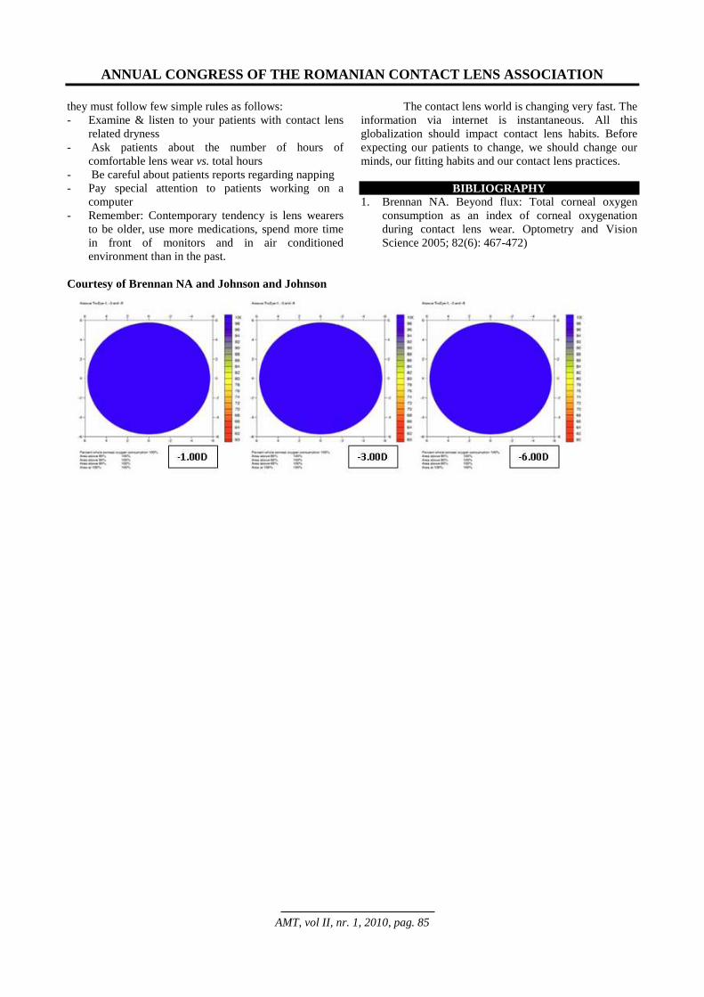
ANNUAL CONGRESS OF THE ROMANIAN CONTACT LENS ASSOCIATION
AMT, vol II, nr. 1, 2010, pag. 85
they must follow few simple rules as follows:
- Examine & listen to your patients with contact lens
related dryness
- Ask patients about the number of hours of
comfortable lens wear vs. total hours
- Be careful about patients reports regarding napping
- Pay special attention to patients working on a
computer
- Remember: Contemporary tendency is lens wearers
to be older, use more medications, spend more time
in front of monitors and in air conditioned
environment than in the past.
The contact lens world is changing very fast. The
information via internet is instantaneous. All this
globalization should impact contact lens habits. Before
expecting our patients to change, we should change our
minds, our fitting habits and our contact lens practices.
BIBLIOGRAPHY
1. Brennan NA. Beyond flux: Total corneal oxygen
consumption as an index of corneal oxygenation
during contact lens wear. Optometry and Vision
Science 2005; 82(6): 467-472)
Courtesy of Brennan NA and Johnson and Johnson

ANNUAL CONGRESS OF THE ROMANIAN CONTACT LENS ASSOCIATION
AMT, vol II, nr. 1, 2010, pag. 86
CORNEAL STAINING: DO WE UNDERSTAND WHAT WE
ARE SEEING?
SILVIA BELCHEVA1, CHRISTINA N GRUPCHEVA2
1Private Practice, Bulgaria, 2Medical University Varna, Bulgaria
Keywords: corneal staining, fluorescein, staining in
contact lens wearers
Sodium fluorescein (‘fluorescein’) is widely
utilised for the evaluation of ocular surface integrity. It is
preferred for anterior ocular surface visualisation due to
its fluorescent properties and its high visibility at low
concentrations. Ocular surface fluorescence observed
clinically is affected by a number of factors including
concentration of fluorescein, thickness of the fluorescein
layer, the wavelength of the exciting light source and
whether or not a barrier filter is used as part of the
viewing system. Three potential cellular mechanisms are
involved in corneal surface fluorescence: surface pooling,
uptake by cells and ingress around cells. Despite the
widespread adoption of use of fluorescein for the
assessment of the ocular surface, the clinical
understanding and interpretation of corneal surface
fluorescence is based upon clinical intuition rather than
underpinning the basic causative mechanisms of this
phenomenon, particularly important for contact lens
wearers.
It is important to understand and to interpret the
corneal staining in all cases of adaptation of contact
lenses, including silicone hydrogel lenses. It demonstrates
chronic injury of the corneal epithelium. The intact
epithelium is the best protection against corneal invasion.
Corneal staining means “clinical procedure allowing the
observation of the vitality and integrity of the corneal
epithelium cells using dye (fluorescein)”. The observation
of the corneal surface with the blue filter of the slit lamp
after instillation of fluorescein allows visualisation of
lesions with different localisation and importance.
What is the value of staining in contact lens wearers
and non wearers?
According to different clinical studies the
corneal staining observed in patients non wearing contact
lenses varies between 4 to 79% depending on the method.
It is usually under first grade or if it is of first grade, is
situated in the half of the cases in the inferior area of the
cornea. It is a result of the impaired tear film. In 30 to
50% of contact lens wearers the staining is under second
grade and in 5 to 10 % of them – above second grade.
Corneal staining is usually asymptomatic, but must be a
part of the routine contact lens examination.
Description and quantitative assessment of corneal
staining The description is according:
Depth: superficial and deep;
Localisation: the cornea is divided into five areas:
central, superior, inferior, nasal and temporal.
Corneal staining grades grade 0 = no staining,
grade 1 = micropunctate staining, clinically
insignificant,
grade 2 = macropunctate staining, it must be noted
and followed up,
grade 3 = moderate coalescent macropunctate
staining, requires treatment,
grade 4 = marked patch staining, requires immediate
treatment.
The visual acuity is usually unaffected except for
the central form of grade 4.
Corneal staining Grading Scales, most widely used

ANNUAL CONGRESS OF THE ROMANIAN CONTACT LENS ASSOCIATION
AMT, vol II, nr. 1, 2010, pag. 87
Clinical forms of corneal staining
Punctate corneal staining: tiny superficial dots –
keratitis punctata superficialis.
Localised or diffuse staining of the cornea with
coalescent dots.
Staining at 3 and 9 o’clock, classical form of staining
induced by hard contact lenses. A temporal and nasal
staining is observed; the reason is the slow renewal of
the tear film between two blinking.
Staining like “smile”: arched epithelial staining
inferiorly between 4 and 8 o’clock, observed in soft
contact lens wearers. It is related with instable tear
layer between the lens and the cornea. It is observed
in 3,3% of soft contact lens wearers.
Superior arcuate epithelial lesion
This is frequent and asymptomatic complication
of silicone hydrogel contact lenses. It is situated in the
superior area of the cornea, covered by the upper eyelid at
2-3 mm of the limbus between 10 and 2 o’clock.The
silicone hydrogel lenses must be replaced with lenses with
lower modulus.
Diffuse staining
Peripheral staining
Inferior and superior staining
Limbal transition pooling
Etiology
There are six known etiologies: mechanical,
expositional, metabolite, infectious, toxic and allergic.
However ,in many cases the reason remains unknown.
Mechanical factors
Defective lens, badly adapted (especially rigid
lens) or badly maintained with deposits on its back
surface, foreign body under the lens and other causes for
microtrauma of the corneal epithelium.
Expositional factors
Typically it reveals like an arcuate lesion (like
smile) in the inferior third of the cornea and it is due to
exposure of the corneal epithelium which causes
dessication of epithelial cells. It is also observed in cases
of upper decentration of rigid lens with diminished
efficacy of blinking. Dry keratitis, related to dehydration
of the lens, could be observed in wearing high hydrophilic
soft contact lens. The staining presents with central
localisation. The staining at 3 and 9 o’clock is related
with this etiology as well.
Metabolic factors
The wearing of soft contact lens could induce
hypoxia followed by synthesis of lactic acid. This chronic
metabolic stress leads to damage of deep cells of corneal
epithelium with formation of microcysts. They produce
superficial diffuse and always bilateral staining. The
silicon hydrogel material allows to limit the hypoxia and
consequently to reduce these metabolic problems.
Infectious origin
A small corneal ulcer is usually observed
(foreign body under the lens, inappropriate night wear),
accompanied by corneal pain. The fluorescence is limited
in the area of the lesion, but characteristic sign is the
diffuse spread in the depth of stroma – posterior halo.
Toxic and/or allergic etiology
Superficial keratitis, related to toxic and allergic
reactions to components of maintaining products, are
observed. The improvement of maintaining systems (lack
of polyhexamethilen biguanide, РНМВ, tiomersal,
benzalkonium chloride, hexidine) should lead to
disappearance of punctate lesions of the cornea.
Is there a relation between corneal staining and
bacterial keratitis?
Obviously, the breaking of the epithelial barrier
of the cornea is “an open door” for infection, allowing
adhesion and later penetration.
By now the causative relation is not clearly proved.
The role of multipurpose solutions for appearing of
infections is not fully elucidated but it is always
strongly recommended the use of products with
minimal deleterious potential.
Conclusions
Corneal staining is a “must” evaluation for a good
contact lens practice.
Grading scales must be used to unify the findings and
examination repeatability.
Staining should be interpreted into the clinical
context.
Further corneal/contact lens solution interactions
should be elucidated.
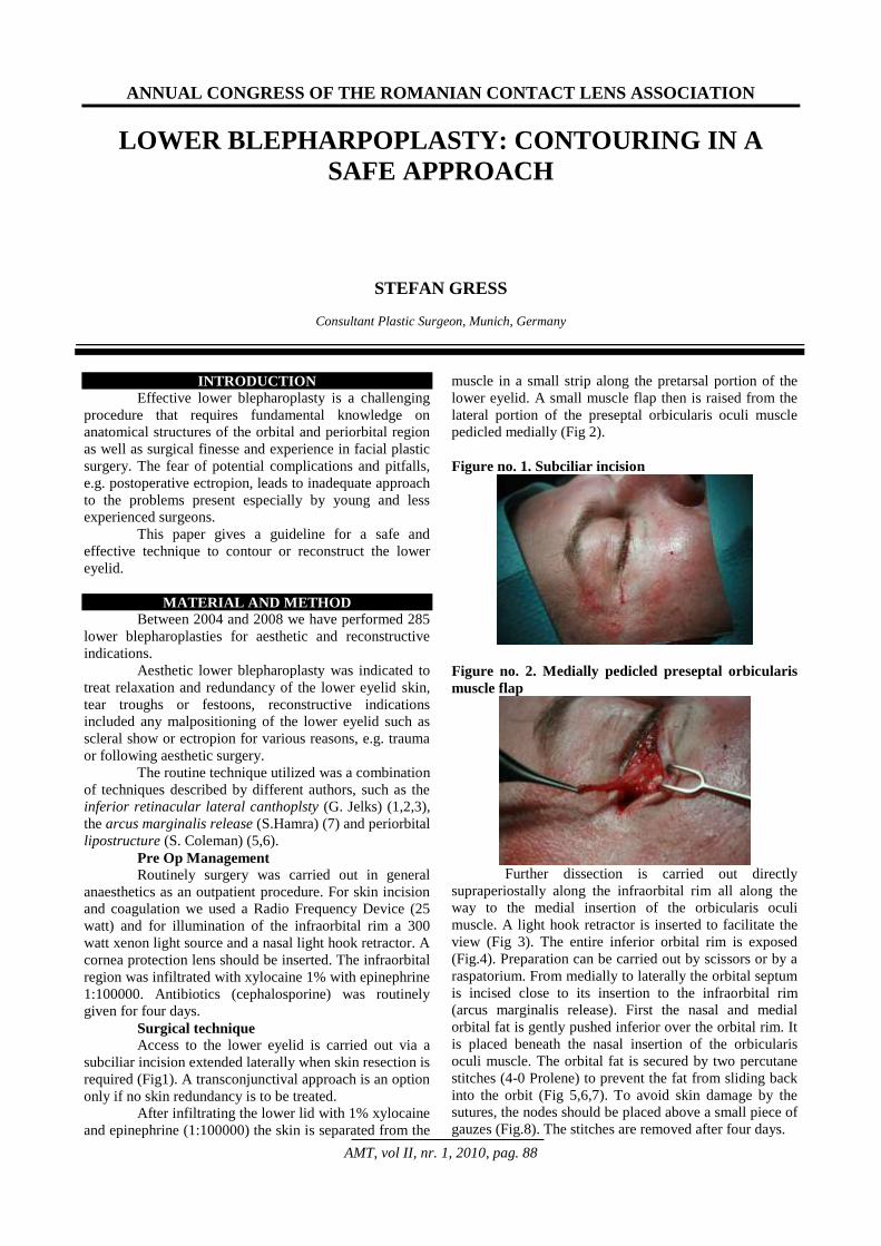
ANNUAL CONGRESS OF THE ROMANIAN CONTACT LENS ASSOCIATION
AMT, vol II, nr. 1, 2010, pag. 88
LOWER BLEPHARPOPLASTY: CONTOURING IN A
SAFE APPROACH
STEFAN GRESS
Consultant Plastic Surgeon, Munich, Germany
INTRODUCTION
Effective lower blepharoplasty is a challenging
procedure that requires fundamental knowledge on
anatomical structures of the orbital and periorbital region
as well as surgical finesse and experience in facial plastic
surgery. The fear of potential complications and pitfalls,
e.g. postoperative ectropion, leads to inadequate approach
to the problems present especially by young and less
experienced surgeons.
This paper gives a guideline for a safe and
effective technique to contour or reconstruct the lower
eyelid.
MATERIAL AND METHOD
Between 2004 and 2008 we have performed 285
lower blepharoplasties for aesthetic and reconstructive
indications.
Aesthetic lower blepharoplasty was indicated to
treat relaxation and redundancy of the lower eyelid skin,
tear troughs or festoons, reconstructive indications
included any malpositioning of the lower eyelid such as
scleral show or ectropion for various reasons, e.g. trauma
or following aesthetic surgery.
The routine technique utilized was a combination
of techniques described by different authors, such as the
inferior retinacular lateral canthoplsty (G. Jelks) (1,2,3),
the arcus marginalis release (S.Hamra) (7) and periorbital
lipostructure (S. Coleman) (5,6).
Pre Op Management
Routinely surgery was carried out in general
anaesthetics as an outpatient procedure. For skin incision
and coagulation we used a Radio Frequency Device (25
watt) and for illumination of the infraorbital rim a 300
watt xenon light source and a nasal light hook retractor. A
cornea protection lens should be inserted. The infraorbital
region was infiltrated with xylocaine 1% with epinephrine
1:100000. Antibiotics (cephalosporine) was routinely
given for four days.
Surgical technique
Access to the lower eyelid is carried out via a
subciliar incision extended laterally when skin resection is
required (Fig1). A transconjunctival approach is an option
only if no skin redundancy is to be treated.
After infiltrating the lower lid with 1% xylocaine
and epinephrine (1:100000) the skin is separated from the
muscle in a small strip along the pretarsal portion of the
lower eyelid. A small muscle flap then is raised from the
lateral portion of the preseptal orbicularis oculi muscle
pedicled medially (Fig 2).
Figure no. 1. Subciliar incision
Figure no. 2. Medially pedicled preseptal orbicularis
muscle flap
Further dissection is carried out directly
supraperiostally along the infraorbital rim all along the
way to the medial insertion of the orbicularis oculi
muscle. A light hook retractor is inserted to facilitate the
view (Fig 3). The entire inferior orbital rim is exposed
(Fig.4). Preparation can be carried out by scissors or by a
raspatorium. From medially to laterally the orbital septum
is incised close to its insertion to the infraorbital rim
(arcus marginalis release). First the nasal and medial
orbital fat is gently pushed inferior over the orbital rim. It
is placed beneath the nasal insertion of the orbicularis
oculi muscle. The orbital fat is secured by two percutane
stitches (4-0 Prolene) to prevent the fat from sliding back
into the orbit (Fig 5,6,7). To avoid skin damage by the
sutures, the nodes should be placed above a small piece of
gauzes (Fig.8). The stitches are removed after four days.

ANNUAL CONGRESS OF THE ROMANIAN CONTACT LENS ASSOCIATION
AMT, vol II, nr. 1, 2010, pag. 89
Figure no. 3. Light hook retractor
Figure no. 4. Exposure of the inferior orbital rim
Figure no. 5. Orbital fat placed over the orbital rim
Figure no. 6 Orbital fat fixed by suture
If the amount of orbital fat is not sufficient to
create a nice contour of the lower eyelid or when the
orbital rim can not entirely be covered by orbital fat,
additional fat grafting can be performed (lipostructure).
The fat can be infiltrated from the subciliary approach or
percutaneously by a sharp or blunt needle (Fig 9).
However it should be placed above the periosteum and
not above the muscle. Overcorrection should be avoided.
The free fat grafts can be used to shape the entire cheek or
midface whenever volume replacement is necessary.
The lateral infraorbital fat pad, when pushed
over the orbital rim, is fixed by 5-0 Vicryl stitches.
Figure no. 7. Percutane fixation of the transpositioned
orbital fat
Figure no. 8. Knod placed over gauzes
Figure no. 9. Percutane fat injection
Prior to skin resection, the lower eyelid needs to
be secured and supported to avoid postoperative ectropion
or scleral show. We utilize the technique of Glenn Jelks,
the inferior retinacular lateral canthoplasty, which is
more a canthopexy procedure:
The superificial lamella of the lateral canthal
tendon is released at its lateral insertion to the orbital rim.
The dissection in continued inferiorly in a subperiostal
plane until it joins the lateral- inferior edge of the orbit
(that now is covered by the inferior lateral orbital fat pad).
Fibrous attachments that run from the lateral canthus into
the lateral inferior orbit are released to achieve free
movement of the lower lid. The lower lid component of
the lateral retinaculum (the component of the anterior
lamella of the lateral canthal tendon that forms the roof
the inferior lateral fat pocket) now is grasped and fixed
vertically into the inner aspect of the supero-lateral orbital
rim by a 5-0 Prolene suture (Fig 10,11). The elevation of
the lower eyelid should appear slightly overcorrected.
When canthoplasty is performed, the muscle flap
raised at the beginning of the procedure is now fixed
under moderate tension in an oblique superior direction to
the periosteum of the lateral orbital rim by 5-0 Vicryl (Fig

ANNUAL CONGRESS OF THE ROMANIAN CONTACT LENS ASSOCIATION
AMT, vol II, nr. 1, 2010, pag. 90
12) sutures. Before skin removal, muscle incisions are
closed by 5-0 Vicryl stiches.
Figure no. 10. Suture passing through the lateral
inferior retinaculum
Figure no. 11. Fixation of the lower lateral
retinaculum to the supero-lateral orbital rim
Figure no. 12. Fixation of the muscle flap to the lateral
orbit
Excess skin can safely be removed now. This
should be performed on open eyes to avoid overresection
(Fig 13). For skin closure 6-0 Prolene is used.
A steri-strip dressing is applied using an
additional long strip laterally to release tension of the
cheek on the incision (Fig 14).
Figure no. 13. Measuring skin excess prior to removal
Post Op management
Stitches are removed after four days, however
the steri- strip dressing should be continued 10- 14 days
following surgery. Any tension on the cheek and lower
eyelid should strictly be avoided for four weeks and the
patient should sleep on his back during that period of
time. We routinely give cephalosporine antibiotics for
four to five days
Figure no. 14. Steri strip dressing
Complications
The most common postoperative problem
encountered is chemosis to various degrees. Chemosis is
not treated actively, local steroids do not help
significantly (Fig 15).
Figure no. 15. Severe chemosis with haemorrage
Overresection of skin and hypertrophic scar
formation can lead to malpositioning of the lower eyelid,
e.g. to a round eye deformation, scleral show or ectropion.
This is seldomly seen when the procedure is performed
correctly and skin closure is carried out without tension.
In some cases granulomas can occur around the
permanent 5-0 Prolene suture that fixes the canthopexy. If
these reactions lead to irritations of the globe or the
patient feels uncomfortable, the suture can be removed
after four weeks.
RESULTS
Figure no. 16. 35 year old patient after lower lid
contouring. Additional lipostructure of the cheek and
midface was performed

ANNUAL CONGRESS OF THE ROMANIAN CONTACT LENS ASSOCIATION
AMT, vol II, nr. 1, 2010, pag. 91
Figure no. 17. 75 year old patient following upper and
lower blepharoplasty
Figure no. 18. 42 year old patient after contouring the
lower eyelid and lipostructure of the nasojugual groofe
Figure no. 19. 48 year old patient after contouring the
lower eyelid and upper blepharoplasty
Figure no. 20. 68 year old patient after contouring the
left lower lid malpositioning following a blow out
fracture
CONCLUSION
For contouring the lower eyelid, the combination
of reshaping inferior orbital fat, fat grafting and muscle
and skin tightening are all together effective tools to
achieve functionally and aesthetically pleasing results in
lower blepharoplasty. To prevent pitfalls and
complications such as postoperative ectropion, a safe
support of the lower eyelid is mandatory. Therefore we
routinely utilize the inferior retinacular lateral
canthoplasty, described by Glenn Jelks. We also utilize
the “no touch technique” that does not allow complete
transsection of the orbicularis oculi muscle along the
lower eyelid for better access to the orbit, because it
would lead to denervation of the pretarsal portion of the
orbicularis oculi muscle likely emerging into a round eye
deformity.
When the guidelines presented in this paper are
perfomed correctly, the lower blepharoplasty will be
successful.
REFERENCES
1. Jelks, G.W. The Inferior Retinacular Lateral
Canthoplasty: A New Technique. Plast. Reconstr.
Surg. 100:1262,1997
2. Jelks, G.W., and Jelks, E.B. Repair of lower lid
deformities. Clin. Plast. Surg. 20: 417, 1993
3. Zide, B.M, and Jelks, G.W. Surgical Anatomy of the
Orbit. New York: Raven Press, 1985
4. Muzaffar, A.R., Mendelson, B.C. Surgical Anatomy
of the Ligamentous Attachments of the Lower Lid
and Lateral Canthus. Plast. Reconstr. Surg. 110: 873,
2001
5. Coleman, S.R., Guerrerosantos, J. Volumetric
Considerations in Facial Augmentation. Journal for
Aesthetic Surgery, 2: 2003
6. Coleman, S.R., Facial recontouring with
lipostructure. Clin. Plast. Surg. 24(2): 347, 1997
7. Hamra, S.T., The role of septal reset in creating a
youthful eyelid- cheek complex in facial
rejuvenation. Plast.Reconstr. Surg., 113 (7): 2124,
2004