Embed Size (px)
Citation preview
University of New EnglandDUNE: DigitalUNE
Nurse Anesthesia Capstones School of Nurse Anesthesia
6-2019
Amniotic Fluid Embolism In The Parturient PatientVanessa PasquarielloUniversity of New England
Follow this and additional works at: https://dune.une.edu/na_capstones
Part of the Anesthesiology Commons, and the Nursing Commons
© 2019 Vanessa Pasquariello
This Capstone is brought to you for free and open access by the School of Nurse Anesthesia at DUNE: DigitalUNE. It has been accepted for inclusionin Nurse Anesthesia Capstones by an authorized administrator of DUNE: DigitalUNE. For more information, please contact [email protected].
Recommended CitationPasquariello, Vanessa, "Amniotic Fluid Embolism In The Parturient Patient" (2019). Nurse Anesthesia Capstones. 27.https://dune.une.edu/na_capstones/27
Running head: AMNIOTIC FLUID EMBOLISM 1
Amniotic Fluid Embolism in the Parturient Patient
Vanessa Pasquariello
University of New England
AMNIOTIC FLUID EMBOLISM 2
Abstract
An amniotic fluid embolism (AFE) occurs when amniotic fluid enters maternal
circulation, causing a physical obstruction or an anaphylactoid reaction, both of which are often
detrimental to the parturient patient. This paper reviews a series of studies to examine the
incidence, risk factors, presentation, and management of an AFE in the healthcare setting.
Diagnosing an AFE remains difficult as a universal diagnostic criterion does not exist (aside
from in reported research); thus, its identification is often made when another differential
diagnosis fails to manifest. The presentation of the following biomarkers: squamous cell
carcinoma antigen, CK13, and CK10/13 can aid in the investigation of an AFE event.
Management of an AFE takes a comprehensive approach with considerations to cardiac
resuscitation, post-cardiac arrest care, hemodynamic support, coagulopathy, and uterine atony.
New and unconventional methods in the treatment of an AFE have been suggested in three case
studies using C1INH, lipid emulsion therapy, and atropine/ondansetron/ketorolac.
AMNIOTIC FLUID EMBOLISM 3
Amniotic Fluid Embolism in the Parturient Patient
AFE Pathophysiology
According to Kanayama & Tamura (2014), lanugo, meconium, squamous cells, tissue
factors, and vernix are fetal materials that make up amniotic fluid. An amniotic fluid embolism
(AFE) develops as a direct result of amniotic fluid entering maternal circulation. Kanayama &
Tamura (2014), explain that this process occurs when the amniotic fluid leaks out of its sac and
joins maternal circulation via ruptured vessels in the uterine cavity or muscle. As this fluid
travels into the maternal circulation, its contents can cause physical obstructions or ensure an
anaphylactoid reaction that can develop into disseminated intravascular coagulation (DIC)
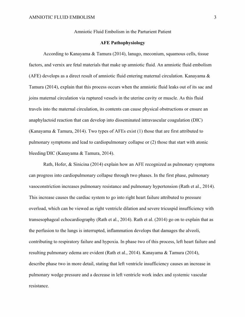
(Kanayama & Tamura, 2014). Two types of AFEs exist (1) those that are first attributed to
pulmonary symptoms and lead to cardiopulmonary collapse or (2) those that start with atonic
bleeding/DIC (Kanayama & Tamura, 2014).
Rath, Hofer, & Sinicina (2014) explain how an AFE recognized as pulmonary symptoms
can progress into cardiopulmonary collapse through two phases. In the first phase, pulmonary
vasoconstriction increases pulmonary resistance and pulmonary hypertension (Rath et al., 2014).
This increase causes the cardiac system to go into right heart failure attributed to pressure
overload, which can be viewed as right ventricle dilation and severe tricuspid insufficiency with
transesophageal echocardiography (Rath et al., 2014). Rath et al. (2014) go on to explain that as
the perfusion to the lungs is interrupted, inflammation develops that damages the alveoli,
contributing to respiratory failure and hypoxia. In phase two of this process, left heart failure and
resulting pulmonary edema are evident (Rath et al., 2014). Kanayama & Tamura (2014),
describe phase two in more detail, stating that left ventricle insufficiency causes an increase in
pulmonary wedge pressure and a decrease in left ventricle work index and systemic vascular
resistance.
AMNIOTIC FLUID EMBOLISM 4
The other type of AFE that exists presents with atonic bleeding/DIC. Rath et al. (2014)
state that the procoagulant substances within the amniotic fluid (tissue factor and
phosphatidylserine), can directly or indirectly cause the cascading effects of DIC. These
procoagulant substances occur directly via cytokines or indirectly by complement activation
from the extrinsic coagulation cascade leading to hyperfibrinolysis (Rath et al., 2014). Kanayama
& Tamura (2014) discuss this further by saying that when amniotic fluid associates with allergy-
associated cells, such as mast cells located within the cervix, it produces bradykinin and
inflammatory cytokines. The production of these mediators facilitates uterine muscle relaxation
and edema that causes incoagulable vaginal bleeding after delivery (Kanayama & Tamura,
2014).
Figure 1. Summarizing the two main types of AFEs and their related pathophysiology. Reprinted from “AFE: diagnosis and management” by Pacheco, L. D., Saade, G., Hankins, G. D., Clark, S. L., & Society for Maternal-Fetal Medicine SMFM, 2016, American journal of obstetrics and gynecology, 215(2), B16-B24.
AMNIOTIC FLUID EMBOLISM 5
Literature Review
Bonnet et al. (2018)
Bonnet et al. (2018) conducted a retrospective study over four years with the goals of
applying a new criterion for AFE and describing the epidemiology, characteristics, and
management of an AFE. The diagnostic criteria, proposed by the Society for Maternal-Fetal
Medicine and the AFE Foundation, was comprised of four conditions that must be present to
diagnose the patient as having an AFE. The four principles are as follows: (1) abrupt
cardiopulmonary arrest, or hypotension (systolic blood pressure less than ninety millimeters of
mercury) with respiratory compromise, (2) documentation of DIC after appearance of
preliminary symptoms with coagulopathy detected before significant loss of blood, (3) onset
during labor or within a half hour of placenta delivery, and (4) no fever (fever defined as greater
than thirty-eight degrees Celsius) (Bonnet et al., 2018).
Results.
Incidence. Bonnet et al. (2018) reported that there were close to four million live births
during their study period. Out of these live births, four hundred twenty-nine maternal deaths
occurred, thirty-nine of which being attributed to an AFE (Bonnet et al., 2018). Of the thirty-nine
deaths, thirty-six contained a complete medical record, and thus data was reported on these
thirty-six cases (Bonnet et al., 2018).
Risk Factors. In considering maternal age in death from an AFE, forty-two percent of
women were over the age of thirty-five and had a body mass index less than thirty before
pregnancy (Bonnet et al., 2018). Bonnet et al. (2018) pointed out that those in between one and
three parities represented fifty-eight percent of the patient population. Out of the thirty-two
patients who went into labor, sixteen had their labor induced, nine of which were induced with
prostaglandins (Bonnet et al., 2018). Out of the thirty-two patients who went into labor, eighteen
AMNIOTIC FLUID EMBOLISM 6
received oxytocin (Bonnet et al., 2018). Additionally, Bonnet et al. (2018) looked at the mode of
delivery; the majority of patients who suffered an AFE underwent a cesarean section (n=nineteen
during labor and three before labor, netting a total of twenty-two patients or about sixty-nine
percent). Interestingly, when looking at fetal characteristics, sixty-six percent of fetuses were of
male gender (Bonnet et al., 2018). Also, an AFE was less common with a gestational age shorter
than thirty-seven weeks (Bonnet et al., 2018).
Presentation. Out of the thirty-six patients that had an AFE, eighty-one percent exhibited
premonitory signs, such as neurological/respiratory signs, fainting, and fetal heart rate
abnormalities (Bonnet et al., 2018). Bonnet et al. (2018) looked into more detail about the
symptoms exhibited by these patients; ninety-seven percent of patients collapsed (n=eighteen as
a first sign), eighty-three percent showed signs of a clinical coagulopathy (n=eight as a first
sign), forty-four percent of patients had respiratory compromise (n=thirteen as a first sign), and
thirty-three percent suffered from a seizure (n=eleven as a first sign).
When looking at hemorrhage, Bonnet et al. (2018) stated that twenty patients exhibited
this symptom immediately, with eight patients displaying it at a later time. Out of the twenty-
four patients who had their platelet count drawn, fifty-percent showed a platelet count greater
than one hundred thousand (Bonnet et al., 2018). According to Bonnet et al. (2018), twenty-two
patients had a prolonged prothrombin time (PTT) and fibrinogen level drawn. Out of these
twenty-two patients described by Bonnet et al. (2018), nearly all showed more than a fifty
percent increase in their PTT (n=nineteen) and the majority had a fibrinogen level less than one
(n=sixteen).
The National Experts Committee concluded that fifty-six percent of patients diagnosed
with an AFE had suboptimal care (Bonnet et al., 2018). Bonnet et al. (2018) state that the most
popular reasons for suboptimal care included obstetrical care (n=sixteen/forty-four percent),
AMNIOTIC FLUID EMBOLISM 7
inadequate hemorrhage management (n=fifteen/forty-two percent), and delayed hysterectomy or
no hysterectomy when clinically indicated (n=thirteen/thirty-six percent). Of the six patients who
were in the intensive care unit, Bonnet et al. (2018) state suboptimal care was exhibited by a
delayed blood transfusion (n=six), insufficient blood transfusion (n-three), lack of coagulation
products while lab work suggested coagulopathy (n=three), and cardiopulmonary resuscitation
being stopped early (n=two).
Fitzpatrick, Tuffnell, Kurinczuk, & Knight (2016)
Fitzpatrick, Tuffnell, Kurinczuk, & Knight (2016) conducted a population-based cohort
and nested case-control study via the UK Obstetric Surveillance System from two thousand five
to two thousand fourteen. Their goals included describing the incidence, risk factors,
management, and outcomes of an AFE and determining if any maternal outcome trends existed
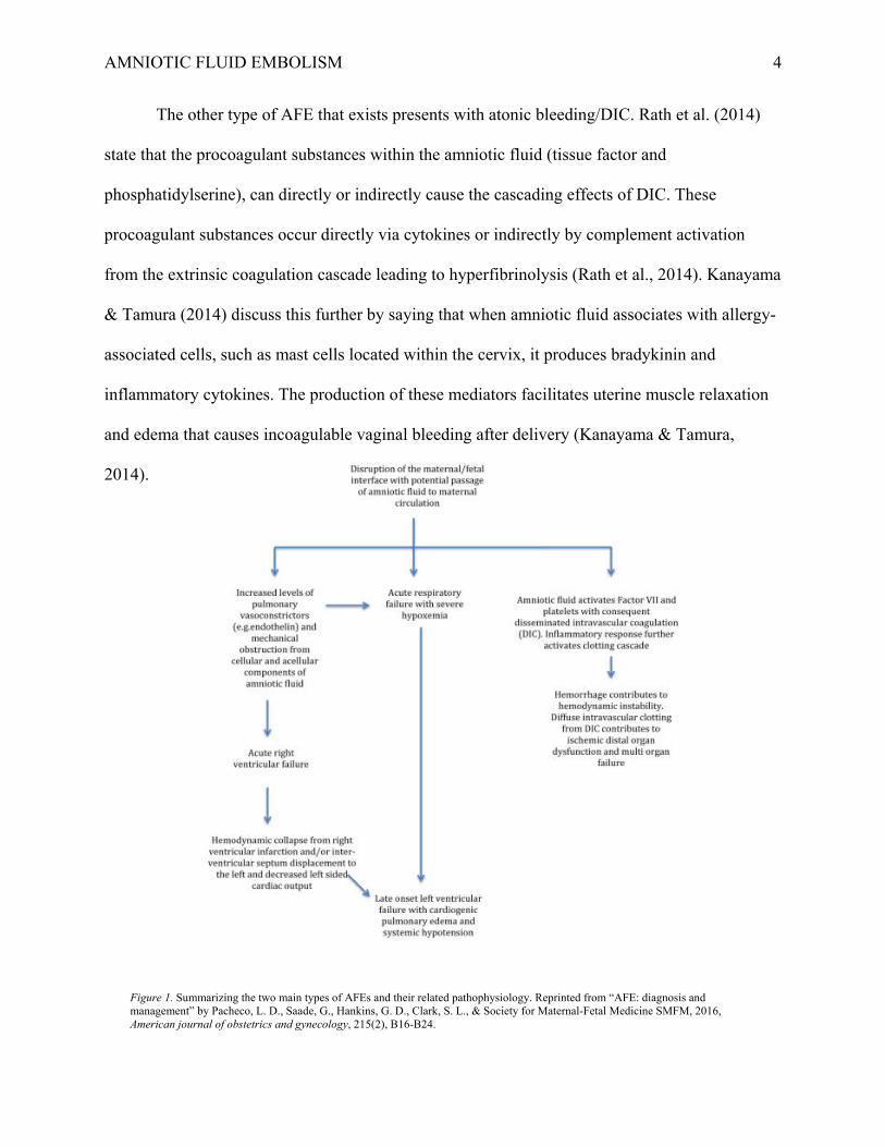
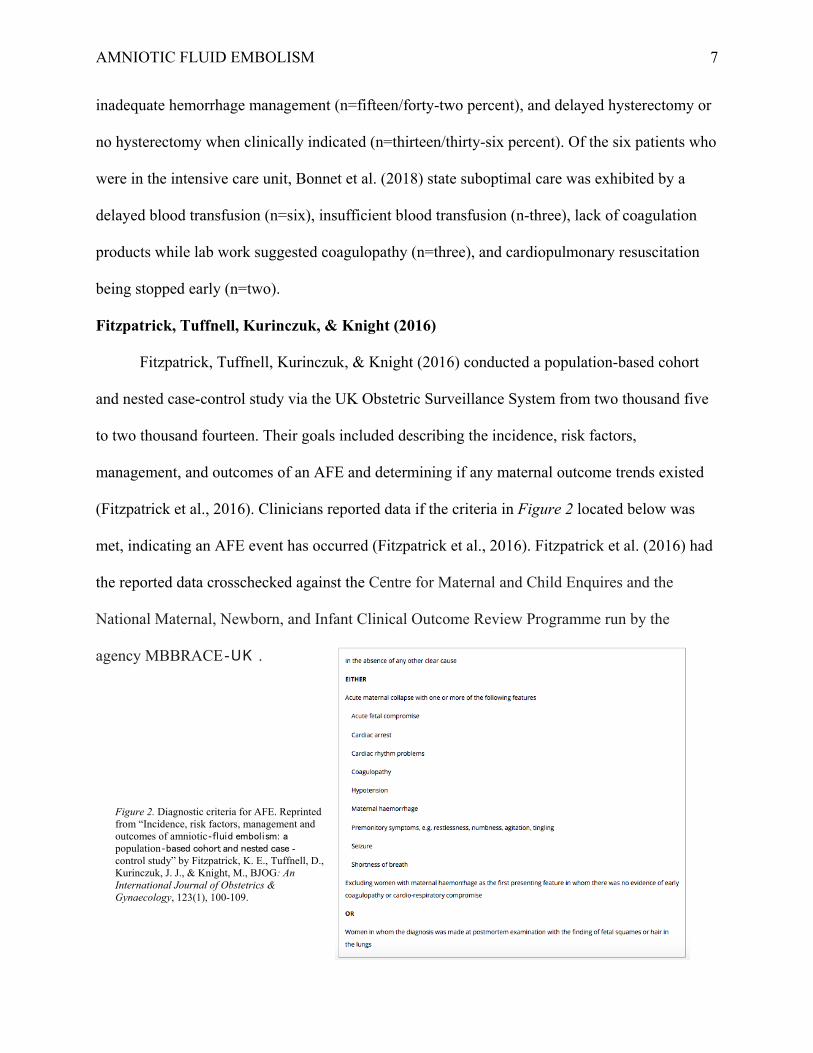
(Fitzpatrick et al., 2016). Clinicians reported data if the criteria in Figure 2 located below was
met, indicating an AFE event has occurred (Fitzpatrick et al., 2016). Fitzpatrick et al. (2016) had
the reported data crosschecked against the Centre for Maternal and Child Enquires and the
National Maternal, Newborn, and Infant Clinical Outcome Review Programme run by the
agency MBBRACE‐UK .
Figure 2. Diagnostic criteria for AFE. Reprinted from “Incidence, risk factors, management and outcomes of amniotic‐fluid embolism: a population‐based cohort and nested case -control study” by Fitzpatrick, K. E., Tuffnell, D., Kurinczuk, J. J., & Knight, M., BJOG: An International Journal of Obstetrics & Gynaecology, 123(1), 100-109.
AMNIOTIC FLUID EMBOLISM 8
Results.
Incidence. Fitzpatrick et al. (2016) data showed that out of over seven million births, a
total of one hundred twenty confirmed cases of AFE transpired with twenty-three cases deemed
fatal. Fitzpatrick et al. (2016) data represent a total incidence of an AFE occurring one-point-
seven times per one hundred thousand people, and a fatality incidence occurring zero-point-three
times per one hundred thousand people.
Risk Factors. Similar to Bonnet et al. (2018) study, Fitzpatrick et al. (2016) study
showed that the majority of woman who suffered an AFE were less than thirty-five years old,
had a body mass index less than thirty, and had a parity of one or higher. Fitzpatrick et al. (2016)
looked further into the ethnicity of affected individuals, the majority being white (n=ninety-
two/seventy-seven percent) compared to black and other ethnic groups (n=twenty-five/twenty-
one percent). Also, Fitzpatrick et al. (2016) had a higher incidence of an AFE in those who never
smoked or quit smoking. Patients who were pregnant with one fetus were more likely than those
who were pregnant with multiple fetuses to develop an AFE (Fitzpatrick et al., 2016).
Additionally, Fitzpatrick et al. (2016) state that forty-nine patients (forty-one percent) had labor
induced. When looking at additional maternal conditions, all three patients who suffered from
placenta previa died from an AFE (Fitzpatrick et al., 2016).
As said by Fitzpatrick et al. (2016) in comparing the above risk factors, all results were
similar among those who survived and died from an AFE. Fitzpatrick et al. (2016) examined the
incidence of AFE with induction of labor. They found that there was an increased incidence of
AFE in women who received prostaglandin without oxytocin, oxytocin without prostaglandin,
and who received both prostaglandin and oxytocin (Fitzpatrick et al., 2016). Out of the women
diagnosed with an AFE and who received prostaglandin, Fitzpatrick et al. (2016) report that
ninety-five percent of patients received dinoprostone, and only one patient received misoprostol.
AMNIOTIC FLUID EMBOLISM 9
Presentation. Fitzpatrick et al. (2016) state that more than half of those diagnosed
(n=sixty-two) had an AFE present before/at delivery, compared to those (n=fifty-five) who
presented at a median of nineteen minutes post-delivery. In those diagnosed with an AFE before,
during, or post-delivery, the median age of gestation was thirty-nine weeks, and diagnosis ranged
from zero minutes to two days, with a median of thirty-three minutes (Fitzpatrick et al., 2016).
Women that presented with cardiac arrest as the first recognized symptom of an AFE were more
likely to die or have a permanent neurological injury (Fitzpatrick et al., 2016). Also, most
women (n=one hundred eight/ninety-one percent) had their membranes ruptured before AFE
presentation (Fitzpatrick et al., 2016). Similar to Bonnet et al. (2018) study, the highest incidence
of AFE was in those that delivered via cesarean section (Fitzpatrick et al., 2016). Those also at
an increased risk of having an AFE included those who delivered vaginally with instrumentation,
followed by spontaneous vaginal delivery (Fitzpatrick et al., 2016).
Management.
Coagulation. Fitzpatrick et al. (2016) mention that out of the women diagnosed with an
AFE, forty-four (thirty-seven percent) did not have any coagulopathy support. Of those women
that did have coagulopathy support, thirty-one patients (twenty-six percent) received only one
method of support, while twenty-one patients (eighteen percent) received more than one method
of coagulopathy management (Fitzpatrick et al., 2016). Cryoprecipitate was used in six cases
(twenty-percent) of those who died from an AFE, compared to forty-one cases (forty-six percent)
of those who survived an AFE (Fitzpatrick et al., 2016). According to Fitzpatrick et al. (2016),
platelets were used in eight cases (twenty-seven percent) of those who died from an AFE,
compared to thirty-six cases (forty percent) of those who survived an AFE. Fresh frozen plasma
was more likely to be used than cryoprecipitate and platelets as evidenced by its use in seventeen
cases (fifty-seven percent) of those who died from an AFE, compared to fifty-two cases (fifty-
AMNIOTIC FLUID EMBOLISM 10
eight percent) of those who survived an AFE (Fitzpatrick et al., 2016). Factor VIIa and
fibrinogen were less commonly used compared to the coagulopathy management methods used
above. Fitzpatrick et al. (2016) show the administration of Factor VIIa appeared in six cases
(twenty-percent) of those who died from an AFE, compared to twenty cases (twenty-two
percent) of those who survived an AFE. Similarly, fibrinogen was used in one case (three
percent) of those who died from an AFE, compared to one case (one percent) of those who
survived an AFE (Fitzpatrick et al., 2016).
Other. Aside from attempting to control an AFE with the coagulopathy methods
discussed above, additional management strategies exist. Fitzpatrick et al. (2016) indicate that
performing a hysterectomy was most common in those that died and survived an AFE (forty
percent and twenty-one percent respectively). Exchange transfusions, plasma exchanges, and
embolization were not a method used in those who died from an AFE but were used in those who
survived (Fitzpatrick et al., 2016). Misoprostol and Hemabate were two other management
techniques employed. Fitzpatrick et al. (2016) show the administration of Misoprostol occurred
in two cases (seven percent) of those who died from an AFE, compared to four cases (four
percent) of those who survived an AFE. Similarly, Hemabate was used in two instances (seven
percent) of those who died from an AFE, compared to six cases (seven percent) of those who
survived an AFE (Fitzpatrick et al., 2016). Fitzpatrick et al. (2016) express that intrauterine
balloons were common in those that survived an AFE compared to those who died (n=six and
n=one respectively). Tranexamic acid was used in one patient who died from an AFE, while it
was more frequently (n=five) used in patients who survived (Fitzpatrick et al., 2016).
AMNIOTIC FLUID EMBOLISM 11
Guillaume et al. (2013)
Guillaume et al. (2013) conducted a retrospective study over ten years with the goals of
providing up to date statistics on AFEs and determining ways to advance current practice
guidelines. Guillaume et al. (2013) defined an AFE as exhibiting the following symptoms: (1)
circulatory and/or cardiopulmonary arrest, (2) hypoxia with oxygen saturation less than ninety
percent, (3) a neurological component such as loss of consciousness/agitation/seizures/coma, and
(4) hemorrhage associated with DIC.
Results.
Incidence. According to Guillaume et al. (2013), data for this study was gathered based
upon the coding system of hospitals in conjunction with histology and pathology reports. During
the study period, there was close to thirty-nine thousand births, eleven of which met the
definition of an AFE (Guillaume et al., 2013). Using this data, Guillaume et al. (2013) concluded
that the incidence of an AFE was twenty-eight in one hundred thousand. Out of the eleven
patients who suffered an AFE, there were three fatalities, carrying a maternal mortality rate of
seven point eight in one hundred thousand people (Guillaume et al., 2013).
Risk Factors. Like discussed in the studies by Bonnet et al. (2018) and Fitzpatrick et al.
(2016), Guillaume et al. (2013) had similar risk factors that were noted in women who suffered
an AFE in regard to maternal age (median of thirty-two years), parity (median one point seven),
and gestational age (median forty-weeks). Similar to Bonnet et al. (2018) study, Guillaume et al.
(2013) reported that the majority of fetuses were male gender.
When looking at if inducing labor played a role in increasing the odds of an AFE,
Guillaume et al. (2013) state that six patients had labor induced. Three of these patients received
misoprostol; two received dinoprostone, and one received oxytocin (Guillaume et al., 2013).
Also, Guillaume et al. (2013) state that oxytocin was used to enhance labor in nine patients. This
AMNIOTIC FLUID EMBOLISM 12
data is similar to that of Fitzpatrick et al. (2016) study which showed that there was an increased
risk of developing an AFE in women who received prostaglandin without oxytocin, oxytocin
without prostaglandin, and both prostaglandin and oxytocin. Different from Fitzpatrick et al.
(2016) study was that more patients received misoprostol than dinoprostone (Guillaume et al.,
2013). Lastly, the patient’s membranes had been ruptured, either spontaneously or artificially, in
all cases of AFE (Guillaume et al., 2013).
Presentation. Guillaume et al. (2013) state that eight-cases of an AFEs occurred during
the peripartum period, two cases transpired in the postpartum period, with one incident taking
place during a scheduled cesarean section. This data is different from Bonnet et al. (2018) and
Fitzpatrick et al. (2016) studies which showed that AFEs were more likely to occur in those
undergoing a cesarean section. In the eight cases of AFE appearing in the peripartum period,
Guillaume et al. (2013) report that delivery of the fetus was performed in all cases within twenty
minutes, with five of those cases being within five minutes of when symptoms of an AFE first
appeared. Delivery methods included vacuum-assisted delivery (n=one) and emergency cesarean
sections (n=seven) (Guillaume et al., 2013). When Guillaume et al. (2013) looked at presenting
symptoms, six patients had a cardiopulmonary arrest, with this being the first symptom in two
patients. In every case of AFE, symptoms progressed to post-partum hemorrhage, with ten out of
the eleven patients having DIC (Guillaume et al., 2013). When looking at cytology and histology
reports, Guillaume et al. (2013) state that eight patients had fetal squamous cells present, with
one of these cases also manifesting meconium.
Management. Guillaume et al. (2013) report that seven of the eleven patients who
experienced post-partum hemorrhage required surgical intervention to establish hemostasis.
Methods to secure hemostasis included an immediate hysterectomy (five patients) and delayed
hysterectomy after ligation of uterine/hypogastric arteries failed (two patients who both later
AMNIOTIC FLUID EMBOLISM 13
died) (Guillaume et al., 2013). Out of the six patients who coded, cardiopulmonary resuscitation
was sufficient in one patient, and defibrillation was required in five patients (Guillaume et al.,
2013). Guillaume et al. (2013) explain that extracorporeal membrane oxygenation with veno-
venous cannulation was utilized in two patients, both of which later died. In every patient,
transfusions took place with a median of eleven units of packed red blood cells, eleven units of
fresh frozen plasma, and two units of platelets (Guillaume et al., 2013). Guillaume et al. (2013)
also say that the administration of fibrinogen occurred in six patients along with Factor VIIa in
two patients. In comparing the products transfused in this study to Fitzpatrick et al. (2016) study,
the use of fibrinogen was more commonly used in Guillaume et al. (2013) study.
McDonnell et al. (2015)
McDonnell et al. (2015) conducted a population-based descriptive study specific to the
countries of Australia and New Zealand as AFEs are one of the leading causes of maternal death,
yet there is no national population study currently in existence in these areas. To collect their
data, McDonnell et al. (2015) utilized the Australasian Maternity Outcomes Surveillance System
which they state represents about ninety-six percent of women delivering in Australia and one
hundred percent of women giving birth in hospitals in New Zealand. After identifying an AFE
event, a case-specific questionnaire was then filled out (McDonnell et al., 2015). McDonnell et
al. (2015) collected their data throughout two years, from the beginning of two thousand ten to
the end of two thousand eleven. They defined an AFE clinically by the presence of acute
hypotension/cardiac arrest, acute hypoxia, and coagulopathy without a source of explanation or
via lab tests in the postmortem period (McDonnell et al., 2015).
Results.
Incidence. McDonnell et al. (2015) results showed that out of over six hundred thousand
women giving birth, thirty-three confirmed cases of an AFE existed, with five cases resulting in
AMNIOTIC FLUID EMBOLISM 14
death. This data shows an incidence of an AFE occurring five point four times in one hundred
thousand people and a mortality rate of zero point eight deaths in one hundred thousand people
(McDonnell et al., 2015). When comparing these rates to studies previously mentioned, they are
higher than reported in the Fitzpatrick et al. (2016) study but less than reported in Guillaume et
al. (2013) study.
Risk Factors. McDonnell et al. (2015) state that gestational age (seventy-three percent
exceeded thirty-seven weeks) was a risk factor for developing an AFE. Two characteristics that
differed in comparison to the studies previously mentioned are that the majority of patients (fifty-
two percent) were over the age of thirty-five and (fifty-two percent) had a parity of zero
(McDonnell et al., 2015). Similar to Bonnet et al. (2018) and Fitzpatrick et al. (2016) studies,
body mass index was taken into consideration, but in this study body mass index was reported as
less than thirty-five (thirty-one patients) making it impossible to identify an association with the
other studies. Comparable to Fitzpatrick et al. (2016) study, nonsmokers were shown to have an
increased risk, and placenta previa (fifteen percent) was also established as a risk factor
(McDonnell et al., 2015). Moreover, consistent with Bonnet et al. (2018) and Guillaume et al.
(2013) study, the majority of fetus’s (sixty-four percent) were of male gender (McDonnell et al.,
2015). Furthermore, McDonnell et al. (2015) looked at the number of women who received
assistance with conceiving, which represented twenty-four percent. Lastly, induction of labor did
prove to be a risk factor for AFE as evidenced by fourteen women having labor induced or
augmented (McDonnell et al., 2015).
Presentation. McDonnell et al. (2015) looked at the location of where the AFE took
place, the majority of which took place in the operating room or birthing suite (both had fifteen
cases), with two events occurring at home, and one incident not being recorded. Five cases
occurred during the pre-labor period, four cases in the first stage of labor, four cases during the
AMNIOTIC FLUID EMBOLISM 15
second stage of labor, and the majority (seventeen cases) during the postpartum period
(McDonnell et al., 2015). The onset of an AFE was noted to occur within five minutes before or
after delivery in forty-eight percent of patients (McDonnell et al., 2015). McDonnell et al. (2015)
recorded the signs and symptoms that patients most commonly presented with as premonitory
(twenty-seven percent), hypotension (twenty-one percent), shortness of breath (fifteen percent),
and fetal compromise (fifteen percent). They went on to report that twenty-two patients (sixty-
seven percent) had a cesarean section, of which six were elective, eight were emergent, and eight
did not have a recorded reason (McDonnell et al., 2015). Like Bonnet et al. (2018) and
Fitzpatrick et al. (2016) studies, this showed a higher incidence of AFE with cesarean sections.
Associated features included seventy-nine percent experiencing hemorrhage, seventy-six percent
hypotension, seventy-three percent coagulopathy, thirty-nine percent cardiac arrest, twenty-four
percent fetal compromise, and nine percent seizures (McDonnell et al., 2015).
Of the five women who died from an AFE, one had labor induced, one had labor
augmented, and one had been induced with labor later augmented (McDonnell et al., 2015). Of
the five deaths, McDonnell et al. (2015) reported that four had an unplanned cesarean section,
and one had a vacuum assisted delivery. When looking at duration until death, four women
passed within thirty-five to one hundred eight minutes after the AFE event, with one lasting three
weeks until death occurred (McDonnell et al., 2015).
Management. McDonnell et al. (2015) identified six patients (eighteen percent) who
received a hysterectomy, fourteen patients (forty-two percent) who needed intubation/ventilation,
fourteen patients (forty-two percent) who required cardiopulmonary resuscitation, and two
patients (six percent) that underwent plasmapheresis. They also looked at the worst recorded lab
results for hemoglobin, fibrinogen, platelets, and international normalized ratio. McDonnell et al.
(2015) found that fifty-two percent had a hemoglobin less than seven, sixty-four percent had a
AMNIOTIC FLUID EMBOLISM 16
fibrinogen level less than two, sixty-one percent had a platelet level less than one hundred
thousand, and forty-eight percent had an international normalized ratio (INR) higher than one
point five. When McDonnell et al. (2015) looked at blood product usage, twenty-eight patients
(eighty-five percent) required some form of blood product replacement, all of which needed
packed red blood cells. Twenty-four patients (seventy-three percent) received fresh frozen
plasma, twenty-four patients (seventy-three percent) cryoprecipitate, nineteen patients (fifty-
eight percent) platelets, and eight patients (twenty-four percent) Factor VIIa (McDonnell et al.,
2015). McDonnell et al. (2015) reported that the majority of patients (sixty-one percent) required
admission to the intensive care unit, and eight patients (twenty-four percent) required admission
to a high dependency unit.
Mu et al. (2016)
Mu et al. (2016) conducted a retrospective population-based study via data from the
National Maternal Mortality Surveillance System from nineteen ninety-six to two thousand
thirteen. They defined an AFE clinically as having symptoms that could not be explained by
another diagnosis (acute hypotension/cardiac arrest, acute hypoxia/pulmonary embolism, DIC,
and renal failure) or by pathology reports displaying fetal debris (Mu et al., 2016). Information
was reviewed by multiple committees which had an ascertainment rate of ninety-six percent for
all maternal deaths.
Results.
Incidence. Over the study period, Mu et al. (2016) reported that there were over twenty
million births, with six hundred sixty-four deaths attributed to an AFE, and documents sufficient
enough to gather data on six hundred forty cases. Utilizing this data, Mu et al. (2016) calculated
the maternal mortality rate for an AFE at one in over thirty thousand births. In comparing the
data from nineteen ninety-six to two thousand thirteen, it showed a decrease in maternal
AMNIOTIC FLUID EMBOLISM 17
mortality rate from AFE but displayed an increase in the proportion of maternal deaths related to
an AFE.
Risk Factors. Similar to the studies by Bonnet et al. (2018), Fitzpatrick et al. (2016), and
Guillaume et al. (2013), Mu et al. (2016) reported that the average maternal age was less than
thirty-five years old (over seventy-three percent), and women had more than one parity (over
fifty-one percent). According to Mu et al. (2016), over seventy-eight percent of women had their
membranes ruptured before the onset of an AFE like seen in Fitzpatrick et al. (2016) and
Guillaume et al. (2013) studies. Mu et al. (2016) had the highest percentage of women who
experienced an AFE deliver vaginally without instrumentation, similar to the results of
Guillaume et al. (2013) study.
Presentation. Mu et al. (2016) and McDonnell et al. (2015) shared similar results
regarding the patient’s location of presenting symptoms and the patient's stage of labor. Mu et al.
(2016) reported that the majority of signs (over thirty-five percent) presented in the delivery suite
with the bulk of AFEs occurring post-delivery (over fifty-two percent). There was next to no
difference in the first versus second stage of labor (both just above seventeen percent), and
women in pre-labor were least likely to have an AFE occur (Mu et al., 2016). Mu et al. (2016)
looked at the time from delivery until there was an onset of symptoms and noted that the
majority occurred between zero and a half hour after birth (over fifty-five percent). Mu et al.
(2016) stated that the most common presenting feature in all stages of labor were premonitory
symptoms (twenty-nine percent) followed by acute fetal compromise (twenty-eight percent). Mu
et al. (2016) detail premonitory symptoms to include shortness of breath, chest pain,
lightheadedness, restlessness, chills, panic, and nausea/vomiting.
Management. Mu et al. (2016) listed cardiopulmonary resuscitation (seventy-five
percent) as being the primary form of treatment of an AFE, followed by intubation/ventilation
AMNIOTIC FLUID EMBOLISM 18
(forty percent), blood products (thirty-seven percent), and hysterectomy (eighteen percent).
These treatment options are different from Guillaume et al. (2013) study in which the majority of
patients had a hysterectomy, but similar to the percent of patients in McDonnell et al. (2015)
study who received a hysterectomy/needed intubation/ventilation.
Shen, Wang, Yang, & Chen (2016)
Shen, Wang, Yang, & Chen (2016) conducted a retrospective study to look at the cases of
an AFE in the city of Suzhou over ten years from the beginning of January two thousand four to
the end of December two thousand thirteen. Shen et at. (2016) collected their data from the
Center for Disease Control, reviewed each patient’s medical record, and then confirmed the
presence of an AFE based upon the definition described by Clark et al. (1995).
Results.
Incidence. Shen et al. (2016) identified fifty-three cases of AFEs, with seventeen cases
resulting in death, out of over seven hundred thousand deliveries during the study period. The
incidence of an AFE was estimated as six-point-nine in one hundred thousand births, while the
case mortality rate was just over thirty-two percent (Shen et al., 2016). In comparing this data to
the studies previously discussed, the incidence of an AFE was close to that reported in
McDonnell et al. (2015) research, less than stated in Guillaume et al. (2013) study, and higher
than described in Fitzpatrick et al. (2016) study.
Risk Factors/Presentation.
Similar to the studies of Bonnet et al. (2018), Fitzpatrick et al. (2016), Guillaume et al.
(2013), and Mu et al. (2016), an age less than thirty-five years old (seventy-two percent) and a
parity of more than one (sixty-six percent) proved to be risk factors for those who suffered an
AFE (Shen et al., 2016). Shen et al. (2016) reported that nine percent of patients had a placenta
issue such as abrupto placenta or placenta previa, and thirteen percent of patients had severe
AMNIOTIC FLUID EMBOLISM 19
preeclampsia. Induction of labor, as discussed in the studies by Fitzpatrick et al. (2016),
Guillaume et al. (2013), and McDonnell et al. (2015), did also prove to be a risk factor for AFE
as evidenced twenty-one percent having labor induced (Shen et al., 2016). Shen et al. (2016)
went on to state that sixty-two percent of patients experienced an AFE during labor/cesarean
section, with thirty-four percent occurring before labor, and four percent taking place after
delivery. In comparing the patients who underwent a cesarean section (twenty-five percent) to
Bonnet et al. (2018), Fitzpatrick et al. (2016), Guillaume et al. (2013), and McDonnell et al.
(2015) study, this risk was of a lower percentage. Matching the results of Bonnet et al. (2018),
Guillaume et al. (2013), and McDonnell et al. (2015) studies, the majority (seventy-one percent)
of newborns were of male gender (Shen et al., 2016). Shen et al. (2016) listed the most common
presenting symptoms of an AFE as shortness of breath, cyanosis, acute hypotension, cardiac
arrest, and seizures.
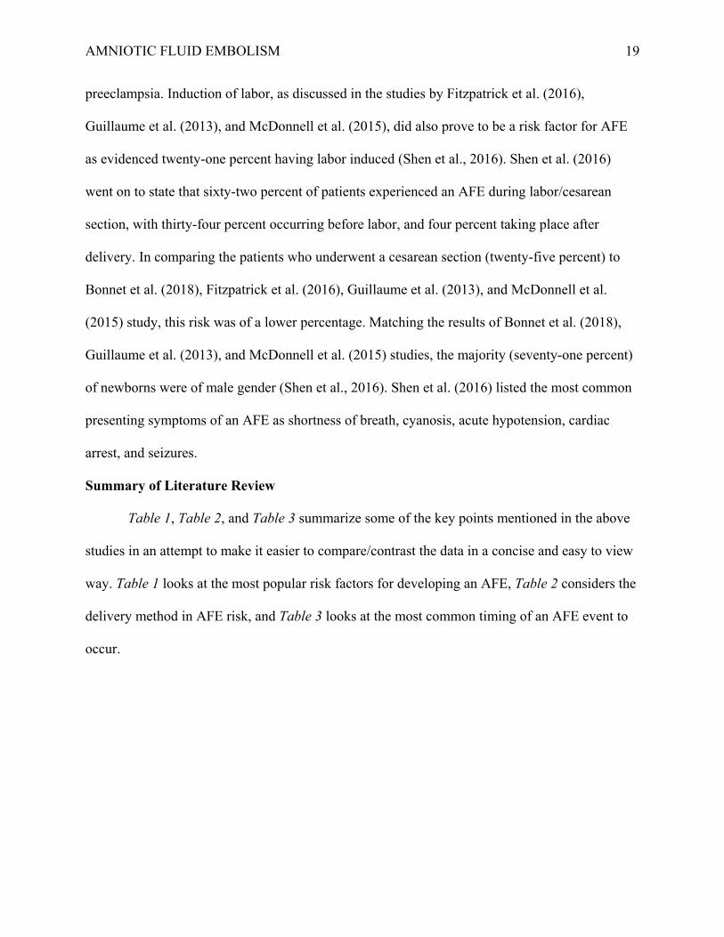
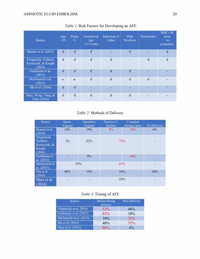
Summary of Literature Review
Table 1, Table 2, and Table 3 summarize some of the key points mentioned in the above
studies in an attempt to make it easier to compare/contrast the data in a concise and easy to view
way. Table 1 looks at the most popular risk factors for developing an AFE, Table 2 considers the
delivery method in AFE risk, and Table 3 looks at the most common timing of an AFE event to
occur.
AMNIOTIC FLUID EMBOLISM 20
Table 1. Risk Factors for Developing an AFE
Table 2. Methods of Delivery
Table 3. Timing of AFE
Source
Age <35
Parity
1+
Gestational
age >37 weeks
Induction of
Labor
Male
Newborn
Nonsmoker
BMI <30 prior to
pregnancy
Bonnet et al. (2018) √
√
√
- √
- √
Fitzpatrick, Tuffnell, Kurinczuk, & Knight
(2016)
√
√
√
√
- √ √
Guillaume et al. (2013)
√
√
√
√
√
- -
McDonnell et al. (2015)
× × √
√
√
√ -
Mu et al. (2016) √
√
- - - - -
Shen, Wang, Yang, & Chen (2016)
√
√
√
√
√
- -
Source Spont. Vaginal
Operative Vaginal
Elective C-Section
C-section during Labor
No Delivery
Bonnet et al. (2018)
14% 19% 8% 53% 6%
Fitzpatrick, Tuffnell, Kurinczuk, & Knight (2016)
1%
22%
71%
-
Guillaume et al. (2013)
- 9% 64% -
McDonnell et al. (2015)
33% 67% -
Mu et al. (2016)
45% 15% 16% 24%
Shen et al. (2016)
- - 25% -
Source Before/during delivery
Post Delivery
Fitzpatrick et al. 2016) 52% 46% Guillaume et al. (2013) 82% 18% McDonnell et al. (2015) 39% 52% Mu et al. (2016) 40% 52% Shen et al. (2016) 96% 4%
AMNIOTIC FLUID EMBOLISM 21
Amniotic Fluid Embolism
Risk Factors
As mentioned in the studies previously discussed, those diagnosed with an amniotic fluid
embolism exhibit several risk factors, commonly including an age less than thirty-five years old,
a parity greater than one, a gestation age greater than thirty-seven weeks, induction of labor,
male gender newborn, nonsmoking status, issues revolving around the placenta, and delivery via
cesarean section.
Diagnosis
Diagnosing an AFE is difficult as a universal diagnostic criterion does not exist across the
field of healthcare. With no ability to make a clear-cut diagnosis, identification of an AFE is
often made based on the presence of specific symptoms that lack any other causative
explanation. In the research studies discussed in this paper, each author had their interpretation of
what traits, although similar, categorized an AFE.
Proposed Diagnostic Criteria for Research. The Society for Maternal-Fetal Medicine
and the Amniotic Fluid Embolism Foundation tasked Clark et al. (2016) to establish a diagnostic
criterion for an AFE to use in the research reporting of these cases. The committee determined
that a patient must exhibit the following four measures to confirm an AFE occurrence which
includes: insults to the (1) cardiac/respiratory and (2) hematological body systems, (3) clinical
onset time and (4) lack of fever (Clark et al., 2016).
When looking at the cardiovascular/respiratory systems, Clark et al. (2016) states that the
patient must experience a sudden cardiopulmonary arrest, or a combination of hypotension
(systolic blood pressure is less than ninety millimeters of mercury) with respiratory compromise
(shortness of breath, oxygen saturation less than ninety-percent, or cyanosis). Clark et al. (2016)
explain that a coagulopathy issue must be identified before significant blood loss to confirm a
AMNIOTIC FLUID EMBOLISM 22
diagnosis of DIC. Clark et al. (2016) modified the Scientific and Standardization Committee on
DIC of the International Society on Thrombosis and Hemostasis scoring system, to attribute for
coagulation changes seen during pregnancy. To make a diagnosis of DIC, a score higher than
three points must be achieved based upon platelet count, PTT/INR, and fibrinogen level (Clark et
al., 2016).
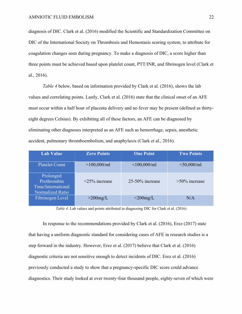
Table 4 below, based on information provided by Clark et al. (2016), shows the lab
values and correlating points. Lastly, Clark et al. (2016) state that the clinical onset of an AFE
must occur within a half hour of placenta delivery and no fever may be present (defined as thirty-
eight degrees Celsius). By exhibiting all of these factors, an AFE can be diagnosed by
eliminating other diagnoses interpreted as an AFE such as hemorrhage, sepsis, anesthetic
accident, pulmonary thromboembolism, and anaphylaxis (Clark et al., 2016).
Table 4. Lab values and points attributed in diagnosing DIC for Clark et al. (2016)
In response to the recommendations provided by Clark et al. (2016), Erez (2017) state
that having a uniform diagnostic standard for considering cases of AFE in research studies is a
step forward in the industry. However, Erez et al. (2017) believe that Clark et al. (2016)
diagnostic criteria are not sensitive enough to detect incidents of DIC. Erez et al. (2016)
previously conducted a study to show that a pregnancy-specific DIC score could advance
diagnostics. Their study looked at over twenty-four thousand people, eighty-seven of which were
Lab Value Zero Points One Point Two Points
Platelet Count >100,000/ml <100,000/ml <50,000/ml
Prolonged Prothrombin
Time/International Normalized Ratio
<25% increase
25-50% increase
>50% increase
Fibrinogen Level >200mg/L <200mg/L N/A
AMNIOTIC FLUID EMBOLISM 23
noted to have DIC (Erez, 2017). Like Clark et al. (2016) DIC criteria, Erez (2017) DIC criteria
considered platelet count, prothrombin time, and fibrinogen level. Erez (2017) also had a scoring
system in place, and a score greater than twenty-six had a sensitivity of eighty-eight percent and
specificity of ninety-six percent for having DIC. Erez (2017) tested the criteria modified by
Clark et al. (2016) on the patients determined to have DIC in their study. Out of the eighty-seven
patients who had DIC, only thirteen of these patients were considered to have DIC according to
Clark et al. (2016) modified DIC criteria (Erez, 2017). Erez (2017) does state that while their
study did not look at any patients suffering from an AFE, they believe that some of those who
suffered from DIC could have been missed using Clark et al. (2017) criteria.
Biomarkers in Diagnosing AFE
Squamous cell carcinoma antigen. Koike, Oi, Naruse, Kanayama, & Kobayashi (2017)
conducted a study to see if using the squamous cell carcinoma antigen could be a diagnostic
marker for AFE. Koike et al. (2017) took remaining blood specimens from four patients that
were proven to have an AFE based on autopsy results, and sixteen patients who were determined
to have an AFE based on clinical manifestations. These patients were found in the Japan
Amniotic Fluid Embolism Registration Center from 2008-2013 (Koike et al., 2017). For
enrollment into this study, patients must have had one of the following symptoms: cardiac
arrest/respiratory arrest/coagulopathy occurring during pregnancy/labor/cesarean section or
within twelve hours post-partum without any other causative illness (Koike et al., 2017). Koike
et al. (2017) also included a control group of seventy-four healthy women and obtained their
blood samples within two hours of standard delivery. In comparing the levels of squamous cell
carcinoma antigen in each of the populations, Koike et al. (2017) found that those patients who
had an AFE diagnosed via autopsy (mean level 35.1nanogram/milliliter) were much higher than
those diagnosed based on clinical manifestations (mean level 6.1nanogram/milliliter). Both
AMNIOTIC FLUID EMBOLISM 24
levels, according to Koike et al. (2017), whether from the autopsy or clinical manifestation group
had higher levels of squamous cell carcinoma antigen compared to the control group (mean level
4.2nanogram/milliliter).
Koike et al. (2017) also estimated the effectiveness of measuring squamous cell
carcinoma antigen as a means of diagnosing an AFE. Their analysis suggested using seven and
fifteen-tenths nanogram/milliliter as a cut-off value which represented a sensitivity of sixty-
percent and sensitivity of eighty-nine and two tenths-percent (Koike et al., 2017). In diagnosing
an AFE in the autopsy group, Koike et al. (2017) stated that the squamous cell carcinoma antigen
had a sensitivity of one hundred percent and a specificity of eighty-nine and two tenths-percent.
Despite their limitation of a small sample size, Koike et al. (2017) was the first study to show
that squamous cell carcinoma antigen is higher in patients diagnosed with an AFE compared to a
healthy control population.
CK13 and CK10/13. Wang et al. (2014) looked at the prevalence of CK13 and CK10/13
in cases of AFE. In this study, Wang et al. (2014) took lung tissue specimens from the autopsy
cases of nineteen maternal deaths, eight of which diagnosed with having an AFE. According to
Wang et al. (2014), those diagnosed with an AFE showed fetal squamous cells/debris in the
pulmonary vessels of their lung samples. Debris were evident by using a mouse monoclonal anti-
CK10/13 antibody and a rabbit monoclonal anti-CK13 antibody for immunohistochemistry
(Wang et al., 2014). Using these antibody’s, Wang et al. (2014) showed that amniotic fluid
squamous cells stained positive while endothelial/alveolar epithelial cells stained negative. Also,
APM staining was able to portray squamous cells, mucus, and microthrombi in amniotic fluid
(Wang et al., 2014). Wang et al. (2014) concluded that the combination of CK13 and CK10/13
immunohistochemistry and APM staining could advance the diagnosis of an AFE.
AMNIOTIC FLUID EMBOLISM 25
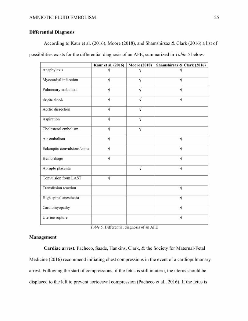
Differential Diagnosis
According to Kaur et al. (2016), Moore (2018), and Shamshirsaz & Clark (2016) a list of
possibilities exists for the differential diagnosis of an AFE, summarized in Table 5 below.
Table 5. Differential diagnosis of an AFE
Management
Cardiac arrest. Pacheco, Saade, Hankins, Clark, & the Society for Maternal-Fetal
Medicine (2016) recommend initiating chest compressions in the event of a cardiopulmonary
arrest. Following the start of compressions, if the fetus is still in utero, the uterus should be
displaced to the left to prevent aortocaval compression (Pacheco et al., 2016). If the fetus is
Kaur et al. (2016) Moore (2018) Shamshirsaz & Clark (2016) Anaphylaxis √ √ √
Myocardial infarction √ √
√
Pulmonary embolism √
√
√
Septic shock √
√
√
Aortic dissection √
√
Aspiration √
√
Cholesterol embolism √
√
Air embolism √
√
Eclamptic convulsions/coma √
√
Hemorrhage √
√
Abrupto placenta √
√
Convulsion from LAST √
Transfusion reaction √
High spinal anesthesia √
Cardiomyopathy √
Uterine rupture √
AMNIOTIC FLUID EMBOLISM 26
considered viable (twenty-three weeks gestation), Pacheco et al. (2016) indicate prompt delivery.
Additionally, there is a theory that defibrillation in the presence of fetal monitoring devices
causes electric arching, in which case these devices can be removed while cardiopulmonary
resuscitation is in progress (Pacheco et al., 2016). Pacheco et al. (2016) also state that there are
no dose adjustments needed for resuscitation medications or defibrillation doses in the pregnant
patient. If unable to obtain spontaneous circulation after four minutes of maternal resuscitation,
perimortem cesarean delivery is indicated to decrease fetal hypoxia (Pacheco et al., 2016).
Pacheco et al. (2016) state that venoarterial extracorporeal membrane oxygenation unresponsive
to resuscitation efforts is controversial as the anticoagulation needed can worsen bleeding in the
already coagulopathic patient.
Post-resuscitation considerations. If successful resuscitation of the patient occurs, post-
cardiac arrest management is critical. Hemodynamic instability often necessitates fluids and
vasopressors/inotropes to maintain a mean arterial pressure of greater than sixty-five millimeters
of mercury (Pacheco et al., 2016). One hundred percent oxygen should not be used as Pacheco et
al. (2016) explain that hyperoxia can exacerbate ischemia-reperfusion injuries. Pacheco et al.
(2016) recommend that because of this, pulse oximetry readings should be between ninety-four
and ninety-eight percent. Serum glucose levels must be kept in a range of one hundred forty
milligrams per deciliter to of one hundred eighty milligrams per deciliter, with the use of insulin
if needed (Pacheco et al., 2016). To increase the chances of a favorable neurological outcome,
Pacheco et al. (2016) recommend the use of therapeutic hypothermia between thirty-two degrees
Celsius and thirty-six degrees Celsius. The main concern with therapeutic hypothermia is that it
can increase hemorrhage risk. If patients do not exhibit signs of DIC, consideration of
therapeutic hypothermia should occur. (Pacheco et al., 2016). If there is an increased risk of
AMNIOTIC FLUID EMBOLISM 27
hemorrhage, Pacheco et al. (2016) recommend considering a higher temperature, closer to thirty-
six degrees Celsius.
Hemodynamic support. As discussed in the pathophysiology of an AFE, the first phase
consists of right ventricular failure. Pacheco et al. (2016) state that transthoracic or
transesophageal echocardiography provides valuable information to guide hemodynamic
interventions. Pacheco et al. (2016) explain that an increase in pulmonary vascular resistance
worsens right heart failure, which is produced by hypoxia/hypercarbia and acidosis. Dobutamine
and milrinone are inotropes that can improve right ventricular output by causing pulmonary
vasodilation (Pacheco et al., 2016). Pacheco et al. (2016) suggest other ways to decrease
pulmonary vascular resistance include sildenafil, prostacyclin, and nitric oxide. As hypotension
from these treatment modalities will present themselves, treatment consists of vasopressors such
as norepinephrine or vasopressin (Pacheco et al., 2016).
As mentioned previously, the second phase of an AFE consists of left ventricular failure.
During this phase excess fluid administration should be avoided. Pacheco et al. (2016) state that
if the patient has a dilated right ventricle, overdistention from excess fluid will increase the size
of the right ventricle and lead to a right-sided myocardial infarction. Additionally, overdistension
of the right ventricle can cause the intraventricular septum to deviate to the left which inhibits
cardiac output (Pacheco et al., 2016). As the right ventricular function starts to improve, Pacheco
et al. (2016) say that left ventricular failure with pulmonary edema usually becomes apparent.
Pacheco et al. (2016) recommend that left-sided heart failure is treated by augmenting preload,
using vasopressors to maintain coronary perfusion pressure in instances of hypotension, and
increase left ventricular contractility (inotropes such as dobutamine and milrinone). If pulmonary
edema does not respond to diuretics, fluid removal through dialysis may be needed (Pacheco et
al., 2016).
AMNIOTIC FLUID EMBOLISM 28
Coagulopathy. Issues with coagulation exhibit themselves simultaneously with the onset
of an AFE or present themselves shortly after. Pacheco et al. (2016) state that assessing patients
clotting status should occur early on and hemorrhage should be treated promptly with massive
transfusion protocols. Pacheco et al. (2016) recommend packed red blood cells, fresh frozen
plasma, and platelets to be administered in a ratio of one to one to one. Kaur et al. (2016)
recommend that platelets be transfused at one to three units per ten kilograms per day if platelets
are less than twenty thousand or if there is bleeding and platelets are between twenty and fifty
thousand. Also, Kaur et al. (2016) state that each unit of cryoprecipitate increases the fibrinogen
level by ten milligrams per deciliter and should be administered at fibrinogen levels less than one
hundred milligrams per deciliter. Some cases treat coagulation issues stemming from an AFE
with recombinant activated factor VII, but concerns exist that the administration of this drug can
lead to excessive thrombosis and organ failure in patients who have DIC (Pacheco et al., 2016).
Therefore, Pacheco et al. (2016) recommend that recombinant activated factor VII be used as a
last resort when bleeding continues with blood product replacement/surgical interventions. If
thromboelastography is available, Pacheco et al. (2016) say it can assist in identifying patients
who would benefit from the administration of antifibrinolytics such as tranexamic acid.
Performing frequent blood sampling makes atrial catheterization beneficial to limit the number
of puncture sites the patient receives and to have an accurate means of blood pressure monitoring
Uterine atony. Uterine atony is seen with an AFE, and should be treated with
uterotonics, ergot derivatives, and prostaglandins when necessary (Pacheco et al., 2016). Most
commonly used, as Kaur et al. (2016) state, is oxytocin as it decreases inflammation by reversing
capillary permeability and stopping the movement of polymorphonuclear leukocytes. Methergine
is beneficial as it causes a sustained tetanic effect on uterine smooth muscle to decrease bleeding
(Kaur et al., 2016). Kaur et al. (2016) also state that Carboprost reduces postpartum bleeding by
AMNIOTIC FLUID EMBOLISM 29
promoting hemostasis at the placental site through myometrial contractions. If these drugs prove
to be unsuccessful in contracting the uterus, Pacheco et al. (2016) recommend considering
uterine tamponade with packing or intrauterine balloons. If still ineffective in tightening the
womb, more invasive treatments include uterine artery ligation and hysterectomy (Pacheco et al.,
2016). It is vital if the patient delivered vaginally to inspect the cervix/vagina for lacerations that
could be the cause of bleeding (Pacheco et al., 2016). If surgical interventions cannot control
bleeding in patients delivering by cesarean section, Pacheco et al. (2016) recommend packing the
pelvis.
Case Studies Suggesting New Treatment for AFE
C1 Esterase Inhibitor
Akasaka et al. (2018) wrote a case report on the proposed effectiveness of using a C1
esterase inhibitor in patients suffering from an AFE. Akasaka et al. (2018) state that C1 esterase
inhibitors (C1INH) are a protein that assists in stopping complement, coagulation, and kinin
pathways. They also mention that there is a decreased C1INH concentration in women who
suffer from an AFE (Akasaka et al., 2018).
Akasaka et al. (2018) case report was a thirty-two-year-old Japanese woman who was
thirty-eight weeks pregnant with her first child (a boy). The patient came to the hospital due to an
elevated blood pressure of one hundred seventy-one over ninety-six, protein in the urine, and
bilateral leg edema (Akasaka et al., 2018). She was given magnesium sulfate for suspected
preeclampsia (Akasaka et al., 2018). Based on this suspicion and having an unfavorable cervix,
Akasaka et al. (2018) say that the patient had an emergency cesarean section with an estimated
blood/amniotic fluid loss of just over fifteen hundred milliliters. According to Akasaka et al.
(2018), immediately after delivery, the patient’s vital signs were stable. Thirty minutes following
delivery, about two hundred milliliters of uterine blood were noted, and oxytocin was
AMNIOTIC FLUID EMBOLISM 30
administered in response (Akasaka et al., 2018). At the one-hour mark since delivery, an
additional seventy milliliters of uterine blood was noted, at which time the patients’ blood
pressure had dropped to 88/46 (Akasaka et al., 2018). In response, Akasaka et al. (2018) state
with the administration of prostaglandin F2α, the patients' blood pressure normalized to 130/68.
Akasaka et al. (2018) drew labs at the time of prostaglandin administration and compared
them to the patients preop values. In comparison, there was a decrease in hemoglobin from
eleven-point-four to six-point-five, platelets from one hundred thirty-three thousand to eighty-
five thousand, and fibrinogen from three hundred thirty-one to ninety-two, with an increase in
PTT from eleven-point-one seconds to fourteen-point-five seconds and activated partial
thromboplastin time from twenty-nine seconds to fifty-five seconds. Akasaka et al. (2018)
interpreted these results as the patient has developed DIC, while also exhibiting a soft uterus.
Akasaka et al. (2018) listed the following unsuccessful interventions including administration of
cryoprecipitate (followed by a deterioration in the level of consciousness), and then five minutes
later the administration of fresh frozen plasma and packed red blood cells (blood pressure
continued to drop to sixty over thirty-one). At this time, they administered one thousand units of
C1INH concentrate and within minutes the patients' level of consciousness improved, and her
uterus became firm (Akasaka et al., 2018).
After ten minutes of C1INH concentrate injection, Akasaka et al. (2018) state the
patients’ blood pressure normalized to one hundred nine over seventy-six. Lab values were
obtained an hour and a half after the patient started to deteriorate in which her hemoglobin
increased from six-point-five to eight-point-two, platelets decreased from eighty-five thousand to
sixty-three thousand, and fibrinogen increased from ninety-two to one hundred forty-one
(Akasaka et al., 2018). Akasaka et al. (2018) noted that ten units of fresh frozen plasma and
cryoprecipitate were needed to get the fibrinogen level to reach one hundred forty-one. C1INH
AMNIOTIC FLUID EMBOLISM 31
levels before injection were thirty-six percent, and after injection rose to seventy-five percent
(Akasaka et al., 2018). From their case study, Akasaka et al. (2018) interpreted the following
results, (1) C1INH concentrate can improve deteriorating vital signs and contract the uterus in
minutes and (2) C1INH concentrate can decrease the number of transfused blood products
needed to stabilize the patient.
Lipid Emulsion Therapy
McAllister, Lay, & Culp (2018) wrote a response to Akasaka et al. (2018) study stating
that C1INH concentrate proved to be an expensive form of treatment (estimated cost of twenty-
five hundred dollars), required evidence to pharmacy to use this drug (off-label usage), and
because of these factors would take a long time to retrieve in an emergency situation. Instead,
McAllister et al. (2018) recommend the use of lipid emulsion therapy which was proven an
effective treatment for an AFE in their study by Lynch, McAllister, Lay, & Culp (2017).
McAllister et al. (2018) also made the comparison that lipid emulsion was proven to be much
more cost effective (around twenty-five dollars) and readily available in anesthesia departments
with familiarity.
Lynch, McAllister, Lay, & Culp (2017) detail lipid emulsion therapy to be effective in a
case study of a twenty-eight-year-old non-smoking woman, who was forty-one weeks of
gestation at the hospital to have her labor induced. She was induced with misoprostol, had an
epidural placed, and then had labor augmented with an oxytocin infusion (Lynch et al., 2017).
Shortly after the initiation of an oxytocin infusion, Lynch et al. (2017) report that fetal
decelerations were noted which resolved after stopping the drip. Soon after, bleeding was
pointed out from the epidural site, without a history of any coagulation issues (Lynch et al.,
2017). Lynch et al. (2017) state that after placing a pressure dressing over the catheter site,
bleeding was still noted from this area one hour later. At this time, lab values were drawn,
AMNIOTIC FLUID EMBOLISM 32
followed again by fetal decelerations (Lynch et al., 2017). On inspection of the patient, Lynch et
al. (2017) report that she was fully dilated with complete effacement, and thus delivered
vaginally with vacuum assistance. After delivery, the patient developed hemorrhage that did not
respond to the administration of oxytocin, misoprostol, carboprost, or methylergonovine (Lynch
et al., 2017). Lynch et al. (2017) suspected DIC when lab results came back with a PTT of
twenty-three seconds and an INR of two.
As a result, Lynch et al. (2017) state that administering two units of packed red blood
cells and placing an intrauterine balloon aided in an attempt to stop the bleeding. A perineal
laceration that occurred during delivery was sutured, even though it was not suspected to be the
source of bleeding (Lynch et al., 2017). Forty-seven minutes after the transfusion, and before
transfer to the intensive care unit, Lynch et al. (2017) report the patient experienced shortness of
breath, chest pain, and an altered level of consciousness, which progressed to hypotension and
tachycardia. The patient was emergently intubated and then found to have no pulse (Lynch et al.,
2017). Lynch et al. (2017) state that cardiopulmonary resuscitation was started, followed by the
administration of six units of packed red blood cells and four units of fresh frozen plasma, with
the insertion of a femoral central line and an arterial catheter. A transesophageal noted the
following abnormalities: tricuspid regurgitation, depressed left ventricular systolic function, and
a potential thrombus one centimeter in diameter in the inferior vena cava near the liver (Lynch et
al., 2017).
Despite adequate cardiopulmonary resuscitation over forty minutes with the
administration of vasopressin, sodium bicarbonate, calcium chloride, atropine, and six
milligrams of epinephrine, the patient's heart rate switched between extreme bradycardia and
asystole (Lynch et al., 2017). Lynch et al. (2017) considered a diagnosis of local anesthetic
systemic toxicity as a last-ditch effort, although there was no evidence of epidural pump
AMNIOTIC FLUID EMBOLISM 33
malfunction, and the amount of local anesthetic infused was well within a normal range. Lipid
twenty-percent emulsion was administered at a dose of one-point-five milliliters per kilogram
(Lynch et al., 2017). Thirty to ninety seconds after administration, Lynch et al. (2017) reports
there was a return of spontaneous circulation (ROSC) with normal sinus rhythm and improved
left and right ventricular function as seen by transesophageal echocardiogram. However, minutes
later the patient had another episode of asystole that was treated with cardiopulmonary
resuscitation and another dose of twenty-percent lipid emulsion therapy (one-point-five
milliliters per kilogram) followed by a twenty-percent lipid emulsion infusion at zero-point-
twenty-five milliliters per kilogram per minute (Lynch et al., 2017). After thirty to ninety
seconds, Lynch et al. (2017) stated there was ROSC, normal sinus rhythm, and patient
movement. Shortly after transferring the patient to the intensive care unit, she regained
consciousness and was able to follow commands; thus, lipid emulsion infusion was discontinued
(Lynch et al., 2017).
Lynch et al. (2017) reported that lab results showed a fibrinogen level of less than sixty
and a d-dimer level greater than twenty micrograms per milliliter. Due to continued bleeding, six
units of packed red blood cells, five units of fresh frozen plasma, one unit of platelets and two
units of cryoprecipitate were administered (Lynch et al., 2017). Lynch et al. (2017) state that the
patient remained intubated overnight and required vasopressor support with epinephrine and
norepinephrine. The following morning the patient was extubated, able to follow commands, and
showed no signs of cognitive impairment (Lynch et al., 2017). Due to reperfusion injuries, Lynch
et al. (2017) mentioned that the patient received four runs of dialysis.
A-OK Protocol
Rezai, Hughes, Larsen, Fuller, & Henderson (2017) detail a case report in which the
administration of Atropine, Ondansetron, and Ketorolac (A-OK) was effective in stabilizing a
AMNIOTIC FLUID EMBOLISM 34
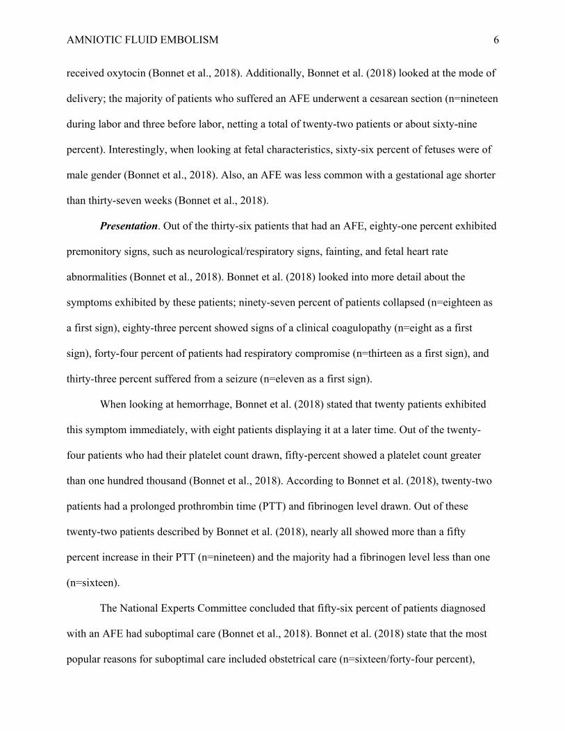
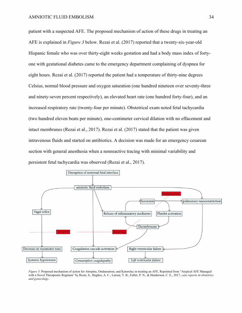
patient with a suspected AFE. The proposed mechanism of action of these drugs in treating an
AFE is explained in Figure 3 below. Rezai et al. (2017) reported that a twenty-six-year-old
Hispanic female who was over thirty-eight weeks gestation and had a body mass index of forty-
one with gestational diabetes came to the emergency department complaining of dyspnea for
eight hours. Rezai et al. (2017) reported the patient had a temperature of thirty-nine degrees
Celsius, normal blood pressure and oxygen saturation (one hundred nineteen over seventy-three
and ninety-seven percent respectively), an elevated heart rate (one hundred forty-four), and an
increased respiratory rate (twenty-four per minute). Obstetrical exam noted fetal tachycardia
(two hundred eleven beats per minute), one-centimeter cervical dilation with no effacement and
intact membranes (Rezai et al., 2017). Rezai et al. (2017) stated that the patient was given
intravenous fluids and started on antibiotics. A decision was made for an emergency cesarean
section with general anesthesia when a nonreactive tracing with minimal variability and
persistent fetal tachycardia was observed (Rezai et al., 2017).
Figure 3: Proposed mechanism of action for Atropine, Ondansetron, and Ketorolac in treating an AFE. Reprinted from “Atypical AFE Managed with a Novel Therapeutic Regimen” by Rezai, S., Hughes, A. C., Larsen, T. B., Fuller, P. N., & Henderson, C. E., 2017, case reports in obstetrics and gynecology.
AMNIOTIC FLUID EMBOLISM 35
Rezai et al. (2017) reported that immediately after delivery of the fetus and before the
placenta was delivered the patient had the following vital signs: oxygen saturation of seventy-
two percent, blood pressure of seventy-two over forty-eight, and no end-tidal carbon dioxide
(equipment checked with no disconnections). Within one minute of these symptoms, two-tenths
of a milligram of Atropine, eight milligrams of Ondansetron, and fifteen milligrams of Ketorolac
were administered (Rezai et al., 2017). Rezai et al. (2017) state that within two to three minutes
of the patient receiving the A-OK medications the following vital signs were present: oxygen
saturation of ninety-seven percent, blood pressure of one hundred thirty-eight over sixty-eight,
and end-tidal carbon dioxide of thirty-two. Fifty units of Oxytocin, two doses of Carboprost,
three units of packed red blood cells, one unit of fresh frozen plasma, and three and a half liters
of fluid were used to treat uterine atony and hemorrhage as evidenced by an estimated blood loss
of two liters (Rezai et al., 2017). Rezai et al. (2017) report that the only vasopressor used was
phenylephrine with a total of eighteen hundred micrograms being administered in fifty-seven
minutes (before the start of the incision to the end of surgery). The patient was admitted to the
intensive care unit, extubated the following day, and sent home on a postoperative day three
(Rezai et al., 2017). Rezai et al. (2017) reported that a chest computed tomography scan, chest x-
ray, doppler ultrasound for thrombosis, lab work to suggest disseminated intravascular
coagulation, blood/urine/sputum cultures, and placenta pathology came back all negative.
Conclusion
The occurrence of an amniotic fluid embolism (AFE) causes a physical obstruction or an
anaphylactoid reaction within the parturient patient. Symptoms present in two forms: as either
cardiopulmonary collapse or DIC, both frequently leading to maternal death. An examination of
current research studies analyzed the incidence, risk factors, presentation, and management of an
AFE in the healthcare setting. From these studies, it shows that an AFE has an incidence rate
AMNIOTIC FLUID EMBOLISM 36
ranging between 1.7-28:100,000 women. Significant risk factors predisposing women to an AFE
include a maternal age less than thirty-five years old, a gestational age greater than thirty-seven
weeks, a parity greater than one, induction of labor, cesarean section, nonsmoking, and male
gender fetus. Additionally, it is shown that an AFE most often occurs prior to or during delivery
compared to the post-delivery period.
Making an in-hospital clinical diagnosis of an AFE remains challenging as a universal
diagnostic criterion has yet to exist. Thus, further research into identifying a diagnostic tool
would be beneficial. As there have been attempts made to establish a uniform guideline to
identify AFE events when reporting in research, this can be used as a stepping stone to build on.
It appears that the presentation of squamous cell carcinoma antigen, CK13, and CK10/13
biomarkers can aid in the investigation/confirmation of an AFE event. Management of an AFE
takes a comprehensive approach with attention to cardiac resuscitation, post-cardiac arrest care,
hemodynamic support, coagulopathy, and uterine atony. In three unique case reports, the
treatment of an AFE was proven successful with using C1INH, lipid emulsion therapy, and
atropine/ondansetron/ketorolac. By applying the information discussed into a nurse anesthetists’
clinical practice, it allows for a better understanding of what an AFE entails, and how to be
prepared in treating the multitude of symptoms that occur in an AFE event.
AMNIOTIC FLUID EMBOLISM 37
References
Akasaka, M., Osato, K., Sakamoto, M., Kihira, T., Ikeda, T., & Yamawaki, T. (2018). Practical
use of C1 esterase inhibitor concentrate for clinical AFE. Journal of Obstetrics and
Gynaecology Research, 44(10), 1995-1998. doi:10.1002/ccr3.316
Bonnet, M. P., Zlotnik, D., Saucedo, M., Chassard, D., Bouvier-Colle, M. H., & Deneux-
Tharaux, C. (2018). Maternal death due to AFE: a national study in France. Anesthesia &
Analgesia, 126(1), 175-182. doi:10.1213/ANE.0000000000002511
Clark, S. L., Romero, R., Dildy, G. A., Callaghan, W. M., Smiley, R. M., Bracey, A. W., ... &
Vadhera, R. B. (2016). Proposed diagnostic criteria for the case definition of amniotic
fluid embolism in research studies. American journal of obstetrics and
gynecology, 215(4), 408-412. doi:10.1016/j.ajog.2016.06.037
Erez, O. (2017). Proposed diagnostic criteria for the case definition of AFE
in research studies. American Journal of Obstetrics & Gynecology, 217(2), 228-229.
doi:10.1016/j.ajog.2017.04.009
Fitzpatrick, K. E., Tuffnell, D., Kurinczuk, J. J., & Knight, M. (2016). Incidence, risk factors,
management and outcomes of amniotic‐fluid embolism: a population‐ based cohort and
nested case-control study. BJOG: An International Journal of Obstetrics &
Gynaecology, 123(1), 100-109. doi:0.1111/1471-0528.13300
Guillaume, A., Sananes, N., Akladios, C. Y., Boudier, E., Diemunsch, P., Averous, G., ... &
Langer, B. (2013). AFE: 10-year retrospective study in a level III maternity
hospital. European Journal of Obstetrics & Gynecology and Reproductive
Biology, 169(2), 189-192. doi:10.1016/j.ejogrb.2013.02.017
Kanayama, N., & Tamura, N. (2014). AFE: pathophysiology and new strategies for
AMNIOTIC FLUID EMBOLISM 38
management. Journal of Obstetrics and Gynaecology Research, 40(6), 1507-1517.
doi:10.1111/jog.12428
Kaur, K., Bhardwaj, M., Kumar, P., Singhal, S., Singh, T., & Hooda, S. (2016). Amniotic fluid
embolism. Journal of Anaesthesiology, clinical pharmacology, 32(2), 153.
doi:10.4103/0970-9185.173356 Koike, N., Oi, H., Naruse, K., Kanayama, N., & Kobayashi, H. (2017). Squamous cell carcinoma
antigen as a novel candidate marker for AFE. Journal of Obstetrics
and Gynaecology Research, 43(12), 1815-1820. doi:10.1111/jog.13453 Lynch, W., McAllister, R. K., Lay, J. F., & Culp, W. C. (2017). Lipid emulsion rescue of
AFE-induced cardiac arrest: a case report. A&A Practice, 8(3), 64-66.
doi:10.1213/XAA.0000000000000427
McAllister, R. K., Lay Jr, J. F., & Culp Jr, W. C. (2018). Letter to 'Practical use of C1 esterase
inhibitor concentrate for clinical AFE'. The journal of obstetrics and
gynaecology research. Retrieved from https://obgyn.onlinelibrary.wiley.com/doi
/full/10.1111/jog.13821
McDonnell, N., Knight, M., Peek, M. J., Ellwood, D., Homer, C. S., McLintock, C., ... &
Sullivan, E. (2015). AFE: an Australian-New Zealand population-
based study. BMC pregnancy and childbirth, 15(1), 352. doi:10.1186/s12884-015-
0792-9.
Moore, L. E. (2018). AFE Differential Diagnoses. Medscape. Retrieved November 16, 2018
from https://emedicine.medscape.com/article/253068-differential
Mu, Y., McDonnell, N., Li, Z., Liang, J., Wang, Y., Zhu, J., & Sullivan, E. (2016). Amniotic
fluid embolism as a cause of maternal mortality in China between 1996 and 2013: a
population-based retrospective study. BMC pregnancy and childbirth, 16(1), 316.
AMNIOTIC FLUID EMBOLISM 39
doi:10.1186/s12884-016-1106-6
Pacheco, L. D., Saade, G., Hankins, G. D., Clark, S. L., & Society for Maternal-Fetal Medicine
SMFM. (2016). AFE: diagnosis and management. American journal of obstetrics and
gynecology, 215(2), B16-B24. doi:10.1016/j.ajog.2016.03.012
Rath, W. H., Hofer, S., & Sinicina, I. (2014). AFE: an interdisciplinary challenge: epidemiology,
diagnosis and treatment. Deutsches Ärzteblatt International, 111(8), 126.
doi:10.3238/arztebl.2014.0126 Rezai, S., Hughes, A. C., Larsen, T. B., Fuller, P. N., & Henderson, C. E. (2017). Atypical
AFE Managed with a Novel Therapeutic Regimen. Case reports in obstetrics and
gynecology, 2017. https://doi.org/10.1155/2017/8458375
Shamshirsaz, A. A., & Clark, S. L. (2016). AFE. Obstetrics and Gynecology Clinics, 43(4), 779-
790. doi:10.1016/j.ogc.2016.07.001 Shen, F., Wang, L., Yang, W., & Chen, Y. (2016). From appearance to essence: 10 years review
of atypical AFE. Archives of gynecology and obstetrics, 293(2), 329-334.
doi:10.1007/s00404-015-3785-z
Wang, J., Lai, Q., Pan, H., Sun, D., Yu, C., Zhang, W., ... & Zhou, R. (2014). Evaluation of
specific marker CK13 and CK10/13 combined with APM staining for the diagnosis of
AFE and aspiration. Forensic science international, 238, 108-112. doi:10.1016/j.
forsciint.2014.02.032