Embed Size (px)
Citation preview

Amira Software for Life & Biomedical SciencesUniversal 2D–5D software for discovery workflows

Thermo Scientific™ Amira™ Software is a powerful, universal 2D–5D solution for visualizing, analyzing and understanding life science and biomedical research data from many image modalities, including Optical and Electron Microscopy, CT, MRI and other techniques.
With incredible speed and flexibility, Amira Software supports advanced 2D–5D bioimaging workflows in research areas ranging from structural and cellular biology to tissue imaging, neuroscience, preclinical imaging and bioengineering.
On the cover: an Amira Software 3D visualization of a Golgi apparatus from the green alga Chlamydomonas reinhardtii. Data courtesy of Dr. Benjamin Engel, Dept. of Molecular Structural Biology, Max Planck Institute for Biochemistry, Martinsried, Germany.

3
• Structural biology
• Cell biology
• Tissue biology
• Preclinical and clinical research
• Neuroscience and brain research
• Dental research
• Orthopedics and 3D printing

4
From any 3D image data, including time series and multi-channel, Amira Software delivers a comprehensive range of data visualization, processing and analysis capabilities. Amira Software allows life science and biomedical researchers to gain invaluable insights into their data, at different scales and from any modality.
From sample to knowledge
Data acquisition
• Optical and electron microscopy
• X-ray tomography
• Other techniques (MRI, etc.)
Molecular visualization
• Dedicated visualization and analysis tools
• Molecular surfaces
• Sequence alignment
• Configuration density computation
• Molecule trajectory animation
Import
• Bio-Formats
• Medical and neuroimage formats
• Very large data
Object tracking
• Custom detection workflows
• Automated tracking
• Precision tracking of thousands of cells
• Automatic selection of motion model, event detection, gap closing
• 40+ track measures
Filtering and pre-processing
• Noise reduction
• Image artifact reduction
• Background correction
Filament tracing
• Automatic extraction of centerlines
• Flexible editing of centerlines
• Interactive tracing of filaments
• Versatile label editor for annotations
• Network measures
Visualization
• Interactive high-quality visualization
• Direct manipulation of objects
Meshing for FEA/CFD
• Generation of 3D mesh direct from 3D images
• Extremely fast generation
• Large dataset handling
• Export to FE-solvers
Digital workflow
Specialized tools
Courtesy of Leica Microsystems

5
Segmentation
• State-of-the-art automatic segmentation algorithms
• Productive environment for supervised segmentation
Diffusion tensor imaging
• MRI/DTI gradient imaging and fiber tracking workflow
• Motion artifact minimization
• Visualization options for grayscale images, tensor fields and fiber tracts
Measurement and analysis
• 200+ measures available
• Custom measures
• Statistics
3D Registration
• Automatic
• Multimodal
• Multiscale
Presentation
• Snapshots and 3D videos
• Advanced presentation scenario
• 3D stereo devices
Biomaterials deformation analysis
• 3D visualization and quantification of deformation induced in biomaterials
La
rge
Da
ta M
an
ag
em
en
t
Au
tom
ati
on
Cu
sto
miz
ati
on

6
From imaging to understanding mechanisms of life, Amira Software supports entire discovery workflows. Process and visualize data from any imaging modality, at any scale, of any size. Perform routine and advanced analyses from a single, universal software application.
Amira Software for life & biomedical sciences
Structural biologyStructural biology researchers are able to obtain cellular structure insights with textbook quality, but they are still challenged by the sheer amount of particles, filaments and microtubules that require manual segmentation with traditional tools. Additionally, and unfortunately, visualization results can still appear fuzzy.
Amira Software reduces the labor effort to a minimum while improving visualization. How is this possible? By employing a fully automated workflow containing fewer parameters for filament and microtubule tracing.
In this workflow, template-matching tools automate filament and microtubule detection, while molecular-matching tools use mature registration algorithms to replace image data with models for improved visualization. As proof of efficacy, all components of the Amira Software structural biology workflow have been successfully applied in several research studies, with results appearing in multiple publications.
Visualization of the nuclear periphery of a HeLa cell revealed by cryo-electron tomography. Data courtesy of Dr. J. Mahamid, Department of Molecular Structural Biology, Max Planck Institute for Biochemistry, Martinsried, Germany.

7
Cell biologyResearchers who study dynamic cellular and intracellular processes face the dual challenges of processing dynamically acquired data with hundreds of time steps and imaging thousands of objects to be detected and tracked.
Amira Software for Cell Biology offers a comprehensive toolbox of image processing methods that can be assembled into custom detection workflows. These workflows can then be automated and applied to large time series data. Subsequently, these objects can be tracked using fully automated methods powered by the academic standard, u-track 3D.
In addition, correlative studies can be conducted using inter-modality registration methods that allow freedom in workflow design. And the efficacy of these methods has been proven through an extensive array of applications spanning numerous fields in the life sciences.
Learn more at thermofisher.com/amira-avizo
Tissue biologyResearchers who study the 3D architecture of cells and tissue in their natural context often face image data amounts that quickly surpass their system memory. Making matters worse, this data is often acquired over several days, or possibly weeks, without researchers even knowing if the data will prove usable. Then, ultimately, the usable data must be reconstructed into a 3D volume for further analysis.
Presenting a novel solution is the combination of the Thermo Scientific™ VolumeScope™ SEM and Amira Software. This hybrid large data management solution offers on-the-fly image quality monitoring during acquisition.
Additionally, interaction-free image reconstruction using our novel hybrid file format avoids data duplication while retaining the original TIFF data format, providing researchers full flexibility of image format choice.
This solution was developed completely in-house, bringing together the industry-leading VolumeScope SEM team with the image analysis and data handling experts from Amira Software to ensure an ease of use that avoids the majority of common pitfalls of large data handling in EM.
C Elegans cell and its nuclei. Images collected with TLS-SPIM, Liang Gao lab, Stony Brook University.
Volume reconstruction of a mouse brain acquired with combination of physical and optical sections in high-vacuum mode. Sample courtesy of P. Laserstein and P. Bastians, Helmstaedter Lab, MPI Brain Research, Germany.

8
Neuroscience and brain researchNeuroscientists who study brain functionality are often interested in neuronal connectivity inside the brain and spinal cord. However, to best understand how brain regions are structured and connected, these researchers sometimes must compile atlases of these regions and trace thousands of neurons in 3D image volumes.
To meet this challenge, Amira Software offers neuroscientists a multi-volume infrastructure that allows them to create brain atlases by averaging multiple image volumes. Additionally, Amira Software provides the tools needed to trace neurons in order to determine the connectivity between such regions. Plus, it includes full annotation capabilities. Used in many academic publications, Amira Software has been proving its worth to the neuroscience research community for nearly two decades.
Biomedical researchers who strive to improve their understanding of the human brain are often interested in the connectivity between different brain regions. The only option for observing this connectivity in living subjects, however, is through MRI/DTI gradient imaging and consecutive fiber tracking. Unfortunately, such workflows are often available only in the software that accompanies dedicated medical treatment devices, which are unaffordable for regular research.
Now, Amira Software offers the entire fiber tracking workflow in an independent, affordable, commercially supported research tool.
Volume rendering of cleared spinal cord imaged with 2-photon microscopy. Image is courtesy of Ali Ertürk, Max Planck Institute of Neurobiology.
Dedicated to Thermo Scientific TEM, SEM, and DualBeam Systems, as well as Thermo Scientific correlative workflows, Amira Software for Thermo Scientific Systems edition has been developed to fit with Thermo Scientific systems data acquisition and specific workflows.
This comprehensive solution also includes robust image registration tools for motion artifact minimization. It also provides a comprehensive library of visualization options for grayscale images, tensor fields and fiber tracts. Amira Software’s robust implementation of well-accepted algorithms has empowered many researchers to move their human brain research forward.
9
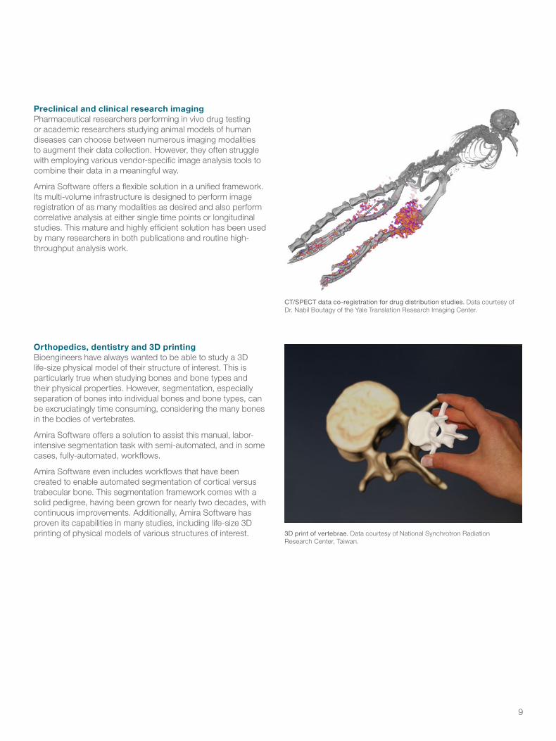
Preclinical and clinical research imagingPharmaceutical researchers performing in vivo drug testing or academic researchers studying animal models of human diseases can choose between numerous imaging modalities to augment their data collection. However, they often struggle with employing various vendor-specific image analysis tools to combine their data in a meaningful way.
Amira Software offers a flexible solution in a unified framework. Its multi-volume infrastructure is designed to perform image registration of as many modalities as desired and also perform correlative analysis at either single time points or longitudinal studies. This mature and highly efficient solution has been used by many researchers in both publications and routine high-throughput analysis work.
Orthopedics, dentistry and 3D printingBioengineers have always wanted to be able to study a 3D life-size physical model of their structure of interest. This is particularly true when studying bones and bone types and their physical properties. However, segmentation, especially separation of bones into individual bones and bone types, can be excruciatingly time consuming, considering the many bones in the bodies of vertebrates.
Amira Software offers a solution to assist this manual, labor-intensive segmentation task with semi-automated, and in some cases, fully-automated, workflows.
Amira Software even includes workflows that have been created to enable automated segmentation of cortical versus trabecular bone. This segmentation framework comes with a solid pedigree, having been grown for nearly two decades, with continuous improvements. Additionally, Amira Software has proven its capabilities in many studies, including life-size 3D printing of physical models of various structures of interest.
CT/SPECT data co-registration for drug distribution studies. Data courtesy of Dr. Nabil Boutagy of the Yale Translation Research Imaging Center.
3D print of vertebrae. Data courtesy of National Synchrotron Radiation Research Center, Taiwan.

Key featuresImport and process image data
• Handle any modality, at any scale, of any size:
– Bio-Formats
– Bitmap formats
– Microscopy: electron and optical
– Medical and neuroimage formats
– Molecular formats
– Other acquisition devices (MRI, radiography, etc.)
– Finite element modeling, geometric modeling, CAD
• Support for multi-data/multi-view, multi-channel, time series, very large data
• Scaling, calibration, conversion, re-sampling
• Image enhancement, comprehensive filtering and convolution, Fourier frequency transforms
• Artifact reduction algorithms
• Advanced multi-mode 2D/3D automatic registration
• Image stack alignment, arithmetic, correlation, fusion
Visualize and explore
• Interactive high-quality volume and multichannel visualization
• Orthogonal, oblique, cylindrical and curved slicing
• Contouring and iso-surface extraction
• Maximum Intensity or other types of projections
• Vector and tensor visualization
• Objects and tracks
• Molecular visualization
Segment
• Thresholding and auto-segmentation, object separation, automatic labeling
• Region growing, snakes, interpolation, wrapping, smoothing
• Morphological processing, including watershed and basins
• Machine Learning-based segmentation
• Automatic tracing of individual fibers and filaments
• Skeletonization and filament network extraction
• Interactive tools for generation or editing of segmentation and spatial graphs
• 3D surface reconstruction
• Grid generation for FEA/CFD
Analyze and quantify
• Intuitive recipe creation, customization, automated replay
• Built-in measurements, including counts, volumes, areas, perimeters, aspect ratios and orientations
• User-defined measures
• Results viewer with spreadsheet tool and charting
• Automatic individual feature measurements, 3D localization and spreadsheet selection
• Automated statistics, distribution graphs
• Feature filtering using any measurement criterion
• Data registration, deformation, comparison and measurements
Present
• Animation and video generation
• Advanced key frame and object animation
• Mix images, geometric models, measurements and simulations
• Annotations, measures legends, histograms and curve plots
• Export spreadsheets, 3D models, high-quality images
• Active and passive 3D stereo vision
• Single and tiled screen display
• Immersive environment
Access ecosystems
• Python scripting API
• Custom C++ modules development
• MATLAB™ bridge

11
We offer a comprehensive set of professional services. From training to consulting or custom development, our professional services experts are dedicated to helping you maximize your productivity with Amira Software.
Professional services
TrainingOur custom training is designed to provide you with immediate and practical skills while keeping your specific goals in sight. We can help you quickly and effectively master all of Amira Software’s capabilities through focused training.
Various courses can be arranged, with typical durations ranging from one to three days. We can customize our training to best fit your needs. The training can be arranged on-site at your location or may also be delivered at one of our facilities.
ConsultingOur experts will help you get the best out of the constant innovations introduced in Amira Software so you can benefit from them in your daily work.
We are your partner in creating solutions using Amira Software. Custom-made consulting sessions can be performed at your facilities or remotely, depending on your needs. Our consultants can help you analyze your specific tasks and workflows and leverage your knowledge and specific expertise to get them implemented in Amira Software
Custom developmentWith 30 years of experience in 3D and image processing and hundreds of projects delivered to small and large organizations, we can provide you with a solution tailored to fit your specific needs.
We have the ability to customize and expand our software solutions at various levels, including, but not limited to:
• Building simple push-button solutions from entire workflows
• Integrating specific workflows
• Implementing our solutions into an existing process
• Creating support for custom file formats

For current certifications, visit thermofisher.com/certifications. Amira is for Research Use Only. Not for use in diagnostic procedures. © 2018 Thermo Fisher Scientific Inc. All rights reserved. All trademarks are the property of Thermo Fisher Scientific and its subsidiaries unless otherwise specified. MATLAB is a trademark of MathWorks. BR0084-EN-12-2018
Find out more at thermofisher.com/amira-avizo