Embed Size (px)
Citation preview

ACG CASE REPORTS JOURNAL
acgcasereports.gi.org ACG Case Reports Journal | Volume 2 | Issue 2 | January 2015116
CASE REPORT | LIVER
Amiodarone-Induced Liver Injury and CirrhosisJonathan Buggey, MD,1 Matthew Kappus, MD,2 Anand S. Lagoo, MD, PhD,3 and Carla W. Brady, MD, MHS2
1Department of Internal Medicine, Duke University Medical Center, Durham, NC2Division of Gastroenterology, Duke University Medical Center, Durham, NC3Department of Pathology, Duke University Medical Center, Durham, NC
AbstractWe present a case report of an 80-year-old woman with volume overload thought initially to be secondary to heart failure, but determined to be amiodarone-induced acute and chronic liver injury leading to submassive necrosis and bridging fibrosis consistent with early cirrhosis. Her histopathology was uniquely absent of steatosis and phospholipidosis, which are commonly seen in AIC.
IntroductionAmiodarone has become one of the most frequently prescribed antiarrhythmics in the United States. It pos-sesses a significant side effect profile that includes hepatotoxicity and cirrhosis. Amiodarone-induced cirrhosis (AIC) is a rare and challenging diagnosis, and established mortality rates for AIC can be as high as 60% at 5 months.1 The most common reported symptoms of AIC are generalized weakness, abdominal pain, and dis-tention, with nearly one-quarter of patients presenting with some degree of abdominal distention or ascites.1
Case ReportAn 80-year-old white woman presented with complaints of nausea, fatigue, anorexia, 4.5 kg of weight gain, and lower extremity and abdominal swelling over the last 4 weeks. Her past medical history was significant for coronary artery disease, ischemic cardiomyopathy, amiodarone-induced hypothyroidism, atrial fibrillation, and chronic kidney disease. Noteworthy medications were amiodarone and furosemide. She had an estimated cumulative amiodarone dosage of 412.5 g over 3.5 total years. During this time period, she was appropriately followed by current prescribing recommendations that suggest AST and ALT monitoring every 6 months with medication discontinuation if levels reach 2 times the upper limit of normal.2 She had a distant history of tobacco use, without any alcohol or illicit drug use. She denied exertional dyspnea, orthopnea, or paroxysmal nocturnal dyspnea. She was admitted to the hospital for concern of heart failure exacerbation and acute-on-chronic kidney injury. Her exam was notable for a blood pressure of 70/44 mm Hg, a paced heart rate of 78 beats per minute, pitting edema in her bilateral lower extremities, flat neck veins, and a benign cardiac and pulmonary examina-tion. She had a firm, distended, non-tender abdomen, and fluid wave. A chest radiograph revealed a possible small right pleural effusion. Admission labs were notable for pro-brain natriuretic peptide (BNP) 2812 pg/mL, urinalysis with 1+ protein, creatinine 3.4 mg/dL, fractional excretion of sodium of 0.64%, and an abnormal hepatic panel (Table 1). Echocardiogram was normal, and right heart catheterization revealed low-normal filling pressures and normal cardiac output. She was started on intravenous fluids and her creatinine improved.
ACG Case Rep J 2015;2(2):116-118. doi:10.14309/crj.2015.23. Published online: January 16, 2015.
Correspondence: Jonathan Buggey, MD; Duke Medical Hospital, Medical Res Office/Rm 8254DN, 2301 Erwin Rd, Durham, NC 27710 ([email protected]).
Copyright: © 2015 Buggey et al. This work is licensed under a Creative Commons Attribution-NonCommercial-NoDerivatives 4.0 International License. To view a copy of this license, visit http://creativecommons.org/licenses/by-nc-nd/4.0.
Amiodarone-Induced Liver Injury and Cirrhosis
acgcasereports.gi.org ACG Case Reports Journal | Volume 2 | Issue 2 | January 2015
Buggey et al
117
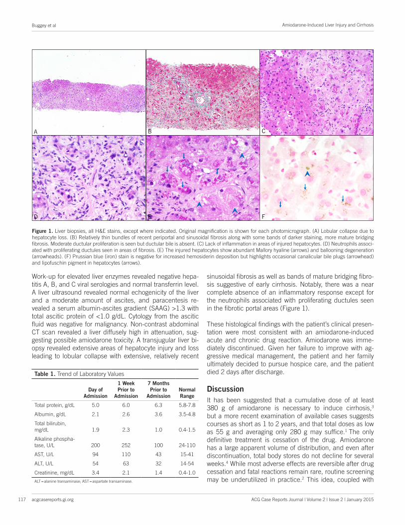
Work-up for elevated liver enzymes revealed negative hepa-titis A, B, and C viral serologies and normal transferrin level. A liver ultrasound revealed normal echogenicity of the liver and a moderate amount of ascites, and paracentesis re-vealed a serum albumin-ascites gradient (SAAG) >1.3 with total ascitic protein of <1.0 g/dL. Cytology from the ascitic fluid was negative for malignancy. Non-contrast abdominal CT scan revealed a liver diffusely high in attenuation, sug-gesting possible amiodarone toxicity. A transjugular liver bi-opsy revealed extensive areas of hepatocyte injury and loss leading to lobular collapse with extensive, relatively recent
sinusoidal fibrosis as well as bands of mature bridging fibro-sis suggestive of early cirrhosis. Notably, there was a near complete absence of an inflammatory response except for the neutrophils associated with proliferating ductules seen in the fibrotic portal areas (Figure 1).
These histological findings with the patient’s clinical presen-tation were most consistent with an amiodarone-induced acute and chronic drug reaction. Amiodarone was imme-diately discontinued. Given her failure to improve with ag-gressive medical management, the patient and her family ultimately decided to pursue hospice care, and the patient died 2 days after discharge.
DiscussionIt has been suggested that a cumulative dose of at least 380 g of amiodarone is necessary to induce cirrhosis,3 but a more recent examination of available cases suggests courses as short as 1 to 2 years, and that total doses as low as 55 g and averaging only 280 g may suffice.1 The only definitive treatment is cessation of the drug. Amiodarone has a large apparent volume of distribution, and even after discontinuation, total body stores do not decline for several weeks.4 While most adverse effects are reversible after drug cessation and fatal reactions remain rare, routine screening may be underutilized in practice.2 This idea, coupled with
Figure 1. Liver biopsies, all H&E stains, except where indicated. Original magnification is shown for each photomicrograph. (A) Lobular collapse due to hepatocyte loss. (B) Relatively thin bundles of recent periportal and sinusoidal fibrosis along with some bands of darker staining, more mature bridging fibrosis. Moderate ductular proliferation is seen but ductular bile is absent. (C) Lack of inflammation in areas of injured hepatocytes. (D) Neutrophils associ-ated with proliferating ductules seen in areas of fibrosis. (E) The injured hepatocytes show abundant Mallory hyaline (arrows) and ballooning degeneration (arrowheads). (F) Prussian blue (iron) stain is negative for increased hemosiderin deposition but highlights occasional canalicular bile plugs (arrowhead) and lipofuschin pigment in hepatocytes (arrows).
A B C
D E F
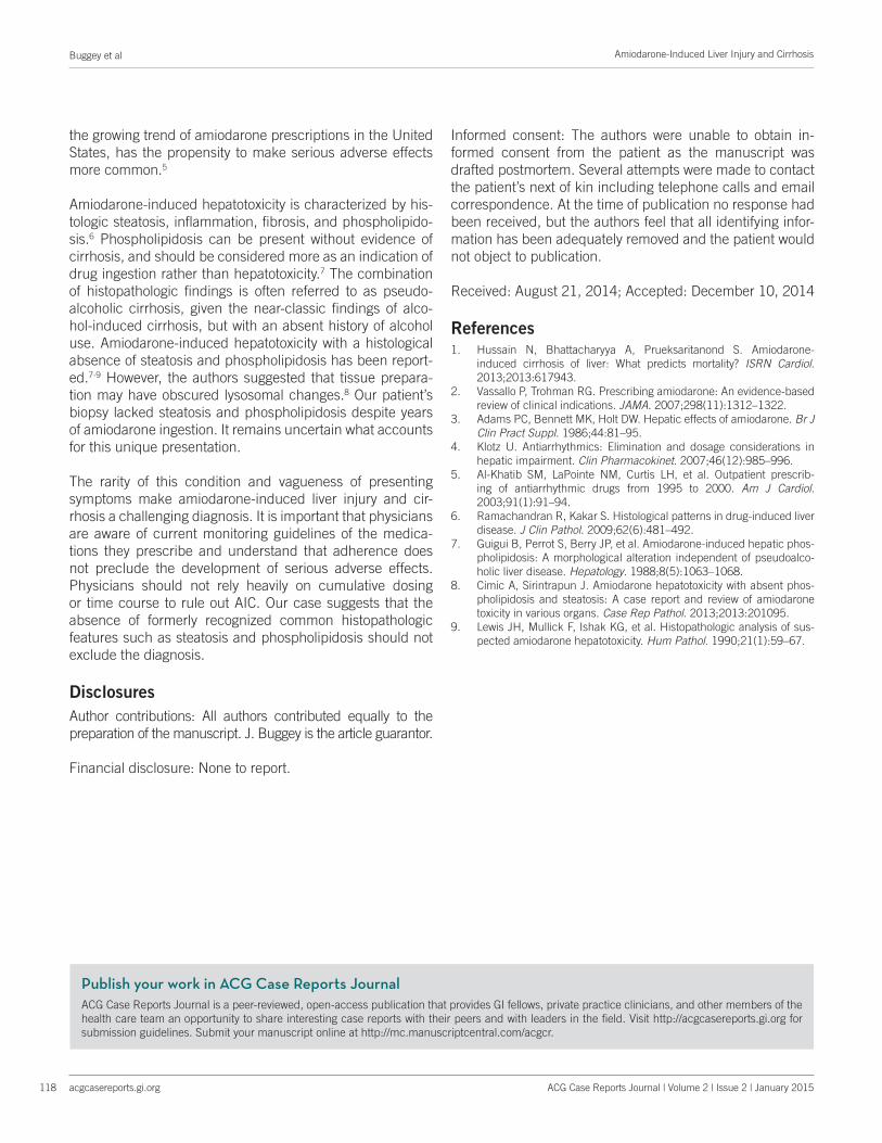
Table 1. Trend of Laboratory Values
Day of Admission
1 Week Prior to
Admission
7 Months Prior to
AdmissionNormal Range
Total protein, g/dL 5.0 6.0 6.3 5.8-7.8
Albumin, g/dL 2.1 2.6 3.6 3.5-4.8
Total bilirubin, mg/dL 1.9 2.3 1.0 0.4-1.5
Alkaline phospha-tase, U/L 200 252 100 24-110
AST, U/L 94 110 43 15-41
ALT, U/L 54 63 32 14-54
Creatinine, mg/dL 3.4 2.1 1.4 0.4-1.0
ALT = alanine transaminase; AST = aspartate transaminase.
Publish your work in ACG Case Reports JournalACG Case Reports Journal is a peer-reviewed, open-access publication that provides GI fellows, private practice clinicians, and other members of the health care team an opportunity to share interesting case reports with their peers and with leaders in the field. Visit http://acgcasereports.gi.org for submission guidelines. Submit your manuscript online at http://mc.manuscriptcentral.com/acgcr.
Buggey et al
acgcasereports.gi.org
Amiodarone-Induced Liver Injury and Cirrhosis
118 ACG Case Reports Journal | Volume 2 | Issue 2 | January 2015
the growing trend of amiodarone prescriptions in the United States, has the propensity to make serious adverse effects more common.5
Amiodarone-induced hepatotoxicity is characterized by his-tologic steatosis, inflammation, fibrosis, and phospholipido-sis.6 Phospholipidosis can be present without evidence of cirrhosis, and should be considered more as an indication of drug ingestion rather than hepatotoxicity.7 The combination of histopathologic findings is often referred to as pseudo-alcoholic cirrhosis, given the near-classic findings of alco-hol-induced cirrhosis, but with an absent history of alcohol use. Amiodarone-induced hepatotoxicity with a histological absence of steatosis and phospholipidosis has been report-ed.7-9 However, the authors suggested that tissue prepara-tion may have obscured lysosomal changes.8 Our patient’s biopsy lacked steatosis and phospholipidosis despite years of amiodarone ingestion. It remains uncertain what accounts for this unique presentation.
The rarity of this condition and vagueness of presenting symptoms make amiodarone-induced liver injury and cir-rhosis a challenging diagnosis. It is important that physicians are aware of current monitoring guidelines of the medica-tions they prescribe and understand that adherence does not preclude the development of serious adverse effects. Physicians should not rely heavily on cumulative dosing or time course to rule out AIC. Our case suggests that the absence of formerly recognized common histopathologic features such as steatosis and phospholipidosis should not exclude the diagnosis.
Disclosures
Author contributions: All authors contributed equally to the preparation of the manuscript. J. Buggey is the article guarantor.
Financial disclosure: None to report.
Informed consent: The authors were unable to obtain in-formed consent from the patient as the manuscript was drafted postmortem. Several attempts were made to contact the patient’s next of kin including telephone calls and email correspondence. At the time of publication no response had been received, but the authors feel that all identifying infor-mation has been adequately removed and the patient would not object to publication.
Received: August 21, 2014; Accepted: December 10, 2014 References1. Hussain N, Bhattacharyya A, Prueksaritanond S. Amiodarone-
induced cirrhosis of liver: What predicts mortality? ISRN Cardiol. 2013;2013:617943.
2. Vassallo P, Trohman RG. Prescribing amiodarone: An evidence-based review of clinical indications. JAMA. 2007;298(11):1312–1322.
3. Adams PC, Bennett MK, Holt DW. Hepatic effects of amiodarone. Br J Clin Pract Suppl. 1986;44:81–95.
4. Klotz U. Antiarrhythmics: Elimination and dosage considerations in hepatic impairment. Clin Pharmacokinet. 2007;46(12):985–996.
5. Al-Khatib SM, LaPointe NM, Curtis LH, et al. Outpatient prescrib-ing of antiarrhythmic drugs from 1995 to 2000. Am J Cardiol. 2003;91(1):91–94.
6. Ramachandran R, Kakar S. Histological patterns in drug-induced liver disease. J Clin Pathol. 2009;62(6):481–492.
7. Guigui B, Perrot S, Berry JP, et al. Amiodarone-induced hepatic phos-pholipidosis: A morphological alteration independent of pseudoalco-holic liver disease. Hepatology. 1988;8(5):1063–1068.
8. Cimic A, Sirintrapun J. Amiodarone hepatotoxicity with absent phos-pholipidosis and steatosis: A case report and review of amiodarone toxicity in various organs. Case Rep Pathol. 2013;2013:201095.
9. Lewis JH, Mullick F, Ishak KG, et al. Histopathologic analysis of sus-pected amiodarone hepatotoxicity. Hum Pathol. 1990;21(1):59–67.