Embed Size (px)
Citation preview
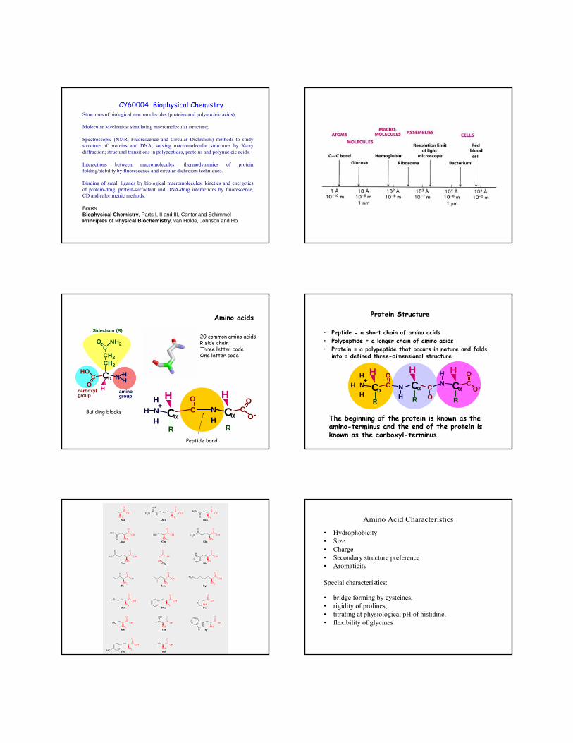
Structures of biological macromolecules (proteins and polynucleic acids);
Molecular Mechanics: simulating macromolecular structure;
Spectroscopic (NMR, Fluorescence and Circular Dichroism) methods to study structure of proteins and DNA; solving macromolecular structures by X-ray diffraction; structural transitions in polypeptides, proteins and polynucleic acids.
Interactions between macromolecules: thermodynamics of protein folding/stability by fluorescence and circular dichroism techniques.
Binding of small ligands by biological macromolecules: kinetics and energeticsof protein-drug, protein-surfactant and DNA-drug interactions by fluorescence, CD and calorimetric methods.
Books : Biophysical Chemistry, Parts I, II and III, Cantor and SchimmelPrinciples of Physical Biochemistry, van Holde, Johnson and Ho
CY60004 Biophysical Chemistry
CαCHO
ON H
H
H
CH2
CH2
CO NH2
carboxylgroup
aminogroup
Sidechain (R)
CαC
ON
RH
CαCO
NH
HR
+H H
O-H
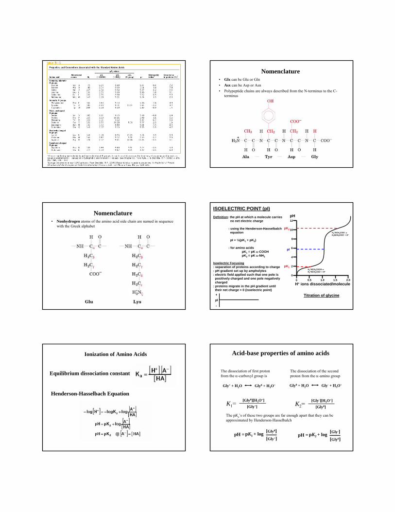
20 common amino acids R side chainThree letter codeOne letter code
Amino acids
Building blocks
Peptide bond
HCα
CO
NH
HR
H +HCα C
ON
RO-
H
NH H
CαCO
R
The beginning of the protein is known as the amino-terminus and the end of the protein is known as the carboxyl-terminus.
• Peptide = a short chain of amino acids• Polypeptide = a longer chain of amino acids• Protein = a polypeptide that occurs in nature and folds
into a defined three-dimensional structure
Protein Structure
Amino Acid Characteristics• Hydrophobicity• Size• Charge• Secondary structure preference• Aromaticity
Special characteristics:
• bridge forming by cysteines, • rigidity of prolines, • titrating at physiological pH of histidine, • flexibility of glycines
Nomenclature • Glx can be Glu or Gln• Asx can be Asp or Asn• Polypeptide chains are always described from the N-terminus to the C-
terminus
Nomenclature • Nonhydrogen atoms of the amino acid side chain are named in sequence
with the Greek alphabet
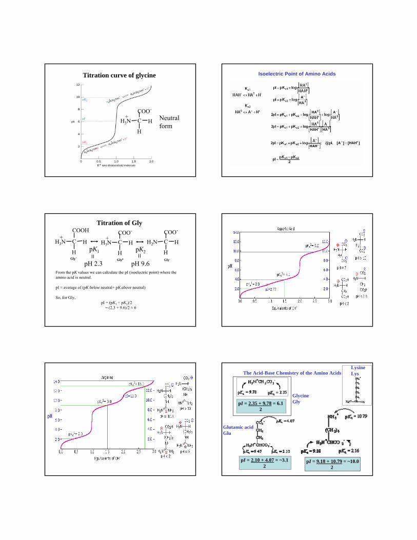
ISOELECTRIC POINT (pI)Definition: the pH at which a molecule carries
no net electric charge
: using the Henderson-Hasselbalchequation
pI = ½(pK1 + pK2)
: for amino acidspK1 = pK α-COOHpK2 = pK α-NH3
Isoelectric Focusing: separation of proteins according to charge: pH gradient set up by ampholytes: electric field applied such that one pole is
positively charged and one pole negativelycharged
: proteins migrate in the pH gradient untiltheir net charge = 0 (isoelectric point)
pK1
pK2
pI
pH
0
2
4
6
8
10
12
0 0.5 1.0 1.5 2.0H+ ions dissociated/molecule
Titration of glycine
H3+NCH2COOH ⇌
H3+NCH2COO- + H+
H3+NCH2COO- ⇌
H2NCH2COO- + H+
pI+
-
Ionization of Amino Acids
Henderson-Hasselbach Equation
Equilibrium dissociation constant
Acid-base properties of amino acids
K1=
Gly+ + H2O Gly0 + H3O+
[Gly0][H3O+][Gly+]
Gly0 + H2O Gly- + H3O+
K2=[Gly-][H3O+]
[Gly0]
The dissociation of first proton from the α-carboxyl group is
The dissociation of the second proton from the α-amino group
The pKa’s of these two groups are far enough apart that they can be approximated by Henderson-Hasselbalch
pK1 + logpH =[Gly0][Gly+]
pK2 + logpH =[Gly-][Gly0]
Titration curve of glycine
H
C H
COO-
H3N+ Neutral
form
Isoelectric Point of Amino Acids
Titration of Gly
H
C H
COO-
H3N+
H
C H
COO-
H2N
H
C H
COOH
H3N+
pK1 pK2
Gly0Gly+Gly-
pH 2.3 pH 9.6From the pK values we can calculate the pI (isoelectric point) where the amino acid is neutral.
pI ≈ average of (pK below neutral+ pK above neutral)
So, for Gly,pI = (pK1 + pK2)/2
= (2.3 + 9.6)/2 ≈ 6
The Acid-Base Chemistry of the Amino Acids
pI = 2.35 + 9.78 = 6.1 2
pI = 9.18 + 10.79 = ~10.02
LysineLys
pI = 2.10 + 4.07 = ~3.1 2
GlycineGly
Glutamic acidGlu
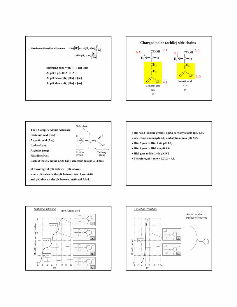
Buffering zone = pK +/- 1 pH unit
At pH = pK, [HA] = [A-]
At pH below pK, [HA] > [A-]
At pH above pK, [HA] < [A-]
Henderson-Hasselbach Equation
C H
COOH
H3N+
Aspartic acid
Asp
D
C
CH2
O OH
C H
COOH
H3N+
CH2
Glutamic acid
Glu
E
C
CH2
O OH
Charged polar (acidic) side chains
2.1
4.1
9.5 2.0
3.9
9.8
The 5 Complex Amino Acids are:
Glutamic acid (Glu)
Aspartic acid (Asp)
Lysine (Lys)
Arginine (Arg)
Histidine (His).
Each of these 5 amino acids has 3 ionizable groups ⇒ 3 pKs.
pI = average of (pK-below) + (pK-above)
where pK-below is the pK between AA+1 and AA0
and pK-above is the pK between AA0 and AA-1.
C
RR
Cα
H
N
O
OHH
H
Aminogroup
Carboxylgroup
Side chain
H +
• His has 3 ionizing groups, alpha-carboxylic acid (pK 1.8),
• side-chain amino (pK 6.0) and alpha-amino (pK 9.2):
• His+2 goes to His+1 via pK 1.8;
• His+1 goes to His0 via pK 6.0;
• His0 goes to His-1 via pK 9.2.
• Therefore, pI = (6.0 + 9.2)/2 = 7.6.
Histidine Titration
3
2
1
00 2 4 6 8 10 12 14
pKa=1.8
pKa=6.0
pKa=9.3
pH
Mol
sO
H-ad
ded
per m
ol h
istid
ine -1
0
+1
+2
N
N
CH 2C
COO -
NH 2
H
H
N
N
CH 2C
COO -
NH 3+
H
H
N +
N
CH 2C
COO -
NH 3+
H
H
H
N +
N
CH 2C
COOH
NH 3+
H
H
H
Free Amino acid
00 2 4 6 8 10 12 14
pKa=6.0
pH
Mol
sO
H-ad
ded 0
+1
Histidine Titration
Amino acid on surface of enzyme
N
N
CH2
C
CO
NH
H
H
N+
N
CH2
C
CO
NH
H
H
H
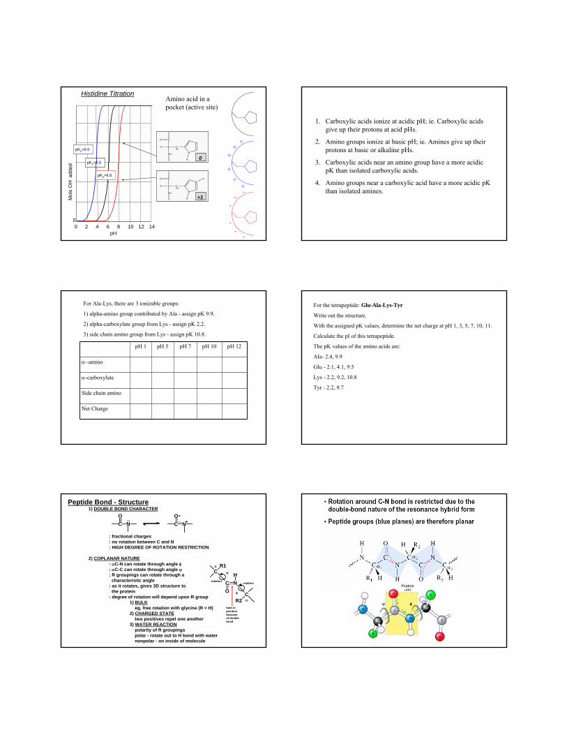
Histidine TitrationAmino acid in a pocket (active site)
+
++
+
+
+
-
-
-
-
-
-
00 2 4 6 8 10 12 14
pKa=6.0
pH
Mol
sO
H-ad
ded
0
+1
N
N
CH2
C
CO
NH
H
H
N+
N
CH2
C
CO
NH
H
H
HpKa>6.0
pKa<6.0
1. Carboxylic acids ionize at acidic pH; ie. Carboxylic acids give up their protons at acid pHs.
2. Amino groups ionize at basic pH; ie. Amines give up their protons at basic or alkaline pHs.
3. Carboxylic acids near an amino group have a more acidic pK than isolated carboxylic acids.
4. Amino groups near a carboxylic acid have a more acidic pKthan isolated amines.
For Ala-Lys, there are 3 ionizable groups:
1) alpha-amino group contributed by Ala - assign pK 9.9.
2) alpha-carboxylate group from Lys - assign pK 2.2.
3) side chain amino group from Lys - assign pK 10.8.
pH 1 pH 5 pH 7 pH 10 pH 12
α−amino
α-carboxylate
Side chain amino
Net Charge
For the tetrapeptide: Glu-Ala-Lys-Tyr
Write out the structure.
With the assigned pK values, determine the net charge at pH 1, 3, 5, 7, 10, 11.
Calculate the pI of this tetrapeptide.
The pK values of the amino acids are:
Ala- 2.4, 9.9
Glu - 2.1, 4.1, 9.5
Lys - 2.2, 9.2, 10.8
Tyr - 2.2, 9.7
Peptide Bond - Structure1) DOUBLE BOND CHARACTER
: fractional charges: no rotation between C and N: HIGH DEGREE OF ROTATION RESTRICTION
2) COPLANAR NATURE: αC-N can rotate through angle φ: αC-C can rotate through angle ψ: R groupings can rotate through a
characteristic angle: as it rotates, gives 3D structure to
the protein: degree of rotation will depend upon R group
1) BULKeg. free rotation with glycine (R = H)
2) CHARGED STATEtwo positives repel one another
3) WATER REACTIONpolarity of R groupingspolar - rotate out to H bond with waternonpolar - on inside of molecule
C NO
C NO..
-+
C NO
H
CR2
CR1α
α
ψ
φ
held inpositionbecauseof doublebond
rotationrotation
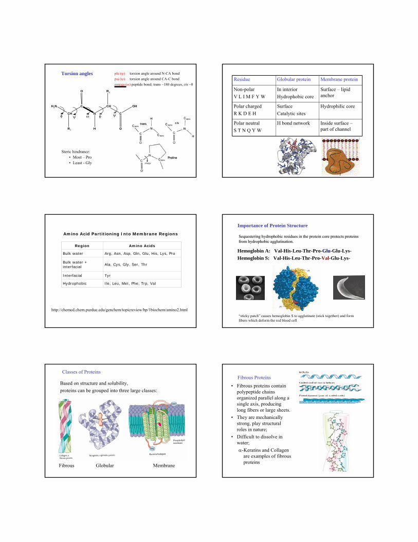
Torsion angles phi (ϕ) torsion angle around N-CA bondpsi (ψ) torsion angle around CA-C bond omega (ω) peptide bond; trans ~180 degrees, cis ~0
Steric hindrance:• Most – Pro• Least - Gly
NH2
CH
C
O
R1
N
CH
C
O
OH
R2
H
φ ωψ φ ψ
Residue Globular protein Membrane protein
Non-polarV L I M F Y W
In interiorHydrophobic core
Surface – lipid anchor
Polar chargedR K D E H
SurfaceCatalytic sites
Hydrophilic core
Polar neutralS T N Q Y W
H bond network Inside surface –part of channel
Amino Acid Partitioning Into Membrane Regions
Region Amino Acids
Bulk water Arg, Asn, Asp, Gln, Glu, His, Lys, Pro
Bulk water + interfacial
Ala, Cys, Gly, Ser, Thr
Interfacial Tyr
Hydrophobic Ile, Leu, Met, Phe, Trp, Val
http://chemed.chem.purdue.edu/genchem/topicreview/bp/1biochem/amino2.html
Sequestering hydrophobic residues in the protein core protects pSequestering hydrophobic residues in the protein core protects proteins roteins from hydrophobic agglutination.from hydrophobic agglutination.
Hemoglobin A: Val-His-Leu-Thr-Pro-Glu-Glu-Lys-Hemoglobin S: Val-His-Leu-Thr-Pro-Val-Glu-Lys-
“sticky patch” causes hemoglobin S to agglutinate (stick together) and form fibers which deform the red blood cell
Importance of Protein Structure
Fibrous Globular Membrane
Classes of Proteins
Based on structure and solubility, proteins can be grouped into three large classes:
Fibrous Proteins• Fibrous proteins contain
polypeptide chains organized parallel along a single axis, producing long fibers or large sheets.
• They are mechanically strong, play structural roles in nature;
• Difficult to dissolve in water;α-Keratins and Collagen
are examples of fibrous proteins
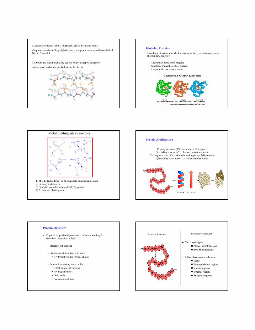
α-keratins are found in hair, fingernails, claws, horns and beaks;
•Sequence consists of long alpha helical rod segments capped with non-helical N- and C-termini
β-keratins are found in silk and consist of gly-ala repeat sequences;
•Ala is small and can be packed within the sheets
Globular Proteins• Globular proteins are classified according to the type and arrangement
of secondary structure
– Antiparallel alpha helix proteins – Parallel or mixed beta sheet proteins – Antiparallel beta sheet proteins
Metal binding sites examples
1) M is Fe (rubredoxin) or Zn (aspartate transcarbamoylase) 2) Carboxypeptidase A 3) Catalytic ion in liver alcohol dehydrogenase4) Azurin and plastocyanin
Primary structure (1°) : the amino acid sequence. Secondary structure (2°) : helices, sheets and turns.
Tertiary structure (3°) : side chain packing in the 3-D structure. Quaternary structure (4°) : association of subunits.
Protein Architecture
• Physical properties of protein that influence stability & therefore, determine its fold:
– Rigidity of backbone
– Amino acid interaction with water• Hydropathy index for side chains
– Interactions among amino acids• Electrostatic interactions• Hydrogen bonds• S-S bonds• Volume constraints
Protein Structure
Primary Structure
Two major types:Alpha Helical RegionsBeta Sheet Regions
• Other classification schemes:TurnsTransmembrane regionsInternal regionsExternal regionsAntigenic regions
Secondary Structure
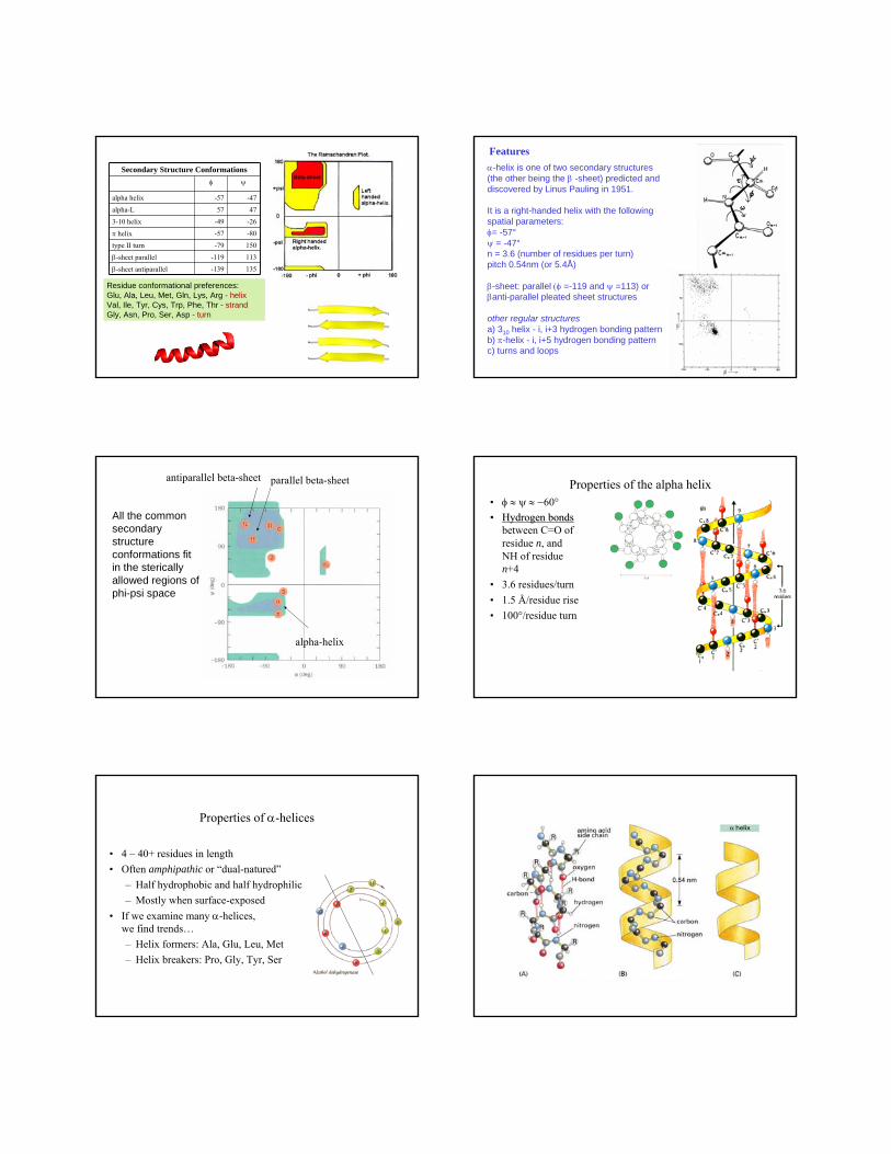
Residue conformational preferences:Glu, Ala, Leu, Met, Gln, Lys, Arg - helixVal, Ile, Tyr, Cys, Trp, Phe, Thr - strandGly, Asn, Pro, Ser, Asp - turn
Secondary Structure Conformationsφ ψ
alpha helix -57 -47
alpha-L 57 47
3-10 helix -49 -26
π helix -57 -80
type II turn -79 150
β-sheet parallel -119 113
β-sheet antiparallel -139 135
α-helix is one of two secondary structures(the other being the β -sheet) predicted and discovered by Linus Pauling in 1951.
It is a right-handed helix with the following spatial parameters: φ= -57°ψ = -47°n = 3.6 (number of residues per turn) pitch 0.54nm (or 5.4Å)
β-sheet: parallel (φ =-119 and ψ =113) or βanti-parallel pleated sheet structures
other regular structuresa) 310 helix - i, i+3 hydrogen bonding patternb) π-helix - i, i+5 hydrogen bonding patternc) turns and loops
Features
All the commonsecondary structureconformations fit in the stericallyallowed regions of phi-psi space
parallel beta-sheetantiparallel beta-sheet
alpha-helix
Properties of the alpha helix• φ ≈ ψ ≈ −60°•• Hydrogen bondsHydrogen bonds
between C=O ofresidue n, andNH of residuen+4
• 3.6 residues/turn• 1.5 Å/residue rise• 100°/residue turn
Properties of α-helices
• 4 – 40+ residues in length• Often amphipathic or “dual-natured”
– Half hydrophobic and half hydrophilic– Mostly when surface-exposed
• If we examine many α-helices,we find trends…– Helix formers: Ala, Glu, Leu, Met– Helix breakers: Pro, Gly, Tyr, Ser
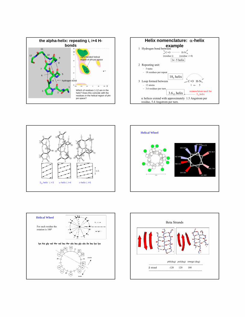
the alpha-helix: repeating i, i+4 H-bonds
2
1
3
4
5
7
8
9
6
10
11
12
Which of residues 1-12 are in the helix? Does this coincide with the residues in the helical region of phi-psi space?
right-handed helical region of phi-psi space
hydrogen bond
Helix nomenclature: α-helix example
1 Hydrogen bond betweenC=O H-N
(residue i) (residue i+4)
1←5 helix2 Repeating unit:
– 5 turns – 18 residues per repeat
185 helix3 Loop formed between C=O H-N
– 13 atoms 1 ← 5– 3.6 residues per turn
3.613 helixα helices extend with approximately 1.5 Angstrom per residue, 5.4 Angstrom per turn.
nomenclature used for 310 helix
310 helix i, i+3 π-helix i, i+5α-helix i, i+4
Helical Wheel
lys his gly val thr val leu thr ala leu gly ala ile leu lys lys
Helical Wheel
For each residue therotation is 1000
Beta Strands
phi(deg) psi(deg) omega (deg)------------------------------------------------------------------β strand -120 120 180 -----------------------------------------------------------------
beta strands/sheets
parallel or anti-parallel sheet?
49
50
51
52
53
54
57
56
beta-strand region of phi-psi space
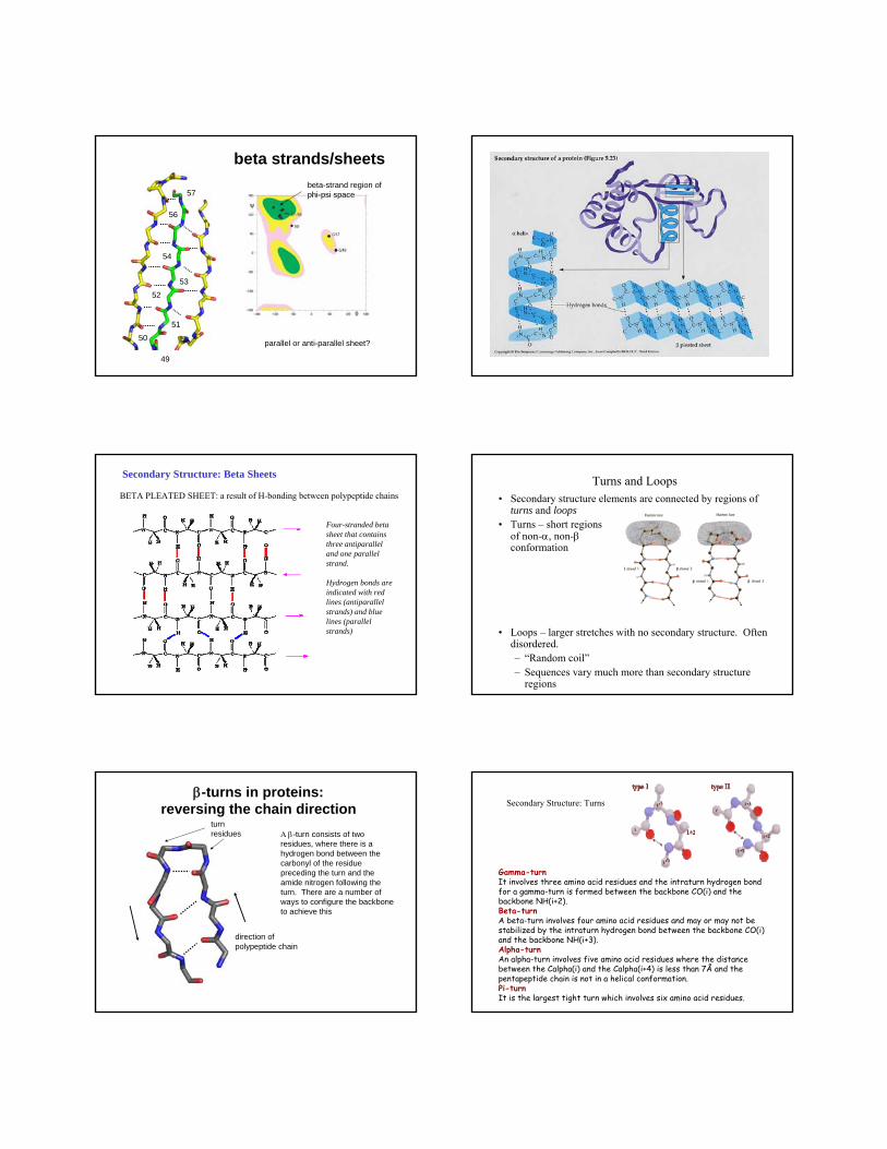
Secondary Structure: Beta Sheets
BETA PLEATED SHEET: a result of H-bonding between polypeptide chains
Four-stranded beta sheet that contains three antiparalleland one parallel strand.
Hydrogen bonds are indicated with red lines (antiparallelstrands) and blue lines (parallel strands)
Turns and Loops• Secondary structure elements are connected by regions of
turns and loops• Turns – short regions
of non-α, non-βconformation
• Loops – larger stretches with no secondary structure. Often disordered.– “Random coil”– Sequences vary much more than secondary structure
regions
β-turns in proteins:reversing the chain direction
Α β-turn consists of two residues, where there is a hydrogen bond between the carbonyl of the residue preceding the turn and the amide nitrogen following the turn. There are a number of ways to configure the backbone to achieve this
turn residues
direction ofpolypeptide chain
Secondary Structure: Turns
Gamma-turnIt involves three amino acid residues and the intraturn hydrogen bond for a gamma-turn is formed between the backbone CO(i) and the backbone NH(i+2). Beta-turnA beta-turn involves four amino acid residues and may or may not be stabilized by the intraturn hydrogen bond between the backbone CO(i) and the backbone NH(i+3). Alpha-turnAn alpha-turn involves five amino acid residues where the distance between the Calpha(i) and the Calpha(i+4) is less than 7Å and the pentapeptide chain is not in a helical conformation. Pi-turnIt is the largest tight turn which involves six amino acid residues.
Tertiary Structure: Aggregation of individual protein.
1. Hydrophobic attraction: the close association attraction of hydrocarbon side-chains.
2. Ionic bond: between positively charged groups and negatively charged groups.
3. Hydrogen bonds
4. Disulfide bonds
A protein has size and shape as well as unique arrangement through hydrogen, ionic, hydrophobic and disulfide bonds.
Tertiary Structure of Protein
Side chainconformation
• side chains differ in their number of degrees of conformational freedom
•but side chains of very different size can have the same number of χangles.
Side chain conformations--canonical staggered forms
t=trans, g=gauchename of conformation
IUPAC nomenclature:http://www.chem.qmw.ac.uk/iupac/misc/biop.html
glutamate
Side chain angles are defined moving outward from the backbone, startingwith the N atom: so the χ1 angle is N–Cα–Cβ–Cγ, the χ2 angle is Cα–Cβ–Cγ –Cδ ...
Hβ
Hβ CγCO
HαNHβ
Cγ HβCO
HαNCγ
Hβ HβCO
HαN
NHCHC
CH2
O
CH2
C
O
O
χ1
χ2
χ3
β
γ
α
δ
χ1 = 180° χ1 = +60° χ1 = -60°
t g+ g–
Newman projections for χ1 of glutamate:
Rotamers• a particular combination of side chain torsional angles χ1, χ2, etc. for a
particular residue is known as a rotamer. • for example, for aspartate, if one considers only the canonical staggered
forms, there are nine (32) possible rotamers: g+g-, g+g+, g-g-, g-g+, tg+, g+t, tg-, g-t, tt
• not all rotamers are equally likely. • for example, valine prefers
its t rotamerdistribution ofvaline rotamersin protein structures
180χ1=0 360
Cγ2
Hβ Cγ1
CO
HαN
χ1=180°, trans or t
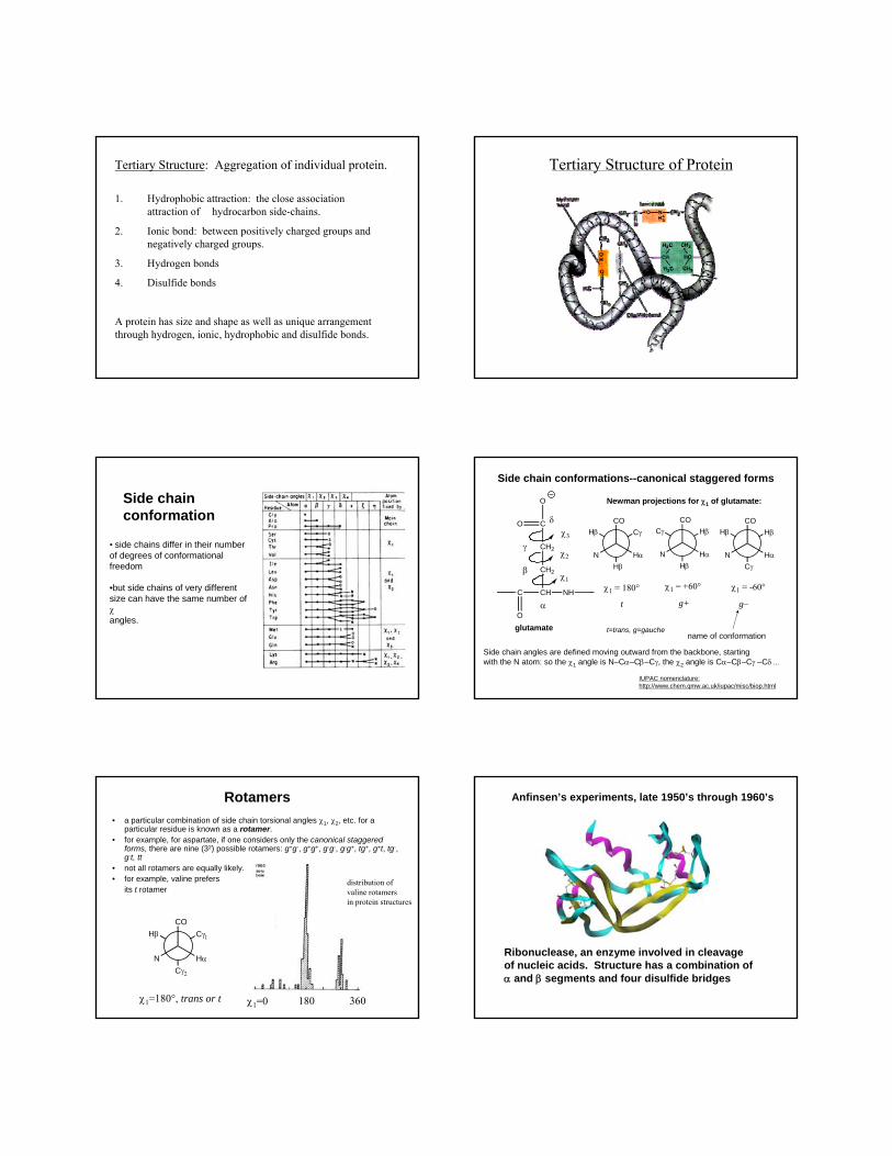
Anfinsen’s experiments, late 1950’s through 1960’s
Ribonuclease, an enzyme involved in cleavageof nucleic acids. Structure has a combination ofα and β segments and four disulfide bridges
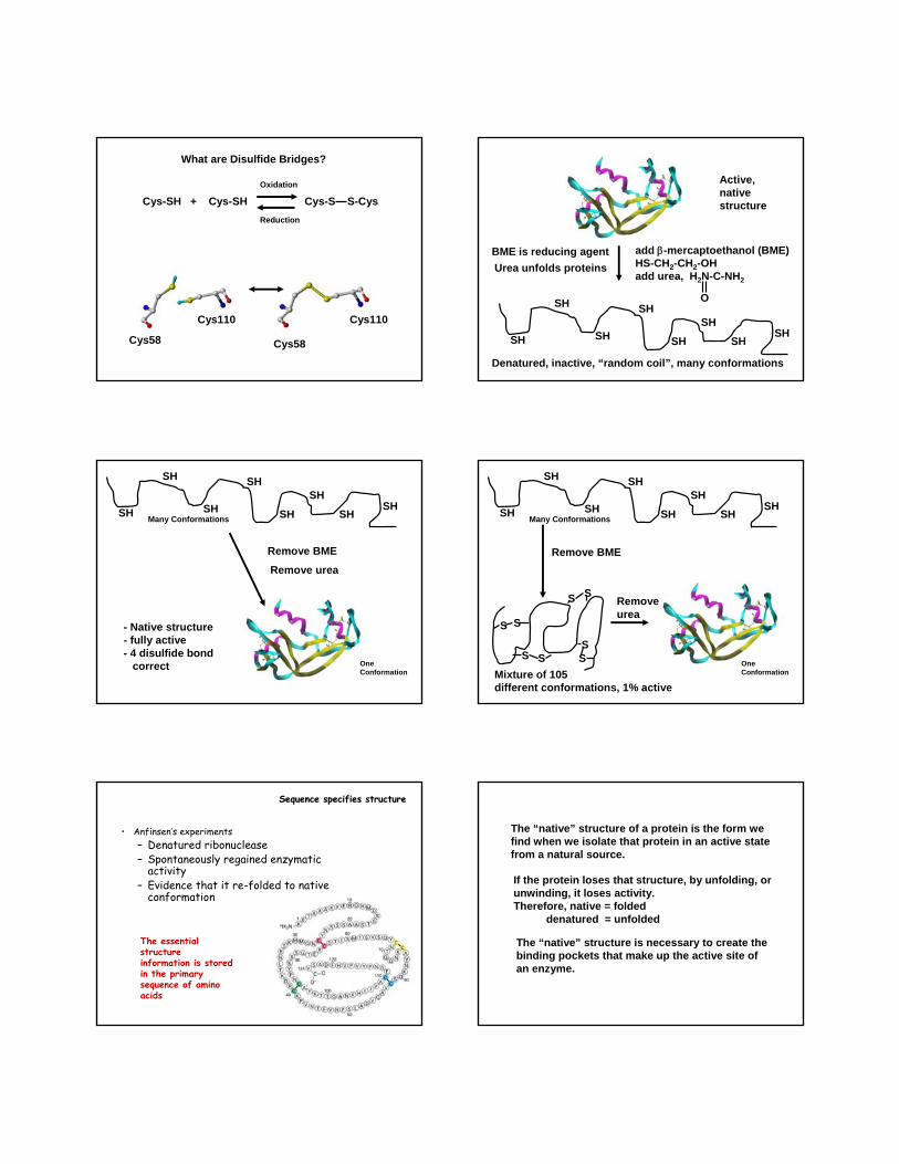
What are Disulfide Bridges?
Cys-SH + Cys-SH Cys-S S-CysOxidation
Reduction
Cys58 Cys58
Cys110 Cys110
add β-mercaptoethanol (BME)HS-CH2-CH2-OHadd urea, H2N-C-NH2
O
SH
SH
SH
SH
SH
SH
SHSH
Active, nativestructure
Denatured, inactive, “random coil”, many conformations
BME is reducing agentUrea unfolds proteins
SH
SH
SH
SH
SH
SH
SHSH
Remove BME
Remove urea
- Native structure- fully active- 4 disulfide bond
correct
Many Conformations
OneConformation
SH
SH
SH
SH
SH
SH
SHSH
Remove BME
S S
S S
SS
SS
Mixture of 105different conformations, 1% active
Removeurea
Many Conformations
OneConformation
• Anfinsen’s experiments– Denatured ribonuclease– Spontaneously regained enzymatic
activity– Evidence that it re-folded to native
conformation
Sequence specifies structureSequence specifies structure
The essential structureinformation is stored in the primary sequence of amino acids
The “native” structure of a protein is the form wefind when we isolate that protein in an active statefrom a natural source.
If the protein loses that structure, by unfolding, orunwinding, it loses activity.Therefore, native = folded
denatured = unfolded
The “native” structure is necessary to create thebinding pockets that make up the active site ofan enzyme.
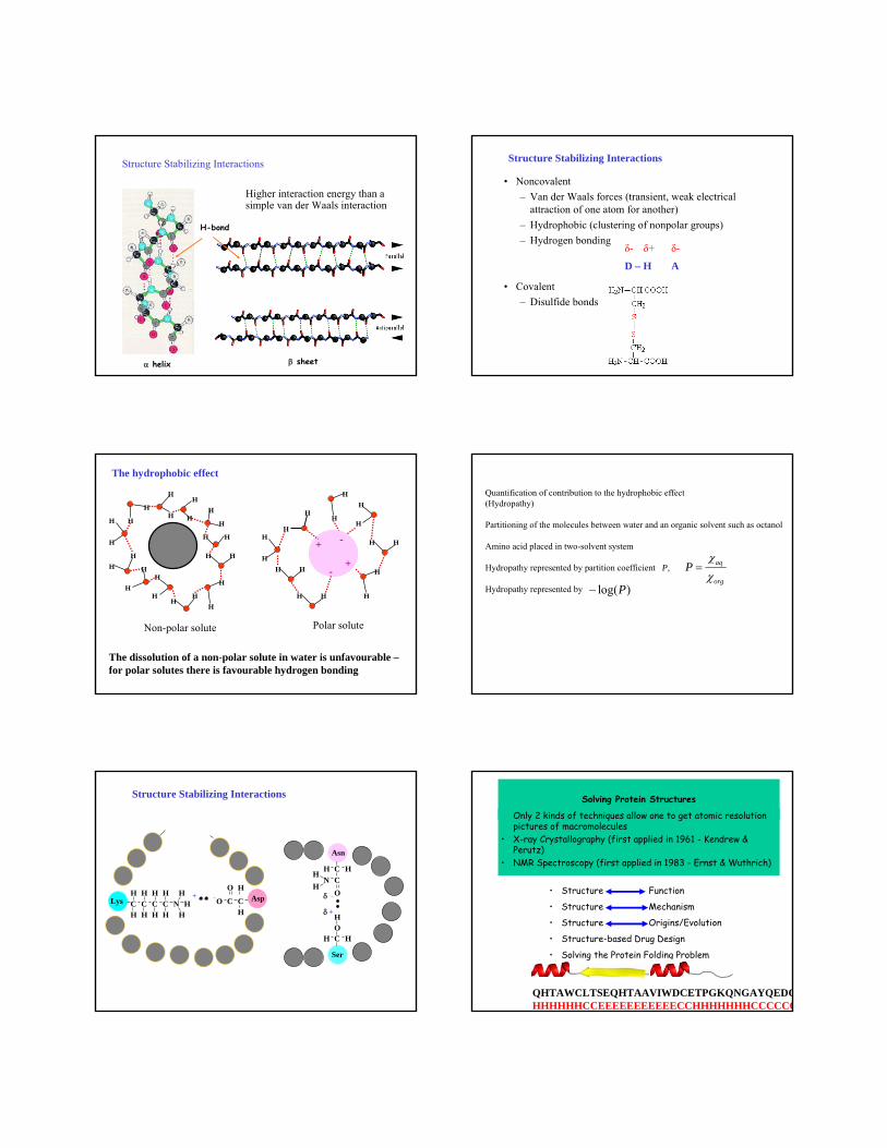
H-bond
α helix β sheet
Higher interaction energy than a simple van der Waals interaction
Structure Stabilizing Interactions Structure Stabilizing Interactions
• Noncovalent– Van der Waals forces (transient, weak electrical
attraction of one atom for another)– Hydrophobic (clustering of nonpolar groups)– Hydrogen bonding
• Covalent– Disulfide bonds
D – H A
δ- δ+ δ-
HH
H
HH
H
H
H
H
HH
H
H
H
H
H
H
HH
H
H
H
H
H
HH
+
+
-
-
HH
H
H
H
H
H
H
HH
HHH
H
Non-polar solute Polar solute
The hydrophobic effect
The dissolution of a non-polar solute in water is unfavourable –for polar solutes there is favourable hydrogen bonding
Quantification of contribution to the hydrophobic effect(Hydropathy)
Partitioning of the molecules between water and an organic solvent such as octanol
Amino acid placed in two-solvent system
Hydropathy represented by partition coefficient P,
Hydropathy represented by
aq
org
Pχχ
=
log( )P−
C C C C NH
HH
+H H HH
HHH H
C
HO
COH
AspLys
H+
H H
δ
CO
H CCN
H
OH
H
δ
Asn
Ser
Structure Stabilizing Interactions
• Structure Function
• Structure Mechanism
• Structure Origins/Evolution
• Structure-based Drug Design
• Solving the Protein Folding Problem
Solving Protein Structures
Only 2 kinds of techniques allow one to get atomic resolution pictures of macromolecules
• X-ray Crystallography (first applied in 1961 - Kendrew & Perutz)
• NMR Spectroscopy (first applied in 1983 - Ernst & Wuthrich)
QHTAWCLTSEQHTAAVIWDCETPGKQNGAYQEDCHHHHHHCCEEEEEEEEEEECCHHHHHHHCCCCCC
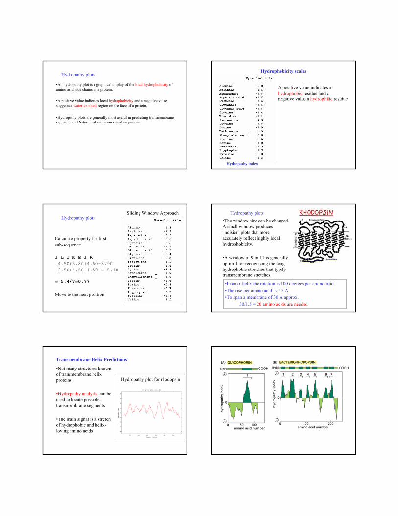
Hydropathy plots
•An hydropathy plot is a graphical display of the local hydrophobicity of amino acid side chains in a protein.
•A positive value indicates local hydrophobicity and a negative value suggests a water-exposed region on the face of a protein.
•Hydropathy plots are generally most useful in predicting transmembranesegments and N-terminal secretion signal sequences.
Hydrophobicity scales
Hydropathy index
A positive value indicates a hydrophobic residue and a negative value a hydrophilic residue
Sliding Window Approach
Calculate property for first sub-sequence
I L I K E I R4.50+3.80+4.50-3.90
-3.50+4.50-4.50 = 5.40
= 5.4/7=0.77
Move to the next position
Hydropathy plots •The window size can be changed. A small window produces "noisier" plots that more accurately reflect highly local hydrophobicity.
•A window of 9 or 11 is generally optimal for recognizing the long hydrophobic stretches that typify transmembrane stretches.
Hydropathy plots
•In an α-helix the rotation is 100 degrees per amino acid•The rise per amino acid is 1.5 Å•To span a membrane of 30 Å approx.
30/1.5 = 20 amino acids are needed
Transmembrane Helix Predictions
•Not many structures known of transmembrane helix proteins
•Hydropathy analysis can be used to locate possible transmembrane segments
•The main signal is a stretch of hydrophobic and helix-loving amino acids
Hydropathy plot for rhodopsin