Embed Size (px)
Citation preview
O R I G I N A L A R T I C L E
Amelogenesis imperfecta caused by N-terminal
enamelin point mutations in mice and men is driven
by endoplasmic reticulum stressSteven J. Brookes1,*,†, Martin J. Barron2,†, Claire E.L. Smith3, James A. Poulter3,Alan J. Mighell4, Chris F. Inglehearn3, Catriona J. Brown5, Helen Rodd6,Jennifer Kirkham1 and Michael J. Dixon2
1Department of Oral Biology, School of Dentistry, Wellcome Trust Brenner Building University Of Leeds, StJames’s University Hospital, Leeds LS9 7TF, UK, 2Faculty of Biology, Medicine & Health, Manchester AcademicHealth Sciences Centre, University of Manchester, Michael Smith Building, Manchester M13 9PT, UK,3Department of Oral Medicine, School of Dentistry, University of Leeds, Leeds, UK, 4Leeds Institute ofBiomedical and Clinical Sciences, St James’s University Hospital, University of Leeds, Leeds LS9 7TF, UK,5Birmingham Dental Hospital and School of Dentistry, Birmingham B5 7EG, UK and 6Unit of Oral Health andDevelopment, School of Clinical Dentistry, University of Sheffield, Sheffield, UK
*To whom correspondence should be addressed. Tel: 01133436159; Email: [email protected]
Abstract‘Amelogenesis imperfecta’ (AI) describes a group of inherited diseases of dental enamel that have major clinical impact. Here,we identify the aetiology driving AI in mice carrying a p.S55I mutation in enamelin; one of the most commonly mutatedproteins underlying AI in humans. Our data indicate that the mutation inhibits the ameloblast secretory pathway leading toER stress and an activated unfolded protein response (UPR). Initially, with the support of the UPR acting in pro-survival mode,Enamp.S55I heterozygous mice secreted structurally normal enamel. However, enamel secreted thereafter was structurally ab-normal; presumably due to the UPR modulating ameloblast behaviour and function in an attempt to relieve ER stress.Homozygous mutant mice failed to produce enamel. We also identified a novel heterozygous ENAMp.L31R mutation causing AIin humans. We hypothesize that ER stress is the aetiological factor in this case of human AI as it shared the characteristicphenotype described above for the Enamp.S55I mouse. We previously demonstrated that AI in mice carrying the Amelxp.Y64H
mutation is a proteinopathy. The current data indicate that AI in Enamp.S55I mice is also a proteinopathy, and based on com-parative phenotypic analysis, we suggest that human AI resulting from the ENAMp.L31R mutation is another proteinopathicdisease. Identifying a common aetiology for AI resulting from mutations in two different genes opens the way for developingpharmaceutical interventions designed to relieve ER stress or modulate the UPR during enamel development to amelioratethe clinical phenotype.
†These authors contributed equally to the research.Received: November 22, 2016. Revised: February 1, 2017. Accepted: March 2, 2017
VC The Author 2017. Published by Oxford University Press.This is an Open Access article distributed under the terms of the Creative Commons Attribution License (http://creativecommons.org/licenses/by/4.0/),which permits unrestricted reuse, distribution, and reproduction in any medium, provided the original work is properly cited.
1863
Human Molecular Genetics, 2017, Vol. 26, No. 10 1863–1876
doi: 10.1093/hmg/ddx090Advance Access Publication Date: 11 March 2017Original Article
IntroductionMature dental enamel, which is composed of approximately 96%mineral and 4% organic material and water compared to approxi-mately 70% mineral and 30% organic material and water found indentine and bone, represents the most extreme form of mamma-lian biomineralization. Enamel mineral is a substituted hydroxy-apatite (HA) and is the hardest and most resilient of the skeletaltissues (1). The wear resistance of dental enamel is a conse-quence of its highly ordered structure; the mineral crystallites areorganized into interlocking prismatic, or rod, structures inter-spersed with interprismatic crystallites (1,2). Amelogenesis, theformation of dental enamel, is presumed to be mediated by theinteractions of enamel extracellular matrix proteins coupled withthe regulated deposition of mineral (3). Amelogenesis occurs intwo broad stages and is mediated by a specialized epitheliumcomprised of ameloblast cells (3). In the early, secretory stage ofamelogenesis, the ameloblasts elaborate the enamel extracellularmatrix proteins composed principally of amelogenin (>90%),ameloblastin and enamelin (3). Post-secretory enzymatic process-ing of the matrix proteins leads to the self-directed organizationof the enamel extracellular matrix and delineates the three-dimensional structure of the nascent enamel (3). Once the fullthickness of the enamel has been deposited, the ameloblastsbegin to degrade the enamel extracellular organic matrix andsubsequently increase transport of mineral ions into the formingenamel (3). Removal of the proteinaceous matrix allows the initialHA crystallites to grow laterally until the full mineral content ofthe tissue is achieved (3).
Inherited defects of dental enamel biomineralization, amelo-genesis imperfecta (AI; MIM PS104500), constitute a commongroup of genetic diseases that occur with an incidence as highas 1:700 live births (4). Affected individuals frequently experi-ence severe problems with self-esteem due to the appearanceof their teeth as well as pain due to sensitivity and require sub-stantial and often complex dental care to manage the condition(5). The critical role played by the proteins of the extracellularmatrix in enamel biomineralization is highlighted by the dis-covery that many of the genes mutated in AI encode matrix pro-teins or the enzymes that modify them (6,7). For example,mutations in the amelogenin (AMELX) gene underlie X-linkedforms of AI (MIM #301200), while mutations in the gene encod-ing enamelin (ENAM) are associated with both autosomal dom-inant and autosomal recessive AI phenotypes (MIM #104500 and#204650) (6,7). The importance of enamelin for normal amelo-genesis is clear because ENAM mutations are relatively commonin AI probands causing predominantly autosomal dominant,and more rarely, autosomal recessive AI (8–18). Enamelin is a186 kDa phosphorylated glycoprotein secreted as a precursorinto the developing enamel extracellular matrix, where itundergoes a series of proteolytic cleavages (19,20). Its low abun-dance (1–5% of matrix protein) and localization at the secretoryfront of the enamel matrix have led to the suggestion that it isimportant for the regulation of mineral growth during the earlystages of enamel formation (21).
Similarly, mutations in the genes encoding matrix metallo-peptidase 20 (MMP20) and kallikrein related peptidase 4 (KLK4)result in autosomal recessive hypomaturation AI (MIM #612529and #204700) (6,7). Recently, mutations in genes encoding otherclasses of proteins, distinct from those of the enamel matrix(e.g. FAM83H, WDR72, DLX3, LAMB3, C4orf26 and SLC24A4), havealso been shown to be associated with AI, underlining the im-portance of intracellular proteins in enamel biomineralization(22–27).
Recently, we described a mouse manifesting an AI pheno-type that was due to a mutation in Amelx (Amelxp.Y64H). We wenton to show that affected animals demonstrated chronic activa-tion of the unfolded protein response (UPR). AI in these animalswas an example of a proteinopathy giving rise to ER stress, anactivated UPR and ultimately ameloblast cell death (28). To testthe hypothesis that ER stress and UPR-mediated cell deathmight be involved in the more frequently observed form of AIcaused by ENAM mutations, and so represent a major aetiolo-gical mechanism in AI, we have characterized the M100395mouse model for AI. This mouse harbours a G-T transversionthat changes a polar serine residue to a hydrophobic isoleucineat position 55 of the translated enamelin peptide sequence(ENAMp.S55I) (29). Here, we report the detailed characterizationof the Enamp.S55I mouse phenotype and identify a novel hetero-zygous ENAMp.L31R mutation in human AI. Comparison of thephenotype of affected enamel of both species strongly suggeststhat the underlying pathological mechanism is similar and in-volves ER stress.
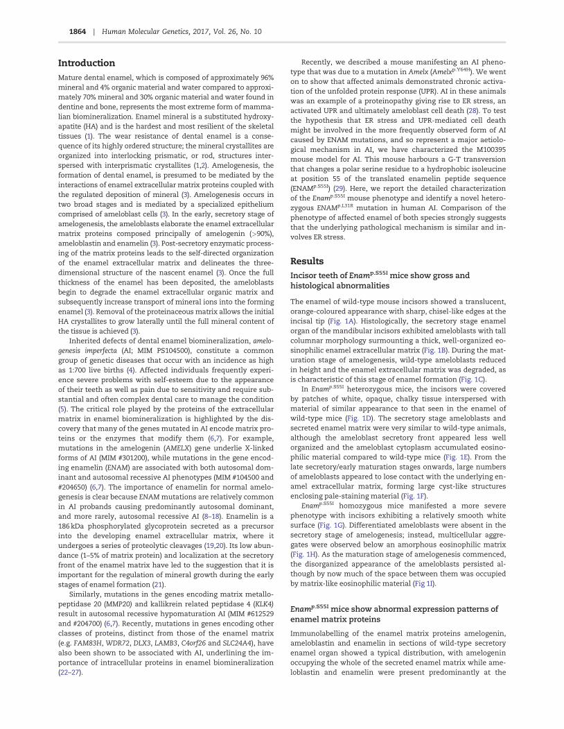
ResultsIncisor teeth of Enamp.S55I mice show gross andhistological abnormalities
The enamel of wild-type mouse incisors showed a translucent,orange-coloured appearance with sharp, chisel-like edges at theincisal tip (Fig. 1A). Histologically, the secretory stage enamelorgan of the mandibular incisors exhibited ameloblasts with tallcolumnar morphology surmounting a thick, well-organized eo-sinophilic enamel extracellular matrix (Fig. 1B). During the mat-uration stage of amelogenesis, wild-type ameloblasts reducedin height and the enamel extracellular matrix was degraded, asis characteristic of this stage of enamel formation (Fig. 1C).
In Enamp.S55I heterozygous mice, the incisors were coveredby patches of white, opaque, chalky tissue interspersed withmaterial of similar appearance to that seen in the enamel ofwild-type mice (Fig. 1D). The secretory stage ameloblasts andsecreted enamel matrix were very similar to wild-type animals,although the ameloblast secretory front appeared less wellorganized and the ameloblast cytoplasm accumulated eosino-philic material compared to wild-type mice (Fig. 1E). From thelate secretory/early maturation stages onwards, large numbersof ameloblasts appeared to lose contact with the underlying en-amel extracellular matrix, forming large cyst-like structuresenclosing pale-staining material (Fig. 1F).
Enamp.S55I homozygous mice manifested a more severephenotype with incisors exhibiting a relatively smooth whitesurface (Fig. 1G). Differentiated ameloblasts were absent in thesecretory stage of amelogenesis; instead, multicellular aggre-gates were observed below an amorphous eosinophilic matrix(Fig. 1H). As the maturation stage of amelogenesis commenced,the disorganized appearance of the ameloblasts persisted al-though by now much of the space between them was occupiedby matrix-like eosinophilic material (Fig 1I).
Enamp.S55I mice show abnormal expression patterns ofenamel matrix proteins
Immunolabelling of the enamel matrix proteins amelogenin,ameloblastin and enamelin in sections of wild-type secretoryenamel organ showed a typical distribution, with amelogeninoccupying the whole of the secreted enamel matrix while ame-loblastin and enamelin were present predominantly at the
1864 | Human Molecular Genetics, 2017, Vol. 26, No. 10
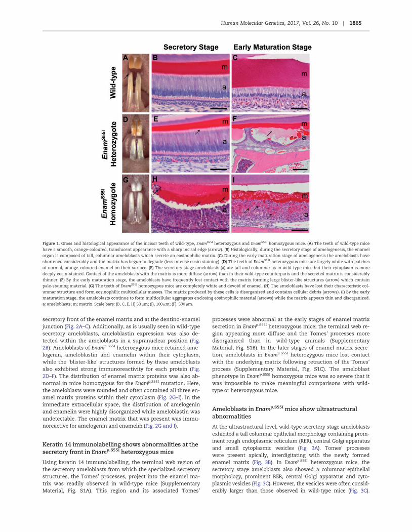
secretory front of the enamel matrix and at the dentino-enameljunction (Fig. 2A–C). Additionally, as is usually seen in wild-typesecretory ameloblasts, ameloblastin expression was also de-tected within the ameloblasts in a supranuclear position (Fig.2B). Ameloblasts of Enamp.S55I heterozygous mice retained ame-logenin, ameloblastin and enamelin within their cytoplasm,while the ‘blister-like’ structures formed by these ameloblastsalso exhibited strong immunoreactivity for each protein (Fig.2D–F). The distribution of enamel matrix proteins was also ab-normal in mice homozygous for the Enamp.S55I mutation. Here,the ameloblasts were rounded and often contained all three en-amel matrix proteins within their cytoplasm (Fig. 2G–I). In theimmediate extracellular space, the distribution of amelogeninand enamelin were highly disorganized while ameloblastin wasundetectable. The enamel matrix that was present was immu-noreactive for amelogenin and enamelin (Fig. 2G and I).
Keratin 14 immunolabelling shows abnormalities at thesecretory front in Enamp.S55I heterozygous mice
Using keratin 14 immunolabelling, the terminal web region ofthe secretory ameloblasts from which the specialized secretorystructures, the Tomes’ processes, project into the enamel ma-trix was readily observed in wild-type mice (SupplementaryMaterial, Fig. S1A). This region and its associated Tomes’
processes were abnormal at the early stages of enamel matrixsecretion in Enamp.S55I heterozygous mice; the terminal web re-gion appearing more diffuse and the Tomes’ processes moredisorganized than in wild-type animals (SupplementaryMaterial, Fig. S1B). In the later stages of enamel matrix secre-tion, ameloblasts in Enamp.S55I heterozygous mice lost contactwith the underlying matrix following retraction of the Tomes’process (Supplementary Material, Fig. S1C). The ameloblastphenotype in Enamp.S55I homozygous mice was so severe that itwas impossible to make meaningful comparisons with wild-type or heterozygous mice.
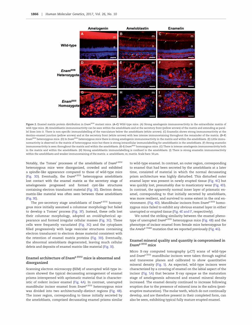
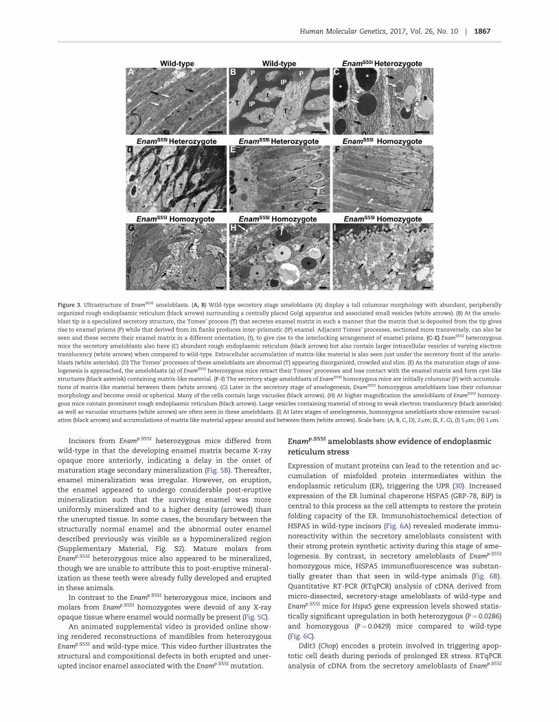
Ameloblasts in Enamp.S55I mice show ultrastructuralabnormalities
At the ultrastructural level, wild-type secretory stage ameloblastsexhibited a tall columnar epithelial morphology containing prom-inent rough endoplasmic reticulum (RER), central Golgi apparatusand small cytoplasmic vesicles (Fig. 3A). Tomes’ processeswere present apically, interdigitating with the newly formedenamel matrix (Fig. 3B). In Enamp.S55I heterozygous mice, thesecretory stage ameloblasts also showed a columnar epithelialmorphology, prominent RER, central Golgi apparatus and cyto-plasmic vesicles (Fig. 3C). However, the vesicles were often consid-erably larger than those observed in wild-type mice (Fig. 3C).
Figure 1. Gross and histological appearance of the incisor teeth of wild-type, EnamS55I heterozygous and EnamS55I homozygous mice. (A) The teeth of wild-type mice
have a smooth, orange-coloured, translucent appearance with a sharp incisal edge (arrow). (B) Histologically, during the secretory stage of amelogenesis, the enamel
organ is composed of tall, columnar ameloblasts which secrete an eosinophilic matrix. (C) During the early maturation stage of amelogenesis the ameloblasts have
shortened considerably and the matrix has begun to degrade (less intense eosin staining). (D) The teeth of EnamS55I heterozygous mice are largely white with patches
of normal, orange-coloured enamel on their surface. (E) The secretory stage ameloblasts (a) are tall and columnar as in wild-type mice but their cytoplasm is more
deeply eosin-stained. Contact of the ameloblasts with the matrix is more diffuse (arrow) than in their wild-type counterparts and the secreted matrix is considerably
thinner. (F) By the early maturation stage, the ameloblasts have frequently lost contact with the matrix forming large blister-like structures (arrow) which contain
pale-staining material. (G) The teeth of EnamS55I homozygous mice are completely white and devoid of enamel. (H) The ameloblasts have lost their characteristic col-
umnar structure and form eosinophilic multicellular masses. The matrix produced by these cells is disorganized and contains cellular debris (arrows). (I) By the early
maturation stage, the ameloblasts continue to form multicellular aggregates enclosing eosinophilic material (arrows) while the matrix appears thin and disorganized.
a: ameloblasts; m; matrix. Scale bars: (B, C, E, H) 50 lm; (I), 100 lm; (F), 500 lm.
1865Human Molecular Genetics, 2017, Vol. 26, No. 10 |
Notably, the Tomes’ processes of the ameloblasts of Enamp.S55I
heterozygous mice were disorganized, crowded and exhibiteda spindle-like appearance compared to those of wild-type mice(Fig. 3D). Eventually, the Enamp.S55I heterozygous ameloblastslost contact with the enamel matrix as the secretory stage ofamelogenesis progressed and formed cyst-like structurescontaining electron translucent material (Fig. 3E). Electron dense,matrix-like material was often seen between these ameloblasts(Fig. 3E).
The pre-secretory stage ameloblasts of Enamp.S55I homozy-gous mice initially assumed a columnar morphology but failedto develop a Tomes’ process (Fig. 3F). Instead, these cells losttheir columnar morphology, adopted an ovoid/spherical ap-pearance and formed irregular cellular masses (Fig. 3G). Thesecells were frequently vacuolated (Fig. 3G) and the cytoplasmfilled progressively with large vesicular structures containingelectron translucent to electron dense material consistent withthe retention of enamel matrix proteins (Fig. 3H). Eventually,the abnormal ameloblasts degenerated, leaving much cellulardebris and deposits of enamel matrix-like material (Fig. 3I).
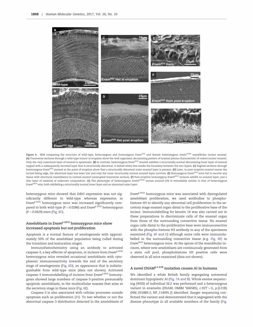
Enamel architecture of Enamp.S55I mice is abnormal anddisorganized
Scanning electron microscopy (SEM) of unerupted wild-type in-cisors showed the typical decussating arrangement of enamelprisms interspersed with aprismatic material that is character-istic of rodent incisor enamel (Fig. 4A). In contrast, uneruptedmandibular incisor enamel from Enamp.S55I heterozygous micewas divided into two architecturally-distinct regions (Fig. 4B).The inner region, corresponding to tissue initially secreted bythe ameloblasts, comprised decussating enamel prisms similar
to wild-type enamel. In contrast, an outer region, correspondingto enamel that had been secreted by the ameloblasts at a latertime, consisted of material in which the normal decussatingprism architecture was highly disturbed. This disturbed outerenamel layer was present in newly erupted tissue (Fig. 4C) butwas quickly lost, presumably due to masticatory wear (Fig. 4D).In contrast, the apparently normal inner layer of prismatic en-amel, corresponding to that initially secreted by ameloblasts,was more resilient, and survived to some extent in the oral en-vironment. (Fig. 4D). Mandibular incisors from Enamp.S55I homo-zygous mice failed to exhibit any obvious enamel layer in eitherunerupted or erupted tissue (Fig. 4E and F, respectively).
We noted the striking similarity between the enamel pheno-type of unerupted Enamp.S55I heterozygous mice (Fig. 4B) and thephenotype of incisor enamel from female mice heterozygous forthe Amelxp.Y64H mutation that we reported previously (Fig. 4G).
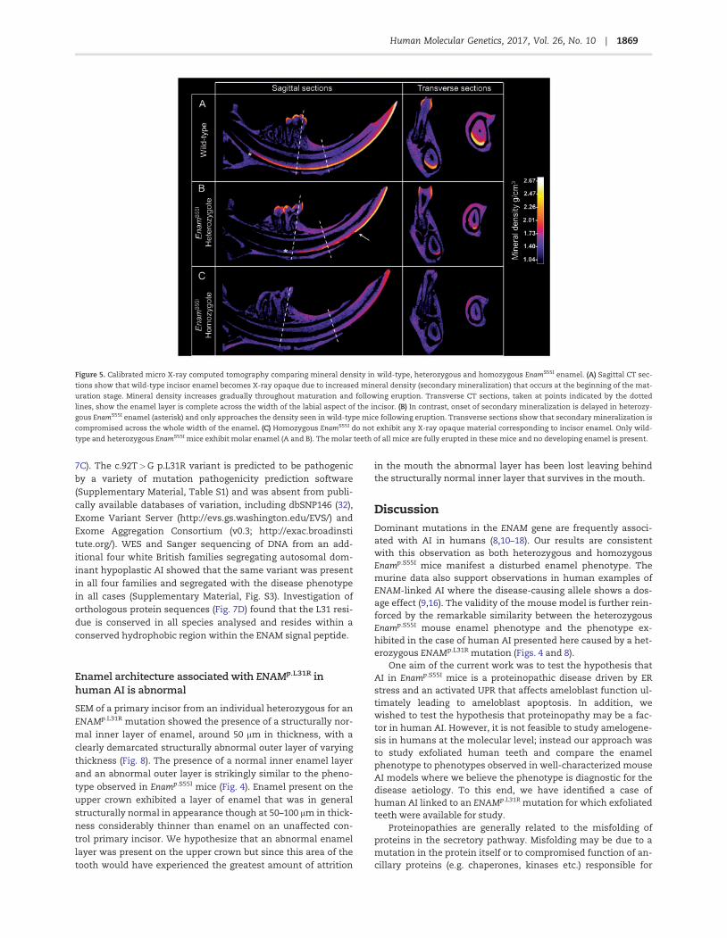
Enamel mineral quality and quantity is compromised inEnamp.S55I mice
Micro X-ray computed tomography (mCT) scans of wild-typeand Enamp.S55I mandibular incisors were taken through sagittaland transverse planes and calibrated to show quantitativemineral density (Fig. 5). As expected, wild-type incisors werecharacterized by a covering of enamel on the labial aspect of theincisor (Fig. 5A) that became X-ray opaque as the maturationstage of amelogenesis advanced and enamel mineral densityincreased. The enamel density continued to increase followingeruption due to the presence of mineral ions in the saliva (post-eruptive maturation). The molar teeth, which do not continuallydevelop, and are therefore present in their completed form, canalso be seen, exhibiting typical fully mature erupted enamel.
Figure 2. Enamel matrix protein distribution in EnamS55I mutant mice. (A–C) Wild-type mice. (A) Strong amelogenin immunoreactivity in the extracellular matrix of
wild-type mice. (B) Ameloblastin immunoreactivity can be seen within the ameloblasts and at the secretory front (yellow arrows) of the matrix and extending as paral-
lel lines into it. There is non-specific immunolabelling of the vasculature below the ameloblasts (white arrows). (C) Enamelin shows strong immunoreactivity at the
dentino-enamel junction (yellow arrows) and at the secretory front (white arrows) with less intense immunostaining throughout the remainder of the matrix. (D–F)
EnamS55I heterozygous mice. (D) In EnamS55I heterozygous mice there is strong amelogenin immunoreactivity in the matrix and within the ameloblasts. (E) Little immu-
noreactivity is observed in the matrix of heterozygous mice but there is strong intracellular immunolabelling for ameloblastin in the ameloblasts. (F) Strong enamelin
immunoreactivity is seen throughout the matrix and within the ameloblasts. (G–I) EnamS55I homozygous mice. (G) There is intense amelogenin immunoreactivity both
in the matrix and within the ameloblasts. (H) Strong ameloblastin immunolabelling is confined to the ameloblasts. (I) There is strong enamelin immunoreactivity
within the ameloblasts and weaker immunostaining of the matrix. a: ameloblasts; m; matrix. Scale bars: 50 lm.
1866 | Human Molecular Genetics, 2017, Vol. 26, No. 10
Incisors from Enamp.S55I heterozygous mice differed fromwild-type in that the developing enamel matrix became X-rayopaque more anteriorly, indicating a delay in the onset ofmaturation stage secondary mineralization (Fig. 5B). Thereafter,enamel mineralization was irregular. However, on eruption,the enamel appeared to undergo considerable post-eruptivemineralization such that the surviving enamel was moreuniformly mineralized and to a higher density (arrowed) thanthe unerupted tissue. In some cases, the boundary between thestructurally normal enamel and the abnormal outer enameldescribed previously was visible as a hypomineralized region(Supplementary Material, Fig. S2). Mature molars fromEnamp.S55I heterozygous mice also appeared to be mineralized,though we are unable to attribute this to post-eruptive mineral-ization as these teeth were already fully developed and eruptedin these animals.
In contrast to the Enamp.S55I heterozygous mice, incisors andmolars from Enamp.S55I homozygotes were devoid of any X-rayopaque tissue where enamel would normally be present (Fig. 5C).
An animated supplemental video is provided online show-ing rendered reconstructions of mandibles from heterozygousEnamp.S55I and wild-type mice. This video further illustrates thestructural and compositional defects in both erupted and uner-upted incisor enamel associated with the Enamp.S55I mutation.
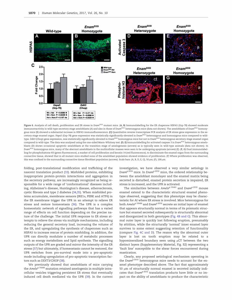
Enamp.S55I ameloblasts show evidence of endoplasmicreticulum stress
Expression of mutant proteins can lead to the retention and ac-cumulation of misfolded protein intermediates within theendoplasmic reticulum (ER), triggering the UPR (30). Increasedexpression of the ER luminal chaperone HSPA5 (GRP-78, BiP) iscentral to this process as the cell attempts to restore the proteinfolding capacity of the ER. Immunohistochemical detection ofHSPA5 in wild-type incisors (Fig. 6A) revealed moderate immu-noreactivity within the secretory ameloblasts consistent withtheir strong protein synthetic activity during this stage of ame-logenesis. By contrast, in secretory ameloblasts of Enamp.S55I
homozygous mice, HSPA5 immunofluorescence was substan-tially greater than that seen in wild-type animals (Fig. 6B).Quantitative RT-PCR (RTqPCR) analysis of cDNA derived frommicro-dissected, secretory-stage ameloblasts of wild-type andEnamp.S55I mice for Hspa5 gene expression levels showed statis-tically significant upregulation in both heterozygous (P¼ 0.0286)and homozygous (P¼ 0.0429) mice compared to wild-type(Fig. 6C).
Ddit3 (Chop) encodes a protein involved in triggering apop-totic cell death during periods of prolonged ER stress. RTqPCRanalysis of cDNA from the secretory ameloblasts of Enamp.S55I
Figure 3. Ultrastructure of EnamS55I ameloblasts. (A, B) Wild-type secretory stage ameloblasts (A) display a tall columnar morphology with abundant, peripherally
organized rough endoplasmic reticulum (black arrows) surrounding a centrally placed Golgi apparatus and associated small vesicles (white arrows). (B) At the amelo-
blast tip is a specialized secretory structure, the Tomes’ process (T) that secretes enamel matrix in such a manner that the matrix that is deposited from the tip gives
rise to enamel prisms (P) while that derived from its flanks produces inter-prismatic (IP) enamel. Adjacent Tomes’ processes, sectioned more transversely, can also be
seen and these secrete their enamel matrix in a different orientation, (t), to give rise to the interlocking arrangement of enamel prisms. (C–E) EnamS55I heterozygous
mice the secretory ameloblasts also have (C) abundant rough endoplasmic reticulum (black arrows) but also contain larger intracellular vesicles of varying electron
translucency (white arrows) when compared to wild-type. Extracellular accumulation of matrix-like material is also seen just under the secretory front of the amelo-
blasts (white asterisks). (D) The Tomes’ processes of these ameloblasts are abnormal (T) appearing disorganized, crowded and slim. (E) As the maturation stage of ame-
logenesis is approached, the ameloblasts (a) of EnamS55I heterozygous mice retract their Tomes’ processes and lose contact with the enamel matrix and form cyst-like
structures (black asterisk) containing matrix-like material. (F–I) The secretory stage ameloblasts of EnamS55I homozygous mice are initially columnar (F) with accumula-
tions of matrix-like material between them (white arrows). (G) Later in the secretory stage of amelogenesis, EnamS55I homozygous ameloblasts lose their columnar
morphology and become ovoid or spherical. Many of the cells contain large vacuoles (black arrows). (H) At higher magnification the ameloblasts of EnamS55I homozy-
gous mice contain prominent rough endoplasmic reticulum (black arrows). Large vesicles containing material of strong to weak electron translucency (black asterisks)
as well as vacuolar structures (white arrows) are often seen in these ameloblasts. (I) At later stages of amelogenesis, homozygous ameloblasts show extensive vacuol-
ation (black arrows) and accumulations of matrix like material appear around and between them (white arrows). Scale bars: (A, B, C, D), 2 lm; (E, F, G), (I) 5 lm; (H) 1 lm.
1867Human Molecular Genetics, 2017, Vol. 26, No. 10 |
heterozygous mice showed that Ddit3 expression was not sig-nificantly different to wild-type whereas expression inEnamp.S55I homozygous mice was increased significantly com-pared to both wild-type (P¼ 0.0286) and Enamp.S55I heterozygous(P¼ 0.0429) mice (Fig. 6C).
Ameloblasts in Enamp.S55I homozygous mice showincreased apoptosis but not proliferation
Apoptosis is a normal feature of amelogenesis with approxi-mately 50% of the ameloblast population being culled duringthe transition and maturation stages.
Immunohistochemistry using an antibody to activatedcaspase-3, a key effector of apoptosis, in incisors from Enamp.S55I
heterozygous mice revealed occasional ameloblasts with cyto-plasmic immunoreactivity towards the end of the secretorystage of amelogenesis (Fig. 6D), an appearance that is indistin-guishable from wild-type mice (data not shown). Activatedcaspase-3 immunolabelling of incisors from Enamp.S55I homozy-gotes showed large numbers of caspase-3-positive presumablyapoptotic ameloblasts, in the multicellular masses that arise atthe secretory stage in these mice (Fig. 6E).
Caspase-3 is also associated with cellular processes outsideapoptosis such as proliferation (31). To test whether or not theabnormal caspase-3 distribution detected in the ameloblasts of
Enamp.S55I homozygous mice was associated with dysregulatedameloblast proliferation, we used antibodies to phospho-histone H3 to identify any abnormal cell proliferation in the se-cretory stage enamel organ distal to the proliferative base of theincisor. Immunolabelling for keratin 14 was also carried out inthese preparations to discriminate cells of the enamel organfrom those of the surrounding connective tissue. No enamelorgan cells distal to the proliferative base were immunoreactivewith the phospho-histone H3 antibody in any of the specimensexamined (Fig. 6F and G) although some cells were immunola-belled in the surrounding connective tissue (e.g. Fig. 6F) inEnamS55I heterozygous mice. At the apices of the mandibular in-cisors, where new ameloblasts are continuously generated froma stem cell pool, phosphohistone H3 positive cells wereobserved in all mice examined (data not shown).
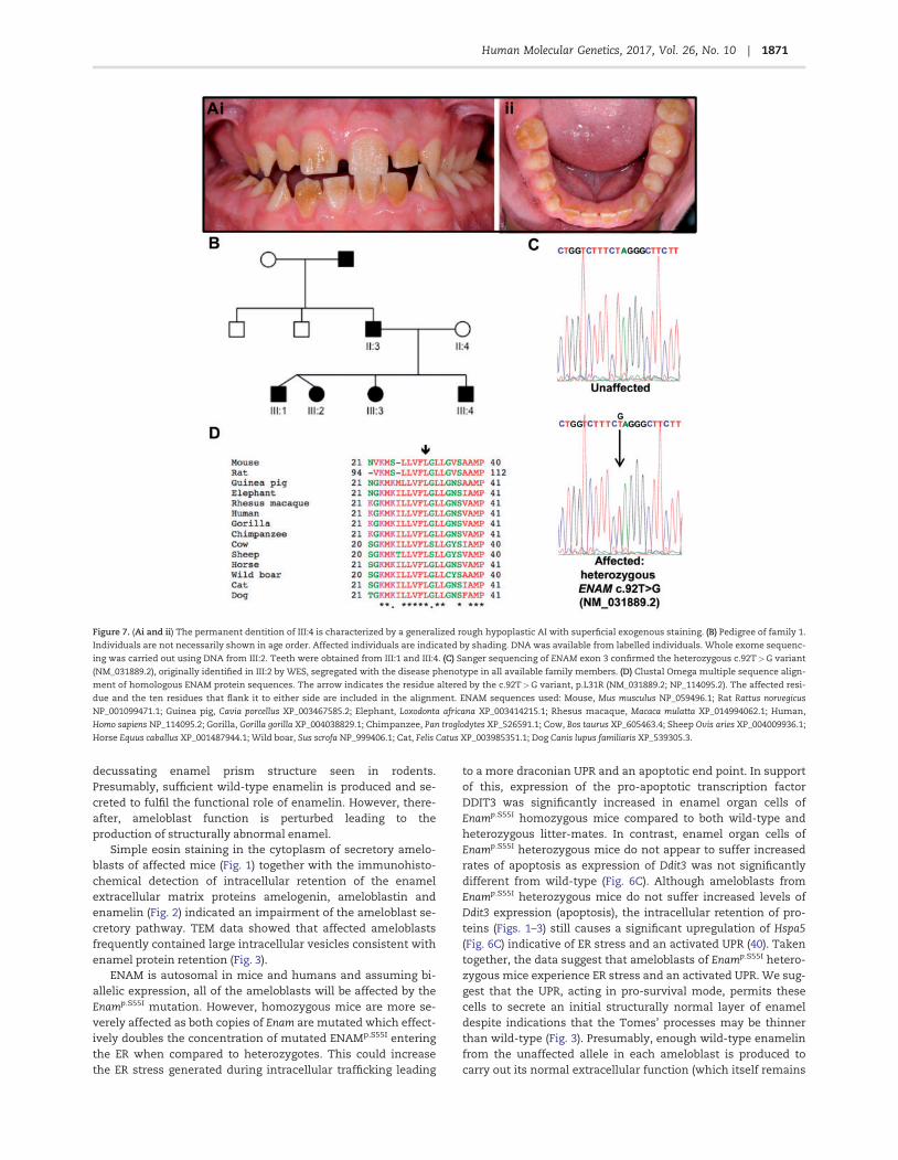
A novel ENAMp.L31R mutation causes AI in humans
We identified a white British family segregating autosomaldominant hypoplastic AI (Fig. 7A and B). Whole exome sequenc-ing (WES) of individual III:2 was performed and a heterozygousvariant in enamelin (ENAM; OMIM *606585), c.92T>G, p.(L31R)(NM_031889.2; NP_114095.2) identified. Sanger sequencing con-firmed the variant and demonstrated that it segregated with thedisease phenotype in all available members of the family (Fig.
Figure 4. SEM comparing the structure of wild-type, heterozygous and homozygous EnamS55I and female heterozygous AmelxY64H mandibular incisor enamel.
(A) Transverse sections through a wild-type incisor at eruption show the well organized, decussating pattern of enamel prisms characteristic of rodent incisor enamel.
Only the very outermost layer of enamel is aprismatic. (B) In contrast, heterozygous EnamS55I enamel exhibits a structurally normal decussating inner layer of enamel
topped with a subsequently secreted layer that is structurally abnormal. A dotted white line marks the boundary between the two layers. (C) Sagittal sections through
heterozygous EnamS55I enamel at the point of eruption show that a structurally abnormal outer enamel layer is present. (D) Later, in post-eruptive enamel nearer the
incisal biting edge, the abnormal layer has been lost and only the inner structurally normal enamel layer survives. (E) Homozygous EnamS55I mice fail to secrete any
tissue with structural resemblance to normal enamel (unerupted transverse section). (F) Post-eruptive homozygous EnampS55I incisors exhibit no enamel layer; just a
thin layer of material of unknown composition. (G) The phenotype of heterozygous AmelxpY64H mouse enamel (39) is remarkably similar to that of heterozygous
EnampS55I mice; both exhibiting a structurally normal inner layer and an abnormal outer layer.
1868 | Human Molecular Genetics, 2017, Vol. 26, No. 10
7C). The c.92T>G p.L31R variant is predicted to be pathogenicby a variety of mutation pathogenicity prediction software(Supplementary Material, Table S1) and was absent from publi-cally available databases of variation, including dbSNP146 (32),Exome Variant Server (http://evs.gs.washington.edu/EVS/) andExome Aggregation Consortium (v0.3; http://exac.broadinstitute.org/). WES and Sanger sequencing of DNA from an add-itional four white British families segregating autosomal dom-inant hypoplastic AI showed that the same variant was presentin all four families and segregated with the disease phenotypein all cases (Supplementary Material, Fig. S3). Investigation oforthologous protein sequences (Fig. 7D) found that the L31 resi-due is conserved in all species analysed and resides within aconserved hydrophobic region within the ENAM signal peptide.
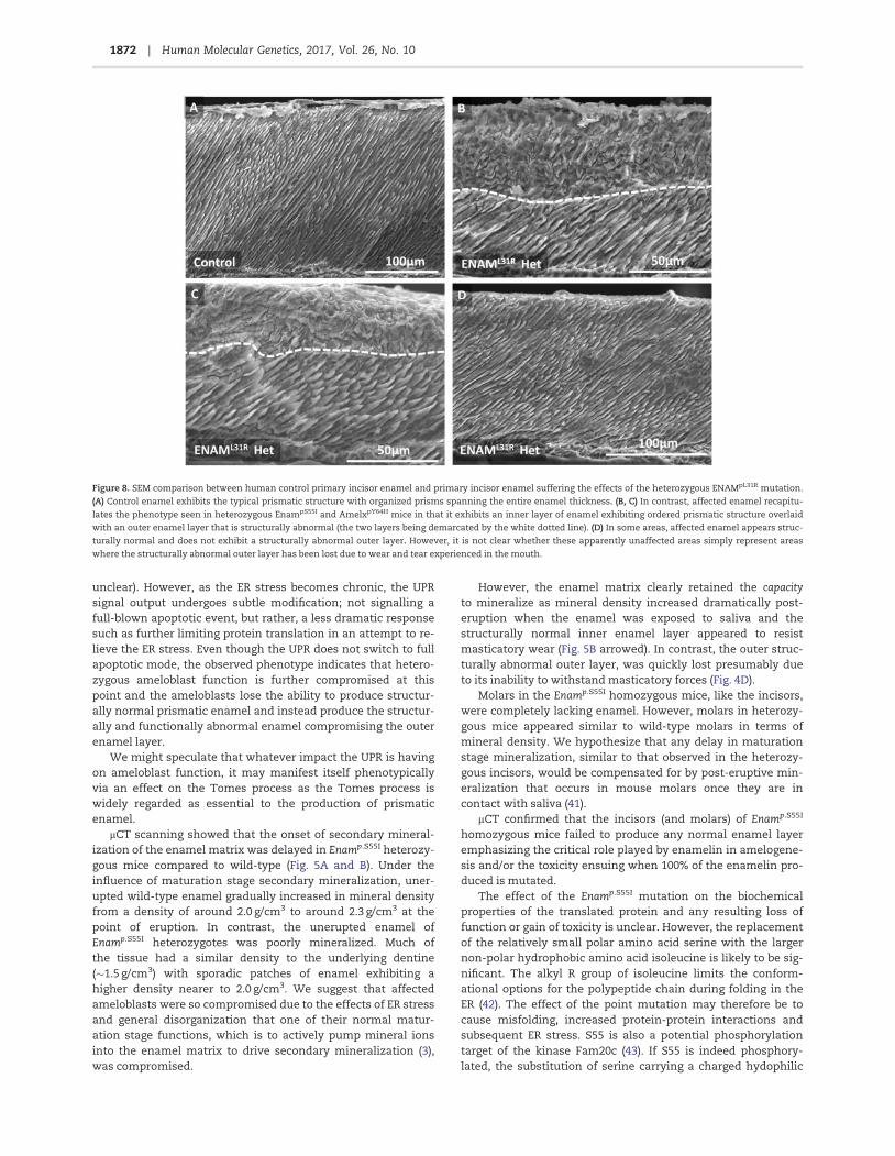
Enamel architecture associated with ENAMp.L31R inhuman AI is abnormal
SEM of a primary incisor from an individual heterozygous for anENAMp.L31R mutation showed the presence of a structurally nor-mal inner layer of enamel, around 50 mm in thickness, with aclearly demarcated structurally abnormal outer layer of varyingthickness (Fig. 8). The presence of a normal inner enamel layerand an abnormal outer layer is strikingly similar to the pheno-type observed in Enamp.S55I mice (Fig. 4). Enamel present on theupper crown exhibited a layer of enamel that was in generalstructurally normal in appearance though at 50–100 mm in thick-ness considerably thinner than enamel on an unaffected con-trol primary incisor. We hypothesize that an abnormal enamellayer was present on the upper crown but since this area of thetooth would have experienced the greatest amount of attrition
in the mouth the abnormal layer has been lost leaving behindthe structurally normal inner layer that survives in the mouth.
DiscussionDominant mutations in the ENAM gene are frequently associ-ated with AI in humans (8,10–18). Our results are consistentwith this observation as both heterozygous and homozygousEnamp.S55I mice manifest a disturbed enamel phenotype. Themurine data also support observations in human examples ofENAM-linked AI where the disease-causing allele shows a dos-age effect (9,16). The validity of the mouse model is further rein-forced by the remarkable similarity between the heterozygousEnamp.S55I mouse enamel phenotype and the phenotype ex-hibited in the case of human AI presented here caused by a het-erozygous ENAMp.L31R mutation (Figs. 4 and 8).
One aim of the current work was to test the hypothesis thatAI in Enamp.S55I mice is a proteinopathic disease driven by ERstress and an activated UPR that affects ameloblast function ul-timately leading to ameloblast apoptosis. In addition, wewished to test the hypothesis that proteinopathy may be a fac-tor in human AI. However, it is not feasible to study amelogene-sis in humans at the molecular level; instead our approach wasto study exfoliated human teeth and compare the enamelphenotype to phenotypes observed in well-characterized mouseAI models where we believe the phenotype is diagnostic for thedisease aetiology. To this end, we have identified a case ofhuman AI linked to an ENAMp.L31R mutation for which exfoliatedteeth were available for study.
Proteinopathies are generally related to the misfolding ofproteins in the secretory pathway. Misfolding may be due to amutation in the protein itself or to compromised function of an-cillary proteins (e.g. chaperones, kinases etc.) responsible for
Figure 5. Calibrated micro X-ray computed tomography comparing mineral density in wild-type, heterozygous and homozygous EnamS55I enamel. (A) Sagittal CT sec-
tions show that wild-type incisor enamel becomes X-ray opaque due to increased mineral density (secondary mineralization) that occurs at the beginning of the mat-
uration stage. Mineral density increases gradually throughout maturation and following eruption. Transverse CT sections, taken at points indicated by the dotted
lines, show the enamel layer is complete across the width of the labial aspect of the incisor. (B) In contrast, onset of secondary mineralization is delayed in heterozy-
gous EnamS55I enamel (asterisk) and only approaches the density seen in wild-type mice following eruption. Transverse sections show that secondary mineralization is
compromised across the whole width of the enamel. (C) Homozygous EnamS55I do not exhibit any X-ray opaque material corresponding to incisor enamel. Only wild-
type and heterozygous EnamS55I mice exhibit molar enamel (A and B). The molar teeth of all mice are fully erupted in these mice and no developing enamel is present.
1869Human Molecular Genetics, 2017, Vol. 26, No. 10 |
folding, post-translational modification and trafficking of thenascent translation product (33). Misfolded proteins, exhibitinginappropriate protein-protein interactions and aggregation inthe secretory pathway, are increasingly recognized as being re-sponsible for a wide range of ‘conformational’ diseases includ-ing, Alzheimer’s disease, Huntington’s disease, atherosclerosis,cystic fibrosis and type 2 diabetes (34,35). When misfolded pro-teins accumulate, membrane sensors (IRE1a, PERK and ATF6) inthe ER membrane trigger the UPR in an attempt to relieve ERstress and restore homeostasis (36). The UPR is a complexhomeostatic network of signalling pathways that has a variedrange of effects on cell function depending on the precise na-ture of the challenge. The initial UPR response to ER stress at-tempts to relieve the stress via multiple mechanisms, includingreducing the general secretory load, increasing the volume ofthe ER, and upregulating the synthesis of chaperones such asHSPA5 to increase rescue of protein misfolding. In addition, theUPR can directly modulate a number of metabolic processessuch as energy metabolism and lipid synthesis. The signallingoutputs of the UPR are graded and mirror the intensity of the ERstress (37) but ultimately, if homeostasis cannot be restored, theUPR switches from pro-survival mode into a pro-apoptoticmode including upregulation of pro-apoptotic transcription fac-tors such as DDIT3/CHOP (38).
We previously showed that ameloblasts of mice carryingthe Amelxp.Y64H mutation retained amelogenin in multiple intra-cellular vesicles triggering persistent ER stress that eventuallyinduced cell death mediated via the UPR (39). In the current
investigation, we have observed a very similar aetiology inEnamp.S55I mice. In Enamp.S55I mice, the ordered relationship be-tween the ameloblast monolayer and the enamel matrix beingsecreted is disturbed, enamel protein secretion is impaired, ERstress is increased, and the UPR is activated.
The similarities between Amelxp.Y64H and Enamp.S55I mouseenamel extend to the characteristic structural enamel pheno-type observed, suggesting that this phenotype may be charac-teristic for AI where ER stress is involved. Mice heterozygous forboth Amelxp.Y64H and Enamp.S55I secrete an initial layer of enamelthat appears structurally normal in terms of its prismatic struc-ture but enamel secreted subsequently is structurally abnormaland disorganized in both genotypes (Fig. 4B and G). This abnor-mal outer layer is quickly lost following eruption, presumablyby attrition, while the structurally ‘normal’ inner enamel layersurvives to some extent suggesting retention of functionality(compare Fig. 4C and D). The reason why the abnormal outerlayer is lost on tooth eruption may be related to ahypomineralized boundary seen using mCT between the twodistinct layers (Supplementary Material, Fig. S2) representing a‘fault line’ susceptible to the shear forces encountered duringmastication.
Clearly, any proposed aetiological mechanism operating inthe Enamp.S55I heterozygous mice needs to account for the en-amel phenotype described above. The fact that approximately50 mm of structurally normal enamel is secreted initially indi-cates that Enamp.S55I translation products have little or no im-pact on the ability of ameloblasts to produce the characteristic
Figure 6. Analysis of cell death, proliferation and ER stress in EnamS55I mutant mice. (A, B) Immunolabelling for the ER chaperone HSPA5 (Grp-78) showed moderate
immunoreactivity in wild-type secretory stage ameloblasts (A) and also in those of EnamS55I heterozygous mice (data not shown). The ameloblasts of EnamS55I homozy-
gous mice (B) showed a substantial increase in HSPA5 immunofluorescence. (C) Quantitative reverse transcriptase PCR analysis of ER stress gene expression in the se-
cretory stage enamel organ. Hspa5 (Grp-78) gene expression was statistically significantly elevated in EnamS55I heterozygous and homozygous mice compared to wild-
type. Ddit3 (Chop) gene expression, was statistically significantly elevated in EnamS55I homozygous mice but not in EnamS55I heterozygous secretory stage enamel organ
compared to wild-type. The data was analysed using the one-sided Mann-Whitney test. (D, E) Immunolabelling for activated caspase 3 in EnamS55I heterozygous amelo-
blasts (D) shows occasional apoptotic ameloblasts at the transition stage of amelogenesis (arrows) as is typically seen in wild-type animals (data not shown). In
EnamS55I homozygous mice, many of the aberrant ameloblasts in the multicellular masses were seen to be undergoing apoptosis (arrows) (E). (F, G) Dual immunolabel-
ling for phosphohistone H3 (green fluorescence), a marker of cell proliferation and keratin 14 (red fluorescence), to discriminate the enamel organ from the surrounding
connective tissue, showed that in all mutant mice studied none of the ameloblast population showed evidence of proliferation. (F) Where proliferation was observed,
this was confined to the surrounding connective tissue fibroblast population (arrows). Scale bars: (A, B, D, E, G), 50 lm; (F), 100 lm.
1870 | Human Molecular Genetics, 2017, Vol. 26, No. 10
decussating enamel prism structure seen in rodents.Presumably, sufficient wild-type enamelin is produced and se-creted to fulfil the functional role of enamelin. However, there-after, ameloblast function is perturbed leading to theproduction of structurally abnormal enamel.
Simple eosin staining in the cytoplasm of secretory amelo-blasts of affected mice (Fig. 1) together with the immunohisto-chemical detection of intracellular retention of the enamelextracellular matrix proteins amelogenin, ameloblastin andenamelin (Fig. 2) indicated an impairment of the ameloblast se-cretory pathway. TEM data showed that affected ameloblastsfrequently contained large intracellular vesicles consistent withenamel protein retention (Fig. 3).
ENAM is autosomal in mice and humans and assuming bi-allelic expression, all of the ameloblasts will be affected by theEnamp.S55I mutation. However, homozygous mice are more se-verely affected as both copies of Enam are mutated which effect-ively doubles the concentration of mutated ENAMp.S55I enteringthe ER when compared to heterozygotes. This could increasethe ER stress generated during intracellular trafficking leading
to a more draconian UPR and an apoptotic end point. In supportof this, expression of the pro-apoptotic transcription factorDDIT3 was significantly increased in enamel organ cells ofEnamp.S55I homozygous mice compared to both wild-type andheterozygous litter-mates. In contrast, enamel organ cells ofEnamp.S55I heterozygous mice do not appear to suffer increasedrates of apoptosis as expression of Ddit3 was not significantlydifferent from wild-type (Fig. 6C). Although ameloblasts fromEnamp.S55I heterozygous mice do not suffer increased levels ofDdit3 expression (apoptosis), the intracellular retention of pro-teins (Figs. 1–3) still causes a significant upregulation of Hspa5(Fig. 6C) indicative of ER stress and an activated UPR (40). Takentogether, the data suggest that ameloblasts of Enamp.S55I hetero-zygous mice experience ER stress and an activated UPR. We sug-gest that the UPR, acting in pro-survival mode, permits thesecells to secrete an initial structurally normal layer of enameldespite indications that the Tomes’ processes may be thinnerthan wild-type (Fig. 3). Presumably, enough wild-type enamelinfrom the unaffected allele in each ameloblast is produced tocarry out its normal extracellular function (which itself remains
Figure 7. (Ai and ii) The permanent dentition of III:4 is characterized by a generalized rough hypoplastic AI with superficial exogenous staining. (B) Pedigree of family 1.
Individuals are not necessarily shown in age order. Affected individuals are indicated by shading. DNA was available from labelled individuals. Whole exome sequenc-
ing was carried out using DNA from III:2. Teeth were obtained from III:1 and III:4. (C) Sanger sequencing of ENAM exon 3 confirmed the heterozygous c.92T>G variant
(NM_031889.2), originally identified in III:2 by WES, segregated with the disease phenotype in all available family members. (D) Clustal Omega multiple sequence align-
ment of homologous ENAM protein sequences. The arrow indicates the residue altered by the c.92T>G variant, p.L31R (NM_031889.2; NP_114095.2). The affected resi-
due and the ten residues that flank it to either side are included in the alignment. ENAM sequences used: Mouse, Mus musculus NP_059496.1; Rat Rattus norvegicus
NP_001099471.1; Guinea pig, Cavia porcellus XP_003467585.2; Elephant, Loxodonta africana XP_003414215.1; Rhesus macaque, Macaca mulatta XP_014994062.1; Human,
Homo sapiens NP_114095.2; Gorilla, Gorilla gorilla XP_004038829.1; Chimpanzee, Pan troglodytes XP_526591.1; Cow, Bos taurus XP_605463.4; Sheep Ovis aries XP_004009936.1;
Horse Equus caballus XP_001487944.1; Wild boar, Sus scrofa NP_999406.1; Cat, Felis Catus XP_003985351.1; Dog Canis lupus familiaris XP_539305.3.
1871Human Molecular Genetics, 2017, Vol. 26, No. 10 |
unclear). However, as the ER stress becomes chronic, the UPRsignal output undergoes subtle modification; not signalling afull-blown apoptotic event, but rather, a less dramatic responsesuch as further limiting protein translation in an attempt to re-lieve the ER stress. Even though the UPR does not switch to fullapoptotic mode, the observed phenotype indicates that hetero-zygous ameloblast function is further compromised at thispoint and the ameloblasts lose the ability to produce structur-ally normal prismatic enamel and instead produce the structur-ally and functionally abnormal enamel compromising the outerenamel layer.
We might speculate that whatever impact the UPR is havingon ameloblast function, it may manifest itself phenotypicallyvia an effect on the Tomes process as the Tomes process iswidely regarded as essential to the production of prismaticenamel.
mCT scanning showed that the onset of secondary mineral-ization of the enamel matrix was delayed in Enamp.S55I heterozy-gous mice compared to wild-type (Fig. 5A and B). Under theinfluence of maturation stage secondary mineralization, uner-upted wild-type enamel gradually increased in mineral densityfrom a density of around 2.0 g/cm3 to around 2.3 g/cm3 at thepoint of eruption. In contrast, the unerupted enamel ofEnamp.S55I heterozygotes was poorly mineralized. Much ofthe tissue had a similar density to the underlying dentine(�1.5 g/cm3) with sporadic patches of enamel exhibiting ahigher density nearer to 2.0 g/cm3. We suggest that affectedameloblasts were so compromised due to the effects of ER stressand general disorganization that one of their normal matur-ation stage functions, which is to actively pump mineral ionsinto the enamel matrix to drive secondary mineralization (3),was compromised.
However, the enamel matrix clearly retained the capacityto mineralize as mineral density increased dramatically post-eruption when the enamel was exposed to saliva and thestructurally normal inner enamel layer appeared to resistmasticatory wear (Fig. 5B arrowed). In contrast, the outer struc-turally abnormal outer layer, was quickly lost presumably dueto its inability to withstand masticatory forces (Fig. 4D).
Molars in the Enamp.S55I homozygous mice, like the incisors,were completely lacking enamel. However, molars in heterozy-gous mice appeared similar to wild-type molars in terms ofmineral density. We hypothesize that any delay in maturationstage mineralization, similar to that observed in the heterozy-gous incisors, would be compensated for by post-eruptive min-eralization that occurs in mouse molars once they are incontact with saliva (41).
mCT confirmed that the incisors (and molars) of Enamp.S55I
homozygous mice failed to produce any normal enamel layeremphasizing the critical role played by enamelin in amelogene-sis and/or the toxicity ensuing when 100% of the enamelin pro-duced is mutated.
The effect of the Enamp.S55I mutation on the biochemicalproperties of the translated protein and any resulting loss offunction or gain of toxicity is unclear. However, the replacementof the relatively small polar amino acid serine with the largernon-polar hydrophobic amino acid isoleucine is likely to be sig-nificant. The alkyl R group of isoleucine limits the conform-ational options for the polypeptide chain during folding in theER (42). The effect of the point mutation may therefore be tocause misfolding, increased protein-protein interactions andsubsequent ER stress. S55 is also a potential phosphorylationtarget of the kinase Fam20c (43). If S55 is indeed phosphory-lated, the substitution of serine carrying a charged hydophilic
Figure 8. SEM comparison between human control primary incisor enamel and primary incisor enamel suffering the effects of the heterozygous ENAMpL31R mutation.
(A) Control enamel exhibits the typical prismatic structure with organized prisms spanning the entire enamel thickness. (B, C) In contrast, affected enamel recapitu-
lates the phenotype seen in heterozygous EnampS55I and AmelxpY64H mice in that it exhibits an inner layer of enamel exhibiting ordered prismatic structure overlaid
with an outer enamel layer that is structurally abnormal (the two layers being demarcated by the white dotted line). (D) In some areas, affected enamel appears struc-
turally normal and does not exhibit a structurally abnormal outer layer. However, it is not clear whether these apparently unaffected areas simply represent areas
where the structurally abnormal outer layer has been lost due to wear and tear experienced in the mouth.
1872 | Human Molecular Genetics, 2017, Vol. 26, No. 10
phosphate group for isoleucine would change the biochemicalcharacter of this site even more dramatically.
Although almost impossible to prove without access to af-fected developing human AI teeth, we believe our data providesevidence that the novel heterozygous ENAMp.L31R human muta-tion reported here may also drive ER stress. The heterozygoushuman ENAMp.L31R phenotype closely mimics the highly charac-teristic phenotype seen in heterozygous Enamp.S55I mice wherethe enamel layer is bifurcated into a structurally normal innerlayer and a structurally abnormal outer layer. The similarphenotypes suggest that AI linked to the heterozygousENAMp.L31R mutation shares a similar disease mechanism basedon an evolving UPR that perturbs ameloblast function at a spe-cific time point resulting in a switch from producing structurallynormal enamel to producing structurally abnormal enamel.Sequence analysis of wild-type ENAM using the Phobius predic-tion tool (44) places residue L31 in the hydrophobic core region(H-region comprising residues 25–33) of the 39 residue ENAMsignal sequence. The H-region is a critical component in the tar-geting of secretory proteins. Once the signal sequence emergesfrom the ribosome, the H-region interacts with the signal recog-nition particle and translation is paused while the complex de-livered to the ER membrane where the ribosome and nascentpeptide are transferred to the translocon. Translation resumesand the growing peptide is translocated into the ER lumen.Once translation is complete the signal sequence is usuallycleaved and the protein is released. Mutations in the H regioncan abolish translocation and the correct cleavage of the signalpeptide. In the presence of the L31R mutation Phobius no longerpredicted the presence of a signal peptide. The actual impact ofthe ENAMp.L31R mutation on enamelin translocation and secre-tion needs to be verified experimentally. However, highly rele-vant to the ENAMp.L31R mutation, is a previously publishedreport detailing a C18R substitution in the H-region of the signalsequence in human preproparathyroid hormone. This mutationstill permitted parathyroid hormone to be translocated into theER but subsequently the protein became trapped in the ER trig-gering ER stress and classic UPR induced apoptosis (45).
In summary, our data indicate that cellular stress provokedby a failure of mutant enamelin to adopt the correct conform-ation within the ER is an aetiological driver leading to AI inEnamp.S55I mice. This mutation is therefore another example ofAI as a conformational disease. A number of pharmacologicalagents are available to overcome the effects of ER stress actingin various ways such as increasing folding protein folding cap-acity or inhibiting apoptosis (46). We have already used onesuch compound, 4-phenybutyrate, in Amelxp.Y64H heterozygousfemale mice, to rescue the AI phenotype (39). The EnamS55I
mouse therefore will provide a further opportunity for us to testthe efficacy of such compounds as potential treatments for AIand provide information on the wider applicability of such com-pounds for treating proteopathic disease in general. Such infor-mation could have clinical value, since the human case of AIreported here linked to a mutation in the H-region of the enam-elin signal sequence may be based on a similar ER stress-linkedaetiology. It is noteworthy that phenylbutyrate reduced theintracellular accumulation of human preproparathyroid hor-mone exhibiting the C18R substitution in the H-region of thesignal sequence and protected cells against ER stress linkedapoptosis (45). Pharmacological treatment of AI in the decidu-ous dentition would require delivering a drug to the developingfoetus in utero. Phenylbutyrate is contraindicated during preg-nancy but it is licensed for use in neonates and children up to18 years of age to treat urea cycle disorders where it is given in
large doses to act as a nitrogen scavenger to treat hyperammo-naemia (by providing an alternative pathway for ammonia se-cretion) (47). Pharmaceutical intervention based onphenylbutyrate would therefore only be useful to rescue thepermanent dentition. In addition, the fact that we observedpost eruptive mineralization of affected enamel in Enamp.S55I
heterozygotes suggests that enamel in some forms of AI couldbenefit from strategies designed to increase/accelerate post-eruptive maturation in humans. The ability to use mouse mod-els to understand AI mechanism in this way coupled with fu-ture genetic screening will provide clinicians with informationupon which clinical management of their patients’ conditioncan be predicated.
Materials and MethodsAnimals
The mutant mouse line M100395 (Enamp.S55I) was obtained from
RIKENGSC (http://www.gsc.riken.jp/Mouse/) and maintained ona C57Bl/6J genetic background. All procedures were performedin accordance with the UK Animals (Scientific Procedures) Act,1986.
Human tissue samples
An exfoliated human primary incisor affected by AI linked to anENAMp.L31R mutation was collected in the UK with the approvalof the relevant Research Ethics Committee (reference 13/YH/0028). Unaffected control primary incisors were obtained fromthe Skeletal Tissues Research Bank (School of Dentistry,University of Leeds) and used with ethical approval and patientconsent.
Histological and immunofluorescence analysis of mouseenamel organ and developing enamel
Mandibles from 2- to 3-month-old heterozygous and homozy-gous Enamp.S55I mutant and wild-type mice (n¼ 4 in each cat-egory) were dissected following cervical dislocation and fixed in4% paraformaldehyde in PBS, pH 7.4, at room temperature for48 h. Following fixation, the mandibles were decalcified in 0.5 MEDTA, dehydrated through a graded ethanol series, cleared inchloroform, embedded as hemi-mandibles in paraffin wax, sec-tioned and stained with haematoxylin and eosin. Sections
were examined using a DMRB microscope (Leica) with SpotTMdigital camera and associated software (RTKE/SE DiagnosticInstruments Inc.). Immunofluorescence analysis was performedon mandibles prepared as above using antibodies raised againstactivated caspase 3 (Abcam, Cambridge, UK), amelogenin FL191,(Santa Cruz Biotechnology, Santa Cruz, CA), ameloblastin (C17,Santa Cruz Biotechnology, Santa Cruz, CA), enamelin, phospho-histone H3 (Abcam, Cambridge, UK), keratin 14 (Abcam,Cambridge, UK) and GRP-78 (Abcam, Cambridge, UK).
Primary antibodies were detected using biotinylated second-ary antibodies (Vector laboratories) followed by Cy-3-conjugated streptavidin (Sigma, Poole, UK) or an AlexaFluor488-conjugated secondary antibody (Abcam, Cambridge, UK) andthe sections were mounted in fluorescence mountant contain-ing DAPI (Vector laboratories).
1873Human Molecular Genetics, 2017, Vol. 26, No. 10 |
Quantitative reverse transcriptase-PCR of mouse enamelorgan mRNA
For quantitative RT-PCR analysis total RNA was extracted usingthe RNeasy kit (Qiagen, Crawley, UK) from micro-dissected se-cretory-stage enamel organs (4 wild-type male, 4 Enamp.S55I het-erozygous and 4 Enamp.S55I homozygous mice) and quantifiedusing a NanoDrop 2000 spectrophotometer (ThermoShandon,Runcorn, UK), then reverse transcribed to complementary DNA.Quantitative RT–PCR was performed according to the manufac-turer’s instructions on a StepOne Plus machine using SYBRGreen master mix (Life Technologies, Paisley, UK) and analysedusing the DD-Ct method, normalized to b-actin (levels of whichwere consistent in all samples). Results were analysed using aone-tailed Mann-Whitney U test. The following primer pairswere used for the analysis: Hspa5 5’-ATCTTTGGTTGCTTGTCGCT-3’, 5’- ATGAAGGAGACTGCTGAGGC-3’; Ddit3 5’- GACCAGGTTCTGCTTTCAGG-3’, 5’- CAGCGACAGAGCCAGAATAA-3’; Actb 5’-CTAAGGCCAACCGTGAAAAGAT-3’, 5’- GCCTGGATGGCTACGTACATG-3’.
Identification of ENAM mutation in human AI
Genomic DNA was obtained from saliva collected usingOrageneVR DNA Sample Collection kits (DNA Genotek, Ottawa,ON, Canada) as detailed in the manufacturer’s instructions.
Three micrograms of genomic DNA was prepared forwhole-exome sequencing using the SureSelect All Exon v5 XTreagent (Agilent Technologies, Santa Clara, CA, USA).Sequencing was performed on an Illumina Hi-Seq 2500sequencing platform (Illumina, San Diego, CA, USA), using a100 bp paired-end protocol. Fastq files were aligned to thehuman reference genome (GRCh37) using Novoalign software(Novocraft Technologies, Selangor, Malaysia). The resultingalignment was processed in the SAM/BAM format using theSAMtools, Picard (http://picard.sourceforge.net) and GATKprograms to correct alignments around indel sites and markpotential PCR duplicates (48,49).
Indel and single-nucleotide variants were called in the VCFformat using the Unified Genotyper function of the GATK pro-gram. Using the dbSNP database at NCBI, any variants presentin dbSNP142 with a minor allele frequency (MAF)�1% were thenexcluded and the remaining variants were annotated using in-house software freely available at http://sourceforge.net/projects/vcfhacks/. Variants in genes known to cause AI wereprioritized for investigation.
PCR and sanger sequencing
The variant was confirmed and segregation was tested in allavailable family members. Primer sequences can be found inSupplementary Material, Table S2. PCR mastermix HotShotDiamond (Clent Life Science, Stourbridge, UK) was used to amp-lify sequences. Sanger sequencing was performed using theBigDye Terminator v3.1 kit (Life Technologies, Carlsbad, CA,USA) according to manufacturer’s instructions and resolved onan ABI3130xl sequencer (Life Technologies). Results were ana-lysed using SeqScape v2.5 (Life Technologies).
The ENAM variant identified in this study has been submit-ted to the Leiden Open Variant Database at http://dna2.leeds.ac.uk/LOVD/ variant ID: ENAM000018 and ClinVar, accession num-ber SCV000328938.
Transmission electron microscopy
Two-month-old mouse mandibles (3 wild-type male, 3Enamp.S55I heterozygous and 3 Enamp.S55I I homozygous mice)were dissected following cervical dislocation and fixed in 2%paraformaldehyde/2% glutaraldehyde prepared in 0.1 M cacody-late buffer containing 0.15 M sucrose and 2 mM calcium chloride(pH 7.3) at 4 �C overnight. Hemi-mandibles were demineralizedas above and dissected axially into posterior, medial and anter-ior portions. Following dissection, the samples were washedwith cacodylate buffer, post-fixed in 1% osmium tetroxide,dehydrated through a graded ethanol series, cleared in propyl-ene oxide and embedded in Epoxy resin (100 resin, AgarScientific Ltd, Stanstead, UK)). Ultrathin sections were con-trasted with uranyl acetate and lead citrate, and examined on aPhilips model 400 transmission electron microscope.
Scanning electron microscopy
Three hemi-mandibles from 2- to 3-month-old heterozygousand homozygous Enamp.S55I mutant and wild-type mice weredissected following cervical dislocation and fixed in 4% parafor-maldehyde in PBS, pH 7.4, at room temperature for 48 h. Eruptedincisors or mandibles were cut transversely with a scalpel andthe cut surfaces polished using1000 grit wet and dry carborun-dum paper followed by final polishing with 12 000 grit nail file toobtain transverse sections through either the erupted or uner-upted incisor enamel. Smear layers were removed by etchingthe ground surface in 30% phosphoric acid for 20 s followed bythorough rinsing in excess de-ionized water. Teeth were driedovernight under vacuum and sputter coated with gold.Specimens were observed using a Hitachi S-3400N scanningelectron microscope (in secondary electron imaging mode)operated at an accelerating voltage of 20 kV and an emissioncurrent of 80 mA.
An exfoliated human primary incisor affected by AI linked toan ENAMp.L31R mutation was fractured along the buccal-lingualmid line. The fractured surface was polished, etched, sputtercoated and examined by SEM as described above.
Micro X-ray computed tomography
Three hemi-mandibles from 2- to 3-month-old heterozygousand homozygous Enamp.S55I mutant and wild-type mice weredissected and fixed as described for scanning electron micros-copy followed by mounting in sealed polypropylene micro tubes(to prevent drying out) together with hydroxyapatite standards.Micro X-ray computed tomography was carried out using aSkyscan 1172 CT scanner (Bruker, Kontich, Belgium). Scans wereobtained using a tube voltage of 75 kV at a constant power of 10W. Image pixel resolution was 8–10 mm and a 0.5 mm alumin-ium filter was used to reduce beam hardening. The scanner wasused in oversize scan mode which allowed hydroxyapatitestandards to be imaged along with each individual sample formineral density calibration purposes. The standards used hadrelative densities of 0.25, 0.75 (Bruker, Kontich, Belgium) and 2.9(Himed, Old Bethpage, USA). Projection images were recon-structed using Recon software (Bruker, Kontich, Belgium).Quantitative mineral density maps were generated usingImageJ software (http://imagej.nih.gov/ij/) and 3D rendered vid-eos produced using CTVox software (Bruker, Kontich, Belgium).
The exfoliated human AI tooth and a corresponding healthycontrol tooth along were scanned as described above except the
1874 | Human Molecular Genetics, 2017, Vol. 26, No. 10
tube voltage was increased to 100kV and the aluminium andcopper filter were engaged to reduce beam hardening.
Supplementary MaterialSupplementary Material is available at HMG online.
Acknowledgements
We thank Samantha Forbes of the electron microscope core fa-cility, Faculty of Biology, Medicine & Health, University ofManchester for her assistance with the transmission electronmicroscopy.
Conflict of Interest statement. None declared.
FundingThis work was supported by a Wellcome Trust programmegrant (grant number 075945), a Wellcome Trust project grant(grant number 093113) and the Wellcome Trust InstitutionalStrategic Support Fund (grant number 105610). J.K. is supportedby the Leeds NIHR Musculoskeletal Biomedical Research Unit.Funding to pay the Open Access publication charges for this art-icle was provided by the Wellcome Trust UK.
References1. Nanci, A. (2007) Ten Cate’s Oral Histology: Development,
Structure And Function. Mosby, St. Louis, MO.2. Bartlett, J.D., Ganss, B., Goldberg, M., Moradian-Oldak, J.,
Paine, M.L., Snead, M.L., Wen, X., White, S.N. and Zhou, Y.L.(2006) 3. Protein-protein interactions of the developing en-amel matrix. Curr. Top. Dev. Biol., 74, 57–115.
3. Smith, C.E. (1998) Cellular and chemical events during en-amel maturation. Crit. Rev. Oral Biol. Med., 9, 128–161.
4. Backman, B. and Holm, A.K. (1986) Amelogenesis imperfecta:prevalence and incidence in a northern Swedish county.Commun. Dent. Oral Epidemiol., 14, 43–47.
5. Coffield, K.D., Phillips, C., Brady, M., Roberts, M.W., Strauss,R.P. and Wright, J.T. (2005) The psychosocial impact of devel-opmental dental defects in people with hereditary amelo-genesis imperfecta. J. Am. Dent. Assoc., 136, 620–630.
6. Hu, J.C., Chun, Y.H., Al Hazzazzi, T. and Simmer, J.P. (2007)Enamel formation and amelogenesis imperfecta. Cells,Tissues Organs, 186, 78–85.
7. Stephanopoulos, G., Garefalaki, M.E. and Lyroudia, K. (2005)Genes and related proteins involved in amelogenesis imper-fecta. J. Dent. Res., 84, 1117–1126.
8. Rajpar, M.H., Harley, K., Laing, C., Davies, R.M. and Dixon,M.J. (2001) Mutation of the gene encoding the enamel-specific protein, enamelin, causes autosomal-dominantamelogenesis imperfecta. Hum. Mol. Genet., 10, 1673–1677.
9. Hart, T.C., Hart, P.S., Gorry, M.C., Michalec, M.D., Ryu, O.H.,Uygur, C., Ozdemir, D., Firatli, S., Aren, G. and Firatli, E. (2003)Novel ENAM mutation responsible for autosomal recessiveamelogenesis imperfecta and localised enamel defects. J.Med. Genet., 40, 900–906.
10. Mardh, C.K., Backman, B., Holmgren, G., Hu, J.C., Simmer, J.P.and Forsman-Semb, K. (2002) A nonsense mutation in theenamelin gene causes local hypoplastic autosomal domin-ant amelogenesis imperfecta (AIH2). Hum. Mol. Genet., 11,1069–1074.
11. Chan, H.C., Mai, L., Oikonomopoulou, A., Chan, H.L.,Richardson, A.S., Wang, S.K., Simmer, J.P. and Hu, J.C. (2010)Altered enamelin phosphorylation site causes amelogenesisimperfecta. J. Dent. Res., 89, 695–699.
12. Gutierrez, S., Torres, D., Briceno, I., Gomez, A.M. andBaquero, E. (2012) Clinical and molecular analysis of theenamelin gene ENAM in Colombian families with autosomaldominant amelogenesis imperfecta. Genet. Mol. Biol., 35,557–566.
13. Kang, H.Y., Seymen, F., Lee, S.K., Yildirim, M., Tuna, E.B.,Patir, A., Lee, K.E. and Kim, J.W. (2009) Candidate gene strat-egy reveals ENAM mutations. J. Dent. Res., 88, 266–269.
14. Kim, J.W., Seymen, F., Lin, B.P., Kiziltan, B., Gencay, K.,Simmer, J.P. and Hu, J.C. (2005) ENAM mutations inautosomal-dominant amelogenesis imperfecta. J. Dent. Res.,84, 278–282.
15. Lindemeyer, R.G., Gibson, C.W. and Wright, T.J. (2010)Amelogenesis imperfecta due to a mutation of the enamelingene: clinical case with genotype-phenotype correlations.Pediatr. Dent., 32, 56–60.
16. Ozdemir, D., Hart, P.S., Firatli, E., Aren, G., Ryu, O.H. and Hart,T.C. (2005) Phenotype of ENAM mutations is dosage-depend-ent. J. Dent. Res., 84, 1036–1041.
17. Simmer, S.G., Estrella, N.M., Milkovich, R.N. and Hu, J.C.(2013) Autosomal dominant amelogenesis imperfecta asso-ciated with ENAM frameshift mutation p.Asn36Ilefs56. Clin.Genet., 83, 195–197.
18. Kida, M., Ariga, T., Shirakawa, T., Oguchi, H. and Sakiyama,Y. (2002) Autosomal-dominant hypoplastic form of amelo-genesis imperfecta caused by an enamelin gene mutation atthe exon-intron boundary. J. Dent. Res., 81, 738–742.
19. Hu, J.C., Zhang, C.H., Yang, Y., Karrman-Mardh, C., Forsman-Semb, K. and Simmer, J.P. (2001) Cloning and characteriza-tion of the mouse and human enamelin genes. J. Dent. Res.,80, 898–902.
20. Brookes, S.J., Lyngstadaas, S.P., Robinson, C., Shore, R.C.,Wood, S.R. and Kirkham, J. (2002) Enamelin compartmental-ization in developing porcine enamel. Connect. Tissue Res., 43,477–481.
21. Hu, J.C., Hu, Y., Smith, C.E., McKee, M.D., Wright, J.T.,Yamakoshi, Y., Papagerakis, P., Hunter, G.K., Feng, J.Q.,Yamakoshi, F. et al. (2008) Enamel defects and ameloblast-specific expression in Enam knock-out/lacz knock-in mice. J.Biol. Chem., 283, 10858–10871.
22. Kim, J.W., Lee, S.K., Lee, Z.H., Park, J.C., Lee, K.E., Lee, M.H.,Park, J.T., Seo, B.M., Hu, J.C. and Simmer, J.P. (2008) FAM83Hmutations in families with autosomal-dominant hypocalci-fied amelogenesis imperfecta. Am. J. Hum. Genet., 82,489–494.
23. El-Sayed, W., Parry, D.A., Shore, R.C., Ahmed, M., Jafri, H.,Rashid, Y., Al-Bahlani, S., Al Harasi, S., Kirkham, J.,Inglehearn, C.F. et al. (2009) Mutations in the beta propellerWDR72 cause autosomal-recessive hypomaturation amelo-genesis imperfecta. Am. J. Hum. Genet., 85, 699–705.
24. Dong, J., Amor, D., Aldred, M.J., Gu, T., Escamilla, M. andMacDougall, M. (2005) DLX3 mutation associated with auto-somal dominant amelogenesis imperfecta with taurodont-ism. Am. J. Med. Genet. A, 133A, 138–141.
25. Poulter, J.A., El-Sayed, W., Shore, R.C., Kirkham, J.,Inglehearn, C.F. and Mighell, A.J. (2014) Whole-exomesequencing, without prior linkage, identifies a mutation inLAMB3 as a cause of dominant hypoplastic amelogenesisimperfecta. Eur. J. Hum. Genet., 22, 132–135.
1875Human Molecular Genetics, 2017, Vol. 26, No. 10 |
26. Parry, D.A., Brookes, S.J., Logan, C.V., Poulter, J.A., El-Sayed,W., Al-Bahlani, S., Al Harasi, S., Sayed, J., Raif el, M., Shore,R.C. et al. (2012) Mutations in C4orf26, encoding a peptidewith in vitro hydroxyapatite crystal nucleation and growthactivity, cause amelogenesis imperfecta. Am. J. Hum. Genet.,91, 565–571.
27. Parry, D.A., Poulter, J.A., Logan, C.V., Brookes, S.J., Jafri, H.,Ferguson, C.H., Anwari, B.M., Rashid, Y., Zhao, H., Johnson,C.A. et al. (2013) Identification of mutations in SLC24A4,encoding a potassium-dependent sodium/calcium exchan-ger, as a cause of amelogenesis imperfecta. Am. J. Hum.Genet., 92, 307–312.
28. Barron, M.J., Brookes, S.J., Kirkham, J., Shore, R.C., Hunt, C.,Mironov, A., Kingswell, N.J., Maycock, J., Shuttleworth, C.A.and Dixon, M.J. (2010) A mutation in the mouse Amelx tri-tyrosyl domain results in impaired secretion of amelogeninand phenocopies human X-linked amelogenesis imperfecta.Hum. Mol. Genet., 19, 1230–1247.
29. Masuya, H., Shimizu, K., Sezutsu, H., Sakuraba, Y., Nagano,J., Shimizu, A., Fujimoto, N., Kawai, A., Miura, I., Kaneda, H.et al. (2005) Enamelin (Enam) is essential for amelogenesis:ENU-induced mouse mutants as models for different clinicalsubtypes of human amelogenesis imperfecta (AI). Hum. Mol.Genet., 14, 575–583.
30. Schroder, M. and Kaufman, R.J. (2005) ER stress and the un-folded protein response. Mutat. Res., 569, 29–63.
31. Lamkanfi, M., Festjens, N., Declercq, W., Vanden Berghe, T.and Vandenabeele, P. (2007) Caspases in cell survival, prolif-eration and differentiation. Cell Death Diff., 14, 44–55.
32. Sherry, S.T., Ward, M.H., Kholodov, M., Baker, J., Phan, L.,Smigielski, E.M. and Sirotkin, K. (2001) dbSNP: the NCBI data-base of genetic variation. Nucl. Acids Res., 29, 308–311.
33. Chiti, F. and Dobson, C.M. (2006) Protein misfolding, func-tional amyloid, and human disease. Annu. Rev. Biochem., 75,333–366.
34. Ellisdon, A.M. and Bottomley, S.P. (2004) The role of proteinmisfolding in the pathogenesis of human diseases. IUBMBLife, 56, 119–123.
35. Ozcan, L. and Tabas, I. (2012) Role of endoplasmic reticulumstress in metabolic disease and other disorders. Annu. Rev.Med., 63, 317–328.
36. Merksamer, P.I. and Papa, F.R. (2010) The UPR and cell fate ata glance. J. Cell Sci., 123, 1003–1006.
37. Southwood, C.M., Garbern, J., Jiang, W. and Gow, A. (2002)The unfolded protein response modulates disease severityin Pelizaeus-Merzbacher disease. Neuron, 36, 585–596.
38. Hetz, C. (2012) The unfolded protein response: controllingcell fate decisions under ER stress and beyond. Nat. Rev. Mol.Cell Biol., 13, 89–102.
39. Brookes, S.J., Barron, M.J., Boot-Handford, R., Kirkham, J. andDixon, M.J. (2014) Endoplasmic reticulum stress in amelo-genesis imperfecta and phenotypic rescue using 4-phenyl-butyrate. Hum. Mol. Genet., 23, 2468–2480.
40. Cawley, K., Deegan, S., Samali, A. and Gupta, S. (2011) Assaysfor detecting the unfolded protein response. MethodsEnzymol., 490, 31–51.
41. Lyngstadaas, S.P., Moinichen, C.B. and Risnes, S. (1998)Crown morphology, enamel distribution, and enamel struc-ture in mouse molars. Anat. Rec., 250, 268–280.
42. Betts, M.J. and Russell, R.B. (2003) Amino acid properties andconsequences of substitutions. In Barnes, M.R. and Gray I.C.(eds), Bioinformatics for Geneticists. Wiley, Chichester, UK, pp.289–316.
43. Cui, J., Xiao, J., Tagliabracci, V.S., Wen, J., Rahdar, M. andDixon, J.E. (2015) A secretory kinase complex regulates extra-cellular protein phosphorylation. Elife, 4, e06120.
44. Kall, L., Krogh, A. and Sonnhammer, E.L. (2007) Advantagesof combined transmembrane topology and signal peptideprediction–the Phobius web server. Nucl. Acids Res., 35,W429–W432.
45. Datta, R., Waheed, A., Shah, G.N. and Sly, W.S. (2007) Signalsequence mutation in autosomal dominant form of hypo-parathyroidism induces apoptosis that is corrected by achemical chaperone. Proc. Natl. Acad. Sci. USA, 104,19989–19994.
46. Hetz, C., Chevet, E. and Harding, H.P. (2013) Targeting the un-folded protein response in disease. Nat. Rev. Drug Discov., 12,703–719.
47. Paediatric Formulary Committee. BNF for Children (online).BMJ Group, Pharmaceutical Press, and RCPCH Publications,London. Available at: http://www.medicinescomplete.com(Accessed on 25th January 2017).
48. McKenna, A., Hanna, M., Banks, E., Sivachenko, A., Cibulskis,K., Kernytsky, A., Garimella, K., Altshuler, D., Gabriel, S.,Daly, M. et al. (2010) The Genome Analysis Toolkit: aMapReduce framework for analyzing next-generation DNAsequencing data. Genome. Res., 20, 1297–1303.
49. DePristo, M.A., Banks, E., Poplin, R., Garimella, K.V., Maguire,J.R., Hartl, C., Philippakis, A.A., del Angel, G., Rivas, M.A.,Hanna, M. et al. (2011) A framework for variation discoveryand genotyping using next-generation DNA sequencingdata. Nat. Genet., 43, 491–498.
1876 | Human Molecular Genetics, 2017, Vol. 26, No. 10