Embed Size (px)
Citation preview
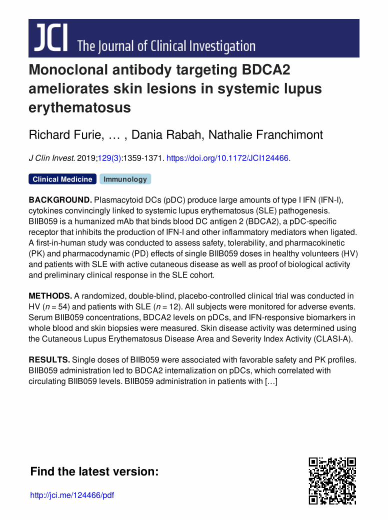
Monoclonal antibody targeting BDCA2ameliorates skin lesions in systemic lupuserythematosus
Richard Furie, … , Dania Rabah, Nathalie Franchimont
J Clin Invest. 2019;129(3):1359-1371. https://doi.org/10.1172/JCI124466.
BACKGROUND. Plasmacytoid DCs (pDC) produce large amounts of type I IFN (IFN-I),cytokines convincingly linked to systemic lupus erythematosus (SLE) pathogenesis.BIIB059 is a humanized mAb that binds blood DC antigen 2 (BDCA2), a pDC-specificreceptor that inhibits the production of IFN-I and other inflammatory mediators when ligated.A first-in-human study was conducted to assess safety, tolerability, and pharmacokinetic(PK) and pharmacodynamic (PD) effects of single BIIB059 doses in healthy volunteers (HV)and patients with SLE with active cutaneous disease as well as proof of biological activityand preliminary clinical response in the SLE cohort.
METHODS. A randomized, double-blind, placebo-controlled clinical trial was conducted inHV (n = 54) and patients with SLE (n = 12). All subjects were monitored for adverse events.Serum BIIB059 concentrations, BDCA2 levels on pDCs, and IFN-responsive biomarkers inwhole blood and skin biopsies were measured. Skin disease activity was determined usingthe Cutaneous Lupus Erythematosus Disease Area and Severity Index Activity (CLASI-A).
RESULTS. Single doses of BIIB059 were associated with favorable safety and PK profiles.BIIB059 administration led to BDCA2 internalization on pDCs, which correlated withcirculating BIIB059 levels. BIIB059 administration in patients with […]
Clinical Medicine Immunology
Find the latest version:
http://jci.me/124466/pdf
The Journal of Clinical Investigation C L I N I C A L M E D I C I N E
1 3 5 9jci.org Volume 129 Number 3 March 2019
IntroductionSystemic lupus erythematosus (SLE) is a chronic autoimmune disease characterized by autoantibody production, inflamma-
tion, and tissue damage in multiple organs resulting from the activation of numerous proinflammatory pathways (1, 2). Clin-ical manifestations vary widely among patients and commonly include rash, arthritis, and nephritis, but the central nervous sys-tem and other major organ systems can also be affected (3).
Therapies used to treat SLE, such as antimalarials, corticoste-roids, and immunosuppressive drugs, are only partially effective and have a wide range of toxicities (4–7). Thus, there is an unmet need for more efficacious and safer therapies that specifically target critical pathways in SLE. Type I IFN (IFN-I) plays a major role in the pathogenesis of SLE (8, 9), and several approaches for targeting the IFN-I pathway have been explored in clinical trials. These include neutralizing mAbs against subtypes of IFN-I (10). Positive results from phase II SLE clinical trials with anti–IFN-α mAb (sifalimumab) and anti–IFN-αβ receptor (anifrolumab) sup-port the pathogenic role of the IFN-I pathway in SLE (11, 12).
Plasmacytoid DCs (pDCs) are a specialized population of bone marrow–derived cells that synthesize large amounts of IFN-I
BACKGROUND. Plasmacytoid DCs (pDC) produce large amounts of type I IFN (IFN-I), cytokines convincingly linked to systemic lupus erythematosus (SLE) pathogenesis. BIIB059 is a humanized mAb that binds blood DC antigen 2 (BDCA2), a pDC-specific receptor that inhibits the production of IFN-I and other inflammatory mediators when ligated. A first-in-human study was conducted to assess safety, tolerability, and pharmacokinetic (PK) and pharmacodynamic (PD) effects of single BIIB059 doses in healthy volunteers (HV) and patients with SLE with active cutaneous disease as well as proof of biological activity and preliminary clinical response in the SLE cohort.
METHODS. A randomized, double-blind, placebo-controlled clinical trial was conducted in HV (n = 54) and patients with SLE (n = 12). All subjects were monitored for adverse events. Serum BIIB059 concentrations, BDCA2 levels on pDCs, and IFN-responsive biomarkers in whole blood and skin biopsies were measured. Skin disease activity was determined using the Cutaneous Lupus Erythematosus Disease Area and Severity Index Activity (CLASI-A).
RESULTS. Single doses of BIIB059 were associated with favorable safety and PK profiles. BIIB059 administration led to BDCA2 internalization on pDCs, which correlated with circulating BIIB059 levels. BIIB059 administration in patients with SLE decreased expression of IFN response genes in blood, normalized MxA expression, reduced immune infiltrates in skin lesions, and decreased CLASI-A score.
CONCLUSIONS. Single doses of BIIB059 were associated with favorable safety and PK/PD profiles and robust target engagement and biological activity, supporting further development of BIIB059 in SLE. The data suggest that targeting pDCs may be beneficial for patients with SLE, especially those with cutaneous manifestations.
TRIAL REGISTRATION. ClinicalTrials.gov NCT02106897.
FUNDING. Biogen Inc.
Monoclonal antibody targeting BDCA2 ameliorates skin lesions in systemic lupus erythematosusRichard Furie,1 Victoria P. Werth,2 Joseph F. Merola,3 Lauren Stevenson,4 Taylor L. Reynolds,4 Himanshu Naik,4 Wenting Wang,4 Romy Christmann,4 Agnes Gardet,4 Alex Pellerin,4 Stefan Hamann,4 Pavan Auluck,4 Catherine Barbey,4 Parul Gulati,4 Dania Rabah,4 and Nathalie Franchimont4
1Division of Rheumatology, Zucker School of Medicine at Hofstra/Northwell, Great Neck, New York, USA. 2Department of Dermatology, Perelman School of Medicine, University of Pennsylvania and Corporal
Michael J. Crescenz VA Medical Center, Philadelphia, Pennsylvania, USA. 3Department of Dermatology and Department of Medicine, Division of Rheumatology, Brigham and Women’s Hospital, Harvard
Medical School, Boston, Massachusetts, USA. 4Biogen, Cambridge, Massachusetts, USA.
Related Commentary: p. 958
Authorship note: DR and NF are co–senior authors.Conflict of interest: RF is a consultant and investigator for Biogen. VPW is a consultant for and has received research grants from Biogen. JFM is a consultant and/or investigator for Biogen, AbbVie, Amgen, Eli Lilly, Novartis, Pfizer, Janssen, UCB, Samumed, Science 37, Celgene, Sanofi Regeneron, Merck, and GSK. LS, TLR, HN, WW, RC, AG, AP, SH, CB, PG, DR, and NF are employees and shareholders of Biogen. PA is currently affiliated with the National Institute of Mental Health, NIH, is not a Biogen shareholder, and has no financial interests to report. The University of Pennsylvania owns the copyright for the Cutaneous Lupus Activity and Severity Index.License: Copyright 2019, American Society for Clinical Investigation.Submitted: September 6, 2018; Accepted: January 10, 2019.Reference information: J Clin Invest. 2019;129(3):1359–1371. https://doi.org/10.1172/JCI124466.
The Journal of Clinical Investigation C L I N I C A L M E D I C I N E
1 3 6 0 jci.org Volume 129 Number 3 March 2019
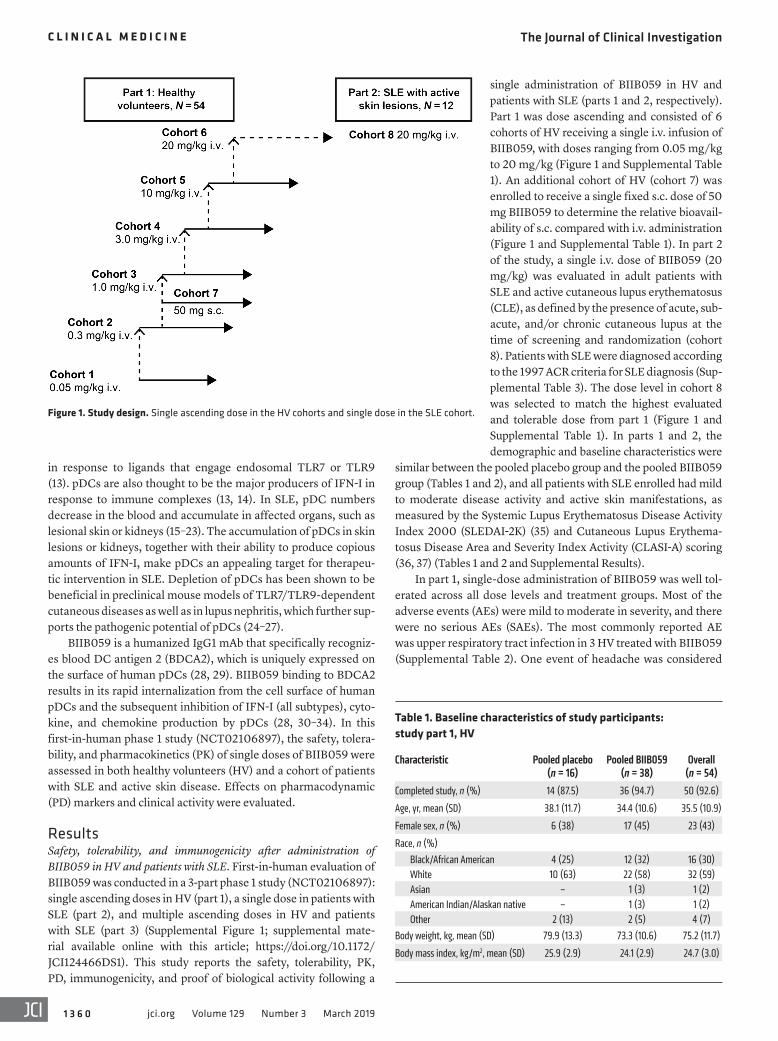
single administration of BIIB059 in HV and patients with SLE (parts 1 and 2, respectively). Part 1 was dose ascending and consisted of 6 cohorts of HV receiving a single i.v. infusion of BIIB059, with doses ranging from 0.05 mg/kg to 20 mg/kg (Figure 1 and Supplemental Table 1). An additional cohort of HV (cohort 7) was enrolled to receive a single fixed s.c. dose of 50 mg BIIB059 to determine the relative bioavail-ability of s.c. compared with i.v. administration (Figure 1 and Supplemental Table 1). In part 2 of the study, a single i.v. dose of BIIB059 (20 mg/kg) was evaluated in adult patients with SLE and active cutaneous lupus erythematosus (CLE), as defined by the presence of acute, sub-acute, and/or chronic cutaneous lupus at the time of screening and randomization (cohort 8). Patients with SLE were diagnosed according to the 1997 ACR criteria for SLE diagnosis (Sup-plemental Table 3). The dose level in cohort 8 was selected to match the highest evaluated and tolerable dose from part 1 (Figure 1 and Supplemental Table 1). In parts 1 and 2, the demographic and baseline characteristics were
similar between the pooled placebo group and the pooled BIIB059 group (Tables 1 and 2), and all patients with SLE enrolled had mild to moderate disease activity and active skin manifestations, as measured by the Systemic Lupus Erythematosus Disease Activity Index 2000 (SLEDAI-2K) (35) and Cutaneous Lupus Erythema-tosus Disease Area and Severity Index Activity (CLASI-A) scoring (36, 37) (Tables 1 and 2 and Supplemental Results).
In part 1, single-dose administration of BIIB059 was well tol-erated across all dose levels and treatment groups. Most of the adverse events (AEs) were mild to moderate in severity, and there were no serious AEs (SAEs). The most commonly reported AE was upper respiratory tract infection in 3 HV treated with BIIB059 (Supplemental Table 2). One event of headache was considered
in response to ligands that engage endosomal TLR7 or TLR9 (13). pDCs are also thought to be the major producers of IFN-I in response to immune complexes (13, 14). In SLE, pDC numbers decrease in the blood and accumulate in affected organs, such as lesional skin or kidneys (15–23). The accumulation of pDCs in skin lesions or kidneys, together with their ability to produce copious amounts of IFN-I, make pDCs an appealing target for therapeu-tic intervention in SLE. Depletion of pDCs has been shown to be beneficial in preclinical mouse models of TLR7/TLR9-dependent cutaneous diseases as well as in lupus nephritis, which further sup-ports the pathogenic potential of pDCs (24–27).
BIIB059 is a humanized IgG1 mAb that specifically recogniz-es blood DC antigen 2 (BDCA2), which is uniquely expressed on the surface of human pDCs (28, 29). BIIB059 binding to BDCA2 results in its rapid internalization from the cell surface of human pDCs and the subsequent inhibition of IFN-I (all subtypes), cyto-kine, and chemokine production by pDCs (28, 30–34). In this first-in-human phase 1 study (NCT02106897), the safety, tolera-bility, and pharmacokinetics (PK) of single doses of BIIB059 were assessed in both healthy volunteers (HV) and a cohort of patients with SLE and active skin disease. Effects on pharmacodynamic (PD) markers and clinical activity were evaluated.
ResultsSafety, tolerability, and immunogenicity after administration of BIIB059 in HV and patients with SLE. First-in-human evaluation of BIIB059 was conducted in a 3-part phase 1 study (NCT02106897): single ascending doses in HV (part 1), a single dose in patients with SLE (part 2), and multiple ascending doses in HV and patients with SLE (part 3) (Supplemental Figure 1; supplemental mate-rial available online with this article; https://doi.org/10.1172/JCI124466DS1). This study reports the safety, tolerability, PK, PD, immunogenicity, and proof of biological activity following a
Figure 1. Study design. Single ascending dose in the HV cohorts and single dose in the SLE cohort.
Table 1. Baseline characteristics of study participants: study part 1, HV
Characteristic Pooled placebo (n = 16)
Pooled BIIB059 (n = 38)
Overall (n = 54)
Completed study, n (%) 14 (87.5) 36 (94.7) 50 (92.6)
Age, yr, mean (SD) 38.1 (11.7) 34.4 (10.6) 35.5 (10.9)
Female sex, n (%) 6 (38) 17 (45) 23 (43)
Race, n (%)Black/African American 4 (25) 12 (32) 16 (30)White 10 (63) 22 (58) 32 (59)Asian – 1 (3) 1 (2)American Indian/Alaskan native – 1 (3) 1 (2)Other 2 (13) 2 (5) 4 (7)
Body weight, kg, mean (SD) 79.9 (13.3) 73.3 (10.6) 75.2 (11.7)
Body mass index, kg/m2, mean (SD) 25.9 (2.9) 24.1 (2.9) 24.7 (3.0)
The Journal of Clinical Investigation C L I N I C A L M E D I C I N E
1 3 6 1jci.org Volume 129 Number 3 March 2019
(Supplemental Figure 4 and data not shown). There were no clini-cally significant changes in physical examinations or ECG, and no dose-dependent patterns of abnormalities were observed. None of the study participants discontinued the study treatment owing to AEs in either part 1 or part 2.
Overall, 6 of 38 HV and no patients with SLE receiving BIIB059 tested positive for anti-BIIB059 binding Ab. Of these responses, 4 were transient responses and 2 were persistent, low-titer responses (data not shown). These antidrug Ab did not affect BIIB059 exposure (PK) or PD, and there was no association with safety events.
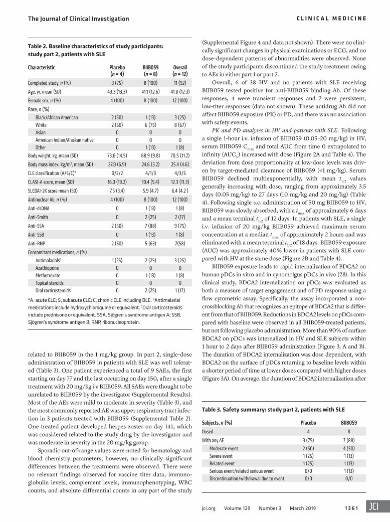
PK and PD analyses in HV and patients with SLE. Following a single 1-hour i.v. infusion of BIIB059 (0.05–20 mg/kg) in HV, serum BIIB059 Cmax and total AUC from time 0 extrapolated to infinity (AUC
∞) increased with dose (Figure 2A and Table 4). The
deviation from dose proportionality at low-dose levels was driv-en by target-mediated clearance of BIIB059 (<1 mg/kg). Serum BIIB059 declined multiexponentially, with mean t1/2 values generally increasing with dose, ranging from approximately 3.5 days (0.05 mg/kg) to 27 days (10 mg/kg and 20 mg/kg) (Table 4). Following single s.c. administration of 50 mg BIIB059 to HV, BIIB059 was slowly absorbed, with a tmax of approximately 6 days and a mean terminal t1/2 of 12 days. In patients with SLE, a single i.v. infusion of 20 mg/kg BIIB059 achieved maximum serum concentration at a median tmax of approximately 2 hours and was eliminated with a mean terminal t1/2 of 18 days. BIIB059 exposure (AUC) was approximately 40% lower in patients with SLE com-pared with HV at the same dose (Figure 2B and Table 4).
BIIB059 exposure leads to rapid internalization of BDCA2 on human pDCs in vitro and in cynomolgus pDCs in vivo (28). In this clinical study, BDCA2 internalization on pDCs was evaluated as both a measure of target engagement and of PD response using a flow cytometric assay. Specifically, the assay incorporated a non-crossblocking Ab that recognizes an epitope of BDCA2 that is differ-ent from that of BIIB059. Reductions in BDCA2 levels on pDCs com-pared with baseline were observed in all BIIB059-treated patients, but not following placebo administration. More than 90% of surface BDCA2 on pDCs was internalized in HV and SLE subjects within 1 hour to 2 days after BIIB059 administration (Figure 3, A and B). The duration of BDCA2 internalization was dose dependent, with BDCA2 on the surface of pDCs returning to baseline levels within a shorter period of time at lower doses compared with higher doses (Figure 3A). On average, the duration of BDCA2 internalization after
related to BIIB059 in the 1 mg/kg group. In part 2, single-dose administration of BIIB059 in patients with SLE was well tolerat-ed (Table 3). One patient experienced a total of 9 SAEs, the first starting on day 77 and the last occurring on day 150, after a single treatment with 20 mg/kg i.v BIIB059. All SAEs were thought to be unrelated to BIIB059 by the investigator (Supplemental Results). Most of the AEs were mild to moderate in severity (Table 3), and the most commonly reported AE was upper respiratory tract infec-tion in 3 patients treated with BIIB059 (Supplemental Table 2). One treated patient developed herpes zoster on day 141, which was considered related to the study drug by the investigator and was moderate in severity in the 20 mg/kg group.
Sporadic out-of-range values were noted for hematology and blood chemistry parameters; however, no clinically significant differences between the treatments were observed. There were no relevant findings observed for vaccine titer data, immuno-globulin levels, complement levels, immunophenotyping, WBC counts, and absolute differential counts in any part of the study
Table 2. Baseline characteristics of study participants: study part 2, patients with SLE
Characteristic Placebo (n = 4)
BIIB059 (n = 8)
Overall (n = 12)
Completed study, n (%) 3 (75) 8 (100) 11 (92)
Age, yr, mean (SD) 43.3 (13.3) 41.1 (12.6) 41.8 (12.3)
Female sex, n (%) 4 (100) 8 (100) 12 (100)
Race, n (%)Black/African American 2 (50) 1 (13) 3 (25)White 2 (50) 6 (75) 8 (67)Asian 0 0 0American Indian/Alaskan native 0 0 0Other 0 1 (13) 1 (8)
Body weight, kg, mean (SD) 73.6 (14.5) 68.9 (9.8) 70.5 (11.2)
Body mass index, kg/m2, mean (SD) 27.0 (6.9) 24.6 (3.2) 25.4 (4.6)
CLE classification (A/S/C)A 0/2/2 4/1/3 4/3/5
CLASI-A score, mean (SD) 16.3 (19.2) 10.4 (5.4) 12.3 (11.3)
SLEDAI-2K score mean (SD) 7.5 (3.4) 5.9 (4.7) 6.4 (4.2 )
Antinuclear Ab, n (%) 4 (100) 8 (100) 12 (100)
Anti-dsDNA 0 1 (13) 1 (8)
Anti-Smith 0 2 (25) 2 (17)
Anti-SSA 2 (50) 7 (88) 9 (75)
Anti-SSB 0 1 (13) 1 (8)
Anti-RNP 2 (50) 5 (63) 7(58)
Concomitant medications, n (%)AntimalarialsB 1 (25) 2 (25) 3 (25)Azathioprine 0 0 0Methotrexate 0 1 (13) 1 (8)Topical steroids 0 0 0Oral corticosteroidsC 0 2 (25) 1 (17)
AA, acute CLE; S, subacute CLE; C, chronic CLE including DLE. BAntimalarial medications include hydroxychloroquine or equivalent. COral corticosteroids include prednisone or equivalent. SSA, Sjögren’s syndrome antigen A; SSB, Sjögren’s syndrome antigen B; RNP, ribonucleoprotein.
Table 3. Safety summary: study part 2, patients with SLE
Subjects, n (%) Placebo BIIB059Dosed 4 8
With any AE 3 (75) 7 (88)Moderate event 2 (50) 4 (50)Severe event 1 (25) 1 (13)Related event 1 (25) 1 (13)Serious event/related serious event 0/0 1 (13)Discontinuation/withdrawal due to event 0/0 0/0

The Journal of Clinical Investigation C L I N I C A L M E D I C I N E
1 3 6 2 jci.org Volume 129 Number 3 March 2019
suggest that BIIB059 administration leads to BDCA2 internaliza-tion without a sustained decrease in pDC numbers.
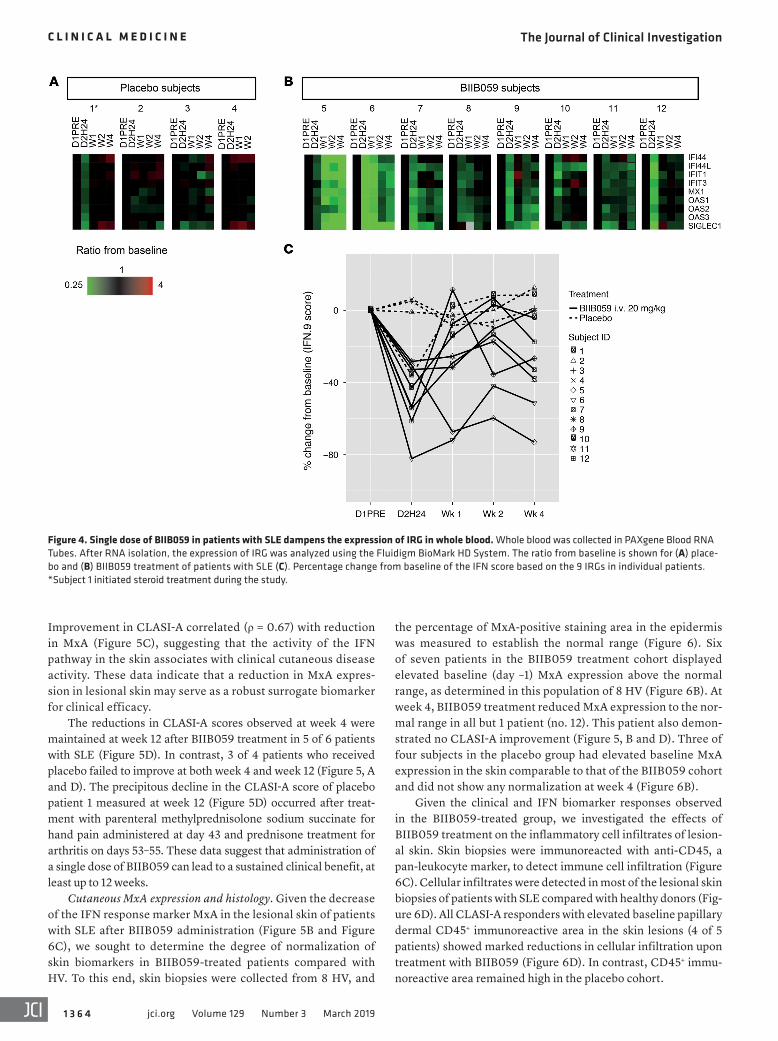
IFN response markers in whole blood and lesional skin. Since BIIB059-mediated BDCA2 internalization is functionally linked to inhibition of IFN-I production by pDCs, we assessed whether BIIB059 modulated the activation of the IFN pathway in patients with SLE. To this end, we investigated the expression of IFN response genes (IRG) from whole blood (Supplemental Figure 3) and hallmark IFN response proteins in lesional skin.
IRG were initially selected on the basis of previously pub-lished IRG data. IRG selection was further refined based on the separation of expression between HV and SLE subjects, the cor-relations among genes, and expression in an internal SLE cohort (Supplemental Figure 3). Their expression was measured in whole blood at baseline and several time points after BIIB059 administration using quantitative PCR. All enrolled patients with SLE showed elevated expression in IRG compared with HV at baseline (Supplemental Figure 3). Single doses of BIIB059 at 20 mg/kg i.v. led to rapid, partial neutralization (~50%) of IRG expression within 24 hours after BIIB059 administration (Figure 4, B and C) compared with placebo (Figure 4A). Since BIIB059 treatment didn’t affect the immune cell composition in whole blood of treated patients, as measured by differential cell count (Supplemental Figure 4), changes in IRG likely reflect the spe-
a single injection of BIIB059 was 14 days at the lowest dose (0.05 mg/kg) in HV, whereas at the highest dose (20 mg/kg), BDCA2 con-tinued to be internalized in most subjects at the last time point tested (112 days) in HV (Figure 3A). Comparisons of individual exposure data and BDCA2 levels on pDC cell surfaces for all treated subjects indicated that circulating BIIB059 must drop below a threshold of approximately 1 μg/ml before BDCA2 on pDC cell surfaces starts returning to baseline levels (data not shown). Since the BIIB059 exposure (AUC) was lower in patients with SLE compared with HV, BIIB059 serum concentration dropped below the 1 μg/ml threshold on days 84 and 112 in some patients, and therefore BDCA2 levels on pDCs started recovering at these time points (Figure 3B).
Internalization of BDCA2 correlated with circulating levels of BIIB059 in both HV (Figure 3C) and patients with SLE (Figure 3D), establishing a PK/PD relationship in vivo.
Reduction from baseline in the number of circulating pDCs was observed following BIIB059 administration, even at the lowest dose level tested (Supplemental Figure 2). The observed reduction was transient, with approximately 50% recovery in average pDC num-bers by week 2 in BIIB059-treated HV and patients with SLE (Sup-plemental Figure 2, B–F). In the 20 mg/kg treatment groups (HV and SLE), recovery in pDC numbers was observed in the presence of more than 100 μg/ml of BIIB059 at week 2, when BDCA2 was still fully internalized on pDCs (Supplemental Figure 2G). These data
Figure 2. BIIB059 PK profile in HV and a cohort of patients with SLE with active cutaneous lupus. BIIB059 serum levels were measured using ELISA. (A) PK of single ascending dose of BIIB059 in HV (n = 38) and (B) PK of 20 mg/kg BIIB059 in HV (black line) (n = 6) and patients with SLE (red line) (n = 8). Arithmetic mean values are represented. conc., concentrations.
Table 4. PK parameters
HV Patients with SLEVariable, mean (% CV)
0.05 mg/kg i.v.
0.3 mg/kg i.v.
1 mg/kg i.v.
3 mg/kg i.v.
10 mg/kg i.v.
20 mg/kg i.v.
50 mg s.c.
20 mg/kg i.v.
AUCt, μg/ml/hr 119 (50.8) 1865 (36.8) 10246 (21.2) 30758 (30.2) 111535 (21.9) 225560 (17.8) 3013 (32.5) 158191.8 (36)AUC∞, μg/ml/hr 229 (NC) 2188 (45.5) 11105 (21.3) 32056 (31.6) 121191 (21.1) 246903 (13.3) 3508 (27.5) 162499.4 (37)Cmax, μg/ml 1.68 (9.8) 10.9 (25.4) 31.7 (11.7) 82.5 (12.1) 280 (23) 603 (10.6) 5.56 (16.1) 473 (31.6)tmax, hours NA NA NA NA NA NA 144 NAt1/2, days 3.5 (27) 8.5 (38.8) 16.8 (34) 18.6 (47.4) 27.3 (20.3) 27.2 (14.6) 11.8 (36.3) 18.1 (40.8)
CV, coevfficient of variation; NC, not calculated.
The Journal of Clinical Investigation C L I N I C A L M E D I C I N E
1 3 6 3jci.org Volume 129 Number 3 March 2019
to a robust and sustained inhibition of the IFN pathway in a fre-quently involved organ (skin) in SLE and results additionally in partial neutralization of the IFN signature in the blood.
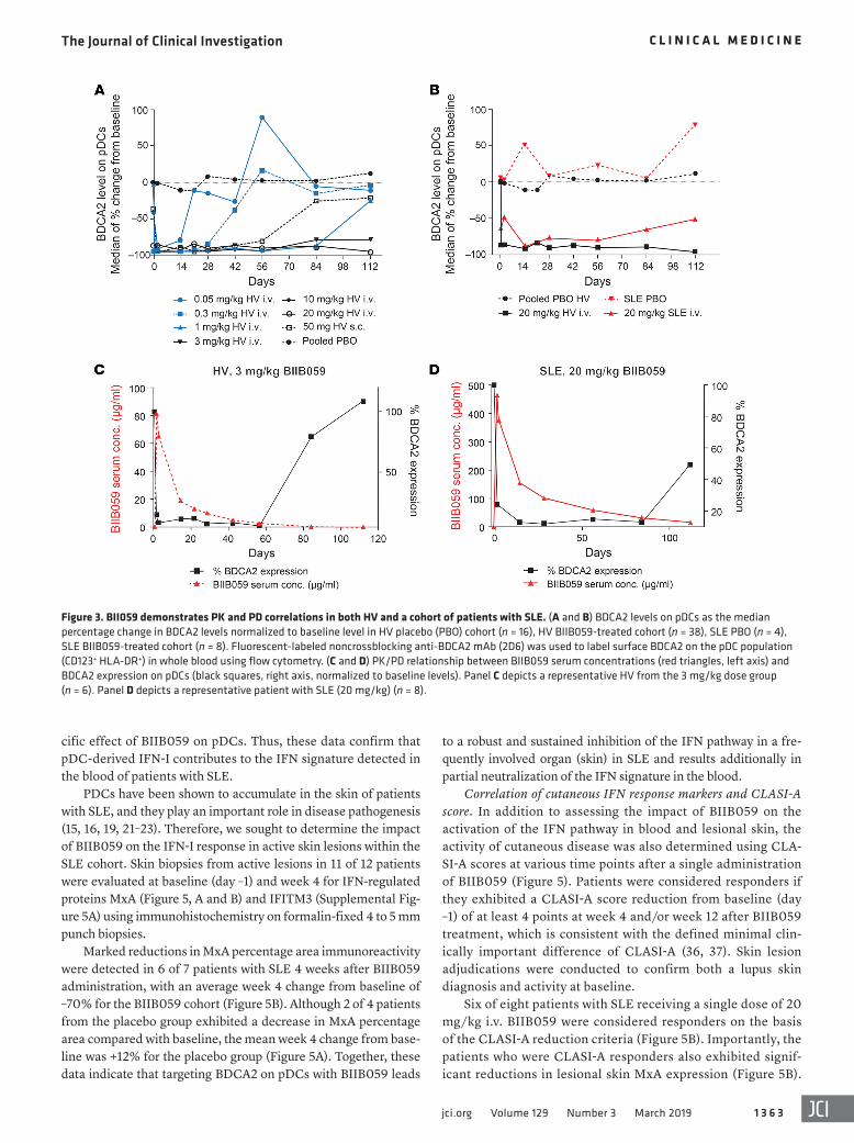
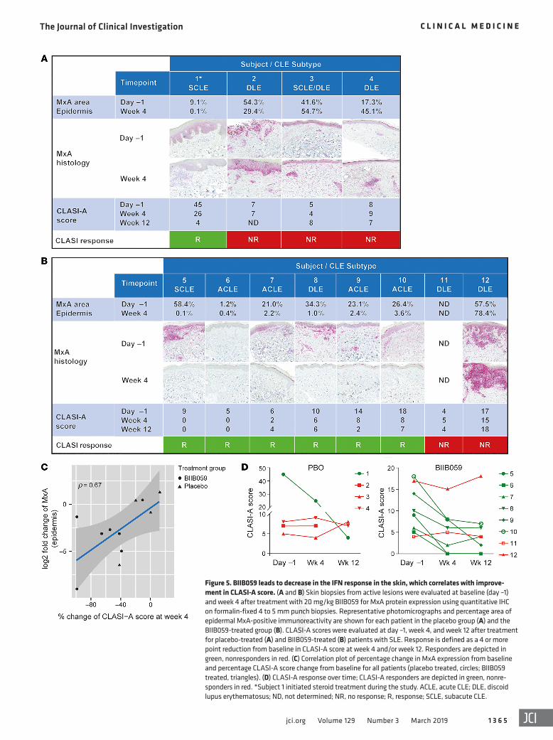
Correlation of cutaneous IFN response markers and CLASI-A score. In addition to assessing the impact of BIIB059 on the activation of the IFN pathway in blood and lesional skin, the activity of cutaneous disease was also determined using CLA-SI-A scores at various time points after a single administration of BIIB059 (Figure 5). Patients were considered responders if they exhibited a CLASI-A score reduction from baseline (day –1) of at least 4 points at week 4 and/or week 12 after BIIB059 treatment, which is consistent with the defined minimal clin-ically important difference of CLASI-A (36, 37). Skin lesion adjudications were conducted to confirm both a lupus skin diagnosis and activity at baseline.
Six of eight patients with SLE receiving a single dose of 20 mg/kg i.v. BIIB059 were considered responders on the basis of the CLASI-A reduction criteria (Figure 5B). Importantly, the patients who were CLASI-A responders also exhibited signif-icant reductions in lesional skin MxA expression (Figure 5B).
cific effect of BIIB059 on pDCs. Thus, these data confirm that pDC-derived IFN-I contributes to the IFN signature detected in the blood of patients with SLE.
PDCs have been shown to accumulate in the skin of patients with SLE, and they play an important role in disease pathogenesis (15, 16, 19, 21–23). Therefore, we sought to determine the impact of BIIB059 on the IFN-I response in active skin lesions within the SLE cohort. Skin biopsies from active lesions in 11 of 12 patients were evaluated at baseline (day –1) and week 4 for IFN-regulated proteins MxA (Figure 5, A and B) and IFITM3 (Supplemental Fig-ure 5A) using immunohistochemistry on formalin-fixed 4 to 5 mm punch biopsies.
Marked reductions in MxA percentage area immunoreactivity were detected in 6 of 7 patients with SLE 4 weeks after BIIB059 administration, with an average week 4 change from baseline of –70% for the BIIB059 cohort (Figure 5B). Although 2 of 4 patients from the placebo group exhibited a decrease in MxA percentage area compared with baseline, the mean week 4 change from base-line was +12% for the placebo group (Figure 5A). Together, these data indicate that targeting BDCA2 on pDCs with BIIB059 leads
Figure 3. BII059 demonstrates PK and PD correlations in both HV and a cohort of patients with SLE. (A and B) BDCA2 levels on pDCs as the median percentage change in BDCA2 levels normalized to baseline level in HV placebo (PBO) cohort (n = 16), HV BIIB059-treated cohort (n = 38), SLE PBO (n = 4), SLE BIIB059-treated cohort (n = 8). Fluorescent-labeled noncrossblocking anti-BDCA2 mAb (2D6) was used to label surface BDCA2 on the pDC population (CD123+ HLA-DR+) in whole blood using flow cytometry. (C and D) PK/PD relationship between BIIB059 serum concentrations (red triangles, left axis) and BDCA2 expression on pDCs (black squares, right axis, normalized to baseline levels). Panel C depicts a representative HV from the 3 mg/kg dose group (n = 6). Panel D depicts a representative patient with SLE (20 mg/kg) (n = 8).
The Journal of Clinical Investigation C L I N I C A L M E D I C I N E
1 3 6 4 jci.org Volume 129 Number 3 March 2019
Improvement in CLASI-A correlated (ρ = 0.67) with reduction in MxA (Figure 5C), suggesting that the activity of the IFN pathway in the skin associates with clinical cutaneous disease activity. These data indicate that a reduction in MxA expres-sion in lesional skin may serve as a robust surrogate biomarker for clinical efficacy.
The reductions in CLASI-A scores observed at week 4 were maintained at week 12 after BIIB059 treatment in 5 of 6 patients with SLE (Figure 5D). In contrast, 3 of 4 patients who received placebo failed to improve at both week 4 and week 12 (Figure 5, A and D). The precipitous decline in the CLASI-A score of placebo patient 1 measured at week 12 (Figure 5D) occurred after treat-ment with parenteral methylprednisolone sodium succinate for hand pain administered at day 43 and prednisone treatment for arthritis on days 53–55. These data suggest that administration of a single dose of BIIB059 can lead to a sustained clinical benefit, at least up to 12 weeks.
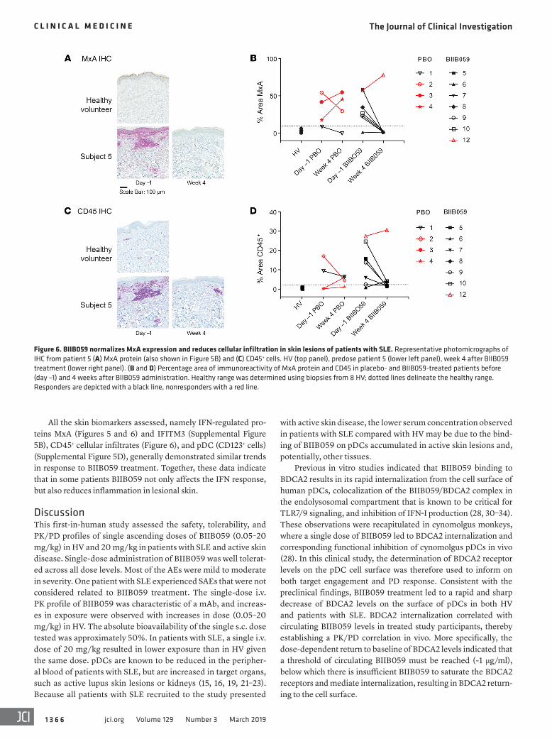
Cutaneous MxA expression and histology. Given the decrease of the IFN response marker MxA in the lesional skin of patients with SLE after BIIB059 administration (Figure 5B and Figure 6C), we sought to determine the degree of normalization of skin biomarkers in BIIB059-treated patients compared with HV. To this end, skin biopsies were collected from 8 HV, and
the percentage of MxA-positive staining area in the epidermis was measured to establish the normal range (Figure 6). Six of seven patients in the BIIB059 treatment cohort displayed elevated baseline (day –1) MxA expression above the normal range, as determined in this population of 8 HV (Figure 6B). At week 4, BIIB059 treatment reduced MxA expression to the nor-mal range in all but 1 patient (no. 12). This patient also demon-strated no CLASI-A improvement (Figure 5, B and D). Three of four subjects in the placebo group had elevated baseline MxA expression in the skin comparable to that of the BIIB059 cohort and did not show any normalization at week 4 (Figure 6B).
Given the clinical and IFN biomarker responses observed in the BIIB059-treated group, we investigated the effects of BIIB059 treatment on the inflammatory cell infiltrates of lesion-al skin. Skin biopsies were immunoreacted with anti-CD45, a pan-leukocyte marker, to detect immune cell infiltration (Figure 6C). Cellular infiltrates were detected in most of the lesional skin biopsies of patients with SLE compared with healthy donors (Fig-ure 6D). All CLASI-A responders with elevated baseline papillary dermal CD45+ immunoreactive area in the skin lesions (4 of 5 patients) showed marked reductions in cellular infiltration upon treatment with BIIB059 (Figure 6D). In contrast, CD45+ immu-noreactive area remained high in the placebo cohort.
Figure 4. Single dose of BIIB059 in patients with SLE dampens the expression of IRG in whole blood. Whole blood was collected in PAXgene Blood RNA Tubes. After RNA isolation, the expression of IRG was analyzed using the Fluidigm BioMark HD System. The ratio from baseline is shown for (A) place-bo and (B) BIIB059 treatment of patients with SLE (C). Percentage change from baseline of the IFN score based on the 9 IRGs in individual patients. *Subject 1 initiated steroid treatment during the study.
The Journal of Clinical Investigation C L I N I C A L M E D I C I N E
1 3 6 5jci.org Volume 129 Number 3 March 2019
Figure 5. BIIB059 leads to decrease in the IFN response in the skin, which correlates with improve-ment in CLASI-A score. (A and B) Skin biopsies from active lesions were evaluated at baseline (day –1) and week 4 after treatment with 20 mg/kg BIIB059 for MxA protein expression using quantitative IHC on formalin-fixed 4 to 5 mm punch biopsies. Representative photomicrographs and percentage area of epidermal MxA-positive immunoreactivity are shown for each patient in the placebo group (A) and the BIIB059-treated group (B). CLASI-A scores were evaluated at day –1, week 4, and week 12 after treatment for placebo-treated (A) and BIIB059-treated (B) patients with SLE. Response is defined as a 4 or more point reduction from baseline in CLASI-A score at week 4 and/or week 12. Responders are depicted in green, nonresponders in red. (C) Correlation plot of percentage change in MxA expression from baseline and percentage CLASI-A score change from baseline for all patients (placebo treated, circles; BIIB059 treated, triangles). (D) CLASI-A response over time; CLASI-A responders are depicted in green, nonre-sponders in red. *Subject 1 initiated steroid treatment during the study. ACLE, acute CLE; DLE, discoid lupus erythematosus; ND, not determined; NR, no response; R, response; SCLE, subacute CLE.
The Journal of Clinical Investigation C L I N I C A L M E D I C I N E
1 3 6 6 jci.org Volume 129 Number 3 March 2019
with active skin disease, the lower serum concentration observed in patients with SLE compared with HV may be due to the bind-ing of BIIB059 on pDCs accumulated in active skin lesions and, potentially, other tissues.
Previous in vitro studies indicated that BIIB059 binding to BDCA2 results in its rapid internalization from the cell surface of human pDCs, colocalization of the BIIB059/BDCA2 complex in the endolysosomal compartment that is known to be critical for TLR7/9 signaling, and inhibition of IFN-I production (28, 30–34). These observations were recapitulated in cynomolgus monkeys, where a single dose of BIIB059 led to BDCA2 internalization and corresponding functional inhibition of cynomolgus pDCs in vivo (28). In this clinical study, the determination of BDCA2 receptor levels on the pDC cell surface was therefore used to inform on both target engagement and PD response. Consistent with the preclinical findings, BIIB059 treatment led to a rapid and sharp decrease of BDCA2 levels on the surface of pDCs in both HV and patients with SLE. BDCA2 internalization correlated with circulating BIIB059 levels in treated study participants, thereby establishing a PK/PD correlation in vivo. More specifically, the dose-dependent return to baseline of BDCA2 levels indicated that a threshold of circulating BIIB059 must be reached (~1 μg/ml), below which there is insufficient BIIB059 to saturate the BDCA2 receptors and mediate internalization, resulting in BDCA2 return-ing to the cell surface.
All the skin biomarkers assessed, namely IFN-regulated pro-teins MxA (Figures 5 and 6) and IFITM3 (Supplemental Figure 5B), CD45+ cellular infiltrates (Figure 6), and pDC (CD123+ cells) (Supplemental Figure 5D), generally demonstrated similar trends in response to BIIB059 treatment. Together, these data indicate that in some patients BIIB059 not only affects the IFN response, but also reduces inflammation in lesional skin.
DiscussionThis first-in-human study assessed the safety, tolerability, and PK/PD profiles of single ascending doses of BIIB059 (0.05–20 mg/kg) in HV and 20 mg/kg in patients with SLE and active skin disease. Single-dose administration of BIIB059 was well tolerat-ed across all dose levels. Most of the AEs were mild to moderate in severity. One patient with SLE experienced SAEs that were not considered related to BIIB059 treatment. The single-dose i.v. PK profile of BIIB059 was characteristic of a mAb, and increas-es in exposure were observed with increases in dose (0.05–20 mg/kg) in HV. The absolute bioavailability of the single s.c. dose tested was approximately 50%. In patients with SLE, a single i.v. dose of 20 mg/kg resulted in lower exposure than in HV given the same dose. pDCs are known to be reduced in the peripher-al blood of patients with SLE, but are increased in target organs, such as active lupus skin lesions or kidneys (15, 16, 19, 21–23). Because all patients with SLE recruited to the study presented
Figure 6. BIIB059 normalizes MxA expression and reduces cellular infiltration in skin lesions of patients with SLE. Representative photomicrographs of IHC from patient 5 (A) MxA protein (also shown in Figure 5B) and (C) CD45+ cells. HV (top panel), predose patient 5 (lower left panel), week 4 after BIIB059 treatment (lower right panel). (B and D) Percentage area of immunoreactivity of MxA protein and CD45 in placebo- and BIIB059-treated patients before (day –1) and 4 weeks after BIIB059 administration. Healthy range was determined using biopsies from 8 HV; dotted lines delineate the healthy range. Responders are depicted with a black line, nonresponders with a red line.
The Journal of Clinical Investigation C L I N I C A L M E D I C I N E
1 3 6 7jci.org Volume 129 Number 3 March 2019
in the skin of patients with SLE were observed at week 4 after a single administration. Biopsies were collected only at week 4 after treatment; therefore, based on the sustained improve-ment in CLASI-A responses in some patients (at least up to week 12), it is possible that the BIIB059-mediated reduction in IFN-I response in the skin is maintained for a longer period.
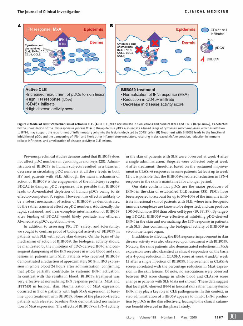
Our data confirm that pDCs are the major producers of IFN-I in the skin of established CLE lesions (38). PDCs have been reported to account for up to 5%–10% of the immune infil-trate in lesional skin of patients with SLE, where interferogenic immune complexes are known to be deposited, and can produce 1000-fold more IFN than other cell types (19, 38, 39). By target-ing BDCA2, BIIB059 was effective at inhibiting pDC-derived IFN-I in the skin and normalizing the IFN response in patients with SLE, thus confirming the biological activity of BIIB059 in vivo in the target organ.
In addition to affecting the IFN response, improvement in skin disease activity was also observed upon treatment with BIIB059. Notably, the same patients who demonstrated reductions in MxA expression were also classified as clinical responders on the basis of a 4-point reduction in CLASI-A score at week 4 and/or week 12 after a single injection of BIIB059. Improvement in CLASI-A scores correlated with the percentage reduction in MxA expres-sion in the skin lesions. Of note, no associations were observed between IRG score change in whole blood and CLASI-A score change in patients with SLE (data not shown). These data suggest that local pDC-derived IFN-I in lesional skin rather than systemic IFN-I may play a key role in CLE pathogenesis. In this context, in vivo administration of BIIB059 appears to inhibit IFN-I produc-tion by pDCs in the skin effectively, leading to the clinical cutane-ous benefit observed in this phase I study.
Previous preclinical studies demonstrated that BIIB059 does not affect pDC numbers in cynomolgus monkeys (28). Admin-istration of BIIB059 to human subjects resulted in a transient decrease in circulating pDC numbers at all dose levels in both HV and patients with SLE. Although the main mechanism of action of BIIB059 is the engagement of the inhibitory receptor BDCA2 to dampen pDC responses, it is possible that BIIB059 leads to Ab-mediated depletion of human pDCs owing to its effector-competent Fc region. However, this effect is unlikely to be a robust mechanism of action of BIIB059, as demonstrated by the rather transient effect on pDC numbers. Additionally, the rapid, sustained, and near-complete internalization of BIIB059 after binding of BDCA2 would likely preclude any efficient Ab-mediated pDC depletion in vivo.
In addition to assessing PK, PD, safety, and tolerability, we sought to confirm proof of biological activity of BIIB059 in patients with SLE with active skin disease. On the basis of the mechanism of action of BIIB059, the biological activity should be manifested by the inhibition of pDC-derived IFN-I and con-sequent dampening of the IFN response in whole blood and skin lesions in patients with SLE. Patients who received BIIB059 demonstrated a reduction of approximately 50% in IRG expres-sion in whole blood 24 hours after administration, confirming that pDCs partially contribute to systemic IFN-I activation. In contrast with the results in blood, BIIB059 treatment was very effective at normalizing IFN response proteins (MxA and IFITM3) in lesional skin. Normalization of MxA expression occurred in 5 of 6 patients with high MxA expression at base-line upon treatment with BIIB059. None of the placebo-treated patients with elevated baseline MxA demonstrated normaliza-tion of MxA expression. The effects of BIIB059 on IFN-I activity
Figure 7. Model of BIIB059 mechanism of action in CLE. (A) In CLE, pDCs accumulate in skin lesions and produce IFN-I and IFN-λ (large arrow), as detected by the upregulation of the IFN-responsive protein MxA in the epidermis. pDCs also secrete a broad range of cytokines and chemokines, which in addition to IFN-I, may support the recruitment of inflammatory cells into the lesions (depicted by CD45+ cells). (B) Treatment with BIIB059 leads to the functional inhibition of pDCs and the dampening of IFN-I and likely other inflammatory mediators, resulting in decreased MxA expression, reduction in immune cellular infiltrates, and amelioration of disease activity in CLE lesions.
The Journal of Clinical Investigation C L I N I C A L M E D I C I N E
1 3 6 8 jci.org Volume 129 Number 3 March 2019
Seven cohorts, comprising 6–9 HV each, were randomized to receive either i.v. (cohorts 1–6) or s.c. (cohort 7) BIIB059 or matching placebo in a 2:1 or 3:1 ratio (Figure 1). Cohorts 1–6 received a single i.v. dose of placebo or 0.05, 0.3, 1, 3, 10, or 20 mg/kg BIIB059. Cohort 7 received a single s.c. dose of placebo or 50 mg BIIB059. The 12 patients with SLE in cohort 8 were randomized 2:1 to receive a single i.v. dose of placebo (n = 4) or 20 mg/kg BIIB059 (n = 8). The study included a 28-day screening period, a 3-day treatment and outpatient observation period (dose administered on day 1), a 16-week follow-up period for both part 1 and part 2, and an additional 12-week extended follow-up period for part 2. The extended follow-up period consisted of telephone calls to subjects for reporting and monitoring SAEs and AEs of interest only.
Eligibility criteriaEligible HV were aged 18–55 years inclusive at the time of informed consent, in good health as determined by the investigator, and had a body mass index between 18 and 30 kg/m2 and body weight of 45 kg or more. Eligible patients with SLE in part 2 of the study were age 18 years or more at the time of informed consent, had an SLE diagnosis according to the 1997 American College of Rheumatology classifi-cation criteria for SLE (41), had a disease duration of 6 months or longer, had a documented history of antinuclear Ab (titer ≥1:80) or anti-dsDNA Ab prior to screening, and had the presence of active lupus skin disease, including acute, subacute, and/or chronic cutane-ous lupus at the time of screening and randomization. Subjects were also willing to have at least 2 skin biopsies of the affected skin area. Final CLE diagnosis was adjudicated by 2 dermatologists. No specific CLASI-A or SLEDAI-2K score was required at baseline. Patients with active lupus nephritis or active neuropsychiatric SLE were excluded as well as patients at high risk of infection. Stable oral prednisone (or equivalent) at a dose of up to 15 mg and treatment with antimalarials, methotrexate (maximum 20 mg/week), or azathioprine (maximum 200 mg/day) were allowed if the dose regimen was stable for at least 28 days prior to randomization. Other SLE standard of care medica-tions were not allowed.
Efficacy assessmentIn part 2, exploratory efficacy analyses of SLE disease activity were performed using the disease instruments: CLASI (36, 37) and the SLE-DAI-2K (35).
Safety profileSafety endpoints included AEs; AEs of interest (viral infections); SAEs; vital signs; 12-lead ECG; and laboratory safety assessments, including hematology with complete blood count with differential and platelet count; blood chemistry (total protein, albumin, cre-atinine, blood urea nitrogen, uric acid bilirubin total and direct, alkaline phosphatase, alanine aminotransferase, aspartate trans-aminase, γ-glutamyl transferase, glucose, calcium, phosphorus, bicarbonate, chloride, sodium, and potassium; urinalysis with dip-stick for blood, protein, and glucose, microscopic examination); C-reactive protein; immunophenotyping; WBC count and absolute differential count of monocytes, neutrophils, and lymphocytes; quantitative immunoglobulins (IgA, IgM, and IgG); complement levels (C3, C4, and Ch50); vaccine titers (tetanus, diphtheria, and pneumococcus); and serum antibodies to BIIB059.
We also observed that CLASI-A responders with elevated base-line CD45+ cellular infiltration in their skin lesions exhibited reduc-tions in immune cellular infiltration upon treatment with BIIB059. This finding supports the hypothesis that pDCs are critical for maintaining the inflammatory environment in SLE skin lesions. BIIB059-mediated functional inhibition of pDCs was shown to inhibit all pDC-derived cytokines and chemokines in vitro, includ-ing IFN-λ1 (type III IFN) (Supplemental Figure 6). Type III IFN, comprising IFN-λ1 (IL-29), IFN-λ2 (IL28a), and IFN-λ3 (IL-28b), binds to a different receptor than IFN-I, but induces a similar sig-naling cascade and IFN response, including driving MxA expres-sion (40). Although pDCs are not the major producers of the non–IFN-I inflammatory mediators, accumulation of pDCs in affected skin could make them an appreciable source capable of supporting the recruitment of inflammatory cells into the lesions and amplify-ing the IFN response (MxA) via IFN-λ in addition to IFN-I. Thus, the overall effect of BIIB059 on skin immune infiltration could be mediated by the inhibition of both pDC-derived IFN-I and proin-flammatory mediators, including chemokines, and BIIB059 may provide a broad therapeutic benefit in SLE (Figure 7, A and B).
In this small phase I clinical study, 2 patients treated with BIIB059 were classified as nonresponders on the basis of CLA-SI-A reduction criteria at week 4 and/or week 12. One of the 2 nonresponders, for whom a skin biopsy was available, did not display reductions in IFN response or inflammatory infiltration in the skin. This lack of response in skin biomarkers and disease activity could not be explained by inadequate drug exposure. A multiple-dose regimen of BIIB059 and a study with a larger num-ber of patients are needed to understand better and confirm the potential clinical benefit of BIIB059 in the skin, but also possibly in other target organs affected by lupus. In this small phase I study, the number of SLE patients with extracutaneous manifestations was extremely low, precluding any meaningful conclusions of the effect of BIIB059 on other systemic manifestations of the disease or analyses of the SLEDAI-2K. BIIB059 is currently being evaluat-ed in a phase 2 clinical trial in patients with active SLE and patients with active CLE (NCT02847598).
BIIB059 is the first therapy, to our knowledge, to specifical-ly target pDCs in patients with SLE. The primary objective of this first-in-human study was achieved by establishing PK/PD correla-tions in vivo and demonstrating tolerability and a favorable safety profile for BIIB059. We opted for an organ-specific approach by studying the effects of pDC inhibition in the skin of patients with SLE. With the caveat that the SLE cohort had a small number of patients with SLE (12 total), our results highlight the central role of pDC-derived IFN-I in the pathogenesis of skin manifestations in SLE and suggest that targeting pDCs with BIIB059 is a promising therapy for SLE and CLE.
Methods
Study designThis study (internal sponsor study code 230LE101) was a randomized, multicenter, phase Ib, placebo-controlled, single ascending dose and multiple ascending dose trial of BIIB059 in HV (parts 1 and 3, respec-tively) or patients with cutaneous SLE (parts 2 and 3, respectively). Only parts 1 and 2 are reported here.
The Journal of Clinical Investigation C L I N I C A L M E D I C I N E
1 3 6 9jci.org Volume 129 Number 3 March 2019
Real-time PCR gene-expression analysis. The expression of 37 genes associated with IFN activity was analyzed using the BioMark HD System (Fluidigm). Owing to the number of assays, expression analysis for each sample was performed across 2 separate runs using independent assay panels. The first panel contained 26 assays as well as assays for 4 housekeeping genes (ACTB, GAPDH, UBC, and YWHAZ), while the second panel contained 11 assays and 3 house-keeping gene assays (UBC, YWHAZ, and B2M). Custom TaqMan Expression Assays (Thermo Fisher Scientific) and samples were prepared according to Fluidigm recommendations and loaded into 96.96 Dynamic Array Integrated Fluidic Circuits (IFC) (Fluidigm). Samples were loaded singly, and assays were loaded in triplicate. IFC plates were primed and loaded using the Controller HX (Fluidigm) and then transferred to the BioMark HD for real-time PCR data col-lection. The following thermocycling program was used: 70°C for 30 minutes, 25°C for 10 minutes, 95°C for 1 minute followed by 45 cycles of 96°C for 5 seconds and 60°C for 20 seconds.
Sample data were normalized to the mean housekeeping gene value for each run, and any sample with an SD greater than 0.3 Ct was omitted from analysis. An endogenous quality control (EQC) was prepared for each assay panel and used to evaluate run accep-tance. Panel 1 EQC was prepared by combining cDNA from 4 SLE donors, while panel 2 EQC was prepared by combining cDNA from 3 SLE donors and 2 Sjögren’s donors. Each EQC was preamplified 10× and then diluted into the appropriate range for the assay. The performance of each EQC was evaluated over 5 independent runs, and acceptance criteria for each assay were set at ±0.3 ΔCt from the mean ΔCt of each EQC assay result.
PK methodsPK assay. The assay to quantify BIIB059 in serum samples was a sandwich ELISA that utilized a BIIB059 anti-idiotypic Ab as capture reagent and HRP-mouse antihuman IgG Fc conjugate for detection. Substrate (3,3′,5,5′-tetramethylbenzidine) was added to plate wells, and color development was subsequently stopped by the addition of 1N sulfuric acid. OD450 was read on a plate reader, and concentra-tions were determined by interpolation from a standard curve using a 5-parameter curve fit.
Dose selection and PK methods. The starting dose of 0.05 mg/kg (i.v. infusion) was expected to have a low pharmacological response based on the estimated human in vivo drug concentration that resulted in 10% of maximal response (EC10) for BDCA2 internaliza-tion and the projected human observed Cmax. The mean EC10 value from preclinical experiments in cynomolgus monkeys was approxi-mately 0.064 μg/ml. To account for the 2-fold higher frequency of pDCs in humans and 8-fold higher BDCA2 density on human pDCs compared with those of cynomolgus monkeys, the estimated human in vivo EC10 for BDCA2 internalization was approximately 1.03 μg/ml. A BIIB059 serum concentration profile following a single 0.05 mg/kg dose (for 70 kg body weight) via a 30-minute i.v. infusion was simulated and gave a projected human Cmax of approximately 1.12 μg/ml. Because this was similar to the estimated human in vivo EC10 for BDCA2 internalization (1.03 μg/ml), this starting dose was expected to have a low pharmacological response in humans and therefore was the appropriate starting dose for the single ascending dose part of the study. Furthermore, substantial safety margins (rel-ative to the no observed adverse effect level [NOAEL] of 125 mg/kg
MaterialsFor flow cytometry, the following were used: BD FACS Lysing Solu-tion 10× Concentrate (BD Biosciences, catalog 349202); BD Trucount Absolute Counting Tubes (BD Biosciences, catalog 340334); and quantum molecules of equivalent soluble fluorochrome (MESF) FITC Calibration Beads (Bangs Laboratories, catalog 555).
BIIB059 (clone 24F4A) is a fully humanized Ab produced in Chi-nese hamster ovary cells as previously described (28). BIIB059 is sup-plied as a sterile liquid drug product containing 50 mg/ml BIIB059, 10 mM sodium citrate, 140 mM l-arginine hydrochloride, and 0.05% polysorbate-80 at pH 6.0. It is provided in 5 ml vials containing 250 mg of BIIB059 per vial.
The following antibodies were used: mouse IgG1κ-FITC (clone X40, BD Biosciences, catalog 349041); anti-BDCA2-FITC (clone 2D6, Biogen-generated mAb, ref. 28, FITC conjugation by BioLeg-end); antihuman CD123-PE (clone 9F5, BD Biosciences, catalog 340545); antihuman HLA-DR-PerCP (clone L243, BD Biosciences, catalog 347364); antihuman lineage cocktail–APC (clones UCHT1, HCD14, 3G8, HIB19, 2H7, HCD56, BioLegend, catalog 348803); mouse monoclonal anti-CD45 (clone MEM-28, Abcam, catalog ab8216); mouse monoclonal anti-MxA (clone M143, provided by G. Kochs, University Medical Center, Freiburg, Germany,); rabbit poly-clonal anti-IFITM3 (Thermo Scientific, catalog Pa5-30382); rab-bit monoclonal anti-CD31 (clone SP38, Spring Bioscience, catalog M3384); and mouse monoclonal anti-CD123 (clone 7G3, BD Biosci-ences, catalog 554527).
BDCA2 expression and pDC countHuman whole blood (250 μl) was mixed with fluorochrome-con-jugated Ab panels containing either anti-BDCA2 or mouse IgG1κ isotype control. The contents were incubated on ice and in the dark for 30 minutes. RBCs were lysed for 10 minutes at room tempera-ture using 5 ml of 1× FACS Lysing Solution (BD). Isolated cells were washed twice with 5 ml of PBS + 1% BSA. Following wash steps, cells were resuspended in 500 μl of PBS + 1% BSA and transferred to BD Trucount Absolute Counting Tubes. Sample acquisition was performed on a BD FACSCalibur flow cytometer (BD Biosciences). For the purpose of standardizing fluorescence intensity, Quan-tum MESF FITC Calibration Beads (Bangs Laboratories) were also acquired under the same instrument gain settings at the time of sample acquisition. BDCA2 expression on the pDC population (CD123+ HLA-DR+) was expressed as an MESF value. The absolute number of pDCs (cells/μl) was determined by comparing cellular events with the known number of fluorescent beads present in the BD Trucount Absolute Counting Tubes.
Ab cocktail panels. Ab cocktail panels were as follows: mouse IgG1κ-FITC or BDCA2-FITC CD123-PE HLA-DR-PerCP; lineage cocktail (CD3, CD14, CD16, CD19, CD20, CD56)-APC.
IFN signature assaysRNA isolation and reverse transcription. Blood was collected in PAXgene Blood RNA Tubes (BD Biosciences) and stored at –20°C until ready for use. Intracellular RNA was isolated using the PAXgene Blood RNA Kit (BD Biosciences) and quantified using a NanoDrop spectrophotome-ter (Thermo Fisher Scientific). RNA was normalized to 20 ng/ml and then reverse transcribed using a High Capacity cDNA Reverse Tran-scription Kit (Thermo Fisher Scientific).
The Journal of Clinical Investigation C L I N I C A L M E D I C I N E
1 3 7 0 jci.org Volume 129 Number 3 March 2019
lots, and IHC and image analysis protocols used on HV specimens were matched, and assay variation for epidermal MxA protein was found to be 7%, well within the accepted variation range for this assay.
For further information, see Supplemental Methods.
StatisticsThe safety analysis set included all the subjects who were random-ized and received at least 1 dose of BIIB059 or placebo. The PD anal-ysis set included all the subjects who were randomized and received at least 1 dose of BIIB059 or placebo and had at least 1 sample bio-marker or other data collected after BIIB059 administration. The PK analysis set included all individuals in the safety analysis set for whom at least 1 primary PK parameter could be calculated. The study was not powered for any formal testing, but the sample size of this study was considered sufficient to characterize the single-dose and multiple-dose safety, tolerability, and PK and PD profiles of BIIB059. The association between log2 fold change of MxA and per-centage change of CLASI-A was assessed by Spearman’s correlation. No formal hypothesis testing was performed. The upper limit of the normal range was determined by taking the maximum percentage area immunoreactivity in a skin compartment for the HV cohort rounded to the nearest 0.1%.
Study approvalInformed consent was obtained from all subjects in accordance with the human subjects institutional review boards of all the participating centers. The study was performed in accordance with the Declaration of Helsinki and the International Conference on Harmonization Note for Guidance on Good Clinical Practice (ICH Topic E6, 1996).
Author contributionsRF designed and conducted the study, analyzed the data, and edited the manuscript. VPW and JFM designed the study, ana-lyzed the data, and edited the manuscript. LS, TLR, WW, AG, and PG designed the experiments, analyzed the data, and edited the manuscript. HN, RC, and CB analyzed the data and edited the manuscript. AP designed the experiments and edited the manu-script. SH and PA designed and/or performed experiments and analyzed the data. DR designed the study, designed the experi-ments, analyzed the data, and wrote the manuscript. NF designed the study, designed the experiments, analyzed the data, and edit-ed the manuscript. All authors critically reviewed the manuscript.
AcknowledgmentsThe authors would like to thank the study participants, Vinshala Chindalore and Craig Curtis, all participating site personnel, and Theresa Podrebarac, Jo Viney, Amy Kao, David Martin, Cristina Musselli, David Dai, Karen Smirnakis, Cynthia Carillio-Infante, Pratapa Prasad, Chase Shen, Douglas Donaldson, Denitza Raitcheva, Eris Bame, Kim Zinnack, Robert Dunstan, Marian Themeles, Xueli Zhang, Daniel Sofia, Luke Jandreski, Shanqin Xu, Galina Marsh, Siva Vobhilineni, and Hannah Kerns for their valuable contributions to this study.
Address correspondence to: Dania Rabah, Biogen Inc., 225 Binney St, Cambridge, Massachusetts 02142, USA. Phone: 617.914.1205; Email: [email protected].
i.v. in cynomolgus monkey) of approximately 1000- to 3600-fold were expected with this starting dose.
The planned maximum dose in parts 1 and 2 was a 20 mg/kg i.v. infusion. This dose was selected to ensure adequate characterization of the safety and tolerability of single ascending doses of BIIB059 with the aim of identifying the maximum tolerated dose, if feasible. Based on the projected Cmax and AUC
∞, the safety margins for this planned
maximum dose (relative to the NOAEL of 125 mg/kg i.v.) were approx-imately 2.5- to 9-fold.
A fixed s.c. dose (50 mg at a concentration of 50 mg/ml) rather than a body weight–based s.c. dose was chosen to minimize the vari-ability in the injection volume of BIIB059.
PK parameter estimation method. All subjects with observed PK con-centrations sufficient for estimating the PK parameters were included in the PK analyses. PK parameters for BIIB059 were estimated on the basis of standard noncompartmental methods using the Phoenix version 6.2 (Certara Corp.) software package. The Cmax and tmax were determined directly from the plasma concentration-time profile for BIIB059. The terminal phase elimination rate constant (λz) was estimat-ed by log-linear regression of concentration-time data in the terminal portion of the PK profiles. The t1/2 was estimated as ln(2)/λz. The AUC from zero time to last observed time point (AUCt) was calculated using the trapezoidal rule. The AUC extrapolated from the last data point to infinity (AUCt-∞) was estimated by dividing the concentration at the last point by the elimination rate constant (λz). The total AUC (AUC
∞) was
calculated as AUCt + AUCt-∞. The clearance (CL) and volume of distri-bution (Vz) were calculated as CL = dose/AUC
∞ and Vz = (CL)/λz (for s.c.
dosing, it is estimated as CL/F and Vz/F). The relative bioavailability was estimated by comparing the AUC values following administration of single i.v. (1 mg/kg) and s.c. (50 mg) doses of BIIB059.
IHC methodsPunch biopsies of 4 to 6 mm in formalin from affected skin were received from the 12 patients with SLE. Biopsies were examined and bisected by a pathologist, then processed into paraffin blocks. H&E-stained sections at various intervals were examined to ensure that IHC was performed on sections within lesions. Punch biopsies of 4 mm were collected from sun-exposed skin of HVs matched approxi-mately by age and ethnicity to the BIIB059-treated cohort (mean age = 34; range, 28–37; White, 6/8; Asian, 1/8; Black or African American, 1/8; female, 8/8) by Folio Biosciences, then processed into paraffin blocks for transfer to Biogen. IHC for MxA, IFITM3, CD45, and dual IHC for CD123/CD31 were performed on 1 section from each speci-men on Ventana Discovery Ultra or Ventana Discovery XT automated staining instruments.
Slides were digitized with a Panoramic P250 scanner at ×200 reso-lution, and skin biopsies were hand annotated to define epidermis, pap-illary dermis, and reticular dermis; follicular epithelium and adnexa were included with epidermis, and sebaceous glands were excluded. Using a combination of color deconvolution and median filters, cus-tomized image analysis algorithms were generated in Visiopharm to distinguish immunopositive areas from immunonegative tissue. Sub-sequently, percentage area values were calculated for each biomarker and sample. CD123 was quantified by first excluding pixels also immu-nopositive for CD31 to differentiate capillaries from pDCs. IHC and image analysis of the HV cohort specimens were performed approxi-mately 2 years after analysis of the BIIB059/placebo specimens. Ab, Ab
The Journal of Clinical Investigation C L I N I C A L M E D I C I N E
1 3 7 1jci.org Volume 129 Number 3 March 2019
1. Liu Z, Davidson A. Taming lupus-a new under-standing of pathogenesis is leading to clinical advances. Nat Med. 2012;18(6):871–882.
2. Tsokos GC, Lo MS, Costa Reis P, Sullivan KE. New insights into the immunopathogenesis of systemic lupus erythematosus. Nat Rev Rheuma-tol. 2016;12(12):716–730.
3. Rahman A, Isenberg DA. Systemic lupus erythe-matosus. N Engl J Med. 2008;358(9):929–939.
4. Yildirim-Toruner C, Diamond B. Current and novel therapeutics in the treatment of systemic lupus erythematosus. J Allergy Clin Immunol. 2011;127(2):303–312; quiz 313–314.
5. Gladman DD, Urowitz MB, Rahman P, Ibañez D, Tam LS. Accrual of organ damage over time in patients with systemic lupus erythematosus. J Rheumatol. 2003;30(9):1955–1959.
6. Doria A, Gatto M, Zen M, Iaccarino L, Punzi L. Optimizing outcome in SLE: treating-to-target and definition of treatment goals. Autoimmun Rev. 2014;13(7):770–777.
7. Petri M, et al. Burden of corticosteroid use in patients with systemic lupus erythema-tosus: results from a Delphi panel. Lupus. 2014;23(10):1006–1013.
8. Eloranta ML, Rönnblom L. Cause and conse-quences of the activated type I interferon system in SLE. J Mol Med (Berl). 2016;94(10):1103–1110.
9. Crow MK. Advances in understanding the role of type I interferons in systemic lupus erythemato-sus. Curr Opin Rheumatol. 2014;26(5):467–474.
10. Crow MK, Olferiev M, Kirou KA. Targeting of type I interferon in systemic autoimmune diseas-es. Transl Res. 2015;165(2):296–305.
11. Khamashta M, et al. Sifalimumab, an anti- interferon-α monoclonal antibody, in moderate to severe systemic lupus erythematosus: a randomised, double-blind, placebo-controlled study. Ann Rheum Dis. 2016;75(11):1909–1916.
12. Furie R, et al. Anifrolumab, an anti-interferon-α receptor monoclonal antibody, in moder-ate-to-severe systemic lupus erythematosus. Arthritis Rheumatol. 2017;69(2):376–386.
13. Kim JM, Park SH, Kim HY, Kwok SK. A plas-macytoid dendritic cells-type I interferon axis is critically implicated in the pathogenesis of systemic lupus erythematosus. Int J Mol Sci. 2015;16(6):14158–14170.
14. Davison LM, Jorgensen TN. New treatments for systemic lupus erythematosus on the hori-zon: targeting plasmacytoid dendritic cells to inhibit cytokine production. J Clin Cell Immunol. 2017;8(6):534.
15. Blomberg S, Eloranta ML, Cederblad B, Nordlin K, Alm GV, Rönnblom L. Presence of cutaneous interferon-alpha producing cells in patients
with systemic lupus erythematosus. Lupus. 2001;10(7):484–490.
16. Farkas L, Beiske K, Lund-Johansen F, Brandtzaeg P, Jahnsen FL. Plasmacytoid dendritic cells (natural interferon- alpha/beta-producing cells) accumulate in cutaneous lupus erythematosus lesions. Am J Pathol. 2001;159(1):237–243.
17. Tucci M, Quatraro C, Lombardi L, Pellegrino C, Dammacco F, Silvestris F. Glomerular accumu-lation of plasmacytoid dendritic cells in active lupus nephritis: role of interleukin-18. Arthritis Rheum. 2008;58(1):251–262.
18. Fiore N, et al. Immature myeloid and plasma-cytoid dendritic cells infiltrate renal tubuloint-erstitium in patients with lupus nephritis. Mol Immunol. 2008;45(1):259–265.
19. Tomasini D, et al. Plasmacytoid dendritic cells: an overview of their presence and distribution in different inflammatory skin diseases, with spe-cial emphasis on Jessner’s lymphocytic infiltrate of the skin and cutaneous lupus erythematosus. J Cutan Pathol. 2010;37(11):1132–1139.
20. Luk KC, Kehm VM, Zhang B, O’Brien P, Tro-janowski JQ, Lee VM. Intracerebral inoculation of pathological α-synuclein initiates a rapidly pro-gressive neurodegenerative α-synucleinopathy in mice. J Exp Med. 2012;209(5):975–986.
21. Vermi W, et al. Cutaneous distribution of plas-macytoid dendritic cells in lupus erythematosus. Selective tropism at the site of epithelial apoptotic damage. Immunobiology. 2009;214(9-10):877–886.
22. Braunstein I, Klein R, Okawa J, Werth VP. The interferon-regulated gene signature is elevated in subacute cutaneous lupus erythematosus and discoid lupus erythematosus and correlates with the cutaneous lupus area and severity index score. Br J Dermatol. 2012;166(5):971–975.
23. Kirchhof MG, Dutz JP. The immunopathology of cutaneous lupus erythematosus. Rheum Dis Clin North Am. 2014;40(3):455–474, viii.
24. Yokogawa M, et al. Epicutaneous application of toll-like receptor 7 agonists leads to systemic autoimmunity in wild-type mice: a new model of systemic lupus erythematosus. Arthritis Rheuma-tol. 2014;66(3):694–706.
25. Guiducci C, et al. Autoimmune skin inflamma-tion is dependent on plasmacytoid dendritic cell activation by nucleic acids via TLR7 and TLR9. J Exp Med. 2010;207(13):2931–2942.
26. Sisirak V, et al. Genetic evidence for the role of plasmacytoid dendritic cells in systemic lupus ery-thematosus. J Exp Med. 2014;211(10):1969–1976.
27. Rowland SL, et al. Early, transient depletion of plasmacytoid dendritic cells ameliorates autoimmunity in a lupus model. J Exp Med. 2014;211(10):1977–1991.
28. Pellerin A, et al. Anti-BDCA2 monoclonal anti-body inhibits plasmacytoid dendritic cell activa-tion through Fc-dependent and Fc-independent mechanisms. EMBO Mol Med. 2015;7(4):464–476.
29. Dzionek A, et al. BDCA-2, BDCA-3, and BDCA-4: three markers for distinct subsets of dendritic cells in human peripheral blood. J Immunol. 2000;165(11):6037–6046.
30. Dzionek A, et al. BDCA-2, a novel plasmacytoid dendritic cell-specific type II C-type lectin, medi-ates antigen capture and is a potent inhibitor of interferon alpha/beta induction. J Exp Med. 2001;194(12):1823–1834.
31. Cao W, et al. BDCA2/Fc epsilon RI gamma com-plex signals through a novel BCR-like pathway in human plasmacytoid dendritic cells. PLoS Biol. 2007;5(10):e248.
32. Jähn PS, Zänker KS, Schmitz J, Dzionek A. BDCA-2 signaling inhibits TLR-9-agonist-induced plas-macytoid dendritic cell activation and antigen presentation. Cell Immunol. 2010;265(1):15–22.
33. Fanning SL, et al. Receptor cross-linking on human plasmacytoid dendritic cells leads to the regulation of IFN-alpha production. J Immunol. 2006;177(9):5829–5839.
34. Röck J, et al. CD303 (BDCA-2) signals in plasma-cytoid dendritic cells via a BCR-like signalosome involving Syk, Slp65 and PLCgamma2. Eur J Immunol. 2007;37(12):3564–3575.
35. Gladman DD, Ibañez D, Urowitz MB. Systemic lupus erythematosus disease activity index 2000. J Rheumatol. 2002;29(2):288–291.
36. Albrecht J, Werth VP. Development of the CLASI as an outcome instrument for cuta-neous lupus erythematosus. Dermatol Ther. 2007;20(2):93–101.
37. Klein R, et al. Development of the CLASI as a tool to measure disease severity and responsiveness to therapy in cutaneous lupus erythematosus. Arch Dermatol. 2011;147(2):203–208.
38. Patsinakidis N, Gambichler T, Lahner N, Moel-lenhoff K, Kreuter A. Cutaneous characteristics and association with antinuclear antibodies in 402 patients with different subtypes of lupus erythematosus. J Eur Acad Dermatol Venereol. 2016;30(12):2097–2104.
39. Siegal FP, et al. The nature of the principal type 1 interferon-producing cells in human blood. Sci-ence. 1999;284(5421):1835–1837.
40. Zanoni I, Granucci F, Broggi A. Interferon (IFN)-λ Takes the Helm: Immunomodulatory Roles of Type III IFNs. Front Immunol. 2017;8:1661.
41. Hochberg MC. Updating the American College of Rheumatology revised criteria for the classifi-cation of systemic lupus erythematosus. Arthritis Rheum. 1997;40(9):1725.