Embed Size (px)
Citation preview
J Appl Oral Sci. 2010;18(1):100-4100
www.scielo.br/jaos
L
ABSTRACT
Amalgam tattoo: a cause of sinusitis?
José Luiz Santos PARIZI1, Gisele Alborghetti NAI2
1- DDS, Professor, Department of Pathology, University of Western São Paulo, Presidente Prudente, SP, Brazil.2- MD, PhD, Professor, Department of Pathology, University of Western São Paulo, Presidente Prudente, SP, Brazil.
Corresponding address: Gisele Alborghetti Nai - Laboratório de Anatomia Patológica e Citopatologia - Universidade do Oeste Paulista (UNOESTE) - RuaJosé Bongiovani, 700 - 19050-680 - Presidente Prudente, SP - Brasil - Phone: +55-18-3229-1059 - Fax: +55-18-3229-1194 - e-mail: [email protected].
Received: November 24, 2008 - Modification: July 19, 2009 - Accepted: August 11, 2009
ittle attention has been paid to the toxicity of silver amalgam fillings, which have been usedover the centuries in Dentistry. Amalgam particles may accidentally and/or traumatically beembedded into the submucosal tissue during placement of a restoration and perpetuate insuch area. This article presents a case of amalgam tattoo and investigates whether it is relatedto the patient’s repeated episodes of sinusitis. The patient was a 46-year-old woman with a 2mm diameter radiopaque lesion in the right oral mucosa detected on a panoramic radiographand presented as a black macula clinically. A complete surgical resection was carried out. Thehistopathological examination revealed deposits of dark-brownish pigments lining the submucosaltissue with adjacent lymphocytic inflammatory infiltrate and multinucleated giant cellsphagocyting pigments. There was a negative staining for both iron and melanin. One yearafter lesion removal, the patient reported that the sinusitis crises had ceased after repeatedepisodes for years. It may be speculated that the inflammatory process related to amalgamtattoo seems to lead to a local immune response that causes sinusitis because it enhances thehuman leukocyte antigen DR (HLA-DR) tissue expression.
Key words: Amalgam. Tattoo. Dental restorative material. Sinusitis. HLA-DR.
INTRODUCTION
Amalgam pigmentation, generally called
amalgam tattoo, is a relatively common finding in
the oral mucosa1,8,13. Gingiva and alveolar mucosa
are the most common locations, and the
mandibular region is more frequently affected than
the maxillary region1,10,13.
Tissue reaction to amalgam can vary
considerably and arise as a macrophage or chronic
inflammatory response, usually in the form of
foreign body reaction, or even not develop any
response1.
This article presents a case of amalgam tattoo
and investigates whether it is related to the patient’s
repeated episodes of sinusitis.
CASE REPORT
A 46-year-old female patient presented with a
2 mm black macule on the oral mucosa of the
posterior fornix on the right. The patient reported
that a tooth with an amalgam filling had been
extracted from that region 15 years before.
Differential clinical diagnosis suggested were
melanoma, melanocytic nevus, hematoma,
hemangioma and amalgam tattoo.
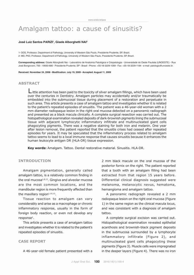
A panoramic radiograph revealed a 2 mm
radiopaque lesion on the right oral mucosa (Figure
1) in the same region as the clinical macula locus,
and was consistent with a diagnosis of amalgam
tattoo.
A complete surgical excision was carried out.
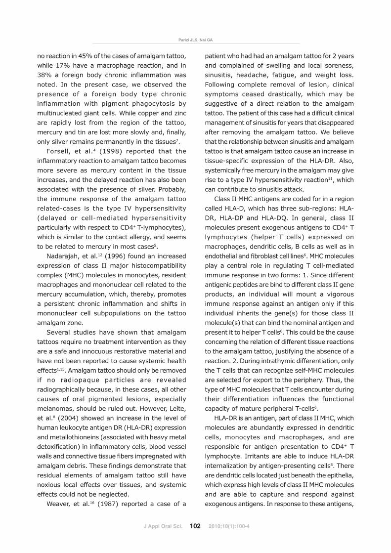
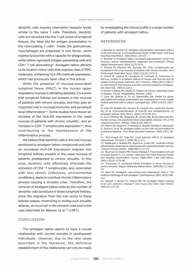
Histopathological examination revealed epithelial
acanthosis and brownish-black pigment deposits
in the submucosa surrounded by a lymphocyte
inflammatory infiltrate (Figure 2), and
multinucleated giant cells phagocyting these
pigments (Figure 3). Muscle cells were impregnated
in the deeper layers (Figure 4). There was no iron
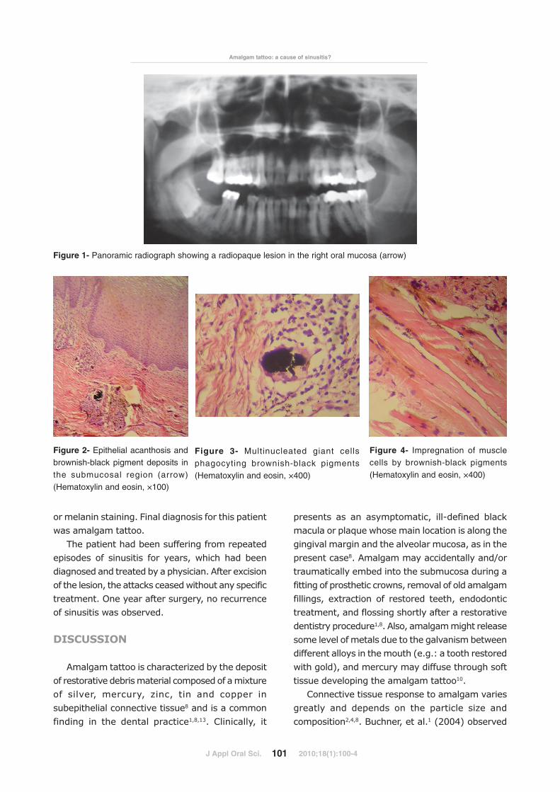
or melanin staining. Final diagnosis for this patient
was amalgam tattoo.
The patient had been suffering from repeated
episodes of sinusitis for years, which had been
diagnosed and treated by a physician. After excision
of the lesion, the attacks ceased without any specific
treatment. One year after surgery, no recurrence
of sinusitis was observed.
DISCUSSION
Amalgam tattoo is characterized by the deposit
of restorative debris material composed of a mixture
of silver, mercury, zinc, tin and copper in
subepithelial connective tissue8 and is a common
finding in the dental practice1,8,13. Clinically, it
presents as an asymptomatic, ill-defined black
macula or plaque whose main location is along the
gingival margin and the alveolar mucosa, as in the
present case8. Amalgam may accidentally and/or
traumatically embed into the submucosa during a
fitting of prosthetic crowns, removal of old amalgam
fillings, extraction of restored teeth, endodontic
treatment, and flossing shortly after a restorative
dentistry procedure1,8. Also, amalgam might release
some level of metals due to the galvanism between
different alloys in the mouth (e.g.: a tooth restored
with gold), and mercury may diffuse through soft
tissue developing the amalgam tattoo10.
Connective tissue response to amalgam varies
greatly and depends on the particle size and
composition2,4,8. Buchner, et al.1 (2004) observed
J Appl Oral Sci. 2010;18(1):100-4101
Amalgam tattoo: a cause of sinusitis?
Figure 3- Multinucleated giant cells
phagocyting brownish-black pigments(Hematoxylin and eosin, ×400)
Figure 4- Impregnation of musclecells by brownish-black pigments
(Hematoxylin and eosin, ×400)
Figure 2- Epithelial acanthosis andbrownish-black pigment deposits in
the submucosal region (arrow)
(Hematoxylin and eosin, ×100)
Figure 1- Panoramic radiograph showing a radiopaque lesion in the right oral mucosa (arrow)
no reaction in 45% of the cases of amalgam tattoo,
while 17% have a macrophage reaction, and in
38% a foreign body chronic inflammation was
noted. In the present case, we observed the
presence of a foreign body type chronic
inflammation with pigment phagocytosis by
multinucleated giant cells. While copper and zinc
are rapidly lost from the region of the tattoo,
mercury and tin are lost more slowly and, finally,
only silver remains permanently in the tissues7.
Forsell, et al.4 (1998) reported that the
inflammatory reaction to amalgam tattoo becomes
more severe as mercury content in the tissue
increases, and the delayed reaction has also been
associated with the presence of silver. Probably,
the immune response of the amalgam tattoo
related-cases is the type IV hypersensitivity
(delayed or cell-mediated hypersensitivity
particularly with respect to CD4+ T-lymphocytes),
which is similar to the contact allergy, and seems
to be related to mercury in most cases5.
Nadarajah, et al.12 (1996) found an increased
expression of class II major histocompatibility
complex (MHC) molecules in monocytes, resident
macrophages and mononuclear cell related to the
mercury accumulation, which, thereby, promotes
a persistent chronic inflammation and shifts in
mononuclear cell subpopulations on the tattoo
amalgam zone.
Several studies have shown that amalgam
tattoos require no treatment intervention as they
are a safe and innocuous restorative material and
have not been reported to cause systemic health
effects1,15. Amalgam tattoo should only be removed
if no radiopaque particles are revealed
radiographically because, in these cases, all other
causes of oral pigmented lesions, especially
melanomas, should be ruled out. However, Leite,
et al.8 (2004) showed an increase in the level of
human leukocyte antigen DR (HLA-DR) expression
and metallothioneins (associated with heavy metal
detoxification) in inflammatory cells, blood vessel
walls and connective tissue fibers impregnated with
amalgam debris. These findings demonstrate that
residual elements of amalgam tattoo still have
noxious local effects over tissues, and systemic
effects could not be neglected.
Weaver, et al.16 (1987) reported a case of a
patient who had had an amalgam tattoo for 2 years
and complained of swelling and local soreness,
sinusitis, headache, fatigue, and weight loss.
Following complete removal of lesion, clinical
symptoms ceased drastically, which may be
suggestive of a direct relation to the amalgam
tattoo. The patient of this case had a difficult clinical
management of sinusitis for years that disappeared
after removing the amalgam tattoo. We believe
that the relationship between sinusitis and amalgam
tattoo is that amalgam tattoo cause an increase in
tissue-specific expression of the HLA-DR. Also,
systemically free mercury in the amalgam may give
rise to a type IV hypersensitivity reaction11, which
can contribute to sinusitis attack.
Class II MHC antigens are coded for in a region
called HLA-D, which has three sub-regions: HLA-
DR, HLA-DP and HLA-DQ. In general, class II
molecules present exogenous antigens to CD4+ T
lymphocytes (helper T cells) expressed on
macrophages, dendritic cells, B cells as well as in
endothelial and fibroblast cell lines6. MHC molecules
play a central role in regulating T cell-mediated
immune response in two forms: 1. Since different
antigenic peptides are bind to different class II gene
products, an individual will mount a vigorous
immune response against an antigen only if this
individual inherits the gene(s) for those class II
molecule(s) that can bind the nominal antigen and
present it to helper T cells6. This could be the cause
concerning the relation of different tissue reactions
to the amalgam tattoo, justifying the absence of a
reaction. 2. During intrathymic differentiation, only
the T cells that can recognize self-MHC molecules
are selected for export to the periphery. Thus, the
type of MHC molecules that T cells encounter during
their differentiation influences the functional
capacity of mature peripheral T-cells6.
HLA-DR is an antigen, part of class II MHC, which
molecules are abundantly expressed in dendritic
cells, monocytes and macrophages, and are
responsible for antigen presentation to CD4+ T
lymphocyte. Irritants are able to induce HLA-DR
internalization by antigen-presenting cells8. There
are dendritic cells located just beneath the epithelia,
which express high levels of class II MHC molecules
and are able to capture and respond against
exogenous antigens. In response to these antigens,
J Appl Oral Sci. 2010;18(1):100-4102
Parizi JLS, Nai GA
dendritic cells express chemokine receptor levels
similar to the naive T cells. Therefore, dendritic
cells are recruited into the T-cell zones of lymphoid
tissues, the ideal site for antigen presentation to
the recirculating T cells6. Inside the granulomas,
macrophages are presented in two forms: some
express lysozymes with a capacity for phagocytosis,
while others represent antigen-presenting cells and
CD4+ T cell-stimulating6. Amalgam tattoo attracts
to its location many cells that express class II MHC
molecules, enhancing HLA-DR molecule expression,
which has previously been cited in this article.
While the presence of mucosa-associated
lymphoid tissue (MALT) in the human upper
respiratory mucosa is still being debated, it is known
that lymphoid follicles are present in the mucosa
of patients with chronic sinusitis, and they play an
important role in mucosal immunity and persistent
local inflammation14. Some studies have shown an
increase of the HLA-DR expression in the nasal
mucosa of patients with chronic sinusitis7, and an
increase in CD4+ T lymphocytes expression3,9, thus
contributing to the maintenance of the
inflammatory process.
We believe that dendritic cells in the oral mucosa
sensitized to amalgam tattoo compounds and with
an increased HLA-DR expression migrate into
lymphoid follicles present in the nasal mucosa of
patients predisposed to chronic sinusitis. In this
area, dendritic cells effectively stimulate the
activation of CD4+ T lymphocytes, and, associated
with new stimuli (infections, environmental
conditions), leads to a profuse chronic inflammatory
process causing a sinusitis crisis. Therefore, the
removal of amalgam tattoo reduces the number of
dendritic cells sensitized in these lymphoid follicles,
since the migration from the oral cavity to these
follicles ceases, minimizing or ending such sinusitis
attacks, as occurred in the present case and in the
case described by Weaver, et al.16 (1987).
CONCLUSION
The amalgam tattoo seems to have a crucial
relationship with chronic sinusitis in predisposed
individuals. However, due to the rare cases
described in the literature, the definitive
establishment of this relationship can only be made
by investigating the clinical profile in a large number
of patients with amalgam tattoo.
REFERENCES
1- Buchner A, Hansen LS. Amalgam pigmentation (amalgam tattoo)
of the oral mucosa: a clinicopathologic study of 268 cases. Oral Surg
Oral Med Oral Pathol. 1980;49(2):139-47.
2- Buchner A. Amalgam tattoo (amalgam pigmentation) of the oral
mucosa: clinical manifestations, diagnosis and treatment. Refuat
Hapeh Vehashinayim 2004;21(3):25-8.
3- Driscoll PV, Naclerio RM, Baroody FM. CD4+ lymphocytes are
increased in the sinus mucosa of children with chronic sinusitis. Arch
Otolaryngol Head Neck Surg. 1996;122(10):1071-6.
4- Forsell M, Larsson B, Ljungqvist A, Carlmark B, Johansson O.
Mercury content in amalgam tattoos of human oral mucosa and its
relation to local tissue reactions. Eur J Oral Sci. 1998;106(1):582-7.
5- Holmstrup P. Oral mucosa and skin reactions related to amalgam.
Adv Dent Res. 1992;6:120-4.
6- Kumar V, Abbas AK, Fausto N. Robbins & Cotran: pathologic basis
of disease. 7th ed. Philadelphia: Elsevier; 2005.
7- Lane AP, Saatian B, Yu XY, Spannhake EW. mRNA for genes
associated with antigen presentation are expressed by human middle
meatal epithelial cells in culture. Laryngoscope. 2004;114(10):1827-
32.
8- Leite CM, Botelho AS, Oliveira JR, Cardoso SV, Loyola AM, Gomez
RS, et al. Immunolocalization of HLA-DR and metallothionein on
amalgam tattoos. Braz Dent J. 2004;15(2):99-103.
9- Liu LY, Marthur SK, Sedgwick JB, Jarjour NN, Busse WW, Kelly EA.
Human airway and peripheral blood eosinophils enhance Th1 e Th2
cytokine secretion. Allergy. 2006;61(5):589-97.
10- Martin JM, Nagore E, Cremades A, Botella-Estrada R, Sanmartin
O, Sevila A, et al. An amalgam tattoo on the oral mucosa related to
a dental prosthesis. J Eur Acad Dermatol Venereol. 2005;19(1):90-
2.
11- McCullough MJ, Tyas MJ. Local adverse effects of amalgam
restorations. Int Dent J. 2008;58(1):3-9.
12- Nadarajah V, Neiders ME, Aguirre A, Cohen RE. Localized cellular
inflammatory responses to subcutaneously implanted dental mercury.
J Toxicol Environ Health. 1996;49(2):113-25.
13- Shulman JD, Beach MM, Rivera-Hidalgo F. The prevalence of oral
mucosal lesions in U.S. adults: data from the Third National Health
and Nutrition Examination Survey 1988-1994. J Am Dent Assoc.
2004;135(9):1279-86.
14- Tsurumaru H. Lymphoid follicle formation in sinus mucosa of
chronic sinusitis. Nippon Jibiinkoka Gakkai Kaiho. 1996;99(11):1662-
75.
15- Wahl MJ. Amalgam: resurrection and redemption. Part 2: The
medical mythology of anti-amalgam. Quintessence. 2001;32(9):696-
710.
16- Weaver T, Auclair PL, Taybos GM. An amalgam tattoo causing
local and systemic disease? Oral Surg Oral Med Oral Pathol.
1987;63:137-40.
J Appl Oral Sci. 2010;18(1):100-4103
Amalgam tattoo: a cause of sinusitis?