Embed Size (px)
Citation preview
8/8/2019 Am J Physiol Cell Physiol 2001 Yamaguchi C382 93
http://slidepdf.com/reader/full/am-j-physiol-cell-physiol-2001-yamaguchi-c382-93 1/13
280:C382-C393, 2001. Am J Physiol Cell PhysiolVassilev, Jennifer L. Sanders and Edward M. BrownToru Yamaguchi, Naibedya Chattopadhyay, Olga Kifor, Chianping Ye, Peter M.human osteoblastic MG-63 cell lineExpression of extracellular calcium-sensing receptor in
You might find this additional info useful...
49 articles, 20 of which can be accessed free at:This article citeshttp://ajpcell.physiology.org/content/280/2/C382.full.html#ref-list-1
9 other HighWire hosted articles, the first 5 are:This article has been cited by
[PDF][Full Text][Abstract], March, 1 2003; 284 (3): R835-R852. Am J Physiol Regul Integr C omp Physiol
J. F. Staub, E. Foos, B. Courtin, R. Jochemsen and A. M. Perault-StaubmetabolismA nonlinear compartmental model of Sr metabolism. II. Its physiological relevance for Ca
[PDF][Full Text][Abstract], December, 2 2005; 280 (48): 40201-40209. J. Biol. Chem.
Fontilla-Poole, Robert W. Mays, Kurt R. Brunden, John J. Harrington and L. Darryl QuarlesMin Pi, Pieter Faber, George Ekema, P. David Jackson, Anthony Ting, Nancy Wang, MichelleIdentification of a Novel Extracellular Cation-sensing G-protein-coupled Receptor
[PDF][Full Text][Abstract], March, 1 2006; 290 (3): H1165-H1171. Am J Physiol Heart Circ Phy siol
Haunso and Soren P. SheikhJacob Tfelt-Hansen, Jakob Lerche Hansen, Sanela Smajilovic, Ernest F. Terwilliger, StigCalcium receptor is functionally expressed in rat neonatal ventricular cardiomyocytes
[PDF][Full Text][Abstract], October, 2009; 297 (4): E915-E923. Am J Physiol Endocrinol Me tabMartin R. Pollak, David Goltzman and Edward M. Brown
Lakshmi Kantham, Steven J. Quinn, Ogo I. Egbuna, Khanjan Baxi, Robert Butters, Jian L. Pang,regulation of parathyroid hormone secretionThe calcium-sensing receptor (CaSR) defends against hypercalcemia independently of its
[PDF][Full Text][Abstract], October, 2010; 1 0 (5): 305-312. Mol Interv
Pierre J. MarieCell BiologyStrontium Ranelate in Osteoporosis and Beyond: Identifying Molecular Targets in Bone
including high resolution figures, can be found at:Updated information and serviceshttp://ajpcell.physiology.org/content/280/2/C382.full.html
can be found at: AJP - Cell PhysiologyaboutAdditional material and informationhttp://www.the-aps.org/publications/ajpcell
This infomation is current as of December 14, 2010.
American Physiological Society. ISSN: 0363-6143, ESSN: 1522-1563. Visit our website at http://www.the-aps.org/.a year (monthly) by the American Physiological Society, 9650 Rockville Pike, Bethesda MD 20814-3991. Copyright © 2001 by the
is dedicated to innovative approaches to the study of cell and molecular physiology. It is published 12 times AJP - Cell Physiology
8/8/2019 Am J Physiol Cell Physiol 2001 Yamaguchi C382 93
http://slidepdf.com/reader/full/am-j-physiol-cell-physiol-2001-yamaguchi-c382-93 2/13
Expression of extracellular calcium-sensing receptorin human osteoblastic MG-63 cell line
TORU YAMAGUCHI, NAIBEDYA CHATTOPADHYAY, OLGA KIFOR, CHIANPING YE,PETER M. VASSILEV, JENNIFER L. SANDERS, AND EDWARD M. BROWN Endocrine-Hypertension Division, Department of Medicine, Brigham and Women’s Hospital, Harvard Medical School, Boston, Massachusetts 02115Received 12 May 2000; accepted in nal form 15 September 2000
Yamaguchi, Toru, Naibedya Chattopadhyay, OlgaKifor, Chianping Ye, Peter M. Vassilev, Jennifer L.Sanders, and Edward M. Brown. Expression of extracel-lular calcium-sensing receptor in human osteoblastic MG-63cell line. Am J Physiol Cell Physiol 280: C382–C393,2001.—We have previously shown the expression of the ex-tracellular calcium (Ca o
2 )-sensing receptor (CaR) in osteo-blast-like cell lines, and others have documented its expres-
sion in sections of murine, bovine, and rat bone. Theexistence of the CaR in osteoblasts remains controversial,however, since some studies have failed to document itsexpression in the same osteoblast-like cell lines. The goals of the present study were twofold. 1) We sought to determinewhether the CaR is expressed in the human osteoblast-likecell line, MG-63, which has recently been reported by othersnot to express this receptor. 2) We investigated whether theCaR, if present in MG-63 cells, is functionally active, sincemost previous studies have not proven the role of the CaR inmediating known actions of Ca o
2 on osteoblast-like cells. Weused immunocytochemistry and Western blotting with thespecic, afnity-puried anti-CaR antiserum 4637 as well asNorthern blot analysis and RT-PCR using a riboprobe andPCR primers specic for the human CaR, respectively, to
show readily detectable CaR protein and mRNA expressionin MG-63 cells. Finally, we employed the patch-clamp tech-nique to show that an elevation in Ca o
2 as well as thespecic, allosteric CaR activator NPS R-467 (0.5 M), but notits less active stereoisomer NPS S-467 (0.5 M), activate anoutward K channel in MG-63 cells, strongly suggesting thatthe CaR in MG-63 cells is not only expressed but is function-ally active.
G protein-coupled receptor; potassium channel; Northernanalysis; reverse transcriptase-polymerase chain reaction;Western analysis; immunocytochemistry
MAINTAINING THE EXTRACELLULAR CALCIUM CONCENTRATION
(Ca o2
) within a narrow physiological range is crucialfor numerous cellular processes, including the mainte-nance of membrane potential as well as cellular prolif-eration, differentiation, and secretion (4). Precise reg-ulation of Ca o
2 is afforded by a G protein-coupled,Ca o
2 -sensing receptor (CaR) that was originally clonedfrom bovine parathyroid gland and senses Ca o
2 as an
extracellular rst messenger (6). The CaR was laterisolated from rat C cells (18, 21) and kidney (38) andalso shown to be present in the intestine (11), therebyimplicating it in maintaining Ca o
2 homeostasis notonly through its actions on the secretion of Ca o
2 -regu-lating hormones (i.e., parathyroid hormone and calci-tonin) but also through its effects on tissues translo-cating Ca 2 into or out of the extracellular uid (e.g.,kidney and intestine).
Bone, like parathyroid, kidney, and intestine, partic-ipates in systemic Ca o
2 homeostasis (5). Thus the CaRcould also potentially play some role(s) within theskeleton by sensing local changes in Ca o
2 owing tobone remodeling. Bone formation during skeletal turn-over is preceded by the migration of macrophage-likemononuclear cells to sites of recent bone resorptionduring the “reversal” phase that precedes the layingdown of new bone (3). Preosteoblasts subsequentlymigrate to the same sites, differentiate into matureosteoblasts, and eventually deposit and mineralize os-teoid protein (36, 37). Bone resorption can produce
local increases in Ca o
2
beneath resorbing osteoclaststhat reach levels as high as 8–40 mM (39). The lattercould, therefore, provide both macrophage-like mono-nuclear cells and preosteoblasts in the local microen- vironment with a signal that modulates their subse-quent physiological responses, such as migration andproliferation. In fact, high Ca o
2 induces chemotaxis of human peripheral blood monocytes (41) and both che-motaxis and proliferation of mouse osteoblasticMC3T3-E1 cells (44). These two cell types have thecapacity, respectively, to differentiate into mature os-teoclasts (19) and osteoblasts (40) under appropriateconditions in culture.
Whether the CaR mediates the known actions of
Ca o2
on osteoblasts, however, remains controversial.We previously showed expression of this receptor indiverse cell types within human bone marrow, includ-ing alkaline phosphatase (ALP)-positive, putative os-teoblast precursors (23). Using multiple detectionmethods (i.e., immunocytochemistry, Western andNorthern analyses, and RT-PCR), we subsequently
Address for reprint requests and other correspondence: E. M.Brown, Endocrine-Hypertension Div., Brigham and Women’s Hospi-tal, 221 Longwood Ave., Boston, MA 02115 (E-mail: embrown @rics.bwh.harvard.edu).
The costs of publication of this article were defrayed in part by thepayment of page charges. The article must therefore be herebymarked ‘‘ advertisement ’’ in accordance with 18 U.S.C. Section 1734solely to indicate this fact.
Am J Physiol Cell Physiol280: C382–C393, 2001.
0363-6143/01 $5.00 Copyright © 2001 the American Physiological Society http://www.ajpcell.orgC382
8/8/2019 Am J Physiol Cell Physiol 2001 Yamaguchi C382 93
http://slidepdf.com/reader/full/am-j-physiol-cell-physiol-2001-yamaguchi-c382-93 3/13
identied CaR protein and mRNA in the osteoblast-like cell lines UMR-106 and SAOS-2 (45). Recently, wealso found that the murine ST-2 stromal cell line (43)and the murine MC3T3-E1 osteoblastic cell line (44)express the CaR. Others have likewise shown in recentstudies that the CaR is expressed in the latter cell line(25) as well as in most osteoblasts in sections of mu-rine, rat, and bovine bone (10). Furthermore, CaRagonists stimulate chemotaxis and proliferation of both ST-2 and MC3T3-E1 cells (43, 45), suggesting thatthe receptor could potentially represent the molecularmediator of some or even all of the previously docu-mented actions of high Ca o
2 on osteoblasts and/or theirprecursors.
However, some investigators have failed to detectCaR expression in osteoblast-like cells (33, 35). Indeed,Quarles and coworkers (35) have suggested that theeffects of elevated levels of Ca o
2 on MC3T3-E1 cells aremediated by a Ca o
2 -sensing mechanism distinct fromthe CaR, based, in part, on their failure to detect theCaR by RT-PCR or Western blot analysis in U-2OS,SAOS-2, and MG-63 osteoblast-like cells in a recentstudy (33). Thus whether or not the CaR and/or otherCa o
2 sensors are expressed in osteoblasts still remainscontroversial.
The goals of the present study were twofold: 1) todetermine whether the CaR is expressed in MG-63cells, an osteoblast-like cell line that we have not stud-ied previously; and 2) to evaluate whether the CaR, if expressed in this cell line, is functionally active. Ourresults demonstrate readily detectable expression of afunctionally active CaR on the cell surface of MG-63cells, thereby further supporting our previous evidencethat the CaR mediates as least some of the knownactions of Ca o
2 on osteoblast-like cells.
MATERIALS AND METHODS
Materials. All routine culture media were obtained fromGIBCO BRL (Grand Island, NY). NPS R-467 and NPS S-467were generous gifts of Dr. Edward F. Nemeth, NPS Pharma-ceuticals, Salt Lake City, UT.
Preparation of bovine parathyroid cells. Parathyroidglands from calves were collected on ice, minced into smallfragments, and digested at 37°C for 75 min with collagenaseand DNase as described previously (7, 27). Dispersed cellswere subsequently utilized for preparation of cellular pro-teins to be employed for Western blotting as described below.
Culture and maintenance of CaR transfected and untrans- fected HEK-293 cells. We obtained a clonal line of human
embryonic kidney (HEK-293) cells stably transfected withthe cDNA encoding the human parathyroid CaR [hPCaR4.0(20)] (referred to here as HEKCaR cells) as well as untrans-fected HEK-293 cells (designated as HEK-293 cells) as gen-erous gifts from Dr. Kimberly Rogers, NPS Pharmaceuticals.We have previously shown that untransfected HEK-293 cellsdo not express an endogenous CaR and are unresponsive toCaR agonists, whereas HEK-293 cells transiently or stablytransfected with the CaR show robust, high Ca o
2 -evokedincreases in the cytosolic Ca 2 concentration (Ca i
2 ),accumulation of inositol phosphates, and regulation of otherCaR-dependent signaling pathways (1, 26). Cells weregrown in Dulbecco’s modied Eagle’s medium with 10%
fetal bovine serum (FBS) without pyruvate and with 200g/ml hygromycin.Cell culture. MG-63 cells, established as an osteoblastic
cell line from a human osteosarcoma, were obtained from Dr.Nancy Weigel, Baylor College of Medicine, Houston, TX, whoobtained the cells originally from American Type CultureCollection (Manassas, VA). MG-63 cells were grown in -Ea-gle’s minimum essential medium (Ca 2 and 1.8 mM; Mg 2
and 0.81 mM; H 2 PO 4 , 1.0 mM) supplemented with 10% FBS(Hyclone, Logan, UT) and 1% penicillin/streptomycin in 5%CO2 at 37°C. The medium was changed twice weekly, and thecells were subcultured into 25-cm 2 culture asks by detach-ing them gently with a cell scraper after reaching subconu-ency. For morphological evaluation, MG-63 cells were platedonto 12-mm circular glass coverslips in 12-well (2.0 cm 2
plates. After 24 h of culture, the medium was discarded, andeach coverslip with adherent cells was washed once withphosphate-buffered saline (PBS), xed with 4% formalde-hyde in PBS for 5 min, and washed with PBS once again.Coverslips were stored for 1 to 7 days at 4°C before immu-nocytochemistry or immunouorescence was performed,which had no effect on the results observed.
Immunocytochemistry for the CaR in MG-63 cells. Immu-nocytochemistry was performed using either mouse monoclo-nal anti-CaR antibody ADD, which was raised to a syntheticpeptide within the CaR corresponding to residues 214–235,or polyclonal anti-CaR antiserum 4637, and the latter wasafnity puried by us as described previously (27). Bothantisera were generously provided by NPS Pharmaceuticals.Monoclonal antibody ADD (1, 2, 21) was raised to a peptidecorresponding to amino acids 214–235 within the predictedextracellular domain of the human CaR, while antiserum4637 was raised to a peptide corresponding to amino acids345–359 of the bovine CaR (21, 27). Optimal stability of afnity-puried antiserum 4637 was obtained by storing it at
20°C in PBS, pH 7.4, that contained 50% glycerol, 1 mg/mlbovine serum albumin (BSA), and 1.5 mM sodium azide.Under these conditions, the titer of the antiserum was un-changed for at least 2 yr as assessed by Western blotting of proteins in crude plasma membranes prepared from CaR-transfected HEK-293 cells.
MG-63 cells xed as described above were treated withDako peroxidase blocking reagent (Dako, Carpenteria, CA)for 10 min to inhibit endogenous peroxidases and then withDako protein block serum-free solution (Dako) for 1 h. Theywere subsequently incubated overnight at 4°C with eitherthe monoclonal ADD antibody or the afnity-puried 4637polyclonal antiserum at concentrations of 5 and 10 g/mlrespectively, in blocking solution (23, 43). Negative controlswere carried out by incubating the cells with the respectiveantisera after they had been preabsorbed with 10 g/ml othe specic peptide against which each had been raised. Afterthe cells were washed three times with 0.5% BSA in PBS for10 min each, peroxidase-coupled sheep anti-mouse or goatanti-rabbit IgG (1:200; Sigma Chemical), respectively, wereadded and incubated for 1 h at room temperature. The cellswere then washed with PBS three times, and the colorreaction was developed using the Dako AEC substrate sys-tem (Dako) for about 10 min. The color reaction was stoppedby washing the slides three times with PBS and once withwater.
Detection of CaR by immunouorescence and uorescenceimmunolocalization by confocal microscopy. For detection of the CaR by immunouorescence using antiserum 4637, form-aldehyde-xed MG-63 or HEKCaR cells were rst treated for10 min with PBS that contained 0.1 mM glycine to quenchintrinsic uorescence due to xation. To block nonspecic
C383C AR IN OSTEOBLASTIC MG-63 CELLS
8/8/2019 Am J Physiol Cell Physiol 2001 Yamaguchi C382 93
http://slidepdf.com/reader/full/am-j-physiol-cell-physiol-2001-yamaguchi-c382-93 4/13
antibody binding sites, the cells were then incubated withPBS that contained 1% BSA and 1% normal goat serum for30 min at room temperature. After blocking, the cells wereincubated overnight at 4°C with 10 g/ml of the antiserum inthe blocking solution. On the following day, the cells werewashed three times with 50% blocking solution in PBS andincubated with a secondary goat anti-rabbit antiserumtagged with the uorophore Alexa 568 (Molecular Probes,Eugene, OR) for 3 h in the dark at room temperature. Thecells were subsequently washed three times with PBS andthree times with deionized water and mounted using anti-fading mounting uid (Vector Laboratories, Burlingame,CA). For identication of plasma membrane localization of the CaR, some preparations of MG-63 cells were doubleimmunostained for the CaR and for either of the two plasmamembrane markers, Na -K -ATPase or alkaline phospha-tase (ALP) (located on the cell surface of osteoblasts). Afterblocking the nonspecic background staining, the cells wereincubated with a mixture of rabbit polyclonal anti-CaR anti-serum 4637 and chicken anti-Na -K -ATPase (ChemiconInternational, Temecula, CA) or mouse monoclonal anti-al-kaline phosphatase (Chemicon International) antibodies inblocking solution overnight at 4°C in a humidied chamber.The next day, after being washed as above, the cells wereincubated with a mixture of secondary antibodies [i.e., AlexaFluor 568-conjugated goat anti-rabbit IgG (MolecularProbes)/FITC-conjugated anti-chicken IgG (Sigma) or AlexaFluor 568 goat anti-rabbit IgG/Alexa 488-conjugated goatanti-mouse IgG (Molecular Probes)] for 3 h in the dark atroom temperature. Images of the resultant uorescence werethen collected on a Bio-Rad MRC 1024.2P multiphoton con-focal microscope in the Brigham and Women’s Hospital Con-focal Microscope Core Facility. The system is equipped withkrypton and argon lasers that can produce excitation wave-lengths of 488, 568, and 647 nm. Alexa 568 was excited at 568nm, which produces a red signal, while uorescein or Alexa488 was excited at 488 nm, which yields a green signal. Theautouorescence of the samples was minimal and was sub-tracted from the values obtained during measurements. Be-cause peptide blocking of anti-CaR antiserum 4637 was per-formed for both immunoperoxidase staining and Westernblotting to establish its specicity, this control was not re-peated in the studies using immunouorescence. Photomicro-graphy was carried out at 1,000.
Western analysis of the CaR in MG-63 cells. Dispersedbovine parathyroid cells or conuent monolayers of HEKCaRor MG-63 cells that had been cultured in six-well plasticcluster plates were rinsed with ice-cold PBS and scraped onice into lysis buffer that contained 10 mM Tris HCl, pH 7.4,1 mM EGTA, 1 mM EDTA, 0.25 M sucrose, 1% Triton X-100,1 mM dithiothreitol, and a cocktail of protease inhibitors (10
g/ml each of aprotinin, leupeptin, and calpain inhibitor aswell as 100 g/ml Pefabloc). The cells were then passedthough a 22-gauge needle 10 times. Nuclei and cell debriswere removed by low-speed centrifugation (1,000 g for 10min), and the resultant total cellular lysate in the superna-tant was used either directly for SDS-PAGE or stored at
80°C.Immunoblot analysis was performed essentially as de-
scribed before (27, 28). Aliquots of supernatant fractionscontaining the total cellular lysate (20 g of protein fromHEKCaR and bovine parathyroid cells and 40 g from MG-63cells) were mixed with an equal volume of 2 SDS-Laemmligel loading buffer containing 100 mM dithiothreitol, incu-bated at 37°C for 15 min, and resolved electrophoretically onlinear 3–10% gradient gels. The separated proteins werethen transferred to nitrocellulose lters (Schleicher and
Schuel, Keene, NH) and incubated with blocking solution(PBS with 0.25% Triton X-100 and 5% dry milk) for 1 h atroom temperature. The blots were subsequently incubatedovernight at 4°C with afnity-puried polyclonal antiserum4637 at 1 g/ml with or without preincubation with twice theconcentration (e.g., 2 g/ml) of the peptide against which theantiserum was raised (as a control for nonspecic binding) inblocking solution with 1% dry milk. The blots were subse-quently washed ve times with PBS that contained 0.25%Triton X-100 and 0.1% dry milk (washing solution) at roomtemperature for 10 min each. The blots were further incu-bated with a 1:2,000 dilution of horseradish peroxidase-cou-pled goat anti-rabbit IgG (Sigma) in blocking solution with1% dry milk for 1 h at room temperature. The blots werenally washed ve times with the washing solution, andprotein bands were detected using an enhanced chemilumi-nescence system (Renaissance kit, DuPont-NEN).
Detection of CaR transcripts in MG-63 cells by Northernblot analysis. To determine the size of the CaR transcript(s)in MG-63 cells, Northern blot analysis was employed onaliquots of 2 g poly(A) RNA obtained using oligo(dT) cel-lulose chromatography of total RNA (11). RNA samples weredenatured and electrophoresed in 2.2 M formaldehyde-1%agarose gels along with a 0.24-kb to 9.5-kb RNA ladder(GIBCO BRL) and transferred overnight to nylon mem-branes (Duralon; Stratagene, La Jolla, CA). A 486-bp KpnI- Xba I fragment corresponding to nucleotides 1745 to 2230of the human parathyroid CaR cDNA (1, 20) was subclonedinto the pBluescript SK vector. The plasmid was then lin-earized with Kpn I, and a 32 P-labeled riboprobe was synthe-sized with the MAXIscript T3 kit (Pharmacia Biotech, Pisca-taway, NJ) using T3 polymerase and [ 32 P]UTP. Nylonmembranes were prehybridized for 2 h at 55°C in a solutionconsisting of 50% formamide, 4 Denhardt’s solution (50Denhardt’s 5 g Ficoll, 5 g polyvinylpyrrolidone, and 5 gBSA/50 ml), 5 sodium chloride-sodium phosphate-EDTA(SSPE) (20 SSPE 2.98 M NaCl and 0.02 M EDTA in 0.2M phosphate buffer, pH 7.0), 0.5% SDS, 10% dextran sulfate,250 g/ml yeast tRNA, and 200 g/ml calf thymus DNA.Labeled cRNA probe (2 10 6 cpm/ml) was then added, andthe membranes were hybridized overnight at 68°C. Washingwas carried out at high stringency [0.1 SSC (20 SSC 3M NaCl and 0.3 M Na 3 -citrate 2H 2 O) and 0.1% SDS at 68°C]for 30 min (6). The membranes were sealed in plastic andexposed to a PhosphorImager screen. The screens were ana-lyzed on a PhosphorImager (Molecular Dynamics, Sunny- vale, CA) using the ImageQuant program.
RT-PCR of CaR transcripts in MG-63 cells. Total RNA wasprepared from monolayers of MG-63 cells in 25-cm 2 cultureasks with the TRIzol reagent (GIBCO BRL). One micro-gram of total RNA was used for the synthesis of single-stranded cDNA (cDNA synthesis kit, GIBCO BRL). The re-sultant rst-stranded cDNA was used for the PCR procedure(16, 45). PCR was performed in a buffer that contained thefollowing nal concentrations of the listed reagents: 20 mMTris HCl (pH 8.4), 50 mM KCl, 1.8 mM MgCl 2 , 0.2 mMdNTP, 0.4 M of forward primer, 0.4 M of reverse primer,and 1 l of ELONGASE enzyme mix (a Taq / Pyrococcus spe-cies GB-D DNA polymerase mixture; GIBCO BRL) (6, 10,13–15). A human parathyroid CaR (20) sense primer, 5CGGGGTACCTTAAGCACCTACGGCATCTAA-3 , and anti-sense primer, 5 - GCTCTAGAGTTAACGCGATCCCAAAGG-GCTC-3 , were employed for the reaction. This set of primerswas designed to span two introns of the human CaR gene toavoid amplication of a similar-sized product from contami-nating genomic DNA. To perform hot-start PCR, the enzymewas added during the initial 3-min denaturation and was
C384 C AR IN OSTEOBLASTIC MG-63 CELLS
8/8/2019 Am J Physiol Cell Physiol 2001 Yamaguchi C382 93
http://slidepdf.com/reader/full/am-j-physiol-cell-physiol-2001-yamaguchi-c382-93 5/13
followed by 35 cycles of amplication (30-s denaturation at94°C, 30-s annealing at 55°C, and 1-min extension at 72°C).The reaction was completed with an additional 10-min incu-bation at 72°C to allow completion of extension. PCR prod-ucts were fractionated on 1.2% agarose gels. The presence of a 425-bp amplied product was consistent with a positivePCR reaction arising from CaR-related sequence withincDNA. The PCR product in the reaction mixture was thenpuried using the QIAquick PCR purication kit (Qiagen,Santa Clarita, CA) and subjected to direct, bidirectionalsequencing employing the same primer pairs used for PCR bymeans of an automated sequencer (AB377; Applied Biosys-tems, Foster City, CA) in the DNA Sequence Faculty of theUniversity of Maine (Orono, ME) using dideoxy terminatorTaq technology.
Electrophysiological measurements. Channel activitieswere measured in cell-attached and inside-out patches incontinuously superfused cells using the patch-clamp tech-nique, as described previously (49). The extracellular bathsolution contained, unless otherwise specied (in mM): 140NaCl, 4.0 KCl, 0.75 CaCl 2 , 1.0 MgCl 2 , 10 glucose, and 10HEPES, pH 7.4. Solutions containing CaR agonists, activa-tors, or other agents were applied to the MG-63 cells bysuperfusion. The pipette solution contained, unless otherwisespecied (in mM): 87.0 NaCl, 55.0 KCl, 1.0 CaCl 2 , 1.0 MgCl 2 ,10 glucose, and 10 HEPES, pH 7.4. When lled with thisexternal solution, pipette tip resistances were 5–10 M . Appropriate concentrations of EGTA were added to achievenal concentrations of 0.1 or 0.5 M free Ca 2 in studies withinside-out patches. Currents were measured using an inte-grating patch-clamp amplier, and single-channel currentswere ltered at 3 kHz. Voltage stimuli were applied, andsingle-channel currents were digitized (200 s per point) andanalyzed using programs that were based on pCLAMP (AxonInstruments, Foster City, CA). The baseline current wasmonitored frequently to ensure proper analysis of single-channel currents.
The resting potential ( V r ) of these cells had a mean valuethat averaged 70 mV in an extracellular solution thatcontained 5.4 mM K . V r was measured in separate experi-
ments or at the end of some single-channel recordings bybreaking the membrane patch with negative pressure. Theobserved mean value of 70 mV was assumed for calculationof membrane potential ( V m ) for experiments in which V r wasnot measured. In experiments using cell-attached patches,V m was expressed as V r plus the voltage applied to the patchpipette ( vp ). Upward deections in the current records rep-resent positive outward currents.
The probability of channel opening ( Po) was calculatedfrom 20-s segments of current records using the equation Po I / N i, where I is the time-averaged current passingthrough the channels for a given period of time, N is thenumber of channels functioning independently within themembrane patch, and i is the single-channel current (49).Single-channel measurements were recorded at 0.75 mMCa o
2 or following an increase in the level of Ca o2 to 2.75 mM.
Single-channel traces were also taken at various voltagesbefore and after addition of the calcimimetic CaR activator,NPS R-467 (0.5 M), or its less active stereoisomer, NPSS-467 (0.5 M), at a level of Ca o
2 of 1.0 mM. Statisticalanalyses of electrophysiological data were carried out usingone-way analysis of variance (ANOVA). Signicant treat-ment effects were further evaluated by Fisher’s protectedleast-signicant difference test of multiple comparisons uti-lizing one-way ANOVA. A P value of 0.05 was considered toindicate a statistically signicant result.
RESULTS
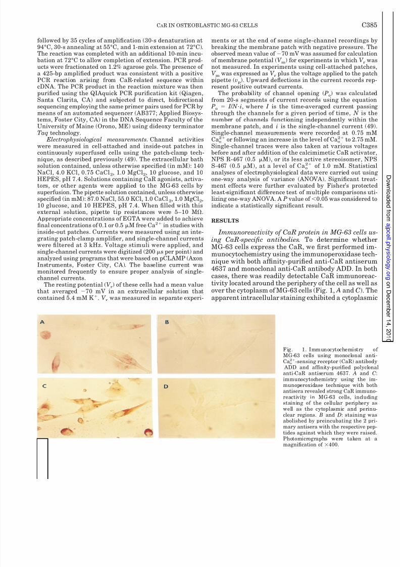
Immunoreactivity of CaR protein in MG-63 cells us-ing CaR-specic antibodies. To determine whetherMG-63 cells express the CaR, we rst performed im-munocytochemistry using the immunoperoxidase tech-nique with both afnity-puried anti-CaR antiserum4637 and monoclonal anti-CaR antibody ADD. In bothcases, there was readily detectable CaR immunoreac-tivity located around the periphery of the cell as well asover the cytoplasm of MG-63 cells (Fig. 1, A and C). The
apparent intracellular staining exhibited a cytoplasmic
Fig. 1. Immunocytochemistry of MG-63 cells using monoclonal anti-Ca o
2 -sensing receptor (CaR) antibody ADD and afnity-puried polyclonalanti-CaR antiserum 4637. A and Cimmunocytochemistry using the im-munoperoxidase technique with bothantisera revealed strong CaR immuno-
reactivity in MG-63 cells, includingstaining of the cellular periphery aswell as the cytoplasmic and perinu-clear regions. B and D: staining wasabolished by preincubating the 2 pri-mary antisera with the respective pep-tides against which they were raised.Photomicrographs were taken at amagnication of 400.
C385C AR IN OSTEOBLASTIC MG-63 CELLS
8/8/2019 Am J Physiol Cell Physiol 2001 Yamaguchi C382 93
http://slidepdf.com/reader/full/am-j-physiol-cell-physiol-2001-yamaguchi-c382-93 6/13
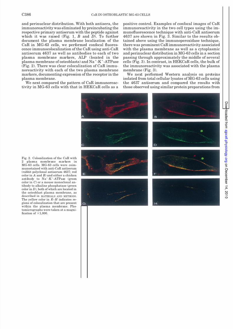
and perinuclear distribution. With both antisera, theimmunoreactivity was eliminated by preincubating therespective primary antiserum with the peptide againstwhich it was raised (Fig. 1, B and D). To furtherdocument the plasma membrane localization of theCaR in MG-63 cells, we performed confocal uores-cence immunocolocalization of the CaR using anti-CaRantiserum 4637 as well as antibodies to each of twoplasma membrane markers, ALP (located in theplasma membrane of osteoblasts) and Na -K -ATPase(Fig. 2). There was clear colocalization of CaR immu-noreactivity with each of the two plasma membranemarkers, documenting expression of the receptor in theplasma membrane.
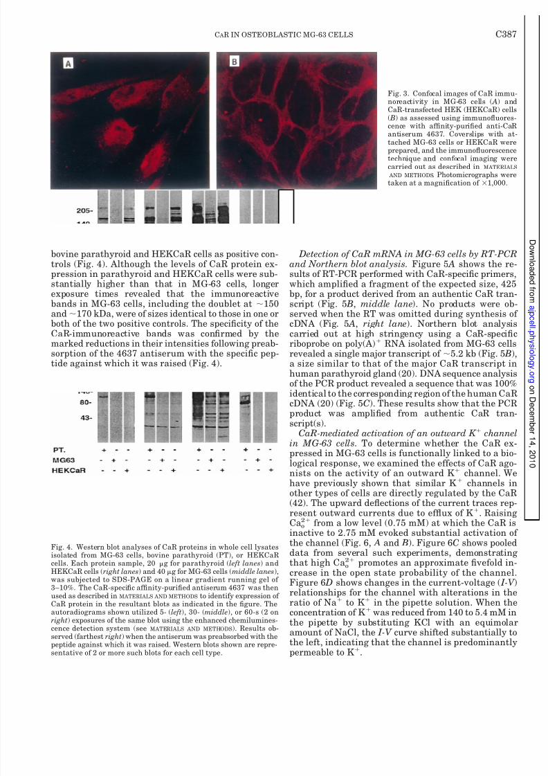
We next compared the pattern of CaR immunoreac-tivity in MG-63 cells with that in HEKCaR cells as a
positive control. Examples of confocal images of CaRimmunoreactivity in the two cell types using the im-munouorescence technique with anti-CaR antiserum4637 are shown in Fig. 3. Similar to the results ob-tained above using the immunoperoxidase technique,there was prominent CaR immunoreactivity associatedwith the plasma membrane as well as a cytoplasmicand perinuclear distribution in MG-63 cells in a sectionpassing through approximately the middle of severalcells (Fig. 3). In contrast, in HEKCaR cells, the bulk of the immunoreactivity was associated with the plasmamembrane (Fig. 3).
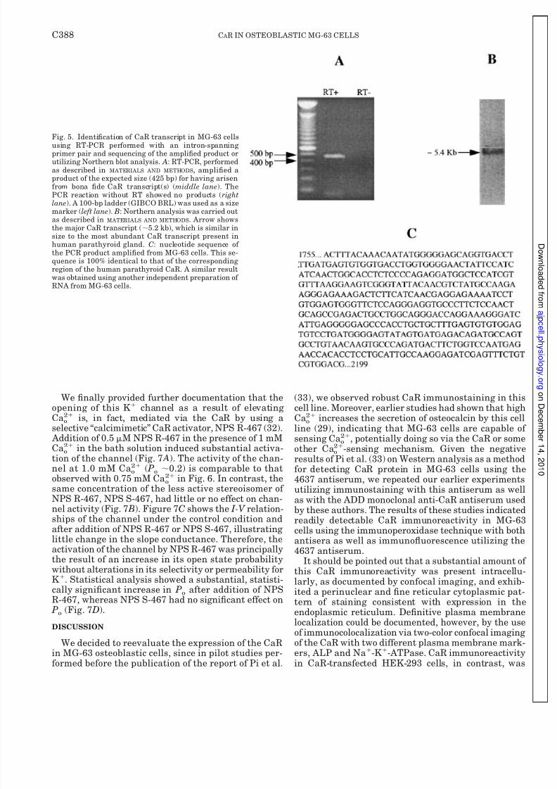
We next performed Western analysis on proteinsisolated from total cellular lysates of MG-63 cells usingthe 4637 antiserum and compared the results withthose observed using similar protein preparations from
Fig. 2. Colocalization of the CaR with2 plasma membrane markers inMG-63 cells. MG-63 cells were coim-munostained with anti-CaR antiserum
(rabbit polyclonal antiserum 4637; redcolor in A and B) and either a chickenantibody to Na -K -ATPase (greencolor in C) or a mouse monoclonal an-tibody to alkaline phosphatase (greencolor in D), both of which are located inthe osteoblast plasma membrane, asdescribed in MATERIALS AND METHODS .The yellow color in E–H indicates re-gions of colocalization that are presentwithin the plasma membrane. Pho-tomicrographs were taken at a magni-cation of 1,000.
C386 C AR IN OSTEOBLASTIC MG-63 CELLS
8/8/2019 Am J Physiol Cell Physiol 2001 Yamaguchi C382 93
http://slidepdf.com/reader/full/am-j-physiol-cell-physiol-2001-yamaguchi-c382-93 7/13
bovine parathyroid and HEKCaR cells as positive con-trols (Fig. 4). Although the levels of CaR protein ex-pression in parathyroid and HEKCaR cells were sub-stantially higher than that in MG-63 cells, longerexposure times revealed that the immunoreactivebands in MG-63 cells, including the doublet at 150and 170 kDa, were of sizes identical to those in one orboth of the two positive controls. The specicity of theCaR-immunoreactive bands was conrmed by themarked reductions in their intensities following preab-sorption of the 4637 antiserum with the specic pep-tide against which it was raised (Fig. 4).
Detection of CaR mRNA in MG-63 cells by RT-PCRand Northern blot analysis. Figure 5 A shows the re-sults of RT-PCR performed with CaR-specic primers,which amplied a fragment of the expected size, 425bp, for a product derived from an authentic CaR tran-script (Fig. 5 B, middle lane ). No products were ob-served when the RT was omitted during synthesis of cDNA (Fig. 5 A, right lane ). Northern blot analysiscarried out at high stringency using a CaR-specicriboprobe on poly(A) RNA isolated from MG-63 cellsrevealed a single major transcript of 5.2 kb (Fig. 5 B)a size similar to that of the major CaR transcript inhuman parathyroid gland (20). DNA sequence analysisof the PCR product revealed a sequence that was 100%identical to the corresponding region of the human CaR
cDNA (20) (Fig. 5 C). These results show that the PCRproduct was amplied from authentic CaR tran-script(s).
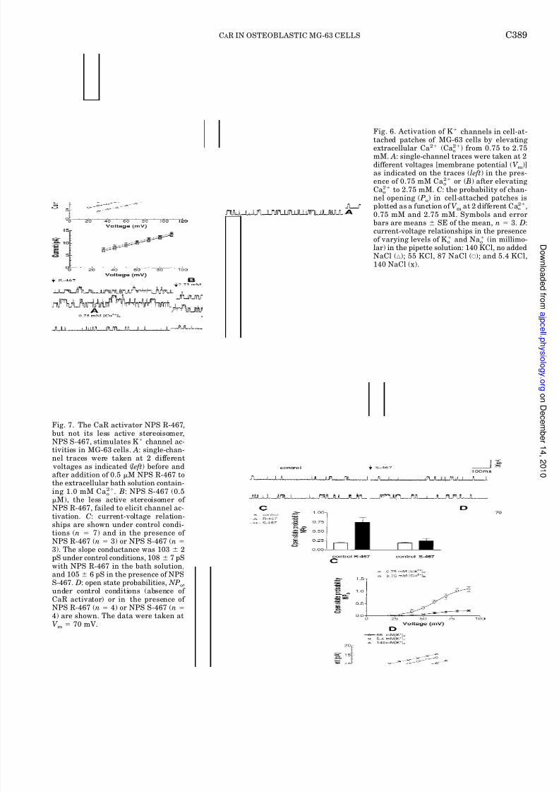
CaR-mediated activation of an outward K channelin MG-63 cells. To determine whether the CaR ex-pressed in MG-63 cells is functionally linked to a bio-logical response, we examined the effects of CaR ago-nists on the activity of an outward K channel. Wehave previously shown that similar K channels inother types of cells are directly regulated by the CaR(42). The upward deections of the current traces rep-resent outward currents due to efux of K . RaisingCa o
2 from a low level (0.75 mM) at which the CaR isinactive to 2.75 mM evoked substantial activation of
the channel (Fig. 6, A and B). Figure 6 C shows pooleddata from several such experiments, demonstratingthat high Ca o
2 promotes an approximate vefold in-crease in the open state probability of the channel.Figure 6 D shows changes in the current-voltage ( I-V relationships for the channel with alterations in theratio of Na to K in the pipette solution. When theconcentration of K was reduced from 140 to 5.4 mM inthe pipette by substituting KCl with an equimolaramount of NaCl, the I-V curve shifted substantially tothe left, indicating that the channel is predominantlypermeable to K .
Fig. 3. Confocal images of CaR immu-noreactivity in MG-63 cells ( A) andCaR-transfected HEK (HEKCaR) cells( B) as assessed using immunouores-cence with afnity-puried anti-CaR
antiserum 4637. Coverslips with at-tached MG-63 cells or HEKCaR wereprepared, and the immunouorescencetechnique and confocal imaging werecarried out as described in MATERIALS AND METHODS . Photomicrographs weretaken at a magnication of 1,000.
Fig. 4. Western blot analyses of CaR proteins in whole cell lysatesisolated from MG-63 cells, bovine parathyroid (PT), or HEKCaRcells. Each protein sample, 20 g for parathyroid ( left lanes ) andHEKCaR cells ( right lanes ) and 40 g for MG-63 cells ( middle lanes ),was subjected to SDS-PAGE on a linear gradient running gel of 3–10%. The CaR-specic afnity-puried antiserum 4637 was thenused as described in MATERIALS AND METHODS to identify expression of CaR protein in the resultant blots as indicated in the gure. Theautoradiograms shown utilized 5- ( left ), 30- (middle ), or 60-s (2 onright ) exposures of the same blot using the enhanced chemilumines-cence detection system (see MATERIALS AND METHODS ). Results ob-served (farthest right ) when the antiserum was preabsorbed with thepeptide against which it was raised. Western blots shown are repre-sentative of 2 or more such blots for each cell type.
C387C AR IN OSTEOBLASTIC MG-63 CELLS
8/8/2019 Am J Physiol Cell Physiol 2001 Yamaguchi C382 93
http://slidepdf.com/reader/full/am-j-physiol-cell-physiol-2001-yamaguchi-c382-93 8/13
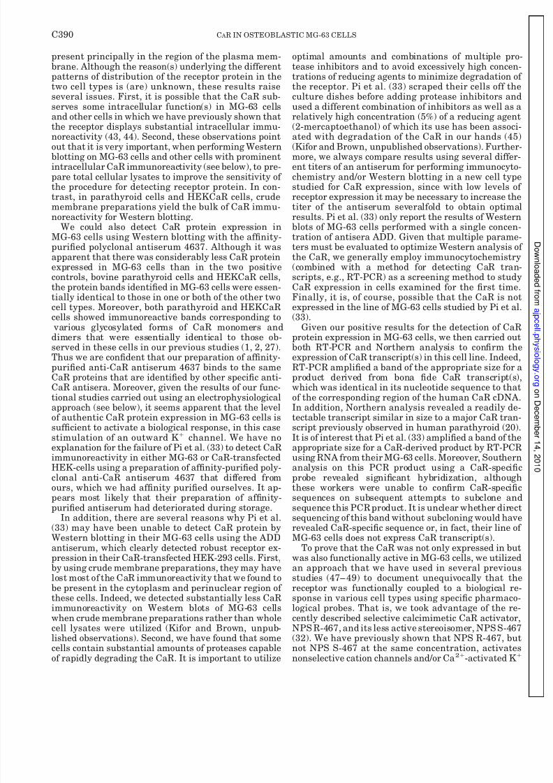
We nally provided further documentation that theopening of this K channel as a result of elevatingCa o
2 is, in fact, mediated via the CaR by using aselective “calcimimetic” CaR activator, NPS R-467 (32). Addition of 0.5 M NPS R-467 in the presence of 1 mMCa o
2 in the bath solution induced substantial activa-tion of the channel (Fig. 7 A). The activity of the chan-nel at 1.0 mM Ca o
2 ( Po 0.2) is comparable to thatobserved with 0.75 mM Ca o
2 in Fig. 6. In contrast, thesame concentration of the less active stereoisomer of NPS R-467, NPS S-467, had little or no effect on chan-nel activity (Fig. 7 B). Figure 7 C shows the I-V relation-ships of the channel under the control condition andafter addition of NPS R-467 or NPS S-467, illustratinglittle change in the slope conductance. Therefore, the
activation of the channel by NPS R-467 was principallythe result of an increase in its open state probabilitywithout alterations in its selectivity or permeability forK . Statistical analysis showed a substantial, statisti-cally signicant increase in P o after addition of NPSR-467, whereas NPS S-467 had no signicant effect on Po (Fig. 7 D).
DISCUSSION
We decided to reevaluate the expression of the CaRin MG-63 osteoblastic cells, since in pilot studies per-formed before the publication of the report of Pi et al.
(33), we observed robust CaR immunostaining in thiscell line. Moreover, earlier studies had shown that highCa o
2 increases the secretion of osteocalcin by this cellline (29), indicating that MG-63 cells are capable of sensing Ca o
2 , potentially doing so via the CaR or someother Ca o
2 -sensing mechanism. Given the negativeresults of Pi et al. (33) on Western analysis as a methodfor detecting CaR protein in MG-63 cells using the4637 antiserum, we repeated our earlier experimentsutilizing immunostaining with this antiserum as wellas with the ADD monoclonal anti-CaR antiserum usedby these authors. The results of these studies indicatedreadily detectable CaR immunoreactivity in MG-63cells using the immunoperoxidase technique with bothantisera as well as immunouorescence utilizing the
4637 antiserum.It should be pointed out that a substantial amount of this CaR immunoreactivity was present intracellu-larly, as documented by confocal imaging, and exhib-ited a perinuclear and ne reticular cytoplasmic pat-tern of staining consistent with expression in theendoplasmic reticulum. Denitive plasma membranelocalization could be documented, however, by the useof immunocolocalization via two-color confocal imagingof the CaR with two different plasma membrane mark-ers, ALP and Na -K -ATPase. CaR immunoreactivityin CaR-transfected HEK-293 cells, in contrast, was
Fig. 5. Identication of CaR transcript in MG-63 cellsusing RT-PCR performed with an intron-spanningprimer pair and sequencing of the amplied product orutilizing Northern blot analysis. A: RT-PCR, performedas described in MATERIALS AND METHODS , amplied aproduct of the expected size (425 bp) for having arisenfrom bona de CaR transcript(s) ( middle lane ). ThePCR reaction without RT showed no products ( rightlane ). A 100-bp ladder (GIBCO BRL) was used as a sizemarker ( left lane ). B: Northern analysis was carried outas described in MATERIALS AND METHODS . Arrow showsthe major CaR transcript ( 5.2 kb), which is similar insize to the most abundant CaR transcript present inhuman parathyroid gland. C: nucleotide sequence of the PCR product amplied from MG-63 cells. This se-quence is 100% identical to that of the correspondingregion of the human parathyroid CaR. A similar resultwas obtained using another independent preparation of RNA from MG-63 cells.
C388 C AR IN OSTEOBLASTIC MG-63 CELLS
8/8/2019 Am J Physiol Cell Physiol 2001 Yamaguchi C382 93
http://slidepdf.com/reader/full/am-j-physiol-cell-physiol-2001-yamaguchi-c382-93 9/13
Fig. 6. Activation of K channels in cell-attached patches of MG-63 cells by elevatingextracellular Ca 2 (Ca o
2 ) from 0.75 to 2.75mM. A: single-channel traces were taken at 2different voltages [membrane potential ( V m )as indicated on the traces ( left ) in the pres-ence of 0.75 mM Ca o
2 or ( B) after elevatingCa o
2 to 2.75 mM. C: the probability of chan-nel opening ( Po ) in cell-attached patches isplotted as a function of V m at 2 different Ca o
2
0.75 mM and 2.75 mM. Symbols and errorbars are means SE of the mean, n 3. Dcurrent-voltage relationships in the presenceof varying levels of K o and Na o (in millimo-lar) in the pipette solution: 140 KCl, no addedNaCl ( ‚ ); 55 KCl, 87 NaCl ( E ); and 5.4 KCl,140 NaCl (x).
Fig. 7. The CaR activator NPS R-467,but not its less active stereoisomer,NPS S-467, stimulates K channel ac-tivities in MG-63 cells. A: single-chan-nel traces were taken at 2 different voltages as indicated ( left ) before andafter addition of 0.5 M NPS R-467 tothe extracellular bath solution contain-ing 1.0 mM Ca o
2 . B : NPS S-467 (0.5M), the less active stereoisomer of
NPS R-467, failed to elicit channel ac-tivation. C: current-voltage relation-ships are shown under control condi-tions ( n 7) and in the presence of NPS R-467 ( n 3) or NPS S-467 ( n3). The slope conductance was 103 2pS under control conditions, 108 7 pSwith NPS R-467 in the bath solution,and 105 6 pS in the presence of NPSS-467. D: open state probabilities, NP o ,under control conditions (absence of CaR activator) or in the presence of NPS R-467 ( n 4) or NPS S-467 ( n4) are shown. The data were taken atV m 70 mV.
C389C AR IN OSTEOBLASTIC MG-63 CELLS
8/8/2019 Am J Physiol Cell Physiol 2001 Yamaguchi C382 93
http://slidepdf.com/reader/full/am-j-physiol-cell-physiol-2001-yamaguchi-c382-93 10/13
present principally in the region of the plasma mem-brane. Although the reason(s) underlying the differentpatterns of distribution of the receptor protein in thetwo cell types is (are) unknown, these results raiseseveral issues. First, it is possible that the CaR sub-serves some intracellular function(s) in MG-63 cellsand other cells in which we have previously shown thatthe receptor displays substantial intracellular immu-noreactivity (43, 44). Second, these observations pointout that it is very important, when performing Westernblotting on MG-63 cells and other cells with prominentintracellular CaR immunoreactivity (see below), to pre-pare total cellular lysates to improve the sensitivity of the procedure for detecting receptor protein. In con-trast, in parathyroid cells and HEKCaR cells, crudemembrane preparations yield the bulk of CaR immu-noreactivity for Western blotting.
We could also detect CaR protein expression inMG-63 cells using Western blotting with the afnity-puried polyclonal antiserum 4637. Although it wasapparent that there was considerably less CaR proteinexpressed in MG-63 cells than in the two positivecontrols, bovine parathyroid cells and HEKCaR cells,the protein bands identied in MG-63 cells were essen-tially identical to those in one or both of the other twocell types. Moreover, both parathyroid and HEKCaRcells showed immunoreactive bands corresponding to various glycosylated forms of CaR monomers anddimers that were essentially identical to those ob-served in these cells in our previous studies (1, 2, 27).Thus we are condent that our preparation of afnity-puried anti-CaR antiserum 4637 binds to the sameCaR proteins that are identied by other specic anti-CaR antisera. Moreover, given the results of our func-tional studies carried out using an electrophysiological
approach (see below), it seems apparent that the levelof authentic CaR protein expression in MG-63 cells issufcient to activate a biological response, in this casestimulation of an outward K channel. We have noexplanation for the failure of Pi et al. (33) to detect CaRimmunoreactivity in either MG-63 or CaR-transfectedHEK-cells using a preparation of afnity-puried poly-clonal anti-CaR antiserum 4637 that differed fromours, which we had afnity puried ourselves. It ap-pears most likely that their preparation of afnity-puried antiserum had deteriorated during storage.
In addition, there are several reasons why Pi et al.(33) may have been unable to detect CaR protein byWestern blotting in their MG-63 cells using the ADD
antiserum, which clearly detected robust receptor ex-pression in their CaR-transfected HEK-293 cells. First,by using crude membrane preparations, they may havelost most of the CaR immunoreactivity that we found tobe present in the cytoplasm and perinuclear region of these cells. Indeed, we detected substantially less CaRimmunoreactivity on Western blots of MG-63 cellswhen crude membrane preparations rather than wholecell lysates were utilized (Kifor and Brown, unpub-lished observations). Second, we have found that somecells contain substantial amounts of proteases capableof rapidly degrading the CaR. It is important to utilize
optimal amounts and combinations of multiple pro-tease inhibitors and to avoid excessively high concen-trations of reducing agents to minimize degradation of the receptor. Pi et al. (33) scraped their cells off theculture dishes before adding protease inhibitors andused a different combination of inhibitors as well as arelatively high concentration (5%) of a reducing agent(2-mercaptoethanol) of which its use has been associ-ated with degradation of the CaR in our hands (45)(Kifor and Brown, unpublished observations). Further-more, we always compare results using several differ-ent titers of an antiserum for performing immunocyto-chemistry and/or Western blotting in a new cell typestudied for CaR expression, since with low levels of receptor expression it may be necessary to increase thetiter of the antiserum severalfold to obtain optimalresults. Pi et al. (33) only report the results of Westernblots of MG-63 cells performed with a single concen-tration of antisera ADD. Given that multiple parame-ters must be evaluated to optimize Western analysis of the CaR, we generally employ immunocytochemistry(combined with a method for detecting CaR tran-scripts, e.g., RT-PCR) as a screening method to studyCaR expression in cells examined for the rst time.Finally, it is, of course, possible that the CaR is notexpressed in the line of MG-63 cells studied by Pi et al.(33).
Given our positive results for the detection of CaRprotein expression in MG-63 cells, we then carried outboth RT-PCR and Northern analysis to conrm theexpression of CaR transcript(s) in this cell line. Indeed,RT-PCR amplied a band of the appropriate size for aproduct derived from bona de CaR transcript(s),which was identical in its nucleotide sequence to thatof the corresponding region of the human CaR cDNA.
In addition, Northern analysis revealed a readily de-tectable transcript similar in size to a major CaR tran-script previously observed in human parathyroid (20).It is of interest that Pi et al. (33) amplied a band of theappropriate size for a CaR-derived product by RT-PCRusing RNA from their MG-63 cells. Moreover, Southernanalysis on this PCR product using a CaR-specicprobe revealed signicant hybridization, althoughthese workers were unable to conrm CaR-specicsequences on subsequent attempts to subclone andsequence this PCR product. It is unclear whether directsequencing of this band without subcloning would haverevealed CaR-specic sequence or, in fact, their line of MG-63 cells does not express CaR transcript(s).
To prove that the CaR was not only expressed in butwas also functionally active in MG-63 cells, we utilizedan approach that we have used in several previousstudies (47–49) to document unequivocally that thereceptor was functionally coupled to a biological re-sponse in various cell types using specic pharmaco-logical probes. That is, we took advantage of the re-cently described selective calcimimetic CaR activator,NPS R-467, and its less active stereoisomer, NPS S-467(32). We have previously shown that NPS R-467, butnot NPS S-467 at the same concentration, activatesnonselective cation channels and/or Ca 2 -activated K
C390 C AR IN OSTEOBLASTIC MG-63 CELLS
8/8/2019 Am J Physiol Cell Physiol 2001 Yamaguchi C382 93
http://slidepdf.com/reader/full/am-j-physiol-cell-physiol-2001-yamaguchi-c382-93 11/13
channels in several cell lines (13–15, 42). Moreover, we validated this approach further by showing that theseresponses to CaR activators were not present in cellsderived from mice homozygous for targeted disruptionof the CaR gene (22, 46–49). Indeed, the results of ourpresent studies indicate that in MG-63 cells raisingCa o
2 or addition of NPS R-467 but not NPS S-467activates an outward K channel, similar to our earlierreports in other cells (12–15, 42). Thus the CaRpresent in the MG-63 cells employed by us is func-tional, at least as assessed by its coupling to activationof this K channel.
Therefore, our results strongly support the expres-sion of both CaR mRNA and functional CaR protein inthe MG-63 cell line studied here. These data furthersupport our earlier results (44, 45) and those of others(25) that the CaR is expressed in several osteoblasticcell lines. We recognize, however, that MG-63 cellswere originally obtained from a human osteosarcomaand may not represent an optimal model for studyingthe functional relevance of the CaR in osteoblasts (17).The MG-63 osteosarcoma cell line shows increases in ALP and osteocalcin expression following treatmentwith 1 ,25-dihydroxyvitamin D 3 (30), which are re-sponses characteristic of relatively undifferentiated os-teoblast precursors. In contrast, another human osteo-blastic cell line, SAOS-2, shows high constitutive ALPexpression but little or no osteocalcin expression, ei-ther with or without addition of 1 ,25-dihydroxyvita-min D 3 (30). The mouse osteoblastic cell line, MC3T3-E1, is known to exhibit properties of osteoprogenitorcells and preosteoblasts in their actively growing stage.Following growth arrest, however, they differentiateand develop markers of mature osteoblasts, includingthe expression of high levels of ALP and the capacity to
form mineralized bone matrix (44). Together, ourpresent and prior studies, which show expression of theCaR in osteoblastic cell lines differing in their pheno-types and apparent stages of differentiation, suggestthat this receptor is expressed in osteoblasts varyingsubstantially in their developmental stages.
It should be pointed out that the MG-63 cell line alsoretains a broblast-like character, with its abundantexpression of type III collagen (24) and low constitutiveexpression of ALP (17, 30). It is of interest in thisregard that McNeil et al. (31) found that rat-1 bro-blasts express the CaR. Indeed, broblasts arise fromthe same mesenchymal stem cell (8) that gives rise toseveral types of CaR-expressing cells involved in bone
growth and/or turnover, including chondrocytes (9),osteoblasts (44, 45), and stromal cells (43). Therefore,expression of the CaR appears to be characteristic of several cell types within this lineage.
Our present and prior studies (44, 45) have clearlyshown that the CaR is expressed in clonal osteoblasticcell lines. Moreover, recent studies have documentedthe expression of CaR mRNA and protein in osteo-blasts within sections of bovine, rat, and murine bone(10), providing additional evidence that bona de os-teoblasts express the CaR. Nevertheless, it is possiblethat other Ca 2 sensors molecularly distinct from the
CaR (34, 35) are also present in osteoblasts and par-ticipate in their cation-sensing capacity. For instance,Pi et al. (32a) have recently isolated primary osteo-blast-like cells from mice with targeted disruption of the CaR that still showed mitogenic and other biolog-ical responses to polyvalent cations. Additional studiesare needed, therefore, utilizing genetic models of gen-eralized (22) or tissue-selective “knock out” of the CaRas well as techniques that downregulate the function of the endogenous osteoblastic CaR, e.g., CaR antagonistsor transfection with dominant negative CaR constructs(31), to establish denitively the CaR’s role in modu-lating the full range of osteoblastic functions that areresponsive to Ca o
2 . The outcome of these experimentsshould answer the question of whether the CaR is theprincipal Ca o
2 sensor in osteoblasts and/or whetherthey possess other Ca o
2 sensors/receptors that alsocontribute to the regulation of osteoblast function byCa o
2 .
The authors gratefully acknowledge generous grant support fromNational Institute of Diabetes and Digestive and Kidney DiseasesGrants DK-41415, DK-48330, and DK-52005, NPS Pharmaceuticals,The St. Giles Foundation, and the National Space Bioscience Re-search Institute (to E. M. Brown), the Mochida Memorial FoundationGrant for Medical and Pharmaceutical Research (to T. Yamaguchi),and the Yamanouchi Foundation Grant for Research on MetabolicDisorders (to T. Yamaguchi).
Present address of T. Yamaguchi: Third Div., Dept. of Medicine,Kobe Univ. School of Medicine, Kobe 650-0017, Japan.
REFERENCES
1. Bai M, Quinn S, Trivedi S, Kifor O, Pearce SHS, PollakMR, Krapcho K, Hebert SC, and Brown EM. Expression andcharacterization of inactivating and activating mutations in thehuman Ca o
2 -sensing receptor. J Biol Chem 271: 19537–19545,1996.
2. Bai M, Trivedi S, and Brown EM. Dimerization of the extra-cellular calcium-sensing receptor (CaR) on the cell surface of CaR-transfected HEK293 cells. J Biol Chem 273: 23605–23610,1998.
3. Baron R. Anatomy and ultrastructure of bone. In: Primer on the Metabolic Bone Diseases and Disorders of Mineral Metabolism(3rd ed.), edited by Favus MJ. Philadelphia, PA: Lippincott-Raven, 1996, p. 3–10.
4. Brown EM. Extracellular Ca 2 sensing, regulation of parathy-roid cell function, and role of Ca 2 and other ions as extracellular(rst) messengers. Physiol Rev 71: 371–411, 1991.
5. Brown EM, Chen CJ, Kifor O, Leboff MS, El-Hajj G, Faj-tova V, and Rubin LT. Ca 2 -sensing, second messengers, andthe control of parathyroid hormone secretion. Cell Calcium 11333–337, 1990.
6. Brown EM, Gamba G, Riccardi D, Lombardi M, Butters R,Kifor O, Sun A, Hediger MA, Lytton J, and Hebert SC.Cloning and characterization of an extracellular Ca 2 -sensingreceptor from bovine parathyroid. Nature 366: 575–580, 1993.
7. Brown EM, Hurwitz S, and Aurbach GD. Preparation of viable isolated bovine parathyroid cells. Endocrinology 99: 1582–1588, 1976.
8. Caplan AI and Dennis JE. Mesenchymal stem cells: progeni-tors, progeny, and pathways. J Bone Miner Res 14: 193–201,1996.
9. Chang W, Tu C, Bajra R, Komuves L, Miller S, Strewler G,and Shoback D. Calcium sensing in cultured chondrogenicRCJ3.1C518 cells. Endocrinology 140: 1911–1919, 1999.
10. Chang W, Tu C, Chen TH, Komuves L, Oda Y, Pratt S,Miller S, and Shoback D. Expression and signal transductionof calcium-sensing receptors in cartilage and bone. Endocrinol-ogy 140: 5883–5893, 1999.
C391C AR IN OSTEOBLASTIC MG-63 CELLS
8/8/2019 Am J Physiol Cell Physiol 2001 Yamaguchi C382 93
http://slidepdf.com/reader/full/am-j-physiol-cell-physiol-2001-yamaguchi-c382-93 12/13
11. Chattopadhyay N, Cheng I, Rogers K, Riccardi D, Hall A,Diaz R, Hebert SC, Soybel DI, and Brown EM. Identicationand localization of extracellular Ca 2 -sensing receptor in ratintestine. Am J Physiol Gastrointest Liver Physiol 274: G122–G130, 1998.
12. Chattopadhyay N, Ye C, Singh DP, Kifor O, Vassilev PM,Shinohara T, Chylack LT Jr, and Brown EM. Expression of extracellular calcium-sensing receptor by human lens epithelialcells. Biochem Biophys Res Commun 233: 801–805, 1997.
13. Chattopadhyay N, Ye C, Yamaguchi T, Nakai M, Kifor O, Vassilev PM, Nishimura RN, and Brown EM. The extracel-lular calcium-sensing receptor is expressed in rat microglia andmodulates an outward K channel. J Neurochem 72: 1915–1922,1999.
14. Chattopadhyay N, Ye CP, Yamaguchi T, Kifor O, VassilevPM, Nishimura R, and Brown EM. Extracellular calcium-sensing receptor in rat oligodendrocytes: expression and poten-tial role in regulation of cellular proliferation and an outward K channel. Glia 24: 449–458, 1998.
15. Chattopadhyay N, Ye CP, Yamaguchi T, Vassilev PM, andBrown EM. Evidence for extracellular calcium-sensing receptormediated opening of an outward K channel in a human astro-cytoma cell line (U87). Glia 26: 64–72, 1999.
16. Cheng I, Klingensmith ME, Chattopadhyay N, Kifor O,Butters RR, Soybel DI, and Brown EM. Identication andlocalization of the extracellular calcium-sensing receptor in hu-
man breast. J Clin Endocrinol Metab 83: 703–707, 1998.17. Clover J and Gowen M. Are MG-63 and HOS TE85 humanosteosarcoma cell lines representative models of the osteoblasticphenotype? Bone 15: 585–591, 1994.
18. Freichel M, Zink-Lorenz A, Holloschi A, Hafner M, Flock-erzi V,and RaueF. Expression of a calcium-sensing receptor ina human medullary thyroid carcinoma cell line and its contribu-tion to calcitonin secretion. Endocrinology 137: 3842–3848,1996.
19. Fujikawa Y, Quinn JMW, Sabokbar A, McGee JOD, and Athanasou NA. The human osteoclast precursor circulates inthe monocyte fraction. Endocrinology 137: 4058–4060, 1996.
20. Garrett JE, Capuano IV, Hammerland LG, Hung BC,Brown EM, Hebert SC, Nemeth EF, and Fuller F. Molecularcloning and functional expression of human parathyroid calciumreceptor cDNAs. J Biol Chem 270: 12919–12925, 1995.
21. Garrett JE, Tamir H, Kifor O, Simin RT, Rogers KV,Mithal A, Gagel RF, and Brown EM. Calcitonin-secretingcells of the thyroid express an extracellular calcium receptorgene. Endocrinology 136: 5202–5211, 1995.
22. Ho C, Conner DA, Pollak MR, Ladd DJ, Kifor O, WarrenHB, Brown EM, Seidman JG, and Seidman CE. A mousemodel of human familial hypocalciuric hypercalcemia and neo-natal severe hyperparathyroidism. Nat Genet 11: 389–394, 1995.
23. House MG, Kohlmeier L, Chattopadhyay N, Kifor O, Yamaguchi T, Leboff MS, Glowacki J, and Brown EM.Expression of an extracellular calcium-sensing receptor in hu-man and mouse bone marrow cells. J Bone Miner Res 12: 1959–1970, 1997.
24. Jukkola A, Risteli L, Melkko J, and Risteli J. Procollagensynthesis and extracellular matrix deposition in MG-63 osteo-sarcoma cells. J Bone Miner Res 8: 651–657, 1993.
25. Kanatani M, Sugimoto T, Kanzawa M, Yano S, and Chi-hara K. High extracellular calcium inhibits osteoclast-like cellformation by directly acting on the calcium-sensing receptorexisting in osteoclast precursor cells. Biochem Biophys Res Com-mun 261: 144–148, 1999.
26. Kifor O, Diaz R, Butters R, and Brown EM. The Ca 2 -sensing receptor (CaR) activates phospholipases C, A 2 , and D inbovine parathyroid and CaR-transfected, human embryonic kid-ney (HEK293) cells. J Bone Miner Res 12: 715–725, 1997.
27. Kifor O, Diaz R, Butters R, Kifor I, and Brown EM. Thecalcium-sensing receptor is localized in caveolin-rich plasmamembrane domains of bovine parathyroid cells. J Biol Chem 273:21708–21713, 1998.
28. Kifor O, Moore FD Jr, Wang P, Goldstein M, Vassilev P,Kifor I, Hebert SC, and Brown EM. Reduced immunostaining
for the extracellular Ca 2 -sensing receptor in primary and ure-mic secondary hyperparathyroidism. J Clin Endocrinol Metab81: 1598–1606, 1996.
29. Lajeunesse D, Kiebzak GM, Frondoza C, and Sacktor B.Regulation of osteocalcin secretion by human primary bone cellsand by the human osteosarcoma cell line MG-63. Bone Miner 14237–250, 1991.
30. Mahonen A, Pirskanen A, Keinanen R, and Maenpaa PH.Effect of 1,25(OH) 2 D3 on its receptor mRNA levels and osteocal-cin synthesis in human osteosarcoma cells. Biochim Biophys Acta 1048: 30–37, 1990.
31. McNeil SE, Hobson SA, Nipper V, and Rodland KD. Func-tional calcium-sensing receptors in rat broblasts are requiredfor activation of SRC kinase and mitogen-activated protein ki-nase in response to extracellular calcium. J Biol Chem 2731114–1120, 1998.
32. Nemeth EF, Steffey ME, Hammerland LG, Hung BC, VanWagenen BC, DelMar EG, and Balandrin MF. Calcimimeticswith potent and selective activity on the parathyroid calciumreceptor. Proc Natl Acad Sci USA 95: 4040–4045, 1998.
32a. Pi M, Garner SC, Flannery P, Spurney RF, and QuarlesLD. Sensing of extracellular cations in CasR-decient osteo-blasts. Evidence for a novel cation-sensing mechanism. J BiolChem 275: 3256–3263, 2000.
33. Pi M, Hinson TK, and Quarles L. Failure to detect the extra-cellular calcium-sensing receptor (CasR) in human osteoblast
cell lines. J Bone Miner Res 14: 1310–1319, 1999.34. Quarles LD. Cation-sensing receptors in bone: a novel para-digm for regulating bone remodeling? J Bone Miner Res 121971–1974, 1997.
35. Quarles DL, Hartle JE II, Siddhanti SR, Guo R, and Hin-son TK. A distinct cation-sensing mechanism in MC3T3–E1osteoblasts functionally related to the calcium receptor. J Bone Miner Res 12: 393–402, 1997.
36. Raisz LG and Kream BE. Regulation of bone formation. N Engl J Med 309: 29–35, 1983.
37. Raisz LG and Kream BE. Regulation of bone formation (sec-ond of two parts). N Engl J Med 309: 83–89, 1983.
38. Riccardi D, Park J, Lee WS, Gamba G, Brown EM, andHebert SC. Cloning and functional expression of a rat kidneyextracellular calcium/polyvalent cation-sensing receptor. Proc Natl Acad Sci USA 92: 131–135, 1995.
39. Silver IA, Murrils RJ, and Etherington DJ. Microlectrodestudies on the acid microenvironment beneath adherent macro-phages and osteoclasts. Exp Cell Res 175: 266–276, 1988.
40. Sudo H, Kodama H, Amagai Y, Yamamoto S, and Kasai S.In vitro differentiation and calcication in a new clonal osteo-genic cell line derived from newborn mouse calvaria. J Cell Biol96: 191–198, 1983.
41. Sugimoto T, Kanatani M, Kano J, Kaji H, Tsukamoto T, Yamaguchi T, Fukase M, and Chihara K. Effects of highcalcium concentration on the functions and interactions of osteo-blastic cells and monocytes and on the formation of osteoclast-like cells. J Bone Miner Res 8: 1445–1452, 1993.
42. Vassilev PM, Ho-Pao CL, Kanazirska MP, Ye C, Hong K,Seidman CE, Seidman JG, and Brown EM. Ca o -sensingreceptor (CaR)-mediated activation of K channels is blunted inCaR gene-decient mouse neurons. Neuroreport 8: 1411–1416,1997.
43. Yamaguchi T, Chattopadhyay N, Kifor O, and Brown EM.Extracellular calcium (Ca o
2 )-sensing receptor in a murine bonemarrow-derived stromal cell line (ST2): potential mediator of theactions of Ca o
2 on the function of ST2 cells. Endocrinology 1393561–3568, 1998.
44. Yamaguchi T, Chattopadhyay N, Kifor O, Butters RR Jr,Sugimoto T, and Brown EM. Mouse osteoblastic cell line(MC3T3-E1) expresses extracellular calcium (Ca o
2 )-sensing re-ceptor and its agonists stimulate chemotaxis and proliferation of MC3T3-E1 cells. J Bone Miner Res 13: 1530–1538, 1998.
45. Yamaguchi T, Kifor O, Chattopadhyay N, and Brown EM.Expression of extracellular calcium (Ca o
2 )-sensing receptor inthe clonal osteoblast-like cell lines, UMR-106 and SAOS-2. Bio-chem Biophys Res Commun 243: 753–757, 1998.
C392 C AR IN OSTEOBLASTIC MG-63 CELLS
8/8/2019 Am J Physiol Cell Physiol 2001 Yamaguchi C382 93
http://slidepdf.com/reader/full/am-j-physiol-cell-physiol-2001-yamaguchi-c382-93 13/13
46. Ye C, Ho-Pao CL, Kanazirska M, Quinn S, Rogers K, Seid-man CE, Seidman JG, Brown EM, and Vassilev PM. Amy-loid-beta proteins activate Ca 2 -permeable channels throughcalcium-sensing receptors. J Neurosci Res 47: 547–554, 1997.
47. Ye C, Ho-Pao CL, Kanazirska M, Quinn S, Seidman CE,Seidman JG, Brown EM, and Vassilev PM. Decient cationchannel regulation in neurons from mice with targeted disrup-tion of the extracellular Ca 2 -sensing receptor gene. Brain Res Bull 44: 75–84, 1997.
48. Ye C, Kanazirska M, Quinn S, Brown EM, and VassilevPM. Modulation by polycationic Ca 2 -sensing receptor agonistsof nonselective cation channels in rat hippocampal neurons. Biochem Biophys Res Commun 224: 271–280, 1996.
49. Ye C, Rogers K, Bai M, Quinn SJ, Brown EM, and VassilevPM. Agonists of the Ca 2 -sensing receptor (CaR) activate non-selective cation channels in HEK293 cells stably transfectedwith the human CaR. Biochem Biophys Res Commun 226: 572–579, 1996.
C393C AR IN OSTEOBLASTIC MG-63 CELLS


![Am J Physiol Heart Circ Physiol 2011[1]](https://img.pdfslide.us/doc/110x75/577ce0031a28ab9e78b28109/am-j-physiol-heart-circ-physiol-20111.jpg)