Embed Size (px)
Citation preview
RESEARCH ARTICLE Open Access
Altered lipid composition in Streptococcuspneumoniae cpoA mutantsMarina Meiers1†, Carsten Volz1,2†, Jessica Eisel1, Patrick Maurer3, Bernhard Henrich1 and Regine Hakenbeck1*
Abstract
Background: Penicillin-resistance in Streptococcus pneumoniae is mainly due to alterations in genes encodingthe target enzymes for beta-lactams, the penicillin-binding proteins (PBPs). However, non-PBP genes are alteredin beta-lactam-resistant laboratory mutants and confer decreased susceptibility to beta-lactam antibiotics. Twopiperacillin resistant laboratory mutants of Streptococcus pneumoniae R6 contain mutations in the putativeglycosyltransferase gene cpoA. The CpoA gene is part of an operon including another putative glycosyltransferase genespr0982, both of which being homologous to glycolipid synthases present in other Gram-positive bacteria.
Results: We now show that the cpoA mutants as well as a cpoA deletion mutant are defective in the synthesis ofgalactosyl-glucosyl-diacylglycerol (GalGlcDAG) in vivo consistent with the in vitro function of CpoA as α-GalGlcDAGsynthase as shown previously. In addition, the proportion of phosphatidylglycerol increased relative tocardiolipin in cpoA mutants. Moreover, cpoA mutants are more susceptible to acidic stress, have an increasedrequirement for Mg2+ at low pH, reveal a higher resistance to lysis inducing conditions and are hypersensitiveto bacitracin.
Conclusions: The data show that deficiency of the major glycolipid GalGlcDAG causes a pleitotropic phenotype ofcpoA mutant cells consistent with severe membrane alterations. We suggest that the cpoA mutations selected withpiperacillin are directed against the lytic response induced by the beta-lactam antibiotic.
Keywords: Streptococcus pneumoniae, Glycolipids, Penicillin resistance, Glycosyltransferase, CpoA, Phospholipid
BackgroundDevelopment of resistance to beta-lactam antibiotics inStreptococcus pneumoniae involves alterations in the targetproteins, the penicillin-binding proteins (PBPs) which resultin decreased affinity to beta-lactams. In order to identifyindividual mutations in S. pneumoniae that are related tothe resistance phenotype, a series of independent mutantfamilies has been selected in the laboratory using stepwiseincreasing concentrations of antibiotics [1]. Two beta-lactams were chosen for selection: piperacillin, whichinduces rapid lysis in the bacteria, and cefotaxime whichdoes not interact with PBP2b and leads to a tolerantresponse [2]. Point mutations in pbp2b from piperacillin-resistant mutants and in pbp2x from cefotaxime resistantmutants have been described [3-5]. Surprisingly, a decrease
in antibiotic susceptibility in some mutants correlated witha mutation in non-PBP genes [6]. In two piperacillin-resistant mutants, P106 and P104, obtained independentlyafter the first selection step before the introduction of PBPmutations, the putative glycosyltransferase (GT) gene cpoAwas affected [7]. Decreased susceptibility for piperacillinof the cpoA mutants was accompanied by a pleiotropicphenotype such as a defect in genetic competence andreduced amount of PBP1a. This indicated a novel mech-anism directed against the activity of lytic β-lactams in S.pneumoniae distinct from target-mediated resistance.The CpoA gene spr0981 and the adjacent gene spr0982
encode putative GTs which belong to the GTB-type super-family (GT1-YqgM-like family). Members of this GT familyare anchored in the membrane cytoplasmic interface byhydrophobic and charge interactions [8,9] and transfer asugar moiety to an acceptor molecule located in the innerleaflet of the membrane. Therefore, it had been proposedthat CpoA perfoms a similar function in S. pneumoniae [7].Meanwhile, in vitro studies revealed that both proteins are
* Correspondence: [email protected]†Equal contributors1Department of Microbiology, University of Kaiserslautern,Gottlieb-Daimler-Strasse, Gebäude 23, D-67663 Kaiserslautern, GermanyFull list of author information is available at the end of the article
© 2014 Meiers et al.; licensee BioMed Central Ltd. This is an Open Access article distributed under the terms of the CreativeCommons Attribution License (http://creativecommons.org/licenses/by/2.0), which permits unrestricted use, distribution, andreproduction in any medium, provided the original work is properly cited. The Creative Commons Public Domain Dedicationwaiver (http://creativecommons.org/publicdomain/zero/1.0/) applies to the data made available in this article, unless otherwisestated.
Meiers et al. BMC Microbiology 2014, 14:12http://www.biomedcentral.com/1471-2180/14/12
involved in the synthesis of glycolipids, with Spr0982 actingas α-monoglucosyl-diacylglycerol (GlcDAG) synthase andCpoA as a α-galactosyl-glucosyl-diacylgylcerol (GalGlc-DAG) synthase [9,10]. These two glycolipids occur at aratio of approximately 1:2.5 in the S. pneumoniae mem-brane [11], in addition to phosphatidyl glycerol and cardi-olipin which constitute the major phospholipids [12].By consecutively synthesizing one nonbilayer-prone
(mono-glucosyl-DAG) and one bilayer-forming glycolipid(di-glycosyl-DAG), the function of the GTs is crucial for thebilayer spontaneous curvature which affects the physicalproperties of the cytoplasmic membrane [13]. An exampleis the mycoplasma Acholeplasma laidlawii, where bilayercurvature is extensively regulated by two closely relatedGTs consecutively synthesizing monoglucosyl-DAG anddiglucosyl-DAG [9,13], enzymes that are homologous toS. pneumoniae Spr0982 and CpoA. Thus it is most likelythat CpoA and Spr0982 play a critical role in S. pneumo-niae related to membrane associated functions in agree-ment with the pleiotropic phenotype of the CpoA mutantsmentioned above. GlcDAG is the proposed lipid anchor ofthe essential choline-containing lipoteichoic acid (LTA) ofS. pneumoniae [14]. In fact, spr0982 has been listed amongessential genes of this organism [15].In the present report, a cpoA deletion mutant was
constructed and compared to the CpoA mutants P106and P104; moreover, the cpoA operon was investigatedby mutational analysis. The aim of this study was toexamine the function of CpoA in vivo, and to furtherour understanding on the physiological consequencesof cpoA mutations.
ResultsThe CpoA gene is part of an operon with fivedownstream genesP104 and P106 are spontaneous piperacillin-resistantlaboratory mutants isolated independently after one selec-tion step from the laboratory strain S. pneumoniae R6 [4,7].Both mutants contain a mutation affecting CpoA: in P104,a transversion within cpoA GTA to GGA led to a Gly12Valexchange in the predicted protein product, whereas inP106, one adenine nucleotide was deleted 15 base pairs(bp) upstream of the proposed cpoA start codon (ATG2
in Figure 1) [7]. Although ATG2 is not preceded by aclassical Shine Dalgarno sequence, this deletion wassuspected to affect the efficiency of ribosome binding tothe cpoA transcript [7]. However, the possibility remainedthat translation actually starts at an alternative start codon(ATG1 in Figure 1) 27 bp upstream of ATG2 which ispreceded by a perfect −10 region. In this case, the deletionin P106 would lead to a frameshift in the 5th codon andthus to the production of a nonsense peptide.To first clarify this issue, the expression signals of cpoA
were mapped. The 5' end of cpoA mRNA was determined
by RACE, and shown to be located 27 bp upstream ofATG2 (Figure 1B). Since this is exactly the position of thealternative start codon ATG1, translation initiation at ATG1
would imply that the cpoA transcript is leaderless [16]. Inorder to see whether ATG1 is indeed functional or whetherATG2 is required for translation, three plasmids wereconstructed in which the inferred promoter PcpoA togetherwith either both, ATG1 and ATG2 (PcpoA-ATG12), ATG1
plus a mutated ATG2 (PcpoA-ATG1ATA2), or ATG1 only(PcpoA-ATG1), was translationally fused with the lacZreporter gene. After single-copy integration of the resultingreporter constructs at the bgaA locus of R6, the expressionof lacZ was determined in two transformants in up to threeexperiments. Beta-galactosidase activity was similar inR6PcpoA-ATG1ATG2 and R6PcpoA-ATG1ATA2 (190–330Miller Units), and slightly lower in R6PcpoA-ATG1 contain-ing a shorter region upstream of lacZ (140–150 MillerUnits), clearly documenting that ATG1 is the translationinitiation site of cpoA and that the cpoA transcript isindeed leaderless. In this case, P106 contains a deletionwithin the structural gene resulting in a frameshift withinthe 5th codon consistent with the failure to detect CpoAin P106 with a specific anti-CpoA antiserum [7], and themutation in P104 is Gly21Val.Comparison with the genetic organization of cpoA and
upstream regions of the closely related species S. mitisB6 and S. oralis Uo5 of known genome sequence [17,18]revealed an almost perfect conservation of cpoA includingthe −10 region in these species (Figure 1B).The arrangement of genes and expression signals pre-
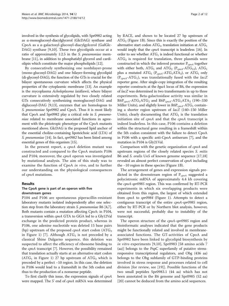
dicted in the downstream region of PcpoA suggested apolycistronic mRNA of approximately 4.4 kb coveringthe cpoA-spr0985 region. This was confirmed by RT-PCRexperiments in which six overlapping products wereobtained from this region, the largest of which extendedfrom cpoA to spr0984 (Figure 1). Attempts to detect acontiguous transcript of the entire cpoA-spr0985 region,either by RT-PCR or by Northern blot analysis, however,were not successful, probably due to instability of thetranscript.The operon structure of the cpoA-spr0985 region and
bioinformatic analyses indicated that the gene productsmight be functionally related and involved in membrane-associated functions. The GT-activities of CpoA andSpr0982 have been linked to glycolipid biosynthesis byin vitro experiments [9,10], Spr0983 [58 amino acids 7(aa)] belongs to the PspC superfamily of putative stress-responsive transcriptional regulators, and Obg (436 aa)belongs to the Obg subfamily of GTP-binding proteinsinvolved in stress response and processes related to celldivision [for review, see [19]]. Possible functions of thetwo small peptides Spr0983.1 (44 aa) which has notbeen annotated in the R6 genome and Spr0985 (52 aa)[20] cannot be deduced from the amino acid sequences.
Meiers et al. BMC Microbiology 2014, 14:12 Page 2 of 12http://www.biomedcentral.com/1471-2180/14/12
Mutational analysis of the cpoA operonTo assess the importance of these gene products, we aimedto construct deletions in each gene. A previous attemptto delete cpoA by insertion-duplication mutagenesis usinga non-replicative plasmid vector had been unsuccessful[7]. This suggested that either cpoA is essential, or thatinsertion of the vector had affected the expression of thedownstream gene spr0982 which has been listed amongessential genes of S. pneumoniae [15]. To avoid such polareffects, a different deletion strategy was applied which wasbased on the construction of in-frame deletions using theJanus cassette (Figure 1). R6 mutants in which 108 centralcodons of cpoA (specifying the GT domain) were replacedwith the Janus cassette were obtained with common effi-ciencies (0.2%), demonstrating that cpoA is a non-essentialgene. Deletions in spr0983 and spr0985 were also ob-tained. However, the generation times of R6ΔcpoA andR6Δspr0985 (with 46–48 min) were significantly longercompared to R6 and R6Δspr0983 (38 min), suggesting thatCpoA and Spr0985 are involved in important functions. Incontrast, transformants carrying deletions in spr0982 andobg occurred only at 1,000- and respectively 10,000-fold
reduced frequencies. This is in agreement with an essentialfunction of the spr0982 product as reported previously [15],and strongly suggested that also obg is indispensable. Therare recovery of transformants carrying deletions inthese genes probably was the result of co-selection ofcompensatory mutations at unknown secondary sites.
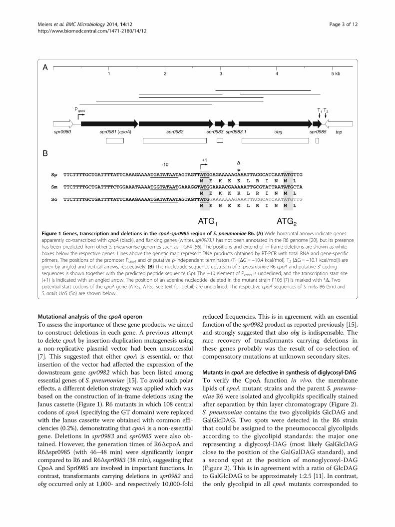
Mutants in cpoA are defective in synthesis of diglycosyl-DAGTo verify the CpoA function in vivo, the membranelipids of cpoA mutant strains and the parent S. pneumo-niae R6 were isolated and glycolipids specifically stainedafter separation by thin layer chromatograpy (Figure 2).S. pneumoniae contains the two glycolipids GlcDAG andGalGlcDAG. Two spots were detected in the R6 strainthat could be assigned to the pneumococcal glycolipidsaccording to the glycolipid standards: the major onerepresenting a diglycosyl-DAG (most likely GalGlcDAGclose to the position of the GalGalDAG standard), anda second spot at the position of monoglycosyl-DAG(Figure 2). This is in agreement with a ratio of GlcDAGto GalGlcDAG to be approximately 1:2.5 [11]. In contrast,the only glycolipid in all cpoA mutants corresponded to
B
1 2 3 4 5 kb
spr0980 spr0981 (cpoA) spr0982 spr0983 obg spr0985 tnp
PcpoA T2T1
spr0983.1
ATG1 ATG2
A
Figure 1 Genes, transcription and deletions in the cpoA-spr0985 region of S. pneumoniae R6. (A) Wide horizontal arrows indicate genesapparently co-transcribed with cpoA (black), and flanking genes (white). spr0983.1 has not been annotated in the R6 genome [20], but its presencehas been predicted from other S. pneumoniae genomes such as TIGR4 [56]. The positions and extend of in-frame deletions are shown as whiteboxes below the respective genes. Lines above the genetic map represent DNA products obtained by RT-PCR with total RNA and gene-specificprimers. The positions of the promoter PcpoA and of putative ρ-independent terminators (T1 [ΔG = −10.4 kcal/mol], T2 [ΔG = −10.1 kcal/mol]) aregiven by angled and vertical arrows, respectively. (B) The nucleotide sequence upstream of S. pneumoniae R6 cpoA and putative 3'-codingsequences is shown together with the predicted peptide sequence (Sp). The −10 element of PcpoA is underlined, and the transcription start site(+1) is indicated with an angled arrow. The position of an adenine nucleotide, deleted in the mutant strain P106 [7] is marked with *Δ. Twopotential start codons of the cpoA gene (ATG1, ATG2; see text for detail) are underlined. The respective cpoA sequences of S. mitis B6 (Sm) andS. oralis Uo5 (So) are shown below.
Meiers et al. BMC Microbiology 2014, 14:12 Page 3 of 12http://www.biomedcentral.com/1471-2180/14/12
the position of the monoglycosyl-DAG (Figure 2). Thisconfirms that CpoA is required for the synthesis of thediglycosyl-DAG in S. pneumoniae in agreement withthe in vitro GalGlcDAG-synthase activity of CpoA, anddocuments that both mutants, P104 and P106, do notcontain a functional CpoA.
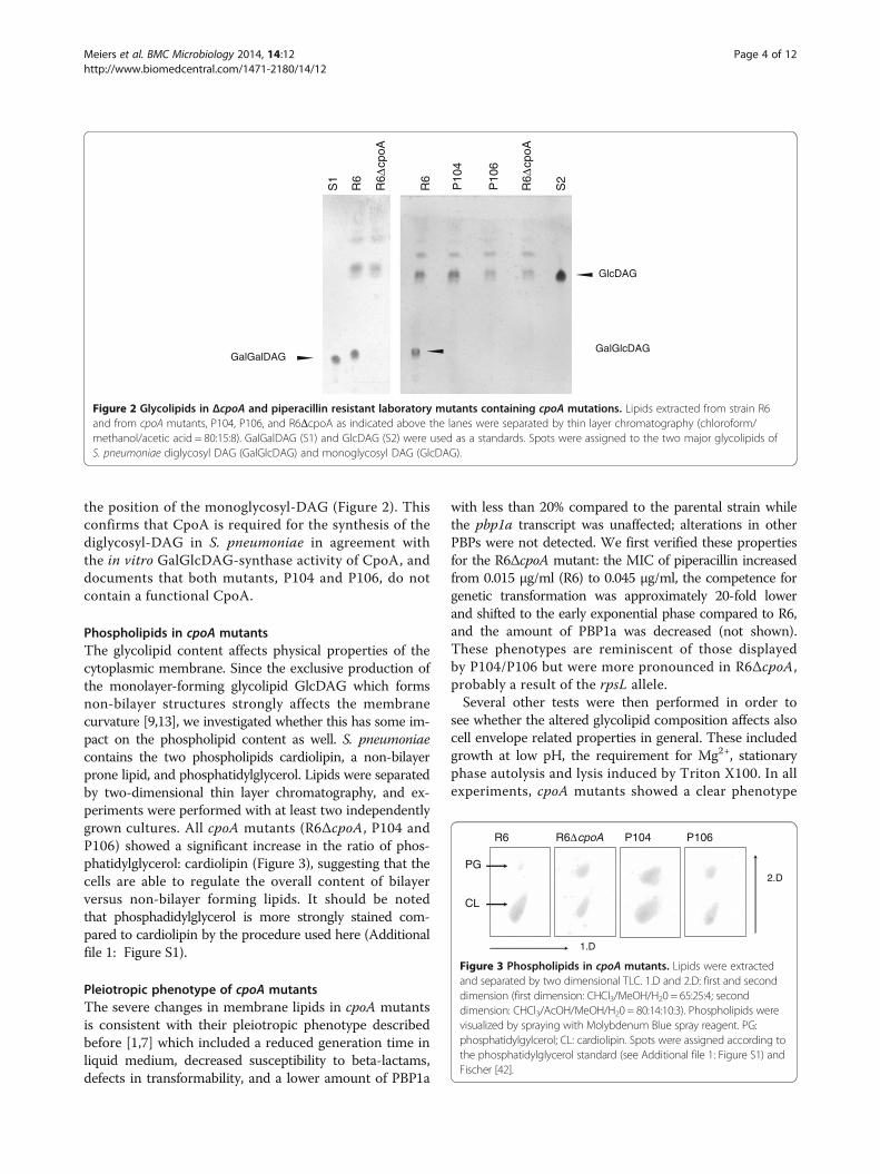
Phospholipids in cpoA mutantsThe glycolipid content affects physical properties of thecytoplasmic membrane. Since the exclusive production ofthe monolayer-forming glycolipid GlcDAG which formsnon-bilayer structures strongly affects the membranecurvature [9,13], we investigated whether this has some im-pact on the phospholipid content as well. S. pneumoniaecontains the two phospholipids cardiolipin, a non-bilayerprone lipid, and phosphatidylglycerol. Lipids were separatedby two-dimensional thin layer chromatography, and ex-periments were performed with at least two independentlygrown cultures. All cpoA mutants (R6ΔcpoA, P104 andP106) showed a significant increase in the ratio of phos-phatidylglycerol: cardiolipin (Figure 3), suggesting that thecells are able to regulate the overall content of bilayerversus non-bilayer forming lipids. It should be notedthat phosphadidylglycerol is more strongly stained com-pared to cardiolipin by the procedure used here (Additionalfile 1: Figure S1).
Pleiotropic phenotype of cpoA mutantsThe severe changes in membrane lipids in cpoA mutantsis consistent with their pleiotropic phenotype describedbefore [1,7] which included a reduced generation time inliquid medium, decreased susceptibility to beta-lactams,defects in transformability, and a lower amount of PBP1a
with less than 20% compared to the parental strain whilethe pbp1a transcript was unaffected; alterations in otherPBPs were not detected. We first verified these propertiesfor the R6ΔcpoA mutant: the MIC of piperacillin increasedfrom 0.015 μg/ml (R6) to 0.045 μg/ml, the competence forgenetic transformation was approximately 20-fold lowerand shifted to the early exponential phase compared to R6,and the amount of PBP1a was decreased (not shown).These phenotypes are reminiscent of those displayedby P104/P106 but were more pronounced in R6ΔcpoA,probably a result of the rpsL allele.Several other tests were then performed in order to
see whether the altered glycolipid composition affects alsocell envelope related properties in general. These includedgrowth at low pH, the requirement for Mg2+, stationaryphase autolysis and lysis induced by Triton X100. In allexperiments, cpoA mutants showed a clear phenotype
P10
6
P10
4
GalGlcDAG
R6
R6Δ
cpoA
GlcDAG
S2
R6
R6Δ
cpoA
GalGalDAG
S1
Figure 2 Glycolipids in ΔcpoA and piperacillin resistant laboratory mutants containing cpoA mutations. Lipids extracted from strain R6and from cpoA mutants, P104, P106, and R6ΔcpoA as indicated above the lanes were separated by thin layer chromatography (chloroform/methanol/acetic acid = 80:15:8). GalGalDAG (S1) and GlcDAG (S2) were used as a standards. Spots were assigned to the two major glycolipids ofS. pneumoniae diglycosyl DAG (GalGlcDAG) and monoglycosyl DAG (GlcDAG).
R6 P104 P106R6ΔcpoA
CL
PG
1.D
2.D
Figure 3 Phospholipids in cpoA mutants. Lipids were extractedand separated by two dimensional TLC. 1.D and 2.D: first and seconddimension (first dimension: CHCl3/MeOH/H20 = 65:25:4; seconddimension: CHCl3/AcOH/MeOH/H20 = 80:14:10:3). Phospholipids werevisualized by spraying with Molybdenum Blue spray reagent. PG:phosphatidylgylcerol; CL: cardiolipin. Spots were assigned according tothe phosphatidylglycerol standard (see Additional file 1: Figure S1) andFischer [42].
Meiers et al. BMC Microbiology 2014, 14:12 Page 4 of 12http://www.biomedcentral.com/1471-2180/14/12
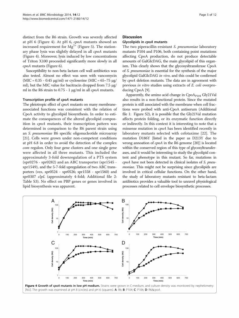
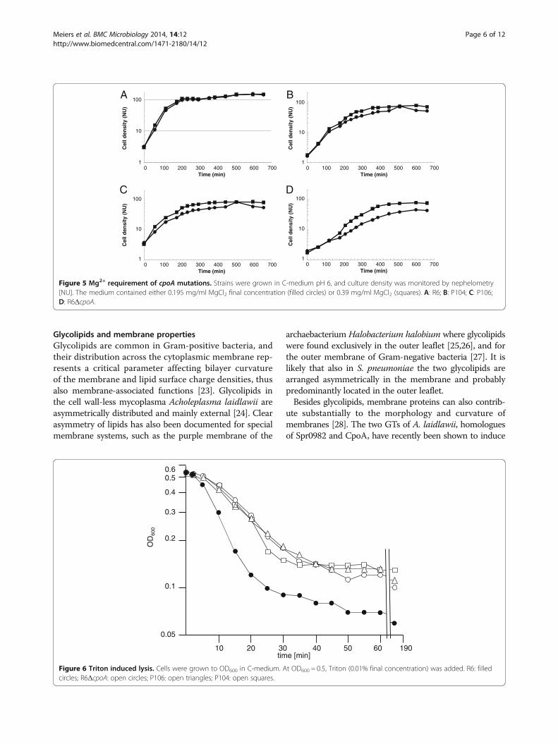
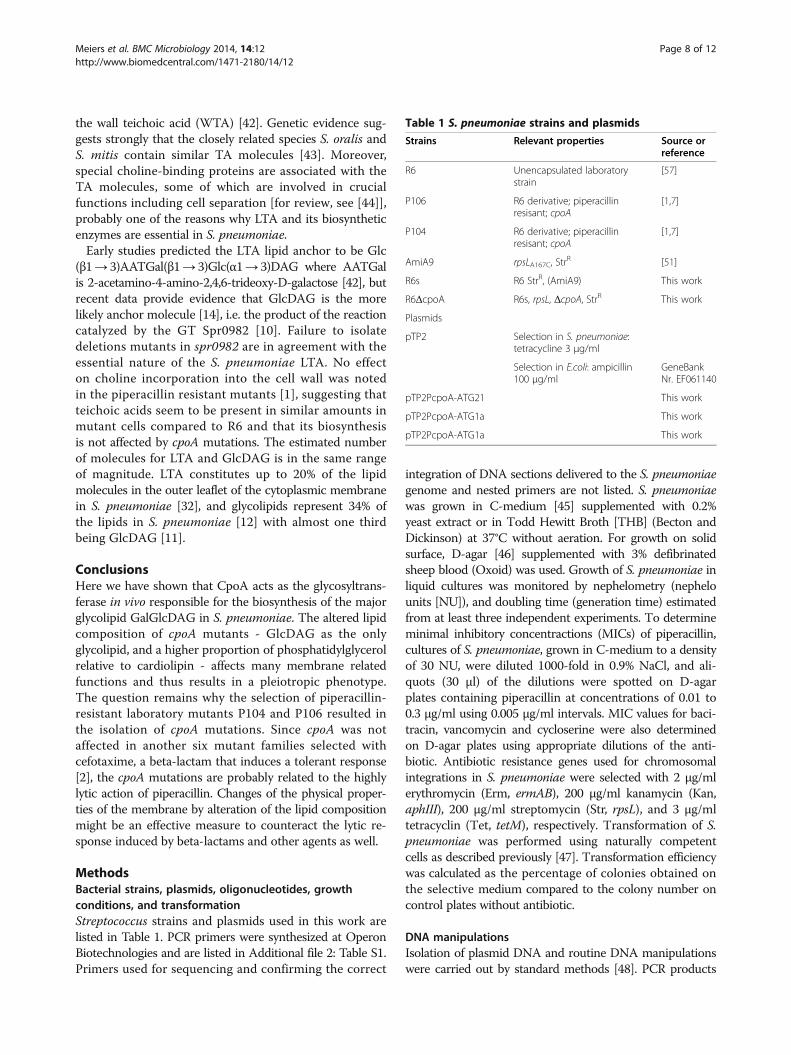
distinct from the R6 strain. Growth was severely affectedat pH 6 (Figure 4). At pH 6, cpoA mutants showed anincreased requirement for Mg2+ (Figure 5). The station-ary phase lysis was slightly delayed in all cpoA mutants(Figure 4). Moreover, lysis induced by low concentrationsof Triton X100 proceeded significantly more slowly in allcpoA mutants (Figure 6).Susceptibility to non-beta lactam cell wall antibiotics was
also tested. Almost no effect was seen with vancomycin(MIC = 0.35 - 0.45 μg/ml) or cycloserine (MIC = 65–75 μg/ml), but the MIC value for bacitracin dropped from 7.5 μg/ml in the R6 strain to 0.75 - 1 μg/ml in all cpoA mutants.
Transcription profile of cpoA mutantsThe pleiotropic effect of cpoA mutants on many membrane-associated functions was consistent with the relation ofCpoA activity to glycolipid biosynthesis. In order to esti-mate the consequences of the altered glycolipid compos-ition in cpoA mutants, their transcription pattern wasdetermined in comparison to the R6 parent strain usingan S. pneumoniae R6 specific oligonucleotide microarray[21]. Cells were grown under non-competent conditionsat pH 6.8 in order to avoid the detection of the complexcom regulon. Only four gene clusters and one single genewere affected in all three mutants. This included theapproximately 3-fold downregulation of a PTS system(spr0276 - spr0282) and an ABC transporter (spr1545 -spr1549), and the 5-7-fold upregulation of two ABC trans-porters (vex, spr0524 - spr0526; spr1558 - spr1560) andspr0307 clpL (approximately 4-fold; Additional file 2:Table S3). No effect on PBP genes or genes involved inlipid biosynthesis was apparent.
DiscussionGlycolipids in cpoA mutantsThe two piperacillin-resistant S. pneumoniae laboratorymutants P104 and P106, both containing point mutationsaffecting CpoA production, do not produce detectableamounts of GalGlcDAG, the main glycolipid of this organ-ism. This clearly shows that the glycosysltransferase CpoAof S. pneumoniae is essential for the synthesis of the majorglycolipid GalGlcDAG in vivo, and this could be confirmedby cpoA deletion mutants. The data are in agreement withprevious in vitro studies using extracts of E. coli overpro-ducing CpoA [9].Apparently, the amino acid change in CpoAP104 Gly21Val
also results in a non-functional protein. Since the mutatedprotein is still associated with the membrane when cell frac-tions were probed with anti-CpoA antiserum (Additionalfile 1: Figure S2), it is possible that the Gly21Val mutationaffects protein folding, or its enzymatic function directlyor indirectly. In this context it is interesting to note that amissense mutation in cpoA has been identified recently inlaboratory mutants selected with cefotaxime [22]. Themutation D186Y [listed in the paper as D213Y due towrong annoation of cpoA in the R6 genome [20]] is locatedwithin the conserved region of this type of glycosyltransfer-ases, and it would be interesting to study the glycolipid con-tent and phenotype in this mutant. So far, mutations incpoA have not been detected in clinical isolates of S. pneu-moniae. This might not be surprising since glycolipids areinvolved in critical cellular functions. On the other hand,the study of laboratory mutants resistant to beta-lactamantibiotics provides a valuable tool to unravel physiologicalprocesses related to cell envelope biosynthetic processes.
A B
C D
1
10
100
0 100 200 300 400 500 600 700
Cel
l den
sity
(N
U)
Time (min)
1
10
100
0 100 200 300 400 500 600 700
Cel
l den
sity
(N
U)
Time (min)
1
10
100
0 100 200 300 400 500 600 700
Cel
l den
sity
(N
U)
Time (min)
1
10
100
0 100 200 300 400 500 600 700
Cel
l den
sity
(N
U)
Time (min)
Figure 4 Growth of cpoA mutants in low pH medium. Strains were grown in C-medium, and culture density was monitored by nephelometry[NU]. The growth was examined at pH 8 (circles) and pH 6 (squares). A: R6; B: P104; C: P106; D: R6ΔcpoA.
Meiers et al. BMC Microbiology 2014, 14:12 Page 5 of 12http://www.biomedcentral.com/1471-2180/14/12
Glycolipids and membrane propertiesGlycolipids are common in Gram-positive bacteria, andtheir distribution across the cytoplasmic membrane rep-resents a critical parameter affecting bilayer curvatureof the membrane and lipid surface charge densities, thusalso membrane-associated functions [23]. Glycolipids inthe cell wall-less mycoplasma Acholeplasma laidlawii areasymmetrically distributed and mainly external [24]. Clearasymmetry of lipids has also been documented for specialmembrane systems, such as the purple membrane of the
archaebacterium Halobacterium halobium where glycolipidswere found exclusively in the outer leaflet [25,26], and forthe outer membrane of Gram-negative bacteria [27]. It islikely that also in S. pneumoniae the two glycolipids arearranged asymmetrically in the membrane and probablypredominantly located in the outer leaflet.Besides glycolipids, membrane proteins can also contrib-
ute substantially to the morphology and curvature ofmembranes [28]. The two GTs of A. laidlawii, homologuesof Spr0982 and CpoA, have recently been shown to induce
1
10
100
0 100 200 300 400 500 600 700
Cel
l den
sity
(N
U)
Time (min)
1
10
100
0 100 200 300 400 500 600 700
Cel
l den
sity
(N
U)
Time (min)
1
10
100
0 100 200 300 400 500 600 700
Cel
l den
sity
(N
U)
Time (min)
1
10
100
0 100 200 300 400 500 600 700
Cel
l den
sity
(N
U)
Time (min)
A B
C D
Figure 5 Mg2+ requirement of cpoA mutations. Strains were grown in C-medium pH 6, and culture density was monitored by nephelometry[NU]. The medium contained either 0.195 mg/ml MgCl2 final concentration (filled circles) or 0.39 mg/ml MgCl2 (squares). A: R6; B: P104; C: P106;D: R6ΔcpoA.
0.5
0.4
0.3
0.2
0.1
0.05
10 20 30 40 50 60 190time [min]
OD
600
0.6
Figure 6 Triton induced lysis. Cells were grown to OD600 in C-medium. At OD600 = 0.5, Triton (0.01% final concentration) was added. R6: filledcircles; R6ΔcpoA: open circles; P106: open triangles; P104: open squares.
Meiers et al. BMC Microbiology 2014, 14:12 Page 6 of 12http://www.biomedcentral.com/1471-2180/14/12

membrane vesiculation upon overproduction in E. coli [29].These enzymes are monotopic, i.e. anchored in the mem-brane cytoplasmic interface by hydrophobic and chargeinteractions in a SecYEG-independent manner [8,9]. Thedata of Wikström et al. [29] strongly suggest that the GTsthemselves are capable of inducing vesiculation, i.e. con-vex bending of the membrane. This implies some possibleconsequences when CpoA is absent, i.e. in P106 and inR6ΔcpoA, in that elimination of CpoA itself could affectthe curvature of the membrane.
Phenotypes of cpoA mutantsFailure to synthesize GalGlcDAG, the bilayerformingdi-glycosyl-glycolipid, must affect the physical propertiesof the cytoplasmic membrane considerably, consistent withthe pleiotropic phenotype associated with cpoA mutants.Introduction of the cpoA point mutations present in P104and P106 into the parental R6 strain conferred the samephenotypes, strongly suggesting that no other mutationsbesides cpoA are present in P104 and P106 (not shown).This included higher susceptibility to acidic stress andincreased requirement for Mg2+ at low pH, as well asreduced lysis rate under lysis inducing conditions. More-over, an altered proportion of the two pneumococcal phos-pholipids was observed in the cpoA mutants. Whereascardiolipin is the major phospholipid in the parental R6strain, all cpoA mutants contained a considerable higheramount of phosphatidylglycerol relative to cardiolipin asshown in Figure 3. Interestingly, mutations in the geneencoding the cardiolipin synthase have been identified incefotaxime resistant laboratory mutants but have not beeninvestigated further [22]. Since GlcDAG, the only glyco-lipid in cpoAmutants, is non-bilayer prone and cardiolipinas well, apparently the cells are capable to regulate theamounts of lipids to ensure sufficient bilayer structure ofthe cytoplasmic membrane. Cross-regulation in membranelipid pathways has already been suggested in B. subtilismutants defective in the cardiolipin synthase gene [30].MIC values of vancomycin or cycloserine inhibiting lateand early stages of peptidogylcan synthesis were notaffected in cpoA mutants, an indication that the cell wallbiochemistry is not affected.Interestingly, cpoA mutants were ten-fold more sus-
ceptible to bacitracin, which targets the lipid moleculebactoprenol. The cpoA mutants expressed an alteredtranscription profile compared to that of the R6 strain,mainly by genes encoding membrane proteins such asPTS systems or ABC transporters that represent minorcomponents of the bacterial cell. On the other hand,we could not detect significant changes of the proteinprofile of cytoplasmic or membrane proteins on SDS-polyacrylamide gels, i.e. no major protein componentswere affected in terms of quantity (not shown). It isconceivable that the transcriptional changes might be an
indirect effect of the altered membrane composition. Werecently reported that a higher susceptibility to bacitracinwas also noted in S. pneumoniae containing a mutatedABC transporter [31]. It is possible that the altered lipidcomposition of the cpoA mutants indirectly affects theABC transporter function and thus bacitracin MIC.
Glycolipids as anchor molecules in Gram-positive bacteriaGlycolipids represent the membrane anchor of importantmembrane-bound cell wall polymers in Gram-positive bac-teria. They function as the lipid anchor for LTA and alsofor another class of membrane-associated cell wall glycopo-lymers, lipoglycans, which seem to replace LTA in the highGC division of Gram-positive bacteria [32,33]. Listeriacontain the same glycolipids as S. pneumoniae, whereasGlcDAG and GlcGlcDAG represent the major glycolipidsin Bacillus, Staphylococcus and Enterococcus. However,these species differ in their biosynthetic enzymes. InBacillus and Staphylococcus, both glycolipids are synthe-sized by one single GT YpfP [34-36], whereas two putativeGTs are involved in glycolipid biosynthesis in Listeria,Streptococcus and Enterococcus [9,10,37,38]. In this contextit is remarkable that the structure of the cpoA operonwhich includes obg and several putative small peptideencoding genes is only maintained within Streptococcusspp., and that other Gram-positive bacteria contain cpoA(plus spr0982 in case of Listeria and Enterococcus) and obghomologues at distinct positions in the genome. Thereason for this is not clear. Several studies revealed thatObg proteins play a role in many important processes,including DNA replication, chromosome segregation,and regulation of stress responses, but their actual functionremains unknown [for review, see [19]].Most of the species mentioned above contain a polygly-
cerophosphate LTA backbone which is anchored to thedi-glycosyl-DAG lipid. Thus, interference of the biosyn-thesis of this glycolipid severely affects LTA and accordinglycell wall integrity as was shown for mutants in the S. aureusGT YpfP [34,35], the (1 → 2)GTs LafA in Listeria, IagA ingroup B streptococci, and E. faecalis BgsA [37-39]. Deletionmutants of S. aureus ypfP produced LTA which was prob-ably attached directly to DAG [34,35]. In the GC-richorganism M. luteus, dimannosyl-DAG is the lipid anchor ofthe essential lipomannan cell wall polymer [40]. Therefore,temperature sensitive mutants defective in lipomannanassembly were isolated of M. luteus, and one of them(mms1) contained a reduced amount of dimannosyl-DAGwhereas the amount of monomannosyl-DAG was increased[41]. The corresponding M. luteus gene encoding a putativeGT is unknown; according to BLAST analysis, the GTencoded by mlut_06690 is a likely CpoA homologue.In contrast to these organisms, the LTA of S. pneumoniae
is unique in that it includes choline and unusual sugarmoieties in its repeating unit which is identical to that of
Meiers et al. BMC Microbiology 2014, 14:12 Page 7 of 12http://www.biomedcentral.com/1471-2180/14/12
the wall teichoic acid (WTA) [42]. Genetic evidence sug-gests strongly that the closely related species S. oralis andS. mitis contain similar TA molecules [43]. Moreover,special choline-binding proteins are associated with theTA molecules, some of which are involved in crucialfunctions including cell separation [for review, see [44]],probably one of the reasons why LTA and its biosyntheticenzymes are essential in S. pneumoniae.Early studies predicted the LTA lipid anchor to be Glc
(β1→ 3)AATGal(β1→ 3)Glc(α1→ 3)DAG where AATGalis 2-acetamino-4-amino-2,4,6-trideoxy-D-galactose [42], butrecent data provide evidence that GlcDAG is the morelikely anchor molecule [14], i.e. the product of the reactioncatalyzed by the GT Spr0982 [10]. Failure to isolatedeletions mutants in spr0982 are in agreement with theessential nature of the S. pneumoniae LTA. No effecton choline incorporation into the cell wall was notedin the piperacillin resistant mutants [1], suggesting thatteichoic acids seem to be present in similar amounts inmutant cells compared to R6 and that its biosynthesisis not affected by cpoA mutations. The estimated numberof molecules for LTA and GlcDAG is in the same rangeof magnitude. LTA constitutes up to 20% of the lipidmolecules in the outer leaflet of the cytoplasmic membranein S. pneumoniae [32], and glycolipids represent 34% ofthe lipids in S. pneumoniae [12] with almost one thirdbeing GlcDAG [11].
ConclusionsHere we have shown that CpoA acts as the glycosyltrans-ferase in vivo responsible for the biosynthesis of the majorglycolipid GalGlcDAG in S. pneumoniae. The altered lipidcomposition of cpoA mutants - GlcDAG as the onlyglycolipid, and a higher proportion of phosphatidylglycerolrelative to cardiolipin - affects many membrane relatedfunctions and thus results in a pleiotropic phenotype.The question remains why the selection of piperacillin-resistant laboratory mutants P104 and P106 resulted inthe isolation of cpoA mutations. Since cpoA was notaffected in another six mutant families selected withcefotaxime, a beta-lactam that induces a tolerant response[2], the cpoA mutations are probably related to the highlylytic action of piperacillin. Changes of the physical proper-ties of the membrane by alteration of the lipid compositionmight be an effective measure to counteract the lytic re-sponse induced by beta-lactams and other agents as well.
MethodsBacterial strains, plasmids, oligonucleotides, growthconditions, and transformationStreptococcus strains and plasmids used in this work arelisted in Table 1. PCR primers were synthesized at OperonBiotechnologies and are listed in Additional file 2: Table S1.Primers used for sequencing and confirming the correct
integration of DNA sections delivered to the S. pneumoniaegenome and nested primers are not listed. S. pneumoniaewas grown in C-medium [45] supplemented with 0.2%yeast extract or in Todd Hewitt Broth [THB] (Becton andDickinson) at 37°C without aeration. For growth on solidsurface, D-agar [46] supplemented with 3% defibrinatedsheep blood (Oxoid) was used. Growth of S. pneumoniae inliquid cultures was monitored by nephelometry (nephelounits [NU]), and doubling time (generation time) estimatedfrom at least three independent experiments. To determineminimal inhibitory concentractions (MICs) of piperacillin,cultures of S. pneumoniae, grown in C-medium to a densityof 30 NU, were diluted 1000-fold in 0.9% NaCl, and ali-quots (30 μl) of the dilutions were spotted on D-agarplates containing piperacillin at concentrations of 0.01 to0.3 μg/ml using 0.005 μg/ml intervals. MIC values for baci-tracin, vancomycin and cycloserine were also determinedon D-agar plates using appropriate dilutions of the anti-biotic. Antibiotic resistance genes used for chromosomalintegrations in S. pneumoniae were selected with 2 μg/mlerythromycin (Erm, ermAB), 200 μg/ml kanamycin (Kan,aphIII), 200 μg/ml streptomycin (Str, rpsL), and 3 μg/mltetracyclin (Tet, tetM), respectively. Transformation of S.pneumoniae was performed using naturally competentcells as described previously [47]. Transformation efficiencywas calculated as the percentage of colonies obtained onthe selective medium compared to the colony number oncontrol plates without antibiotic.
DNA manipulationsIsolation of plasmid DNA and routine DNA manipulationswere carried out by standard methods [48]. PCR products
Table 1 S. pneumoniae strains and plasmids
Strains Relevant properties Source orreference
R6 Unencapsulated laboratorystrain
[57]
P106 R6 derivative; piperacillinresisant; cpoA
[1,7]
P104 R6 derivative; piperacillinresisant; cpoA
[1,7]
AmiA9 rpsLA167C, StrR [51]
R6s R6 StrR, (AmiA9) This work
R6ΔcpoA R6s, rpsL, ΔcpoA, StrR This work
Plasmids
pTP2 Selection in S. pneumoniae:tetracycline 3 μg/ml
Selection in E.coli: ampicillin100 μg/ml
GeneBankNr. EF061140
pTP2PcpoA-ATG21 This work
pTP2PcpoA-ATG1a This work
pTP2PcpoA-ATG1a This work
Meiers et al. BMC Microbiology 2014, 14:12 Page 8 of 12http://www.biomedcentral.com/1471-2180/14/12
and DNA recovered after restriction endonuclease di-gestions were purified using the JETquick spin columntechnique kit. Restriction enzymes and T4 DNA ligase werepurchased from Roche Applied Science or New EnglandBiolabs and used according to the manufacturer’s instruc-tions. PCRs were performed using either Goldstar RedTaq polymerase (Eurogentec) or iProof High-Fidelity DNApolymerase (Bio-Rad) according to the manufacturer’sinstructions. Nucleotide sequencing was performed usingthe ABI Prism BigDye Terminator Ready Reaction cyclesequencing kit, version 3.1 (Perkin Elmer-ABI). Nucleotidesequences were analyzed by using the CloneManager andPhred/Phrap/Consed software.
Identification of transcription start siteThe start point of cpoA transcription was determined byrapid amplification of cDNA ends (5' RACE) as describedpreviously [49] using RNA of S. pneumoniae R6 isolatedat a culture density of 40 NU. The primer cpoARACE2was used for reverse transcription of RNA ligated to theRNA adapter, and the nested primer and cpoARACE1 wasused for amplification of cDNA (for primers, see Additionalfile 2: Table S1 and S2).
Construction of delivery cassettes, plasmids and mutantsTo identify the initiation site of cpoA translation, fusionsof two DNA fragments with the lacZ reporter gene wereconstructed. They contained PcpoA (i) together either withtwo potential start codons (ATG1 and ATG2 in Figure 1B),(ii) with a mutation in ATG2 (ATA), or (iii) with ATG1
only. The three fragments were amplified from chromo-somal DNA of S. pneumoniae R6 by using the primer pairsPcpoA_Eco_f/PcpoA_r2, PcpoA_Eco_f/PcpoABam_r1a andPcpoA_Eco_f/PcpoABam_r1b, cleaved with EcoRI andBamHI, and ligated with the EcoRI/BamHI-digestedtranslation probe vector pTP2. The desired plasmids,pTP2PcpoA-ATG21, pTP2PcpoA-ATG1a and pTP2PcpoA-ATG1b were isolated after transformation of E. coli DH5αand subsequently used to transform S. pneumoniae R6;alternatively plasmids were directly transformed into S.pneumoniae R6. DNA from TetR transformants was PCR-amplified and sequenced to confirm the presence of thelacZ fusions in the resulting strains R6-PcpoA-ATG21,R6-PcpoA-ATG1a and R6-PcpoA-ATG1b.In-frame deletions in cpoA, spr0982, spr0983, obg, or
spr0985 were constructed via a two-step process in whichthe central part of the respective gene(s) was first replacedwith the Janus cassette [50] that confers a KanR StrS
phenotype in a StrR background. In the second step, theJanus cassette was deleted, thus restoring the original StrR
phenotype. The constituents of ‘replacement fragments’and ‘deletion fragments’ used in the first and second stepsof each deletion were amplified from chromosomal DNAof S. pneumoniae R6 by using the primer pairs listed in
Additional file 2: Table S2. To generate a ‘replacementfragment’, two PCR products of 0.7 to 1 kb (‘upstream’ and‘downstream fragment’) flanking the desired deletion werejoined with the two ends of the Janus cassette either bythe use of appropriate restriction sites added to the endsof the respective primers or by overlap extension PCRwith nested primers. The ‘replacement fragment’ was usedto transform a StrR derivative of S. pneumoniae R6 (R6s)obtained by transformation of R6 with chromosomalDNA carrying the AmiA9 resistance marker [51]. In theresulting KanR StrS transformants, the correct position ofthe Janus cassette was confirmed by DNA extraction andPCR with appropriate primers. To generate a ‘deletionfragment’ (containing the desired deletion), the respective‘upstream’ and ‘downstream fragments’ were directly joinedwith each other either by the use of appropriate restrictionsites added to the primers or by overlap extension PCRwith nested primers. The ‘deletion fragment’ was used totransform a derivative of R6s carrying the Janus cassette atthe site of the desired deletion. DNA from transformantsdisplaying a KanS StrR phenotype was PCR-amplified andsequenced to confirm the presence of the deletion in theresulting mutant.
Determination of β-galactosidase activityPreparation of cell extracts from cultures of S. pneumoniae,grown to a density of OD600 = 0.8 in C-medium, anddetermination of specific β-galactosidase activities wereperformed as described [52].
Lipid extraction and analysisLipids were extracted from S. pneumoniae essentially asdescribed [53]. Briefly, cells harvested by centrifugationof liquid cultures grown to a density of about 70 NUwere resuspended in 0.8 ml H2O per gram wet weightand subsequently mixed with 3 ml of chloroform/methanol (1:2) per gram wet weight. After gentle agitationfor 2 h at 4°C, chloroform (1 volume) and H2O (1 volume)were added and mixed. The samples were centrifuged at4,000 × g and 4°C for 5 min, the organic phases wererecovered, mixed with 1 volume of H2O equilibrated withchloroform/methanol (1:2), and centrifuged as before.Recovered organic phases were completely evaporated,and the remainders were dissolved in 50 to 100 μl ofchloroform/methanol (80:15). Glycolipids were separatedby one-dimensional thin layer chromatography in chloro-form/methanol/acetic acid (80:15:8) on silica gel G plates(0.025 mm; Merck). For visualization the plates weresprayed with 1-naphthol (3.2% w/v in methanol/H2SO4/H2O = 25:3:2) and heated at 110°C for 10 min. GalGal-DAG (Sigma) and GlcDAG were used as standards.Phospholipids were separated on two-dimensional thinlayer chromatography (first dimension: CHCl3/MeOH/H2O = 65:25:4; second dimension: CHCl3/AcOH/MeOH/
Meiers et al. BMC Microbiology 2014, 14:12 Page 9 of 12http://www.biomedcentral.com/1471-2180/14/12
H2O = 80:14:10:3) and stained with 1.3% molydbenumoxide in 4.2 M sulfuric acid (Molybdenum Blue sprayreagent, Sigma-Aldrich). Spots were assigned according tothe reference lipid phosphatidylglycerol (Sigma) and thepattern described elsewhere for phospholipids [42].
Immunological detection of CpoAS. pneumoniae cells were grown to mid-exponential growthphase (80 NU), harvested by centrifugation (9,000 rpm,15 min, 4°C, Beckman centrifuge J2-21), and washed oncewith 20 mM sodium phosphate buffer pH 7.2. Pellets wereresuspended in 180 μl sodium phosphate buffer and mixedwith 500 mg glass beads per 100 mg wet weight, followedby disruption in a cell mill (Vitrogen-Zellmühle Typ VI-4,Edmund Bühler GmbH) for 20 min. All further steps werecarried out on ice. Glass beads were removed by centrifuga-tion for 6 min (14,000 rpm, 4°C, Hermle Z513K centrifuge).Membranes were separated from cytoplasmic proteinsby ultracentrifugation (Beckman centrifuge, TLA 100.4rotor) for 2 h at 60,000 rpm and 4°C. Pellets were resus-pended in half of the volume of the supernatant, andfractions stored at −80°C. For SDS polyacrylamide gelelectrophoresis, 3 μl per fraction were used. Westernblotting was performed as described previously [54] andCpoA was visualized using a 1:10,000 dilution of rabbitantiserum raised against a purified CpoA-derivative asdescribed [7].
Microarray-based transcriptome analysisExtraction of total RNA from exponentially growing S.pneumoniae cultures (40 NU), reverse transcription ofRNA into labeled cDNA, prehybridization, hybridization,slide washing, scanning, and analysis of the data wereperformed as described previously [55]. For each strain,data sets from at least four hybridizations were used fornormalization and statistical analysis. Only data whichshowed P values below 10-4 in a paired t test, and relativechanges in the transcript amount of greater than threefoldwere considered further. The oligonucleotide microarraycovering genes and intergenic regions of S. pneumoniaeR6/TIGR4 has been described [21].
Accession numberS. pneumoniae R6/TIGR4 oligonucleotide microarray:ArrayDesign R6/TIGR4 ArrayExpres accession numberA-MEXP-1846.
Availability of supporting dataThe data sets supporting the results of this article areincluded within the article and its additional files.
Additional files
Additional file 1: Figure S1. Phospholipids in S. pneumoniae R6. Lipidswere extracted and separated by two dimensional TLC. 1.D and 2.D: firstand second dimension (first dimension: CHCl3/MeOH/H20 = 65:25:4;second dimension: CHCl3/AcOH/MeOH/H20 = 80:14:10:3). Phospholipidswere visualized by spraying with Molybdenum Blue spray reagent. PG:phosphatidylgylcerol; CL: cardiolipin. Standards: PG, 0.3 μMol; CL, 0.17 μmol.Figure S2. Membrane association of CpoA. Membrane (m) and cytoplasmicproteins (s) were separated by SDS-PAGE followed by immunostaining withanti-CpoA antiserum (see Methods for detail). Closed arrows indicate theposition of CpoA in the membrane fractions of S. pneumoniae R6 and P104,the open arrow shows the absence of CpoA in R6ΔcpoA. M: markerproteins.
Additional file 2: Table S1. Primers. Table S2. PCR primer pairs usedfor the construction of in-frame deletions 1. Table S3. Altered transcriptionprofiles in cpoA mutants.
Abbreviationsaa: Amino acids; bp: Base pairs; GalGlcDAG: 1,2-diacyl-3–O–[α-D-glucopyranosyl-(1→ 2)-O–α-D-galactopyranosyl]-sn-glycerol; GlcDAG: 1,2-diacyl-3-O–(α-D-glucopyranosyl)-sn-glycerol; GT: Glycosyltransferase; LTA: Lipoteichoic acid;NU: Nephelo units.
Competing interests- In the past five years have you received reimbursements, fees, funding, orsalary from an organization that may in any way gain or lose financially fromthe publication of this manuscript, either now or in the future? Is such anorganization financing this manuscript (including the article-processingcharge)? no- Do you hold any stocks or shares in an organization that mayin any way gain or lose financially from the publication of this manuscript,either now or in the future? No- Do you hold or are you currently applying for any patents relating to thecontent of the manuscript? Have you received reimbursements, fees,funding, or salary from an organization that holds or has applied for patentsrelating to the content of the manuscript? No- Do you have any other financial competing interests? NoNon-financial competing interests- Are there any non-financial competing interests (political, personal, religious,ideological, academic, intellectual, commercial or any other) to declare inrelation to this manuscript? No
Authors’ contributionsMM, CV and JE carried out the molecular genetic studies and phenotypicanalyses; MM carried out immunoassays and lipid chromatography. RH, BHand PM conceived of the study; RH and BH participated in its design andcoordination and helped to draft the manuscript. All authors read andapproved the final manuscript.
AcknowledgementsThis work was supported by the EU (Intafar LSHM-CT-2004-512138), the DFG(Ha 1011/11-1), and the Stiftung Rheinland-Pfalz für Innovation (15202–3862). We thank Martin Rieger for his assistance in analyzing microarray data,and Reinhold Brückner for helpful discussions.
Author details1Department of Microbiology, University of Kaiserslautern,Gottlieb-Daimler-Strasse, Gebäude 23, D-67663 Kaiserslautern, Germany.2Present address: Department of Microbial Natural Products,Helmholtz-Institute for Pharmaceutical Research Saarland (HIPS), SaarlandUniversity, D-66123 Saarbrücken, Germany. 3Present address: Hochschule fürTechnik und Wirtschaft des Saarlandes, Goebenstrasse 40, D-66117Saarbrücken, Germany.
Received: 4 September 2013 Accepted: 16 January 2014Published: 20 January 2014
Meiers et al. BMC Microbiology 2014, 14:12 Page 10 of 12http://www.biomedcentral.com/1471-2180/14/12
References1. Laible G, Hakenbeck R: Penicillin-binding proteins in β-lactam-resistant
laboratory mutants of Streptococcus pneumoniae. Mol Microbiol 1987,1:355–363.
2. Hakenbeck R, Tornette S, Adkinson NF: Interaction of non-lytic β-lactamswith penicillin-binding proteins in Streptococcus pneumoniae. J GenMicrobiol 1987, 133:755–760.
3. Hakenbeck R, Martin C, Dowson C, Grebe T: Penicillin-binding protein 2bof Streptococcus pneumoniae in piperacillin-resistant laboratory mutants.J Bacteriol 1994, 176:5574–5577.
4. Laible G, Hakenbeck R: Five independent combinations of mutations canresult in low-affinity penicillin-binding protein 2x of Streptococcuspneumoniae. J Bacteriol 1991, 173:6986–6990.
5. Krauß J, van der Linden M, Grebe T, Hakenbeck R: Penicillin-bindingproteins 2x and 2b as primary PBP-targets in Streptococcus pneumoniae.Microb Drug Resist 1996, 2:183–186.
6. Hakenbeck R, Grebe T, Zähner D, Stock JB: β-Lactam resistance inStreptococcus pneumoniae: penicillin-binding proteins and nonpenicillin-binding proteins. Mol Microbiol 1999, 33:673–678.
7. Grebe T, Paik J, Hakenbeck R: A novel resistance mechanism for β-lactamsin Streptococcus pneumoniae involves CpoA, a putativeglycosyltransferases. J Bacteriol 1997, 179:3342–3349.
8. Li L, Storm P, Karlsson OP, Berg S, Wieslander A: Irreversible binding and activitycontrol of the 1,2-diacylglycerol 3-glucosyltransferase from Acholeplasmalaidlawii at an anionic lipid bilayer surface. Biochemistry 2003, 42:9677–9686.
9. Edman M, Berg S, Storm P, Wikström M, Vikström S, Öhmann A, WieslanderA: Structural features of glycosyltransferases synthesizing major bilayerand nonbilayer-prone membrane lipids in Acholeplasma laidlawii andStreptococcus pneumoniae. J Biol Chem 2003, 278:8420–8428.
10. Berg S, Edman M, Li L, Wikström M, Wieslander A: Sequence properties ofthe 1,2-diacylglycerol 3-glucosyltransferase from Acholeplasma laidlawiimembranes. Recognition of a large group of lipid glycosyltransferases ineubacteria and archaea. J Biol Chem 2001, 276:22056–22063.
11. Tatituri RV, Brenner MB, Turk J, Hsu FF: Structural elucidation of diglycosyldiacylglycerol and monoglycosyl diacylglycerol from Streptococcuspneumoniae by multiple-stage linear ion-trap mass spectrometry withelectrospray ionization. J Mass Spectrom 2012, 47:115–123.
12. Brundish DE, Shaw N, Baddiley J: The phospholipids of PneumococcusI-192R, A.T.C.C. 12213. Some structural rearrangements occurring undermild conditions. Biochem J 1967, 104:205–211.
13. Wieslander A, Christiansson A, Rilfors L, Lindblom G: Lipid bilayer stabilityin membranes, Regulation of lipid composition in Acholeplasma laidlawiias governed by molecular shape. Biochemistry 1980, 19:3650–3655.
14. Seo HS, Cartee RT, Pritchard DG, Nahm MH: A new model ofpneumococcal lipoteichoic acid structure resolves biochemical,biosynthetic, and serologic inconsistencies of the current model.J Bacteriol 2008, 190:2379–2387.
15. Song JH, Ko KS, Lee JY, Baek JY, Oh WS, Yoon HS, Jeong JY, Chun J:Identification of essential genes in Streptococcus pneumoniae by allelicreplacement mutagenesis. Mol Cells 2005, 19:365–374.
16. Laursen BS, Sørensen HP, Mortensen KK, Sperling-Petersen HU: Initiation ofprotein synthesis in bacteria. Microbiol Mol Biol Rev 2005, 69:101–123.
17. Denapaite D, Brückner R, Nuhn M, Reichmann P, Henrich B, Maurer P,Schähle Y, Selbmann P, Zimmermann W, Wambutt R, et al: The genome ofStreptococcus mitis B6 - what is a commensal? PLoS ONE 2010, 5:e9426.
18. Reichmann P, Nuhn M, Denapaite D, Brückner R, Henrich B, Maurer P, RiegerM, Klages S, Reinhard R, Hakenbeck R: Genome of Streptococcus oralisstrain Uo5. J Bacteriol 2011, 193:2888–2889.
19. Czyz A, Wegrzyn G: The Obg subfamily of bacterial GTP-binding proteins:essential proteins of largely unknown functions that are evolutionarilyconserved from bacteria to humans. Acta Biochim Pol 2005, 52:35–43.
20. Hoskins J, Alborn WEJ, Arnold J, Blaszczak LC, Burgett S, DeHoff BS, EstremST, Fritz L, Fu D-J, Fuller W, et al: Genome of the bacterium Streptococcuspneumoniae strain R6. J Bacteriol 2001, 183:5709–5717.
21. Sauerbier J, Maurer P, Rieger M, Hakenbeck R: Streptococcus pneumoniaeR6 interspecies transformation: genetic analysis of penicillin resistancedeterminants and genome-wide recombination events. Mol Microbiol2012, 86:692–706.
22. Fani F, Brotherton MC, Leprohon P, Ouellette M: Genomic analysis andreconstruction of cefotaxime resistance in Streptococcus pneumoniae.J Antimicrob Chemother 2013, 68:1718–1727.
23. Shaw N: Bacterial glycolipids. Bacteriol Rev 1970, 34:365–377.24. Rottem S: Transbilayer distribution of lipids in microbial membranes.
Curr Top Membr Trans 1982, 17:235–261.25. Weik M, Patzelt H, Zaccai G, Oesterhelt D: Localization of glycolipids in
membranes by in vivo labeling and neutron diffraction. Mol Cell 1998,1:411–419.
26. Henderson R, Jubb JS, Whytock S: Specific labelling of the protein andlipid on the extracellular surface of purple membrane. J Mol Biol 1978,123:259–274.
27. Kamio Y, Nikaido H: Outer membrane of Salmonella typhimurium:accessibility of phospholipid head groups to phospholipase c andcyanogen bromide activated dextran in the external medium.Biochemistry 1976, 15:2561–2570.
28. Campelo F, McMahon HT, Kozlov MM: The hydrophobic insertionmechanism of membrane curvature generation by proteins. Biophys J2008, 95:2325–2339.
29. Wikström M, Kelly AA, Georgiev A, Eriksson HM, Klement MR, Bogdanov M,Dowhan W, Wieslander A: Lipid-engineered Escherichia coli membranesreveal critical lipid headgroup size for protein function. J Biol Chem 2009,284:954–965.
30. Lopez CS, Alice AF, Heras H, Rivas EA, Sanchez-Rivas C: Role of anionicphospholipids in the adaptation of Bacillus subtilis to high salinity. Micro-biology 2006, 152:605–616.
31. Becker P, Hakenbeck R, Henrich B: An ABC transporter of Streptococcuspneumoniae involved in susceptibility to vancoresmycin and bacitracin.Antimicrob Agents Chemother 2009, 53:2034–2041.
32. Fischer W: Lipoteichoic acid and lipoglycans. In Bacterial Cell Wall. Editedby Ghuysen J-M, Hakenbeck R. Amsterdam: Elsevier Sciences BV;1994:199–211.
33. Rahman O, Dover LG, Sutcliffe IC: Lipoteichoic acid biosynthesis: two stepsforwards, one step sideways? Trends Microbiol 2009, 17:219–225.
34. Fedtke I, Mader D, Kohler T, Moll H, Nicholson G, Biswas B, Henseler K, GötzF, Zähringer U: A Staphylococcus aureus ypfP mutant with stronglyreduced lipoteichoic acid (LTA) content: LTA governs bacterial surfaceproperties and autolysin activity. Mol Microbiol 2007, 65:1078–1091.
35. Kiriukhin MY, Debabov DV, Shinabarger DL, Neuhaus FC: Biosynthesis ofthe glycolipid anchor in lipoteichoic acid of Staphylococcus aureusRN4220: role of YpfP, the diglucosyldiacylglycerol synthase. J Bacteriol2001, 183:3506–3514.
36. Jorasch P, Wolter FP, Zähringer U, Heinz E: A UDP glucosyltransferase fromBacillus subtilis successively transfers up to four glucose residues to1,2-diacylglycerol: expression of ypfP in Escherichia coli and structuralanalysis of its reaction products. Mol Microbiol 1998, 29:419–430.
37. Webb AJ, Karatsa-Dodgson M, Grundling A: Two-enzyme systems forglycolipid and polyglycerolphosphate lipoteichoic acid synthesis in Listeriamonocytogenes. Mol Microbiol 2009, 74:299–314.
38. Doran KS, Engelson EJ, Khosravi A, Maisey HC, Fedtke I, Equils O, MichelsenKS, Arditi M, Peschel A, Nizet V: Blood–brain barrier invasion by group BStreptococcus depends upon proper cell-surface anchoring of lipotei-choic acid. J Clin Invest 2005, 115:2499–2507.
39. Theilacker C, Sanchez-Carballo P, Toma I, Fabretti F, Sava I, Kropec A, HolstO, Huebner J: Glycolipids are involved in biofilm accumulation andprolonged bacteraemia in Enterococcus faecalis. Mol Microbiol 2009,71:1055–1069.
40. Pakkiri LS, Wolucka BA, Lubert EJ, Waechter CJ: Structural and topologicalstudies on the lipid-mediated assembly of a membrane-associatedlipomannan in Micrococcus luteus. Glycobiology 2004, 14:73–81.
41. Pakkiri LS, Waechter CJ: Dimannosyldiacylglycerol serves as a lipid anchorprecursor in the assembly of the membrane-associated lipomannan inMicrococcus luteus. Glycobiology 2005, 15:291–302.
42. Fischer W: Pneumococcal lipoteichoic and teichoic acid. In Streptococcuspneumoniae - Molecular biology and mechanism of disease. Edited byTomasz A. Larchmont, NY: Mary Ann Liebert, Inc; 2000:155–177. 10538.
43. Denapaite D, Brückner R, Hakenbeck R, Vollmer W: Biosynthesis of teichoicacids in Streptococcus pneumoniae and closely related species: lessonsfrom genomes. Microb Drug Resist 2012, 18:344–358.
44. Hakenbeck R, Madhour A, Denapaite D, Brückner R: Versatility of cholinemetabolism and choline binding proteins in Streptococcus pneumoniaeand commensal streptococci. FEMS Microbiol Rev 2009, 33:572–586.
45. Lacks S, Hotchkiss RD: A study of the genetic material determining anenzyme activity in pneumococcus. Biochim Biophys Acta 1960, 39:508–517.
Meiers et al. BMC Microbiology 2014, 14:12 Page 11 of 12http://www.biomedcentral.com/1471-2180/14/12
46. Alloing G, Granadel C, Morrison DA, Claverys J-P: Competence pheromone,oligopeptide permease, and induction of competence in Streptococcuspneumoniae. Mol Microbiol 1996, 21:471–478.
47. Mascher T, Merai M, Balmelle N, de Saizieu A, Hakenbeck R: TheStreptococcus pneumoniae cia regulon: CiaR target sites and transcriptionprofile analysis. J Bacteriol 2003, 185:60–70.
48. Sambrook J, Fritsch EF, Maniatis T: Molecular Cloning: A Laboratory Manual.Plainview, New York: Cold Spring Harbor Laboratory Press; 1989.
49. Kovács M, Halfmann A, Fedtke I, Heintz M, Peschel A, Vollmer W, HakenbeckR, Brückner R: A functional dlt operon, encoding proteins required forincorporation of D-alanine in teichoic acids in gram-positive bacteria,confers resistance to cationic antimicrobial peptides in Streptococcuspneumoniae. J Bacteriol 2006, 188:5797–5805.
50. Sung CK, Li H, Claverys JP, Morrison DA: An rpsL cassette, janus, for genereplacement through negative selection in Streptococcus pneumoniae.Appl Environ Microbiol 2001, 67:5190–5196.
51. Salles C, Creancier L, Claverys JP, Méjean V: The high level streptomycinresistance gene from Streptococcus pneumoniae is a homologue of theribosomal protein S12 gene from Escherichia coli. Nucleic Acids Res 1992,20:6103.
52. Halfmann A, Hakenbeck R, Brückner R: A new integrative reporter plasmidfor Streptococcus pneumoniae. FEMS Microbiol Lett 2007, 268:217–224.
53. Arbogast LY, Henderson TO: Effect of inhibition of protein synthesis onlipid metabolism in Lactobacillus plantarum. J Bacteriol 1975, 123:962–971.
54. Hakenbeck R, Ellerbrok H, Briese T, Handwerger S, Tomasz A: Penicillin-binding proteins of penicillin-susceptible and -resistant pneumococci:immunological relatedness of altered proteins and changes in peptidescarrying the β-lactam binding site. Antimicrob Agents Chemother 1986,30:553–558.
55. McKessar S, Hakenbeck R: The two-component regulatory system TCS08 isinvolved in cellobiose metabolism of Streptococcus pneumoniae R6.J Bacteriol 2007, 189:1342–1350.
56. Tettelin H, Nelson KE, Paulsen IT, Eisen JA, Read TD, Peterson S, HeidelbergJ, DeBoy RT, Haft DH, Dodson RJ, et al: Complete genome sequence of avirulent isolate of Streptococcus pneumoniae. Science 2001, 293:498–506.
57. Ottolenghi E, Hotchkiss RD: Release of genetic transforming agent frompneumococcal cultures during growth and disintegration. J Exp Med1962, 116:491–519.
doi:10.1186/1471-2180-14-12Cite this article as: Meiers et al.: Altered lipid composition inStreptococcus pneumoniae cpoA mutants. BMC Microbiology 2014 14:12.
Submit your next manuscript to BioMed Centraland take full advantage of:
• Convenient online submission
• Thorough peer review
• No space constraints or color figure charges
• Immediate publication on acceptance
• Inclusion in PubMed, CAS, Scopus and Google Scholar
• Research which is freely available for redistribution
Submit your manuscript at www.biomedcentral.com/submit
Meiers et al. BMC Microbiology 2014, 14:12 Page 12 of 12http://www.biomedcentral.com/1471-2180/14/12