Embed Size (px)
Citation preview

ALTERATIONS I N T H E BLOOD COAGULATION SYSTEM I N D U C E D BY BACTERIAL E N D O T O X I N
I. IN VIvo (Gss'~P.AUZED SHWARZmAN RZACZION)
BY DONALD G. McKAY, M.D., AND SANDOR S, SHAPIRO,* M.D.
(From the Departments of Pathology, Obstetrics and Gynecology, Harvard Medical School, Boston, and the Pathology Laboratory, Free Hospital for Women,
Brookline, Massachusetts)
(Received for publication, October 12, 1957)
Histologic studies of rabbits dying after two properly spaced intravenous injections of bacterial endotoxin reveal a deposition in certain arterioles, capillaries, and venules of a material that has been referred to as "fibrinoid" or "hyaline thrombi" (1).
On the l~.,sis of histochemical observations, Booth et al. (2) have suggested that this material is derived from breakdown products of the walls of arterioles or arteries which are presumably damaged primarily by exposure to the toxin. Most students of the Shwartzman reaction have preferred to describe the material as hyaline thrombi or "fibrinoid" with no commitment as to its origin. Several observations have indi- cated that these are actually fibrin thrombi. Histologically the thrombi react with the phosphotungstic acid hematoxylin, periodic aeid-Schiff, and Weigert's stains, none of which is specific for fibrin but all of which react with fibrin. More importantly, it was demonstrated by Good and Thomas (3) and Cluff and Berthrong (4) that when the animals' blood is rendered incoagulable with heparin, the generalized as well as the local Shwartzman reaction cannot be elicited. Brunson et al. (5) using an acid polysaccharide (liquoid), shown by Walton (6) to precipitate fibrinogen in vitro, demonstrated that this material in combination with small amounts of bacterial endotoxin also was capable of producing a pathologic sequence that was entirely similar to the generalized Shwartzman reaction. Thomas (7) further noted a pre- cipitous drop in circulating fibrinogen after the injection of liquoid. With this back- ground he suggested that "fibrinoid" is derived from the circulating blood and that fibrinogen may play an important role in its formation.
These observations suggest that the manifestations of the generalized Shwartzman reaction are in part mediated by one or more episodes of dis- seminated intravascular coagulation. The studies reported herein on the alterations in the hemostatic mechanism during the development of the gen- eralized Shwartzman reaction were undertaken for the following r ea sons : -
* Former Josiah Macy Jr. Student Fellow in Obstetrics.
353
Dow
nloaded from http://rupress.org/jem
/article-pdf/107/3/353/1079257/353.pdf by guest on 07 Decem
ber 2021

354 BACTERIAL ENDOTOXIN AND BLOOD COAGULATION SYSTEM. I
1. To determine whether there is a drop in the level of circulating fibrinogen after the injection of bacterial endotoxin, as noted b y Thomas after the injec- t ion of liquoid.
2. To compare the al terat ions in the blood coagulation system in this reac- t ion with those seen in other experimental conditions such as intravenous thromboplas t in injection, in which disseminated int ravascular coagulat ion is the major pathologic consequence.
3. To determine whether al terat ions in the blood clott ing mechanism could account for the difference in location and extent of thrombosis when two spaced doses of endotoxin instead of one are given intravenously.
Materials and Methods
Hybrid female albino rabbits weighing 1 kg. were injected with Shcar's polysaccharide. I Two doses of 0.2 rag. of polysacclmridc in 2.0 mL of physiologic saline were injected int O the marginal ear vein with a 24 hour interval between the first and second injection. Control animals were injected with saline only. Blood samples were obtained by cardiac puncture, using iodine antisepsis, at the following times: before injection; 1 hour, 4 hours, 24 hours, 25 hours, 28 hours, and 48 hours after the first injection. Three groups of animals were studied: (a) Control group injected with saline, (b) animals given one injection and followed through the 48 hour period; and (c) animals given 2 injections 24 hours apart and followed through the 48 hour period. Three to four mL of blood wcre drawn at each cardiac puncture and the following tests done:--
A. White blood count. B. Piatelet count by the Rees-Ecker technique. C. Whole blood coagulation time. A modified three tube Lee-White procedure in silicone
was used. The test was run at 37°C. and a solid clot in the third tube was the end point. The needles, syringes, and tubes were siliconized with General Electric SC-87 drifilm.
D. One stage prothrombin time. Blood was drawn into plain glass tubes containing po- tossium oxalate; spun at room temperature at 1500 r.p.m, for 15 minutes and a one stage pro- thrombin time was done on the plasma using simplastin (Warner-Chilcott) as the thrombo- plastic agent.
E. Thrombin clotting times. 0.2 ml. of oxalated plasma was added to 0.2 ml. of a solution of fibrinogen (Cohn's fraction I). To this were added 0.2 ml. of saline containing 150, 200, or 250 #g. of bovine thrombin; and the dotting time of the mixture was then measured.
F. Fibrinogen level. The method described by Ratnoff (8) was used. G. Fibrinogenolysis. The method suggested by Crosby (9) was used. A fibrinogen level on
one portion of the plasma sample was done immediately. A 1 ml. aliquot was diluted in saline and buffer in the same manner but placed in a 37°C. incubator for 2 hours. At the end of this time the fibrinogen was clotted and the amount determined in the usual manner. The differ- ence between the incubated and non-incubated fibrinogen levels has been taken as the amount of fibrinogenolysis.
H. Fibrinolysis. Clotted whole blood was placed in the incubator for 48 hours at 37°C. and observed for destruction of the clot.
The animals that died during the experiment were autopsied immediately and all others were sacrificed at the end of the 48 hour period and autopsied immediately. The tissues were
1 We wish to express appreciation to Dr. Murray J. Shear of the National Institute of Health for his generosity in supplying the polysaccharide.
Dow
nloaded from http://rupress.org/jem
/article-pdf/107/3/353/1079257/353.pdf by guest on 07 Decem
ber 2021

D. G. MCKAY AND S. S. SHAPIRO 355
fixed in 10 per cent neutral formalin and paraffin sections were stained with hematoxylin and eosin, and phosphotungstic acid hematoxylin.
OBSERVATIONS
Histology.--Because of the variability of response to different endotoxins and to different concentrations of the same endotoxin, it is necessary first to define in histologic terms, the extent and locations of the intravascular thrombi under the conditions of the experiment.
TABLE I
Lungs Liver Spleen Kidneys
1+* 2+ 3+ 4+
Controls (21 animals)
1 0 0 0 0 0 0 0
i + 2 + 3 + 4 +
Tota l . . 14 (38 per cent)
One injection (37 animals)
4 . 5 2 10 3 5 0 !
9 (24 per cent) 21 (57 per cent) 6 (16 per cent)
1 + 2 + 3 + 4 +
15 11 6 2
5 4 10 5
Two injections (45 animals)
12 6 8 14 9 7 2 2
31 (69 per cent) 29 (64 per cent) Tota l . . 34 (76 per cent) 24 (53 per cent)
* 1-F Thrombosis in one capillary; 2 + many capillary and venular thrombi; 3 + ~/~ to of all capillaries or venules occluded by thrombi; 4 + Virtually all capillaries or venules
contain thrombi.
Although a complete autopsy was performed on each animal, pathologic changes ascribable to the bacterial toxin were observed only in the lungs, liver, spleen, and kidney. Areas of hemorrhage, necrosis, and inflammation were observed in the myocardium of the majority of animals but were undoubtedly related only to the needle puncture sites. The histologic sec- tions were examined for the presence of thrombi in capillaries, arterioles, and venules as well as for areas of necrosis. In this study necrosis was a common accompaniment of the intra- vascular thrombi, but many animals dying shortly after the first or even the second injection showed only thrombosis with no necrosis.
In the lungs the thrombi were found in capillaries, venules, and arterioles. Occasional hemorrhages were observed in the alveolar spaces but no infarcts were found. There was variation in the extent of thrombosis from one case to the next in the lungs as well as in all
Dow
nloaded from http://rupress.org/jem
/article-pdf/107/3/353/1079257/353.pdf by guest on 07 Decem
ber 2021

356 BACTERIAL ENDOTOXIN AND BLOOD COAGULATION SYSTEM. I
the other organs. In the liver, the intravascular deposits were found in the sinusoids and in the hepatic veins. It was quite common to see thrombi in the central veins extending into the intralobular hepatic veins as rounded masses of fibrin containing trapped leukocytcs and a few red blood cells. The areas of necrosis wcrc generally in the central portion of the liver
WBC (IN THOUSANDS/c.mm.) 1 6 ,
12
8
4 ¸
CONTROL . . . . . SINGLE INJECTION
TWO INJECTIONS
ENDOTOXIN I.V. ENDOTOXIN I.V.
HOURS Fro. 1. Changes in circulating leukocytes.
4"8
TABLE II Circulating Leukocytes in Thousands per c.mm.
Hrs ........ 0 1 4 24 25 28 48
Mean°. Range. " (3.74_'s3.s)
Control series (7 animals)
3.9 (3.4.4 5.1 4.4 1 4 4 " 8 t 5.0 (3.0-4.6) 1-5.3) (4.1-6.2) (3.7-5.6)l(4.0-5.7)1(3.9-5.7)
Mean.. Range.
One injection (11 animals) 1.3 1.3 [ 5.3
(0.8-1.9) (0.2-3.1) (5.4-7.9) 5.6 I 7.5 I
(One sample each) 8.9
Two injections (16 animals) Mean ] 4.5 T 1.8 ' 1.4 ] 7.4 .... 2.8 3.3 8.2 Range...[ [ l 1 ( 3 " 1 - 5 " 4 ) (0.9-3,4) (0.6-2.9) (4.8-11.3)(0.9-6.9) (1.2-5.8)(3.5-14.0)
Iobule hut often extended into the periportal region and sometimes involved a portion of a Iobule or even several adjacent Iobules. In the spleen, the intravasculax deposits were found in the sinnsoids of the red pulp and never in the Malpighian corpuscles. In the kidney, the deposits appeared primarily in the glomerular capillaries. Only a few cases exhibited artetiolar
Dow
nloaded from http://rupress.org/jem
/article-pdf/107/3/353/1079257/353.pdf by guest on 07 Decem
ber 2021

D. G. McKAY AND S. S. SHAPIRO 357
or venular thrombi. When necrosis occurred it presented the appearance of bilateral renal cortical necrosis. In order to compare the extent of the thrombosis with the extent of the al- terations in the circulating coagulation factors, the organs were graded from 0 to 4+, based on the relative number of thrombi in the small vessels. Table I indicates the extent, location, and incidence of thrombi in the various organs.
Thrombi appeared following the first injection but the thrombosis was present in fewer animals and was much less extensive than in animacs reeeiv!ng two injections. Although the
PLATELETS ( IN THOUSANDS/c. ram.)
4 0 0
300 '
2 0 0
IO0
. . . . . . CONTROL
I . l SINGLE INJECTION , r e , l i B = TWO INJECTIONS
. . . . .--- ' '"" --'J"x_.
- - " = o
ENOOTOX~N ~.V. ENDOTOX~N t~
HOURS
FIG. 2. Changes in circulating platelets.
4~
TABLE II I Circulating Platelets in Thousands per C. ram.
Control series (8 animals)
Mean ~ .. . . 235 (212608) 6-35 2 6 1 1 2 5 4 [ 238 224 287 Range. . (165-344) (216-336) (208-308) (218--286) (188-254) (184--416)
One injection (11 animals)
Mean .... 319 t73 I 1 8 4 ] 1 9 7 1 2 1 7 ' 1 8 3 1 1 7 3 (56-285) (63-341) (148-226) sample Range. (190-564) (One each)
Two injections (16 animals)
Mean .... 3 0 1 1 1 9 5 xs7 I 144 90 ] 7 3 [ 76 Range... (198-500) (59-386) (48--344) (37-286) (30-270) (30-243) (29-188)
Dow
nloaded from http://rupress.org/jem
/article-pdf/107/3/353/1079257/353.pdf by guest on 07 Decem
ber 2021
358 BACTERIAL ENDOTOXIN AND BLOOD COAGULATION SYSTEM. I
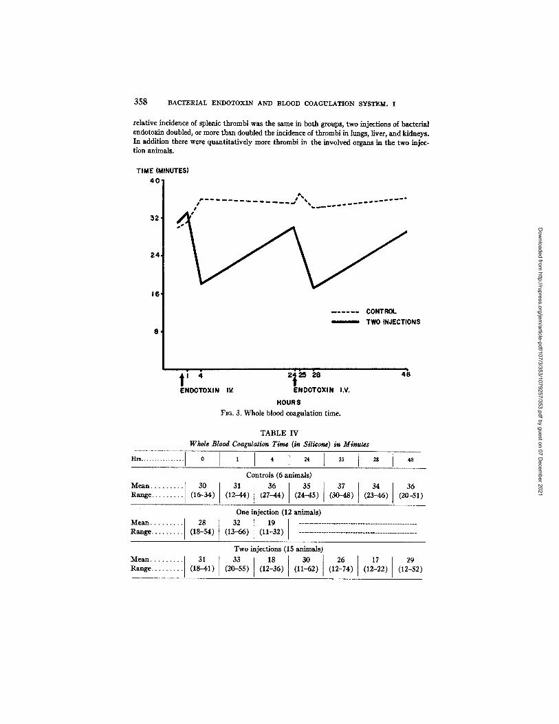
relative incidence of splenic thrombi was the same in both groups, two injections of bacterial endotoxin doubled, or more than doubled the inddence of thrombi in lungs, fiver, and kidneys. In addition there were quantitatively more thrombi in the involved organs in the two injec- tion animals.
TIME (MINUTES) 4 0
I I
52,
24,
16'
. . . . . . CONTROL
TWO INJECTIONS 8 ,
ENDOTOXIN IV. ENDOTOXIN I.V.
HOURS
FIG. 3. Whole blood coasu]ation time.
TABLE IV Whole Blood Coagulation Time (in Silicone) in Miuute~
" " . . . . . . . . . . . . . . . . t o ' ' I ~' ~, . I . Controls (6 animals)
Mean . . . . . . . . . 30 31 36 [ 35 37 3 4 , 36 Range . . . . . . . . . (16-34) (12--44) (27-44) [ (24-45) (30-48) (23--46) [ (20-51)
One injection (12 animals) Mean . . . . . . . . . 28 32 19 . . . . . . . . . . . . . . . . . . . . . . . . . . . . . . . . . . . . . . . . . . . Range . . . . . . . . . (18-54) (13-66) (11-32)
Two injections (15 animals)
Mean . . . . . . . . : 3 1 3 3 1 1 8 3 0 1 2 6 1 7 1 2 9 Range . . . . . . . . (18-41) (20-55) (12-36) (11-62) (12-74) (12-22) (12-52)
Dow
nloaded from http://rupress.org/jem
/article-pdf/107/3/353/1079257/353.pdf by guest on 07 Decem
ber 2021

D. G. McKAY AND S. S. SHAPIRO 359
Circulating Leukocytes.--Table II and Fig. 1 illustrate the changes in the circulating leukocytes during the 48 hour period of observation. The drop in circulating leukocytes after each injection can be related to the histologic observation that the granulocytes are sequestered in the pulmonary and hepatic
rag. FIBRINOGENI I00 mL
9 0 0
$ 0 0
7 0 0
600,
500,
4 0 0 '
300 '
2 0 0 '
I 0 0 .
CONTROL SINGLE INJECTION /
~,, ~ TWO INJECTIONS / *
/ I ~, °/ .7" /
ENDOTOXlN I.V ENOOTOXIN I.V. HOURS
FIG. 4. Changes in circulating fibrinogen.
4~
TABLE V Fibrinogen in Mg./lO0 M1.
Hrs ........ 0 I I I ~, [ 24 I 25 1 28 ] 48
Controls (9 animals)
Mean ... . 215 216 196 281 248 (129-332432) 253 Range... (176-272)(161-275) (144-251) (194-364) (140-349) (167-319)
One injection (11 animals) Mean .... 275 249 226 530 473 [ 474 784
I (474) Range. . (194-477)(204-282) (134-270) (517-624) (422-524) (4,30-1500)
Two injections (12 animals)
] (332-603) (379-623) 304 ( 5 Z 2 5 ) Mean .... 233 209 203 465 484 ' (!06-490) Range... (168-322) (147-258) (164-243)
Dow
nloaded from http://rupress.org/jem
/article-pdf/107/3/353/1079257/353.pdf by guest on 07 Decem
ber 2021

FIBRINOGEN (rag.%) 7 0 0 .
6 0 0 .
500 .
4 0 0 '
300 ,
200.
I 0 0
--', . - ~ V
NO RENAL THROMBI
RENAL THROMBI PRESENT
m I a i i 1' 4 2t ,~ 2s ENOOTOX,, ,.~. ENOOTO~,N ,.~.
HOURS FzG. 5. l~bfinogen levels with and without renal t]~ombi.
46
FIBRINOGEN (toO. %)
4 0 0 .
300.
200,
I00 '
. . . . . CONTROL INCUBATED SAMPLE
ENDOTOXIN I.V. ENDOTOXIN HOURS
Fzo. 6. Fibrinogenolysis.
I.V.
4~
Dow
nloaded from http://rupress.org/jem
/article-pdf/107/3/353/1079257/353.pdf by guest on 07 Decem
ber 2021

D. G. McKAY AND S. S. SHAPIRO 361
vessels during these brief periods. They appear to be subsequently released into the circulating blood. Only one animal was followed during the second 24 hours in the one injection series. The degree of change in circulating leuko- cytes could not be related to the degree of pathologic alteration in individual CaseS.
16
'¢3 t - O
(n
~D ._e I - "
E 2 e-
O.
12
8 -
. . . . Cont ro l Two In jec t ions
I 1 II I I Hours ~1 4 24 25 28 48
T t Endotoxin W. Endotoxin I.V,
FIG. 7. One stage prothrombin times.
TABLE VI Protkrorabin Times in Seconds
H r s . . . 0 [ I ! 4 ..... 24 2 5 1 2 8 1 4 8
Controls (4 animals) Mean .... 8.6 9.1 8.4 8.8 7.2 8.1 7.7 Range... (7.6-9.5) (7.9-10.0)(7.5-9.3) (8.2-9.7) (6.2-8.0)(6.5-9.2) (6.8-8.4)
Two injections (12 animals) • .I 7.7 7.5 7.9 7.0 7.4 8.6 7.3
Mean.. I (6.5-9.2) (6.1-8.9) Range.. (6.8-9.6) (6.9-9.7) (6.2-9.4) (7.2-11.0) (7.3-9.4)
Circulating Platelets.--Table I I I and Fig. 2 indicate that the platelets de- crease after the first injection, and to a lower level after the second injection. As in the white blood cell count there was no direct correlation of the degree of platelet response with the extent of anatomic change.
Whole Blood Coagulation Time.--Table IV and Fig. 3 reveal the shortening of the coagulation time in silicone 4 hours following each injection and the subsequent return to normal 24 hours after each injection.
Dow
nloaded from http://rupress.org/jem
/article-pdf/107/3/353/1079257/353.pdf by guest on 07 Decem
ber 2021

362 BACTERIAL ENDOTOXIN AND BLOOD COAGULATION SYSTEM. I
Fibrinogen.--The alterations in circulating fibrinogen levels are presented in Table V and Fig. 4. The fibrinogen levels in the control animals remained almost constant except for a slight decrease during each 4 hour period following the injection of saline. Since 8 to 10 ral. of blood were removed just prior to this period, hemodilution may account for these apparent drops. They can- not be construed as evidence of intravascular dotting, since no fbrin thrombi were observed in these animals. The most striking changes are the elevations
ANTITHROMBIN TIME (SECONDS)
2 0 . . . . . CONTROL
TWO INJECTIONS
~ I 200~250f
4
ENDOTOXIN LV. ENDOTOXIN I.V.
HOURS
Fzo. 8. A n t i t h r o m b i n t imes .
i i i [
48
TABLE VII Thrombin Clotting Times in Seconds
I Hra ...... 0 1 4 [ 24 25 48 28
Controls (4 animals)
Mean.... 14.2 15.9 17.5 11.7 11.9 11.9 12.8 Range... (12.9-16.0) (15.5--17.3) (16.0-18.6) (11.5-12.0) (10.2-13.6) (10.9-13.9) (11.6-14.8)
Two injections (12 animals)
Mean . . . . 14.5 14.4 15.1 12.5 13.4 13.9 13.? Range... (11.?-19.~) (12.6-16.1) (11.9-17.9) (11.5-13.6) (11.2-15.6) (9.9-22.0) (13.1-15.0)
Dow
nloaded from http://rupress.org/jem
/article-pdf/107/3/353/1079257/353.pdf by guest on 07 Decem
ber 2021

D. G. McKAY AND S. S. SHAPIRO 363
of the fibrinogen levels 24 hours after the first injection of toxin and the sharp decrease in fibrinogen after the second injection in animals developing the generalized Shwartzman reaction. This drop in fibrinogen occurs during the period when the fibrin thrombi develop histologically in the kidney. When the amount of thrombosis on a histologic basis was compared with degree of fibrinogen drop after the second injection of toxin, a close agreement was found.
The injection of two spaced doses of endotoxin did not always produce glomerular thrombi within the 48 hour period. The fibrinogen levels in such animals are presented in Fig. 5 and are compared with those that did develop renal lesions. The very slight drop in fibrinogen in those animals that were free of renal thrombi was of the same order as the drop in the controls and may also have been due to hemodilution.
Fibrinolysis and Fibrinogenolysis.--The possibility that the observed de- creases in circulating fibrinogen might be due to activation of circulating fibrinolytic or fibrinogenolytic enzymes required an answer. None of the whole blood clots incubated for 48 hours showed any evidence of lysis. When ali- quots of plasma samples used in the fibrogen determination were diluted with buffered saline and incubated for 2 hours at 37°C., no significant difference was noted when the two fibrinogen levels were compared. The fibrinogen levels before and after incubation are presented in Fig. 6.
Prothrombin Times.---One stage prothrombin times on blood taken during the course of the generalized Shwartzman reaction are presented in Table VI and Fig. 7. There does not appear to be a significant difference between the controls and the injected animals.
Thrombin Clotting Times.--In addition to the whole blood coagulation times, thrombin clotting times were measured to test for the possible develop- ment of anticoagulant activity during the generalized Shwartzman reaction. The results obtained using 150 #g. of bovine thrombin are presented in Table VII and the results with 150, 200, and 250 #g. are presented in Fig. 8. There is no significant difference between the controls and the toxin-treated animals.
DISCUSSION
These findings demonstrate that when bacterial endotoxins gain access to the blood stream in the proper dose range they have a profound effect on certain dements of the blood coagulation system. With one exposure to endo- toxin there is a progressive elevation in the circulating fibrinogen for 48 hours to a level more than twice the normal. The mechanism of the increase is not known but presumably represents an increased rate of release or production from the liver. With a second injection 24 hours after the first, there was an abrupt decrease in fibrinogen in animals exhibiting the renal thrombi of the generalized Shwartzman reaction. This confirms for bacterial endotoxin
Dow
nloaded from http://rupress.org/jem
/article-pdf/107/3/353/1079257/353.pdf by guest on 07 Decem
ber 2021

3 6 4 BACTERIAL ENDOTOXIN AND BLOOD COAGULATION SYSTEM. I
the effect noted by Thomas (7) after the injection of liquoid and endotoxin. Because the drop occurs at the time that thrombi are formed on a histologic basis, it seems likely that these thrombi are derived from fibrinogen and are therefore composed of fibrin. When the thrombi do not appear in the kidney g!omeruli, the drop in fibrinogen does not occur. The decrease in fibrinogen cannot be attributed to enzymatic destruction since in these experiments there was no evidence of fibrinolytic or fibrinogenolytic activity in the ani- mals' blood.
In attempting to unravel the problem of what constitutes "preparation" for the generalized Shwartzman reaction or the basic reason for the difference in extent of thrombosis between the first and the second injections, several questions are raised by the results obtained thus far. On a histologic basis the difference between the first and second injection is twofold:--
(a) The incidence and extent of lung and liver thrombi are increased (doubled) by the second injection, and
(b) Renal thrombi appear usually only after the second injection. One of the most notable hematologic differences in existence at the time of the second injection is the fact that the fibrinogen level is twice as high as it is at the time of the first injection. This suggests that the more extensive thrombosis fol- lowing the second injection may be due to the presence of greater amounts of fibrinogen. In conformity with this hypothesis it is interesting to note that the two animals developing renal thrombi after only one injection had higher fibrinogen levels to start with (by approximately 100 mg. per cent) than the remainder of the control animals.
Two other factors have been altered by the time of the second injection, namely an increase in circulating leukocytes and a decrease in circulating platelets. As has been previously noted, the extent of thrombosis was not directly related to the extent of thrombocytopenia or leukocytosis in individual animals. The fate of the platelets is not entirely settled. It seems clear that some are clumped and caught in the pulmonary vessels since they are seen in this location in the sections. Stetson (10) has demonstrated that endotoxins cause a clumping of platelets in dvo. The fact that the thrombocytopenia persists after both injections suggests that many are destroyed after being dumped. If platelets are destroyed and their clot-promoting substance re- leased into the plasma, the possibility that they may be responsible for ini- tiating the disseminated intravascular thrombosis must be considered.
The role of the leukocytes in this reaction has received considerable atten- tion. They appear to play an important part since the production of leuko- penia, by pretreatment with nitrogen mustard, prevents the generalized Shwartzman reaction (11-14). Endotoxins are taken up by the circulating leukocytes within 15-30 minutes and the blood is cleared of endotoxin within this time (15-17). In vitro studies have shown that endotoxin-treated leuko-
Dow
nloaded from http://rupress.org/jem
/article-pdf/107/3/353/1079257/353.pdf by guest on 07 Decem
ber 2021

D. G. McKA¥ AND S. S. SHAPIRO 365
cytes (a) disintegrate at a rate 4-5 times that of normal leukocytes (18), (b) clump and adhere to each other (18), (c) do not migrate on slides or in tissue culture preparations as normal leukocytes do (18, 19), and (d) release lysozyme into the surrounding medium (20). The possibility that a clot-promoting agent may be released from damaged leukocytes in the generalized Shwartz- man reaction must be considered. It is clear from the studies reported herein that leukopenia occurs during both periods of thrombosis and that the more extensive thrombosis following the second injection is preceded by an elevation of circulating leukocytes.
Although the whole blood coagulation time is shortened after each intra- venous injection of toxin, it returns to normal by the time of the second injec- tion and therefore cannot be considered, even in a general sense, a "prepara- tory" phenomenon. However, since the shortening occurs during both periods of fibrin deposition, the two phenomena may be related. It may be that a partial polymerization of fibrinogen has already been achieved in vivo, and with fewer steps to go through in ~itro, the time required for clotting is less. The "heparin-precipitable protein" described by Thomas (21) may represent another manifestation of such an altered fibrinogen.
From the hematological standpoint "preparation" for the generalized Shwartzman reaction is accompanied by an increased circulating fibrinogen, leukocytosis, and thrombocytopenia. The actual mechanism by which any or all of these changes react to produce the phenomenon remains to be elicited.
It is useful to compare the disseminated intravascular thrombosis found in the Shwartzman reaction with disseminated thrombosis induced by agents other than bacterial endotoxins. Tissue thromboplastin has been used experi- mentally in extensive studies by Schneider (22). This agent in large quantities induces sudden death with extensive pulmonary vascular thrombi. With sub- lethal amounts some animals exhibited cerebral and hepatic hemorrhages and areas of necrosis. Four major differences exist between the effects of tissue thromboplastin and bacterial endotoxin injected intravenously:--
(a) The amount of bacterial endotoxin used is considerably less than the amount of thromboplastin.
(b) Thrombosis occurs within a few minutes after thromboplastin injection but over a period of several hours with bacterial endotoxin.
(c) Thrombi appear predominantly in the lungs and veins and not in the kidneys following thromboplastin, but predominantly in the kidneys, liver, spleen, and lungs after bacterial endotoxin.
(d) Bilateral renal cortical necrosis has not been found after thromboplastin injection.
These differences indicate that although both these reagents cause intra- vascular thrombosis they do so by different mechanisms.
The injection of incompatible blood has been shown to produce disseminated
Dow
nloaded from http://rupress.org/jem
/article-pdf/107/3/353/1079257/353.pdf by guest on 07 Decem
ber 2021

366 BACTERIAL E N D O T O X I N AND BLOOD COAGULATION SYSTEM. I
intravascular thrombosis (23-25). As in the experiments using thrombo- plastin, the amount of clot-promoting agent was far greater than the amount of endotoxin used. The response of the hemostatic mechanism to the clot- promoting agent released from red blood cells in another animal species, was in some ways similar to but in others different from, the response to bacterial endotoxin. The two reactions share in common an immediate drop in leuko- cytes and platelets, an initial decrease in coagulation time, a decrease in circulating fibrinogen, and no evidence of fibfinolysis or fibrinogenolysis. However, in the incompatible transfusion reaction the coagulation time became prolonged after several hours, a circulating anticoagulant activity appeared, and the prothrombin times were prolonged. It is not certain as to whether these represent differences in species response or differences in the agents used to induce thrombosis. Nevertheless, these histological and hemato- logical comparisons suggest that bacterial endotoxins do not act in the same manner as tissue thromboplastin.
SUMMARY AND CONCLUSIONS
The intravenous injection of bacterial endotoxins alter the coagulation system of rabbits' blood in ~/vo. Twenty-four hours after the first injection the fibrinogen level rises to twice normal values. The second injection at this time causes a 30 to 40 per cent decrease in fibrinogen content in 4 hours. Twenty hours later it again rises to twice normal values. A marked decrease in whole blood coagulation times in silicone occurs 4 hours after both injections but rises to normal values 24 hours following each injection. The circulating platelets drop from average levels of 300,000/c.mm. to 150,000/c.mm. after the first injection. The platelets remain at this low level and decrease to less than I00,000 after the second injection. During this time no fibrinolytic or fibrinogenolytic activity can be detected. Also, there is no significant change in the one stage prothrombin times or antithrombin titres.
The marked decrease in circulating fibrinogen at the time when intracapil- lary thrombi are formed suggests that the "hyaline" thrombi of the generalized Shwartzman reaction are composed, in part, of fibrin.
There appears to be a relationship between the level of circulating fibrinogen at the time of injection of bacterial endotoxin and the extent of the thrombo- sis. The higher the preinjection fibrinogen level, the more extensive is the thrombosis. There is also a relationship between the amount of fibrinogen loss and the extent of thrombosis after the injection. The more extensive the thrombosis the greater is the postinjection decrease in circulating fibrinogen.
A comparison between the response of the hemostatic mechanism to tissue thromboplastin and bacterial endotoxin indicates that the latter acts in a unique manner and not by way of a simple "thromboplastic" activity.
From the hematological standpoint, "preparation" for the generalized
Dow
nloaded from http://rupress.org/jem
/article-pdf/107/3/353/1079257/353.pdf by guest on 07 Decem
ber 2021

D. G. McKAY AND S. S. SHAPIRO 367
Shwartzman reaction is accompanied by an increased circulating fibrinogen, leukocytosis, and thrombocytopenia.
BIBLIOGRAPHY
1. Thomas, L., Rheumatic Fever--A Symposium, Minneapolis, University of Minnesota Press, 1952, 232.
2. Booth, E., Muirhead, E. E., and Montgomery, P. O'B. Arch. Path., 1956, 6t, 169.
3. Good, R. A., and Thomas, L., Y. Exp. Med., 1953, 97, 871. 4. Cluff, L. E., and Berthrong, M., Bull. Johns Hopkins Hosp., 1953, 92, 353. 5. Brunson, J. G., Davis, R. L., and Thomas, L., Am. J. Path., 1955, 31,669. 6. Walton, K. W., Brit. Y. Pkarmacol. and Chemotherap., 1954, 9, I. 7. Thomas, L., Smith, R. T., and Von Korff, R., J. Exp. Med., 1955, 102, 263. 8. Ratnoif, O. D., J. Lab. and Clin. Med., 1951, 37, 316. 9. Crosby, W. H., and Stefanini, M., J. Lab. and Clln. Meal., 1952, 46, 374; also
personal communication. 10. Stetson, C. A., Jr., y. Exp. Med., 1951, 93, 489. 11. Bennett, I .L. , Jr., and Cluff, L. E., Proc. Soc. Exp. Biol. and Med., 1952, SJ.,
304. 12. Stetson, C. A., Jr., and Good, R. A., J. Exp. Med., 1951, 93, 49. 13. Race, G. J., and Reed, D. W., Southern Med., 1953, 46, 207. 14. Schlang, H. A., Proc. Soc. Exp. Biol. and Med., 1952, 81, 274. 15. Bmde, A. I., Carey, F. J., Sutherland, D., and Zalesky, M., Y. Clin. Inv., 1955,
34, 850. 16. Brude, A. I., Carey, F. J., and Zalesky, M., J. Clin. Inv., 1955, 34, 858. 17. Gamble, C. N., and Brunson, J. G., Arch. Path., 1955, 60, 583. 18. Berthrong, M., and Cluff, L. E., J. Exp. Med., 1953, 98, 331. 19. Martin, S. P., and Chaudhuri, S. N., Proc. Soc. Exp. Biol. and Med., 1952, 81,
286. 20. Kerby, G. P., Proc. Soc. Exp. Biol. and Meal., 1952, 81,581. 21. Thomas, L., Smith, R. T., and Von Korff, R. Proc. Soc. Exp. Biol. and Med.
1954, 86, 813. 22. Schneider, C. L., in Toxemia of Pregnancy, Human and Veterinary, Ciba Foun-
dation Symposium, (J. Hammond, F. J. Browne, and G. E. W. Wolstenholme, editors), London, J. and A. Churchill, Ltd., 1950, 163.
23. Hardaway, R. M., III, McKay, D. G., Wahle, G. H., Jr., Tartock, D. E , and Edelstein, R., Am. Y. Surg., 1956, 91, 24.
24. McKay, D. G., Hardaway, R. M., III, and Bums, R., Am. J. Surg., 1956, 91, 31.
25. McKay, D. G., Hardaway, R. M., III, Wahle, G. H. Jr., Edelstein, R., and Tartock, D. E., Am. J. Surg. 1955, 89, 583.
Dow
nloaded from http://rupress.org/jem
/article-pdf/107/3/353/1079257/353.pdf by guest on 07 Decem
ber 2021