Embed Size (px)
Citation preview
ARTICLE
Alterations in CDH15 and KIRREL3 in Patientswith Mild to Severe Intellectual Disability
Kavita Bhalla,1,4,5 Yue Luo,1,4 Tim Buchan,2 Michael A. Beachem,1 Gregory F. Guzauskas,1
Sydney Ladd,1 Shelly J. Bratcher,1 Richard J. Schroer,1 Janne Balsamo,2 Barbara R. DuPont,1,3
Jack Lilien,2 and Anand K. Srivastava1,3,*
Cell-adhesion molecules play critical roles in brain development, as well as maintaining synaptic structure, function, and plasticity. Here
we have found the disruption of two genes encoding putative cell-adhesion molecules, CDH15 (cadherin superfamily) and KIRREL3
(immunoglobulin superfamily), by a chromosomal translocation t(11;16) in a female patient with intellectual disability (ID). We
screened coding regions of these two genes in a cohort of patients with ID and controls and identified four nonsynonymous CDH15
variants and three nonsynonymous KIRREL3 variants that appear rare and unique to ID. These variations altered highly conserved
residues and were absent in more than 600 unrelated patients with ID and 800 control individuals. Furthermore, in vivo expression stud-
ies showed that three of the CDH15 variations adversely altered its ability to mediate cell-cell adhesion. We also show that in neuronal
cells, human KIRREL3 colocalizes and interacts with the synaptic scaffolding protein, CASK, recently implicated in X-linked brain
malformation and ID. Taken together, our data suggest that alterations in CDH15 and KIRREL3, either alone or in combination with
other factors, could play a role in phenotypic expression of ID in some patients.
Introduction
Intellectual disability (ID), also known as intellectual
and developmental disability or mental retardation, is
the most frequently reported developmental disability,
affecting cognitive function in about 1%–3% of people
worldwide. Intellectual disability, often diagnosed as
developmental delay in early childhood, is a genetically
and clinically heterogeneous condition characterized by
below-average intellectual functioning (IQ < 70) in
conjunction with significant limitations in adaptive func-
tioning.1 The causation in at least half of all ID cases is still
unknown.2,3 Among the identifiable genetic causes, chro-
mosome abnormalities, single-gene mutations, and multi-
factorial interactions account for approximately 30% of ID
overall. It is expected that the genetic component of ID, in
part, is due to alterations in molecular pathways involved
in cognitive function.2,4
A large number of genes distributed throughout the
genome are anticipated to cause ID. This is well established
for the X chromosome, where more than 80 genes that
cause syndromal and nonsyndromal ID have been identi-
fied.3–5 Compared with genes on the X chromosome,
very few autosomal genes have been implicated in ID.
The autosomal ID genes identified are primarily involved
in syndromal and metabolic conditions and only five are
involved in nonsyndromal ID.6–12 Only a few families or
unrelated individuals with autosomal-recessive ID have
been found to have mutations in these genes.6
Identification of autosomal genes associated with ID has
proven very difficult primarily because of the lack of large
The American
families for linkage analysis.3 Furthermore, finding ID-
causing gene mutations in candidate genes has been diffi-
cult because of the enormous genetic heterogeneity and
rarity of mutations in any individual gene in the ID popu-
lation. It has been observed that mutations in most cloned
X-linked ID genes have a very low (<1.0%) prevalence in
patients with ID.5 However, a significant contribution of
both common and rare gene variants in disease pheno-
types has been suggested in several recent studies.13–15
Growing evidence indicates that defects in synapse
formation or synaptic plasticity are major causes of
ID.4,16 Cell-adhesion molecules of the cadherin and immu-
noglobulin (Ig) superfamilies play critical roles in brain
development, as well as maintaining synaptic structure,
function, and plasticity.17 In this study, we characterized
a balanced translocation in a female patient with severe
ID that truncates two genes encoding such cell-adhesion
molecules, CDH15 (cadherin superfamily) (MIM 114019)
and KIRREL3 (Ig superfamily) (MIM 607761). These find-
ings prompted us to analyze a large cohort of patients
with ID of unknown cause for alterations in the two genes.
We identified and characterized seven variations in key
functional domains of CDH15 and KIRREL3 in unrelated
patients with ID. We show that rare variants of CDH15
are functionally significant. We also show that in neuronal
cells, KIRREL3 interacts with the synaptic scaffold protein
calmodulin-associated serine/threonin kinase (CASK)
(MIM 300172), recently implicated in X-linked brain
abnormalities and ID.18–20 Consistent with a predicted
role of KIRREL3-CASK in brain function, a role for mouse
Kirrel3 in synaptogenesis has been suggested21 and the
1J.C. Self Research Institute of Human Genetics, Greenwood Genetic Center, 113 Gregor Mendel Circle, Greenwood, SC 29646, USA; 2Department of
Biological Sciences, University of Iowa, Iowa City, IA 52242, USA; 3Department of Genetics & Biochemistry, Clemson University, Clemson, SC 29634, USA4These authors contributed equally to this work5Present address: Division of Endocrinology, Diabetes & Nutrition, University of Maryland, Baltimore, MD 21201, USA
*Correspondence: [email protected]
DOI 10.1016/j.ajhg.2008.10.020. ª2008 by The American Society of Human Genetics. All rights reserved.
Journal of Human Genetics 83, 703–713, December 12, 2008 703
deletion of Cask in mice have been shown to impair
synaptic function.22
Material and Methods
Patients and Control SamplesCMS3377 is a 56-year-old white female with severe ID (with an in-
telligence quotient of 16). She began walking at about 3 years of
age. Physical examination revealed her head circumference to be
54.5 cm (45th centile). She had alternating exotropia, flat midface,
some downslanting of the lower eyelids, a thin nasal bridge,
a rounded nasal tip, a nasal septum below the alae nasi, and small
chin. Other clinical features included short fifth fingers and finger-
nails, broad and short feet with short toes, and 2-3-4 syndactyly on
the right and 2-3 syndactyly on the left. Routine high-resolution
chromosome analysis on blood revealed an apparently balanced
translocation t(11;16)(q24.2;q24). Furthermore, no additional ob-
vious chromosomal rearrangements at or near the chromosomal
breakpoints or unrelated to the translocations were detected by
array-CGH analyses via a targeted GenoSensor Array 300 and the
Human Mapping 250K (NSP) array. No karyotype information
was available from the parents, who are deceased but apparently
normal phenotypically.
Mutation screening was performed in a cohort of 657 unrelated
patients with ID of unknown etiology. These patients were negative
for the FMR1 (MIM 309550) expansion. Control samples consisted
of 800 normal individuals. The study was approved by the Institu-
tional Review Board of the Self Regional Healthcare and the Na-
tional Institutes of Health Office of Human Research Protection.
FISH AnalysisBAC and PAC DNAs were isolated with QIAGEN Mini-Prep
columns and were labeled by incorporation of biotin-11-dUTP
(Sigma) or digoxigenin-11-dUTP (Boehringer Mannheim) by
nick translation with DNA polymerase I (Life Technologies). Meta-
phase chromosome spreads were obtained from lymphoblastoid
cell lines from patients and FISH was performed as described
previously.23 Chromosome-specific labeled centromeric alphoid
probes were used for chromosome 11 and 16 identification. The
labeled probes were visualized with FITC-avidin (for Biotin) or
rhodamine-labeled anti-digoxigenin and the chromosomes were
counterstained with DAPI. Images were examined under a Zeiss
Axiphot fluorescent microscope.
Inverse-PCR and Cloning of Junction FragmentFor inverse-PCR, genomic DNA from patient CMS3377 was di-
gested with Pst1 and electrophoresed on a 0.8% Seakem LE agarose
gel. DNA fragments between 1.0 and 1.1 kb were gel-purified with
QIAquick gel extraction kit. Fragments were self-ligated at 14�C for
14 hr at a concentration of 3 ng/ml with 1 unit of T4 DNA ligase
(Promega, Madison, WI) and amplified with CDH15 exon 2/intron
2 complementary primers (2Fa and 2Ra; Table S1 available online),
in order to amplify flanking sequences. The PCR reaction was per-
formed on 1 ml of self-ligated fragments in a total volume of 30 ml,
containing 10 mM of each primer, 25 mM dNTPs, 13 PCR buffer
(1.5 mM MgCl2), and 1 unit of Taq polymerase, under the follow-
ing conditions: 95�C for 4 min, 35 cycles of 95�C for 30 s, 60�C for
30 s, and 72�C for 2 min, with a final extension of 7 min. A nested
PCR was performed with primers 2Fa2 and 2Ra2 (Table S1) and
PCR mix 1 as the template. The PCR product was gel-purified,
subcloned, and sequenced.
704 The American Journal of Human Genetics 83, 703–713, Decemb
Expression AnalysisTotal RNA from Epstein-Barr virus (EBV)-transformed lymphoblas-
toid cell lines or lymphocytes was isolated with Trizol LS (Life
Technologies) and first-strand synthesis was performed with
SuperScript first-strand synthesis system for RT-PCR (Invitrogen).
4 mg of total RNA was mixed with 50 ng/ml of random hexamers
supplied with the kit in a final volume of 10 ml with DEPC-treated
sterile water. Standard PCR of the first strand cDNA was performed
in a final volume of 10 ml with 0.5 units of Taq DNA polymerase
(Sigma) with 0.02 mM of TaqStart antibody (Clontech), 10 mM of
each primer, 250 mM of dNTPs, and 13 PCR buffer (1.5 mM
MgCl2). Typical PCR conditions were: initial denaturation at 95�C
for 150 s followed by 30–40 cycles of 95�C for 30 s, 60�C–65�C for
30 s, and 72�C for 30–60 s with a final extention of 5 min. 1/10,
1 ml, and 2 ml of the cDNA was used for ACSF3 and ST3GAL4
(MIM 104240), CDH15, and KIRREL3 genes, respectively, to carry
out quantitative PCRs. Primers are listed in Table S1. Initially,
the PCR was performed for various PCR cycles with random lym-
phoblastoid cDNA. The PCR was then carried out in a final volume
of 30 ml. Different volumes of the amplified PCR products were an-
alyzed on the 2% agarose gels with ethidium bromide staining in
13 TBE buffer. Having standardized the number of PCR cycles and
volumes of the respective PCR product to be loaded on the agarose
gel, the normal and patient cDNA were amplified with gene-
specific primers. Quantitative analysis was performed with
AlphaEase software supplied with the AlphaEase FC Imaging
system (IS-2200, Alpha Innotech).
Mutation ScreeningMutations were analyzed with Incorporation PCR SSCP.24 Ampli-
cons were amplified in a 10 ml reaction volume containing
10–20 ng of genomic DNA, 13 PCR buffer (Sigma), 0.125 mM
dNTPs, 1 mCi of [a-32P]dCTP (3000 Ci/mmol), 0.50 mM of each
primer (Table S1), and 1.0 unit of Taq polymerase with the follow-
ing PCR conditions: initial denaturation at 95�C for 300 s, fol-
lowed by 30 s at 95�C, 30 s at 55�C–68�C, and 60 s at 72�C for
30–35 cycles. 32P-labeled PCR products were denatured for 3 min
at 96�C in formamide/NaOH containing loading buffer and ana-
lyzed on 0.53 MDE (BioWhittaker Molecular Applications) gel
run at 8 W for 17–20 hr at room temperature. The radioactive
signal was visualized with X-Omat film (Eastman Kodak).
Both strands of PCR products from patients with abnormally
migrating bands and a normal control were amplified and se-
quenced with DYEnamic ET Dye Terminator Cycle sequencing
Kit (GE Healthcare) and an automated MegaBACE 1000 DNA
Analysis system (GE Healthcare). Sequences were analyzed with
the DNASTAR program (Madison WI).
Site-Directed Mutagenesis and TransfectionE. coli containing the plasmid pOTB7 with the CDH15 insert were
grown overnight and plasmid DNA was isolated with a QIAGEN
Midiprep Kit and ethanol precipitation. Point mutations were in-
troduced into the wild-type sequence with PCR site-directed muta-
genesis. For each mutation, a forward and a reverse primer were
designed containing a Kozak sequence, a start codon, and the de-
sired mutation flanked on both sides by 14 bases homologous to
wild-type CDH15. Purified PCR products were ligated into the
phCMV3 (Gene Therapy Systems, San Diego, CA) at the 30 posi-
tion of the HA tag. DNA from all constructs was sequenced.
L-cells were grown in 60 mm dishes in Dulbecco’s modified
Eagle’s medium (DMEM; Life Technologies Inc., Grand Island, NY)
er 12, 2008
with 10% FBS and 1% P/S, to 60% confluence. The medium was
then changed to OptiMem (Life Technologies Inc.) and cells
transfected with Lipofectamine (Life Technologies Inc.) according
to the manufacturer’s procedures. Stable clones were selected in
culture medium containing 500 mg/ml G418.
Cell-Surface LabelingL-cells expressing CDH15 constructs were grown to confluence in
60 mm culture plates washed three times with ice-cold PBS
(pH 8.0). The cell-membrane-impermeable reagent, NHS-LC-S-S-
Biotin (Pierce, Rockford, IL), was then added to 0.5 mg/ml and in-
cubated at 4�C for 30 min. Cells were washed three times with ice-
cold PBS (pH 8.0) to remove any remaining biotinylation reagent
and lysed in RIPA buffer (50 mM Tris [pH 7.4], 150 mM NaCl, 0.1%
SDS, 0.0 5% DOC, 1% NP-40) containing a cocktail of protease
inhibitors (Calbiochem, San Diego, CA) and 10 mg/ml DNase.
The cleared lysates were then incubated with streptavidin-conju-
gated magnetic beads (Roche, Indianapolis, IN), and the beads
were washed several times and eluted with SDS-sample buffer.
Eluted material was analyzed by western blots with anti-HA anti-
body (Roche).
Cell Aggregation AssayCells grown to confluence in 60 mm plates were washed with
HBSGKCa (25 mM HEPES [pH 7.2], 150 mM NaCl, 3 mM KCl,
2 mM glucose, and 1 mM CaCl2) and released by incubation
with 0.2% trypsin in the same buffer, for 3 min at room tempera-
ture. The cells were collected by centrifugation at 2000 3 g and
resuspended in DMEM/HEPES (pH 7.2) containing 5 mg/ml anti-
pain and 10 mg/ml DNAase. About 103 cells were aliquoted in
35 mm plates, in 3 ml DMEM/HEPES and incubated at 37�C and
70 RPM. After 3 hr, cultures were examined under the microscope
and photographed. The number of aggregates and single cells was
counted in 6 separate aliquots containing z100 particles each.
KIRREL3 and CASK Expression ConstructsKIRREL3 (IMAGE clone 5195277) and CASK (IMAGE clone
40125862) cDNA clones were purchased from Open Biosystems
(Huntsville, AL). The KIRREL3 open reading was subcloned into
pcDNA3.1D/V5-His-TOPO vector which tagged with V5 with
pcDNA 3.1 directional TOPO Expression Kit (Invitrogen, Carlsbad,
CA). The CASK gene was subcloned into pcDNA3.1/N-terminal-
GFP-TOPO vector which tagged with GFP with GFP Fusion
TOPO TA Expression Kit (Invitrogen, Carlsbad, CA).
Neuronal Cell Culture and TransfectionHT22 cells, a mouse hippocampal cell line, were maintained in
DMEM supplemented with 10% fetal calf serum (Atlanta Biologi-
cals, Norcross, GA) and penicillin/streptomycin at 37�C and 5%
CO2. HEK293H cells, generated by transformation of human
embryonic kidney cell cultures, were maintained in the similar
condition of HT22 cells. PC12 cell, a cancer cell line derived
from a pheochromocytoma of the rat adrenal medulla, were main-
tained in DMEM supplemented with 15% horse serum 3% FBS,
L-glutamine, and penicillin/streptomycin at 37�C and 5% CO2.
Cells were transfected with lipofectamine 2000 (Invitrogen, Carls-
bad, CA) with conditions recommended by the supplier. During
transfection, cells were maintained in serum- and antibiotic-free
medium. After 4 hr of exposure to DNA-lipofectamine mixture,
cells were fed with medium containing FBS.
The American
Antibodies and Indirect Immunofluorescence
StainingCells grown on glass coverslips were fixed and permeabilized with
4% paraformaldehyde (Sigma) and 0.1% Triton X-100 (ICN Bio-
medicals) in PBS. Fixed cells were blocked in blocking buffer (2%
horse serum and 0.4% BSA in PBS) for 30 min. Cells were then
incubated with primary antibodies diluted in blocking buffer at
appropriate concentrations for 1 hr. The primary antibodies were
added simultaneously for the double staining. The mouse anti-
V5 antibody and the rabbit anti-GFP antibody were used at
a 1:400 and 1:800 dilution, respectively. Subsequent antibody
detection was carried out with Rhodamine-conjugated anti-mouse
IgG and FITC-conjugated anti-rabbit IgG. Nuclear staining was
performed with Sytox Orange (Molecular Probes) staining 5 min.
Slides were then viewed under the confocal microscope Zeiss
LSM510. The emission filters used for Rhodamine -conjugated
and FITC-conjugated fluorescence were 594 nm and 488 nm,
respectively.
Coimmunoprecipitation and Western Blot AnalysisHEK293H cells grown in 60 mm diameter tissue-culture plates
were transiently transfected with 3 mg of V5-tagged KIRREL3
expression plasmid and/or 3 mg of GFP-tagged CASK expression
plasmid with Lipofectamine 2000 (Life Technologies, Inc.) under
conditions specified by the supplier. 24 hr after transfection, cells
were washed with 13 ice-cold PBS and incubated with 1% NP-40
lysis buffer (1% NP-40; 150 mM NaCl; 50 mM Tris [pH 8.0])
plus protease inhibitor cocktail (Sigma) for at least 10 min on
ice. Protein extracts were then collected by 10 min centrifuge at
10,000 3 g at 4�C. Cleared supernatant were quantified by
Bradford. 500 mg of protein from each experimental group were
incubated with 1 mg anti-V5 antibody (anti-rabbit, Sigma) on
a spinning rotator at 4�C over night. Magnetic beads conjugated
with anti-rabbit antibody (Pierce Inc.) were coated with 1% BSA
in 1% NP40 lysis buffer with protease inhibitor cocktail by incuba-
tion together at room temperature for 1 hr. The beads were added
to the lysate-antibody mixture and incubated on the rotator at 4�C
for 2 hr. The immunoprecipitation reaction was washed six times
with 1% NP40 lysis buffer with protease inhibitor cocktail for
10 min each at room temperature. Bound protein was eluted
with 40 ml 13 sodium dodecyl sulfate (SDS) sample buffer and,
after SDS-PAGE, subjected to western blot analysis to detect
GFP-tagged protein. Primary antibodies used were the rabbit
anti-GFP antibody (Roche) at 1:2000, the mouse anti-V5 antibody
(Invitrogen, Carlsbad, CA) at 1:5000. Immunoreactive bands were
detected by SuperSignal West Dura (Pierce Inc.) with standard
X-ray film (Kodak, Rochester, NY).
Results
Truncation of Two Genes, CDH15 and KIRREL3, by
the Translocation Breakpoints in a Patient with ID
To identify potential candidate autosomal genes for ID, we
analyzed a 56-year-old female patient (CMS3377) with
severe ID who had an apparently balanced translocation
t(11;16)(q24.2;q24). Whole-genome array-CGH analysis
with a Human Mapping 250K Nsp Array revealed no obvi-
ous additional chromosomal imbalance in the patient. We
hypothesized that the ID phenotype in the patient was
likely resulting from either haploinsufficiency or gain of
Journal of Human Genetics 83, 703–713, December 12, 2008 705
function of a gene or genes present at or near the break-
points in 11q and 16q.
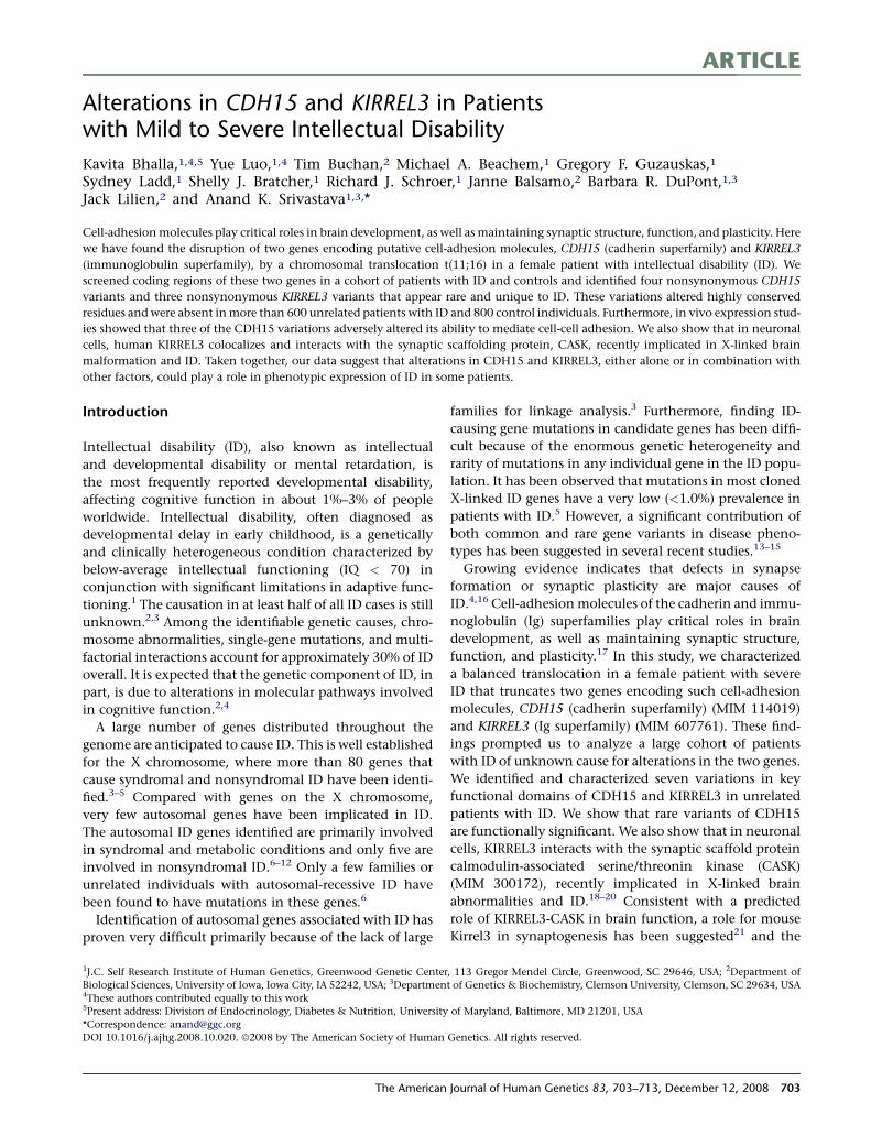
By using fluorescence in situ hybridization (FISH), we lo-
calized the 16q breakpoint to within the P1-artificial clone
(PAC) RP4-754F23 (Figures 1A and 1B). Sequence analysis
of the corresponding genomic region revealed that the
clone RP4-754F23 contains one known gene, cadherin 15
(CDH15),25 and part of a gene, acyl-CoA synthetase family
member 3 (ACSF3) (Figure 1B).
The CDH15 gene has 14 exons spanning about 24.3 kb
of genomic DNA. One of the four overlapping probes
encompassing the open reading frame of the CDH15 gene
detected a novel fragment in Southern analysis of the pa-
tient’s DNA digested with EcoR1, Pst1, or Xba1 (data not
shown). By using a genomic probe, we fine mapped the
16q24 translocation breakpoint to the second intron of
Figure 1. Cloning of the t(11;16)Translocation Breakpoints and Identifi-cation of the Candidate ID Genes(A) Metaphase spread from the femalepatient with the t(11;16) translocationshowing FISH signals obtained with PACclone RP4-754F23 (green), with a chromo-some 16 centromere-specific alphoid se-quence probe (red). The hybridization sig-nals (white arrowheads) on both der(16)and der(11) chromosomes indicate thatthis PAC clone spans the 16q breakpoint.(B) Genes residing in clone RP4-754F23 areshown. Hybridization of a 1.2 kb probecontaining CDH15 exon 2, intron 2, andexon 3 to a Southern blot containing Pst1restricted DNA from the patient (P) andfrom a control male (M) and a controlfemale (F). Novel aberrant restriction frag-ments (1.0 kb, 5.5 kb) corresponding tothe two junction fragments were identifiedin DNA from the patient (arrowheads).Inverse PCR cloning of the 1.0 kb junctionfragment and subsequent sequencing iden-tified the chromosome 11 sequence at thet(11;16) translocation. The chromosome11 breakpoint junction sequence wasmapped in intron 1 of the KIRREL3 gene.
CDH15 and detected two novel Pst1
DNA fragments (1.0 kb and 5.5 kb)
corresponding to two breakpoint
junctions (Figure 1B). We cloned the
1.0 kb junction fragment and se-
quence analysis revealed that it con-
tains the sequence corresponding to
intron 2 of CDH15 as well as sequence
from chromosome 11q24 (Figure 1B).
The chromosome 11 breakpoint junc-
tion sequence mapped to intron 1 of
a large gene, KIRREL3 (KIAA1867),
originally isolated from a fetal brain cDNA library.26 The
KIRREL3 gene has 17 exons spanning a genomic region
of approximately 575 kb. We further confirmed the se-
quence of both translocation breakpoint regions by using
primers that specifically amplified products from the deriv-
ative chromosomes but not from the normal chromo-
somes (Figure S1). A single base-pair deletion was identified
at the 16q24 breakpoint (Figure 1B and data not shown).
Extensive FISH analysis detected no additional genomic
rearrangements over a large genomic interval (approxi-
mately 10 Mb) in the vicinity of the 16q or the 11q break-
points (see above).
Consistent with disruption of one chromosomal copy,
expression of CDH15 and KIRREL3 was 38%–45% lower
in patient CMS3377 (data not shown) but not in two con-
trol individuals. We also analyzed expression of ST3GAL4,
706 The American Journal of Human Genetics 83, 703–713, December 12, 2008
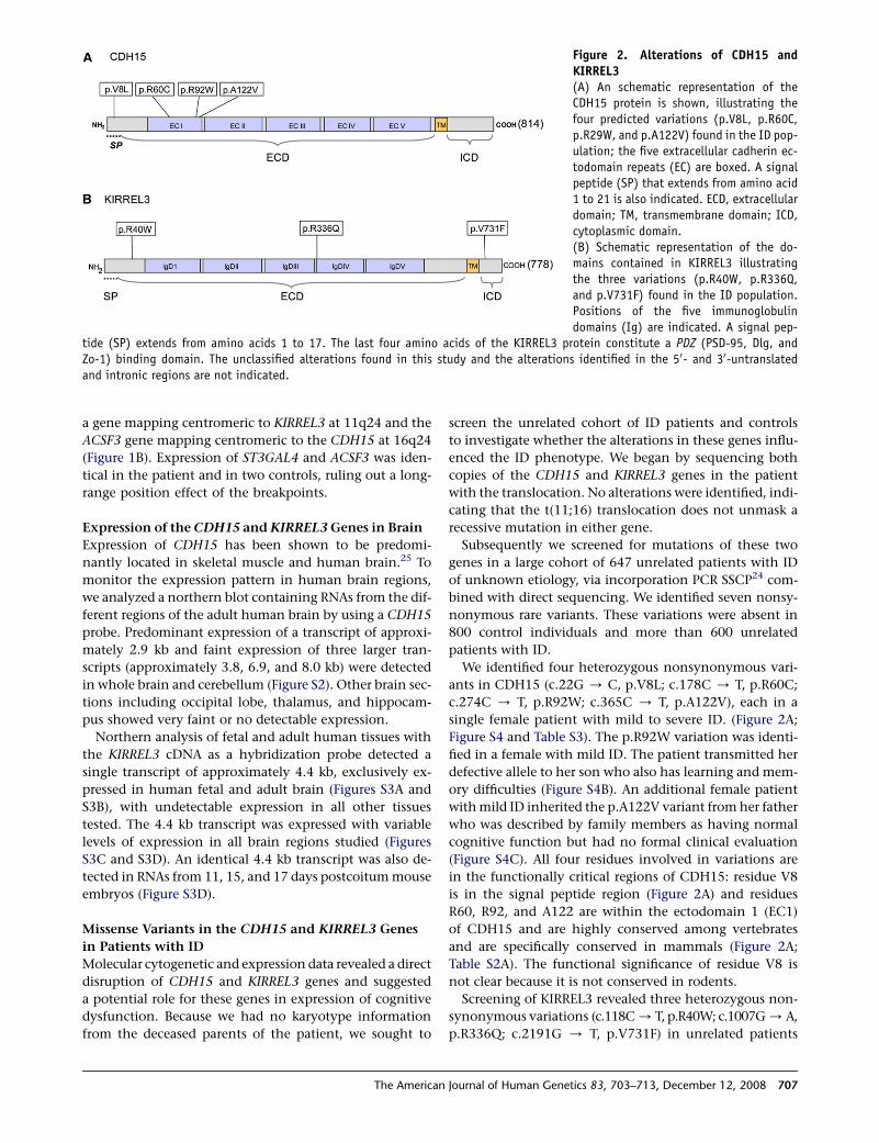
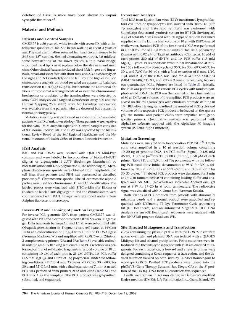
Figure 2. Alterations of CDH15 andKIRREL3(A) An schematic representation of theCDH15 protein is shown, illustrating thefour predicted variations (p.V8L, p.R60C,p.R29W, and p.A122V) found in the ID pop-ulation; the five extracellular cadherin ec-todomain repeats (EC) are boxed. A signalpeptide (SP) that extends from amino acid1 to 21 is also indicated. ECD, extracellulardomain; TM, transmembrane domain; ICD,cytoplasmic domain.(B) Schematic representation of the do-mains contained in KIRREL3 illustratingthe three variations (p.R40W, p.R336Q,and p.V731F) found in the ID population.Positions of the five immunoglobulindomains (Ig) are indicated. A signal pep-
tide (SP) extends from amino acids 1 to 17. The last four amino acids of the KIRREL3 protein constitute a PDZ (PSD-95, Dlg, andZo-1) binding domain. The unclassified alterations found in this study and the alterations identified in the 50- and 30-untranslatedand intronic regions are not indicated.
a gene mapping centromeric to KIRREL3 at 11q24 and the
ACSF3 gene mapping centromeric to the CDH15 at 16q24
(Figure 1B). Expression of ST3GAL4 and ACSF3 was iden-
tical in the patient and in two controls, ruling out a long-
range position effect of the breakpoints.
Expression of the CDH15 and KIRREL3 Genes in Brain
Expression of CDH15 has been shown to be predomi-
nantly located in skeletal muscle and human brain.25 To
monitor the expression pattern in human brain regions,
we analyzed a northern blot containing RNAs from the dif-
ferent regions of the adult human brain by using a CDH15
probe. Predominant expression of a transcript of approxi-
mately 2.9 kb and faint expression of three larger tran-
scripts (approximately 3.8, 6.9, and 8.0 kb) were detected
in whole brain and cerebellum (Figure S2). Other brain sec-
tions including occipital lobe, thalamus, and hippocam-
pus showed very faint or no detectable expression.
Northern analysis of fetal and adult human tissues with
the KIRREL3 cDNA as a hybridization probe detected a
single transcript of approximately 4.4 kb, exclusively ex-
pressed in human fetal and adult brain (Figures S3A and
S3B), with undetectable expression in all other tissues
tested. The 4.4 kb transcript was expressed with variable
levels of expression in all brain regions studied (Figures
S3C and S3D). An identical 4.4 kb transcript was also de-
tected in RNAs from 11, 15, and 17 days postcoitum mouse
embryos (Figure S3D).
Missense Variants in the CDH15 and KIRREL3 Genes
in Patients with ID
Molecular cytogenetic and expression data revealed a direct
disruption of CDH15 and KIRREL3 genes and suggested
a potential role for these genes in expression of cognitive
dysfunction. Because we had no karyotype information
from the deceased parents of the patient, we sought to
The American
screen the unrelated cohort of ID patients and controls
to investigate whether the alterations in these genes influ-
enced the ID phenotype. We began by sequencing both
copies of the CDH15 and KIRREL3 genes in the patient
with the translocation. No alterations were identified, indi-
cating that the t(11;16) translocation does not unmask a
recessive mutation in either gene.
Subsequently we screened for mutations of these two
genes in a large cohort of 647 unrelated patients with ID
of unknown etiology, via incorporation PCR SSCP24 com-
bined with direct sequencing. We identified seven nonsy-
nonymous rare variants. These variations were absent in
800 control individuals and more than 600 unrelated
patients with ID.
We identified four heterozygous nonsynonymous vari-
ants in CDH15 (c.22G / C, p.V8L; c.178C / T, p.R60C;
c.274C / T, p.R92W; c.365C / T, p.A122V), each in a
single female patient with mild to severe ID. (Figure 2A;
Figure S4 and Table S3). The p.R92W variation was identi-
fied in a female with mild ID. The patient transmitted her
defective allele to her son who also has learning and mem-
ory difficulties (Figure S4B). An additional female patient
with mild ID inherited the p.A122V variant from her father
who was described by family members as having normal
cognitive function but had no formal clinical evaluation
(Figure S4C). All four residues involved in variations are
in the functionally critical regions of CDH15: residue V8
is in the signal peptide region (Figure 2A) and residues
R60, R92, and A122 are within the ectodomain 1 (EC1)
of CDH15 and are highly conserved among vertebrates
and are specifically conserved in mammals (Figure 2A;
Table S2A). The functional significance of residue V8 is
not clear because it is not conserved in rodents.
Screening of KIRREL3 revealed three heterozygous non-
synonymous variations (c.118C / T, p.R40W; c.1007G / A,
p.R336Q; c.2191G / T, p.V731F) in unrelated patients
Journal of Human Genetics 83, 703–713, December 12, 2008 707
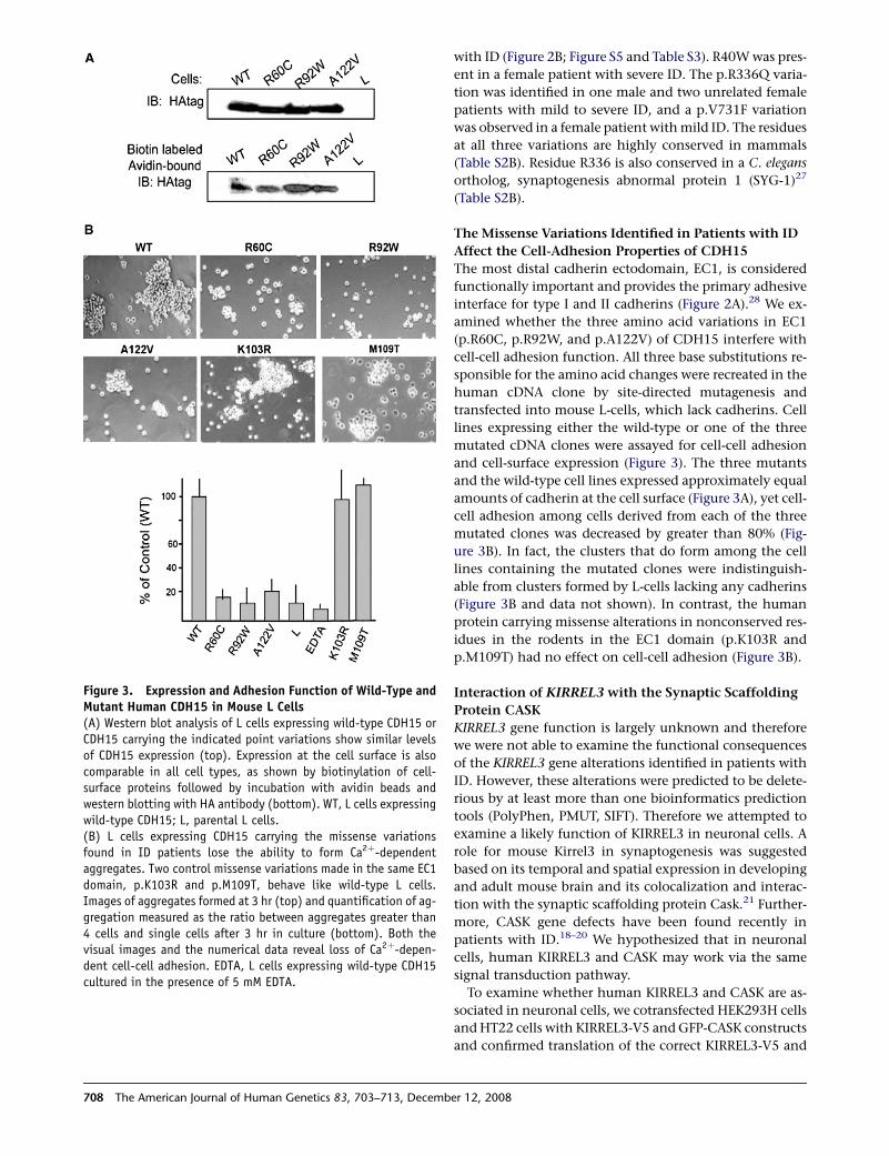
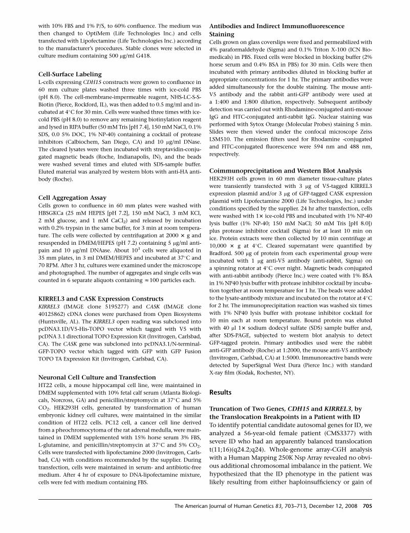
Figure 3. Expression and Adhesion Function of Wild-Type andMutant Human CDH15 in Mouse L Cells(A) Western blot analysis of L cells expressing wild-type CDH15 orCDH15 carrying the indicated point variations show similar levelsof CDH15 expression (top). Expression at the cell surface is alsocomparable in all cell types, as shown by biotinylation of cell-surface proteins followed by incubation with avidin beads andwestern blotting with HA antibody (bottom). WT, L cells expressingwild-type CDH15; L, parental L cells.(B) L cells expressing CDH15 carrying the missense variationsfound in ID patients lose the ability to form Ca2þ-dependentaggregates. Two control missense variations made in the same EC1domain, p.K103R and p.M109T, behave like wild-type L cells.Images of aggregates formed at 3 hr (top) and quantification of ag-gregation measured as the ratio between aggregates greater than4 cells and single cells after 3 hr in culture (bottom). Both thevisual images and the numerical data reveal loss of Ca2þ-depen-dent cell-cell adhesion. EDTA, L cells expressing wild-type CDH15cultured in the presence of 5 mM EDTA.
708 The American Journal of Human Genetics 83, 703–713, Decemb
with ID (Figure 2B; Figure S5 and Table S3). R40W was pres-
ent in a female patient with severe ID. The p.R336Q varia-
tion was identified in one male and two unrelated female
patients with mild to severe ID, and a p.V731F variation
was observed in a female patient with mild ID. The residues
at all three variations are highly conserved in mammals
(Table S2B). Residue R336 is also conserved in a C. elegans
ortholog, synaptogenesis abnormal protein 1 (SYG-1)27
(Table S2B).
The Missense Variations Identified in Patients with ID
Affect the Cell-Adhesion Properties of CDH15
The most distal cadherin ectodomain, EC1, is considered
functionally important and provides the primary adhesive
interface for type I and II cadherins (Figure 2A).28 We ex-
amined whether the three amino acid variations in EC1
(p.R60C, p.R92W, and p.A122V) of CDH15 interfere with
cell-cell adhesion function. All three base substitutions re-
sponsible for the amino acid changes were recreated in the
human cDNA clone by site-directed mutagenesis and
transfected into mouse L-cells, which lack cadherins. Cell
lines expressing either the wild-type or one of the three
mutated cDNA clones were assayed for cell-cell adhesion
and cell-surface expression (Figure 3). The three mutants
and the wild-type cell lines expressed approximately equal
amounts of cadherin at the cell surface (Figure 3A), yet cell-
cell adhesion among cells derived from each of the three
mutated clones was decreased by greater than 80% (Fig-
ure 3B). In fact, the clusters that do form among the cell
lines containing the mutated clones were indistinguish-
able from clusters formed by L-cells lacking any cadherins
(Figure 3B and data not shown). In contrast, the human
protein carrying missense alterations in nonconserved res-
idues in the rodents in the EC1 domain (p.K103R and
p.M109T) had no effect on cell-cell adhesion (Figure 3B).
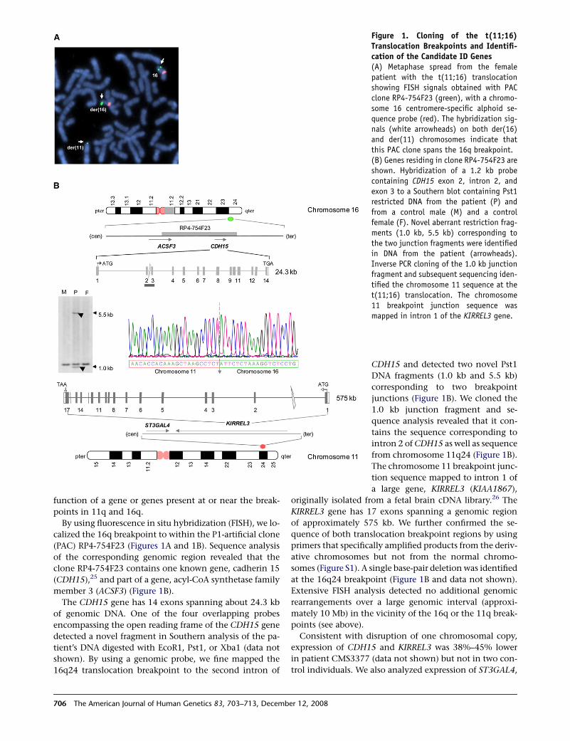
Interaction of KIRREL3 with the Synaptic Scaffolding
Protein CASK
KIRREL3 gene function is largely unknown and therefore
we were not able to examine the functional consequences
of the KIRREL3 gene alterations identified in patients with
ID. However, these alterations were predicted to be delete-
rious by at least more than one bioinformatics prediction
tools (PolyPhen, PMUT, SIFT). Therefore we attempted to
examine a likely function of KIRREL3 in neuronal cells. A
role for mouse Kirrel3 in synaptogenesis was suggested
based on its temporal and spatial expression in developing
and adult mouse brain and its colocalization and interac-
tion with the synaptic scaffolding protein Cask.21 Further-
more, CASK gene defects have been found recently in
patients with ID.18–20 We hypothesized that in neuronal
cells, human KIRREL3 and CASK may work via the same
signal transduction pathway.
To examine whether human KIRREL3 and CASK are as-
sociated in neuronal cells, we cotransfected HEK293H cells
and HT22 cells with KIRREL3-V5 and GFP-CASK constructs
and confirmed translation of the correct KIRREL3-V5 and
er 12, 2008
GFP-CASK fusion proteins by western blot analysis. We
assayed for the presence of CASK in immunoprecipitates
of KIRREL3 (Figure 4D). Results from both cell lines were
consistent: in cells overexpressing CASK and KIRREL3,
GFP-CASK is precipitated by the KIRREL3-V5 but not by
the equally tagged control protein (LaZ-V5). CASK-specific
signal was not detected in control cells expressing only
LacZ-V5 or GFP-CASK. Subsequently, we also confirmed
endogenous expression of CASK in these cells and con-
firmed interaction of the endogenous CASK with KIRREL3-
V5 via a CASK-specific antibody (Figure 4E).
We examined whether KIRREL3 and CASK are also colo-
calized in neuronal cell. First to examine the localization of
KIRREL3 protein in neurons, PC12 cells were transiently
transfected with a KIRREL3-V5 fusion construct and then
stained with V5 antibody. Confocal microscopy showed
KIRREL3-positive staining in a ring shape implying its
expression on cell membrane (Figure 4A). Some likely cyto-
plasmic signal was also noted.
To study colocalization of KIRREL3 and CASK, we co-
transfected PC12 cells with KIRREL3-V5 and GFP-CASK
constructs. Immunostaining reveals the colocalization of
these two proteins (Figure 4B). We also examined the
colocalization of both proteins in HT22 cells (Figure 4C).
Again, CASK expression was found to overlap with the
expression of KIRREL3. CASK is primarily a cytoplasmic
protein that interacts with several membrane proteins29
and as expected, CASK signals were also noted in areas
where KIRREL3 was not expressed (Figures 4B and 4C).
Discussion
Balanced de novo chromosomal rearrangements are associ-
ated with abnormal phenotypes in about 6% of cases30 and
are often found to affect the gene or genes at or near the
breakpoints. Such translocations in patients with ID have
been proven to be valuable resource in the search for genes
causally related to disease. Our study identifies two genes,
CDH15 and KIRREL3, physically disrupted by the translo-
cation breakpoints in a female patient with severe ID.
Both genes are expressed in the brain, and a putative role
for these two genes in brain function critical for learning
and memory has been predicted. We also identified several
nonsynonymous sequence variations in each of these two
genes in unrelated patients with ID. The variations were
absent in 800 control individuals and 600 individuals
with ID. In one case, a CDH15 variation was also identified
in a mother and son, both with learning and memory
problems. We hypothesized that these rare variants are
likely to be functionally important and may influence
gene function in the brain that is critical for learning and
memory. To support this, our in vivo functional studies
provided evidence that only CDH15 variations, identified
in patients with ID, adversely alter its ability to mediate
cell-cell adhesion. Unfortunately, we have no comparable
evidence for the KIRREL3 gene alterations. Nonetheless,
The American
the KIRREL3 alterations identified in patients with ID
were predicted to be deleterious and found to be in con-
served residues. Thus we examined a potential role for
KIRREL3 in neuronal cells and showed that KIRREL3 inter-
acts and colocalizes with the synaptic scaffolding protein
CASK, also recently implicated in ID.
Individuals affected with ID usually do not have children
and thus a large number of mutations causing autosomal-
dominant ID are de novo including chromosomal rear-
rangements associated with ID. Although the karyotypes
of the parents are not known, we strongly suspect that
the translocation is de novo in the patient and speculate
that the disruption of both genes in the patient with the
t(11;16) translocation may have contributed to the severity
of ID. This patient also has finger anomalies, including syn-
dactyly, where none of the patients carrying variants in ei-
ther gene exhibits such anomalies. It is likely that the
anomalies may be due to an additional factor or modifying
gene.
Finding several rare nonsynonymous sequence variations
in each of these two genes in unrelated patients with ID sug-
gests that alterations in either gene are likely to affect brain
function and may contribute to mild to severe ID. However,
some of these rare variants might contribute to disease risk,
and additional factors may be required to complete expres-
sion of ID phenotype. Such a possibility has recently been
noted in studies involving two genes, Neurexin1 (NRXN1)
(MIM 600565) and Contactin associated protein-like 2
(CNTNAP2) (MIM 604569), suggesting the contribution of
conserved rare variants to disease risk.13,14 Moreover, the
significance of rare missense variations in patients remains
critical considering several SNPs in other genes have been
found to be functionally important.31
The first gene, CDH15, encodes a protein of 814 amino
acid residues25 (Figure 2A). It belongs to the type I classic
cadherin superfamily group, all of which are single-pass
transmembrane molecules with five extracellular (EC) re-
peats that mediate calcium-dependent homophilic, cell-
cell adhesion.25,32 Additionally, all members of this group
have a highly conserved cytoplasmic domain that interacts
with b-catenin, which in turn interacts with a-catenin reg-
ulating cytoskeletal function at adherens junctions33 (Fig-
ure 5). The four identified missense alterations in CDH15
are all clustered in the N-terminal region. Residue R60 is
highly conserved in M, N, and R cadherins (Table S2A),
all primarily neural and thus may be critical for neural
function in general. Residue R92 is specific to mammalian
cadherin 15 (M-cadherins) and thus may play a specific
role in CDH15 function. CDH15 is also expressed in skele-
tal muscle. However, none of the patients with CDH15 var-
iations have a skeletal muscle disorder. In one case, we no-
ticed that a 46-year-old patient with the p.R60C CDH15
variation had her finger reported to extend only to neutral
and her elbow did not extend to neutral, and in a second
case, a 44-year-old patient with the p.A122V variation
reportedly has facial muscle hypotonia and short distal
phalanges.
Journal of Human Genetics 83, 703–713, December 12, 2008 709
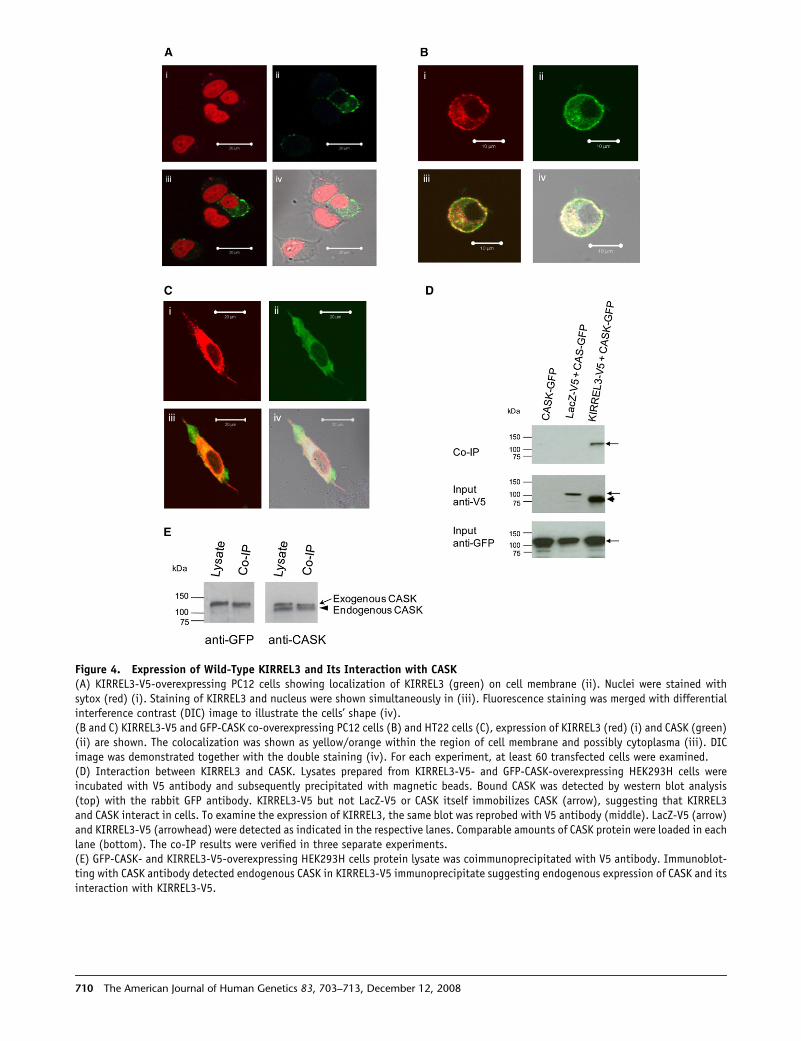
Figure 4. Expression of Wild-Type KIRREL3 and Its Interaction with CASK(A) KIRREL3-V5-overexpressing PC12 cells showing localization of KIRREL3 (green) on cell membrane (ii). Nuclei were stained withsytox (red) (i). Staining of KIRREL3 and nucleus were shown simultaneously in (iii). Fluorescence staining was merged with differentialinterference contrast (DIC) image to illustrate the cells’ shape (iv).(B and C) KIRREL3-V5 and GFP-CASK co-overexpressing PC12 cells (B) and HT22 cells (C), expression of KIRREL3 (red) (i) and CASK (green)(ii) are shown. The colocalization was shown as yellow/orange within the region of cell membrane and possibly cytoplasma (iii). DICimage was demonstrated together with the double staining (iv). For each experiment, at least 60 transfected cells were examined.(D) Interaction between KIRREL3 and CASK. Lysates prepared from KIRREL3-V5- and GFP-CASK-overexpressing HEK293H cells wereincubated with V5 antibody and subsequently precipitated with magnetic beads. Bound CASK was detected by western blot analysis(top) with the rabbit GFP antibody. KIRREL3-V5 but not LacZ-V5 or CASK itself immobilizes CASK (arrow), suggesting that KIRREL3and CASK interact in cells. To examine the expression of KIRREL3, the same blot was reprobed with V5 antibody (middle). LacZ-V5 (arrow)and KIRREL3-V5 (arrowhead) were detected as indicated in the respective lanes. Comparable amounts of CASK protein were loaded in eachlane (bottom). The co-IP results were verified in three separate experiments.(E) GFP-CASK- and KIRREL3-V5-overexpressing HEK293H cells protein lysate was coimmunoprecipitated with V5 antibody. Immunoblot-ting with CASK antibody detected endogenous CASK in KIRREL3-V5 immunoprecipitate suggesting endogenous expression of CASK and itsinteraction with KIRREL3-V5.
710 The American Journal of Human Genetics 83, 703–713, December 12, 2008
Interestingly, in one patient, a p.R92W variation was
transmitted to her son who also has learning and memory
difficulties. Residue A122 is in the middle of the critical
‘‘HAV’’ sequence known to be critical for all type 1 classic
cadherins and this is important for adhesion in general
and differentiates type 1 cadherins from type 2 cadherins.
However, the p.A122V variation in the patient (CMS7914)
was inherited from the father who reportedly lacks an ID
phenotype (Figure S4C). This indicates that the segrega-
tion of this variant might show incomplete penetrance
and may produce ID only upon interaction with other fac-
tors. Nonetheless, our in vivo functional studies showed
that the three variations (p.R60C, p.R92W, and p.A122V)
in the EC1 domain of CDH15 significantly affect its ability
to mediate cell-cell adhesion. Additionally, missense
alterations considered to be polymorphisms (p.K103R
and p.M109T) in two nonconserved residues in the EC1
domain did not alter cell-adhesion function (Figure 3B).
These results support the inference that the residues al-
tered in the ID patients are important for CDH15 function.
In the mouse, Cdh15 gene expression is restricted to the
granular layer of the cerebellar cortex in synapses and
other intercellular junctions.34,35 Indeed, a role for cadher-
ins in synaptic plasticity has long been appreciated.17,36
Recently, defects in two protocadherin genes, PCDH19
(MIM 300460) and PCDH10 (MIM 608286), have been
shown to be associated with cognitive impairment in
humans.37,38
The second gene, KIRREL3, is a mammalian homolog of
the gene kirre (kin of irregular chiasm C-roughest) of
Drosophila melanogaster.39,40 The KIRREL3 gene is predicted
to encode a type 1a membrane protein of 778 amino acids
containing five Ig-like domains in its extracellular portion
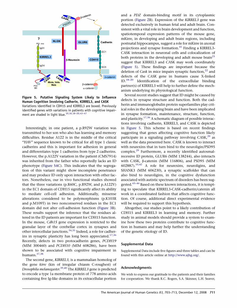
Figure 5. Putative Signaling System Likely to InfluenceHuman Cognition Involving Cadherin, KIRREL3, and CASKVariations identified in CDH15 and KIRREL3 are boxed. Previouslyidentified genes with variations in patients with cognitive impair-ment are shaded in light blue.10,16,18–20,43–47
The American
and a PDZ domain-binding motif in its cytoplasmic
portion (Figure 2B). Expression of the KIRREL3 gene was
detected exclusively in human fetal and adult brain. Con-
sistent with a vital role in brain development and function,
spatiotemporal expression patterns of the mouse gene,
mKirre, in developing and adult brain regions, including
postnatal hippocampus, suggest a role for mKirre in axonal
projections and synapse formation.41 Finding a KIRREL3-
CASK interaction in neuronal cells and colocalization of
both proteins in the developing and adult mouse brain21
suggest that KIRREL3 and CASK may work coordinately
(Figure 5). These findings are important because the
deletion of Cask in mice impairs synaptic function,22 and
defects of the CASK gene in humans cause X-linked
ID.18–20 Identification of the extracellular binding
partner(s) of KIRREL3 will help to further define the mech-
anism underlying its physiological function.
Several recent studies suggest that ID might be caused by
defects in synapse structure and function. Both the cad-
herin and immunoglobulin protein superfamilies play crit-
ical roles in the developing brain and have been implicated
in synapse formation, maintenance, structure, function,
and plasticity.17,36 A schematic diagram of possible interac-
tions involving cadherin, KIRREL3, and CASK is depicted
in Figure 5. This scheme is based on recent findings
suggesting that genes affecting cognitive function likely
participate in a signaling pathway involving CASK,29 as
well as the data presented here. CASK is known to interact
with neurexins that in turn bind to the neuroligin/PSD95
complex.29 Furthermore, a recently identified autosmal-
recessive ID protein, GLUR6 (MIM 138244), also interacts
with CASK, b-catenin (MIM 116806), and PSD95 (MIM
602887).10,42 A role for neurexins, neuroligins, and
SHANK3 (MIM 606230), a synaptic scaffolder that can
also bind to neuroligins, in the cognitive dysfunction
associated with autistic spectrum of disorders has been sug-
gested.43–46 Based on these known interactions, it is tempt-
ing to speculate that KIRREL3-CASK-cadherin/catenin all
work in a coordinated fashion that affects cognitive func-
tion. Of course, additional direct experimental evidence
will be required to support this hypothesis.
Altogether, our studies point to a likely contribution of
CDH15 and KIRREL3 in learning and memory. Further
study in animal models should provide a system to exam-
ine how these two proteins contribute to cognitive func-
tion in humans and may help further the understanding
of the genetic etiology of ID.
Supplemental Data
Supplemental Data include five figures and three tables and can be
found with this article online at http://www.ajhg.org/.
Acknowledgments
We wish to express our gratitude to the patients and their families
for cooperation. We thank R.C. Rogers, S.A. Skinner, L.H. Seaver,
Journal of Human Genetics 83, 703–713, December 12, 2008 711
and K.B. Clarkson for providing clinical details of patients; D.
Bealer and C. Skinner for assistance in obtaining patients’ samples;
T. Moss for help with cell culture; D. Schultz for assistance in
sequencing; L. Allen and J. John for technical assistance in muta-
tion screening; J. Collins for assistance in statistical analysis and
for discussion; K. Franek and J. Norris for assistance in preparation
of expression constructs and confocal microscopy; and C.E.
Schwartz and R.E. Stevenson for helpful discussion. This work
was supported by a grant from the National Institutes of Child
Health and Human Development (R01-HD39331 to A.K.S.) and
from the National Eye Institute (R01-EY013363 to J.L. and J.B.).
The authors report no conflict of interest.
Received: September 15, 2008
Revised: October 18, 2008
Accepted: October 24, 2008
Published online: November 13, 2008
Web Resources
The URLs for data presented herein are as follows:
Ensembl Genome Browser, http://www.ensemble.org/
HUGO nomenclature committee, http://www.genenames.org/
NCBI Human Genome Browser and Database, http://www.ncbi.
nlm.nih.gov/
Online Mendelian Inheritance in Man (OMIM), http://www.ncbi.
nlm.nih.gov/Omim/
PMUT, http://mmb.pcb.ub.es/PMut/
PolyPhen, http://genetics.bwh.harvard.edu/pph/
SIFT, http://blocks.fhcrc.org/sift/SIFT_seq_submit2.html
UCSC Human Genome Browser, http://genome.usc.edu/cgi-bin/
hgGateway
References
1. American Association on Mental Retardation. (2002). Mental
Retardation: Definition, Classification and Systems of Sup-
port, Tenth Edition (Washington, DC: American Association
on Mental Retardation).
2. Inlow, J.K., and Restifo, L.L. (2004). Molecular and compara-
tive genetics of mental retardation. Genetics 166, 835–881.
3. Ropers, H.H. (2008). Genetics of intellectual disability. Curr.
Opin. Genet. Dev. 18, 241–250.
4. Chelly, J., Khelfaoui, M., Francis, F., Cherif, B., and Bienvenu,
T. (2006). Genetics and pathophysiology of mental retarda-
tion. Eur. J. Hum. Genet. 14, 701–713.
5. Chiurazzi, P., Schwartz, C.E., Gecz, J., and Neri, G. (2008).
XLMR genes: Update 2007. Eur. J. Hum. Genet. 16, 422–434.
6. Basel-Vanagaite, L. (2007). Genetics of autosomal recessive
non-syndromic mental retardation: Recent advances. Clin.
Genet. 72, 167–174.
7. Molinari, F., Rio, M., Meskenaite, V., Encha-Razavi, F., Auge, J.,
Bacq, D., Briault, S., Vekemans, M., Munnich, A., Attie-Bitach,
T., et al. (2002). Truncating neurotrypsin mutation in autoso-
mal recessive nonsyndromic mental retardation. Science 298,
1779–1781.
8. Higgins, J.J., Pucilowska, J., Lombardi, R.Q., and Rooney, J.P.
(2004). A mutation in a novel ATP-dependent Lon protease
gene in a kindred with mild mental retardation. Neurology
63, 1927–1931.
712 The American Journal of Human Genetics 83, 703–713, Decemb
9. Basel-Vanagaite, L., Attia, R., Yahav, M., Ferland, R.J., Anteki,
L., Walsh, C.A., Olender, T., Straussberg, R., Magal, N., Taub,
E., et al. (2006). The CC2D1A, a member of a new gene family
with C2 domains, is involved in autosomal recessive nonsyn-
dromic mental retardation. J. Med. Genet. 43, 203–210.
10. Motazacker, M.M., Rost, B.R., Hucho, T., Garshasbi, M.,
Kahrizi, K., Ullmann, R., Abedini, S.S., Nieh, S.E., Amini,
S.H., Goswami, C., et al. (2007). A defect in the ionotropic glu-
tamate receptor 6 gene (GRIK2) is associated with autosomal
recessive mental retardation. Am. J. Hum. Genet. 81, 792–798.
11. Molinari, F., Foulquier, F., Tarpey, P.S., Morelle, W., Boissel, S.,
Teague, J., Edkins, S., Futreal, P.A., Stratton, M.R., Turner, G.,
et al. (2008). Oligosaccharyltransferase-subunit mutations in
nonsyndromic mental retardation. Am. J. Hum. Genet. 82,
1150–1157.
12. Garshasbi, M., Hadavi, V., Habibi, H., Kahrizi, K., Kariminejad,
R., Behjati, F., Tzschach, A., Najmabadi, H., Ropers, H.H., and
Kuss, A.W. (2008). A defect in the TUSC3 gene is associated
with autosomal recessive mental retardation. Am. J. Hum.
Genet. 82, 1158–1164.
13. Bakkaloglu, B., O’Roak, B.J., Louvi, A., Gupta, A.R., Abelson,
J.F., Morgan, T.M., Chawarska, K., Klin, A., Ercan-Sencicek,
A.G., Stillman, A.A., et al. (2008). Molecular cytogenetic anal-
ysis and resequencing of contactin associated protein-like 2 in
autism spectrum disorders. Am. J. Hum. Genet. 82, 165–173.
14. Kim, H.G., Kishikawa, S., Higgins, A.W., Seong, I.S., Donovan,
D.J., Shen, Y., Lally, E., Weiss, L.A., Najm, J., Kutsche, K., et al.
(2008). Disruption of neurexin 1 associated with autism
spectrum disorder. Am. J. Hum. Genet. 82, 199–207.
15. Arking, D.E., Cutler, D.J., Brune, C.W., Teslovich, T.M., West,
K., Ikeda, M., Rea, A., Guy, M., Lin, S., Cook, E.H., et al.
(2008). A common genetic variant in the neurexin superfam-
ily member CNTNAP2 increases familial risk of autism. Am. J.
Hum. Genet. 82, 160–164.
16. Laumonnier, F., Cuthbert, P.C., and Grant, S.G. (2007). The
role of neuronal complexes in human X-linked brain diseases.
Am. J. Hum. Genet. 80, 205–220.
17. Yamagata, M., Sanes, J.R., and Weiner, J.A. (2003). Synaptic
adhesion molecules. Curr. Opin. Cell Biol. 15, 621–632.
18. Najm, J., Horn, D., Wimplinger, I., Golden, J.A., Chizhikov,
V.V., Sudi, J., Christian, S.L., Ullmann, R., Kuechler, A., Haas,
C.A., et al. (2008). Mutations of CASK cause an X-linked brain
malformation phenotype with microcephaly and hypoplasia
of the brainstem and cerebellum. Nat. Genet. 40, 1065–1067.
19. Froyen, G., Van Esch, H., Bauters, M., Hollanders, K., Frints,
S.G., Vermeesch, J.R., Devriendt, K., Fryns, J.P., and Marynen,
P. (2007). Detection of genomic copy number changes in pa-
tients with idiopathic mental retardation by high-resolution
X-array-CGH: important role for increased gene dosage of
XLMR genes. Hum. Mutat. 28, 1034–1042.
20. Piluso, G., D’Amico, F., Saccone, V., Rotundo, L., and Nigro, V.
(2007). A missense mutation in CASK gene causes FG
syndrome in an Italian GFS family. 13th International Workshop
on Fragile X and X-Linked Mental Retardation. (http://xlmr.
interfree.it/home.htm), Abstract P58.
21. Gerke, P., Benzing, T., Hohne, M., Kispert, A., Frotscher, M.,
Walz, G., and Kretz, O. (2006). Neuronal expression and
interaction with the synaptic protein CASK suggest a role for
Neph1 and Neph2 in synaptogenesis. J. Comp. Neurol. 498,
466–475.
22. Atasoy, D., Schoch, S., Ho, A., Nadasy, K.A., Liu, X., Zhang, W.,
Mukherjee, K., Nosyreva, E.D., Fernandez-Chacon, R., Missler,
er 12, 2008
M., et al. (2007). Deletion of CASK in mice is leathal and im-
pairs synaptic function. Proc. Natl. Acad. Sci. USA 104,
2525–2530.
23. Griggs, B.L., Ladd, S., Saul, R.A., DuPont, B.R., and Srivastava,
A.K. (2008). Dedicator of cytokinesis 8 is disrupted in two pa-
tients with mental retardation and developmental disabilities.
Genomics 91, 195–202.
24. Sossey-Alaoui, K., Lyon, J.A., Jones, L., Abidi, F.E., Hartung,
A.J., Hane, B., Schwartz, C.E., Stevenson, R.E., and Srivastava,
A.K. (1999). Molecular cloning and characterization of TRPC5
(HTRP5), the human homologue of a mouse brain receptor-
activated capacitative Ca2þ entry channel. Genomics 60,
330–340.
25. Shimoyama, Y., Shibata, T., Kitajima, M., and Hirohashi, S.
(1998). Molecular cloning and characterization of a novel
human classic cadherin homologous with mouse muscle
cadherin. J. Biol. Chem. 273, 10011–10018.
26. Nagase, T., Nakayama, M., Nakajima, D., Kikuno, R., and
Ohara, O. (2001). Prediction of the coding sequences of un-
identified human genes. XX. The complete sequences of 100
new cDNA clones from brain which code for large proteins
in vitro. DNA Res. 8, 85–95.
27. Shen, K., and Bargmann, C.I. (2003). The immunoglobulin
superfamily protein SYG-1 determines the location of specific
synapses in C. elegans. Cell 112, 619–630.
28. Koch, A.W., Manzur, K.L., and Shan, W. (2004). Structure-
based models of cadherin-mediated cell adhesion: The
evolution continues. Cell. Mol. Life Sci. 61, 1884–1895.
29. Hsueh, Y.P. (2006). The role of the MAGUK protein CASK in
neural development and synaptic function. Curr. Med.
Chem. 13, 1915–1927.
30. Warburton, D. (1991). De novo balanced chromosome
rearrangements and extra marker chromosomes identified at
prenatal diagnosis: Clinical significance and distribution of
breakpoints. Am. J. Hum. Genet. 49, 995–1013.
31. Sethupathy, P., Borel, C., Gagnebin, M., Grant, G.R., Deutsch,
S., Elton, T.S., Hatzigeorgiou, A.G., and Antonarakis, S.E.
(2007). Human microRNA-155 on chromosome 21 differen-
tially interacts with its polymorphic target in the AGTR1 30
untranslated region: A mechanism for functional single-nu-
cleotide polymorphisms related to phenotypes. Am. J. Hum.
Genet. 81, 405–413.
32. Lilien, J., Balsamo, J., Arregui, C., and Xu, G. (2002). Turn-off,
drop-out: Functional state switching of cadherins. Dev. Dyn.
224, 18–29.
33. Yamada, S., Pokutta, S., Drees, F., Weis, W.I., and Nelson, W.J.
(2005). Deconstructing the cadherin-catenin-actin complex.
Cell 123, 889–901.
34. Bahjaoui-Bouhaddi, M., Padilla, F., Nicolet, M., Cifuentes-
Diaz, C., Fellmann, D., and Mege, R.M. (1997). Localized
deposition of M-cadherin in the glomeruli of the granular
layer during the postnatal development of mouse cerebellum.
J. Comp. Neurol. 378, 180–195.
35. Rose, O., Grund, C., Reinhardt, S., Starzinski-Powitz, A., and
Franke, W.W. (1995). Contactus adherens, a special type of
The American
plaque-bearing adhering junction containing M-cadherin, in
the granule cell layer of the cerebellar glomerulus. Proc.
Natl. Acad. Sci. USA 92, 6022–6026.
36. Bamji, S.X. (2005). Cadherins: Actin with the cytoskeleton to
form synapses. Neuron 47, 175–178.
37. Dibbens, L.M., Tarpey, P.S., Hynes, K., Bayly, M.A., Scheffer,
I.E., Smith, R., Bomar, J., Sutton, E., Vandeleur, L., Shoubridge,
C., et al. (2008). X-linked protocadherin 19 mutations cause
female-limited epilepsy and cognitive impairment. Nat.
Genet. 40, 776–781.
38. Morrow, E.M., Yoo, S.Y., Flavell, S.W., Kim, T.K., Lin, Y., Hill,
R.S., Mukaddes, N.M., Balkhy, S., Gascon, G., Hashmi, A.,
et al. (2008). Identifying autism loci and genes by tracing
recent shared ancestry. Science 321, 218–223.
39. Ueno, H., Sakita-Ishikawa, M., Morikawa, Y., Nakano, T.,
Kitamura, T., and Saito, M. (2003). A stromal cell-derived
membrane protein that supports hematopoietic stem cells.
Nat. Immunol. 4, 457–463.
40. Ramos, R.G., Igloi, G.L., Lichte, B., Baumann, U., Maier, D.,
Schneider, T., Brandstatter, J.H., Frohlich, A., and Fischbach,
K.F. (1993). The irregular chiasm C-roughest locus of Drosoph-
ila, which affects axonal projections and programmed cell
death, encodes a novel immunoglobulin-like protein. Genes
Dev. 7, 2533–2547.
41. Tamura, S., Morikawa, Y., Hisaoka, T., Ueno, H., Kitamura, T.,
and Senba, E. (2005). Expression of mKirre, a mammalian ho-
molog of Drosophila Kirre, in the developing and adult mouse
brain. Neuroscience 133, 615–624.
42. Coussen, F., Normand, E., Marchal, C., Costet, P., Choquet, D.,
Lambert, M., Mege, R.M., and Mulle, C. (2002). Recruitment
of the kainate receptor subunit glutamate receptor 6 by cad-
herin/catenin complexes. J. Neurosci. 22, 6426–6436.
43. Autism Genome Project Consortium. (2007). Mapping autism
risk loci using genetic linkage and chromosomal rearrange-
ments. Nat. Genet. 39, 319–328.
44. Durand, C.M., Betancur, C., Boeckers, T.M., Bockmann, J.,
Chaste, P., Fauchereau, F., Nygren, G., Rastam, M., Gillberg,
I.C., Anckarsater, H., et al. (2007). Mutations in the gene en-
coding the synaptic scaffolding protein SHANK3 are associ-
ated with autism spectrum disorders. Nat. Genet. 39, 25–27.
45. Jamain, S., Quach, H., Betancur, C., Rastam, M., Colineaux,
C., Gillberg, I.C., Soderstrom, H., Giros, B., Leboyer, M., Gill-
berg, C., et al. (2003). Mutations of the X-linked genes
encoding neuroligins NLGN3 and NLGN4 are associated
with autism. Nat. Genet. 34, 27–29.
46. Laumonnier, F., Bonnet-Brilhault, F., Gomot, M., Blanc, R., Da-
vid, A., Moizard, M.P., Raynaud, M., Ronce, N., Lemonnier, E.,
Calvas, P., et al. (2004). X-linked mental retardation and
autism are associated with a mutation in the NLGN4 gene,
a member of the neuroligin family. Am. J. Hum. Genet. 74,
552–557.
47. Israely, I., Costa, R.M., Xie, C.W., Silva, A.J., Kosik, K.S., and
Liu, X. (2004). Deletion of the neuron-specific protein delta-
catenin leads to severe cognitive and synaptic dysfunction.
Curr. Biol. 14, 1657–1663.
Journal of Human Genetics 83, 703–713, December 12, 2008 713


![Neuroinformatics and Analysis of Connectomic Alterations ...acm-paper].pdfNeuroinformatics and Analysis of Connectomic Alterations Due to Cerebral Microhemorrhages in Geriatric Mild](https://img.pdfslide.us/doc/110x75/5f7c9596fc19e924393f8ea8/neuroinformatics-and-analysis-of-connectomic-alterations-acm-paperpdf-neuroinformatics.jpg)


![100 80 60 40 20 0 LOVE DESERVESYOUR DOG’S LIFE˜ LASTING · Week 4 Week 5 Week 6 Week 7 Week 8 [dog’s name] Extremely severe Severe Moderate Mild Very mild Normal dog Day 1 Day](https://img.pdfslide.us/doc/110x75/5f3c13c61a22f676b92a4384/100-80-60-40-20-0-love-deservesyour-dogas-lifeoe-lasting-week-4-week-5-week-6.jpg)

![LIVER 3 KISS 2018 [Kompatibilis üzemmód] · 2018. 3. 12. · 2018. 03. 12. 3 Liver alterations associated with pregnancy Acute steatosis in pregnancy –rare, from mild to severe](https://img.pdfslide.us/doc/110x75/5ff28ad5a14fb268c76c9315/liver-3-kiss-2018-kompatibilis-zemmd-2018-3-12-2018-03-12-3-liver.jpg)