Embed Size (px)
Citation preview

University of Wollongong University of Wollongong
Research Online Research Online
University of Wollongong Thesis Collection 1954-2016 University of Wollongong Thesis Collections
2015
Alteration of cholesterol homeostasis in the Huntington's disease brain Alteration of cholesterol homeostasis in the Huntington's disease brain
Fabian Kreilaus University of Wollongong
Follow this and additional works at: https://ro.uow.edu.au/theses
University of Wollongong University of Wollongong
Copyright Warning Copyright Warning
You may print or download ONE copy of this document for the purpose of your own research or study. The University
does not authorise you to copy, communicate or otherwise make available electronically to any other person any
copyright material contained on this site.
You are reminded of the following: This work is copyright. Apart from any use permitted under the Copyright Act
1968, no part of this work may be reproduced by any process, nor may any other exclusive right be exercised,
without the permission of the author. Copyright owners are entitled to take legal action against persons who infringe
their copyright. A reproduction of material that is protected by copyright may be a copyright infringement. A court
may impose penalties and award damages in relation to offences and infringements relating to copyright material.
Higher penalties may apply, and higher damages may be awarded, for offences and infringements involving the
conversion of material into digital or electronic form.
Unless otherwise indicated, the views expressed in this thesis are those of the author and do not necessarily Unless otherwise indicated, the views expressed in this thesis are those of the author and do not necessarily
represent the views of the University of Wollongong. represent the views of the University of Wollongong.
Recommended Citation Recommended Citation Kreilaus, Fabian, Alteration of cholesterol homeostasis in the Huntington's disease brain, Doctor of Philosophy thesis, School of Biological Sciences, University of Wollongong, 2015. https://ro.uow.edu.au/theses/4627
Research Online is the open access institutional repository for the University of Wollongong. For further information contact the UOW Library: [email protected]

Alteration of cholesterol homeostasis
in the Huntington’s disease brain
A thesis submitted in fulfilment of the requirements for the award of the degree
Doctor of Philosophy
from
The University of Wollongong
by
Fabian Kreilaus
(B Biotech. Hons.)
Biological Sciences
2015

ii
Certification
I, Fabian Kreilaus, declare that this thesis, submitted in fulfilment of the requirements for the award of Doctor of Philosophy, in the Department of Biological Sciences, University of Wollongong, is wholly my own work unless otherwise referenced or acknowledged. The document has not been submitted for qualifications at any other academic institution.
Fabian Kreilaus
1st Sep. 2015
iii
Acknowledgements
There are several people I would like to acknowledge for assistance and support through my
candidature. My supervisors Andrew Jenner and Brett Garner for constantly providing excellent
advice and encouragement during all aspects of my research. Henry and Kalani for taking the
time to share their experience and knowledge in the laboratory. This has helped my work run
very smoothly and was much appreciated. Special thanks to Adena for assisting me in many
weeks of tissue collection, where your skilled animal dissections provided me with excellent
brain tissue samples. Also for giving me the most clear-cut advice in terms of laboratory work
and thesis writing, and providing constant emotional support in the last 3 years. I would also like
to acknowledge: the UoW statistical consulting service for providing advice on statistical
analysis; Anthony Hannan for providing R6/1 mice that made research into this animal model
possible; Catriona McLean and the Victorian Brain Bank, supported by The Florey Institute of
Neuroscience and Mental Health, The Alfred and the Victorian Forensic Institute of Medicine
and funded in part by Australia’s National Health & Medical Research Council, Parkinson’s
Victoria and MND Victoria, for providing human post mortem brain tissue.
iv
Publications from this thesis
Fabian Kreilaus, Adena S. Spiro, Catriona A. McLean, Brett Garner, and Andrew M. Jenner, (2015) Evidence for altered cholesterol metabolism in Huntington’s disease post-mortem brain tissue, Neuropathology and Applied Neurobiology, doi: 10.1111/nan.12286
Fabian Kreilaus, Adena S. Spiro, Anthony J. Hannan, Brett Garner, and Andrew M. Jenner, (2015) Brain cholesterol synthesis and metabolism is progressively disturbed in the R6/1 mouse model of Huntington’s disease: A targeted GC-MS/MS sterol analysis, Journal of Huntington’s Disease 4:305-318
Manuscript in preparation
Fabian Kreilaus, Adena S. Spiro, Anthony J. Hannan, Brett Garner, and Andrew M. Jenner, Therapeutic effects of anthocyanins and environmental enrichment in R6/1 Huntington’s disease mice
Conference poster presentations
Fabian Kreilaus, Adena S. Spiro, Andrew M. Jenner, Altered brain cholesterol oxidation and synthesis in the R6/1 mouse model of Huntington’s disease, Joint Meeting of the Societies for Free Radical Research of Australasia and Japan in Sydney (2013)
Fabian Kreilaus, Catriona A. McLean, Andrew M. Jenner, Altered cholesterol metabolism and
increased cholesterol peroxidation in human post mortem Huntington’s disease brain,
Australasian Neuroscience Society meeting (2014)
Fabian Kreilaus, Adena S. Spiro, Catriona A. McLean, Andrew M. Jenner, Brain cholesterol
synthesis and metabolism is altered in human Huntington’s disease brain, Australian Lipid
Meeting (2014)
Fabian Kreilaus, Adena S. Spiro, Catriona A. McLean, Andrew M. Jenner, Human Huntington’s
disease brain exhibits cholesterol synthetic and metabolic alterations, Australian Society for
Medical Research NSW annual scientific meeting (2015)
Table of contents
Certification................................................................................................................................ ii
Acknowledgements ................................................................................................................... iii
Publications from this thesis ..................................................................................................... iv
Conference poster presentations ............................................................................................. iv

v
List of figures ............................................................................................................................. ix
List of tables .............................................................................................................................. xi
List of appendix tables .............................................................................................................. xi
Abbreviations ........................................................................................................................... xii
Abstract ................................................................................................................................... xiv
Chapter 1 Introduction ................................................................................................................. 1
1.1 Cholesterol .......................................................................................................................... 2
1.1.1 Cholesterol in cell membranes .................................................................................... 2
1.1.2 Cholesterol in lipid rafts ............................................................................................... 3
1.2 Cholesterol in the brain ...................................................................................................... 3
1.2.1 Synthesis of cholesterol in the brain ............................................................................ 4
1.2.2 Cholesterol metabolism in the brain ........................................................................... 7
1.2.2.1 Formation of 24(S)-hydroxycholesterol ................................................................ 7
1.2.2.2 Possible regulation of brain cholesterol homeostasis by 24(S)-
hydroxycholesterol ........................................................................................................... 9
1.2.2.3 27-Hydroxycholesterol .......................................................................................... 9
1.2.3 Toxicity of cholesterol metabolites ............................................................................ 11
1.3 Cholesterol oxidation products ......................................................................................... 12
1.4 Phytosterols ...................................................................................................................... 13
1.5 Cholesterol and neurodegenerative disease .................................................................... 15
1.5.1 Huntington's disease .................................................................................................. 15
1.5.2 Symptoms .................................................................................................................. 16
1.5.3 Neuropathology ......................................................................................................... 16
1.5.4 Huntingtin protein ..................................................................................................... 17
1.5.5 Toxicity of mutant huntingtin .................................................................................... 17
1.5.6 Mouse models of Huntington’s disease ..................................................................... 18
1.5.7 Cellular and fly models of Huntington’s disease ........................................................ 20
1.5.8 Alteration of cholesterol homeostasis in Huntington's disease ................................ 20
1.5.9 Alterations of cholesterol biosynthesis in Huntington's disease ............................... 21
1.5.10 Cholesterol levels in Huntington's disease .............................................................. 22
1.5.11 Cholesterol metabolic alterations in Huntington's disease ..................................... 22
1.5.12 Proposed mechanisms altering cholesterol homeostasis in Huntington's disease . 23
1.6 Therapeutics in Huntington's disease mouse models ...................................................... 24
1.6.1 Environmental enrichment and Huntington's disease .............................................. 24
vi
1.6.2 Flavonoid supplementation ....................................................................................... 25
1.6.2.1 Anthocyanins and Huntington's disease ............................................................. 25
1.7 Biological markers of neurodegeneration ........................................................................ 27
1.8 Aims................................................................................................................................... 28
Chapter 2 General materials and methods ................................................................................. 30
2.1 Methods of sterol analysis ................................................................................................ 31
2.1.1 Triple Quadrupole GC-MS .......................................................................................... 32
2.2 Materials ........................................................................................................................... 33
2.3 Methods ............................................................................................................................ 34
2.3.1 Mice............................................................................................................................ 34
2.3.2 RotaRod ...................................................................................................................... 34
2.3.3 Hind paw clasping ...................................................................................................... 35
2.3.4 Tissue collection ......................................................................................................... 35
2.3.5 Lipid extraction .......................................................................................................... 35
2.3.6 Triple quadrupole GC-MS sterol analysis ................................................................... 36
Chapter 3 Association of cholesterol metabolism with Huntington's disease progression in R6/1
transgenic mice ........................................................................................................................... 40
3.2 Materials and Methods ..................................................................................................... 42
3.3 Results ............................................................................................................................... 43
3.3.1 Physical phenotype .................................................................................................... 43
3.3.1.1 Weight loss in R6/1 mouse ................................................................................. 43
3.3.1.2 Hind paw clasping phenotype ............................................................................. 45
3.3.1.3 Motor performance ............................................................................................ 45
3.3.1.4 Brain mass ........................................................................................................... 45
3.3.2 Sterol analysis of R6/1 brain tissue ............................................................................ 49
3.3.2.1 Cholesterol synthetic precursors ........................................................................ 49
3.3.2.2 Cholesterol synthetic precursors during ageing ................................................. 50
3.3.2.3 Cholesterol .......................................................................................................... 53
3.3.2.4 Cholesterol metabolites ...................................................................................... 54
3.3.2.5 Cholesterol oxidation products ........................................................................... 57
3.3.2.6 Phytosterols ........................................................................................................ 57
3.4 Discussion .......................................................................................................................... 58
3.4.1 Cholesterol synthesis and metabolism ...................................................................... 59
3.4.1.1 Cholesterol synthetic precursors ........................................................................ 59
3.4.1.2 Age related changes to cholesterol synthetic precursors ................................... 63

vii
3.4.1.3 Cholesterol levels in the R6/1 mouse ................................................................. 63
3.4.1.4 Age related alteration to cholesterol levels in the brain .................................... 65
3.4.1.5 Cholesterol metabolite changes in ageing and Huntington’s disease ................ 66
3.4.1.6 Cholesterol oxidation product changes in ageing and HD .................................. 68
3.4.1.7 Phytosterol changes during ageing and HD ........................................................ 69
3.4.2 Physical phenotypic changes in the R6/1 mouse ....................................................... 69
3.4.2.1 Weight loss .......................................................................................................... 69
3.4.2.2 Hind paw clasping phenotype ............................................................................. 70
3.4.2.3 Motor performance ............................................................................................ 70
3.4.2.4 Brain mass ........................................................................................................... 72
3.4.3 Correlation of phenotype and sterol changes during HD progression ...................... 72
3.4.4 Conclusion .................................................................................................................. 73
Chapter 4 The effect of environmental enrichment on cholesterol homeostasis and motor
phenotype in the R6/1 HD mouse model ................................................................................... 75
4.1 Introduction ...................................................................................................................... 76
4.2 Materials and Methods ..................................................................................................... 77
4.3 Results ............................................................................................................................... 79
4.3.1 Phenotype .................................................................................................................. 79
4.3.1.1 Weight loss .......................................................................................................... 79
4.3.1.2 Hind paw clasping ............................................................................................... 81
4.3.1.3 RotaRod ............................................................................................................... 83
4.3.1.4 Brain mass ........................................................................................................... 83
4.3.2 Sterol analysis of R6/1 brain tissue ............................................................................ 85
4.3.2.1 Cholesterol oxidation products ........................................................................... 85
4.3.2.2 Cholesterol synthetic precursors, metabolic products and phytosterols ........... 85
4.4 Discussion .......................................................................................................................... 88
4.4.1 Motor performance ................................................................................................... 88
4.4.2 Mechanisms of environmental enrichment changes in the brain ............................. 89
4.4.3 Cholesterol synthesis and metabolism ...................................................................... 91
4.4.4 Cholesterol oxidation products .................................................................................. 92
4.4.5 Weight loss and brain mass ....................................................................................... 93
4.4.6 Conclusion .................................................................................................................. 94
Chapter 5 Berry extract supplementation in the R6/1 transgenic mouse model of Huntington's
disease ........................................................................................................................................ 95
5.1 Introduction ...................................................................................................................... 96
viii
5.2 Materials and Methods ..................................................................................................... 97
5.3 Results ............................................................................................................................... 99
5.3.1 Phenotype .................................................................................................................. 99
5.3.1.1 Body weight ........................................................................................................ 99
5.3.1.2 Hind paw clasping ............................................................................................... 99
5.3.1.3 RotaRod ............................................................................................................. 102
5.3.1.4 Brain mass ......................................................................................................... 102
5.3.2 Sterol analysis of R6/1 brain tissue .......................................................................... 104
5.4 Discussion ........................................................................................................................ 105
5.4.1 Motor performance in R6/1 transgenic mice and possible protective mechanisms of
dietary phenolics in the brain ........................................................................................... 106
5.4.2 Cholesterol homeostasis and BE supplementation in R6/1 mice ............................ 110
5.4.3 The influence of BE on hind paw clasping in R6/1 mouse ....................................... 111
5.4.4 Body weight loss and brain mass of R6/1 mice receiving a BE diet ......................... 112
5.4.5 Therapeutic intervention and bioavailability of phytochemicals ............................ 113
5.4.6 Conclusion ................................................................................................................ 114
Chapter 6 Cholesterol metabolism in Huntington's disease post-mortem brain tissue .......... 115
6.1 Introduction .................................................................................................................... 116
6.2 Materials and methods ................................................................................................... 117
6.3 Results ............................................................................................................................. 120
6.3.1 Sterol analysis .......................................................................................................... 120
6.3.1.1 Cholesterol metabolites .................................................................................... 120
6.3.1.2 Cholesterol oxidation products ......................................................................... 120
6.3.1.3 Cholesterol synthetic precursors ...................................................................... 120
6.3.2 Cholesterol metabolic and synthetic enzymes ........................................................ 121
6.4 Discussion ........................................................................................................................ 124
6.4.1 Alteration of cholesterol metabolism in human Huntington’s disease brain .......... 124
6.4.2 Alteration of cholesterol synthesis in human Huntington’s disease brain .............. 126
6.4.3 Cholesterol oxidation products in human Huntington’s disease brain ................... 128
6.4.4 Consequences of altered cholesterol homeostasis in human Huntington’s disease
brain .................................................................................................................................. 128
6.4.5 Conclusion ................................................................................................................ 129
Chapter 7 General discussion ................................................................................................... 130
7.1 Cholesterol homeostasis alterations in Huntington's disease ........................................ 131
7.2 Brain cholesterol synthesis and metabolism in human and mouse models of HD ........ 131
ix
7.3 Localisation of cholesterol synthetic and metabolic changes in Huntington's disease .. 136
7.4 Sex differences in Huntington's disease ......................................................................... 136
7.5 Future directions studying cholesterol homeostasis in HD ............................................ 137
7.6 Conclusion ....................................................................................................................... 139
References ............................................................................................................................ 141
Appendices ............................................................................................................................ 168
List of figures
Figure 1.1 Chemical structure of cholesterol………………………………………………………………………… 2
Figure 1.2 Simplified pathway showing cholesterol synthesis, metabolism and free radical
oxidation relevant to this thesis ………………………………………………………………………………………….... 5
Figure 1.3 Chemical structure of 24(S)-hydroxycholesterol………………………………………………….. 8
Figure 1.4 A hypothesised mechanism of cholesterol regulation between neurons and
astrocytes…………………………………………………………………………………………………………………….…..….. 10
Figure 1.5 Chemical structure of 27-hydroxycholesterol……………………………………………………….11
Figure 1.6 The major movements of 24(S)-hydroxycholesterol and 27-hydroxycholesterol
across the blood-brain barrier……………………………………………………………………………………………….12
Figure 1.7 Chemical structure of 7β-hydroxycholesterol and 7-ketocholesterol …………………. 13
Figure 1.8 The chemical structure of common phytosterols; campesterol, β-sitosterol,
stigmasterol and brassicasterol …………………………………………………………………………………........... 14
Figure 1.9 The basic chemical structure of an anthocyanidin …………………………………………….. 26
Figure 1.10 The chemical structure of anthocyanins predominantly found in berries
(blueberry, blackberry, black currant) ……………………………………………………………………….………… 27
Figure 2.1 Typical chromatograms of sterol compounds analysed by triple quadrupole GC-
MS……………………………………………………………………………………………………………………………………..38
Figure 2.2 Typical chromatograms of sterol compounds analysed by triple quadrupole GC-
MS……………………………………………………………………………………………………………………………………. 39
Figure 3.1 Weight loss in R6/1 mice during HD progression ..……………………………………………... 44
Figure 3.2 Hind paw clasping phenotype in R6/1 mice………………………………………………………… 46
Figure 3.3 RotaRod motor performance of R6/1 mice …………………………………………………….... 47
x
Figure 3.4 Brain mass of R6/1 mice during disease progression …………………………..…………….. 48
Figure 3.5: Cholesterol synthetic precursor levels in striatum tissue of R6/1 and WT
littermates.…………………………………………………………………………………………………………………………… 51
Figure 3.6: Cholesterol synthetic precursor levels in cortex tissue of R6/1 and WT
littermates……………………………….…………………………………………………………………………………………… 52
Figure 3.7: Cholesterol levels in cortex tissue of R6/1 and WT littermates……….…………………. 53
Figure 3.8: Oxysterol metabolites of cholesterol in striatum tissue of R6/1 and WT
littermates………………………………….………………………………………………………………………………………… 55
Figure 3.9: Oxysterol metabolites of cholesterol in cortex tissue of R6/1 and WT
littermates…………………………………………………………………………………………………………………….……… 56
Figure 4.1 Enriched and control housing conditions………………….…………………………………………. 78
Figure 4.2 The effect of environmental enrichment on weight loss in R6/1 mice ……………….. 80
Figure 4.3 The effect of environmental enrichment on the hind paw clasping phenotype in
R6/1 mice …………………………………………………………………………………………………………………………….. 82
Figure 4.4 The influence of environmental enrichment on RotaRod motor performance of
R6/1 mice …………………………………………………………………………………………………………………………….. 84
Figure 4.5 The influence of environmental enrichment on cholesterol oxidation products in
male cortex ………………………………………………………………………………………………………………………….. 86
Figure 4.6 The influence of environmental enrichment on cholesterol oxidation products in
cortex of WT mice……………………………………………………………………………………………………….…..…… 87
Figure 5.1 The effect of berry extract supplementation on weight loss in R6/1 mice………… 100
Figure 5.2 The effect of berry extract supplementation on the hind paw clasping phenotype
in R6/1 mice…………………………………………………………………………………………………………………..….. 101
Figure 5.3 The effect of berry extract supplementation on the RotaRod motor performance of
R6/1 mice …………………………………………………………………………………………………………………………… 103
Figure 6.1 Sterol levels in human HD putamen, caudate, grey cortex, white cortex and
cerebellum .………………………………………………………………………………………………………………………..122
Figure 6.2 Protein level of cholesterol synthetic and metabolic enzymes in human HD
putamen .………………………………………………………………………………………………………………………….. 123
Figure 6.3 mRNA levels of genes coding for cholesterol synthetic and metabolic enzymes in
human HD putamen..….……………………………………………………………………………………………………… 123
xi
List of tables
Table 3.1 Age/sterol correlation in R6/1 and WT mice …………………………………………………….… 57
Table 4.1 Brain mass of R6/1 and WT mice with environmentally enriched or control
housing………………………………………………………………………………………………………………………………... 83
Table 5.1 Brain mass of R6/1 and WT mice fed a berry extract supplemented diet………….. 102
Table 6.1 Demographic details of control and HD cohorts…………………………………………………. 119
List of appendix tables
Appendix 1a Absolute values of sterols in male R6/1 and WT cortex and striatum…………… 168
Appendix 1b Absolute values of sterols in male R6/1 and WT cortex and striatum ………….. 169
Appendix 2a Absolute values of sterols in female R6/1 and WT cortex and striatum……….. 170
Appendix 2b Absolute values of sterols in female R6/1 and WT cortex and striatum……..….171
Appendix 3a Absolute values of sterols measured in combined sexes R6/1 and WT cortex and
striatum……………………………………………………………………………………………………………………………… 172
Appendix 3b Absolute values of sterols measured in combined sexes R6/1 and WT cortex and
striatum……………………………………………………………………………………………………………………………… 173
Appendix 4 Absolute concentration of sterols measured in the cortex and striatum of male
R6/1 and WT mice treated with environmental enrichment……………………………………………… 174
Appendix 5 Absolute concentration of sterols measured in the cortex and striatum of female
R6/1 and WT mice treated with environmental enrichment…………………………………………….. 175
Appendix 6 Absolute concentration of sterols measured in the cortex and striatum of
combined sexes R6/1 and WT mice treated with environmental enrichment…………...……… 176
Appendix 7 Absolute concentration of sterols measured in the cortex and striatum of male
R6/1 and WT mice receiving dietary supplementation Absolute concentration of sterols
measured in human post-mortem brain tissue………………………………………………………………..… 177
Appendix 8 Absolute concentration of sterols measured in the cortex and striatum of female
R6/1 and WT mice receiving dietary supplementation………………………………………………………. 178
Appendix 9 Absolute concentration of sterols measured in the cortex and striatum of
combined sexes R6/1 and WT mice receiving dietary supplementation............................... 179
Appendix 10 Absolute concentration of sterols in human post-mortem brain tissue………… 180

xii
Abbreviations
24-OHC 24(S)-hydroxycholesterol
27-OHC 27-hydroxycholesterol
7β-OHC 7β-hydroxycholesterol
7-KC 7-ketocholesterol
AD Alzheimer’s disease
ApoE apolipoprotein E
BBB blood brain barrier
BDNF brain-derived neurotrophic factor
BE berry extract
BHT tert-butylhydroxytoluene
BSTFA O-bis(trimethylsilyl) trifluoroacetamide
CAG cytosine-adenine-guanine
CE collision energy
COPs cholesterol oxidation products
CSF cerebrospinal fluid
CYP27A1 cholesterol 27-hydroxylase
CYP46A1 cholesterol 24-hydroxylase
CYP7B1 5-hydroxycholesterol 7-α-hydroxylase
DHCR24 delta(24)-sterol reductase
DHCR7 7-dehydrocholesterol reductase
EE environmental enrichment
ERK extracellular signal-related kinase
GAPDH glyceraldehyde 3-phosphate dehydrogenase
GC-MS gas chromatography - mass spectrometry
GC-MS/MS triple quadrupole gas chromatography - mass spectrometry
HD Huntington's disease
xiii
Hdh HTT mouse homologue
HMG-CoAred 3-hydroxy-3-methylglutaryl-coenzyme-A reducatase
HTT huntingtin
mHTT mutant huntingtin
MRI magnetic resonance imaging
MRM multiple reaction monitoring
MTBE methyl tert-butyl ether
PD Parkinson's disease
PMI post mortem interval
RNU6-6P spliceosomal U6 small nuclear RNA pseudogene
ROS reactive oxygen species
SPE solid-phase extraction
SREBP sterol regulatory element-binding protein
SRM selective reaction monitoring
TMCS trimethylchlorosilane
WT wild type
YAC yeast artificial chromosome
xiv
Abstract
Huntington’s disease is a progressive neurodegenerative disease caused by a mutation in the
huntingtin protein. Although the mutation has been identified, the molecular mechanisms
underlying Huntington’s disease pathology are not fully understood. Dysfunction of cholesterol
homeostasis has been previously associated with Huntington’s disease, however detailed
examination of potential changes has not been undertaken. The aim of this project was to
identify cholesterol homeostatic alterations in HD that may be relevant to mechanisms that
underlie neurodegeneration, or potentially identify associated molecules to be used as
biomarkers of Huntington’s disease. Using a novel triple quadrupole gas chromatography-mass
spectrometry method, we have conducted 3 separate studies in R6/1 mice. Firstly,
comprehensively characterising the physical phenotype and cholesterol homeostatic alterations
during disease progression. These were then used for reference when R6/1 mice were subject
to environmental enrichment, and anthocyanin dietary supplementation. Human HD post
mortem tissue was also analysed for cholesterol synthetic precursors, metabolites and oxidation
products. A progressive dysfunction of cholesterol synthesis was detected in both striatum and
cortex of the R6/1 mouse. At later stages in the disease model, the major brain cholesterol
metabolite, 24(S)-hydroxycholesterol, was also significantly reduced. Novel age-related changes
pertaining to brain cholesterol homeostasis were also detected in these mice. Environmental
enrichment of R6/1 mice attenuated the progression of motor dysfunction in male mice.
Cholesterol oxidation products, markers of oxidative stress, were also reduced in the cortex of
both wild type and R6/1 mice receiving enrichment. Dietary supplementation with anthocyanins
also delayed the onset of motor dysfunction in female R6/1 mice. These studies have highlighted
a potential sex differences in HD. Human HD post mortem tissue revealed a specific disturbance
to cholesterol synthesis in the putamen, as well as elevated cholesterol oxidation products.
Consistent with the R6/1 mouse model, 24(S)-hydroxycholesterol levels were significantly
xv
reduced in the striatum (caudate and putamen). Enzymes involved in brain cholesterol
metabolism (cholesterol 24-hydroxylase) and synthesis (delta(24)-sterol reductase) were also
significantly depleted in the putamen. In conclusion we have identified disturbances in
cholesterol metabolic and synthetic pathways in both human and R6/1 mouse brain tissue. In
addition to being potentially useful biomarkers of disease severity and progression, these
alterations may provide further insight into the effects of lipid alterations in HD pathophysiology,
and potentially other neurodegenerative disorders.

1
Chapter 1
Introduction
2
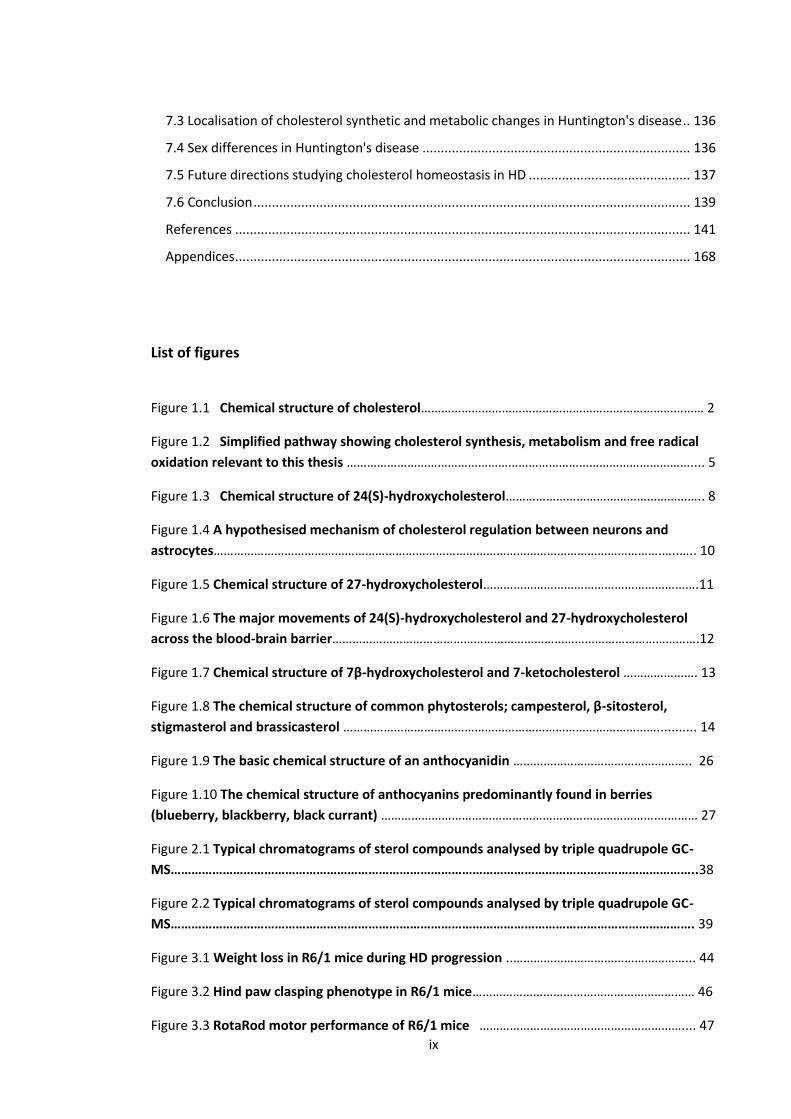
1.1 Cholesterol
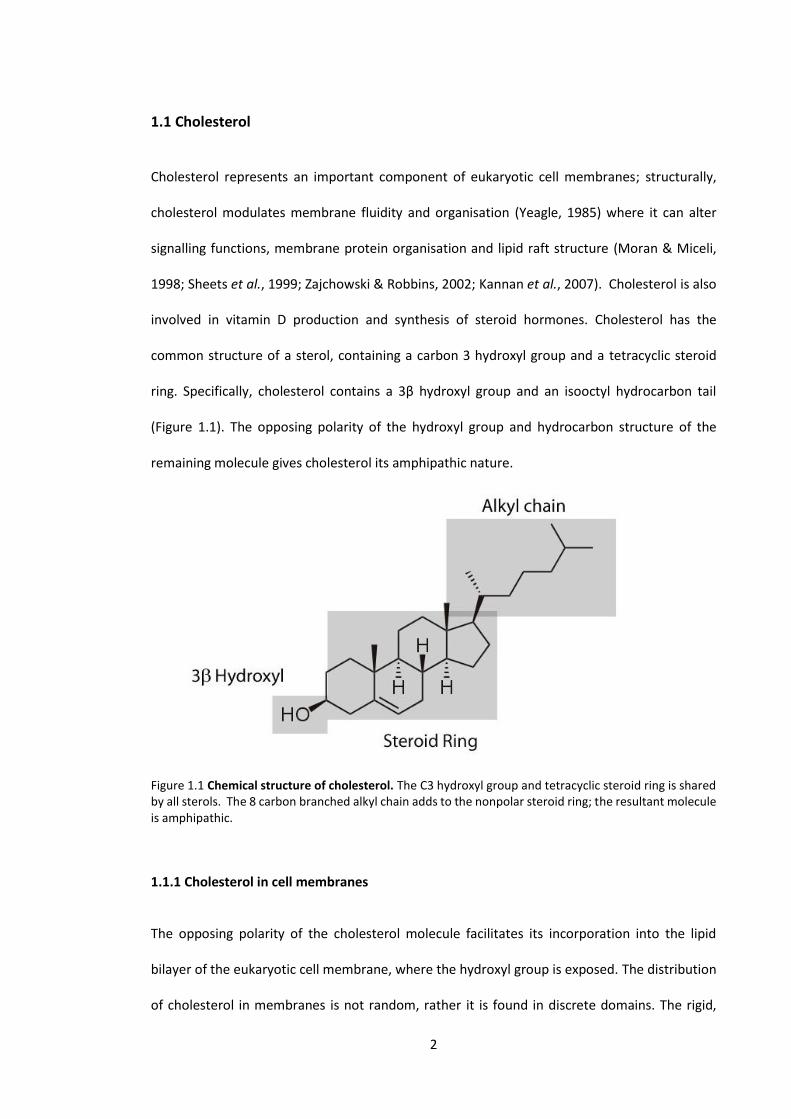
Cholesterol represents an important component of eukaryotic cell membranes; structurally,
cholesterol modulates membrane fluidity and organisation (Yeagle, 1985) where it can alter
signalling functions, membrane protein organisation and lipid raft structure (Moran & Miceli,
1998; Sheets et al., 1999; Zajchowski & Robbins, 2002; Kannan et al., 2007). Cholesterol is also
involved in vitamin D production and synthesis of steroid hormones. Cholesterol has the
common structure of a sterol, containing a carbon 3 hydroxyl group and a tetracyclic steroid
ring. Specifically, cholesterol contains a 3β hydroxyl group and an isooctyl hydrocarbon tail
(Figure 1.1). The opposing polarity of the hydroxyl group and hydrocarbon structure of the
remaining molecule gives cholesterol its amphipathic nature.
Figure 1.1 Chemical structure of cholesterol. The C3 hydroxyl group and tetracyclic steroid ring is shared by all sterols. The 8 carbon branched alkyl chain adds to the nonpolar steroid ring; the resultant molecule is amphipathic.
1.1.1 Cholesterol in cell membranes
The opposing polarity of the cholesterol molecule facilitates its incorporation into the lipid
bilayer of the eukaryotic cell membrane, where the hydroxyl group is exposed. The distribution
of cholesterol in membranes is not random, rather it is found in discrete domains. The rigid,

3
polycyclic structure of cholesterol in the membrane can have the effect of restricting the
movement of neighbouring hydrocarbon chains, introducing order. This can also disrupt tightly
packed alkyl chains that make up the hydrophobic interior of the cell membrane. These chemical
features of cholesterol are important when considering the dynamics of cholesterol in the cell
membrane, and the potential disturbances caused by altered levels of cholesterol.
1.1.2 Cholesterol in lipid rafts
Lipid rafts are highly dynamic, heterogeneous, cholesterol and sphingolipid rich microdomains
found in the lipid bilayer of membranes; functioning to segregate and concentrate proteins that
carry out cellular processes (Simons & Ikonen, 1997; Pike, 2006). Lipid rafts are associated with
essential cellular functions, including signal transduction (Janes et al., 2000), membrane
trafficking (Brown & London, 1998) and membrane associated proteolysis (Vetrivel et al., 2005).
Lipid rafts are essential for normal brain function and have been identified in glia and neurons
(Tsui-Pierchala et al., 2002; Gielen et al., 2006). The dysfunction of lipid rafts may have serious
consequences in the brain, and this has been associated with several neurodegenerative
diseases (Urano et al., 2005; del Toro et al., 2010; Fabelo et al., 2011). Since lipid rafts are
enriched with cholesterol, it has been proposed that altered cholesterol homeostasis in the
brain may lead to a disturbance of lipid raft structure and their associated functions (Rojo et al.,
2006).
1.2 Cholesterol in the brain
The brain contains the highest concentration of cholesterol of any tissue in the body; accounting
for approximately 25% of the total cholesterol, in an organ that only makes up 2% of the total
body mass. The distribution of cholesterol is not homogenous in the brain, 70% is found in the
myelinated axons of white matter, and the remaining 30% in the membranes of neurons and

4
glia (Norton & Autilio, 1965; Snipes & Suter, 1997). Although cholesterol is highly concentrated
in the brain, it is not able to move across the blood brain barrier (BBB) (Jurevics & Morell, 1995).
The isolation of the brain from peripheral sources of cholesterol suggests that strict cholesterol
homeostasis is required within the brain to maintain function. A clear example of this is genetic
mutations in cholesterol synthetic enzymes leading to severe neurological dysfunction
(desmosterolosis and Smith-Lemli-Opitz syndrome) (Wassif et al., 1998; Waterham et al., 2001).
Altered cholesterol metabolism has also been associated with several neurodegenerative
diseases including Alzheimer's disease (AD), Parkinson's disease (PD) and Huntington's disease
(HD) (Wahrle et al., 2002; Cordy et al., 2003; Gibson Wood et al., 2003; Lim et al., 2011). Whether
this is a cause or effect has not been established.
1.2.1 Synthesis of cholesterol in the brain
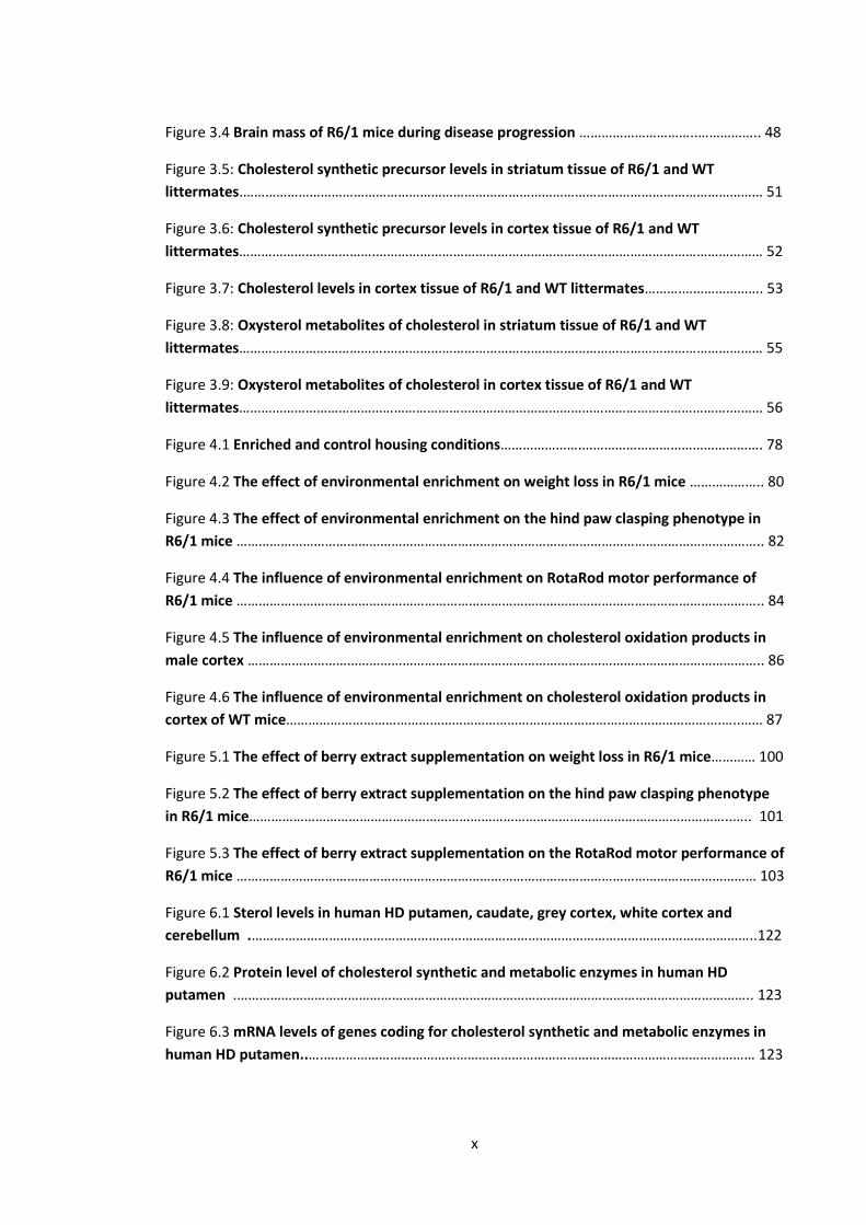
Cholesterol cannot cross the BBB, therefore de novo synthesis is required to supply the brain
with cholesterol (Jurevics & Morell, 1995). Brain cholesterol synthesis follows the same pathway
as in peripheral tissues, a process where acetate is converted to cholesterol in over 20 steps.
The major enzymes and intermediates of the lower ('post-squalene') cholesterol synthetic
pathway are represented in Figure 1.2. The cholesterol synthetic pathway is split, with the last
common precursor lanosterol. The Bloch pathway has a major intermediate of desmosterol,
while the Kandutsch-Russell pathway utilises lathosterol (Bloch, 1965). Squalene occurs earlier
in the pathway before cyclisation of the steroid ring. The rate limiting enzyme in the cholesterol
biosynthetic pathway has been previously identified as 3-hydroxy-3-methylglutaryl-coenzyme-
A reducatase (HMG-CoAred) (Snipes & Suter, 1997), which appears early in the pathway.
Negative feedback regulates HMG-CoAred through degradation (Gardner & Hampton, 1999)
and transcriptional control (Reynolds et al., 1984). As the cholesterol synthetic pathway is
involved in producing multiple products along the pathway, more complex regulation is likely to
occur. Recent studies specifically investigating the regulation of the "post-squalene" cholesterol

5
synthetic pathway, have suggested the synthetic enzyme delta(24)-sterol reducatse (DHCR24)
to have regulatory roles beyond catalysing the final step in the Bloch pathway (Luu et al., 2015).
As these findings are quite recent, the importance of DHCR24 levels in brain cholesterol
homeostatic regulation is unknown.
Figure 1.2 Simplified pathway showing cholesterol synthesis, metabolism and free radical oxidation relevant to this thesis. Major “post-squalene” cholesterol synthetic precursors shown follow a branched pathway, the Kandutsch-Russell pathway or Bloch pathway. Cholesterol can be oxidised enzymatically to form 24(S)-hydroxycholesterol (24-OHC) or 27-hydroxycholesterol (27-OHC) by cholesterol 24-hydroxylase (CYP46A1) and cholesterol 27-hydroxylase (CYP27A1) respectively. Reactive oxygen species (ROS) can oxidise cholesterol to form 7-ketocholesterol and 7β-hydroxycholesterol. The position of delta(24)-sterol reductase (DHCR24), a cholesterol synthetic enzyme is also shown. Broken lines indicate intermediates that have not been shown in this simplified scheme.
6
Although cholesterol synthesis and metabolism in peripheral tissues is relatively well
understood, the difficulty of performing in vivo studies in brain tissue has left many cellular
processes involving cholesterol synthesis and metabolism undefined. However, in vitro studies
examining isolated neurons and glial cells have identified some brain specific processes involved
in cholesterol regulation and trafficking. Embryonic neurons have been identified to synthesise
cholesterol (Saito et al., 1987), however cholesterol synthesis in adult neurons is unsustainably
low (Nieweg et al., 2009). These findings support the hypothesis that cholesterol synthesis is
mostly abandoned in neurons shortly after foetal development during which the majority of
cholesterol is synthesised in the brain (Pfrieger, 2002). As neurons have a high demand for
cholesterol, specifically for axon growth (Hayashi et al., 2004), maintenance of dendrites (Fan et
al., 2002), and synaptogenesis (Mauch et al., 2001); the source of neuronal cholesterol in the
mature brain has been investigated. In vitro studies suggest neurons source cholesterol from
astrocytes, and this has been demonstrated to be essential for neuron growth in vitro (Mauch
et al., 2001; Nagler et al., 2001). Further support for the 'outsourcing' hypothesis has been
demonstrated through the in vivo disruption of squalene synthase (an essential enzyme for
cholesterol synthesis) in adult neurons, resulting in normal brain morphology in mice
(Funfschilling et al., 2007). This indicates adult neurons are able to survive independent of their
own cholesterol synthesis. Pfrieger et al. (2002) proposed that an apolipoprotein shuttle from
astrocytes to neurons is the mechanism by which neurons obtain cholesterol, and there is
evidence that neurons have the capability to process cholesterol in lipoprotein particles through
the endosome-lysosome pathway (Parton et al., 1992; Brown et al., 1997). There may be several
reasons that neurons outsource cholesterol synthesis to astrocytes. The high energy cost, and
the need for a large number of enzymes in different cellular compartments, may explain why
neurons, whose primary function is electrical synaptic transmission, abandoned cholesterol
synthesis after foetal development. The elongated shape of the neuron may also hinder
transport of cholesterol, from where it is produced in the cell body, to where it is needed
7
(synapses). Oligodendrocytes maintain cholesterol synthesis in adulthood and synthesise
cholesterol at a rate that exceeds the level in astrocytes (Saito et al., 1987; Nieweg et al., 2009).
It is unclear if neurodegeneration alters cholesterol synthesis in the brain and if intermediates
accumulate or diverge into different metabolic pathways. There is evidence for decreased levels
of cholesterol synthetic precursors in aging (Thelen et al., 2006) and AD (Kolsch et al., 2010),
however, since many analytical techniques are unable to detect these low level compounds,
much of the current literature does not contain enough information to interpret the extent of
changes occurring.
1.2.2 Cholesterol metabolism in the brain
Cholesterol synthesis in the adult brain is slow, in the order of µg/h (Spady & Dietschy, 1983),
which is surprising, as the brain is one of the most metabolically active tissues in the body.
Although the rate of synthesis is low, excess cholesterol must still be removed from the brain.
The BBB is impermeable to cholesterol, therefore simple diffusion of cholesterol into the plasma
does not occur. It has been suggested that cholesterol can move into the cerebrospinal fluid
(CSF) in apolipoprotein E (ApoE) particles and then into the plasma (Pitas et al., 1987a). However,
this accounts only for a small amount of cholesterol removed to the periphery, indicating other
mechanisms must be at play.
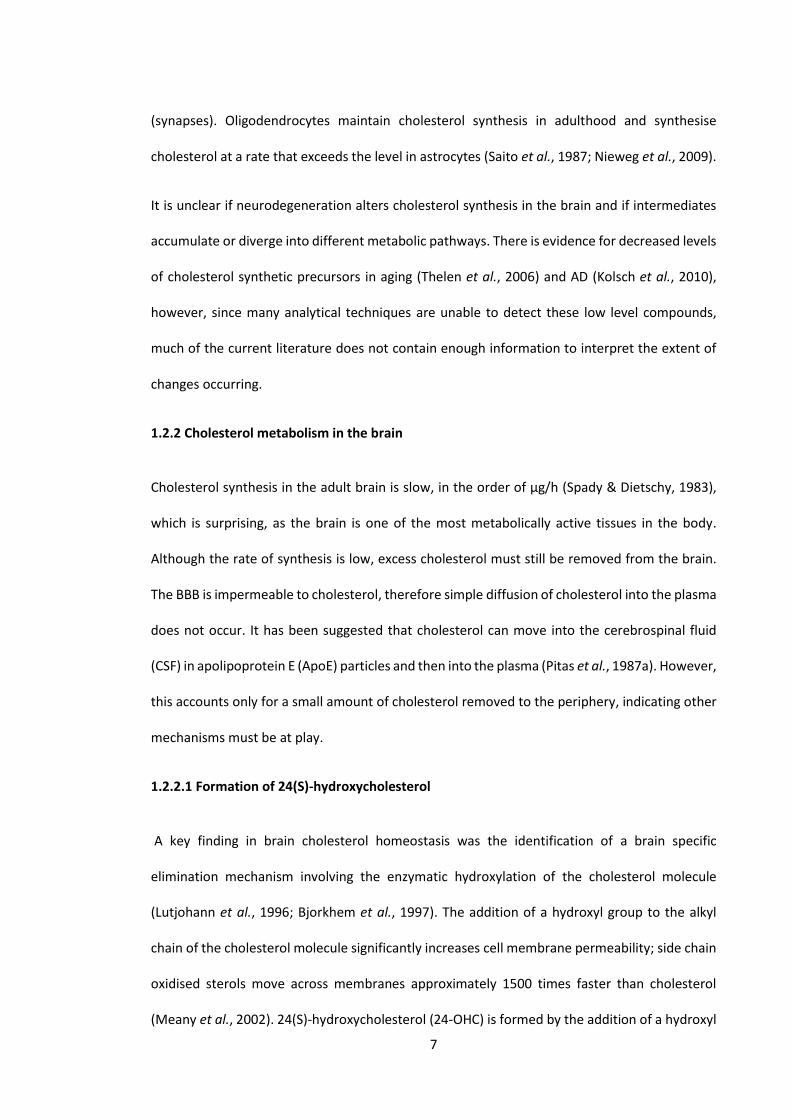
1.2.2.1 Formation of 24(S)-hydroxycholesterol
A key finding in brain cholesterol homeostasis was the identification of a brain specific
elimination mechanism involving the enzymatic hydroxylation of the cholesterol molecule
(Lutjohann et al., 1996; Bjorkhem et al., 1997). The addition of a hydroxyl group to the alkyl
chain of the cholesterol molecule significantly increases cell membrane permeability; side chain
oxidised sterols move across membranes approximately 1500 times faster than cholesterol
(Meany et al., 2002). 24(S)-hydroxycholesterol (24-OHC) is formed by the addition of a hydroxyl
8
group in the 24S position of cholesterol (Figure. 1.3). A cytochrome P-450, cholesterol 24-
hydroxylase (CYP46A1), expressed primarily in neurons, catalyses this reaction (Lund et al.,
1999). Although the liver is the site of most cholesterol metabolism in the body, CYP46A1
expression is almost exclusive to neurons (Lund et al., 1999), suggesting a brain specific role in
cholesterol metabolism. Studies involving CYP46A1 knockout mice (Lund et al., 2003) and 18O2
incorporation into 24-OHC in rats (Bjorkhem et al., 1997), have estimated that 24-OHC is
responsible for 40-60% of the cholesterol eliminated from the brain. A rise in 24-OHC levels
between the brachial artery and jugular vein in human subjects is consistent with these findings
that demonstrate there is a net flux from the brain into circulation (Bjorkhem et al., 1998).
Taking into account mechanisms of cholesterol elimination from the brain, the complete
turnover of cholesterol in the human brain is estimated to be in the order of 5 years (Bjorkhem
et al., 1998).
Figure 1.3 Chemical structure of 24(S)-hydroxycholesterol. 24(S)-Hydroxycholesterol (24-OHC) is the major elimination product of cholesterol from the brain, formed by the enzymatic hydroxylation of cholesterol by cholesterol 24-hydroxylase (CYP46A1).
9
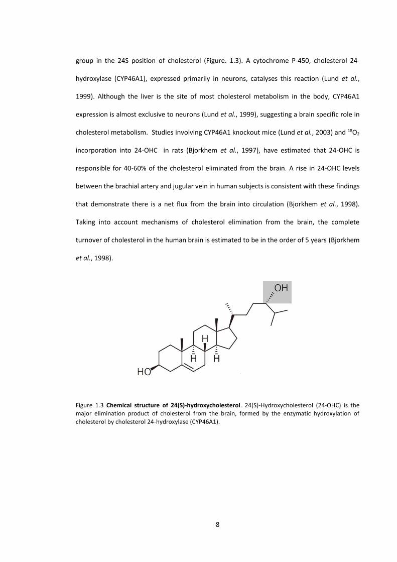
1.2.2.2 Possible regulation of brain cholesterol homeostasis by 24(S)-hydroxycholesterol
The current understanding of cholesterol synthetic regulation is that a complex interplay exists
between sterol sensing elements [sterol regulatory element-binding proteins (SREBPs)] and
transcription factors responsible for producing cholesterol synthetic enzymes (Brown &
Goldstein, 1999). Whether these mechanisms are active in neurons and astrocytes has not been
established. Ong et al. (2000) found that SREBPs are present in the neurons of the neocortex
and hippocampus, however their function in terms of sterol regulation in these regions is
unknown. The hypothesis that neurons import the majority of cholesterol from astrocytes is
supported by several studies (Mauch et al., 2001; Funfschilling et al., 2007), and thus it is
believed that a mechanism is in place to regulate this exchange (Vance & Hayashi, 2010). 24-
OHC is an activator of the nuclear receptor liver X receptor-β, that has the downstream effect of
activating ATP binding cassette transporter A1 (Repa et al., 2000), a cholesterol transport
mediator that resides in astrocytes. A convenient hypothesis suggests that metabolised
cholesterol in the form of 24-OHC promotes the delivery of cholesterol from astrocytes to
neurons (Figure 1.4) (Pfrieger, 2003), however there is evidence that synthesis and delivery of
cholesterol are regulated by separate mechanisms (Abildayeva et al., 2006). Further research is
necessary to elucidate the physiological importance of this mechanism, and potential impacts
of altered cholesterol metabolism in the brain.
1.2.2.3 27-Hydroxycholesterol
27-Hydroxycholesterol (27-OHC) is a major metabolic product of cholesterol in peripheral tissue,
entering the bloodstream to be further metabolised in the liver (Martin et al., 1993; Lund et al.,
1996). 27-OHC is formed by the enzymatic hydroxylation of cholesterol at the 27 carbon position
(Figure 1.5) by the cytochrome P450, cholesterol 27-hydroxylase (CYP27A1). This reaction takes
place in all cells; however the expression of CYP27A1 in the brain is significantly less than in
10
other tissues (Lein et al., 2007). A concentration gradient results in a net movement of 27-OHC
from circulation into the brain (Heverin et al., 2005), where it is quickly metabolised into more
polar products (including dihydroxysterols and cholestenoic acids), catalysed by the enzymes
CYP27A1 and 5-hydroxycholesterol 7-α-hydroxylase (CYP7B1) (Meaney et al., 2007). These
products then move back into circulation where they are efficiently absorbed by the liver (Lund
et al., 1996; Meaney et al., 2007). It has been previously demonstrated that circulating levels of
27-OHC are positively correlated to cholesterol levels in the blood (Babiker et al., 2005). It is
unknown if increased levels of 27-OHC entering the brain from circulation are detrimental, and
this is potentially relevant to neurodegenerative diseases as hypercholesterolemia has been
associated with AD and PD (Kivipelto et al., 2001; Hu et al., 2008).
Figure 1.4 A hypothesised mechanism of cholesterol regulation between neurons and astrocytes
(Pfrieger, 2002). 24(S)-Hydroxycholesterol (24-OHC) binds the nuclear receptor LXR which activates the
cholesterol transport mediator ABCA1. It is unknown what promotes cholesterol synthesis in this
mechanism. Adapted from Bjorkhem (2006).

11
Figure 1.5 Chemical structure of 27-hydroxycholesterol. 27-Hydroxycholesterol (27-OHC) is a cholesterol metabolite primarily produced in peripheral tissue where it enters the bloodstream to be further metabolised in the liver. 27-OHC is formed by the enzymatic hydroxylation of cholesterol by cholesterol 27-hydroxylase (CYP27A1).
1.2.3 Toxicity of cholesterol metabolites
Several in vitro experiments have shown that cholesterol metabolites also have cytotoxic
activity. It is unknown what causes specific changes in brain cholesterol metabolism resulting in
the production, or excess production of potentially toxic oxysterol species. 27-
hydroxycholesterol is toxic to human monocyte-macrophages in vitro (Clare et al., 1995);
similarly 24-OHC has toxic effects towards differentiated neuroblastoma cells (Kolsch et al.,
2001). 24-OHC is a major endogenous cholesterol metabolite in the brain, however, as the
majority of studies rely on in vitro models, the importance of this potential toxicity is yet to be
established in vivo.
12
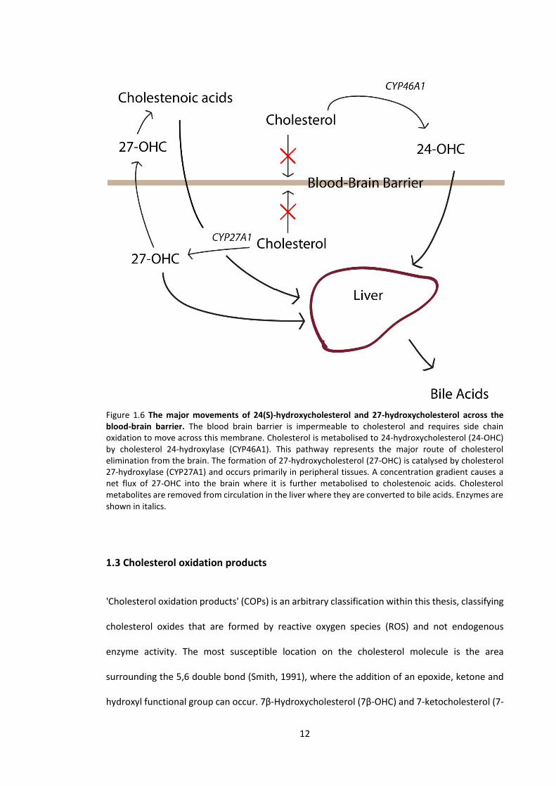
Figure 1.6 The major movements of 24(S)-hydroxycholesterol and 27-hydroxycholesterol across the blood-brain barrier. The blood brain barrier is impermeable to cholesterol and requires side chain oxidation to move across this membrane. Cholesterol is metabolised to 24-hydroxycholesterol (24-OHC) by cholesterol 24-hydroxylase (CYP46A1). This pathway represents the major route of cholesterol elimination from the brain. The formation of 27-hydroxycholesterol (27-OHC) is catalysed by cholesterol 27-hydroxylase (CYP27A1) and occurs primarily in peripheral tissues. A concentration gradient causes a net flux of 27-OHC into the brain where it is further metabolised to cholestenoic acids. Cholesterol metabolites are removed from circulation in the liver where they are converted to bile acids. Enzymes are shown in italics.
1.3 Cholesterol oxidation products
'Cholesterol oxidation products' (COPs) is an arbitrary classification within this thesis, classifying
cholesterol oxides that are formed by reactive oxygen species (ROS) and not endogenous
enzyme activity. The most susceptible location on the cholesterol molecule is the area
surrounding the 5,6 double bond (Smith, 1991), where the addition of an epoxide, ketone and
hydroxyl functional group can occur. 7β-Hydroxycholesterol (7β-OHC) and 7-ketocholesterol (7-
13
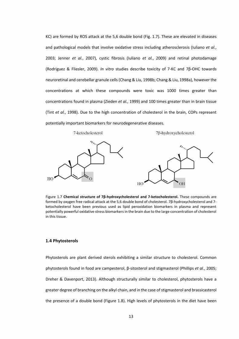
KC) are formed by ROS attack at the 5,6 double bond (Fig. 1.7). These are elevated in diseases
and pathological models that involve oxidative stress including atherosclerosis (Iuliano et al.,
2003; Jenner et al., 2007), cystic fibrosis (Iuliano et al., 2009) and retinal photodamage
(Rodriguez & Fliesler, 2009). In vitro studies describe toxicity of 7-KC and 7β-OHC towards
neuroretinal and cerebellar granule cells (Chang & Liu, 1998b; Chang & Liu, 1998a), however the
concentrations at which these compounds were toxic was 1000 times greater than
concentrations found in plasma (Zieden et al., 1999) and 100 times greater than in brain tissue
(Tint et al., 1998). Due to the high concentration of cholesterol in the brain, COPs represent
potentially important biomarkers for neurodegenerative diseases.
Figure 1.7 Chemical structure of 7β-hydroxycholesterol and 7-ketocholesterol. These compounds are formed by oxygen free radical attack at the 5,6 double bond of cholesterol. 7β-hydroxycholesterol and 7-ketocholesterol have been previous used as lipid peroxidation biomarkers in plasma and represent potentially powerful oxidative stress biomarkers in the brain due to the large concentration of cholesterol in this tissue.
1.4 Phytosterols
Phytosterols are plant derived sterols exhibiting a similar structure to cholesterol. Common
phytosterols found in food are campesterol, β-sitosterol and stigmasterol (Phillips et al., 2005;
Dreher & Davenport, 2013). Although structurally similar to cholesterol, phytosterols have a
greater degree of branching on the alkyl chain, and in the case of stigmasterol and brassicasterol
the presence of a double bond (Figure 1.8). High levels of phytosterols in the diet have been

14
found to compete with intestinal cholesterol absorption, reducing plasma levels of cholesterol
(Ikeda et al., 1988; Katan et al., 2003). The reduction of cholesterol solubility in phospholipid bile
salt micelles in the presence of β-sitosterol demonstrated by Ikeda et al. (1988) is also explained
by the thermodynamically favourable interaction of phytosterols with micelles (Armstrong &
Carey, 1987). Dietary phytosterols present a viable option in lowering cholesterol absorption
from diet, which is also accompanied by increased phytosterol absorption and incorporation
into various tissues in the body (Plat et al., 2008). Phytosterols are able to cross the BBB and
have been measured in animal brain tissue (Plat et al., 2008). The long-term cellular effects of
phytosterols incorporated into the brain have not been established and further studies are
necessary to examine their impact on human health.
Figure 1.8 The chemical structure of common phytosterols; campesterol, β-sitosterol, stigmasterol and brassicasterol. Phytosterols are produced in plants and share a similar structure to cholesterol, differing in the degree of branching and presence of double bonds on the alkyl chain (highlighted). Phytosterols are absorbed from food and accumulate in tissues including the brain.

15
1.5 Cholesterol and neurodegenerative disease
Cholesterol is an essential molecule for cellular function; however, excess cholesterol circulating
in the body can be harmful. It has been established that high levels of cholesterol in the blood
contributes to a number of diseases, in particular atherosclerosis and cardiovascular disease.
The influence of altered cholesterol homeostasis in neurodegenerative diseases is however, not
as well understood. Cholesterol levels have been reported to alter in vitro protein aggregation
relevant to PD (Bar-On et al., 2008). Several studies have also highlighted altered brain
cholesterol levels in AD (Mason et al., 1992; Wahrle et al., 2002; Cordy et al., 2003), and may
influence amyloid beta formation by altering the physical properties of cell membranes (Rojo et
al., 2006). It is also hypothesised that cellular redistribution of cholesterol without changes in
total cholesterol levels may play a role in AD (Gibson Wood et al., 2003). Due to conflicting
reports of cholesterol levels in neurodegenerative brain tissue, the exact influence of altered
cholesterol homeostasis in neurodegenerative diseases is currently unknown.
1.5.1 Huntington's disease
HD is an autosomal dominant, progressive, neurodegenerative disease characterised by the
expansion of a trinucleotide repeat on the N-terminus of the huntingtin protein (HTT). The
cytosine-adenine-guanine (CAG) repeat, coding for glutamine, is located on exon 1 of the HTT
gene (MacDonald et al., 1993). Full penetrance of the disease is observed when an individual
has 42 or more repeats (Brinkman et al., 1997), with 36-41 repeats resulting in incomplete
penetrance (Kremer et al., 1994; Rubinsztein et al., 1996; Brinkman et al., 1997). Although
disease symptoms are not associated with a repeat number of 29-35, CAG repeat expansion in
successive generations is possible (Trottier et al., 1994; Ranen et al., 1995). Expansion of CAG
repeats occurs almost exclusively during spermatogenesis, causing the appearance of HD in

16
individuals without a family history of the disease. Juvenile onset HD, characterised by a long
CAG expansion (~100), is also much more likely from paternal inheritance (Kremer et al., 1995).
1.5.2 Symptoms
HD was previously known as Huntington's chorea due to the involuntary movements exhibited
by patients (Huntingon, 1872). Mutant gene carriers show subtle symptoms before clinical
diagnosis in a period referred to as pre-manifest, pre-symptomatic or pre-diagnostic. Pre-
manifest HD patients have been shown to have dysfunction in tongue force, grip strength and
finger tapping tasks (Bechtel et al., 2010; Reilmann et al., 2010a; Reilmann et al., 2010b). The
first overt symptoms of HD are typically involuntary movements, usually accompanied with
depression (Kirkwood et al., 2001). Other symptoms include abnormal eye movement (Penney
et al., 1990) and clumsiness (Kirkwood et al., 2001). During progression there is weight loss
(Sanberg et al., 1981; Morales et al., 1989; Djousse et al., 2002), cognitive decline, speech
difficulties and memory loss (Kirkwood et al., 2001). In late stages of the disease, HD patients
have difficulty swallowing; the aspiration of food into the lungs is a major cause of death in these
cases (Sorensen & Fenger, 1992). The progression and severity of disease after onset does not
correlate strongly with CAG repeat length in the range of 40-60 (Squitieri et al., 2002). However,
the correlation between the age of onset and CAG repeat length is well established (Ranen et
al., 1995). Rare juvenile cases of HD with 100+ CAG repeats do, however, have accelerated
progression of the disease (Squitieri et al., 2002).
1.5.3 Neuropathology
The neuropathological progression of HD was classified in detail by Vonsattel et al. (1985), and
was adopted to grade HD brain tissue. This revealed the disease progressed from the caudate
nucleus and putamen (striatum) with degeneration moving in a lateral basal direction. Early
changes include moderate astrocytosis of the caudate nucleus and putamen in grade I, followed

17
by macroscopic atrophy and neuronal loss in grade II. Neuron loss continues in grade III with
overt shrinkage of the striatum. Grade IV was classified by extreme shrinkage of the striatum,
including the globus pallidus. Astrocyte numbers were also increased compared to control at
this stage in the disease. Brain mass was also found to be negatively correlated to the disease
grade, with an average 20% reduction in brain mass by grade IV. More sensitive magnetic
resonance imaging (MRI) techniques have also identified volume reduction in the hippocampus,
cerebral cortex and amygdala of HD patients, with the cerebellum relatively spared (Rosas et al.,
2003). Along with neuron loss, astrocytosis and increased oligodendrocyte densities are also
observed in severely affected regions of the HD brain (Myers et al., 1991).
1.5.4 Huntingtin protein
HTT is made up of >3100 amino acid residues and has a molecular mass of approximately 349
kDa depending on the length of the CAG repeat (Gil & Rego, 2008). HTT is expressed throughout
the body, with the highest expression in the brain and testes (Strong et al., 1993;
Landwehrmeyer et al., 1995). It is localised to a number of subcellular compartments and has
functions in intracellular trafficking, clathrin mediated endocytosis, transcriptional regulation
and cell signalling (Harjes & Wanker, 2003; Li & Li, 2004). The protein is believed to be essential
for early development in mice, as knockout of the HTT mouse homologue (Hdh) is embryonically
lethal (Leavitt et al., 2001).
1.5.5 Toxicity of mutant huntingtin
The exact role of the CAG mutation on the HTT protein in HD pathogenesis is still debated. It is
not completely clear if mutant huntingtin (mHTT) has a toxic gain of function or if a loss of
function is responsible for disease, or possibly a combination of both factors. The heterozygous
disruption of the HTT gene does not cause a HD phenotype in human cases, suggesting the loss
of function in one allele is not entirely responsible for disease pathology (Ambrose et al., 1994).

18
Expression of normal length HTT can reduce the toxicity of mHTT in mice (Leavitt et al., 2001),
also suggesting compensation for loss of function. Another confounding finding is that human
mHTT can rescue the embryonic lethality of the Hdh knockout mouse (Leavitt et al., 2001).
However, this may simply be a reflection of the role of HTT in developmental processes, similar
to the human disease where patients develop normally, only manifesting symptoms later in life.
Another debated hypothesis of mHTT toxicity involves protein aggregation. Aggregation of
mHTT is a hallmark of HD, however, this may not be an accurate predictor of cell death.
Aggregate formation is higher in cortical neurons, which are relatively spared compared to the
selectively vulnerable striatal neurons that contain fewer aggregates (Kuemmerle et al., 1999).
In vitro studies investigating mHTT aggregation have also reported neuronal death without the
formation of inclusions, and that cells forming inclusions had reduced risk of death (Arrasate et
al., 2004). In this study, cells forming inclusions had reduced levels of mHTT in the rest of the
cell, suggesting that inclusion formation may be protective. Although inclusions may not be
directly linked to cell death, other lines of evidence suggest that proteasomal disruption in cells
caused by mHTT aggregates may be neurotoxic (Jana et al., 2001; Waelter et al., 2001). It is also
possible that undiscovered neurotoxic aggregates are too small for detection by light microscopy
as suggested by Bates (2003).
1.5.6 Mouse models of Huntington’s disease
Prior to the HTT gene identification in 1993 (MacDonald et al.), the primary rodent models of
HD were based on the production of brain lesions by neurotoxin injections into the striatum.
Injection of glutamic acid, kainic acid (McGeer & McGeer, 1976; Schwarcz & Coyle, 1977) and
quinolinic acid (Beal et al., 1986) replicated some of the biochemical changes observed in the
human HD brain, and highlighted a potential involvement of NMDA mediated excitotoxicity in
HD pathogenesis. Since the identification of the HTT gene and the expanded CAG repeat that
causes HD, genetic models were generated for more accurate replication of the human
19
condition. Common mouse models have used multiple approaches to model HD, including
knock-in models and transgenic models expressing full length and truncated forms of human
HTT.
The R6 mouse models are widely used to study HD pathology and therapeutic interventions.
These mice were generated by the insertion of a truncated form of human HTT that codes for a
CAG expansion and 67 amino acids of exon 1 human HTT under the human promoter (Mangiarini
et al., 1996). The R6/1 mouse expressing truncated HTT with approximately 115-120 CAG
repeats, and the R6/2 with 140-150 repeats, are the best characterised variants of this model.
R6 mice mimic several pathological hallmarks including early striatal degeneration, and physical
phenotypes of HD such as motor dysfunction and uncontrolled movements (Mangiarini et al.,
1996). These features and the relatively short disease onset are reasons why this model has
been widely used in the study of HD. Instability of the CAG repeat between generations can
result in CAG expansion. This can cause variation in the specific CAG repeat length between
colonies (Mangiarini et al., 1997). Selectively breeding mice without expanded repeats is able to
control CAG expansion in subsequent generations.
A model expressing full length expanded human HTT was also generated using a yeast artificial
chromosome (YAC). These mice expressing HTT with a normal number of repeats (18), and
expanded CAG repeats (46, 72, and 128), have also been characterised to have striatal
degeneration, motor deficits, exhibit motor abnormalities and mHTT aggregation (Hodgson et
al., 1999; Slow et al., 2003).
Knock-in of a CAG repeat expansion into the Hdh gene has also been used to generate HD models
that express an expanded form of mouse HTT under endogenous transcriptional control
(Wheeler et al., 1999; Wheeler et al., 2000). Heterozygous and homozygous knock-in mice
expressing 92 (HdhQ92) and 111 (HdhQ111) CAG repeats have been commonly examined in
20
previous literature; knock-in models also exhibit degenerative pathologies (Wheeler et al.,
2002).
There are benefits and drawbacks in each model; this includes the severity of the disease in mice
where R6 models are most representative due to the long CAG repeat that causes a relatively
rapid onset of symptoms. HTT sequences found in transgenic models (R6 and YAC) however,
become randomly inserted into the genome and therefore do not have the endogenous mouse
promoter as well as the endogenous protein still being expressed. Therefore models that knock
in expanded CAG repeats into the mouse HTT sequence (e.g. HdhQ111) more accurately
represent the disease from a genetic perspective.
1.5.7 Cellular and fly models of Huntington’s disease
Several models of HD have been generated in Drosophila where an expanded fragment of exon
1 human HTT is expressed (Httex1pQ93 and Htt128Q). These models have been used in several
studies as they provide a number of phenotypes including photoreceptor degeneration, motor
abnormalities as well as reduced lifespan (Steffan et al., 2001; Lee et al., 2004). A number of cell
models expressing forms of expanded HTT have been developed since the identification of the
affected gene. A model developed in yeast that expresses exon 1 HTT with 75 CAG repeats has
been used to assess HTT aggregation (Ehrnhoefer et al., 2006). A model in a neuronal cell line
(PC12) developed to express exon 1 HTT has also been used to examine the effects of mutant
huntingtin on extracellular signalling (Aiken et al., 2004; Maher et al., 2011).
1.5.8 Alteration of cholesterol homeostasis in Huntington's disease
While it is not fully understood how the polyglutamine expansion causes cellular dysfunction in
HD, mHTT has been identified to alter membrane order (del Toro et al., 2010) and HTT-
phospholipid interactions (Kegel et al., 2009). Despite the genotypic identification of mHTT
carriers, there is a lack of reliable biomarkers to predict HD progression or effectiveness of
21
therapies. Several studies have identified that cholesterol synthesis and metabolism in HD cell
lines and animal models is significantly disturbed and suggest these to be potential biomarkers
of HD (Valenza et al., 2005; Valenza et al., 2007b; Valenza et al., 2010), but the mechanisms and
metabolic pathways affected have not been fully examined in human or mouse models of HD.
1.5.9 Alterations of cholesterol biosynthesis in Huntington's disease
Gene transcription profiles of HTT-inducible cells identified a possible dysregulation of lipid
homeostasis in HD (Sipione et al., 2002). This study reported the reduction of several mRNAs
coding for cholesterol biosynthetic enzymes, including that of 7-dehydrocholesterol reductase
(DHCR7), the final enzyme of the Kandutsch-Russell pathway (Sipione et al., 2002). Further
investigation into HD fibroblasts, R6/2 mice and HD post mortem brain tissue, identified a
consistent reduction of several other mRNAs coding for enzymes in the cholesterol synthetic
pathway (Valenza et al., 2005). The active nuclear form of SREBP was also found to have a 50%
reduction in human HD fibroblasts and in brain tissue of mice (Valenza et al., 2005). Brain lipid
analysis of R6/2 and YAC 128 mice has also revealed a consistent reduction in the cholesterol
synthetic precursors lathosterol and lanosterol (Valenza et al., 2007a; Valenza et al., 2007b).
Further association of brain cholesterol synthesis and mHTT was demonstrated by a CAG repeat
dependant reduction of lathosterol in YAC mice, and reduced levels of lathosterol in the
homozygous knock-in mouse (HdhQ111/111) compared to the heterozygous knock-in (Valenza et
al., 2010). The current understanding of cholesterol biosynthesis in HD has been previously
limited by the analysis of a small number of synthetic precursors in a pathway with over 20 steps.
Analysis of a wider range of synthetic precursors is likely to provide a more complete story of
the potential changes occurring in HD. In addition to this, a detailed analysis of cholesterol
synthetic precursors in human post mortem tissue is yet to be reported in the literature.

22
1.5.10 Cholesterol levels in Huntington's disease
Although a significant reduction of cholesterol biosynthetic precursors were observed
previously in these mouse models, the total level of brain cholesterol remains relatively stable.
Unchanged levels of cholesterol were observed in the striatum or whole brain of R6/1 and R6/2
models (Valenza et al., 2007b; del Toro et al., 2010), while only a small reduction in brain
cholesterol levels were observed in the HdhQ111, YAC72 and YAC128 mice (del Toro et al., 2010;
Valenza et al., 2010). A previous study examining cholesterol levels in human HD post mortem
tissue identified an increase in cholesterol, however the sample size (n = 3) was too small to
draw a firm conclusion regarding cholesterol levels in human HD brain (del Toro et al., 2010).
Mechanisms to eliminate cholesterol may also be downregulated to maintain constant
cholesterol levels in the brain, highlighting the potential involvement of altered cholesterol
metabolism in HD.
1.5.11 Cholesterol metabolic alterations in Huntington's disease
The major elimination product of cholesterol in the brain, 24-OHC, is also believed to have an
important role in cholesterol homeostatic regulation in the brain. Reduced brain levels of 24-
OHC have been consistently demonstrated in multiple rodent models of HD, including knock-in
models and transgenic models expressing truncated and full length HTT (Valenza et al., 2007a;
Valenza et al., 2010). Plasma measurements of 24-OHC in the YAC 128 model reflect the reduced
level in brain (Valenza et al., 2007a); plasma studies in HD patients have also identified reduced
24-OHC levels in symptomatic individuals (Leoni et al., 2008; Leoni et al., 2013). Since CYP46A1
expression is primarily localised to neurons (Lund et al., 1999), it has been suggested that
generation of 24-OHC (as measured in plasma) is a marker of metabolically active neurons in the
brain (Bjorkhem, 2006). Recent evidence suggests that changes in CYP46A1 and 24-OHC brain
levels may play a role in neurodegeneration (Kolsch et al., 2002; Papassotiropoulos et al., 2003;

23
Brown et al., 2004; Tian et al., 2010), however, the exact role this might play in HD pathogenesis
has not been established. Comprehensive examination of cholesterol synthesis and metabolism
in human HD, beyond plasma biomarkers is currently lacking in the literature. This represents a
significant gap in the current knowledge that would help confirm the validity of rodent HD
models examining this particular metabolic pathway in relation to HD pathology.
1.5.12 Proposed mechanisms altering cholesterol homeostasis in Huntington's disease
Consistent demonstration of cholesterol homeostatic perturbation in HD models has led to the
identification of several associations of HTT and mHTT with cholesterol synthetic regulation. A
hypothesised mechanism of reduced cholesterol biosynthesis in HD involves an interaction of
mHTT with SREBPs (Kaltenbach et al., 2007). It has been suggested that the CAG expansion
causes a diminished capacity of mHTT to translocate SREBP to the nucleus where it has functions
to activate cholesterol synthetic genes (Guan et al., 1995; Lloyd & Thompson, 1995). HTT is
known to have cellular trafficking properties, and this hypothesis is consistent with the finding
that transgenic HD cells contain less active nuclear SREBPs than controls (Valenza et al., 2005).
Another factor causing reduced cholesterol synthesis is related to decreased levels of brain-
derived neurotrophic factor (BDNF) that have been observed in human HD and HD mouse
models (Zuccato et al., 2001; Spires et al., 2004; Zuccato et al., 2005). BDNF is known to increase
cholesterol content of lipid rafts in vitro and increase neurotransmitter release dependant on
BDNF-elicited cholesterol (Suzuki et al., 2007). In vivo, abnormal synaptic plasticity in R6/2 mice
(Murphy et al., 2000; Picconi et al., 2006) may also be related to dysfunction of BDNF-mediated
cholesterol levels in the cell.
With the complex control of cholesterol homeostasis in the brain, it is likely that other
undiscovered factors, potentially involved with cholesterol metabolism, may be involved in
altering cholesterol synthesis in the presence of mHTT. Further investigation into cell-type

24
specific cholesterol homeostatic alterations will also contribute to understanding how these
altered pathways affect whole tissue.
1.6 Therapeutics in Huntington's disease mouse models
There is currently no cure or effective therapies for the treatment of HD. Due to the late onset
and possibility of early identification of mHTT carries, there is a relatively long timeframe
(decades) to implement potential therapies for HD suffers. It also allows for close study of the
manifestation of neurodegenerative pathologies that may be relevant to understanding
'sporadic' neurodegenerative diseases such as AD. Mouse models of HD, particularly the R6
models, have been used to examine several therapeutic strategies including dietary
supplementation (Ehrnhoefer et al., 2006; Maher et al., 2011), tissue transplantation (van Dellen
et al., 2001), drug treatments (Ferrante et al., 2002; Schiefer et al., 2002) and gene therapies
(Popovic et al., 2005). This thesis will focus on the use of environmental enrichment (EE) and
dietary supplementation in HD.
1.6.1 Environmental enrichment and Huntington's disease
Increased cognitive activity is believed to be a factor in reducing the risk of developing dementia
and neurodegenerative diseases later in life (Karlsson et al., 1988; Stern et al., 1994; Evans et
al., 1997). Substantial evidence also suggests that increased physical, sensory-motor and
cognitive activity associated with EE has positive effects in the brain to improve memory,
cognition and reduce the severity of neurodegenerative phenotypes in animal models
(Wainwright et al., 1993; Kempermann et al., 2002; Jankowsky et al., 2005; Lazarov et al., 2005).
Recently it has been discovered that EE may also be beneficial in HD. Studies in mouse models
of HD illustrate that EE delays onset of physical phenotypes (van Dellen et al., 2000), improves
motor co-ordination (Hockly et al., 2002; Spires et al., 2004) and improves survival (Carter et al.,
2000), compared to mice with standard housing. Rodent studies utilising EE usually involves a

25
larger cage with a greater number of animals, the addition of novel objects (toys), tunnels,
additional nesting materials and in some cases running wheels (van Praag et al., 1999; van Dellen
et al., 2000; Hockly et al., 2002; Hockly et al., 2003; Spires et al., 2004). A study in human
populations has also identified that environmental factors also influence the age of onset of HD
(Wexler & Res, 2004). Increased neurogenesis in the hippocampus has been identified to explain
some of the positive effects on memory seen in EE animal studies (van Praag et al., 1999; Lazic
et al., 2006), however specific mechanisms improving the disease severity in HD, which involves
striatal atrophy, requires further investigation. Whether cholesterol homeostatic perturbations
observed in mouse models are altered by EE is currently unknown.
1.6.2 Flavonoid supplementation
Flavonoids are phenolic micronutrients in foods derived from plants. A large area of study has
identified multiple health benefits of dietary flavonoids with potential beneficial effects in
cardiovascular diseases (Nakagawa et al., 1999; Ruel et al., 2005) and cancer (Murphy et al.,
2003; Ferguson et al., 2006). Recently dietary supplementation of flavonoids and other plant
derived phytochemicals have revealed an influence on the brain, including positive effects on
cognitive function in human studies (File et al., 2001; Duffy et al., 2003; Casini et al., 2006) and
improvement of neurodegenerative phenotypes in animal models of AD (Li et al., 2015;
Nakajima et al., 2015; Sabogal-Guaqueta et al., 2015).
1.6.2.1 Anthocyanins and Huntington's disease
Anthocyanins are a class of flavonoid that have the backbone structure of an anthocyanidin
(Figure 1.9), with the addition of a glycoside (O-linked sugar). Anthocyanins found in plants are
primarily 3-glycosides of anthocyanidins. In addition to the location and species of sugar,
different structures of anthocyanins are derived from the position of hydroxy and methoxy
functional groups on the anthocyanindin backbone. Berries, including blueberries, blackberries,
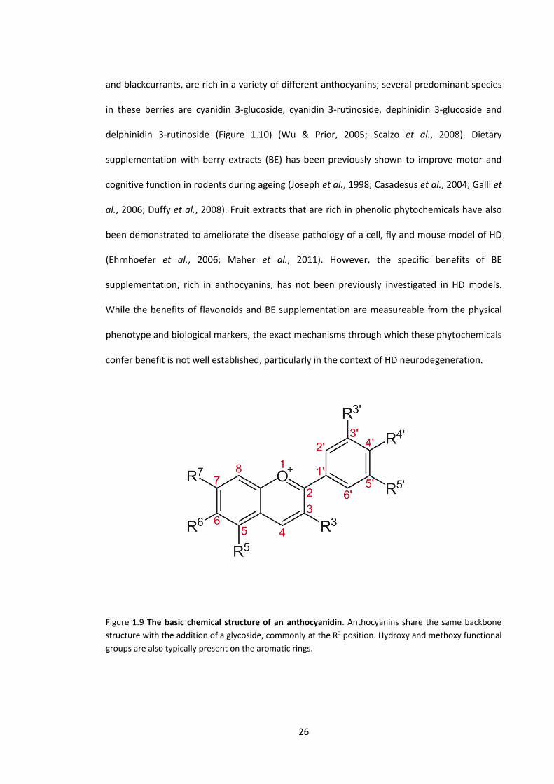
26
and blackcurrants, are rich in a variety of different anthocyanins; several predominant species
in these berries are cyanidin 3-glucoside, cyanidin 3-rutinoside, dephinidin 3-glucoside and
delphinidin 3-rutinoside (Figure 1.10) (Wu & Prior, 2005; Scalzo et al., 2008). Dietary
supplementation with berry extracts (BE) has been previously shown to improve motor and
cognitive function in rodents during ageing (Joseph et al., 1998; Casadesus et al., 2004; Galli et
al., 2006; Duffy et al., 2008). Fruit extracts that are rich in phenolic phytochemicals have also
been demonstrated to ameliorate the disease pathology of a cell, fly and mouse model of HD
(Ehrnhoefer et al., 2006; Maher et al., 2011). However, the specific benefits of BE
supplementation, rich in anthocyanins, has not been previously investigated in HD models.
While the benefits of flavonoids and BE supplementation are measureable from the physical
phenotype and biological markers, the exact mechanisms through which these phytochemicals
confer benefit is not well established, particularly in the context of HD neurodegeneration.
Figure 1.9 The basic chemical structure of an anthocyanidin. Anthocyanins share the same backbone
structure with the addition of a glycoside, commonly at the R3 position. Hydroxy and methoxy functional
groups are also typically present on the aromatic rings.

27
Figure 1.10 The chemical structure of anthocyanins predominantly found in berries (blueberry,
blackberry, blackcurrant). Different structures of anthocyanins arise from the arrangement of hydroxy
and methoxy functional groups on the anthocyanidin backbone, and the location and species of sugar that
is attached to the molecule. In the pictured molecules the glycosides are glucose and rutinose.
1.7 Biological markers of neurodegeneration
Diagnosis is an essential process in order to apply the correct treatment to combat disease.
Currently there is a need for diagnostic tests for neurodegenerative diseases, not only due to
their inevitably fatal nature, but also due to the increasing prevalence of these diseases. Ideally
biomarkers can be obtained from saliva, urine or blood as these require little or no invasive
procedures to obtain. Although more invasive procedures such as CSF samples or a brain biopsy
may result in a faster, more sensitive diagnosis, the stress on the body is usually unjustifiable. In
28
the case of HD where mutant carriers can be easily identified, biomarkers are of most benefit to
therapeutic trials in assessing the efficacy of treatment. In human patients a plasma biomarker
is ideal; however, examining post-mortem brain tissue from human and animal models may give
insight into changes that can be measured in the circulation. Post mortem brain biomarkers are
also useful in assessing treatments in animal trials where brain tissue is available from test
subjects. Since HD, as well as other neurodegenerative diseases have a significant association
with altered cholesterol metabolism in the brain, cholesterol metabolites and synthetic
precursors are potentially useful to track the progression of disease. 24-OHC levels in brain
tissue and plasma reflect turnover of cholesterol in the brain, however further investigation into
this metabolite is required before its clinical usefulness can be established. This is due to 24-
OHC plasma levels being variable in humans, dependant on hepatic metabolism and brain:liver
mass ratio (Bretillon et al., 2000a). It is known that 24-OHC is reduced in plasma of HD patients
and correlates to volume loss in the striatum (Leoni et al., 2008); however longitudinal studies,
that would be most effective in tracking brain metabolic dysfunction over time, currently do not
exist. Little is known about the concentration and location of other cholesterol synthetic
precursors and metabolic products in the HD brain, these being potentially powerful indicators
of disease severity and progression. Further investigation into biomarkers that correlate well
with HD severity will clearly increase the current understanding of fundamental mechanisms of
HD pathology and invariably accelerate the search for effective therapies.
1.8 Aims
The aim of this thesis was to investigate the involvement of brain cholesterol homeostasis in HD,
and identify sterol-related biomarkers associated with the disease using a novel and highly
sensitive analytical technique. We aimed to comprehensively quantify changes in brain
cholesterol metabolites and synthetic precursors during the progression of HD, by investigating

29
the R6/1 mouse model over 5 stages of the disease. We also aimed to investigate the
effectiveness of EE and BE supplementation to attenuate the physical phenotype and
cholesterol homeostatic perturbation in the R6/1 model. Finally we aimed to identify changes
to brain cholesterol homeostasis in human HD by conducting a comprehensive analysis of 5
regions of HD post mortem brain tissue.

30
Chapter 2
General materials and methods

31
2.1 Methods of sterol analysis
The specific method of sterol analysis used in this thesis is a novel and important aspect that
requires further explanation. Qualitative and quantitative analysis of chemical compounds is key
to identifying and understanding reactions in biological systems. Different analytical methods
have various strengths and weaknesses depending on compounds analysed, concentration of
the analytes and the speed and expense of analysis. Only a small number of the established, and
well characterised cholesterol metabolic products and synthetic precursors have been analysed
in vivo and in vitro, therefore data pertaining to a limited number of sterols has been discovered.
A number of techniques have been previously employed, however, sensitivity is crucial for the
analysis of cholesterol metabolites and synthetic precursors that are usually found at
concentrations 103-106 times less than cholesterol (Micheletta & Iuliano, 2006).
Proton nuclear magnetic resonance has been used to quantify cholesterol in bile acids
(Srivastava et al., 2005). Although successful, this method is limited to highly abundant sterols
(µ-mg.mL-1), and may not be sensitive enough for low level intermediates and metabolic
products that typically require measurement of ng.mL-1 concentrations (Dzeletovic et al., 1995).
Gas chromatography coupled with mass spectrometry (GC-MS) has been demonstrated as a
sensitive analytical tool in a study identifying 9 cholesterol oxides in plasma (Dzeletovic et al.,
1995). By using deuterated internal standards corresponding to each analyte, the study was able
to achieve a high level of precision, with the ability to measure concentrations of 0.5-6 ng.mL-1.
This method has been employed in a number of studies that examine the concentration of 24-
OHC and 27-OHC in plasma (Lutjohann et al., 1996; Bjorkhem et al., 1998). These metabolites
are found in higher abundance relative to other known cholesterol metabolites (Dzeletovic et
al., 1995), and thus sensitivity is not a limiting factor. In order to measure the low level
intermediates involved in synthesis, metabolic pathways and mechanisms of oxidative damage,
a higher degree of sensitivity is required.

32
2.1.1 Triple Quadrupole GC-MS
Previously, gas chromatography and mass spectrometry has been used separately for qualitative
and quantitative analysis. However, when coupled, GC-MS presents a powerful analytical tool.
Triple quadrupole GC-MS (GC-MS/MS) operates on the same fundamental principles of a single
quadrupole GC-MS, however, structurally it has two quadrupoles aligned in series, separated by
a collision cell. Specific ions (formed by analyte ionisation) can be filtered by the first quadrupole,
fragmented in the collision cell, and then filtered again in the third quadrupole. The ability to
filter specific ions after two fragmentations gives high specificity towards the analyte of interest
and often increases sensitivity due to an improved signal to noise ratio. GC-MS/MS has not been
widely used as an analytical method for the detection of cholesterol synthetic precursors,
metabolites and oxidation products, therefore the ion transitions required for analysis has not
been previously established. The process of developing a multiple reaction monitoring (MRM)
method for GC-MS/MS is purely experimental, as ion formation and fragmentation is dependent
on chemical structure and difficult to be calculated theoretically. MRM transitions involve the
formation of a precursor ion during ionisation, followed by fragmentation in the collision cell to
form a second fragment known as the product ion. Precursor ions are fragmented by the
collision with inert gas, typically N2 or Ar, at a specified collision energy (CE) (Hopfgartner et al.,
2004). High CEs commonly form small mass fragments; these fragments can have a higher signal
than larger mass fragments, however they are less specific to the compound being measured
(Hopfgartner et al., 2004). One ion transition is used for quantitation, however to increase
confidence in identification, one or more 'qualifier' ion transitions can also be monitored. The
presence and ratio of specific qualifier ion transitions is used to identify and measure
compounds with a high degree of specificity. With this high sensitivity, GC-MS/MS has the
potential to measure in the femtogram range (Agilent, 2008).

33
It is clear that new methods need to be established that have the ability to reliably measure low
concentrations of sterols before any certainty can be reached regarding cholesterol metabolic
and synthetic pathways and how they are disturbed in HD. This is especially crucial for the
measurement of biomarkers which may only show small changes in concentration.
2.2 Materials
Materials
Desmosterol-d6, zymosterol-d5, zymosterol and lanosterol-d6 were obtained from Avanti lipids
(Alabaster, AL, USA). Tert- butylhydroxytoluene (BHT), cholesterol, -cholestane, 7–
hydroxycholesterol, 7-dehydrocholesterol and 7-ketocholesterol and squalene were from Sigma
(St. Louis, MO, USA). Lathosterol, lanosterol, desmosterol, 27-hydroxycholesterol, campesterol,
brassicasterol, -sitosterol and stigmasterol were obtained from Steraloids (Newport, RI, USA).
Campesterol-d3, 7–hydroxycholesterol-d7, lathosterol-d4, -sitosterol-d7 and 7-
ketocholesterol-d7 were purchased from CDN Isotopes (Quebec, Canada). 27-
hydroxycholesterol-d5, 24-hydroxycholesterol and 24-hydroxycholestero-d7 were from Medical
Isotopes, Inc. (Pelham, AL, USA). Squalene-d6 and 24,25-dihydro lanosterol-d6 were obtained
from Toronto research chemicals (TRC, Ontario, Canada). All standards obtained were of the
highest purity (>95%). Methanol, hexane, methyl tert-butyl ether (MTBE), acetonitrile, toluene,
formic acid and NaOH were purchased from Ajax Finechem (Thermo Fisher Scientific, AU).
CUQAX223 UCT Clean-Up QAX2 solid phase extraction columns and N,O-bis(trimethylsilyl)
trifluoroacetamide (BSTFA) + 1% trimethylchlorosilane (TMCS) was purchased from PM
Separations (Qld, Australia).

34
2.3 Methods
2.3.1 Mice
Transgenic R6/1 male mice were provided by Professor Anthony Hannan (The Florey Institute,
Melbourne) and bred with CBB6 (CBA x C57/B6) F1 female mice at Australian Bio-Resources
(Mossvale, Australia). Mice were genotyped at 4 weeks from ear clippings. Equal numbers of
male and female R6/1 and wild type (WT) littermates were transported to the University of
Wollongong animal housing facility at the age of 5 weeks. Unless otherwise stated, mice were
housed in standard small rodent cages with a wire lid (30 cm x 12 cm x 13 cm), containing
sawdust, pine shavings and a PVC tube. Mice were provided with standard rodent diet ("rat &
mouse nut", Vella Stock Feeds, NSW, Australia) and water available ad libitum. All procedures
that were undertaken conformed to the standards of the University of Wollongong ethics
committee (ethics approval number: AE 13/20).
2.3.2 RotaRod
A RotaRod apparatus (TSE systems, Bad Homberg, Germany) was used to measure motor
performance of mice. The RotaRod used consisted of a single rod (30 mm diameter, hard plastic
with longitudinal grooves) divided into 5 lanes, allowing the simultaneous testing of multiple
animals. Mice were acclimatised to the RotaRod over two days before initial testing. During the
acclimatisation, mice were allowed to balance on the RotaRod without any rotation for 5
minutes. The RotaRod was then operated at a slow speed of 4 RPM for 1 minute. Following this,
the accelerating testing program was run (4 RPM for 5 sec followed by a linear acceleration from
4 to 40 RPM over 200 sec). During the testing period mice from the same cage were placed on
the rod before the accelerating testing program was initiated. The latency to fall was measured
automatically by the RotaRod using light beam sensors. Once all mice had fallen from the rod

35
the procedure was repeated (5 times in total) and the average of the two longest run times was
recorded for analysis. The RotaRod apparatus was cleaned with 70% ethanol and dried between
test subjects. Testing was performed between 9 am to 11 am over the testing period.
2.3.3 Hind paw clasping
The hind paw clasping phenotype was tested by suspending the mouse by the tail and applying
a light tap to the animals back with a finger (also known as tail test or tail suspension test). Mice
that clasped both hind paws tightly to their body were considered to have a complete clasp
phenotype. Mice that clasped one paw or both paws in an interrupted manner were considered
to have a "half clasp" phenotype.
2.3.4 Tissue collection
At specific experimental time points mice were euthanised using slow flow CO2 asphyxiation.
Perfusion of the mouse was performed by perforating the right atrium and injecting ice cold 1 x
PBS into the left ventricle until the perfusate was clear (approximately 20-30 mL). The head was
then excised and the whole brain removed and weighted. Cerebral cortex and striatum was
dissected and snap frozen in liquid N2 and stored at -80oC prior to analysis. Brain dissection was
performed on an aluminium block on ice.
2.3.5 Lipid extraction
Frozen brain tissue (~5-10 mg) was weighed directly into a 0.5 mL polypropylene tube containing
5 Zircosil® ceramic beads (1.3 mm) (Klausen Pty Ltd, NSW, Australia), 150 µL methanol (0.01%
BHT) and internal standards (4oC). Tissue was homogenised at 4°C using a Precellys 24
homogeniser (Bertin Technologies) (2 x 20 s at 5,000 rpm) and the homogenate was transferred
to a clean glass vial. The tube and ceramic beads were washed with 100 µL methanol (4°C) and
was added to the homogenate with 250 µL of NaOH (1 M). The sample was hydrolysed at room

36
temperature for 16 h in the absence of light and then acidified with 330 µL of 1 M formic acid.
The sample was made up to a final volume of 3 mL by the addition of 2.2 mL milliQ water. The
final solution had a methanol concentration of 8% (v:v) and pH 4.5. Solid-phase extraction (SPE)
was carried out on a 200 mg mixed C8/anion exchange quaternary amine column (CUQAX223,
UCT Inc.) that had been preconditioned with 2 mL methanol and then 2 mL 40 mM formic acid
buffer (pH 4.5). The lipid extract was loaded and the column washed with 2 mL methanol in 40
mM formic acid (40:60). The SPE column was dried with N2 gas flow for 5 min. Sterols and
oxysterols were eluted with 2 mL hexane followed by 2 mL hexane/MTBE (50:50).
2.3.6 Triple quadrupole GC-MS sterol analysis
The sterol/oxysterol fraction was dried under N2 gas flow at 37°C and derivatised by the
addition of 20 µL acetonitrile and 20 µL BSTFA + 1% TMCS for one hour at 37oC. Samples were
dried under N2 and immediately reconstituted in 40 µL toluene for GC-MS/MS analysis.
Selective reaction monitoring (SRM) analysis of sterols/oxysterols was carried out on an
Agilent 7000B triple quadrupole mass selective detector interfaced with an Agilent 7890A GC
system gas chromatograph. A 20 m long Rxi-5sil MS column (Restek corp., Bellefonte, PA, USA)
was used for chromatography. Analysis of all compounds except cholesterol was performed in
a single chromatographic run using a splitless injection. The GC parameters used were as
follows: Initial temperature of 200oC to 273oC at a rate of 50oC/min. This was followed by an
increase to 300oC at a rate of 2.25oC/min. The flow rate was 0.8 mL/min. The mass
spectrometer source temperature was 280oC with an electron energy of -60 eV. Cholesterol
was quantified using the internal standard α-cholestane in a separate injection and
chromatographic run using a 1:30 split ratio. The GC parameters used were as follows: Initial
temperature of 240oC to 305oC at 50oC/min. This was held for 6 min. The flow rate was 1
mL/min. The mass spectrometer source temperature was 280oC with an electron energy of -70
37
eV. Quantification was performed by Agilent Masshunter Quantitative software (V B.05.00) by
comparison of specific SRM transitions of each compound with their corresponding heavy
isotopes and using relative response factor calibration. A typical chromatogram of each analyte
measured in mouse brain tissue is presented in Figures 1-2. Relative molar response factors of
all analytes were calculated from calibration curves constructed from 5 different
concentrations of authentic oxysterol mixed with deuterated standards in triplicate. The
assayed concentrations - sterols/oxysterols (0.1-300 ng) and cholesterol (25 to 150 ug) covered
the range expected from brain tissue. The linearality of standard curves was excellent as
determined by simple linear regression (r2 > 0.985). Peak area of a single target transition was
used for quantification, with at least one other transitions used for qualification. The
calibration curve for each compound was constructed with the lowest calibration being at least
5-10 fold lower than the analyte measured in the sample. No significant matrix effects that
impacted on analysis were detected. This method has been fully validated for reproducibility,
accuracy and sensitivity according to International Committee for harmony guidelines
(International Council for Harmonisation, 2005). Method validation was performed on 6 mg
tissue aliquots of a single pool of mouse cortex homogenate (60mg/ml), that was spiked with
different concentrations of sterol. Percent recovery ranged between 89-104 %. Intra and inter
day assay CV % was 0.1 - 7.9 and 1.5 – 8.1 respectively (Jenner, A.M. 2015, unpublished data).
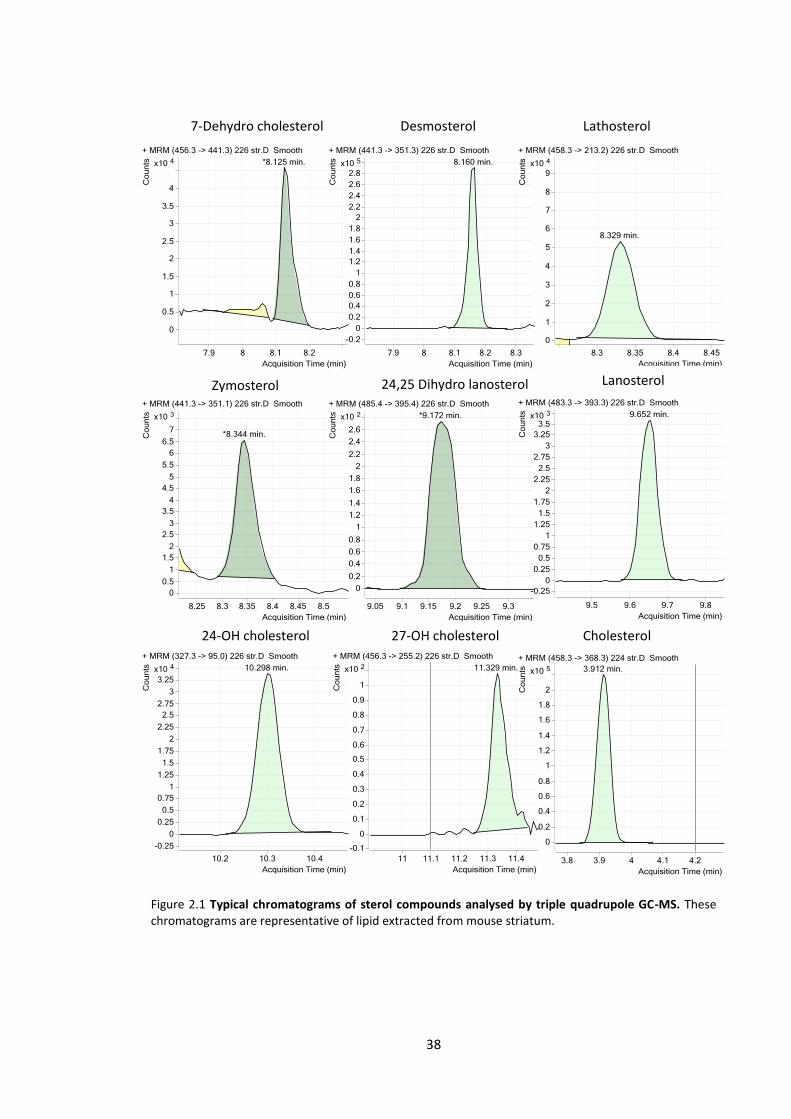
38
Figure 2.1 Typical chromatograms of sterol compounds analysed by triple quadrupole GC-MS. These chromatograms are representative of lipid extracted from mouse striatum.
7-Dehydro cholesterol Desmosterol Lathosterol
Zymosterol 24,25 Dihydro lanosterol Lanosterol
24-OH cholesterol 27-OH cholesterol Cholesterol
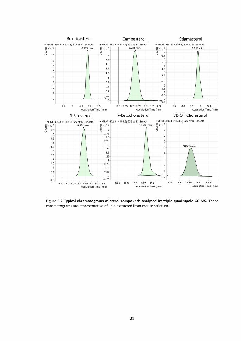
39
Figure 2.2 Typical chromatograms of sterol compounds analysed by triple quadrupole GC-MS. These chromatograms are representative of lipid extracted from mouse striatum.
Brassicasterol Campesterol Stigmasterol
β-Sitosterol 7-Ketocholesterol 7β-OH Cholesterol

40
Chapter 3
Association of cholesterol metabolism
with Huntington's disease progression in
R6/1 transgenic mice
41
3.1 Introduction
Significant research has been invested into determining HD pathology that involves a number of
neurophysiological changes that manifest as behavioural, motor and cognitive impairments.
These changes have been classified over the progression of the disease in humans and in a
limited number of mouse models. Distinct biochemical alterations are also present in the HD
brain. Among these changes is a defect in cholesterol biosynthesis and metabolism that has been
consistently demonstrated in several mouse models. These models include mice expressing an
expanded fragment of human HTT (R6/2), full length human HTT (YAC) and expanded mouse
hungtingtin (hdhQ111). Due to the complexity of the cholesterol synthetic pathway (Bloch, 1965),
previous studies have only measured a small number of synthetic intermediates and are unable
to describe the full extent of the alterations that occur.
In this chapter the R6/1 mouse model expressing a CAG expanded exon 1 of human HTT (~115-
120 repeats) was characterised in terms of cholesterol synthetic and metabolic changes. Physical
phenotypic changes were also examined including, impaired motor performance, weight loss,
reduced brain mass and involuntary clasping. Analysis of multiple time points and brain regions
aimed to establish when specific alterations to the cholesterol synthetic and metabolic pathways
occur, and the potential influence these have in the disease. A comprehensive analysis of sterol
compounds was also undertaken to identify any potential biomarkers associated with HD in R6/1
mice, to be used for future studies.
42
3.2 Materials and Methods
Materials: Materials used in this study are listed in 2.2.
Mice: R6/1 mice were generated as outlined in 2.3.1. Male and female mice (5-6 per group) were
housed in cages of 4-5 mice, with at least 2 mice of each genotype per cage. Standard laboratory
housing was provided (sawdust, pine shavings and a single PVC tube) with standard rodent diet
(see 2.3.1) and water available ad libitum. Mice were acclimatised over 1 week and this included
brief handling and weighing. All animal experiments were conducted with approval from the
University of Wollongong ethics committee (ethics number AE 13/20).
Body weight: Mouse weight was recorded every 4-5 days from 7 weeks of age until sacrifice.
Hind paw clasping: Hind paw clasping was tested every 5-10 days following the procedure
outlined in 2.3.3.
RotaRod: Motor performance was measured using the RotaRod protocol outlined in 2.3.2. The
RotaRod performance of mice was tested at 6, 11, 15, 19 and 23 weeks of age.
Tissue collection: Mice were sacrificed at 5 time points (6, 12, 20, 24 and 28 weeks) using slow
flow CO2 asphyxiation. Brain tissue was dissected and collected as described in 2.3.4.
Lipid extraction and GC-MS/MS analysis of sterols: Quantification of cholesterol synthetic
precursors, metabolites, oxidation products and phytosterols was performed as outlined in 2.3.5
and 2.3.6.
Statistical Analysis: Linear regression analysis was used to identify the rate of weight gain
between genotypes and identify the relationship between age and sterol changes in mice. A 2-
way ANOVA was used to analyse RotaRod performance, body weight, brain lipid and brain mass
data. Bonferroni post-tests were used to compare means at individual time points. A student’s
t test was also used to compare means at individual time points in the sterol data. P < 0.05 was

43
considered significant. All analyses were performed in Graphpad PRISM v5.0 (Graphpad
Software Inc., USA).
3.3 Results
3.3.1 Physical phenotype
The cohort of R6/1 and WT littermates housed until 28 weeks of age (n = 5 per genotype and
sex) were tested periodically for physical changes that characterise the progression of the HD
phenotype. These results are represented in Figures 3.1, 3.2 and 3.3.
3.3.1.1 Weight loss in R6/1 mouse
Mouse body weight was recorded every 4-5 days (Figure 3.1); male and female R6/1 body weight
was significantly reduced compared to WT mice over the course of the study (p < 0.0001). The
mean body weight of R6/1 mice did not increase after 16 weeks of age in males, and 19 weeks
in females. Prior to weight loss (7-19 weeks) the rate of weight gain was significantly less in
female R6/1 mice compared to WT (p = 0.00012). No significant difference in the rate of weight
gain was detected between male R6/1 and WT mice in the 6-16 week period (p = 0.073). By the
28 week time point the average weight increase in R6/1 mice from the earliest measurement (7
weeks) was 8% in males and 19% in females.

44
Figure 3.1 Weight loss in R6/1 mice during HD progression. R6/1 mice had significantly reduced body weight compared to WT over the course of the study (p < 0.0001) (2-way ANOVA). The R6/1 weight loss phenotype is more severe in male mice compared to females. Male R6/1 mice began to lose weight after 16 weeks compared to 19 weeks in females. The average weight gain over 28 weeks was less in male R6/1 mice (8%) compared to female R6/1 mice (19%). The rate of weight gain prior to weight loss was significantly slower in R6/1 females compared to WT (p = 0.00012) (linear regression analysis) while no difference was observed in male mice. n = 5 per group. Error bars represent + SEM.

45
3.3.1.2 Hind paw clasping phenotype
The hind paw clasping phenotype previously observed in R6/1 and R6/2 mice (Mangiarini et al.,
1996) was also tested over the course of the study (Figure 3.2). The clasping phenotype was first
observed at 12 weeks in males, with the percentage of mice showing positive for the phenotype
increasing to 60% at the end of the study. Female R6/1 mice first exhibited the clasping
phenotype at the age of 17 weeks. The percentage of female R6/1 mice showing the phenotype
increased to 60% by the end of the study. A small proportion of WT mice showed positive to a
"half clasp" phenotype at varying times throughout the study.
3.3.1.3 Motor performance
A RotaRod apparatus was used to test motor performance in R6/1 mice (Figure 3.3). The
average latency to fall from the RotaRod was significantly less in R6/1 mice compared to WT in
both males (p = 0.0016) and females (p = 0.0004) over the course of the study. A significant
difference at the 23 week time point was detected between male R6/1 mice and WT (p < 0.01).
No difference in the RotaRod performance was detected between sexes of the same genotype
(p = 0.81).
3.3.1.4 Brain mass
Mouse brain (extracted for sterol analysis at each time point) was weighed prior to dissection.
The brain mass of both male and female R6/1 mice was significantly less than WT littermates at
later time points (Figure 3.4). The difference between WT and R6/1 male mice was highly
significant at 20, 24 and 28 weeks (p < 0.0001). In female mice a significant difference was
detected at 12 and 20 weeks (p < 0.05), becoming highly significant at 24 and 28 weeks (p <
0.0001).
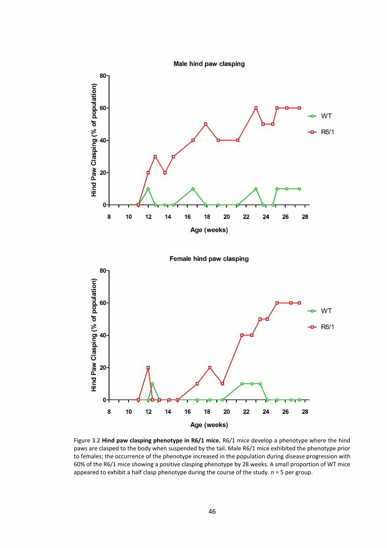
46
Figure 3.2 Hind paw clasping phenotype in R6/1 mice. R6/1 mice develop a phenotype where the hind paws are clasped to the body when suspended by the tail. Male R6/1 mice exhibited the phenotype prior to females; the occurrence of the phenotype increased in the population during disease progression with 60% of the R6/1 mice showing a positive clasping phenotype by 28 weeks. A small proportion of WT mice appeared to exhibit a half clasp phenotype during the course of the study. n = 5 per group.
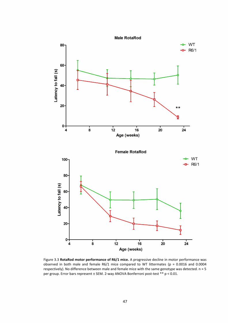
47
Figure 3.3 RotaRod motor performance of R6/1 mice. A progressive decline in motor performance was observed in both male and female R6/1 mice compared to WT littermates (p = 0.0016 and 0.0004 respectively). No difference between male and female mice with the same genotype was detected. n = 5 per group. Error bars represent ± SEM. 2-way ANOVA Bonferroni post-test ** p < 0.01.
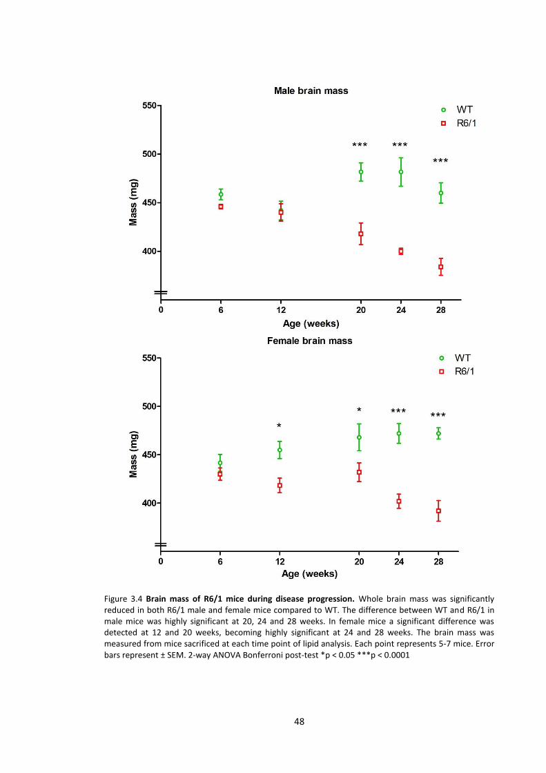
48
Figure 3.4 Brain mass of R6/1 mice during disease progression. Whole brain mass was significantly reduced in both R6/1 male and female mice compared to WT. The difference between WT and R6/1 in male mice was highly significant at 20, 24 and 28 weeks. In female mice a significant difference was detected at 12 and 20 weeks, becoming highly significant at 24 and 28 weeks. The brain mass was measured from mice sacrificed at each time point of lipid analysis. Each point represents 5-7 mice. Error bars represent ± SEM. 2-way ANOVA Bonferroni post-test *p < 0.05 ***p < 0.0001
49
3.3.2 Sterol analysis of R6/1 brain tissue
No major differences in sterols (cholesterol synthetic precursors, metabolites, oxidation
products and phytosterols) were found between sexes in R6/1 or WT mice. To increase statistical
power both sexes were combined and reanalysed. The level of all sterols measured for the
individual sexes can be found in Appendix 1-3. The following results summarise the data for
combined sexes. The sterol related compounds were detected in the brain tissue at
concentrations ranging from 0.1 – 80 ng.mg-1 tissue. This consistent with the several compounds
that have been previously investigated in mouse brain (Valenza et al., 2007b; Valenza et al.,
2010).
3.3.2.1 Cholesterol synthetic precursors
Several cholesterol synthetic precursors were significantly decreased in R6/1 striatum and
cortex compared to WT (Figures 3.5A-D, 3.6A-D). Lathosterol levels were significantly reduced
from the pre-symptomatic age of 6 weeks in R6/1 striatum (p < 0.001) and cortex (p < 0.05)
when compared to WT mice. Lathosterol was consistently reduced in both cortex and striatum
of R6/1 mice compared to WT in all later time points (12, 20, 24 and 28 weeks; p < 0.001).
Lanosterol and zymosterol followed a similar consistent reduction; a significant difference in
lanosterol levels was first detected at 12 weeks in striatum and cortex of R6/1 (cortex p < 0.05,
striatum p < 0.01). R6/1 striatum exhibited an early reduction in zymosterol at 6 weeks (p <
0.01), and at all later time points. The level of zymosterol in cortex tissue was only significantly
reduced compared to WT at 28 weeks of age (p < 0.05). The cholesterol synthetic precursor
24,25 dihydro lanosterol was not significantly altered in the R6/1 striatum or cortex although it
was detected at lower mean levels than WT at all time points examined.
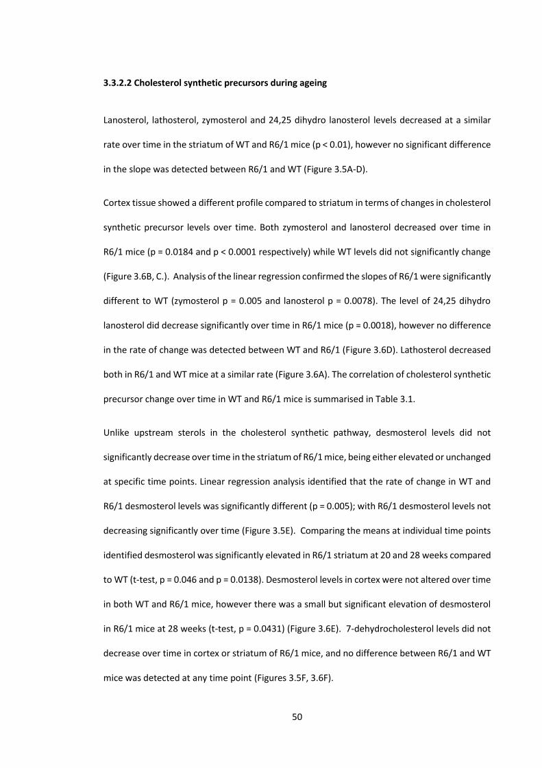
50
3.3.2.2 Cholesterol synthetic precursors during ageing
Lanosterol, lathosterol, zymosterol and 24,25 dihydro lanosterol levels decreased at a similar
rate over time in the striatum of WT and R6/1 mice (p < 0.01), however no significant difference
in the slope was detected between R6/1 and WT (Figure 3.5A-D).
Cortex tissue showed a different profile compared to striatum in terms of changes in cholesterol
synthetic precursor levels over time. Both zymosterol and lanosterol decreased over time in
R6/1 mice (p = 0.0184 and p < 0.0001 respectively) while WT levels did not significantly change
(Figure 3.6B, C.). Analysis of the linear regression confirmed the slopes of R6/1 were significantly
different to WT (zymosterol p = 0.005 and lanosterol p = 0.0078). The level of 24,25 dihydro
lanosterol did decrease significantly over time in R6/1 mice (p = 0.0018), however no difference
in the rate of change was detected between WT and R6/1 (Figure 3.6D). Lathosterol decreased
both in R6/1 and WT mice at a similar rate (Figure 3.6A). The correlation of cholesterol synthetic
precursor change over time in WT and R6/1 mice is summarised in Table 3.1.
Unlike upstream sterols in the cholesterol synthetic pathway, desmosterol levels did not
significantly decrease over time in the striatum of R6/1 mice, being either elevated or unchanged
at specific time points. Linear regression analysis identified that the rate of change in WT and
R6/1 desmosterol levels was significantly different (p = 0.005); with R6/1 desmosterol levels not
decreasing significantly over time (Figure 3.5E). Comparing the means at individual time points
identified desmosterol was significantly elevated in R6/1 striatum at 20 and 28 weeks compared
to WT (t-test, p = 0.046 and p = 0.0138). Desmosterol levels in cortex were not altered over time
in both WT and R6/1 mice, however there was a small but significant elevation of desmosterol
in R6/1 mice at 28 weeks (t-test, p = 0.0431) (Figure 3.6E). 7-dehydrocholesterol levels did not
decrease over time in cortex or striatum of R6/1 mice, and no difference between R6/1 and WT
mice was detected at any time point (Figures 3.5F, 3.6F).

51
Figure 3.5: Cholesterol synthetic precursor levels in striatum tissue of R6/1 and WT littermates. Extracted sterols were quantified using heavy isotope mass dilution GC-MS/MS. (A) Lathosterol, (B) lanosterol and (C) zymosterol levels were significantly reduced early in the R6/1 mice compared to WT. (D) 24,25 dihydro lanosterol showed a trend to be decreased in R6/1 mice however there was no significant difference between WT and R6/1 at any time point. Lanosterol, lathosterol, zymosterol and 24,25 dihydro lanosterol decreased over time in both R6/1 and WT mice (p < 0.01). Genotype had no effect on the rate of change over time. The rate of change over time in (E) desmosterol levels in WT and R6/1 was significantly different (p = 0.005). Desmosterol did not significantly decline in R6/1 mice, and was significantly elevated at 20 and 28 weeks compared to control. (F) 7-Dehydrocholesterol did not significantly decline in R6/1 mice and no significant differences were detected between R6/1 and WT at any time point examined. Each data point represents combined results from male and female mice, n = 10-13 per group. Error bars represent ± SEM. 2-way ANOVA Bonferroni post-test *p < 0.05 **p < 0.01 ***p < 0.0001. t-test Ψp < 0.05
A B
C
E F
D

52
Figure 3.6:
Figure 3.6: Cholesterol synthetic precursor levels in cortex tissue of R6/1 and WT littermates. Extracted sterols were quantified using heavy isotope mass dilution GC-MS/MS. (A) Lathosterol and (B) lanosterol levels were significantly reduced early in R6/1 mice compared to WT. (C) Zymosterol was significantly reduced in R6/1 at the end time point. (D) 24,25 dihydro lanosterol had a trend to be reduced however there was no significant difference between WT and R6/1 at any time point. Zymosterol and lanosterol decreased in R6/1 over time while WT levels remained unchanged (p < 0.01). Lathosterol levels decreased both in R6/1 and WT at a similar rate. 24,25 dihydro lanosterol levels were not detected to change over time. The rate of change in (E) desmosterol levels over time was not significantly different between R6/1 and WT; however desmosterol was elevated in R6/1 mice at 28 weeks. (F) 7-Dehydrocholesterol did not significantly decline in R6/1 mice and no significant differences were detected between R6/1 and WT at any time point examined. Each data point represents combined results from male and female mice, n = 10-13 per group. Error bars represent ± SEM. 2-way ANOVA Bonferroni post-test *p < 0.05 **p < 0.01 ***p < 0.0001. t-test Ψp < 0.05
A B
C D
E F
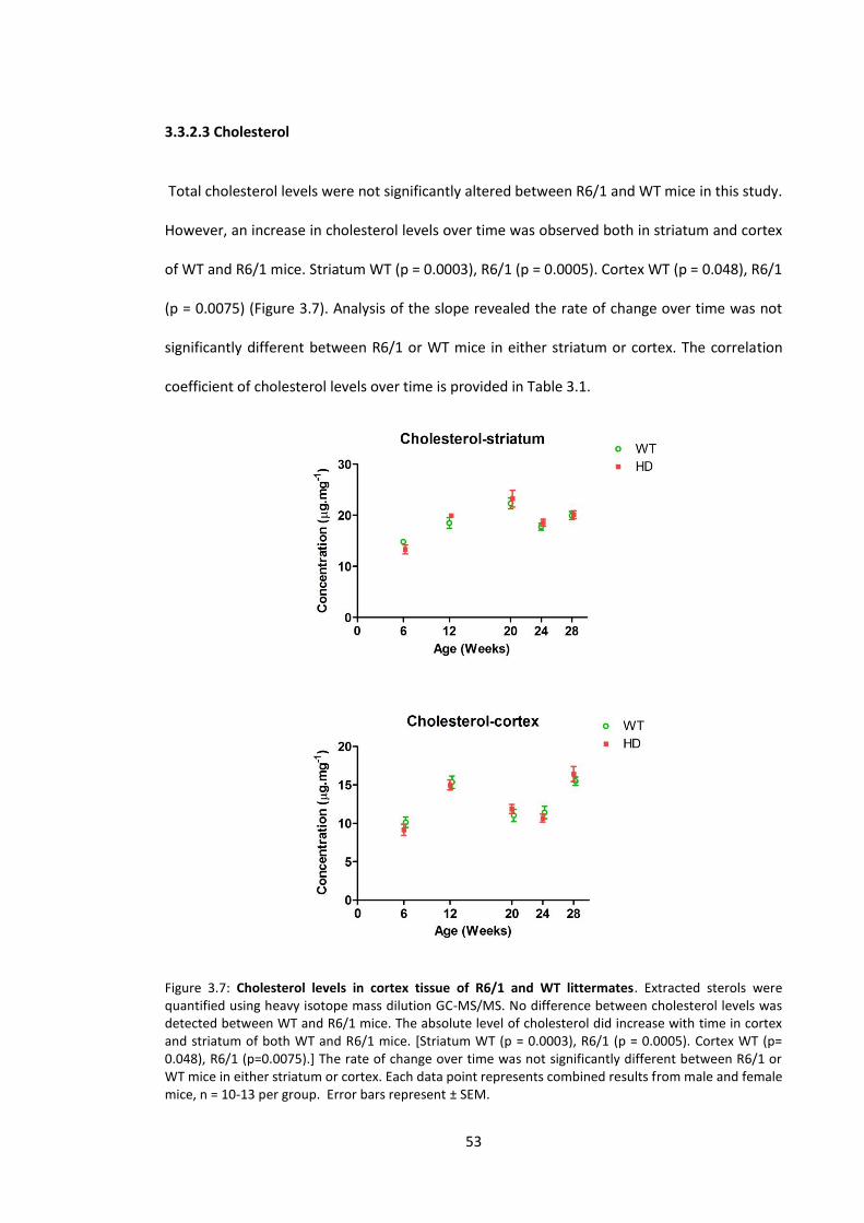
53
3.3.2.3 Cholesterol
Total cholesterol levels were not significantly altered between R6/1 and WT mice in this study.
However, an increase in cholesterol levels over time was observed both in striatum and cortex
of WT and R6/1 mice. Striatum WT (p = 0.0003), R6/1 (p = 0.0005). Cortex WT (p = 0.048), R6/1
(p = 0.0075) (Figure 3.7). Analysis of the slope revealed the rate of change over time was not
significantly different between R6/1 or WT mice in either striatum or cortex. The correlation
coefficient of cholesterol levels over time is provided in Table 3.1.
Figure 3.7: Cholesterol levels in cortex tissue of R6/1 and WT littermates. Extracted sterols were quantified using heavy isotope mass dilution GC-MS/MS. No difference between cholesterol levels was detected between WT and R6/1 mice. The absolute level of cholesterol did increase with time in cortex and striatum of both WT and R6/1 mice. [Striatum WT (p = 0.0003), R6/1 (p = 0.0005). Cortex WT (p= 0.048), R6/1 (p=0.0075).] The rate of change over time was not significantly different between R6/1 or WT mice in either striatum or cortex. Each data point represents combined results from male and female mice, n = 10-13 per group. Error bars represent ± SEM.
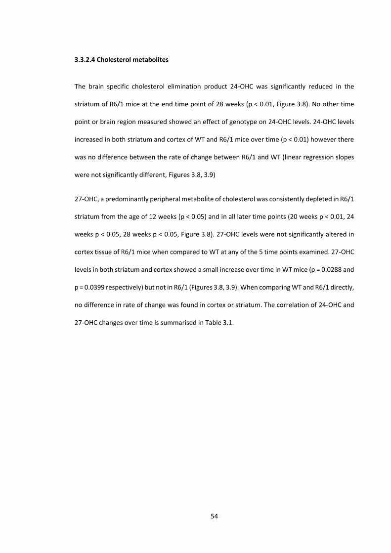
54
3.3.2.4 Cholesterol metabolites
The brain specific cholesterol elimination product 24-OHC was significantly reduced in the
striatum of R6/1 mice at the end time point of 28 weeks (p < 0.01, Figure 3.8). No other time
point or brain region measured showed an effect of genotype on 24-OHC levels. 24-OHC levels
increased in both striatum and cortex of WT and R6/1 mice over time (p < 0.01) however there
was no difference between the rate of change between R6/1 and WT (linear regression slopes
were not significantly different, Figures 3.8, 3.9)
27-OHC, a predominantly peripheral metabolite of cholesterol was consistently depleted in R6/1
striatum from the age of 12 weeks (p < 0.05) and in all later time points (20 weeks p < 0.01, 24
weeks p < 0.05, 28 weeks p < 0.05, Figure 3.8). 27-OHC levels were not significantly altered in
cortex tissue of R6/1 mice when compared to WT at any of the 5 time points examined. 27-OHC
levels in both striatum and cortex showed a small increase over time in WT mice (p = 0.0288 and
p = 0.0399 respectively) but not in R6/1 (Figures 3.8, 3.9). When comparing WT and R6/1 directly,
no difference in rate of change was found in cortex or striatum. The correlation of 24-OHC and
27-OHC changes over time is summarised in Table 3.1.

55
Figure 3.8: Cholesterol metabolites in striatum tissue of R6/1 and WT littermates. Extracted sterols were quantified using heavy isotope mass dilution GC-MS/MS. 24(S)-Hydroxycholesterol was significantly reduced in striatum of R6/1 mice compared to WT at the end stage of the study. A significant reduction in 27-hydroxycholesterol levels was detected early (12 weeks), and was consistently reduced until 28 weeks. The level of 24-OHC increased during ageing in both WT and R6/1 mice (p < 0.01), the rate of change was not different between genotypes. Each data point represents combined results from male and female mice, n = 10-13 per group. Error bars represent ± SEM. Bonferroni post-test *p < 0.05 **p < 0.01
56
Figure 3.9: Cholesterol metabolites in cortex tissue of R6/1 and WT littermates. Extracted sterols were quantified using heavy isotope mass dilution GC-MS/MS. R6/1 cortex did not have significantly altered levels of 24-OHC or 27-OHC at any time point examined. 24-OHC levels increased in WT and R6/1 mice during ageing (p < 0.01), the rate of change was not different between genotypes. Each data point represents combined results from male and female mice, n = 10-13 per group. Error bars represent ± SEM.
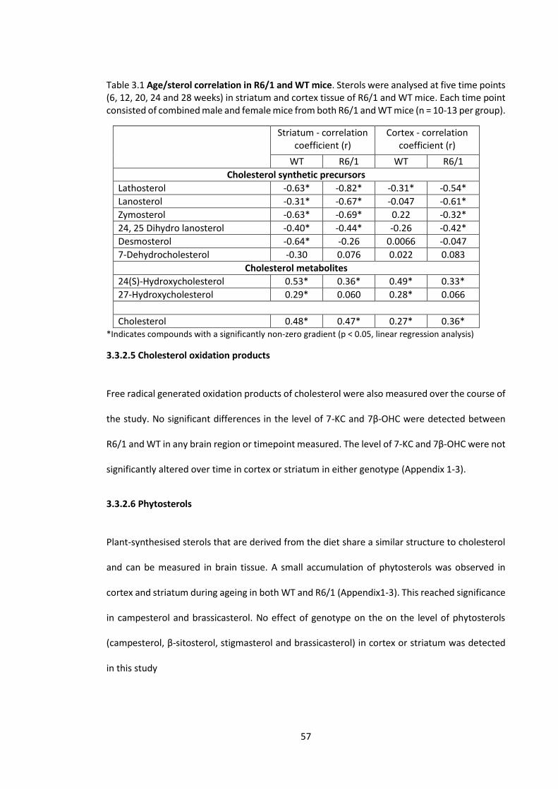
57
Table 3.1 Age/sterol correlation in R6/1 and WT mice. Sterols were analysed at five time points (6, 12, 20, 24 and 28 weeks) in striatum and cortex tissue of R6/1 and WT mice. Each time point consisted of combined male and female mice from both R6/1 and WT mice (n = 10-13 per group).
Striatum - correlation coefficient (r)
Cortex - correlation coefficient (r)
WT R6/1 WT R6/1
Cholesterol synthetic precursors
Lathosterol -0.63* -0.82* -0.31* -0.54*
Lanosterol -0.31* -0.67* -0.047 -0.61*
Zymosterol -0.63* -0.69* 0.22 -0.32*
24, 25 Dihydro lanosterol -0.40* -0.44* -0.26 -0.42*
Desmosterol -0.64* -0.26 0.0066 -0.047
7-Dehydrocholesterol -0.30 0.076 0.022 0.083
Cholesterol metabolites
24(S)-Hydroxycholesterol 0.53* 0.36* 0.49* 0.33*
27-Hydroxycholesterol 0.29* 0.060 0.28* 0.066
Cholesterol 0.48* 0.47* 0.27* 0.36* *Indicates compounds with a significantly non-zero gradient (p < 0.05, linear regression analysis)
3.3.2.5 Cholesterol oxidation products
Free radical generated oxidation products of cholesterol were also measured over the course of
the study. No significant differences in the level of 7-KC and 7β-OHC were detected between
R6/1 and WT in any brain region or timepoint measured. The level of 7-KC and 7β-OHC were not
significantly altered over time in cortex or striatum in either genotype (Appendix 1-3).
3.3.2.6 Phytosterols
Plant-synthesised sterols that are derived from the diet share a similar structure to cholesterol
and can be measured in brain tissue. A small accumulation of phytosterols was observed in
cortex and striatum during ageing in both WT and R6/1 (Appendix1-3). This reached significance
in campesterol and brassicasterol. No effect of genotype on the on the level of phytosterols
(campesterol, β-sitosterol, stigmasterol and brassicasterol) in cortex or striatum was detected
in this study
58
3.4 Discussion
Brain cholesterol homeostasis is maintained by de novo synthesis and brain-specific excretion
pathways. Brain cholesterol is also isolated from peripheral cholesterol levels as the BBB is
impermeable to cholesterol (Bjorkhem & Meaney, 2004). These regulatory mechanisms indicate
that maintaining cholesterol levels is essential for normal brain function. This is indeed the case
as cholesterol has essential functions involving synaptogenesis (Mauch et al., 2001), axon
growth (Hayashi et al., 2004), membrane fluidity (Yeagle, 1985) and lipid raft structure relevant
to cell signalling (Sheets et al., 1999; Zajchowski & Robbins, 2002; Kannan et al., 2007). Genetic
defects in cholesterol synthetic enzymes also cause severe neurological impairment in diseases
such as Smith-Lemli-Optitz (Wassif et al., 1998) and desmosterolosis (Waterham et al., 2001).
A defect in cholesterol synthesis was first described in HD transgenic cells that identified several
genes involved in cholesterol biosynthesis downregulated, including HMGCR and DHCR7
(Sipione et al., 2002). Several HD mouse models have also been generated that express either:
an expanded fragment of human HTT (Mangiarini et al., 1996), an expanded full-length human
HTT (Hodgson et al., 1999; Slow et al., 2003), or an expanded mouse huntingtin (Wheeler et al.,
1999). A similar deficit in cholesterol biosynthesis has generally been reported in these models,
identified through a reduction of cholesterol synthetic precursors (Valenza et al., 2005; Valenza
et al., 2007a; Valenza et al., 2007b; del Toro et al., 2010; Valenza et al., 2010) and, in some cases,
total cholesterol levels (Valenza et al., 2007a).
The current literature has mostly used the R6/2 transgenic mouse model that expresses exon 1
of human HTT with a 140-150 CAG repeat and has a rapid onset of symptoms. This mouse has
been characterised in terms of phenotypic progression and potential changes in only a limited
number of cholesterol synthetic precursors and metabolites. We have however, chosen to
examine the R6/1 model which expresses a shorter CAG expansion (115-120) of exon 1 HTT. The
R6/1 mouse has a later onset of motor dysfunction and a longer life span, therefore providing a
59
model that more accurately reflects the late onset and long timeline (15-30 years) of disease in
human patients (Foroud et al., 1999). The R6/1 model has already been characterised in terms
of behavioural phenotype and some molecular changes including, glycosphingolipid
abnormalities (Denny et al., 2010), gene expression profile alterations (Hodges et al., 2008),
cannabinoid receptor changes (Naver et al., 2003) and serotonin transporter deficits (Pang et
al., 2009). Cholesterol synthesis and metabolism has not been comprehensively examined prior
to experiments undertaken in this chapter.
3.4.1 Cholesterol synthesis and metabolism
Previous studies examining brain cholesterol changes in mouse models typically investigated a
limited number of compounds at one or two time points during disease progression, in a single
brain region or whole brain homogenate. In addition to this, potential differences between sexes
have not been explored. A comprehensive analysis is required to assess whether alterations of
cholesterol synthesis are a driving factor or a secondary event involved in HD pathogenesis.
In this study we have analysed cortex and striatum in male and female R6/1 mice at 5 time points
over the course of the disease, from a pre-symptomatic age (6 weeks) to end stage (28 weeks).
Mass dilution GC-MS/MS analysis was used to quantify a larger number of cholesterol synthetic
precursors, major metabolites, cholesterol oxidation products and phytosterols. This is the first
study to comprehensively examine sterol changes in the R6/1 mouse, and the most
comprehensive sterol analysis of any HD mouse model to date.
3.4.1.1 Cholesterol synthetic precursors
Significant differences between sexes were not detected in the sterol profile of either R6/1 or
WT mice, and for simplicity the following discussion describes changes occurring when sexes
were combined. Analysis of combined sexes also increased the statistical power, and detected
significance in compounds exhibiting small changes. Although this study was not able to detect
60
differences between sexes in sterol levels of R6/1 mice, increasing the "n" number for future
studies may identify small but significant differences that may explain the differences in the
behavioural phenotype observed between sexes.
Sterol analysis of R6/1 cortex and striatum showed a significant reduction in several cholesterol
synthetic precursors over the course of the disease. The significant reduction of lathosterol and
zymosterol in the striatum of R6/1 mice at 6 weeks of age represents the earliest detectable
change in the R6/1 sterol profile, and appears before the onset of overt motor dysfunction
measured on the RotaRod. Changes to zymosterol are more specific to the striatum compared
to other precursors that decrease early in both cortex and striatum. This highlights zymosterol
as a possible specific biomarker of early striatal changes in HD mice. 24,25 Dihydro lanosterol
levels were not significantly altered between R6/1 and WT mice however the mean
concentration in R6/1 mice was consistently lower than WT at all five time points. This sterol is
found at low concentrations in the brain (0.1-0.4 ng/mg tissue), and while the GC-MS/MS
method we have developed can sensitively detect this compound at these levels, it may be
prone to greater concentration variation between individuals at any time, and statistically
significant differences would therefore be difficult to detect with the current study size.
Synthetic precursor levels were more severely affected in striatum compared to cortex in our
study; changes occurred earlier and were of a greater magnitude in striatum (lathosterol
decreased by 80% in striatum compared with 50% in cortex). This observation is consistent with
previous studies in the YAC and R6/2 models (Valenza et al., 2007a; Valenza et al., 2007b), as
well as the striatum in humans being the earliest and most severely affected brain region in HD
(Vonsattel et al., 1985). Absolute cholesterol and cholesterol synthetic precursor levels were
also previously shown to be higher in striatum compared to cortex (Zhang et al., 1996; Valenza
et al., 2007b). For this reason it has been hypothesised that disturbed cholesterol homeostasis
in HD may lead to specific vulnerability of the striatum (Valenza et al., 2007b). Significant sterol

61
changes early in the striatum of the R6/1 mouse suggest that alterations to the cholesterol
biosynthetic pathway in the brain occur very early; with the possibility that these changes may
even be present during embryonic development. This has implications for brain development as
the majority of brain cholesterol is synthesised during prenatal life (Dietschy & Turley, 2004). If
this is the case in HD, neurodegeneration may be seeded early with individuals only becoming
symptomatic later in life.
A current hypothesis suggests that errors in myelination during development may be a factor
in human HD, taking many years to manifest before onset due to compensatory mechanisms
that function early but become overwhelmed in the aged brain (Bartzokis et al., 2007).
Alternatively, early changes may be independent of the pathological processes in HD and
investigating changes that occur during overt motor dysfunction may provide further insight into
the involvement of altered cholesterol synthesis in the disease.
This study has also examined 7-dehydrocholesterol and desmosterol; the immediate precursors
to cholesterol in the Kandutsch-Russell and Bloch pathways respectively (Figure 1.2). Contrary
to a previous finding that desmosterol decreases in YAC 128 mice (Valenza et al., 2007a),
desmosterol levels were significantly elevated in R6/1 striatum and cortex by the end stage in
our study. This may be an artefact of the different mouse models expressing different forms of
mHTT, however it may also be due to the previous study using single quadrupole GC-MS (Valenza
et al., 2007a), a technique that has lower specificity in discriminating between similar sterols
(especially 7-dehydrocholesterol) compared to GC-MS/MS used in our study. However, it is
unlikely that the elevated desmosterol levels we observe are due to a higher throughput of
synthesis, as cholesterol levels did not increase in our study or in a previous study examining
R6/1 mice (del Toro et al., 2010). A plausible explanation for this accumulation is the reduced
activity of the enzyme (DHCR24) that catalyses the conversion of desmosterol to cholesterol.
Previous studies in R6/2 mice do not report desmosterol levels and therefore it is unknown if

62
desmosterol accumulation also occurs in the longer exon 1 CAG repeat mouse model (Valenza
et al., 2007b). 7-Dehydrocholesterol, similarly to desmosterol did not decrease over time as seen
in upstream precursors, and the levels were not significantly altered between WT and R6/1 mice
at any time point or brain region. Considering lathosterol levels are substantially reduced (the
immediate precursor of 7-dehydrocholesterol) and 7-dehydrocholesterol levels were
unchanged, we suggest that the enzyme that converts 7-dehydrocholesterol to cholesterol
(DHCR7) may also be less active or at lower abundance in R6/1 mice. Our current study gives a
novel insight into 7-dehydrocholesterol levels which have not been previously reported in HD
mouse brain; most likely due to analytical difficulties in resolving the structures of desmosterol
and 7-dehydrocholesterol, achieved only in this study by high resolution GC-MS/MS.
Previously it has been suggested that the absolute level of cholesterol synthetic precursors
(lathosterol and lanosterol) in the brain are markers of cholesterol synthesis (Thelen et al., 2006;
Valenza et al., 2007b), however our results which describe an accumulation of penultimate
precursors suggest there is likely to be more complex regulation at various points of the
synthetic pathway than previously believed. How this regulation affects lanosterol and
lathosterol levels is unknown, and therefore the measurement of these precursors alone to
interpret synthetic rate may be potentially confounding. The regulation of "post-squalene"
cholesterol synthesis has been investigated recently showed that the two terminal enzymes in
the pathway interact functionally, DHCR24 regulating the activity of DHCR7 (Luu et al., 2015).
The idea of synthetic enzymes interacting physically supports the hypothesis of a
"cholestesome" of cholesterol synthesis; which would involve a discrete group of enzymes
acting on cholesterol synthetic precursors as they are converted to a final product (Sharpe &
Brown, 2013). DHCR24 is also believed to have further regulatory roles upstream in the pathway
(Luu et al., 2015), however these roles have not been fully established. It is clear that the
cholesterol synthetic pathway has more complex regulation than previously believed, and while

63
recent studies have identified synthetic enzyme interactions in cells derived from the periphery,
the specific details of cholesterol synthetic regulation remains to be fully understood in the
brain.
3.4.1.2 Age related changes to cholesterol synthetic precursors
Cholesterol synthetic precursors lanosterol, lathosterol, zymosterol and 24, 25 dihydro
lanosterol in the striatum decrease at a similar rate during ageing in WT and R6/1 mice. This was
also evident in cortex however the correlation was weaker. Consistent with our results in mouse
brain, a negative correlation in lathosterol and lanosterol levels has been observed in human
brain tissue during ageing (r = -0.5) (Thelen et al., 2006). Whether this suggests an overall
decrease in cholesterol levels with ageing is discussed below (3.4.1.4).
Interestingly 7-dehydrocholesterol levels did not decline over time in WT or R6/1 mice in cortex
or striatum in our study. In contrast, desmosterol levels declined in WT striatum over 28 weeks,
while remaining stable in R6/1. The regional specificity of this change highlights desmosterol
accumulation (or reduction in DHCR24 levels or activity), as a more specific indicator of
cholesterol synthetic alterations in the R6/1 mouse compared to upstream cholesterol synthetic
precursor changes. Desmosterol, as measured in this study may also represent a reliable marker
of DHCR24 activity in brain tissue of HD, or other neurodegenerative disorders.
3.4.1.3 Cholesterol levels in the R6/1 mouse
Our study did not detect any significant alterations in the cholesterol levels of the striatum and
cortex of R6/1 mice compared to WT at any time point or brain region analysed. This is
consistent with previous studies in the striatum of R6/1 (del Toro et al., 2010) and R6/2 mice
(Valenza et al., 2007b). Previous studies also describes conflicting results; a small but significant
decrease in cholesterol levels was measured in whole brain homogenates of YAC 128 mice at 10
months of age (Valenza et al., 2007a), and a significant increase of striatal cholesterol was
64
detected in HdhQ111 mice (del Toro et al., 2010). The apparent contradictions in cholesterol level
changes previously reported could be explained by several factors. The different mouse models
varying in the length and species of huntingtin expressed (YAC, R6 - human HTT, HdhQ111-mouse
huntingtin), may cause different metabolic effects that alter total cholesterol in the brain.
Detection and quantification methodologies (enzymatic, colorimetric, mass spectrometry) and
sample preparation differs between studies, and have been shown to affect the result obtained
(Marullo et al., 2012). Of these methods, mass spectrometry was previously found to be the
most sensitive and reliable method for cholesterol quantification (Marullo et al., 2012). Tissue
samples are also varied between studies; from discrete brain regions such as the striatum, to
whole brain homogenates. Different brain regions vary in total volume and cholesterol content
(10-30 µg/mg tissue) (Zhang et al., 1996; Valenza et al., 2007b). The use of whole brain
homogenates may obscure changes in discrete regions as well as potentially giving a false
positive if non-disease-specific changes occur in a large brain region. Analysis of brain
cholesterol in HD models would benefit from the use of standardised methods in collecting
discrete brain regions and using specific analytical methods such as a mass spectrometry
method developed by our laboratory (Abbott et al., 2013). This would allow more accurate
comparisons to be made between laboratories studying different HD models.
Unchanged levels of cholesterol in the R6/1 brain compared to WT that we observe does not
necessarily rule out an altered cholesterol synthetic rate in this tissue. The concentration of
cholesterol in the brain is relatively high (μg.mg-1), and the rate of synthesis slow in the adult
brain (ng.h-1.mg-1) (Spady & Dietschy, 1983), therefore significant alterations to total cholesterol
levels may not take place during the short life span of these mice. This idea is consistent with
total cholesterol changes only being observed in models with a longer lifespan (Valenza et al.,
2010).
65
Metabolism of cholesterol to 24-OHC occurs mainly in neurons (Lund et al., 1999) and altered
rates of metabolism may compensate for declining synthesis which is discussed in more detail
in 3.4.1.5. It is also possible that although the concentration of cholesterol does not change in
whole tissue samples, cholesterol is redistributed within the cell as previously observed in
cultured striatal cells derived from the R6/2 mouse model (del Toro et al., 2010). The discrete
cellular and regional pools of cholesterol in the brain are diverse and vary in concentration.
Myelin contains 70-80% of cholesterol in the brain (Norton & Autilio, 1965; Snipes & Suter,
1997); it is formed early in development and is possible that this is not greatly affected by the
HD mutation. Neuronal cholesterol and that synthesised in astrocytes represents a smaller mass
of cholesterol however is more dynamic (Bjorkhem et al., 1997). Small alterations in cholesterol
formation and delivery between astrocyte and neuron may not be easily detected in whole
tissue samples and is one limitation of examining whole tissue. Ideally, individual cell types could
be examined from in vivo sources, however this is technically challenging. While in vitro cell
culture experiments provide the ability to control cell types and the extracellular environment,
essential cholesterol shuttling between astrocytes and neurons as well as ageing, cannot be
accurately replicated. This is an important consideration when examining cholesterol
homeostasis in vitro.
3.4.1.4 Age related alteration to cholesterol levels in the brain
Interestingly a small, but significant increase in cholesterol was observed over time in the
striatum and cortex of WT and R6/1 mice over the 28 week study. Mice in this study are not
significantly aged (28 weeks old, representing early adulthood) and therefore it is difficult to
deduce the influence of further ageing on cholesterol levels in these mice. Striatal cholesterol
levels have been reported to increase during ageing in rats; regional age-related differences
were also noted with cerebellum and hippocampus remaining unchanged (Zhang et al., 1996).
Whether cholesterol levels and the biosynthetic rate changes over time in the human brain has

66
not been established; too few studies have examined brain cholesterol during ageing, those that
do have found unchanged levels (Soderberg et al., 1990; Thelen et al., 2006), or a decrease in
cholesterol (Svennerholm et al., 1994). Cholesterol synthetic precursors have been shown to
decrease with age, however a significant alteration of cholesterol levels was not detected due
substantial variation between individuals (Thelen et al., 2006). Previous inconclusive findings are
likely the result of brain regional differences, high variation within the human population and
lower "n" numbers of post-mortem tissues studied in different publications. An association
between cholesterol homeostasis and AD have been highlighted previously (Cutler et al., 2004;
Gamba et al., 2011; Gamba et al., 2014) ; however, no consensus has been reached regarding
cholesterol changes in these settings. Previous literature examining cholesterol changes in
human HD brain is similarly inconclusive, probably due to low numbers of samples and the high
variation between samples leading to a loss of statistical power (del Toro et al., 2010). While
total cholesterol levels in tissue may be unchanged, altered distribution of cholesterol within
the cell may have a very significant influence. Studies depleting cholesterol in cell membranes
observe significant changes to cell signalling (Kabouridis et al., 2000). Primary striatal cultures of
HD mice also show increased cholesterol concentration at the cell membrane while the overall
concentration remains unaltered (del Toro et al., 2010). These findings are potentially important
and require more investigation in the HD context.
3.4.1.5 Cholesterol metabolite changes in ageing and Huntington’s disease
24-OHC has been marked as a molecule of interest after being identified as a major and specific
elimination product of cholesterol from the brain (Lutjohann et al., 1996; Bjorkhem et al., 1998).
24-OHC is formed enzymatically from CNS cholesterol catalysed by CYP46A1, which is primarily
expressed in neurons (Lund et al., 1999). 24-OHC levels in plasma have been suggested as a
biomarker of several neurodegenerative diseases including AD (Bretillon et al., 2000b), PD
(Bjorkhem et al., 2013), multiple sclerosis (Teunissen et al., 2003) and recently HD (Leoni et al.,
67
2008; Leoni et al., 2013). Our study identified a significant (25%) reduction of 24-OHC levels in
the R6/1 striatum at 28 weeks of age. We believe the relative decline of 24-OHC in R6/1 mice
compared to WT is progressive over the course of the disease becoming more pronounced at
later stages. Previous studies have also identified a reduction of this metabolite in the brain and
plasma of several other HD mouse models (Valenza et al., 2007b; Valenza et al., 2010).
Aside from differences between genotypes, 24-OHC levels increase in cortex and striatum of
R6/1 and WT during ageing. This data supports the finding that CYP46A1 enzyme levels increases
during ageing in the mouse brain (Lund et al., 1999). Human CYP46A1 does not show such a
strong age related increase (Lund et al., 1999), this is consistent with plasma 24-OHC
measurements in humans being independent of age after the 2nd decade (Lutjohann et al.,
1996). 24-OHC levels during ageing in the brain have not been measured extensively and our
data constitutes the first evidence of age-related 24-OHC accumulation in HD mouse brain.
Age-related changes during HD are important to examine, as measuring only a single time point
can produce potentially misleading results. When examining one time point in our study, the
reduction of 24-OHC in R6/1 mice could be assumed to compensate for reduced levels of
cholesterol synthetic precursors, therefore maintaining equal cholesterol levels between WT
and R6/1 mice. However by examining multiple time points in our study we have identified that
both 24-OHC and total cholesterol levels increase with age in both R6/1 and WT mice. This
suggests a more complex system maintaining cholesterol levels, potentially involving another
mechanism that regulates cholesterol excretion from the brain via ApoE (Pitas et al., 1987b).
ApoE has been reported to be altered during neurodegeneration (Roses, 1996), and evidence
from mouse models suggests that ApoE is reduced during HD pathology (Valenza et al., 2010).
ApoE has been suggested to have neuroprotective properties (Pedersen et al., 2000; Horsburgh
et al., 2002), with expression trending to increase during ageing (Masliah et al., 1996). Potential
interactions of 24-OHC and ApoE have not been explored in HD, however this represents

68
another possible influence of altered cholesterol metabolism observed in the HD brain. Further
investigation is required to elucidate the underlying mechanisms responsible for cholesterol
metabolic changes during ageing; these studies will ultimately provide greater insight into lipid
alterations occurring during neurodegeneration that typically takes place in the aged brain.
A predominantly peripheral metabolite of cholesterol, 27-OHC, was also significantly reduced in
R6/1 striatum by the age of 12 weeks, while cortex tissue had no significant alterations. 27-OHC
has a net movement from circulation into the brain (Heverin et al., 2005) and reduced levels
seen in R6/1 might be explained by a whole body metabolic dysfunction that is known to occur
in HD (Lodi et al., 2000; Chen et al., 2007). However, this is unlikely as cortex did not exhibit the
same reduction of 27-OHC levels observed in striatum. It is possible that enzymes that further
metabolise 27-OHC are upregulated, or enzymes capable of forming 27-OHC in brain are
downregulated in striatum, however we have no experimental evidence to support this, and
requires further investigation.
3.4.1.6 Cholesterol oxidation product changes in ageing and HD
Oxidation of cholesterol in the 5,6 position generates 7β-OHC and 7-KC. These products
represent stable and reliable markers of cholesterol oxidation, previously assayed in plasma
(Iuliano et al., 2003). In contrast to our results that showed no change in COPs, a study in the
striatum of R6/1 mice did observe a time dependant increase of lipid peroxidation (Perez-
Severiano et al., 2000). While the different methodology used (lipid soluble fluorescence vs GC-
MS/MS in our study) may derive a different result, we would expect that as cholesterol is highly
abundant in the brain, the presence of oxidative stress would increase the formation of the COPs
we have measured. Until multiple laboratories using reliable analytical techniques can
consistently demonstrate the presence of oxidative stress in the brain of R6/1 mice, the role of
lipid peroxidation in the HD pathogenesis of this mouse model is still in question.
69
3.4.1.7 Phytosterol changes during ageing and HD
Phytosterols are derived from the diet and share a similar structure to cholesterol. They are
known to slowly accumulate in brain and other peripheral tissues (Plat et al., 2008). Phytosterol
levels were variable between R6/1 and WT over the course of the study and no significant effect
of genotype was observed. A small accumulation of campesterol and brassicasterol over time in
both WT and R6/1 mice was observed in both regions, consistent with previous studies in rats
(Plat et al., 2008). As these compounds can only be taken up from the circulation, they may
represent a useful post mortem indicator of a disturbed BBB that would result in higher
phytosterols levels present in HD brain tissue.
3.4.2 Physical phenotypic changes in the R6/1 mouse
In this study we have also investigated the R6/1 phenotype in detail over the course of the
disease, from 6 to 28 weeks of age. Previous literature has typically studied either male or female
R6/1 mice, in separate studies. Here we have examined both sexes in parallel to more directly
assess sex differences in the HD phenotype that have been previously reported.
3.4.2.1 Weight loss
Human and mouse metabolism is quite different yet the CAG expansion in transgenic R6/1 mice
causes weight loss in line with observations in human HD patients (Sanberg et al., 1981; Morales
et al., 1989). Weight loss may not be directly involved in neuronal dysfunction; however
monitoring the effect of mHTT on peripheral metabolism may give an insight into relevant
effects in the brain. Consistent with previous literature we have observed a milder weight loss
phenotype in females compared to male R6/1 mice (Clifford et al., 2002; Naver et al., 2003). In
our study female weight gain in R6/1 mice was significantly slower than WT mice prior to weight
loss suggesting a metabolic abnormality from early life; however after a certain age a dramatic
weight loss is seen. Due to a high variation in body weight of male R6/1 mice, a significant
70
difference in weight gain prior to weight loss was not detected. Food intake of R6/1 mice was
not measured in this study to reduce stress on animals being isolated and reunited. We do not
believe the primary cause of weight loss is a lack of feeding as it has been reported that R6/1
mice have a similar food intake to WT mice however still exhibit a distinct weight loss profile
(Mangiarini et al., 1996), similar to that described in our study.
3.4.2.2 Hind paw clasping phenotype
The hind paw clasping phenotype measures an involuntary clasping of the hind paws when R6/1
and R6/2 mice are suspended by the tail (Mangiarini et al., 1996). Female mice had a later onset
of hind paw clasping with a lower percentage of mice exhibiting the phenotype until the very
end of the study where 60% of both male and female R6/1 mice showed a positive phenotype.
Hind paw clasping has been described previously, however the onset is variable. Mangiarini et
al. (1996) and Naver et al. (2003) observe hind paw clasping from 16-20 weeks in R6/1 mice,
while other studies have observed the phenotype much later at 30 weeks (Clifford et al., 2002).
In this study we have categorised hind paw clasping into a full clasp or half clasp. A half clasp is
a less severe phenotype, where one or two hind paws are clasped in an interrupted manner.
This behaviour was seen prior to full clasping (two paws clasped continuously) and was used to
create a distinction between the severities of clasping. Although hind paw clasping is limited to
producing binary data of 'positive' or 'negative', it is a fast and simple test that is able to examine
a reflex response that is not measured by the RotaRod.
3.4.2.3 Motor performance
During the progression of disease in the R6/1 mouse, a progressive motor dysfunction was
observed and this was quantified using data derived from the RotaRod experiments. Both male
and female R6/1 mice showed progressive loss of motor skills that were quantified by the time
taken to fall from a rotating, accelerating rod. While all mice were treated under the same
71
conditions, variation between mice of the same test group did exist, and therefore any sex
differences in the progression of the motor phenotype could not be conclusively demonstrated.
However, the impaired motor performance at the end time point was comparable between
male and female R6/1 mice.
The RotaRod provides an objective and quantitative measure of forced motor function, however
comparison of data obtained in different laboratories can be difficult. Dependant on the
manufacturer, the fundamental design of the apparatus can be slightly different. The RotaRod
manufactured by Ugo Basile (Italy) [used in previous studies (Carter et al., 1999; Hockly et al.,
2003; Spires et al., 2004; Hodges et al., 2008)] and Panlab RotaRod (Barcelona, Spain) are open
designs without an enclosed box around the apparatus. The RotaRod from TSE systems (Bad
Homberg, Germany) and Sandigeo instruments (USA) are enclosed in a darkened box. The rod
diameter for mice is usually 30 mm, however these specifications are not always defined in the
literature. This specification will affect surface speed (as RotaRod is controlled in RPM) and also
the available surface area for the animal to balance on. The material of which the rod is made
can also potentially influence performance. A RotaRod made from hard plastic with longitudinal
grooves has been used (Carter et al., 1999; Spires et al., 2004) and a modified version achieved
by covering the rod with a bicycle inner tube (Hockly et al., 2002; Hodges et al., 2008), has also
been described. The specific acceleration and speed protocol is another important variable that
influences the difficulty of the task. A single standard protocol is not defined in the literature;
past studies have used a range of acceleration profiles [3-44 RPM over 410 sec (Hodges et al.,
2008); 4-40 RPM over 570 sec (Hockly et al., 2002); 3.5-40 RPM over 110 sec (Spires et al., 2004);
4-40 RPM over 180 sec (Lazic et al., 2006); 5-44 RPM with undefined acceleration (Carter et al.,
1999)]. These differences make it difficult to compare absolute run times of mice between
studies. Even if the specific RPM at which a mouse fell, is known, the increased difficulty of faster
acceleration must still be considered. Aside from comparisons between other studies, these
72
factors need to be considered when testing the effects of interventions. A slower accelerating
rod with a rubber covering may be less challenging for HD mice at later stages and may be more
sensitive to detect smaller improvements in motor function. Alternatively this may create a
ceiling effect early in the disease progression, and subtle differences in the age of onset may be
missed. Care should be taken when interpreting and comparing the result of RotaRod
experiments from other laboratories, as well maintaining strict consistency within a particular
experiment.
3.4.2.4 Brain mass
Both male and female R6/1 mice had a similar profile of brain mass changes during disease
progression; being significantly reduced from 20 weeks of age until the 28 week time point,
where both male and female R6/1 mice had a 17% reduction in brain mass compared to WT.
This is similar to the 19% reduction in the brain mass of R6/2 mice that has been previously
reported (Mangiarini et al., 1996). In this study no regionally specific atrophy was observed,
rather an overall shrinkage of the brain was reported (Mangiarini et al., 1996). In a more recent
study utilising MRI, specific shrinkage of the striatum and cortex was detected in R6/1 mice
(Rattray et al., 2013), similar to the atrophy observed in human HD (Vonsattel et al., 1985; Rosas
et al., 2003). Further investigation into specific cell loss and the striatal microenvironment of
R6/1 mice is required to confirm that neuron loss and astrocytosis, as seen in human HD (Myers
et al., 1991), also occurs in the mouse model.
3.4.3 Correlation of phenotype and sterol changes during HD progression
Marked changes in synthetic precursors (lanosterol, lathosterol, zymosterol) were detected at 6
weeks, a point at which the behavioural tests we have performed did not distinguish between
R6/1 and WT mice. A subtle, early difference between the exploratory behaviour of R6/1 and
WT mice at 4 weeks of age has been shown (Bolivar et al., 2004), and suggests that other neural

73
changes are present at this early age. It is possible that the cholesterol synthetic changes we
have detected are independent of HD pathology; however it must also be considered that
progressive reduction in cholesterol synthesis has a cumulative effect that only becomes
apparent after a threshold is reached. If precursor changes do reflect disease, they are
potentially useful post mortem biomarkers for assessing the efficacy of intervention studies.
Currently no studies exist that examine the modulation of the cholesterol synthetic deficit in HD
models through therapeutic intervention. Changes to other synthetic precursors (desmosterol)
and cholesterol metabolites 24-OHC and 27-OHC were detected later in the disease once motor
symptoms were present in our current study. These compounds are potentially useful to assess
the effect of therapy, specifically in preclinical animal trials as measurement of these compounds
must be taken from brain tissue. Examining cholesterol synthetic rate in vivo in the brain is
technically challenging, alterations to specific stages of the cholesterol synthetic pathway is
more so difficult in vivo and has not been previously reported. After assessing the progression
of the R6/1 model used in this study, future experiments examining progressive brain sterol
changes would benefit from investigating a 15-16 week time point that marks the onset of (or
just prior to) hind paw clasping and severe motor decline.
3.4.4 Conclusion
In this study we have comprehensively profiled cholesterol synthetic and metabolic changes in
R6/1 mice using GC-MS/MS and found similar reductions in the limited cholesterol synthetic
precursors previously reported in the R6/2 model. The novel measurement of desmosterol and
7-dehydrocholesterol in this study has highlight that there may be more complex regulation of
the cholesterol synthetic pathway, as not all "post-squalene" precursors were reduced equally
in brain tissue. It has also highlighted the specific accumulation of desmosterol levels in R6/1
mice that is potentially due to a reduced activity of DHCR24. Along with levels of the brain
specific cholesterol metabolite, 24-OHC, being significantly reduced in striatum of R6/1 mice, we

74
have successfully quantified 27-OHC levels and identified a striatum specific depletion of this
metabolite in R6/1 mice. This suggests that multiple pathways of cholesterol metabolism are
perturbed in the R6/1 brain. Phytosterols and COPs were not altered between genotypes,
suggesting an intact BBB and absence of lipid peroxidation in R6/1 mice. The multiple time points
measured in this study have not only avoided potentially misleading conclusions that are
possible in single time point studies, but also identified previously unreported age-related
changes to many sterols in the brain. A milder weight loss and hind paw clasping phenotype was
observed in female R6/1 mice compared to male R6/1 mice, however no significant difference
in the loss of brain mass or motor dysfunction was detected between sexes. The physical
phenotype in these mice did not correlate exactly to cholesterol synthetic and metabolic
alterations, however it is possible that molecular changes are cumulative and reach a threshold
before overt symptoms occur. Comprehensive and sensitive analysis of sterol compounds in the
brain of R6/1 mice has given a new insight into synthetic and metabolic changes occurring in this
model of HD. It has also provided novel data on age-related changes occurring in the brain, which
are not only useful for further investigating HD, but also other neurodegenerative processes that
typically manifest in the aged brain.

75
Chapter 4
The effect of environmental enrichment
on cholesterol homeostasis and motor
phenotype in the R6/1 HD mouse model

76
4.1 Introduction
An active lifestyle is believed to be important in reducing the risk of many peripheral diseases
including, cancers, cardiovascular diseases and diabetes (Duncan et al., 1997; Lee et al., 1999;
Shephard & Balady, 1999). Several epidemiological studies have highlighted that increased
cognitive use and physical activity also reduces the risk of AD, PD and other dementias (Stern et
al., 1994; Evans et al., 1997; Lindsay et al., 2002). However, due to environmental and genetic
heterogeneity in the human population, these studies are limited in their ability to discover
specific aspects of the environment that cause beneficial effects in the brain and the
mechanisms involved. Previous animal studies involving EE report similar findings to
epidemiological studies, delaying disease onset and reducing the severity of neurological
disorders (Kempermann et al., 2002; Jankowsky et al., 2005; Lazarov et al., 2005). Animals
housed with EE exhibit improved memory and learning, and numerous neurophysiological
changes including increased synaptic plasticity, neurogenesis and reduced oxidative stress in the
brain (Jones et al., 1999; van Praag et al., 1999; Fernandez et al., 2004). The use of EE in HD
mouse models has been reported to improve motor function and improve molecular deficits
including amelioration of reduced levels of BDNF in the striatum (Spires et al., 2004). In addition
to essential roles in neuronal growth and survival, BDNF is believed to play a role in cholesterol
homeostasis in the brain, being potentially relevant to HD mouse models that exhibit a
significant cholesterol synthetic and metabolic perturbation. Therefore in this study we have
investigated the influence of EE on the cholesterol homeostatic alterations in the R6/1 mouse
model. The influence of EE on the physical phenotype in R6/1 mice was also examined, which
includes: motor dysfunction, the hind paw clasping phenotype, and brain and body weight loss.
77
4.2 Materials and Methods
Materials: Materials used in this study are listed in 2.2.
Mice: R6/1 mice were generated as outlined in 2.3.1. Mice were randomly assigned into cages
of control or EE housing (4-5 animals per cage, 7-8 animals per group). Both male and female
mice were used in this study. Control housing used a standard small mouse cage (30 cm x 12 cm
x 13 cm) with a wire lid. Control cages contained sawdust, pine shavings and one PVC tube. EE
housing used a larger cage with a wire lid (48 cm x 12 cm x 18cm), and contained novel objects
with different textures and shapes. These included additional bedding material (tissue paper,
wool, paper), cardboard tubes, wooden, metal and plastic objects (See Figure 4.1 for an
example). Novel objects were replaced with new objects twice per week. Mice were provided
with standard rodent diet (see 2.3.1 for details) and water available ad libitum in both housing
conditions. Mice were housed with at least 2 of each genotype per cage.
Body weight: Mouse weight was recorded every 4-5 days from 7 weeks of age until sacrifice.
Hind paw clasping: Hind paw clasping was tested every 5-10 days following the procedure
outlined in 2.3.3.
RotaRod: Motor performance was measured using the RotaRod protocol outlined in 2.3.2. The
RotaRod performance of mice was tested at 6, 11, 15, 19 and 23 weeks of age.
Tissue collection: Mice were sacrificed at 26 weeks of age using slow flow CO2 asphyxiation. Brain
tissue was dissected and collected as described in 2.3.4.
Lipid extraction and GC-MS/MS analysis of sterols: Quantification of cholesterol synthetic
precursors, metabolites, oxidation products and phytosterols was performed as outlined in 2.3.5
and 2.3.6.
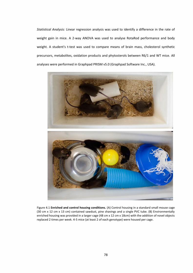
78
Statistical Analysis: Linear regression analysis was used to identify a difference in the rate of
weight gain in mice. A 2-way ANOVA was used to analyse RotaRod performance and body
weight. A student's t-test was used to compare means of brain mass, cholesterol synthetic
precursors, metabolites, oxidation products and phytosterols between R6/1 and WT mice. All
analyses were performed in Graphpad PRISM v5.0 (Graphpad Software Inc., USA).
Figure 4.1 Enriched and control housing conditions. (A) Control housing in a standard small mouse cage (30 cm x 12 cm x 13 cm) contained sawdust, pine shavings and a single PVC tube. (B) Environmentally enriched housing was provided in a larger cage (48 cm x 12 cm x 18cm) with the addition of novel objects replaced 2 times per week. 4-5 mice (at least 2 of each genotype) were housed per cage.

79
4.3 Results
4.3.1 Phenotype
The effect of EE on the progression of the HD phenotype in R6/1 mice was examined from 7-26
weeks of age. Body weight, hind paw clasping and RotaRod performance was tested over this
period (n = 7-8 per group). The brain mass of mice was measured at the completion of the study
at 26 weeks of age when all mice were sacrificed.
4.3.1.1 Weight loss
Body weight was recorded every 4-5 days (Figure 4.2). Progressive weight loss is known to occur
at 15 weeks in R6/1 mice (Mangiarini et al., 1996).
EE did not significantly alter weight gain between male WT and R6/1 mice, or have an overall
effect on the weight loss phenotype although the baseline levels were higher in EE mice. Prior
to weight loss, weight gain in both EE and control R6/1 males was significantly less than WT in
both housing conditions (p < 0.001). EE had no effect on body weight in WT males when
normalised to baseline measurements.
Female R6/1 mice in EE housing had a significantly greater weight gain prior to weight loss (7-15
weeks) when compared to control R6/1 and WT mice (p < 0.0001), and a similar weight gain to
enriched WT female mice. Although the average bodyweight at baseline was higher in EE mice,
when considering the full course of the experiment, the weight loss profile was not significantly
altered between EE and control R6/1 mice. EE significantly increased the rate of weight gain in
WT females compared to control WT female mice (p < 0.0001).

80
Figure 4.2 The effect of environmental enrichment on weight loss in R6/1 mice. Environmental enrichment (EE) did not significantly alter weight gain or weight loss in WT and R6/1 male mice. Female R6/1 mice housed with EE had a significantly greater weight gain prior to weight loss compared to control R6/1 female mice (7-15 weeks) (p < 0.0001), however there was no overall effect by the final time point. EE increased the weight gain in WT female mice compared to control WT females (p < 0.0001) n = 7-8 per group. Error bars represent + SEM

81
4.3.1.2 Hind paw clasping
The percentage of male R6/1 mice housed with EE that exhibited a clasping phenotype was
consistently lower than control R6/1 mice over the course of the study. At 26 weeks, 50% of EE
male R6/1 and 80% of control R6/1 mice were positive for the clasping phenotype (Figure 4.3).
Female R6/1 mice from both EE and control housing had a similar onset of hind paw clasping at
22 weeks of age. The percentage of EE female R6/1 mice exhibiting a clasping phenotype
between 23-26 weeks was less than control mice, however, the difference was relatively small
over this short period (35% vs 50%, Figure 4.3). No influence of housing was observed in WT
mice with several mice from both enriched and control housing showing a half clasp phenotype
at various time points during the study (Figure 4.3).

82
Figure 4.3 The effect of environmental enrichment on the hind paw clasping phenotype in R6/1 mice. R6/1 mice develop a phenotype where the hind paws are clasped to the body when suspended by the tail. Environmental enrichment (EE) did not alter the onset of hind paw clasping in male R6/1 mice, however the percentage of mice showing positive for the phenotype was consistently lower than control R6/1 mice during disease progression. Hind paw clasping at 22 weeks in female R6/1 mice and the small attenuation of hind paw clasping in the EE female R6/1 mice was not considered significant. A small proportion of WT mice appeared to exhibit a half clasp during the course of the study independent of housing treatment. n = 7-8 per group.

83
4.3.1.3 RotaRod
A small but statistically significant improvement in motor performance was detected in EE male
R6/1 mice compared to control R6/1 males (p = 0.0032). While R6/1 males kept in both housing
conditions did show significant motor decline over the entire study, R6/1 mice housed with EE
had a delayed motor decline and a consistently higher average RotaRod time compared to
control R6/1 mice (Figure 4.4). EE housing did not have a significant effect on the RotaRod
performance of female R6/1 mice; both EE control mice had a similar motor decline over the
course of the study (Figure 4.4). A small improvement in the average RotaRod time of EE R6/1
females at 20 and 24 weeks was observed, however this did not reach statistical significance.
The RotaRod performance of WT animals was not influenced by the housing treatment in either
male or female mice.
4.3.1.4 Brain mass
The reduction of brain mass previously observed in R6/1 mice (Chapter 3) was not altered by EE
housing. Male and female R6/1 mice housed in EE or control housing had a highly significant
brain mass reduction of 17-18% compared to WT mice of the same sex and housing treatment
(p < 0.0001). Absolute brain mass values can be found in Table 4.1.
Table 4.1 Brain mass of R6/1 and WT mice with environmentally enriched or control housing. Values represent mg ± SEM. n = 7-8 per group. EE = environmental enrichment
Control housing EE
WT R6/1 WT R6/1
Male 476 ± 6 391 ± 7 493 ± 6 401 ± 5
Female 480 ± 5 399 ± 7 483 ± 8 402 ± 8

84
Figure 4.4 The influence of environmental enrichment on RotaRod motor performance of R6/1 mice. Environmental enrichment (EE) caused a delay in the onset of motor dysfunction in male R6/1 mice compared to control R6/1, and improved the average latency to fall from the RotaRod over the course of the study (p = 0.0032). No effect of housing treatment was detected between female EE and control R6/1 mice. The RotaRod performance of WT animals was not influenced by the housing treatment in either male or female mice. n = 7-8 per group. Error bars represent + SEM.

85
4.3.2 Sterol analysis of R6/1 brain tissue
All mice were sacrificed at 26 weeks of age and solvent extracted lipids from the cortex and
striatum were analysed using GC-MS/MS for cholesterol synthetic precursors, metabolites,
oxidation products and phytosterols. Sterol compounds examined in this study included;
[cholesterol synthetic precursors (lathosterol, lanosterol, zymosterol, 24,25 dihydro lanosterol,
desmosterol, 7-dehydrocholesterol), metabolic products (24-OHC, 27-OHC), oxidation products
(7-KC, 7β-OH), phytosterols (campesterol, β-sitosterol, stigmasterol, brassicasterol)]. Quantified
values for all sterols measured can be found in Appendix 4-6.
4.3.2.1 Cholesterol oxidation products
Male R6/1 mice housed with EE had significantly reduced levels of 7β-OH in cortex tissue
compared to control R6/1 mice (p = 0.0343) (Figure 4.5). Male R6/1 and WT control mice did not
significantly differ in 7β-OH levels (Figure 4.5). Although the effect of EE was not statistically
significant in male WT mice, a similar profile to that of R6/1 male mice was observed. No
difference in the level of cholesterol oxidation products was detected between housing
treatments in female R6/1 mice. Combining the sexes in WT mice revealed a significant
reduction of 7β-OH levels in cortex tissue (p = 0.0389) (Figure 4.6). Significant alterations to 7β-
OH or 7-KC were not detected in any other brain region in either genotype or sex.
4.3.2.2 Cholesterol synthetic precursors, metabolic products and phytosterols
EE had no detectable influence on the level of cholesterol synthetic precursors, metabolic
products or phytosterols in cortex or striatum of male and female R6/1 mice. No differences in
these compounds were detected between EE and control WT mice. The sterol profile of R6/1 vs
WT mice was consistent to the results obtained in Chapter 3 at a similar time-point of 24 weeks.
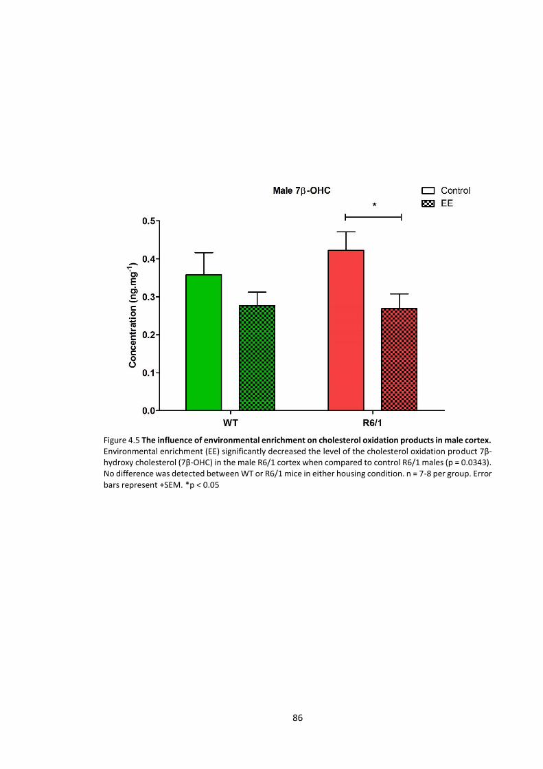
86
Figure 4.5 The influence of environmental enrichment on cholesterol oxidation products in male cortex. Environmental enrichment (EE) significantly decreased the level of the cholesterol oxidation product 7β-hydroxy cholesterol (7β-OHC) in the male R6/1 cortex when compared to control R6/1 males (p = 0.0343). No difference was detected between WT or R6/1 mice in either housing condition. n = 7-8 per group. Error bars represent +SEM. *p < 0.05
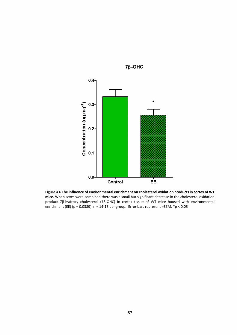
87
Figure 4.6 The influence of environmental enrichment on cholesterol oxidation products in cortex of WT mice. When sexes were combined there was a small but significant decrease in the cholesterol oxidation product 7β-hydroxy cholesterol (7β-OHC) in cortex tissue of WT mice housed with environmental enrichment (EE) (p = 0.0389). n = 14-16 per group. Error bars represent +SEM. *p < 0.05

88
4.4 Discussion
Previous studies investigating EE in R6 mice have reported several neurophysiological
changes, including, increased neurogenesis (Lazic et al., 2006) and neurotrophin
expression (Spires et al., 2004) in EE mice. In this study we have examined the effect of
EE on R6/1 mice by characterising physical phenotypic changes as well as molecular
changes to cholesterol homeostasis and oxidative stress.
4.4.1 Motor performance
In this study we have detected improved motor performance in R6/1 male mice housed with EE.
The improvement observed was in conjunction with a milder hind paw clasping phenotype over
the course of the study. A similar finding was reported where EE delayed the onset and overall
severity of the clasping phenotype in R6/1 mice (van Dellen et al., 2000). Since then, several
studies have found improved motor co-ordination (RotaRod) and grip strength in both R6/1 and
R6/2 mice housed with EE (Hockly et al., 2002; Spires et al., 2004). In the past, studies testing
motor performance using the RotaRod have reported varying results in the latency to fall from
the rod (Spires et al., 2004; Lazic et al., 2006). These differences may result from different
RotaRod protocols (as previously discussed in 3.4.2.3), or differences in husbandry protocols
between laboratories that have been demonstrated to influence animal behaviour (Logge et al.,
2014; Sorge et al., 2014). It may also be the result of different enrichment protocols, where the
type of environment (toys, tunnels, and nesting materials), cage size, and presence of exercise
equipment may result in different outcomes (van Praag et al., 1999; Zajac et al., 2010). This then
poses the question, what is environmental enrichment? It has been defined as 'a sustained
increase in daily, cognitive and sensory-motor and physical activity' by Mattson et al. (2001),
however we also consider that EE could involve increased sensory and cognitive stimulation in
the absence of high levels of physical activity. This is consistent with human epidemiology that

89
finds increased cognitive function (by measure of education level and occupational status)
reduces risk of dementia (Stern et al., 1994; Evans et al., 1997). This question also highlights the
idea that various aspects of EE may have different effects in specific brain regions. This is evident
in a study where the presence of a running wheel alone improved brain neurogenesis
differentially to a large cage containing novel object stimuli (van Praag et al., 1999). Wheel
running and EE have also been reported to modify gene expression differentially in R6/2 mice
(Zajac et al., 2010). Apart from increasing physical fitness, running wheels may provide motor
skill practise and potentially confound RotaRod results when assessing motor performance. In
the current study we have provided enrichment in the absence of a running wheel to examine
whether degeneration of the brain controlling motor function in HD can be achieved by sensory
and cognitive stimulation.
The age of exposure and duration of EE is also likely to play a role in the brain changes. Due to
mice being bred in a separate facility in the current study, EE was started at 7 weeks of age,
while previous studies implemented enrichment at 4 weeks of age (van Dellen et al., 2000;
Hockly et al., 2002; Spires et al., 2004). The difference of several weeks early in development
may be significant as it has been found that early handling (pre-weaning) of rodents alters
behavioural phenotypes (Levine et al., 1967) and age-related brain deficits (Meaney et al., 1991)
compared to later handled (post-weaning) animals (Levine & Otis, 1958). Therefore, in the R6/1
mouse model where biochemical changes are evident at 6 weeks (Chapter 3), we postulate that
earlier exposure to EE would result in the greatest benefit. Several mechanisms have been
proposed to explain the various benefits of EE seen in animal disease models, many of these
being specifically relevant to HD pathology.
4.4.2 Mechanisms of environmental enrichment changes in the brain
EE has been demonstrated to improve behavioural phenotypes in animal studies including,
improved spatial memory, motor function and reduced anxiety (Kempermann et al., 2002;

90
Benaroya-Milshtein et al., 2004; Spires et al., 2004). Neurophysiological changes that occur
alongside these behavioural improvements include; increased dendritic branching (Kolb & Gibb,
1991) and synaptic density (Saito et al., 1994), reduced spontaneous apoptosis (Young et al.,
1999), and increased expression of neurotrophic factors (Turner & Lewis, 2003; Lazarov et al.,
2005). Studies incorporating EE prior to a damaging insult in the brain demonstrate that EE builds
a 'protective' environment that confers resistance to damage (Kolb & Gibb, 1991; Johnson et al.,
2013), while implementing EE after brain damage also demonstrates the potential for repair
and growth (Wainwright et al., 1993; Rampon et al., 2000; Shin et al., 2013).
A simplified hypothesis suggests that neuronal activation leads to increased expression of
neurotrophic factors that promote neurogenesis, plasticity and cell survival (Mattson et al.,
2001). Previous studies have focused on BDNF which is elevated in a variety of rodent models
housed with EE (Ickes et al., 2000; Turner & Lewis, 2003; Lazarov et al., 2005). BDNF has
functions involving in cell survival, differentiation and growth (Binder & Scharfman, 2004), and
is believed to play a role in HD pathology since it is reduced in human HD brain (Zuccato et al.,
2001) as well as several HD mouse models (Spires et al., 2004; Zuccato et al., 2005). BDNF
knockout mice also show a similar gene expression profile to human HD (Strand et al., 2007).
The relevance of BDNF and EE has been highlighted by studies that report rescued BDNF levels
in HD mice exposed to EE, accompanied by improved RotaRod performance (Duan et al., 2003;
Spires et al., 2004). EE restored BDNF levels in the striatum of HD mice and caused a milder
motor phenotype (Spires et al., 2004). As cortical levels of BDNF are unaffected in these HD
mice, it is hypothesised that the corticostriatal transport of BDNF is impaired in HD, which is
ameliorated by EE (Spires et al., 2004). Specific elevation of BDNF in striatum of HD mice housed
with EE, suggests that EE may be specifically beneficial to HD pathology which involves early and
severe striatal atrophy (Vonsattel et al., 1985).

91
Another aspect of EE may also involve increased cognitive activity through more complex social
interactions. Rats housed in isolation have forebrain atrophy and significantly reduced levels of
neurotrophins compared to rats housed in groups (Pham et al., 1999; Ickes et al., 2000).
Enrichment protocols using a larger number of animals in a large cage (10-15) (van Praag et al.,
1999; Hockly et al., 2002), enables more complex social hierarchies and interactions compared
to a small cage of 4 animals. These increased social interactions have been shown to modulate
the endocrine system in rodents (Blanchard et al., 1995; Hardy et al., 2002), and induce
neurological changes (Chao et al., 1993). Benefits through increased social interaction may be
of relevance to HD pathology that has well established neuroendocrine alterations (Popovic et
al., 2004; Wood et al., 2008; Saleh et al., 2009). While the beneficial effects of EE on behaviour
and brain physiologies are evident in previous literature, including our current study, further
work is required to elucidate the specific aspects of an enriched environment that actively confer
health benefits to the CNS.
4.4.3 Cholesterol synthesis and metabolism
Previously we have characterised the progression of the cholesterol synthetic and metabolic
defects in the R6/1 mouse model of HD (Chapter 3). In our current study EE had no detectable
effect in altering the levels of lipid species measured, when compared to control mice. The levels
of all sterol compounds measured were consistent with the later time points (24-28 weeks) in
our previous study (Chapter 3). Cholesterol homeostatic alterations were investigated in this
study since previous literature reports improved BDNF levels in EE HD mice (Spires et al., 2004).
BDNF is believed to influence cholesterol homeostasis in the brain, increasing cholesterol
content and gene expression of cholesterol synthetic enzymes in cultured neurons (Suzuki et al.,
2007). This neurotrophin also promotes DHCR24 expression (Sarchielli et al., 2014), the enzyme
responsible for catalysing desmosterol conversion to cholesterol at the end of the cholesterol
synthetic pathway. In this study (and in Chapter 3) we observed an accumulation of desmosterol
92
in the striatum, which is likely attributed to loss of DHCR24 levels or activity (as discussed in
3.4.1.1). However, EE did not alter desmosterol levels in R6/1 mice. It is possible that EE did not
restore BDNF levels early enough to attenuate the severe cholesterol synthetic deficit in R6/1
mice. The mechanisms behind altered cholesterol homeostasis in HD are still largely undefined
in vivo; it is possible other factors may also play a significant role. Specifically mHTT, which may
diminish the effect of BDNF by interfering with sterol regulatory binding proteins (SREBPs)
(Valenza et al., 2005) that also regulate cholesterol synthesis (Yokoyama et al., 1993).
Further investigation into BDNF and cholesterol homeostasis is required in HD models. A
potentially useful model to investigate whether BDNF can rescue the cholesterol deficit in vivo
is a transgenic HD mouse crossed to overexpress BDNF (Canals et al., 2004; Xie et al., 2010). As
cholesterol synthetic precursors are present in concentrations as low as 0.1 ng/mg in brain
tissue, sensitive analytical tools such as GC-MS/MS are essential to detect the full extent of any
potential changes.
4.4.4 Cholesterol oxidation products
The cholesterol oxidation product 7β-OHC was reduced in the cortex of R6/1 male mice housed
with EE. The difference in oxidation markers observed between EE and control housing is likely
to be a combination of EE reducing oxidative stress in the brain and a control or "non-enriched
environment" promoting it. This is supported by studies reporting reduced markers of oxidative
stress (nitrotyrosine, glutathione) in the brain of rodents housed with EE (Fernandez et al., 2004;
Herring et al., 2010), and studies describing elevated brain lipid peroxidation in rodents under
environmental stress (Liu et al., 1996; Lucca et al., 2009). In our current study mice housed
without EE may have increased levels of stress due to having fewer places to find shelter, while
EE mice have many places to hide and therefore reduced environmental stress.

93
Although WT and R6/1 mice in control housing had comparable levels of 7β-OH, the HD brain
may have diminished capacity to cope with an increased oxidative environment, enhancing the
deleterious effects of a "non-enriched” environment. This was previously observed in the R6/2
HD mouse model which was highly susceptible to an artificially induced oxidative insult while
basal levels of oxidative markers were unaltered between genotypes (Bogdanov et al., 1998).
Interestingly oxidative stress was not significantly elevated in the striatum of R6/1 mice,
consistent with previous analysis in 3.3.2.5. While the pathological hallmark of HD is striatal
atrophy, the cortex does undergo neurodegeneration in later stages of human HD (Vonsattel et
al., 1985; Rosas et al., 2003), and may explain why oxidative stress may be present in this region
of R6/1 mice late in the disease progression (25 weeks). Although oxidative stress in R6/1 mice
has been reported in a previous study (Perez-Severiano et al., 2000), our data suggests that
oxidative stress may not be a primary driving factor in R6/1 HD pathology as it is not specific to
regions that are most susceptible to early degeneration.
4.4.5 Weight loss and brain mass
EE of R6/1 mice in this study did not have significant effects on the overall weight loss and the
reduction in brain mass that is characteristic of these transgenic mice (Mangiarini et al., 1996).
This is consistent with previous findings where weight loss or brain masses were not significantly
altered with EE in the R6/1 mouse (Spires et al., 2004). EE has been shown to increase brain
mass in WT rodents, however subtle differences were found to be regionally specific and only
detectable with very large cohorts (La Torre, 1968; Bennett et al., 1969; Henderson, 1970).
Specific to HD models, EE in R6/2 mice was associated with a small increase in peristriatal volume
(6%) (Hockly et al., 2002), suggesting that any brain mass changes due to EE are most likely small
in magnitude, and only detectable using complex and sensitive brain volume analytical
techniques.
94
4.4.6 Conclusion
EE had a measureable effect on attenuating motor decline and involuntary clasping in male R6/1
mice in this study. Although motor symptoms were delayed, significant and progressive motor
dysfunction was still observed by the end stage of disease in EE mice. Improvements to
perturbed cholesterol homeostasis were not detected at 25 weeks of age once the disease
phenotype was fully present in both EE and control R6/1 mice. Although lipid peroxidation was
reduced in male R6/1 mice housed with EE, a substantial alteration in oxidative stress markers
was not observed when comparing R6/1 and WT mice in our study, and may be a secondary
event associated with HD neurodegeneration (Browne & Beal, 2006). However, it is possible that
although basal levels of oxidative stress were not elevated in R6/1 mice, the HD brain is more
susceptible to damage by stressful stimuli and this is ameliorated by EE. While the main
hypothesis surrounding the benefits of EE in HD involve upregulation of neurotrophic factors,
the benefit of specific aspects of EE on brain function are not fully understood. Previous EE
studies in HD models, and the study described here have provided further insight into HD
pathology, not only to extend the understanding of molecular mechanisms in HD
neurodegeneration, but also highlight a potentially significant suppressor of disease progression
and severity that may be applied to human HD patients.

95
Chapter 5
Berry extract supplementation in the R6/1
transgenic mouse model of Huntington's
disease

96
5.1 Introduction
In recent years sensitive analytical techniques have highlighted many classes of compounds
present in foods that have potentially potent health benefits. Many of these compounds were
previously believed to exert beneficial effects primarily through their antioxidant properties,
however more complex biochemical interactions have been recently highlighted which suggest
that plant derived phytochemicals may alter cell signalling, invoke a hormetic response or inhibit
protein misfolding.
Many plants produce secondary metabolites that are concentrated in fruit, among these are
phenolic compounds that contain multiple aromatic rings in their structure. Recent studies using
berry extract (BE) supplementation have discovered that berries such as blueberries,
blackberries and blackcurrants have potent effects to reduce brain deficits associated with aging
in animal models (Joseph et al., 1998; Casadesus et al., 2004; Galli et al., 2006). Anthocyanins
are a major phenolic component of berries, and are believed to be the main active component
of these fruits. Dietary supplementation of phenolic fruit extracts have also been applied to
neurodegenerative disease, and in the case of HD, several compounds have been identified that
attenuate disease pathology in animal and cell models (Ehrnhoefer et al., 2006; Maher et al.,
2011).
In this chapter we have investigated BE extract supplementation on the R6/1 HD mouse model
and tracked the progression and severity of symptoms. We have assessed the effects of BE
extract supplementation on biochemical changes involved in altered cholesterol homeostasis
and oxidative stress as well as several established phenotypic changes that occur in these mice.
97
5.2 Materials and Methods
Mice: R6/1 mice were generated as outlined in 2.3.1. Equal numbers of male and female R6/1
and WT mice (6 per group) were caged in standard housing (30 cm x 12 cm x 13 cm) containing
a single PVC tube, saw dust and pine shavings. Four mice were housed per cage, two of each
genotype. All procedures that were undertaken conformed to the standards of the University of
Wollongong ethics committee (ethics approval number: AE 13/20).
Diet: Control and BE diets were manufactured by Speciality Feeds Pty Ltd. (Glen Forrest, W.A,
Australia). Control diet comprised of the standard rodent growth diet (AIN-93G) (Reeves et al.,
1993). BE diet contained [control diet + 0.2% blackcurrant extract (NutriPhy Blackcurrant 100,
Chr. Hansen pty. Ltd., Australia)]. Control and BE diet were equal in caloric content. BE diet was
calculated to be equivalent to one serving of whole berries in humans (100-150 g) (Prior et al.,
2001; Scalzo et al., 2008; Tabart et al., 2011; Milivojevic et al., 2012), corrected to body surface
area as described in Reagan-Shaw et al. (2008). The consumption of anthocyanins by mice in this
study (when corrected for body surface area) was similar to previous studies investigating
anthocyanin supplementation in rats (Joseph et al., 1999; Youdim et al., 2000). Both diets were
kept a 4oC in a light sealed box prior to being dispensed for feeding. Food and water was
available ad libitum. Unconsumed diet was removed and replenished from cages every 2 days.
Body weight: Mouse weight was recorded every 4-5 days from 7 weeks of age until sacrifice.
Hind paw clasping: Hind paw clasping was tested every 5-10 days following the procedure
outlined in 2.3.3.
RotaRod: Motor performance was measured using the RotaRod protocol outlined in 2.3.2. The
RotaRod performance of mice was tested at 6, 11, 15, 19 and 23 weeks of age.
98
Tissue collection: Mice were sacrificed at 25 weeks of age using slow flow CO2 asphyxiation. Brain
tissue was dissected and collected as described in 2.3.4.
Lipid extraction and GC-MS/MS analysis of sterols: Quantification of cholesterol synthetic
precursors, metabolites, oxidation products and phytosterols was performed as outlined in 2.3.5
and 2.3.6.
Statistical analysis: Linear regression analysis was used to identify differences in the rate of
weight gain between mice. 2-way ANOVA was used to test for an effect of BE extract
supplementation on RotaRod performance and weight loss. A student's t test was used to
compare mean values of sterol analysis and brain mass. All analyses were performed in
Graphpad PRISM v5.0 (Graphpad Software Inc., USA).
99
5.3 Results
5.3.1 Phenotype
5.3.1.1 Body weight
Supplementation of BE had no effect on the weight loss phenotype in either male or female R6/1
mice when body weight was normalised to baseline levels (Figure 5.1). Both male and female
R6/1 mice had significant weight loss independent of diet when compared to WT mice (p < 0.01).
A significant difference in weight gain prior to weight loss was observed between control and BE
R6/1 males, the control fed mice gaining weight more rapidly (p = 0.004). No other significant
differences in weight gain were detected in WT or female R6/1 mice.
5.3.1.2 Hind paw clasping
A small delay in the onset of the hind paw clasping phenotype was observed in male R6/1 mice
fed a BE supplemented diet (12 vs. 14 weeks of age in control and BE fed mice respectively)
(Figure 5.2). After the initial onset of clasping the percentage of R6/1 control mice showing
positive for clasping was higher on average, however there was variability between time points
and a clear difference in hind paw clasping between diet treatments was not observed. In female
R6/1 mice the hind paw clasping phenotype progressively increased after 19 weeks of age
independent of diet treatment (Figure 5.2). A small percentage of WT mice from both diet
treatments were positive for a half clasp phenotype at various stages of the study.
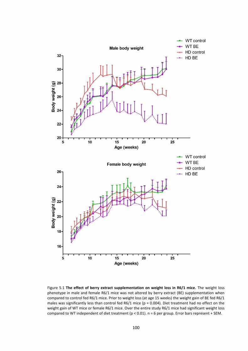
100
Figure 5.1 The effect of berry extract supplementation on weight loss in R6/1 mice. The weight loss phenotype in male and female R6/1 mice was not altered by berry extract (BE) supplementation when compared to control fed R6/1 mice. Prior to weight loss (at age 15 weeks) the weight gain of BE fed R6/1 males was significantly less than control fed R6/1 mice (p = 0.004). Diet treatment had no effect on the weight gain of WT mice or female R6/1 mice. Over the entire study R6/1 mice had significant weight loss compared to WT independent of diet treatment (p < 0.01). n = 6 per group. Error bars represent + SEM.
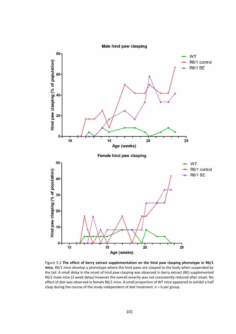
101
Figure 5.2 The effect of berry extract supplementation on the hind paw clasping phenotype in R6/1 mice. R6/1 mice develop a phenotype where the hind paws are clasped to the body when suspended by the tail. A small delay in the onset of hind paw clasping was observed in berry extract (BE) supplemented R6/1 male mice (2 week delay) however the overall severity was not consistently reduced after onset. No effect of diet was observed in female R6/1 mice. A small proportion of WT mice appeared to exhibit a half clasp during the course of the study independent of diet treatment. n = 6 per group.

102
5.3.1.3 RotaRod
The average RotaRod time for female R6/1 mice fed a BE diet was consistently higher than
control fed mice over the course of the study, and a small but significant overall improvement
was detected (p = 0.0349). Male R6/1 mice fed a BE diet did not have a detectable improvement
in performance compared to control fed R6/1 mice. BE supplementation did not alter the
RotaRod performance of male or female WT mice (Figure 5.3).
5.3.1.4 Brain mass
Dietary intervention had no effect in altering the brain mass reduction seen in R6/1 mice (R6/1
male BE vs control p = 0.587, R6/1 female BE vs control p = 0.738) (Table 5.1). Both male and
female R6/1 mice fed control and BE supplemented diets had significantly reduced brain mass
when compared to WT mice (p < 0.001), with a mean reduction of 10-15%.
Control diet BE
WT R6/1 WT R6/1
Male 467 ± 8 397 ± 14 465 ± 8 388 ± 5
Female 460 ± 6 405 ± 13 458 ± 5 410 ± 5
Table 5.1 Brain mass of R6/1 and WT mice fed a berry extract supplemented diet. Values represent mg ± SEM. n = 6 per group. BE = Berry extract
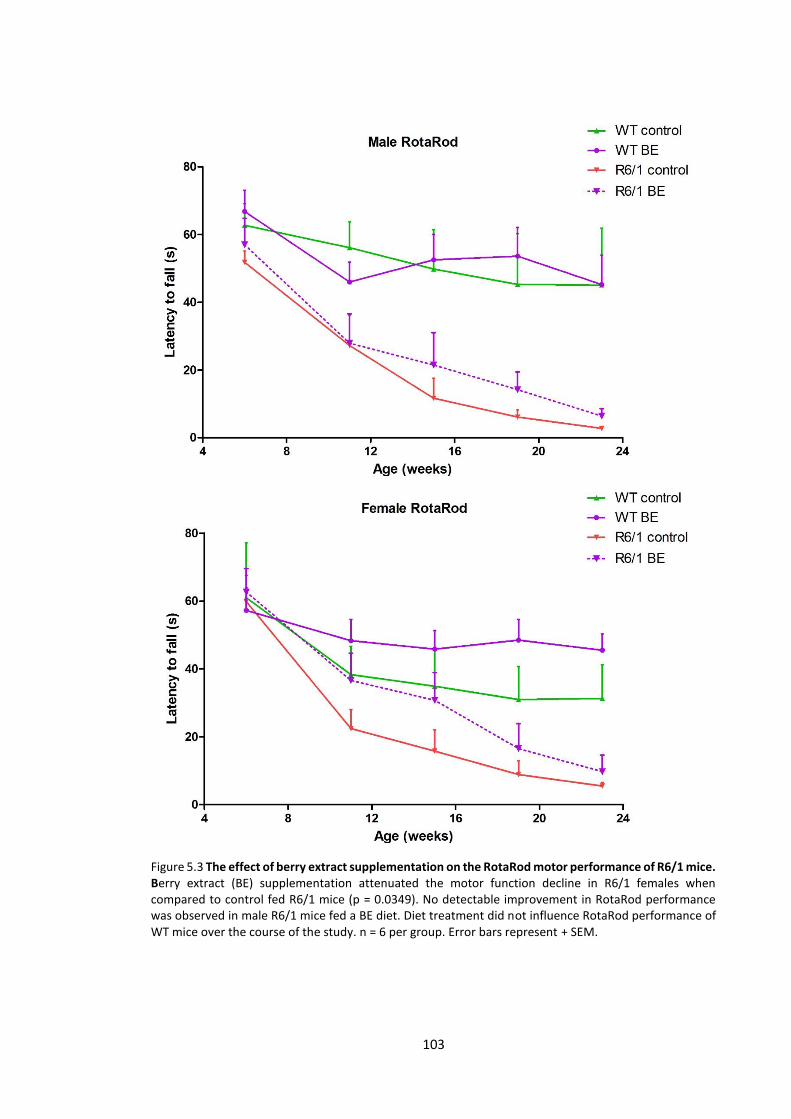
103
Figure 5.3 The effect of berry extract supplementation on the RotaRod motor performance of R6/1 mice. Berry extract (BE) supplementation attenuated the motor function decline in R6/1 females when compared to control fed R6/1 mice (p = 0.0349). No detectable improvement in RotaRod performance was observed in male R6/1 mice fed a BE diet. Diet treatment did not influence RotaRod performance of WT mice over the course of the study. n = 6 per group. Error bars represent + SEM.
104
5.3.2 Sterol analysis of R6/1 brain tissue
At the age of 25 weeks all mice were sacrificed for brain lipid analysis. Striatum and cortex was
dissected from the brain and sterols were analysed using GC-MS/MS. Dietary supplementation
with BE had no detectable influence in WT or R6/1 mice when examining the level of cholesterol
synthetic precursors, metabolic products, oxidation products and phytosterols quantified in this
study. No significant sex differences were observed in WT or R6/1 mice. The sterol profile of
R6/1 vs WT mice was consistent to the results obtained in Chapter 3 and 4 at a similar time-
point of 24 weeks. Sterol compounds examined in this study included; [cholesterol synthetic
precursors (lathosterol, lanosterol, zymosterol, 24,25 dihydro lanosterol, desmosterol, 7-
dehydrocholesterol), metabolic products (24-OHC, 27-OHC), oxidation products (7-KC, 7β-OH),
phytosterols (campesterol, β-sitosterol, stigmasterol, brassicasterol) and cholesterol].
Quantified values for all sterols measured can be found in Appendix 7-9.
105
5.4 Discussion
The R6 mouse models of HD have been previously used to examine a variety of therapeutic
strategies targeting multiple aspects of HD pathology that is manifested in these mice. These
include studies addressing: metabolic impairment, protein misfolding, inflammation,
excitotoxcity and oxidative stress through strategies such as drug treatments, gene therapies,
tissue transplantation, environmental enrichment and dietary interventions (Ferrante et al.,
2000; van Dellen et al., 2000; van Dellen et al., 2001; Ferrante et al., 2002; Wang et al., 2005;
Maher et al., 2011). These interventions have typically generated small improvements in motor
dysfunction and survival of mice, and have increased the current understanding of mechanisms
involved in HD pathology. Novel therapeutics are likely to take many years to develop and test
for safety, hence many studies in animals have trialled already approved drugs (lithium chloride,
remacemide, riluzole) with some success, reducing the severity of motor phenotypes and
increasing survival of animals (Ferrante et al., 2002; Schiefer et al., 2002; Wood & Morton, 2003).
An advantage of dietary supplementation is that the active compounds used to treat disease are
safe and already consumed on a daily basis. A large number of foods containing phytochemicals
believed to have health benefits have been identified recently, among these are anthocyanins,
phenolic compounds that are highly concentrated in berries. BE has been used to reduce age-
related cognitive deficits and improve memory and cognition in rats (Joseph et al., 1999;
Casadesus et al., 2004; Galli et al., 2006; Duffy et al., 2008). The dosing of anthocyanins
consumed by rats in these studies is equivalent to the consumption of approximately 100-150g
of fresh berries (blueberry, blackberry, blackcurrant) per day in humans. Several other plant
derived phenolic compounds have been trailed in animal models of neurodegeneration
including HD (Ehrnhoefer et al., 2006; Maher et al., 2011). Prior to experimentation in this
chapter, BE has not been investigated in HD models. Here we discuss the benefits, and possible

106
protective mechanisms, of phytochemical supplementation in brain related disorders,
specifically in HD and the R6/1 HD mouse model.
5.4.1 Motor performance in R6/1 transgenic mice and possible protective mechanisms of
dietary phenolics in the brain
In this study we describe the novel use of BE to improve motor function in the R6/1 HD mouse
model. Our study was able to identify a significant improvement in the RotaRod performance of
female R6/1 mice. Although there is a trend for improved function in the later stages of disease
in male R6/1 mice, this did not reach statistical significance. It is possible that due to the more
rapid onset of symptoms in male R6/1 mice as reported in Chapter 3, we have not been able to
detect a significant level of improvement in male mice. This is consistent with the greatest effect
of BE supplementation in females occurring at the onset of symptoms at 11-15 weeks, delaying
the onset of motor dysfunction by several weeks. Improvements in motor function have also
been observed in aged rats fed a BE supplemented diet (Shukitt-Hale et al., 2009). Several other
studies using BE supplementation in rats have seen improvements in memory and
neurolophysiological deficits associated with aging in the brain (Joseph et al., 1999; Goyarzu et
al., 2004; Ramirez et al., 2005; Galli et al., 2006; Duffy et al., 2008). In many studies using dietary
phenolic supplementation, young and healthy WT controls do not show the same level of
improvement in brain biochemistry or motor and cognitive performance when compared to
aged or disease models (Duffy et al., 2008; Wang et al., 2010; Willis et al., 2010). This suggests
that phenolic supplementation specifically attenuates disease pathology (or ageing) rather than
an overall improvement of neurological function.
The dose of BE and the anthocyanins contained in this diet is also an important consideration
when conducting and comparing studies, or assessing the feasibility of dietary berry
consumption for therapeutic purposes. Our current study has used a commercially available
blackcurrant anthocyanin extract with a known concentration of anthocyanins. The dose of

107
anthocyanins equated to the consumption of approximately 100-150 g of berries per day in
humans (300 mg total anthocyanins) assuming a daily intake of 3 g of diet in mice. Total
anthocyanin content of berries varies between species; blueberries, blackberries and
blackcurrants contain approximately 1.5-2 mg, 2 mg and 3 mg total anthocyanins per gram fresh
weight respectively (Scalzo et al., 2008). Previous studies in rats have used lyophilised extracts
by homogenising whole blueberries (Joseph et al., 1999; Youdim et al., 2000; Andres-Lacueva et
al., 2005), however the exact anthocyanin content has not been described. Assuming no
anthocyanins have been lost in this process, the dosing is similar to that used in our study;
approximately equal to the consumption of 100-150 g of fresh berries per day in humans. One
aspect that has been overlooked however is the potential loss of anthocyanins during the
extraction process. Oven drying and lyophilisation has been demonstrated to reduce the total
anthocyanin content of blueberries and grape skin (Lohachoompol et al., 2004; de Torres et al.,
2010). It is therefore important to quantify total anthocyanins in extracts to identify the exact
quantity of the active molecule being utilised in diet treatments.
Due to the heterogeneity of phytochemical structures found in food, it is likely that different
classes of compounds or even different compounds of similar structure act through diverse
mechanisms. Several hypothesised mechanisms have been suggested to explain the
neuroprotective properties of BE supplementation. One hypothesis focuses on the antioxidant
properties of BE containing anthocyanins and other phenolic compounds. Several studies using
a BE supplemented diet describe reduced levels of oxidative stress markers in rat brain (Joseph
et al., 1999; Elks et al., 2011). In vitro studies have also reported cells treated with BE and grape
seed extract as having enhanced survival against oxidative stress (Duffy et al., 2008; Wang et al.,
2010). However, the physiological relevance of the dose of phytochemicals used in these cell
culture studies is unknown. Our study did not detect altered lipid peroxidation in the brain of BE
supplemented R6/1 or WT mice when quantifying the cholesterol oxidation products 7-KC and
108
7β-OHC. Lipid peroxidation was not previously identified as being altered in R6/1 compared to
WT mice (Chapter 3) and therefore antioxidant effects by suppressing lipid peroxidation is
unlikely to confer a strong protective effect towards the HD mice in our study.
Hormesis has been suggested as a mechanism through which phytochemicals such as
anthocyanins may confer health benefits. A hormetic response involves a low dose of a toxin (or
stressor) promoting or upregulating protective survival factors in an organism, which then have
enhanced survival prospects against future toxic insults such as increased oxidative stress in
aging or that caused by mHTT in HD. Dietary phytochemicals have been shown to increase the
immune response (Feng et al., 2002), expression of cytoprotective proteins (McWalter et al.,
2004) and have anti-inflammatory properties (Willis et al., 2010). Attenuated age-related
neurological deficits also suggests the upregulation of prosurvival mechanisms by these
compounds (Casadesus et al., 2004; Goyarzu et al., 2004; Galli et al., 2006). However, high
concentrations of phytochemicals can also be toxic as demonstrated by in vitro studies (Liontas
& Yeger, 2004; Fimognari et al., 2005). A previous study feeding BE to rats describes a similar
hormetic response when reactive oxygen and nitrogen species were elevated in short fed
animals term (2 days), while the same markers where significantly decreased in chronic fed
animals (6 and 12 weeks) (Elks et al., 2011). A future study into the survival of BE fed HD mice
would be useful to identify if this supplementation can delay death in R6/1 mice. In the current
study, lipid analysis of brain tissue required all mice to be sacrificed at a predetermined time
point and therefore a survival study could not be conducted.
There is substantial evidence to suggest that BE and anthocyanins also influence cellular
signalling pathways in the brain that may result in neuroprotection and promote proliferation.
BE supplementation has been shown to increase extracellular signal-related kinase (ERK)
phosphorylation in aged rat brains (Williams et al., 2008). The ERK signalling cascade has a role
in altering synaptic plasticity and dendritic morphology relevant to memory (Bailey et al., 1997;
109
Wu et al., 2001), and essential neuronal transcriptional events (Sweatt, 2004). A recent study in
a HD cell and fly model identified increased ERK phosphorylation with supplementation of fisetin
(a flavonoid found in fruit) (Maher et al., 2011). Fisetin was able to reduce photoreceptor
degeneration pathology and increase survival in a dose dependant manner in a HD fly model
(Maher et al., 2011). Fisetin was also supplemented in the diet of R6/2 HD mice that exhibited
improved motor performance and also increased survival (Maher et al., 2011). In addition to
modification of ERK phosphorylation by BE anthocyanins in rats, Williams et al, (2008) found
increased cAMP response element-binding protein (CREB) phosphorylation. CREB is involved
with gene transcriptional regulation (Carlezon et al., 2005), including that of BDNF (Tao et al.,
1998; Yu et al., 2012). The BDNF gene codes for the neurotrophin BDNF, that has functions in
cell survival, differentiation and growth (Binder & Scharfman, 2004), and is significantly reduced
in HD (Zuccato et al., 2001; Zuccato et al., 2005). BDNF levels have not been reported in
phytochemical supplemented HD mouse models, however the current literature on HD
pathophysiology suggests rescued BDNF levels may be one mechanism by which BE
supplementation improves the HD phenotype described.
There is evidence to suggest that mHTT aggregation plays a significant role in HD pathogenesis
(Gutekunst et al., 1999; Yang et al., 2002), and therefore the inhibition of aggregation has been
the target of therapies to reduce HD severity. Dietary supplementation of a green tea-derived
phenolic compound reduced the disease phenotype of a HD fly model (increased movement and
reduced photoreceptor degeneration), and reduced mHTT aggregates in a HD cell model
(Ehrnhoefer et al., 2006). It was therefore proposed that the protective effects of this phenolic
compound are conveyed through inhibition of mHTT aggregation (Ehrnhoefer et al., 2006).
Further evidence from a study using a grape seed extract reported decreased mHTT aggregation
in a HD cell model, increased survival of a HD fly model and improved RotaRod performance of
the R6/2 HD mouse model (Wang et al., 2010). Interestingly the same cell model treated with
110
fisetin did not prevent mHTT aggregation (Maher et al., 2011). Although our current study, or
previous studies in phytochemical supplementation in HD mice have not examined mHTT
aggregation, the available evidence suggests that the beneficial effects of different phenolics
may work through multiple different mechanisms to result in an improved phenotype. Future
investigation into a cocktail of phytochemical supplements would support this idea further and
possibly highlight a supplementation regime that has more potent therapeutic effects than
single compounds or extracts alone.
5.4.2 Cholesterol homeostasis and BE supplementation in R6/1 mice
In this study we also investigated alterations to cholesterol homeostasis in R6/1 mice receiving
BE supplementation. BE supplementation did not result in significant alterations to the sterol
profile (alteration of synthetic and metabolic products of cholesterol) previously identified in
the R6/1 mouse model (Chapter 3). Previously BE supplementation improved ERK
phosphorylation (Williams et al., 2008) and this may influence BDNF levels, and in turn alter
cholesterol homeostasis. However, the cholesterol synthetic deficits began early in the R6/1
model (6 weeks of age, Chapter 3) and therefore the age at which supplementation is initiated
in HD models may significantly affect the relative benefits of the treatment. Several past dietary
studies in HD animal models began supplementation at the embryonic development stage, by
feeding the P1 generation and continuing supplementation after birth (Clifford et al., 2002;
Ehrnhoefer et al., 2006; Wang et al., 2010). Another possible strategy for future dietary studies
is intermittent supplementation; this has been suggested to potentially enhance a hormetic
response that may be caused by these phytochemicals (Mattson, 2008).
111
5.4.3 The influence of BE on hind paw clasping in R6/1 mouse
A small delay in the onset of the hind paw clasping phenotype was observed in BE supplemented
male R6/1 mice, however an overall improvement during the later stages of the study was not
observed in either male or female R6/1 mice. It is possible that the hind paw clasping phenotype
is controlled by a brain region or neurological process that is not specifically improved through
dietary anthocyanin intervention. Hind paw clasping represents a reflex response in contrast to
motor co-ordination measured by the RotaRod apparatus. The idea that different brain regions
control different functions is well established, and the manifestation of broad behavioural
symptoms in HD is likely to be the result of neurodegeneration in multiple brain regions. HD
brain pathology is indeed widespread, following early striatal degeneration, atrophy is found in
the globus pallidus, accumbens, amygdala, thalmus and cerebral cortex (Vonsattel et al., 1985;
Rosas et al., 2003). Tissue transplant studies in the anterior cingulate cortex of R6/1 mice have
identified an improvement in hind paw clasping but no change in RotaRod performance (van
Dellen et al., 2001). Similarly hind paw clasping and several involuntary phenotypes (sniffing,
rearing, grooming) were improved in fatty acid fed R6/1 mice without rescuing striatal pathology
(Clifford et al., 2002). The same fatty acid supplementation has proved effective in mouse
models of dyskinesia (involuntary movement disorder) (Vaddadi et al., 2006), possibly acting to
rescue a similar region involved with choreic movements in HD mice. Although sensitive and
specific volumetric analysis of R6 mouse brain has been conducted to investigate gross cell loss
(Sawiak et al., 2009), future work investigating molecular alterations in HD mouse brain may
benefit from analysing tissue samples derived from micro dissection techniques that facilitates
high resolution sampling.
112
5.4.4 Body weight loss and brain mass of R6/1 mice receiving a BE diet
The weight loss phenotype was not attenuated in male or female R6/1 mice fed a BE
supplemented diet. The only detectable effect was a reduced weight gain in BE male R6/1 mice
when compared to control R6/1 males. However, due to the baseline difference in body weight
of control and BE fed animals (resulting form randomisation), the difference in weight gain is
likely to be a chance event. Previous literature also reports that BE supplementation does not
modify the body weight of rodents (Sweeney et al., 2002; Goyarzu et al., 2004; Duffy et al., 2008;
Rashid et al., 2014). The majority of therapeutic interventions in the R6 mouse models target
brain alterations and therefore do not restore the peripheral metabolic deficit that is a classical
symptom of HD. Studies addressing HD metabolic deficits in R6 mice using dietary restriction
(Duan et al., 2003) or creatine supplementation (Ferrante et al., 2000) have reported a small
improvement in weight loss, however it is not completely rescued. Total food consumption is
also a consideration for dietary invention studies; in the present study it is unlikely that average
food consumption per cage was influenced by diet, similar to a previous BE study in rats (Elks et
al., 2011) and a preliminary study in WT mice (Jenner, A.M., pers. comm. 2015). In this study the
specific food intake of WT and R6/1 mice could not be determined due to equal distribution of
WT and R6/1 genotypes within cages. Separation of mice to measure food consumption was not
performed to prevent potential stress and uncontrolled factors of social hierarchy and
aggression between reunited mice. Previously, feeding habits have been shown to be unaltered
by the mHTT transgene in R6 mice; however the typical weight loss profile is still observed
(Mangiarini et al., 1996). This is consistent with the human HD symptomatology where weight
loss is observed with equal or higher caloric intake (Sanberg et al., 1981; Morales et al., 1989).
This suggests that food intake was most likely not a factor contributing to R6/1 weight loss in
our current study.
113
Previous studies have approached phytochemical supplementation by either feeding animals a
concentrated plant extract (Papandreou et al., 2009; Wang et al., 2010; Maher et al., 2011)
similar to our current study, or a purified phytochemical compound (Ehrnhoefer et al., 2006; Ho
et al., 2010). Experimental designs that supplement fruit extracts containing a mixture of
phytochemicals do not have the specificity required to identify a single molecular mechanism of
action, however fruit extracts are more relevant in a dietary context as they represent the
phytochemical mixture readily available for human consumption.
In this study there was no detectable effect of BE supplementation on the brain mass of R6/1
mice. Improved motor and cognitive performance independent of improved brain mass in R6
mice has been observed in several studies (Spires et al., 2004; van Dellen et al., 2008), and
suggests that significant recovery of the brain mass phenotype in these mice may not be
necessary to improve the symptomatic outcome of HD.
5.4.5 Therapeutic intervention and bioavailability of phytochemicals
Delivery of active molecules and compounds to the site of action is a major challenge to treat
many diseases, and is one factor that explains why many improvements in vitro are not exactly
matched in vivo. Compounds must remain stable and un-modified until the target site is
reached; oral delivery must also survive the low pH in the stomach and be absorbed into the
bloodstream. Addressing neurological disorders and neurodegenerative diseases present
additional complications as therapeutic agents must pass the BBB. Permeability of the BBB is
dependent on several factors including active transport across the membrane, molecular weight
and lipid solubility (Banks, 2009). Phytochemical compounds previously investigated in HD are
relatively small molecules, and in the case of anthocyanins contained in BE, are able to cross the
BBB and penetrate regions which have improved with treatment (Andres-Lacueva et al., 2005;
Kalt et al., 2008). As many phytochemicals, including anthocyanins are consumed daily in a
normal diet, the process of assessing potential toxicity in humans is simplified compared to

114
newly developed drugs. These features make phytochemicals such as anthocyanins potentially
useful therapeutic agents that can be easily implemented to treat disease within the brain.
5.4.6 Conclusion
Dietary phytochemicals, specifically anthocyanins in BE are reported to have substantial
protective effects in the aged and diseased brain. The motor phenotype of R6/1 HD mouse
model was attenuated in female mice through dietary supplementation of BE in our study.
Several mechanisms may be responsible for improvements seen in HD models treated with BE
supplementation. These include; rescued cell signalling, anti-aggregation, antioxidant and anti-
inflammatory mechanisms that may be stimulated by a hormetic response. Different
phytochemicals are likely to work through different mechanisms, and specifically in BE
supplemented HD, may deliver therapeutic benefits to address a dysfunction of motor co-
ordination, but not involuntary reflex movement such as hind paw clasping as seen in this study.
The progressive weight loss and brain mass reduction observed in HD mouse models was not
rescued by BE supplementation; however these phenotypes are not necessarily rescued in
conjunction with motor and cognitive improvements. Our study was unable to detect an
alteration of the cholesterol homeostatic deficit that is observed in R6/1 mice. Nevertheless,
previous findings that consistently demonstrate ameliorated age-related brain deficits with BE
supplementation, and our current finding of improved motor function in R6/1 mice, indicates
this dietary supplement should be investigated more thoroughly in HD.

115
Chapter 6
Cholesterol metabolism in Huntington's
disease post-mortem brain tissue

116
6.1 Introduction
HD is an autosomal dominant, progressive neurodegenerative disease characterised by the
expansion of a glutamine repeat on the N-terminus of HTT. Classic symptoms include involuntary
movement and cognitive dysfunction. The classical neuropathological hallmark of HD is the
severe atrophy of the striatum (caudate and putamen) (Vonsattel et al., 2011), with substantial
volume loss in the order of 50% in late stages of the disease. While these brain regions are most
severely affected, MRI techniques have highlighted that the hippocampus, cerebral cortex,
globus pallidus and amygdala also have reduced volume in HD patients (Rosas et al., 2003).
Despite the genotypic identification of mHTT carriers, there is a lack of reliable biomarkers to
predict HD progression or effectiveness of therapies. The mutation of HTT has been reported to
alter HTT-phospholipid interactions (Kegel et al., 2009), membrane organisation (del Toro et al.,
2010) and gene transcription of lipid metabolic enzymes (Sipione et al., 2002). Several studies
have also identified that cholesterol synthesis and metabolism in HD cell lines and animal models
is significantly disturbed (Valenza et al., 2005; Valenza et al., 2007b; Valenza et al., 2010),
however the current literature contains very limited data obtained from human tissue, which is
required for greater understanding of human HD pathophysiology. In this chapter we have
investigated for the first time the level of cholesterol metabolites, synthetic precursors and
oxidation products in 13 cases of human HD and 13 matched healthy controls across 5 brain
regions, to comprehensively profile sterol changes in human brain. This study also investigated
the levels of two key cholesterol synthetic and metabolic enzymes in HD putamen.
117
6.2 Materials and methods
Materials: Materials used in this study are listed in 2.2.
Human brain tissue: Frozen brain tissues from five brain regions (putamen, caudate, cerebellum,
grey and white frontal cortex) were received from the Victorian Brain Bank Network. The cohort
contained 13 cases of HD and 13 controls from each brain region. Tissue was transported on dry
ice and stored at -80°C until analysis. Demographic and basic clinical data are presented in Table
6.1. The mean age of control cases was 68.9 ± 1.9 y which was not significantly different from
the HD cases with mean age of 67.3 ± 2.2 y. Post mortem interval (PMI) of control cases (41.5 ±
4 h) and tissue pH (6.4 ± 0.1) was not significantly different to HD PMI (37.5 ± 6.2 h) or tissue pH
(6.4 ± 0.04). All brain tissue was from the left hemisphere of Caucasian donors. Ethics approval
was from the University of Wollongong Human Research Ethics Committee (HE10/327). Control
tissue was screened with a full neuropathology examination to determine the absence of
degenerative pathologies. The research was carried out in accordance with the Declaration of
Helsinki (2008) of the World Medical Association.
Western Blotting: Brain tissue (~20 mg) was added to 250 µL ice cold radio-immunoprecipitation
assay (RIPA; 50 mM Tris, 150 nM NaCl, 0.1% SDS, 0.5% sodium deoxycholate, 1% Triton X 100)
buffer containing 1% protease inhibitor (P8340, Sigma) and homogenised at 4°C using a Precellys
24 homogeniser (Bertin Technologies) (2 x 20 s, 6000 rpm). The homogenate was centrifuged at
14 000 x g and the soluble fraction taken for SDS-PAGE. Homogenates were incubated at 95°C
for 10 min with 5x loading dye containing β-mercaptoethanol. Equal amounts of protein (90 µg,
determined by a BCA assay) were loaded onto a 12% acrylamide gel and electrophoresed for 1
h at 150V. Proteins were then transferred onto a 0.4 µm nitrocellulose membrane (BIO-RAD,
Gladesville, NSW, Australia) for 35 min at 100V. To improve antigen detection, boiling phosphate
buffered saline + tween 20 (PBST) (50 mL) was added to the membrane and left to cool to room
temperature. The membrane was blocked with a 10 mL solution of PBST containing 5% skim milk
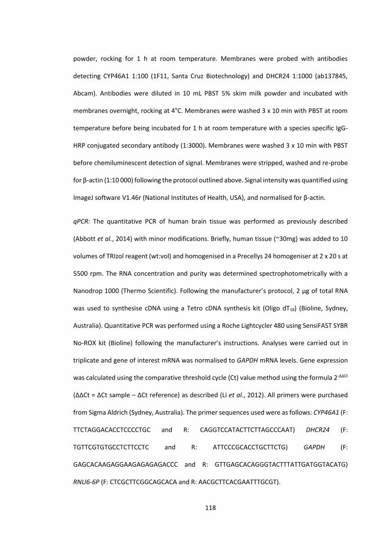
118
powder, rocking for 1 h at room temperature. Membranes were probed with antibodies
detecting CYP46A1 1:100 (1F11, Santa Cruz Biotechnology) and DHCR24 1:1000 (ab137845,
Abcam). Antibodies were diluted in 10 mL PBST 5% skim milk powder and incubated with
membranes overnight, rocking at 4°C. Membranes were washed 3 x 10 min with PBST at room
temperature before being incubated for 1 h at room temperature with a species specific IgG-
HRP conjugated secondary antibody (1:3000). Membranes were washed 3 x 10 min with PBST
before chemiluminescent detection of signal. Membranes were stripped, washed and re-probe
for β-actin (1:10 000) following the protocol outlined above. Signal intensity was quantified using
ImageJ software V1.46r (National Institutes of Health, USA), and normalised for β-actin.
qPCR: The quantitative PCR of human brain tissue was performed as previously described
(Abbott et al., 2014) with minor modifications. Briefly, human tissue (~30mg) was added to 10
volumes of TRIzol reagent (wt:vol) and homogenised in a Precellys 24 homogeniser at 2 x 20 s at
5500 rpm. The RNA concentration and purity was determined spectrophotometrically with a
Nanodrop 1000 (Thermo Scientific). Following the manufacturer’s protocol, 2 µg of total RNA
was used to synthesise cDNA using a Tetro cDNA synthesis kit (Oligo dT18) (Bioline, Sydney,
Australia). Quantitative PCR was performed using a Roche Lightcycler 480 using SensiFAST SYBR
No-ROX kit (Bioline) following the manufacturer’s instructions. Analyses were carried out in
triplicate and gene of interest mRNA was normalised to GAPDH mRNA levels. Gene expression
was calculated using the comparative threshold cycle (Ct) value method using the formula 2-ΔΔCt
(ΔΔCt = ΔCt sample – ΔCt reference) as described (Li et al., 2012). All primers were purchased
from Sigma Aldrich (Sydney, Australia). The primer sequences used were as follows: CYP46A1 (F:
TTCTAGGACACCTCCCCTGC and R: CAGGTCCATACTTCTTAGCCCAAT) DHCR24 (F:
TGTTCGTGTGCCTCTTCCTC and R: ATTCCCGCACCTGCTTCTG) GAPDH (F:
GAGCACAAGAGGAAGAGAGAGACCC and R: GTTGAGCACAGGGTACTTTATTGATGGTACATG)
RNU6-6P (F: CTCGCTTCGGCAGCACA and R: AACGCTTCACGAATTTGCGT).

119
Lipid extraction and GC-MS/MS analysis of sterols: Quantification of cholesterol synthetic
precursors, metabolites, oxidation products and phytosterols was performed as outlined in 2.3.5
and 2.3.6.
Statistical Analysis: All results are expressed as mean ± SEM with a P < 0.05 considered
significant. An unpaired t-test was used to test for significantly different means, an F-test was
used to determine if variances were significantly different. Welch’s correction for unequal
variances was used when variances were found to be significantly different. All analyses were
performed in Graphpad PRISM v5.0 (Graphpad Software Inc., USA).
Table 6.1 Demographic details of control and HD cohorts.
Case# Gender Age (y) PMI (h) pH HD Grade
stage(0-
IV)
Con 1 M 78.3 46 6.54 - Con 2 M 69.1 34 6.31 - Con 3 M 63.9 54.5 6.51 - Con 4 M 81 36.5 6.56 - Con 5 M 64.1 24 6.56 - Con 6 F 59 30 6.84 - Con 7 F 67.3 30 6.23 - Con 8 F 74.8 61.5 6.24 - Con 9 F 68.3 71.5 6.34 - Con 10 F 60.4 49 6.23 - Con 11 M 63.9 32 6.47 - Con 12 M 69.4 24 6.27 - Con 13 M 75.6 46 6.57 - HD 1 M 77 8.5 6.57 IV HD 2 M 68.7 72 6.32 IV HD 3 M 61.1 17 6.54 IV HD 4 M 81.1 50.5 6.23 IV HD 5 M 66.6 37 6.26 IV HD 6 F 57.2 22 6.22 IV HD 7 F 66.7 18.5 6.21 IV HD 8 F 72.2 22 6.43 IV HD 9 F 70.7 70 6.16 IV HD 10 F 51.5 63.5 6.54 IV HD 11 M 62.3 21.5 6.62 IV HD 12 M 65.9 58 6.33 IV HD 13 M 73.9 26.5 6.31 IV

120
6.3 Results
6.3.1 Sterol analysis
Human HD post mortem brain tissue from putamen, caudate, frontal cortex (grey and white)
and cerebellum was analysed using heavy isotope dilution GC-MS/MS. Cholesterol metabolites,
oxidation products and synthetic precursors are presented as percent relative change compared
to control (Figure 6.1). Absolute values of sterols measured are provided in Appendix 10.
6.3.1.1 Cholesterol metabolites
The most significant changes were detected in HD putamen, a region that is severely affected in
HD (Vonsattel et al., 1985). The brain specific cholesterol metabolite 24-OHC, was reduced 59%
(p < 0.0001) in HD putamen (Figure 6.1A). Caudate was the only other region to show changes
in this metabolite with a 66% reduction (p = 0.0009) of 24-OHC in HD tissue compared to control
tissue (Figure 6.1A). 27-OHC, a peripheral cholesterol metabolite was significantly increased by
3-fold in HD putamen (p = 0.0107). 27-OHC was also significantly increased 3-fold in grey frontal
cortex (p = 0.0117) and 2-fold in cerebellum (p = 0.0281) (Figure 6.1A).
6.3.1.2 Cholesterol oxidation products
HD putamen was the only brain region to show evidence of oxidative stress, with a 53% and 69%
increase in the cholesterol oxidation products 7β-OHC (p = 0.0226) and 7-KC (p = 0.0009)
respectively (Figure 6.1B).
6.3.1.3 Cholesterol synthetic precursors
Altered cholesterol synthesis was evident in HD putamen which exhibited an increase in the
cholesterol synthetic precursors, desmosterol (3-fold, p = 0.0046), lathosterol (2-fold, p =
0.0405), zymosterol (3-fold, p = 0.0066) and 24, 25 dihydro lanosterol (2-fold, p = 0.0051) (Figure

121
6.1C). Total cholesterol levels were also significantly increased by 31% in HD putamen (p =
0.0342) (Figure 6.1D). No other brain regions were found to have significant changes in
cholesterol precursors, or cholesterol levels.
6.3.2 Cholesterol metabolic and synthetic enzymes
To further investigate pathways that may contribute to changes in brain cholesterol metabolism
and synthesis observed in putamen, we examined two major enzymes involved by western
blotting. The protein level of CYP46A1 in HD was reduced 90% compared to control (p = 0.0027)
(Figure 6.2A). We also detected a significant (91%) reduction (p = 0.0014) of DHCR24 protein
levels (Figure 6.2B), which is in agreement with the accumulation of desmosterol seen in HD
putamen (Figure 6.1B).
The mRNA levels of the genes CYP46A1 and DHCR24 were also measured in putamen using
quantitative PCR. mRNA levels did not reflect protein levels and were not significantly changed
between HD and control tissue in both CYP46A1 and DHCR2 (Figure 6.3).
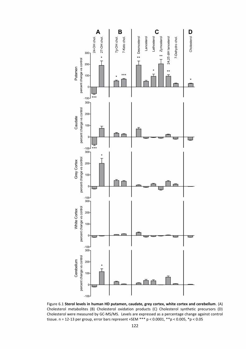
122
Figure 6.1 Sterol levels in human HD putamen, caudate, grey cortex, white cortex and cerebellum. (A) Cholesterol metabolites (B) Cholesterol oxidation products (C) Cholesterol synthetic precursors (D) Cholesterol were measured by GC-MS/MS. Levels are expressed as a percentage change against control tissue. n = 12-13 per group, error bars represent +SEM *** p < 0.0001, **p < 0.005, *p < 0.05
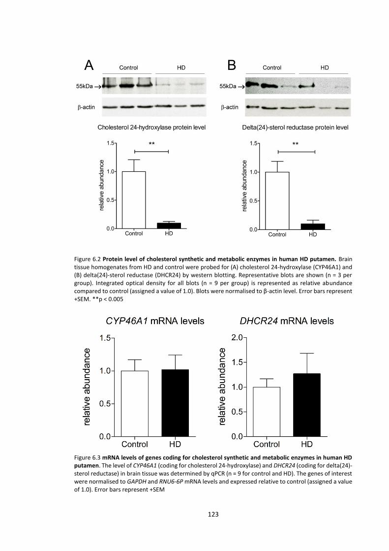
123
Figure 6.2 Protein level of cholesterol synthetic and metabolic enzymes in human HD putamen. Brain tissue homogenates from HD and control were probed for (A) cholesterol 24-hydroxylase (CYP46A1) and (B) delta(24)-sterol reductase (DHCR24) by western blotting. Representative blots are shown (n = 3 per group). Integrated optical density for all blots (n = 9 per group) is represented as relative abundance compared to control (assigned a value of 1.0). Blots were normalised to β-actin level. Error bars represent +SEM. **p < 0.005
Figure 6.3 mRNA levels of genes coding for cholesterol synthetic and metabolic enzymes in human HD putamen. The level of CYP46A1 (coding for cholesterol 24-hydroxylase) and DHCR24 (coding for delta(24)-sterol reductase) in brain tissue was determined by qPCR (n = 9 for control and HD). The genes of interest were normalised to GAPDH and RNU6-6P mRNA levels and expressed relative to control (assigned a value of 1.0). Error bars represent +SEM

124
6.4 Discussion
Alteration of cholesterol metabolism has been recently linked to several neurodegenerative
diseases including AD, PD and HD (Sipione et al., 2002; Puglielli et al., 2003; Cheng et al., 2011).
In particular, the elimination pathway where 24-OHC is formed by the hydroxylation of
cholesterol has been suggested as a contributing factor in neurodegeneration (Bjorkhem et al.,
2006). Neuronal 24-OHC has a major role in the turnover of cholesterol in the brain (Bjorkhem
et al., 1998), and the level of 24-OHC in plasma has been suggested as a measure of CYP46A1
enzymatic activity and thus metabolically active neurons (Bjorkhem, 2006).
6.4.1 Alteration of cholesterol metabolism in human Huntington’s disease brain
Previous studies have found that the plasma levels of 24-OHC in HD patients correlate with
disease severity and brain volume measured by MRI (Leoni et al., 2008; Leoni et al., 2013). Here
for the first time we report the levels of 24-OHC in human HD post-mortem brain tissue. A
significant reduction of 24-OHC was found in HD putamen accompanied by a large (90%)
reduction in the protein level of CYP46A1, the enzyme responsible for 24-OHC formation.
Reduced enzyme levels may be due to neuron loss, where CYP46A1 is primarily located.
Alterations in post-transcriptional regulation may also play a role since no change in mRNA
expression was detected between control and HD. This is potentially relevant as protein
turnover is known to be dysregulated in HD cell models (Hatters, 2008). Caudate (another
striatal region affected in HD) also had reduced levels of 24-OHC, while grey and white frontal
cortex and cerebellum showed no change. Although the cerebral cortex is affected in later stages
of HD (de la Monte et al., 1988; Rosas et al., 2003), the degree of volume loss and astrocytosis
is substantially less compared to the striatum (Sotrel et al., 1991; Vonsattel & DiFiglia, 1998;
Rosas et al., 2003). Reduced 24-OHC levels in HD striatum supports previous human studies that
reported reduced plasma levels of this metabolite in HD patients (Leoni et al., 2008; Leoni et al.,

125
2013). Our data indicates that decreased circulating 24-OHC levels are likely the result of
reduced 24-OHC production in affected regions and not an aberration of BBB 24-OHC flux. In the
mature brain, delivery of cholesterol synthesised in the astrocyte is required for normal
neuronal function (Posse de Chaves et al., 1997; Mauch et al., 2001; Hayashi et al., 2004). It has
been hypothesised that 24-OHC production in neurons acts as a signalling molecule to initiate
the delivery of cholesterol from astrocytes to neurons therefore maintaining homeostasis
(Pfrieger, 2003). There is also evidence to suggest that CYP46A1 may have neuroprotective
properties, being upregulated near plaques (Brown et al., 2004) and in glia of the human AD
brain (Bogdanovic et al., 2001). Whether reduced levels of CYP46A1 in HD is a secondary event
reflecting active neuron loss, or is a pathogenic factor altering cholesterol metabolism and
synthesis remains to be established.
27-Hydroxycholesterol, a metabolite of cholesterol that is produced predominantly in peripheral
tissue, was increased in several regions of human HD brain as compared to controls. While an
increased flux of 27-OHC into the brain may be caused by a disrupted BBB, we believe this is not
the case in this tissue since sterols that are present in higher peripheral abundance such as
campesterol (a dietary derived phytosterol) were not significantly different between HD and
control brain tissues (Appendix 10). As there is a net movement of 27-OHC from circulation into
the brain (Heverin et al., 2005), 27-OHC accumulation may be the result of a decrease in the
activity of enzymes in the brain (such as CYP27A1) that further metabolise this oxysterol to
cholestenoic acids (Meaney et al., 2007). Accumulation of 27-OHC is potentially important in
neurodegeneration since in vitro studies have shown 27-OHC promotes β-amyloidogenesis
(Gamba et al., 2014) as well as being elevated in human AD brain and transgenic mice (Heverin
et al., 2004; Shafaati et al., 2011). While there is little direct in vivo physiological evidence that
cholesterol metabolites promote neuronal damage, further studies are required to investigate

126
the hypothesis that preventing the accumulation of 27-OHC in the brain reduces
neurodegeneration in AD and HD.
6.4.2 Alteration of cholesterol synthesis in human Huntington’s disease brain
A defect in cholesterol synthesis has been previously described in several cell and mouse models
of HD. Specifically; a reduction in the cholesterol synthetic precursors lathosterol and lanosterol
as well as cholesterol in the YAC128 mouse (Valenza et al., 2007a), reduced levels of lathosterol
and lanosterol, but not cholesterol, in the R6/2 mouse model (Valenza et al., 2007b), reduced
mRNA levels of cholesterol synthetic enzymes in R6/2 mouse and human HD fibroblasts (Valenza
et al., 2005) and reduced cholesterol synthetic enzymes in a transgenic HD cell model (Sipione
et al., 2002). In contrast to these findings, our data on end stage human HD putamen shows an
increase in cholesterol synthetic precursors from both the Bloch (desmosterol and 24,25 dihydro
lanosterol) and Kandutsch-Russell (lathosterol and zymosterol) pathways and an increase in
total cholesterol levels. No significant changes in squalene levels were observed (Appendix 10)
suggesting that alterations in the upstream mevalonate pathway had no influence on the
changes that we measured in the Kandutsch-Russell and Bloch pathways. The increase in
cholesterol and synthetic precursors was only observed in the putamen; a region that
degenerates early and severely in the human disease (Vonsattel et al., 1985). In contrast to
macro and microscopic classification of disease pathology, which shows the caudate
degenerates in parallel with the putamen (Vonsattel et al., 1985; Vonsattel & DiFiglia, 1998), we
did not observe any cholesterol synthetic alterations in this region. While the cholesterol
synthetic changes observed in the putamen may suggest an overall increase of cholesterol
synthesis utilising both pathways, we believe this is not the case for the Bloch pathway. In our
study, HD putamen had significantly higher levels of desmosterol, an immediate synthetic
precursor of cholesterol (Bloch pathway) and a substrate of DHCR24. Significantly depleted
protein levels of DHCR24 in human HD putamen (91% reduction) are consistent with the reduced
127
conversion of desmosterol to cholesterol and its subsequent accumulation. Unaltered mRNA
levels of DHCR24 in putamen again suggests enzyme levels are being affected post-
transcriptionally. Previous in vitro examination of cholesterol synthesis in specific cell types
found astrocytes utilise the Bloch pathway via desmosterol, while neurons primarily utilised 7-
dehydro cholesterol and other precursors from the Kandutsch-Russell pathway (Nieweg et al.,
2009). This suggests that in late stage human HD, desmosterol accumulates in astrocytes due to
down regulated DHCR24. An increased density of oligodendrocytes and activated astrocytes is
known to be present in HD (Myers et al., 1991) and is believed to be a compensatory response
to demyelination in HD (Bartzokis et al., 2007). While the specific cell type/s exhibiting elevated
Kandutsch-Russell synthetic precursors is yet to be determined, increased oligodendrocyte
activity attempting remyelination in affected tissue may contribute to this.
Previous studies in HD mouse brain reported a significant reduction of the cholesterol precursor
lathosterol (Valenza et al., 2007a; Valenza et al., 2007b), which is found in the Kandutsch-Russell
pathway. In contrast, the Bloch pathway precursor desmosterol, was either unchanged (Valenza
et al., 2007a) or not reported (Valenza et al., 2007b) in these previous HD mouse studies.
Examining precursors from both arms of the cholesterol synthetic pathway in these mouse
models enables greater insight into the alterations occurring in different cell types, and their
possible response to HD pathology / neurodegeneration. The differences seen between HD
mouse models, in vitro cell models, and human post-mortem tissue may reflect a difference in
the homeostatic response to mutant HTT between organisms and cell types. There is also a
difference between the relative ages of mice being in early adulthood compared to end stage
human HD tissue where the mean age was 67 years old. The differences in the data derived from
our current study and previous mouse models and cell culture models may also reflect that in
this human tissue study we examined end stage HD where a significant number of vulnerable
neurons are predicted to have been lost. Examining earlier stages of the disease in brain tissue
128
is therefore important to consolidate the role of cholesterol synthesis and metabolism in the
early stages of HD in humans.
6.4.3 Cholesterol oxidation products in human Huntington’s disease brain
Along with measuring cholesterol metabolites and precursors, our GC-MS/MS method was able
to sensitively detect COPs, and use these as markers of oxidative stress in HD brain tissue.
Oxidative stress has been previously shown in HD (Browne et al., 1999), and in several other
neurodegenerative diseases (Subbarao et al., 1990; Ferrante et al., 1997; Cheng et al., 2011).
Similar to changes seen in other sterols, significant increases in 7-KC and 7β-OHC were only
observed in putamen. The specific increase in oxidation products in the putamen is consistent
with this region being affected early and severely in HD (Vonsattel et al., 1985). 7-KC and 7β-
OHC have been shown to be stable, and previously used as markers of oxidative stress in plasma
(Iuliano et al., 2003) and CSF (Leoni et al., 2005). Measurement of COPs in HD plasma together
with other sterols may represent a convenient tool to follow progression, quantify severity, and
assess therapeutic effectiveness in patients. Importantly, these markers can be measured
sensitively and reliably by GC-MS/MS techniques.
6.4.4 Consequences of altered cholesterol homeostasis in human Huntington’s disease brain
Homeostasis of cholesterol is essential for neurological function however the precise role of
cholesterol in HD neurodegeneration is still debated. Evidence suggests that cholesterol
accumulation in HD alters membrane organisation and cell signalling, enhancing the
susceptibility of striatal neurons to excitotoxicity (del Toro et al., 2010). Another hypothesis
suggests that impaired cholesterol synthesis drives neurodegeneration by limiting the supply of
cholesterol to neurons (Valenza et al., 2007a). As a result synaptogenesis and dendrite
outgrowth is impaired (Mauch et al., 2001; Fan et al., 2002), and neurotransmission is disrupted
(Koudinov & Koudinova, 2005). It is also believed that a reduction of BDNF in HD (Zuccato et al.,

129
2001) may play a significant role in neurodegeneration (Zuccato & Cattaneo, 2009). BDNF is a
neurotrophin expressed by neurons that promotes cell survival, differentiation and growth
(Binder & Scharfman, 2004). Among these properties, BDNF has been shown to promote
DHCR24 expression (Sarchielli et al., 2014). Therefore, a loss of BDNF action may also contribute
to a reduction in DHCR24, and the cholesterol homeostatic imbalance we have detected in HD
putamen.
6.4.5 Conclusion
Our study reveals for the first time that several cholesterol synthetic and metabolic pathways
are disturbed in multiple brain regions of human HD. This disturbance was identified to be most
severe in the putamen, a region that degenerates early and severely in HD. These data provide
evidence to support previous studies that link altered cholesterol synthesis, metabolism and
oxidative stress with the neuropathological process involved in HD. Since HD shares similar
sterol disturbances as other late-onset neurodegenerative diseases, we believe these changes
represent potential biomarkers for neurodegenerative disease and elucidating cholesterol
related mechanisms of neuropathology may provide targets for therapeutic intervention. The
key finding of reduced CYP46A1 in human HD brain confirms previous studies in HD patient
plasma that indicate 24-OHC as a promising peripheral biomarker to monitor the development
of HD neuropathology.

130
Chapter 7
General discussion

131
7.1 Cholesterol homeostasis alterations in Huntington's disease
The functions of cholesterol biosynthesis and metabolism, although not fully understood in the
brain, have been demonstrated to be essential for proper brain function (Kabouridis et al., 2000;
Mauch et al., 2001; Hayashi et al., 2004); mutations in enzymes of the cholesterol synthetic
pathway having severe neurological consequences in humans (Wassif et al., 1998; Waterham et
al., 2001). The mutation of the HTT protein in HD is associated with alterations to cholesterol
homeostasis and has been demonstrated consistently in multiple rodent and cell models
(Sipione et al., 2002; Valenza et al., 2005; Valenza et al., 2007a; Valenza et al., 2007b; del Toro
et al., 2010). Thorough investigation of cholesterol synthetic and metabolic alterations in human
HD brain has not been conducted prior to the experiments in this thesis. Using sensitive GC-
MS/MS analytical techniques we have identified several alterations that are consistent between
the R6/1 mouse model and human post mortem tissue. We have also identified several
alterations that are not consistent, and require further investigation to determine the relevance
of these changes to HD pathology. Here we discuss our data derived from the R6/1 mouse model
in comparison to that obtained from human HD post-mortem tissue, and highlight HD specific
alterations to cholesterol homeostasis and other pathways that require more detailed
investigation in the HD brain.
7.2 Brain cholesterol synthesis and metabolism in human and mouse models of HD
Although many animal models of neurodegenerative disease are able to replicate some
pathological hallmarks of a disease, they rarely replicate precisely the condition observed in
humans. This is due to fundamental differences between species in; diet, metabolism, ageing as
well as the physical size of the brain. Aside from some of these limitations, mice have been
genetically manipulated to model several neurodegenerative diseases including AD, PD, MND

132
and MS (Mucke et al., 2000; Bettelli et al., 2003; Laird et al., 2008; Gispert et al., 2009). In the
case of HD, generation of several mouse models expressing forms of mutated human HTT
(Mangiarini et al., 1996; Hodgson et al., 1999; Slow et al., 2003) as well as knock-in expanded
mouse huntingtin (Wheeler et al., 1999), have allowed for in vivo study of the mechanisms that
underlie HD. In this study we have investigated the R6/1 mouse model, expressing a CAG
expanded (115-120 repeats) exon 1 of human HTT, similar to the R6/2 model that expresses a
longer CAG repeat (Mangiarini et al., 1996). Multiple aspects of HD pathology have been
investigated in the R6 models as they show many of the molecular and physical phenotypes of
human HD. These include similarities in gene expression changes (Strand et al., 2007), reduced
levels of BDNF (Spires et al., 2004; Zuccato et al., 2005), nuclear inclusions (Hansson et al., 2001;
Meade et al., 2002) and cannabinoid receptor downregulation (Denovan-Wright & Robertson,
2000; Naver et al., 2003; Horne et al., 2013). A physical phenotype indicative of human HD is
also observed in these mice that includes, a progressive motor and cognitive dysfunction, weight
loss, striatal atrophy and chorea (Mangiarini et al., 1996; Hansson et al., 1999; Murphy et al.,
2000; Naver et al., 2003).
In this study we have focused on characterising cholesterol synthetic and metabolic alterations
in HD. Based on several studies in mouse and cell models, cholesterol synthesis (by measure of
cholesterol synthetic precursors lathosterol and lanosterol) is believed to be reduced in HD
(Sipione et al., 2002; Valenza et al., 2005; Valenza et al., 2007a; Valenza et al., 2007b). Prior to
our study, cholesterol homeostasis has not been sufficiently examined in human HD brain tissue,
with three previous studies using a maximum of 3 cases of HD post-mortem tissue (Valenza et
al., 2005; del Toro et al., 2010; Samara et al., 2014). Our study, and previous studies examining
cholesterol levels in aged human brain have shown cholesterol levels to be variable (Thelen et
al., 2006), and studies using small samples may therefore lack statistical power to identify
changes, or are susceptible to identifying false positives. Analysis of 5 regions in 13 cases of late
133
stage human HD post mortem brain tissue in our study has revealed an increase in cholesterol
synthetic precursors of the Kandutsch-Russell pathway (such as lathosterol), and increased total
cholesterol levels, contrary to that demonstrated in previous mouse model studies (Valenza et
al., 2007a; Valenza et al., 2007b), and our study of R6/1 mice (Chapter 3). Several factors may
underlie these differences in cholesterol biosynthetic alterations between the R6 mice and
humans, including the CAG length, HTT protein length (truncated vs full length) and the intact
expression of endogenous murine huntingtin.
The duration of HD in humans compared to the mouse is also likely to be a factor resulting in
differences in cholesterol homeostasis. Although we have used the R6/1 mouse which develops
symptoms slower than the R6/2 model, the onset of disease and disease timeline is rapid
compared to that in humans that manifests over decades (Foroud et al., 1999). The relative age
of affected R6/1 mice is comparable to humans in early adulthood and therefore the effects of
an aged brain may be less apparent in the mouse. A slower disease timeline in humans is likely
to result in some cellular changes in the brain that do not have time to manifest in the mouse
model, and may explain the absence of lipid peroxidation in the R6/1 mice we examined.
Massive neuron loss through apoptosis is not observed in the R6 models, rather a shrinkage of
neurons is a common neuropathological feature in these mice (Hansson et al., 1999; Iannicola
et al., 2000; Klapstein et al., 2001; Petersen et al., 2002). A degenerative process reported to be
'dark cell degeneration' has also been previously reported in R6/1 mice and juvenile human HD
(Iannicola et al., 2000; Turmaine et al., 2000). This process is believed to occur over weeks and
may not manifest completely in the lifespan of these mice (Turmaine et al., 2000). The lack of
significant astrocytosis and increased oligodendrocyte density in R6 mice (Mangiarini et al.,
1996; Hansson et al., 1999; Turmaine et al., 2000), compared to that seen in human HD (Myers
et al., 1991), is potentially relevant to the level Kandutsch-Russell cholesterol synthetic
precursors we observe between human and R6/1 mice. Oligodendrocytes have a high capacity
134
for cholesterol synthesis, and the increased density of these cells believed to be attempting
remyelination in HD (Bartzokis et al., 2007), may explain increased cholesterol levels and
synthetic precursors that we observe in human HD putamen.
Although this difference in Kandutsch-Russell precursors does exist between mouse and human,
we believe the increase of desmosterol (the penultimate sterol of the Bloch pathway) in both
species describes a HD specific dysfunction of the cholesterol synthetic regulation. Desmosterol
levels are likely to reflect the level or activity of DHCR24 (as shown in chapter 6), which is
believed to have regulatory roles in cholesterol synthesis beyond converting desmosterol to
cholesterol at the end of the synthetic pathway (Luu et al., 2015). DHCR24 expression levels
have also been reported to be mediated by BDNF, a trophic factor that is depleted in both human
and animal models of HD (Zuccato et al., 2001; Spires et al., 2004; Zuccato et al., 2005). The
current literature has not investigated brain levels of DHCR24 in detail, and further examination
into desmosterol levels and DHCR24 activity in HD may explain the cholesterol homeostatic
perturbation beyond simply describing the level of Kandutsch-Russell cholesterol synthetic
intermediates.
Human and mouse HD brain tissues we have investigated also exhibited some important
similarities in cholesterol metabolic alterations. Formation of 24-OHC by the enzyme CYP46A1
is a major route of cholesterol elimination from the brain. In our study, the level of 24-OHC is
reduced in the striatum of human and R6/1 mice, this is consistent with previous studies in
rodent brain (Valenza et al., 2010) and human plasma (Leoni et al., 2008). 24-OHC levels were
previously suggested as a biomarker of metabolically active neurons in the brain (Bjorkhem,
2006), however the relevance of this suggestion in a diseased brain state is not fully understood.
Although in human HD, where significant neuron loss occurs, it is unknown whether reduced 24-
OHC levels are simply a reflection of neuron loss, or if a cholesterol metabolic deficit precedes
neurodegeneration. Previously it has been hypothesised that cholesterol metabolism in neurons
135
and delivery from astrocytes may be regulated by feedback from 24-OHC (Pfrieger, 2003;
Bjorkhem, 2006),however a precise mechanisms by which 24-OHC regulates cholesterol
synthesis in the brain has not been identified. Until further investigation is undertaken, it is
unknown if synthetic, or metabolic dysfunction is the primary cause of cholesterol homeostatic
dysfunction in HD.
Our study of human HD tissue has highlighted that the metabolic pathway involving 27-OHC
formation and further metabolism is not consistent with the R6/1 mouse striatum. 27-OHC has
a net movement into the brain from the periphery, where it is quickly metabolised to
cholestenoic acids that diffuse back into the bloodstream (Heverin et al., 2005; Meaney et al.,
2007). It was previously suggested that the cell-rich grey matter in the human brain is the major
site of 24-OHC formation and further metabolism of 27-OHC (Bjorkhem, 2006). Degeneration in
these areas would therefore result in the decrease of 24-OHC levels and increased 27-OHC, as
we observed in human caudate and putamen, and previously described in human AD brain
(Heverin et al., 2004). In the R6/1 mouse model we investigated, a striatum specific decrease in
27-OHC levels contradicts findings in humans. It is possible that faster cholesterol turnover in
the brain of mice compared to humans (Dietschy & Turley, 2004), and lower absolute levels of
27-OHC in this region (0.05ng/mg in mouse vs 0.3 ng/mg in human), causes greater fluctuation
and variation in absolute levels. Interestingly, previous examination of a mouse AD model
revealed a consistent increase of 27-OHC in mouse brain tissue as well as in human AD brain
tissue (Heverin et al., 2004). Previously levels of 27-OHC levels have not been reported in studies
examining cholesterol metabolism in HD mouse models (Valenza et al., 2007a; Valenza et al.,
2007b); further work to identify if this change occurs in other models may clarify if 27-OHC
formation or further metabolism has a role in HD pathogenesis. Future studies of CSF and plasma
may also identify 27-OHC, or further metabolites of 27-OHC, as potential biomarkers of CYP7B1
and CYP27A1 activity in HD brain.

136
7.3 Localisation of cholesterol synthetic and metabolic changes in Huntington's
disease
Neurodegeneration in human HD begins in the striatum, affecting neighbouring regions such as
the globus pallidus, hippocampus, amygdala and cerebral cortex (Vonsattel et al., 1985; Rosas
et al., 2003). The end stage tissue we have examined specifically exhibit a cholesterol metabolic
deficit in the striatum (caudate and putamen), and altered synthesis was only observed in the
putamen. This suggests that these changes may be associated with early events in HD, where
specific degeneration of medium spiny neurons occurs in the striatum (Vonsattel et al., 1985).
Interestingly in the R6/1 and R6/2 HD mouse model, the cholesterol synthetic defect is more
widespread, detected in the striatum and cortex in our study, and the hippocampus in a previous
study (Valenza et al., 2007b). Although changes to Kandutsch-Russell synthetic precursors are
more pronounced in the striatum of R6/1 mice compared to other regions, the early occurrence
of changes suggests these alterations may not be as specific to HD as the reduction of 24-OHC
and accumulation of desmosterol. Both these changes are present in the striatum at later stages
of the disease, and we hypothesise these alterations are not as widespread in the brain of R6/1
mice, similar to our observations in human brain.
7.4 Sex differences in Huntington's disease
In this thesis we have also examined interventions of HD in the R6/1 model. It is of interest that
different interventions had differential effects on the motor phenotype of male and female R6/1
mice. EE improved the motor co-ordination of male R6/1 mice but did not influence females.
The opposite occurred with BE supplementation, where female R6/1 mice had improved motor
co-ordination compared to controls, however males did not improve. We have also previously
identified a sex difference in the progression of the physical phenotype in R6/1 mice (Chapter

137
3). Previous studies examining the R6/1 model typically use males or females in separate studies
(Clifford et al., 2002; Naver et al., 2003), and it is likely that any sex differences have not been
previously observed in the more studied R6/2 model as the progression is more rapid
(Mangiarini et al., 1996). On a molecular level, we have not detected any significant difference
in brain sterols between male and female R6/1 mice with our current study size; it is possible a
larger study may identify potential alterations between mice that reflect phenotypic changes.
Few studies have examined sex-specific changes in HD models, one study reported
neuroprotective effects of a sex hormone (17β-estradiol) in HD transgenic rats (Bode et al.,
2008), another identified sex-dependant alterations to BDNF expression in R6/1 mice when
exposed to environmental enrichment (Zajac et al., 2010). These findings suggest further
investigation into sex differences is warranted in HD models, an area which has been largely
ignored in the past. In human HD patients, a significant influence of sex in the progression and
severity of HD has not been previously identified. Assessing the age of onset, and disease
severity between patients is complex as symptoms can be highly variable between people
(Foroud et al., 1999). Even if overt differences between sexes have not been identified in human
HD patients, it is possible that the response to certain treatments may be sex dependant as
previously identified in mice (Zajac et al., 2010). It is therefore an important consideration to
assess both sexes in studies, not only in mouse models but also in humans.
7.5 Future directions studying cholesterol homeostasis in HD
Our findings constitute the first comprehensive sterol analysis in human HD brain tissue, and we
have identified several alterations to cholesterol homeostasis that were previously not
identified in mouse models of HD. We have examined late stage human PM tissue (grade IV) and
therefore the current understanding of cholesterol homeostasis in human HD would benefit
from analysis of earlier stage HD tissue. However, due to the relatively early onset of HD, and

138
long timeline (Foroud et al., 1999), sufficient early stage cases for a statistically powerful analysis
are very difficult to obtain (McLean, C. A. 2015, pers. comm., 27 July), and is reflected in the past
literature where the majority of analyses are carried out in late stage tissue (Augood et al., 1996;
Arzberger et al., 1997; Petersen et al., 2005; Zuccato et al., 2008; Silvestroni et al., 2009) .
Measurements of peripheral sterols may therefore be required to confirm these cholesterol
homeostatic alterations also occur early in HD.
Currently, methods to directly measure in vivo brain cholesterol synthesis without post-mortem
tissue examination have not been reported. 24-OHC levels in plasma, which originate from the
brain, may be an indirect marker of brain cholesterol synthesis. Formation of 24-OHC constitutes
a major route of cholesterol elimination from the brain (Bjorkhem et al., 1997; Bjorkhem et al.,
1998), and the formation of 24-OHC is believed to be coupled to cholesterol synthesis (Lund et
al., 2003). The reduction of plasma 24-OHC levels in HD patients has been described in the past
(Leoni et al., 2008; Leoni et al., 2013), however naturally high variability in 24-OHC plasma
concentrations between individuals limits the ability of a single time point to fully describe the
changes occurring over disease progression. A very useful future study would be the
examination of HD patient plasma in a longitudinal study beginning in the pre-symptomatic
stage. Tracking changes on an individual basis would help normalise baseline variability in 24-
OHC levels, and improve the power of detection. With highly sensitive analytical techniques,
only low volumes of plasma are required for analysis (maximum 250 µL) (Leoni et al., 2008);
plasma measurements of 24-OHC may therefore be a useful biomarker in a prospective
longitudinal study such as Enroll-HD (CHDI Foundation Inc, 2015).
Another future direction for cholesterol homeostasis in HD is to carry out an even more
comprehensive analysis in animal models of HD. Transgenic large animal models with a larger
brain, may provide an alternative insight into early cholesterol perturbations occurring in human
HD. However, until these transgenic models that have been generated in sheep (Jacobsen et al.,
139
2010), pig (Yang et al., 2010) and rhesus monkey (Yang et al., 2008), are consistently established
and validated, a closer look at mouse models may be more feasible in the short term. Here we
have examined a transgenic mouse expressing truncated human HTT. It would therefore be of
great interest to more closely examine the progression of cholesterol homeostasis in other
models expressing truncated human HTT [N171-82Q (Schilling et al., 1999)], full length human
HTT [YAC128 (Slow et al., 2003)], as well as knock-in HD mice [hdhQ92, hdhQ111 (Wheeler et
al., 1999)] to identify if alterations in cholesterol synthesis and metabolism progress consistently
across these models. Future studies would also benefit from examining several key synthetic
and metabolic enzymes we have highlighted as potentially important in our lipid analysis. These
include CYP27A1, CYP7B1, DHCR24 and CYP46A1. Investigating protein levels in addition to
mRNA expression would produce additional valuable results as we have previously shown that
CYP46A1 and DHCR24 protein levels do not reflect mRNA expression in human HD (Chapter 6).
7.6 Conclusion
Due to HTT having multiple roles in a variety of cellular compartments, it is not surprising that
the HD mutation has been associated with neurodegenerative pathology acting through
multiple mechanisms. The mutation has also been associated with a cholesterol homeostatic
perturbation, causing cholesterol synthetic and metabolic dysfunction in multiple rodent and
cell models. In this study we report the novel reduction of DHCR24 and CYP46A1 in the human
HD putamen. Measurement of cholesterol synthetic precursors and metabolites in human HD
post mortem brain tissue also confirms the cholesterol homeostatic perturbation in animal
models also occurs in human HD. It is not surprising that the R6/1 model and the human HD
brain do not share all disturbances of cholesterol synthesis and metabolism, however
predominant changes to the brain specific metabolic product 24-OHC, and accumulation of
desmosterol are consistent between species and we suggest are important pathways for future
140
investigation. The specific localisation of cholesterol synthetic and metabolic dysfunction to the
putamen in the human HD brain suggests this may be an early event in HD pathogenesis.
Similarly, these alterations were also detected earliest and most severely in the striatum of R6/1
mice. Although we were unable to detect an alteration in the brain sterol profile of R6/1 mice
treated with EE and dietary BE supplementation, a sex-dependant improvement in motor
function was seen in both intervention studies, supporting a growing body of evidence that has
identified sex differences in HD. Both mouse and human tissue have proved valuable in
identifying cholesterol homeostatic alterations in HD. In addition to this, the mouse model has
enabled us to identify fundamental age-related changes in both WT and HD mice, adding to the
currently sparse knowledge of cholesterol synthetic and metabolic changes in the brain during
ageing. Although investigation of isolated compounds and enzymes tells part of the story,
further investigation of the fundamental mechanisms surrounding cholesterol synthesis and
metabolism in the brain will help identify the relevance of these pathways in HD pathogenesis,
and most likely an array of other neurodegenerative diseases.
141
References
Abbott, S. K., Jenner, A. M., Mitchell, T. W., Brown, S. H., Halliday, G. M., Garner, B. (2013). "An improved high-throughput lipid extraction method for the analysis of human brain lipids." Lipids 48: 307-318.
Abbott, S. K., Li, H., Munoz, S. S., Knoch, B., Batterham, M., Murphy, K. E., Halliday, G. M., Garner, B. (2014). "Altered ceramide acyl chain length and ceramide synthase gene expression in Parkinson's disease." Mov Disord 29: 518-526.
Abildayeva, K., Jansen, P. J., Hirsch-Reinshagen, V., Bloks, V. W., Bakker, A. H., Ramaekers, F. C., de Vente, J., Groen, A. K., Wellington, C. L., Kuipers, F., Mulder, M. (2006). "24(S)-hydroxycholesterol participates in a liver X receptor-controlled pathway in astrocytes that regulates apolipoprotein E-mediated cholesterol efflux." Journal of Biological Chemistry 281: 12799-12808.
Agilent. (2008). "Agilent Technologies Introduces Triple Quadrupole GC/MS, Updated Triple Quadrupole LC/MS atAmerican Society for Mass Spectrometry " Retrieved 8/4/2011, from http://www.agilent.com.au/about/newsroom/presrel/2008/02jun-ca08045.html.
Aiken, C. T., Tobin, A. J., Schweitzer, E. S. (2004). "A cell-based screen for drugs to treat Huntington's disease." Neurobiology of Disease 16: 546-555.
Ambrose, C. M., Duyao, M. P., Barnes, G., Bates, G. P., Lin, C. S., Srinidhi, J., Baxendale, S., Hummerich, H., Lehrach, H., Altherr, M., Wasmuth, J., Buckler, A., Church, D., Housman, D., Berks, M., Micklem, G., Durbin, R., Dodge, A., Read, A., Gusella, J., MacDonald, M. E. (1994). "Structure and expression of the Huntington's disease gene: Evidence against simple inactivation due to an expanded CAG repeat." Somatic Cell and Molecular Genetics 20: 27-38.
Andres-Lacueva, C., Shukitt-Hale, B., Galli, R. L., Jauregui, O., Lamuela-Raventos, R. M., Joseph, J. A. (2005). "Anthocyanins in aged blueberry-fed rats are found centrally and may enhance memory." Nutr Neurosci 8: 111-120.
Armstrong, M. J., Carey, M. C. (1987). "Thermodynamic and molecular determinants of sterol solubilities in bile salt micelles." Journal of Lipid Research 28: 1144-1155.
Arrasate, M., Mitra, S., Schweitzer, E. S., Segal, M. R., Finkbeiner, S. (2004). "Inclusion body formation reduces levels of mutant huntingtin and the risk of neuronal death." Nature 431: 805-810.
Arzberger, T., Krampfl, K., Leimgruber, S., Weindl, A. (1997). "Changes of NMDA receptor subunit (NR1, NR2B) and glutamate transporter (GLT1) mRNA expression in Huntington's disease--an in situ hybridization study." Journal of neuropathology and experimental neurology 56: 440-454.
Augood, S. J., Faull, R. L., Love, D. R., Emson, P. C. (1996). "Reduction in enkephalin and substance P messenger RNA in the striatum of early grade Huntington's disease: a detailed cellular in situ hybridization study." Neuroscience 72: 1023-1036.
Babiker, A., Dzeletovic, S., Wiklund, B., Pettersson, N., Salonen, J., Nyyssonen, K., Eriksson, M., Diczfalusy, U., Bjorkhem, I. (2005). "Patients with atherosclerosis may have increased circulating levels of 27-hydroxycholesterol and cholestenoic acid." Scandinavian Journal of Clinical and Laboratory Investigation 65: 365-375.
142
Bailey, C. H., Kaang, B. K., Chen, M., Martin, K. C., Lim, C. S., Casadio, A., Kandel, E. R. (1997). "Mutation in the phosphorylation sites of MAP kinase blocks learning-related internalization of apCAM in Aplysia sensory neurons." Neuron 18: 913-924.
Banks, W. A. (2009). "Characteristics of compounds that cross the blood-brain barrier." BMC Neurology 9 Suppl 1: S3.
Bar-On, P., Crews, L., Koob, A. O., Mizuno, H., Adame, A., Spencer, B., Masliah, E. (2008). "Statins reduce neuronal alpha-synuclein aggregation in in vitro models of Parkinson's disease." Journal of Neurochemistry 105: 1656-1667.
Bartzokis, G., Lu, P. H., Tishler, T. A., Fong, S. M., Oluwadara, B., Finn, J. P., Huang, D., Bordelon, Y., Mintz, J., Perlman, S. (2007). "Myelin breakdown and iron changes in Huntington's disease: pathogenesis and treatment implications." Neurochemical Research 32: 1655-1664.
Bates, G. (2003). "Huntingtin aggregation and toxicity in Huntington's disease." Lancet 361: 1642-1644.
Beal, M. F., Kowall, N. W., Ellison, D. W., Mazurek, M. F., Swartz, K. J., Martin, J. B. (1986). "Replication of the neurochemical characteristics of Huntington's disease by quinolinic acid." Nature 321: 168-171.
Bechtel, N., Scahill, R. I., Rosas, H. D., Acharya, T., van den Bogaard, S. J., Jauffret, C., Say, M. J., Sturrock, A., Johnson, H., Onorato, C. E., Salat, D. H., Durr, A., Leavitt, B. R., Roos, R. A., Landwehrmeyer, G. B., Langbehn, D. R., Stout, J. C., Tabrizi, S. J., Reilmann, R. (2010). "Tapping linked to function and structure in premanifest and symptomatic Huntington disease." Neurology 75: 2150-2160.
Benaroya-Milshtein, N., Hollander, N., Apter, A., Kukulansky, T., Raz, N., Wilf, A., Yaniv, I., Pick, C. G. (2004). "Environmental enrichment in mice decreases anxiety, attenuates stress responses and enhances natural killer cell activity." The European journal of neuroscience 20: 1341-1347.
Bennett, E. L., Rosenzweig, M. R., Diamond, M. C. (1969). "Rat brain: effects of environmental enrichment on wet and dry weights." Science 163: 825-826.
Bettelli, E., Pagany, M., Weiner, H. L., Linington, C., Sobel, R. A., Kuchroo, V. K. (2003). "Myelin oligodendrocyte glycoprotein-specific T cell receptor transgenic mice develop spontaneous autoimmune optic neuritis." The Journal of experimental medicine 197: 1073-1081.
Binder, D. K., Scharfman, H. E. (2004). "Brain-derived neurotrophic factor." Growth Factors 22: 123-131.
Bjorkhem, I. (2006). "Crossing the barrier: oxysterols as cholesterol transporters and metabolic modulators in the brain." Journal of Internal Medicine 260: 493-508.
Bjorkhem, I., Heverin, M., Leoni, V., Meaney, S., Diczfalusy, U. (2006). "Oxysterols and Alzheimer's disease." Acta Neurol Scand Suppl 185: 43-49.
Bjorkhem, I., Lovgren-Sandblom, A., Leoni, V., Meaney, S., Brodin, L., Salveson, L., Winge, K., Palhagen, S., Svenningsson, P. (2013). "Oxysterols and Parkinson's disease: evidence that levels of 24S-hydroxycholesterol in cerebrospinal fluid correlates with the duration of the disease." Neuroscience Letters 555: 102-105.
Bjorkhem, I., Lutjohann, D., Breuer, O., Sakinis, A., Wennmalm, A. (1997). "Importance of a Novel Oxidative Mechanism for Elimination of Brain Cholesterol." Journal of Biological Chemistry 272: 30178-30184.
Bjorkhem, I., Lutjohann, D., Diczfalusy, U., Stahle, L., Ahlborg, G., Wahren, J. (1998). "Cholesterol homeostasis in human brain: turnover of 24S-hydroxycholesterol and
143
evidence for a cerebral origin of most of this oxysterol in the circulation." Journal of Lipid Research 39: 1594-1600.
Bjorkhem, I., Meaney, S. (2004). "Brain cholesterol: long secret life behind a barrier." Arteriosclerosis Thrombosis and Vascular Biology 24: 806-815.
Blanchard, D. C., Spencer, R. L., Weiss, S. M., Blanchard, R. J., McEwen, B., Sakai, R. R. (1995). "Visible burrow system as a model of chronic social stress: behavioral and neuroendocrine correlates." Psychoneuroendocrinology 20: 117-134.
Bloch, K. (1965). The biological synthesis of cholesterol, Nobel Foundation. Bode, F. J., Stephan, M., Suhling, H., Pabst, R., Straub, R. H., Raber, K. A., Bonin, M.,
Nguyen, H. P., Riess, O., Bauer, A., Sjoberg, C., Petersen, A., von Horsten, S. (2008). "Sex differences in a transgenic rat model of Huntington's disease: decreased 17beta-estradiol levels correlate with reduced numbers of DARPP32+ neurons in males." Human Molecular Genetics 17: 2595-2609.
Bogdanov, M. B., Ferrante, R. J., Kuemmerle, S., Klivenyi, P., Beal, M. F. (1998). "Increased vulnerability to 3-nitropropionic acid in an animal model of Huntington's disease." Journal of Neurochemistry 71: 2642-2644.
Bogdanovic, N., Bretillon, L., Lund, E. G., Diczfalusy, U., Lannfelt, L., Winblad, B., Russell, D. W., Bjorkhem, I. (2001). "On the turnover of brain cholesterol in patients with Alzheimer's disease. Abnormal induction of the cholesterol-catabolic enzyme CYP46 in glial cells." Neuroscience Letters 314: 45-48.
Bolivar, V. J., Manley, K., Messer, A. (2004). "Early exploratory behavior abnormalities in R6/1 Huntington's disease transgenic mice." Brain Research 1005: 29-35.
Bretillon, L., Lutjohann, D., Stahle, L., Widhe, T., Bindl, L., Eggertsen, G., Diczfalusy, U., Bjorkhem, I. (2000a). "Plasma levels of 24S-hydroxycholesterol reflect the balance between cerebral production and hepatic metabolism and are inversely related to body surface." Journal of Lipid Research 41: 840-845.
Bretillon, L., Siden, A., Wahlund, L. O., Lutjohann, D., Minthon, L., Crisby, M., Hillert, J., Groth, C. G., Diczfalusy, U., Bjorkhem, I. (2000b). "Plasma levels of 24S-hydroxycholesterol in patients with neurological diseases." Neuroscience Letters 293: 87-90.
Brinkman, R. R., Mezei, M. M., Theilmann, J., Almqvist, E., Hayden, M. R. (1997). "The likelihood of being affected with Huntington disease by a particular age, for a specific CAG size." American Journal of Human Genetics 60: 1202-1210.
Brown, D. A., London, E. (1998). "Functions of Lipid Rafts in Biological Membranes." Annual Review of Cell and Developmental Biology 14: 111-136.
Brown, J., 3rd, Theisler, C., Silberman, S., Magnuson, D., Gottardi-Littell, N., Lee, J. M., Yager, D., Crowley, J., Sambamurti, K., Rahman, M. M., Reiss, A. B., Eckman, C. B., Wolozin, B. (2004). "Differential expression of cholesterol hydroxylases in Alzheimer's disease." Journal of Biological Chemistry 279: 34674-34681.
Brown, M. D., Banker, G. A., Hussaini, I. M., Gonias, S. L., VandenBerg, S. R. (1997). "Low density lipoprotein receptor-related protein is expressed early and becomes restricted to a somatodendritic domain during differentiation in culture." Brain Research 747: 313-317.
Brown, M. S., Goldstein, J. L. (1999). "A proteolytic pathway that controls the cholesterol content of membranes, cells, and blood." Proceedings of the National Academy of Sciences of the United States of America 96: 11041-11048.

144
Browne, S. E., Beal, M. F. (2006). "Oxidative damage in Huntington's disease pathogenesis." Antioxidants & Redox Signaling 8: 2061-2073.
Browne, S. E., Ferrante, R. J., Beal, M. F. (1999). "Oxidative stress in Huntington's disease." Brain Pathology 9: 147-163.
Canals, J. M., Pineda, J. R., Torres-Peraza, J. F., Bosch, M., Martin-Ibanez, R., Munoz, M. T., Mengod, G., Ernfors, P., Alberch, J. (2004). "Brain-derived neurotrophic factor regulates the onset and severity of motor dysfunction associated with enkephalinergic neuronal degeneration in Huntington's disease." The Journal of neuroscience : the official journal of the Society for Neuroscience 24: 7727-7739.
Carlezon, W. A., Jr., Duman, R. S., Nestler, E. J. (2005). "The many faces of CREB." Trends in Neurosciences 28: 436-445.
Carter, R. J., Hunt, M. J., Morton, A. J. (2000). "Environmental stimulation increases survival in mice transgenic for exon I of the Huntington's disease gene." Movement Disorders 15: 925-937.
Carter, R. J., Lione, L. A., Humby, T., Mangiarini, L., Mahal, A., Bates, G. P., Dunnett, S. B., Morton, A. J. (1999). "Characterization of progressive motor deficits in mice transgenic for the human Huntington's disease mutation." The Journal of neuroscience : the official journal of the Society for Neuroscience 19: 3248-3257.
Casadesus, G., Shukitt-Hale, B., Stellwagen, H. M., Zhu, X., Lee, H. G., Smith, M. A., Joseph, J. A. (2004). "Modulation of hippocampal plasticity and cognitive behavior by short-term blueberry supplementation in aged rats." Nutr Neurosci 7: 309-316.
Casini, M. L., Marelli, G., Papaleo, E., Ferrari, A., D'Ambrosio, F., Unfer, V. (2006). "Psychological assessment of the effects of treatment with phytoestrogens on postmenopausal women: a randomized, double-blind, crossover, placebo-controlled study." Fertility and Sterility 85: 972-978.
Chang, J. Y., Liu, L. Z. (1998a). "Neurotoxicity of cholesterol oxides on cultured cerebellar granule cells." Neurochemistry International 32: 317-323.
Chang, J. Y., Liu, L. Z. (1998b). "Toxicity of cholesterol oxides on cultured neuroretinal cells." Curr Eye Res 17: 95-103.
Chao, H. M., Blanchard, D. C., Blanchard, R. J., McEwen, B. S., Sakai, R. R. (1993). "The effect of social stress on hippocampal gene expression." Molecular and cellular neurosciences 4: 543-548.
CHDI Foundation Inc. (2015). "Enroll-HD A worldwide observational study for Huntington's disease families." Retrieved 28.07.15, 2015, from https://www.enroll-hd.org/.
Chen, C. M., Wu, Y. R., Cheng, M. L., Liu, J. L., Lee, Y. M., Lee, P. W., Soong, B. W., Chiu, D. T. (2007). "Increased oxidative damage and mitochondrial abnormalities in the peripheral blood of Huntington's disease patients." Biochemical and Biophysical Research Communications 359: 335-340.
Cheng, D., Jenner, A. M., Shui, G., Cheong, W. F., Mitchell, T. W., Nealon, J. R., Kim, W. S., McCann, H., Wenk, M. R., Halliday, G. M., Garner, B. (2011). "Lipid pathway alterations in Parkinson's disease primary visual cortex." PLoS One 6: e17299.
Clare, K., Hardwick, S. J., Carpenter, K. L. H., Weeratunge, N., Mitchinson, M. J. (1995). "Toxicity of oxysterols to human monocyte-macrophages." Atherosclerosis 118: 67-75.
Clifford, J. J., Drago, J., Natoli, A. L., Wong, J. Y., Kinsella, A., Waddington, J. L., Vaddadi, K. S. (2002). "Essential fatty acids given from conception prevent topographies of
145
motor deficit in a transgenic model of Huntington's disease." Neuroscience 109: 81-88.
Cordy, J. M., Hussain, I., Dingwall, C., Hooper, N. M., Turner, A. J. (2003). "Exclusively targeting beta-secretase to lipid rafts by GPI-anchor addition up-regulates beta-site processing of the amyloid precursor protein." Proceedings of the National Academy of Sciences of the United States of America 100: 11735-11740.
Cutler, R. G., Kelly, J., Storie, K., Pedersen, W. A., Tammara, A., Hatanpaa, K., Troncoso, J. C., Mattson, M. P. (2004). "Involvement of oxidative stress-induced abnormalities in ceramide and cholesterol metabolism in brain aging and Alzheimer's disease." Proceedings of the National Academy of Sciences of the United States of America 101: 2070-2075.
de la Monte, S. M., Vonsattel, J. P., Richardson, E. P., Jr. (1988). "Morphometric demonstration of atrophic changes in the cerebral cortex, white matter, and neostriatum in Huntington's disease." Journal of neuropathology and experimental neurology 47: 516-525.
de Torres, C., Diaz-Maroto, M. C., Hermosin-Gutierrez, I., Perez-Coello, M. S. (2010). "Effect of freeze-drying and oven-drying on volatiles and phenolics composition of grape skin." Analytica Chimica Acta 660: 177-182.
del Toro, D., Xifro, X., Pol, A., Humbert, S., Saudou, F., Canals, J. M., Alberch, J. (2010). "Altered cholesterol homeostasis contributes to enhanced excitotoxicity in Huntington's disease." Journal of Neurochemistry 115: 153-167.
Denny, C. A., Desplats, P. A., Thomas, E. A., Seyfried, T. N. (2010). "Cerebellar lipid differences between R6/1 transgenic mice and humans with Huntington's disease." Journal of Neurochemistry 115: 748-758.
Denovan-Wright, E. M., Robertson, H. A. (2000). "Cannabinoid receptor messenger RNA levels decrease in a subset of neurons of the lateral striatum, cortex and hippocampus of transgenic Huntington's disease mice." Neuroscience 98: 705-713.
Dietschy, J. M., Turley, S. D. (2004). "Thematic review series: brain Lipids. Cholesterol metabolism in the central nervous system during early development and in the mature animal." Journal of Lipid Research 45: 1375-1397.
Djousse, L., Knowlton, B., Cupples, L. A., Marder, K., Shoulson, I., Myers, R. H., Grp, H. S. (2002). "Weight loss in early stage of Huntington's disease." Neurology 59: 1325-1330.
Dreher, M. L., Davenport, A. J. (2013). "Hass avocado composition and potential health effects." Critical Reviews in Food Science and Nutrition 53: 738-750.
Duan, W., Guo, Z., Jiang, H., Ware, M., Li, X. J., Mattson, M. P. (2003). "Dietary restriction normalizes glucose metabolism and BDNF levels, slows disease progression, and increases survival in huntingtin mutant mice." Proceedings of the National Academy of Sciences of the United States of America 100: 2911-2916.
Duffy, K. B., Spangler, E. L., Devan, B. D., Guo, Z., Bowker, J. L., Janas, A. M., Hagepanos, A., Minor, R. K., DeCabo, R., Mouton, P. R., Shukitt-Hale, B., Joseph, J. A., Ingram, D. K. (2008). "A blueberry-enriched diet provides cellular protection against oxidative stress and reduces a kainate-induced learning impairment in rats." Neurobiology of Aging 29: 1680-1689.
Duffy, R., Wiseman, H., File, S. E. (2003). "Improved cognitive function in postmenopausal women after 12 weeks of consumption of a soya extract containing isoflavones." Pharmacology, biochemistry, and behavior 75: 721-729.

146
Duncan, K., Harris, S., Ardies, C. M. (1997). "Running exercise may reduce risk for lung and liver cancer by inducing activity of antioxidant and phase II enzymes." Cancer Letters 116: 151-158.
Dzeletovic, S., Breuer, O., Lund, E., Diczfalusy, U. (1995). "Determination of cholesterol oxidation products in human plasma by isotope dilution-mass spectrometry." Analytical Biochemistry 225: 73-80.
Ehrnhoefer, D. E., Duennwald, M., Markovic, P., Wacker, J. L., Engemann, S., Roark, M., Legleiter, J., Marsh, J. L., Thompson, L. M., Lindquist, S., Muchowski, P. J., Wanker, E. E. (2006). "Green tea (-)-epigallocatechin-gallate modulates early events in huntingtin misfolding and reduces toxicity in Huntington's disease models." Human Molecular Genetics 15: 2743-2751.
Elks, C. M., Reed, S. D., Mariappan, N., Shukitt-Hale, B., Joseph, J. A., Ingram, D. K., Francis, J. (2011). "A blueberry-enriched diet attenuates nephropathy in a rat model of hypertension via reduction in oxidative stress." PLoS One 6: e24028.
Evans, D. A., Hebert, L. E., Beckett, L. A., Scherr, P. A., Albert, M. S., Chown, M. J., Pilgrim, D. M., Taylor, J. O. (1997). "Education and other measures of socioeconomic status and risk of incident Alzheimer disease in a defined population of older persons." Archives of Neurology 54: 1399-1405.
Fabelo, N., Martin, V., Santpere, G., Marin, R., Torrent, L., Ferrer, I., Diaz, M. (2011). "Severe alterations in lipid composition of frontal cortex lipid rafts from Parkinson's disease and incidental Parkinson's disease." Molecular medicine 17: 1107-1118.
Fan, Q. W., Yu, W., Gong, J. S., Zou, K., Sawamura, N., Senda, T., Yanagisawa, K., Michikawa, M. (2002). "Cholesterol-dependent modulation of dendrite outgrowth and microtubule stability in cultured neurons." Journal of Neurochemistry 80: 178-190.
Feng, Y. H., Zhou, W. L., Wu, Q. L., Li, X. Y., Zhao, W. M., Zou, J. P. (2002). "Low dose of resveratrol enhanced immune response of mice." Acta Pharmacologica Sinica 23: 893-897.
Ferguson, P. J., Kurowska, E. M., Freeman, D. J., Chambers, A. F., Koropatnick, J. (2006). "In vivo inhibition of growth of human tumor lines by flavonoid fractions from cranberry extract." Nutrition and Cancer 56: 86-94.
Fernandez, C. I., Collazo, J., Bauza, Y., Castellanos, M. R., Lopez, O. (2004). "Environmental enrichment-behavior-oxidative stress interactions in the aged rat: issues for a therapeutic approach in human aging." Annals of the New York Academy of Sciences 1019: 53-57.
Ferrante, R. J., Andreassen, O. A., Dedeoglu, A., Ferrante, K. L., Jenkins, B. G., Hersch, S. M., Beal, M. F. (2002). "Therapeutic effects of coenzyme Q10 and remacemide in transgenic mouse models of Huntington's disease." The Journal of neuroscience : the official journal of the Society for Neuroscience 22: 1592-1599.
Ferrante, R. J., Andreassen, O. A., Jenkins, B. G., Dedeoglu, A., Kuemmerle, S., Kubilus, J. K., Kaddurah-Daouk, R., Hersch, S. M., Beal, M. F. (2000). "Neuroprotective effects of creatine in a transgenic mouse model of Huntington's disease." The Journal of neuroscience : the official journal of the Society for Neuroscience 20: 4389-4397.
Ferrante, R. J., Browne, S. E., Shinobu, L. A., Bowling, A. C., Baik, M. J., MacGarvey, U., Kowall, N. W., Brown, R. H., Jr., Beal, M. F. (1997). "Evidence of increased oxidative
147
damage in both sporadic and familial amyotrophic lateral sclerosis." Journal of Neurochemistry 69: 2064-2074.
File, S. E., Jarrett, N., Fluck, E., Duffy, R., Casey, K., Wiseman, H. (2001). "Eating soya improves human memory." Psychopharmacology 157: 430-436.
Fimognari, C., Berti, F., Iori, R., Cantelli-Forti, G., Hrelia, P. (2005). "Micronucleus formation and induction of apoptosis by different isothiocyanates and a mixture of isothiocyanates in human lymphocyte cultures." Mutation Research 582: 1-10.
Foroud, T., Gray, J., Ivashina, J., Conneally, P. M. (1999). "Differences in duration of Huntington's disease based on age at onset." Journal of Neurology Neurosurgery & Psychiatry 66: 52-56.
Funfschilling, U., Saher, G., Xiao, L., Mobius, W., Nave, K. A. (2007). "Survival of adult neurons lacking cholesterol synthesis in vivo." BMC Neuroscience 8.
Galli, R. L., Bielinski, D. F., Szprengiel, A., Shukitt-Hale, B., Joseph, J. A. (2006). "Blueberry supplemented diet reverses age-related decline in hippocampal HSP70 neuroprotection." Neurobiology of Aging 27: 344-350.
Gamba, P., Guglielmotto, M., Testa, G., Monteleone, D., Zerbinati, C., Gargiulo, S., Biasi, F., Iuliano, L., Giaccone, G., Mauro, A., Poli, G., Tamagno, E., Leonarduzzi, G. (2014). "Up-regulation of beta-amyloidogenesis in neuron-like human cells by both 24- and 27-hydroxycholesterol: protective effect of N-acetyl-cysteine." Aging Cell 13: 561-572.
Gamba, P., Leonarduzzi, G., Tamagno, E., Guglielmotto, M., Testa, G., Sottero, B., Gargiulo, S., Biasi, F., Mauro, A., Vina, J., Poli, G. (2011). "Interaction between 24-hydroxycholesterol, oxidative stress, and amyloid-B in amplifying neuronal damage in Alzheimer’s disease: three partners in crime." Aging Cell 10: 403-417.
Gardner, R. G., Hampton, R. Y. (1999). "A Highly Conserved Signal Controls Degradation of 3-Hydroxy-3-methylglutaryl-coenzyme A (HMG-CoA) Reductase in Eukaryotes." The Journal of Biological Chemistry 274: 21671-31679.
Gibson Wood, W., Eckert, G. P., Igbavboa, U., Muller, W. E. (2003). "Amyloid beta-protein interactions with membranes and cholesterol: causes or casualties of Alzheimer’s disease." Biochemica et Biophysica Acta 1610: 281-290.
Gielen, E., Baron, W., Vandeven, M., Steels, P., Hoekstra, D., Ameloot, M. (2006). "Rafts in oligodendrocytes: evidence and structure-function relationship." GLIA 54: 499-512.
Gil, J. M., Rego, A. C. (2008). "Mechanisms of neurodegeneration in Huntington's disease." European Journal of Neuroscience 27: 2803-2820.
Gispert, S., Ricciardi, F., Kurz, A., Azizov, M., Hoepken, H. H., Becker, D., Voos, W., Leuner, K., Muller, W. E., Kudin, A. P., Kunz, W. S., Zimmermann, A., Roeper, J., Wenzel, D., Jendrach, M., Garcia-Arencibia, M., Fernandez-Ruiz, J., Huber, L., Rohrer, H., Barrera, M., Reichert, A. S., Rub, U., Chen, A., Nussbaum, R. L., Auburger, G. (2009). "Parkinson phenotype in aged PINK1-deficient mice is accompanied by progressive mitochondrial dysfunction in absence of neurodegeneration." PLoS One 4: e5777.
Goyarzu, P., Malin, D. H., Lau, F. C., Taglialatela, G., Moon, W. D., Jennings, R., Moy, E., Moy, D., Lippold, S., Shukitt-Hale, B., Joseph, J. A. (2004). "Blueberry supplemented diet: effects on object recognition memory and nuclear factor-kappa B levels in aged rats." Nutr Neurosci 7: 75-83.
148
Guan, G., Jiang, G., Koch, R. L., Shechter, I. (1995). "Molecular cloning and functional analysis of the promoter of the human squalene synthase gene." The Journal of Biological Chemistry 270: 21958-21965.
Gutekunst, C. A., Li, S. H., Yi, H., Mulroy, J. S., Kuemmerle, S., Jones, R., Rye, D., Ferrante, R. J., Hersch, S. M., Li, X. J. (1999). "Nuclear and neuropil aggregates in Huntington's disease: relationship to neuropathology." The Journal of neuroscience : the official journal of the Society for Neuroscience 19: 2522-2534.
Hansson, O., Guatteo, E., Mercuri, N. B., Bernardi, G., Li, X. J., Castilho, R. F., Brundin, P. (2001). "Resistance to NMDA toxicity correlates with appearance of nuclear inclusions, behavioural deficits and changes in calcium homeostasis in mice transgenic for exon 1 of the huntington gene." The European journal of neuroscience 14: 1492-1504.
Hansson, O., Petersen, A., Leist, M., Nicotera, P., Castilho, R. F., Brundin, P. (1999). "Transgenic mice expressing a Huntington's disease mutation are resistant to quinolinic acid-induced striatal excitotoxicity." Proceedings of the National Academy of Sciences of the United States of America 96: 8727-8732.
Hardy, M. P., Sottas, C. M., Ge, R., McKittrick, C. R., Tamashiro, K. L., McEwen, B. S., Haider, S. G., Markham, C. M., Blanchard, R. J., Blanchard, D. C., Sakai, R. R. (2002). "Trends of reproductive hormones in male rats during psychosocial stress: role of glucocorticoid metabolism in behavioral dominance." Biology of Reproduction 67: 1750-1755.
Harjes, P., Wanker, E. E. (2003). "The hunt for huntingtin function: interaction partners tell many different stories." Trends in Biochemical Sciences 28: 425-433.
Hatters, D. M. (2008). "Protein misfolding inside cells: the case of huntingtin and Huntington's disease." IUBMB Life 60: 724-728.
Hayashi, H., Campenot, R. B., Vance, D. E., Vance, J. E. (2004). "Glial lipoproteins stimulate axon growth of central nervous system neurons in compartmented cultures." Journal of Biological Chemistry 279: 14009-14015.
Henderson, N. D. (1970). "Brain weight increases resulting from environmental enrichment: a directional dominance in mice." Science 169: 776-778.
Herring, A., Blome, M., Ambree, O., Sachser, N., Paulus, W., Keyvani, K. (2010). "Reduction of cerebral oxidative stress following environmental enrichment in mice with Alzheimer-like pathology." Brain Pathology 20: 166-175.
Heverin, M., Bogdanovic, N., Lutjohann, D., Bayer, T., Pikuleva, I., Bretillon, L., Diczfalusy, U., Winblad, B., Bjorkhem, I. (2004). "Changes in the levels of cerebral and extracerebral sterols in the brain of patients with Alzheimer's disease." Journal of Lipid Research 45: 186-193.
Heverin, M., Meaney, S., Lutjohann, D., Diczfalusy, U., Wahren, J., Bjorkhem, I. (2005). "Crossing the barrier: net flux of 27-hydroxycholesterol into the human brain." Journal of Lipid Research 46: 1047-1052.
Ho, D. J., Calingasan, N. Y., Wille, E., Dumont, M., Beal, M. F. (2010). "Resveratrol protects against peripheral deficits in a mouse model of Huntington's disease." Experimental Neurology 225: 74-84.
Hockly, E., Cordery, P. M., Woodman, B., Mahal, A., van Dellen, A., Blakemore, C., Lewis, C. M., Hannan, A. J., Bates, G. P. (2002). "Environmental enrichment slows disease progression in R61/2 Huntington's disease mice." Annals of Neurology 51: 235-242.
149
Hockly, E., Woodman, B., Mahal, A., Lewis, C. M., Bates, G. (2003). "Standardization and statistical approaches to therapeutic trials in the R6/2 mouse." Brain Research Bulletin 61: 469-479.
Hodges, A., Hughes, G., Brooks, S., Elliston, L., Holmans, P., Dunnett, S. B., Jones, L. (2008). "Brain gene expression correlates with changes in behavior in the R6/1 mouse model of Huntington's disease." Genes Brain and Behavior 7: 288-299.
Hodgson, J. G., Agopyan, N., Gutekunst, C. A., Leavitt, B. R., LePiane, F., Singaraja, R., Smith, D. J., Bissada, N., McCutcheon, K., Nasir, J., Jamot, L., Li, X. J., Stevens, M. E., Rosemond, E., Roder, J. C., Phillips, A. G., Rubin, E. M., Hersch, S. M., Hayden, M. R. (1999). "A YAC mouse model for Huntington's disease with full-length mutant huntingtin, cytoplasmic toxicity, and selective striatal neurodegeneration." Neuron 23: 181-192.
Hopfgartner, G., Varesio, E., Tschappat, V., Grivet, C., Bourgogne, E., Leuthold, L. A. (2004). "Triple Quadrupole Linear Ion Trap Mass Spectrometer for the Analysis of Small Molecules and Macromolecules." Journal of Mass Spectrometry 39: 845-855.
Horne, E. A., Coy, J., Swinney, K., Fung, S., Cherry, A. E., Marrs, W. R., Naydenov, A. V., Lin, Y. H., Sun, X., Keene, C. D., Grouzmann, E., Muchowski, P., Bates, G. P., Mackie, K., Stella, N. (2013). "Downregulation of cannabinoid receptor 1 from neuropeptide Y interneurons in the basal ganglia of patients with Huntington's disease and mouse models." The European journal of neuroscience 37: 429-440.
Horsburgh, K., Macrae, I. M., Carswell, H. (2002). "Estrogen is neuroprotective via an apolipoprotein E-dependent mechanism in a mouse model of global ischemia." Journal of cerebral blood flow and metabolism : official journal of the International Society of Cerebral Blood Flow and Metabolism 22: 1189-1195.
Hu, G., Antikaninen, R., Jousilahta, P., Kivipelto, M., Tuomilehto, J. (2008). "Total cholesterol and the risk of Parkinson disease." Neurology 70: 1972-1979.
Huntingon, G. (1872). "On chorea." Medical and Surgical Reporter of Philadelphia 26: 317-321.
Iannicola, C., Moreno, S., Oliverio, S., Nardacci, R., Ciofi-Luzzatto, A., Piacentini, M. (2000). "Early alterations in gene expression and cell morphology in a mouse model of Huntington's disease." Journal of Neurochemistry 75: 830-839.
Ickes, B. R., Pham, T. M., Sanders, L. A., Albeck, D. S., Mohammed, A. H., Granholm, A. C. (2000). "Long-term environmental enrichment leads to regional increases in neurotrophin levels in rat brain." Experimental Neurology 164: 45-52.
Ikeda, I., Tanaka, K., Sugano, M., Vahouny, G. V., Gallo, L. L. (1988). "Inhibition of Cholesterol Absorption in Rats by Plant Sterols." Journal of Lipid Research 29: 1573-1582.
International Council for Harmonisation. (2005). "Validation of analytical proceedures: Text and methodology Q2(R1)." Retrieved 01.06.15, 2015, from http://www.ich.org/fileadmin/Public_Web_Site/ICH_Products/Guidelines/Quality/Q2_R1/Step4/Q2_R1__Guideline.pdf.
Iuliano, L., Micheletta, F., Natoli, S., Corradini, S. G., Iappelli, M., Elisei, W., Giovannelli, L., Violi, F., Diczfalusy, U. (2003). "Measurement of oxysterols and alpha-tocopherol in plasma and tissue samples as indices of oxidant stress status." Analytical Biochemistry 312: 217-223.
150
Iuliano, L., Monticolo, R., Straface, G., Zullo, S., Galli, F., Boaz, M., Quattrucci, S. (2009). "Association of cholesterol oxidation and abnormalities in fatty acid metabolism in cystic fibrosis." American Journal of Clinical Nutrition 90: 477-484.
Jacobsen, J. C., Bawden, C. S., Rudiger, S. R., McLaughlan, C. J., Reid, S. J., Waldvogel, H. J., MacDonald, M. E., Gusella, J. F., Walker, S. K., Kelly, J. M., Webb, G. C., Faull, R. L., Rees, M. I., Snell, R. G. (2010). "An ovine transgenic Huntington's disease model." Human Molecular Genetics 19: 1873-1882.
Jana, N. R., Zemskov, E. A., Wang, G. H., Nukina, N. (2001). "Altered proteasomal function due to the expression of polyglutamine-expanded truncated N-terminal huntingtin induces apoptosis by caspase activation through mitochondrial cytochrome c release." Human Molecular Genetics 10: 1049-1059.
Janes, P. W., Ley, S. C., Magee, A. I., Kabouridis, P. S. (2000). "The role of lipid rafts in T cell antigen receptor (TCR) signalling." Immunology 12: 23-34.
Jankowsky, J. L., Melnikova, T., Fadale, D. J., Xu, G. M., Slunt, H. H., Gonzales, V., Younkin, L. H., Younkin, S. G., Borchelt, D. R., Savonenko, A. V. (2005). "Environmental enrichment mitigates cognitive deficits in a mouse model of Alzheimer's disease." Journal of Neuroscience 25: 5217-5224.
Jenner, A., Ren, M., Rajendran, R., Ning, P., Huat, B. T., Watt, F., Halliwell, B. (2007). "Zinc supplementation inhibits lipid peroxidation and the development of atherosclerosis in rabbits fed a high cholesterol diet." Free Radical Biology & Medicine 42: 559-566.
Johnson, E. M., Traver, K. L., Hoffman, S. W., Harrison, C. R., Herman, J. P. (2013). "Environmental enrichment protects against functional deficits caused by traumatic brain injury." Frontiers in behavioral neuroscience 7: 44.
Jones, T. A., Chu, C. J., Grande, L. A., Gregory, A. D. (1999). "Motor skills training enhances lesion-induced structural plasticity in the motor cortex of adult rats." The Journal of neuroscience : the official journal of the Society for Neuroscience 19: 10153-10163.
Joseph, J. A., Shukitt-Hale, B., Denisova, N. A., Bielinski, D., Martin, A., McEwen, J. J., Bickford, P. C. (1999). "Reversals of age-related declines in neuronal signal transduction, cognitive, and motor behavioral deficits with blueberry, spinach, or strawberry dietary supplementation." The Journal of neuroscience : the official journal of the Society for Neuroscience 19: 8114-8121.
Joseph, J. A., Shukitt-Hale, B., Denisova, N. A., Prior, R. L., Cao, G., Martin, A., Taglialatela, G., Bickford, P. C. (1998). "Long-term dietary strawberry, spinach, or vitamin E supplementation retards the onset of age-related neuronal signal-transduction and cognitive behavioral deficits." Journal of Neuroscience 18: 8047-8055.
Jurevics, H., Morell, P. (1995). "Cholesterol for synthesis of myelin is made Locally, not imported into the brain." Journal of Neurochemistry 64: 895-901.
Kabouridis, P. S., Janzen, J., Magee, A. L., Ley, S. C. (2000). "Cholesterol depletion disrupts lipid rafts and modulates the activity of multiple signaling pathways in T lymphocytes." European Journal of Immunology 30: 954-963.
Kalt, W., Blumberg, J. B., McDonald, J. E., Vinqvist-Tymchuk, M. R., Fillmore, S. A., Graf, B. A., O'Leary, J. M., Milbury, P. E. (2008). "Identification of anthocyanins in the liver, eye, and brain of blueberry-fed pigs." Journal of Agricultural and Food Chemistry 56: 705-712.
151
Kaltenbach, L. S., Romero, E., Becklin, R. R., Chettier, R., Bell, R., Phansalkar, A., Strand, A., Torcassi, C., Savage, J., Hurlburt, A., Cha, G. H., Ukani, L., Chepanoske, C. L., Zhen, Y., Sahasrabudhe, S., Olson, J., Kurschner, C., Ellerby, L. M., Peltier, J. M., Botas, J., Hughes, R. E. (2007). "Huntingtin interacting proteins are genetic modifiers of neurodegeneration." PLoS genetics 3: e82.
Kannan, K. B., Barlos, D., Hauser, C. J. (2007). "Free cholesterol alters lipid raft structure and function regulating neutrophil Ca2+ entry and respiratory burst: correlations with calcium channel raft trafficking." Journal of Immunology 178: 5253-5261.
Karlsson, I., Brane, G., Melin, E., Nyth, A. L., Rybo, E. (1988). "Effects of Environmental Stimulation on Biochemical and Psychological Variables in Dementia." Acta Psychiatrica Scandinavica 77: 207-213.
Katan, M. B., Grundy, S. M., Jones, P., Law, M., Miettinen, T., Paoletti, R., Stresa Workshop, P. (2003). "Efficacy and safety of plant stanols and sterols in the management of blood cholesterol levels." Mayo Clinic Proceedings 78: 965-978.
Kegel, K. B., Sapp, E., Alexander, J., Valencia, A., Reeves, P., Li, X., Masso, N., Sobin, L., Aronin, N., DiFiglia, M. (2009). "Polyglutamine expansion in huntingtin alters its interaction with phospholipids." Journal of Neurochemistry 110: 1585-1597.
Kempermann, G., Gast, D., Gage, F. H. (2002). "Neuroplasticity in old age: sustained fivefold induction of hippocampal neurogenesis by long-term environmental enrichment." Annals of Neurology 52: 135-143.
Kirkwood, S. C., Su, J. L., Conneally, P. M., Foroud, T. (2001). "Progression of symptoms in the early and middle stages of Huntington disease." Archives of Neurology 58: 273-278.
Kivipelto, M., Helkala, E. L., Laakso, M. P., Hanninen, T., Hallikainen, M., Alhainen, K., Soininen, H., Tuomilehto, J., Nissinen, A. (2001). "Midlife vascular risk factors and Alzheimer's disease in later life: longitudinal, population based study." BMJ 322: 1447-1451.
Klapstein, G. J., Fisher, R. S., Zanjani, H., Cepeda, C., Jokel, E. S., Chesselet, M. F., Levine, M. S. (2001). "Electrophysiological and morphological changes in striatal spiny neurons in R6/2 Huntington's disease transgenic mice." Journal of neurophysiology 86: 2667-2677.
Kolb, B., Gibb, R. (1991). "Environmental enrichment and cortical injury: behavioral and anatomical consequences of frontal cortex lesions." Cerebral cortex 1: 189-198.
Kolsch, H., Heun, R., Jessen, F., Popp, J., Hentschel, F., Maier, W., Lutjohann, D. (2010). "Alterations of cholesterol precursor levels in Alzheimer's disease." Biochimica et Biophysica Acta 1801: 945-950.
Kolsch, H., Ludwig, M., Lutjohann, D., Rao, M. L. (2001). "Neurotoxicity of 24-hydroxycholesterol, an important cholesterol elimination product of the brain, may be prevented by vitamin E and estradiol-17â." Journal of Neural Transmission 108: 477-488.
Kolsch, H., Lutjohann, D., Ludwig, M., Schulte, A., Ptok, U., Jessen, F., von Bergmann, K., Rao, M. L., Maier, W., Heun, R. (2002). "Polymorphism in the cholesterol 24S-hydroxylase gene is associated with Alzheimer's disease." Molecular Psychiatry 7: 899-902.
Koudinov, A. R., Koudinova, N. V. (2005). "Cholesterol homeostasis failure as a unifying cause of synaptic degeneration." Journal of the Neurological Sciences 229-230: 233-240.

152
Kremer, B., Almqvist, E., Theilmann, J., Spence, N., Telenius, H., Goldberg, Y. P., Hayden, M. R. (1995). "Sex-dependent mechanisms for expansions and contractions of the CAG repeat on affected Huntington disease chromosomes." American Journal of Human Genetics 57: 343-350.
Kremer, B., Goldberg, P., Andrew, S. E., Theilmann, J., Telenius, H., Zeisler, J., Squitieri, F., Lin, B., Bassett, A., Almqvist, E., et al. (1994). "A worldwide study of the Huntington's disease mutation. The sensitivity and specificity of measuring CAG repeats." New England Journal of Medicine 330: 1401-1406.
Kuemmerle, S., Gutekunst, C. A., Klein, A. M., Li, X. J., Li, S. H., Beal, M. F., Hersch, S. M., Ferrante, R. J. (1999). "Huntington aggregates may not predict neuronal death in Huntington's disease." Annals of Neurology 46: 842-849.
La Torre, J. C. (1968). "Effect of differential environmental enrichment on brain weight and on acetylcholinesterase and cholinesterase activities in mice." Experimental Neurology 22: 493-503.
Laird, F. M., Farah, M. H., Ackerley, S., Hoke, A., Maragakis, N., Rothstein, J. D., Griffin, J., Price, D. L., Martin, L. J., Wong, P. C. (2008). "Motor neuron disease occurring in a mutant dynactin mouse model is characterized by defects in vesicular trafficking." The Journal of neuroscience : the official journal of the Society for Neuroscience 28: 1997-2005.
Landwehrmeyer, G. B., McNeil, S. M., Dure, L. S. t., Ge, P., Aizawa, H., Huang, Q., Ambrose, C. M., Duyao, M. P., Bird, E. D., Bonilla, E., et al. (1995). "Huntington's disease gene: regional and cellular expression in brain of normal and affected individuals." Annals of Neurology 37: 218-230.
Lazarov, O., Robinson, J., Tang, Y. P., Hairston, I. S., Korade-Mirnics, Z., Lee, V. M., Hersh, L. B., Sapolsky, R. M., Mirnics, K., Sisodia, S. S. (2005). "Environmental enrichment reduces Abeta levels and amyloid deposition in transgenic mice." Cell 120: 701-713.
Lazic, S. E., Grote, H. E., Blakemore, C., Hannan, A. J., van Dellen, A., Phillips, W., Barker, R. A. (2006). "Neurogenesis in the R6/1 transgenic mouse model of Huntington's disease: effects of environmental enrichment." The European journal of neuroscience 23: 1829-1838.
Leavitt, B. R., Guttman, J. A., Hodgson, J. G., Kimel, G. H., Singaraja, R., Vogl, A. W., Hayden, M. R. (2001). "Wild-type huntingtin reduces the cellular toxicity of mutant huntingtin in vivo." American Journal of Human Genetics 68: 313-324.
Lee, I. M., Hennekens, C. H., Berger, K., Buring, J. E., Manson, J. E. (1999). "Exercise and risk of stroke in male physicians." Stroke; a journal of cerebral circulation 30: 1-6.
Lee, W. C., Yoshihara, M., Littleton, J. T. (2004). "Cytoplasmic aggregates trap polyglutamine-containing proteins and block axonal transport in a Drosophila model of Huntington's disease." Proceedings of the National Academy of Sciences of the United States of America 101: 3224-3229.
Lein, E. S., Hawrylycz, M. J., Ao, N., Ayres, M., Bensinger, A., Bernard, A., Boe, A. F., Boguski, M. S., Brockway, K. S., Byrnes, E. J., Chen, L., Chen, L., Chen, T.-M., Chi Chin, M., Chong, J., Crook, B. E., Czaplinska, A., Dang, C. N., Datta, S., Dee, N. R., Desaki, A. L., Desta, T., Diep, E., Dolbeare, T. A., Donelan, M. J., Dong, H.-W., Dougherty, J. G., Duncan, B. J., Ebbert, A. J., Eichele, G., Estin, L. K., Faber, C., Facer, B. A., Fields, R., Fischer, S. R., Fliss, T. P., Frensley, C., Gates, S. N., Glattfelder, K. J., Halverson, K. R., Hart, M. R., Hohmann, J. G., Howell, M. P., Jeung, D. P., Johnson,

153
R. A., Karr, P. T., Kawal, R., Kidney, J. M., Knapik, R. H., Kuan, C. L., Lake, J. H., Laramee, A. R., Larsen, K. D., Lau, C., Lemon, T. A., Liang, A. J., Liu, Y., Luong, L. T., Michaels, J., Morgan, J. J., Morgan, R. J., Mortrud, M. T., Mosqueda, N. F., Ng, L. L., Ng, R., Orta, G. J., Overly, C. C., Pak, T. H., Parry, S. E., Pathak, S. D., Pearson, O. C., Puchalski, R. B., Riley, Z. L., Rockett, H. R., Rowland, S. A., Royall, J. J., Ruiz, M. J., Sarno, N. R., Schaffnit, K., Shapovalova, N. V., Sivisay, T., Slaughterbeck, C. R., Smith, S. C., Smith, K. A., Smith, B. I., Sodt, A. J., Stewart, N. N., Stumpf, K.-R., Sunkin, S. M., Sutram, M., Tam, A., Teemer, C. D., Thaller, C., Thompson, C. L., Varnam, L. R., Visel, A., Whitlock, R. M., Wohnoutka, P. E., Wolkey, C. K., Wong, V. Y., Wood, M., Yaylaoglu, M. B., Young, R. C., Youngstrom, B. L., Feng Yuan, X., Zhang, B., Zwingman, T. A., Jones, A. R. (2007). "Genome-wide atlas of gene expression in the adult mouse brain." Nature 445: 168-176.
Leoni, V., Long, J. D., Mills, J. A., Di Donato, S., Paulsen, J. S., group, P.-H. s. (2013). "Plasma 24S-hydroxycholesterol correlation with markers of Huntington disease progression." Neurobiology of Disease 55: 37-43.
Leoni, V., Lutjohann, D., Masterman, T. (2005). "Levels of 7-oxocholesterol in cerebrospinal fluid are more than one thousand times lower than reported in multiple sclerosis." Journal of Lipid Research 46: 191-195.
Leoni, V., Mariotti, C., Tabrizi, S. J., Valenza, M., Wild, E. J., Henley, S. M. D., Hobbs, N. Z., Mandelli, M. L., Grisoli, M., Bjorkhem, I., Cattaneo, E., Di Donato, S. (2008). "Plasma 24S-hydroxycholesterol and caudate MRI in pre-manifest and early Huntingtons disease." Brain 131: 2851-2859.
Levine, S., Haltmeyer, G. C., Karas, G. G., Denenberg, V. H. (1967). "Physiological and behavioral effects of infantile stimulation." Physiology & Behavior 2: 55-59.
Levine, S., Otis, L. S. (1958). "The effects of handling before and after weaning on the resistance of albino rats to later deprivation." Canadian journal of psychology 12: 103-108.
Li, C., Zug, C., Qu, H., Schluesener, H., Zhang, Z. (2015). "Hesperidin ameliorates behavioral impairments and neuropathology of transgenic APP/PS1 mice." Behavioural Brain Research 281: 32-42.
Li, H., Evin, G., Hill, A. F., Hung, Y. H., Bush, A. I., Garner, B. (2012). "Dissociation of ERK signalling inhibition from the anti-amyloidogenic action of synthetic ceramide analogues." Clinical Science (London) 122: 409-419.
Li, S. H., Li, X. J. (2004). "Huntingtin-protein interactions and the pathogenesis of Huntington's disease|." Trends in Genetics 20: 146-154.
Lim, L., Jackson-Lewis, V., Wong, L. C., Shui, G. H., Goh, A. X. H., Kesavapany, S., Jenner, A. M., Fivaz, M., Przedborski, S., Wenk, M. R. (2011). "Lanosterol Induces Mitochondrial Uncoupling and Protects Dopaminergic Neurons from Cell Death in a Model for Parkinson’s Disease." Cell Death and Differentiation: Online http://www.nature.com/cdd/journal/vaop/ncurrent/suppinfo/cdd2011105s2011101.html.
Lindsay, J., Laurin, D., Verreault, R., Hebert, R., Helliwell, B., Hill, G. B., McDowell, I. (2002). "Risk factors for Alzheimer's disease: a prospective analysis from the Canadian Study of Health and Aging." American Journal of Epidemiology 156: 445-453.

154
Liontas, A., Yeger, H. (2004). "Curcumin and resveratrol induce apoptosis and nuclear translocation and activation of p53 in human neuroblastoma." Anticancer Research 24: 987-998.
Liu, J., Wang, X., Shigenaga, M. K., Yeo, H. C., Mori, A., Ames, B. N. (1996). "Immobilization stress causes oxidative damage to lipid, protein, and DNA in the brain of rats." FASEB journal : official publication of the Federation of American Societies for Experimental Biology 10: 1532-1538.
Lloyd, D. B., Thompson, J. F. (1995). "Transcriptional modulators affect in vivo protein binding to the low density lipoprotein receptor and 3-hydroxy-3-methylglutaryl coenzyme A reductase promoters." The Journal of Biological Chemistry 270: 25812-25818.
Lodi, R., Schapira, A. H., Manners, D., Styles, P., Wood, N. W., Taylor, D. J., Warner, T. T. (2000). "Abnormal in vivo skeletal muscle energy metabolism in Huntington's disease and dentatorubropallidoluysian atrophy." Annals of Neurology 48: 72-76.
Logge, W., Kingham, J., Karl, T. (2014). "Do individually ventilated cage systems generate a problem for genetic mouse model research?" Genes, brain, and behavior 13: 713-720.
Lohachoompol, V., Srzednicki, G., Craske, J. (2004). "The change of total anthocyanins in blueberries and their antioxidant effect after drying and freezing." Journal of Biomedicine & Biotechnology 2004: 248-252.
Lucca, G., Comim, C. M., Valvassori, S. S., Reus, G. Z., Vuolo, F., Petronilho, F., Dal-Pizzol, F., Gavioli, E. C., Quevedo, J. (2009). "Effects of chronic mild stress on the oxidative parameters in the rat brain." Neurochemistry International 54: 358-362.
Lund, E., Andersson, O., Zhang, J., Babiker, A., Ahlborg, G., Diczfalusy, U., Einarsson, K., Sjovall, J., Bjorkhem, I. (1996). "Importance of a novel oxidative mechanism for elimination of intracellular cholesterol in humans." Arteriosclerosis, Thrombosis, and Vascular Biology 16: 208-212.
Lund, E. G., Guileyardo, J. M., Russell, D. W. (1999). "cDNA cloning of cholesterol 24-hydroxylase, a mediator of cholesterol homeostasis in the brain." Proceedings of the National Academy of Sciences of the United States of America 96: 7238-7243.
Lund, E. G., Xie, C., Kotti, T., Turley, S. D., Dietschy, J. M., Russell, D. W. (2003). "Knockout of the cholesterol 24-hydroxylase gene in mice reveals a brain-specific mechanism of cholesterol turnover." Journal of Biological Chemistry 278: 22980-22988.
Lutjohann, D., Breuer, O., Ahlborg, G., Nennesmo, I., Siden, I., Diczfalusy, U., Bjorkhem, I. (1996). "Cholesterol homeostasis in human brain: Evidence for an age-dependent flux of 24S-hydroxycholesterol from the brain into the circulation." Proceedings of the National Academy of Sciences of the United States of America 93: 9799-9804.
Luu, W., Hart-Smith, G., Sharpe, L. J., Brown, A. J. (2015). "The terminal enzymes of cholesterol synthesis, DHCR24 and DHCR7, interact physically and functionally." Journal of Lipid Research 56: 888-897.
MacDonald, M. E., Ambrose, C. M., Duyao, M. P., Myers, R. H., Lin, C., Srinidhi, L., Barnes, G., Taylor, S. A., James, M., Groot, N., MacFarlane, H., Jenkins, B., Anderson, M. A., Wexler, N. S., Gusella, J. F., Bates, G. P., Baxendale, S., Hummerich, H., Kirby, S., North, M., Youngman, S., Mott, R., Zehetner, G., Sedlacek, Z., Poustka, A., Frischauf, A.-M., Lehrach, H., Buckler, A. J., Church, D., Doucette-Stamm, L., O'Donovan, M. C., Riba-Ramirez, L., Shah, M., Stanton, V. P., Strobel, S. A., Draths,

155
K. M., Wales, J. L., Dervan, P., Housman, D. E., Altherr, M., Shiang, R., Thompson, L., Fielder, T., Wasmuth, J. J., Tagle, D., Valdes, J., Elmer, L., Allard, M., Castilla, L., Swaroop, M., Blanchard, K., Collins, F. S., Snell, R., Holloway, T., Gillespie, K., Datson, N., Shaw, D., Harper, P. S. (1993). "A novel gene containing a trinucleotide repeat that is expanded and unstable on Huntington's disease chromosomes." Cell 72: 971-983.
Maher, P., Dargusch, R., Bodai, L., Gerard, P. E., Purcell, J. M., Marsh, J. L. (2011). "ERK activation by the polyphenols fisetin and resveratrol provides neuroprotection in multiple models of Huntington's disease." Human Molecular Genetics 20: 261-270.
Mangiarini, L., Sathasivam, K., Mahal, A., Mott, R., Seller, M., Bates, G. P. (1997). "Instability of highly expanded CAG repeats in mice transgenic for the Huntington's disease mutation." Nature Genetics 15: 197-200.
Mangiarini, L., Sathasivam, K., Seller, M., Cozens, B., Harper, A., Hetherington, C., Lawton, M., Trottier, Y., Lehrach, H., Davies, S. W., Bates, G. P. (1996). "Exon 1 of the HD gene with an expanded CAG repeat is sufficient to cause a progressive neurological phenotype in transgenic mice." Cell 87: 493-506.
Martin, K. O., Budai, K., Javitt, N. B. (1993). "Cholesterol and 27-hydroxycholesterol 7 alpha-hydroxylation: evidence for two different enzymes." Journal of Lipid Research 34: 581-588.
Marullo, M., Valenza, M., Leoni, V., Caccia, C., Scarlatti, C., De Mario, A., Zuccato, C., Di Donato, S., Carafoli, E., Cattaneo, E. (2012). "Pitfalls in the detection of cholesterol in Huntington's disease models." PLoS Curr 4: e505886e505889a501968.
Masliah, E., Mallory, M., Veinbergs, I., Miller, A., Samuel, W. (1996). "Alterations in apolipoprotein E expression during aging and neurodegeneration." Progress in neurobiology 50: 493-503.
Mason, R. P., Shoemaker, W. J., Shajenko, L., Chambers, T. E., Herbette, L. G. (1992). "Evidence of Changes in the Alzheimer's Disease Brain Cortical Membrane Structure Mediated by Cholesterol." Neurobiology of Aging 13: 413-419.
Mattson, M. P. (2008). "Dietary factors, hormesis and health." Ageing Research Reviews 7: 43-48.
Mattson, M. P., Duan, W., Lee, J., Guo, Z. (2001). "Suppression of brain aging and neurodegenerative disorders by dietary restriction and environmental enrichment: molecular mechanisms." Mechanisms of Ageing and Development 122: 757-778.
Mauch, D. H., Nagler, K., Schumacher, S., Goritz, C., Muller, E. C., Otto, A., Pfrieger, F. W. (2001). "CNS synaptogenesis promoted by glia-derived cholesterol." Science 294: 1354-1357.
McGeer, E. G., McGeer, P. L. (1976). "Duplication of biochemical changes of Huntington's chorea by intrastriatal injections of glutamic and kainic acids." Nature 263: 517-519.
McWalter, G. K., Higgins, L. G., McLellan, L. I., Henderson, C. J., Song, L., Thornalley, P. J., Itoh, K., Yamamoto, M., Hayes, J. D. (2004). "Transcription factor Nrf2 is essential for induction of NAD(P)H:quinone oxidoreductase 1, glutathione S-transferases, and glutamate cysteine ligase by broccoli seeds and isothiocyanates." The Journal of nutrition 134: 3499S-3506S.
Meade, C. A., Deng, Y. P., Fusco, F. R., Del Mar, N., Hersch, S., Goldowitz, D., Reiner, A. (2002). "Cellular localization and development of neuronal intranuclear inclusions
156
in striatal and cortical neurons in R6/2 transgenic mice." The Journal of comparative neurology 449: 241-269.
Meaney, M. J., Aitken, D. H., Bhatnagar, S., Sapolsky, R. M. (1991). "Postnatal handling attenuates certain neuroendocrine, anatomical, and cognitive dysfunctions associated with aging in female rats." Neurobiology of Aging 12: 31-38.
Meaney, S., Heverin, M., Panzenboeck, U., Ekstrom, L., Axelsson, M., Andersson, U., Diczfalusy, U., Pikuleva, I., Wahren, J., Sattler, W., Bjorkhem, I. (2007). "Novel route for elimination of brain oxysterols across the blood-brain barrier: conversion into 7alpha-hydroxy-3-oxo-4-cholestenoic acid." Journal of Lipid Research 48: 944-951.
Meany, S., Bodin, K., Diczfalusy, U., Bjorkhem, I. (2002). "On the Rate of Translocation in vitro and Kinetics in vivo of the Major Oxysterols in Human Circulation: Critical Importance of the Position of the Oxygen Function." Journal of Lipid Research 44: 2130-2135.
Micheletta, F., Iuliano, L. (2006). "Free radical attack on cholesterol: oxysterols as markers of oxidative stress and as bioactive molecules." Immunology, Endocrine & Metabolic Agents in Medicinal Chemistry 6: 305-316.
Milivojevic, J., Slatnar, A., Mikulic-Petkovsek, M., Stampar, F., Nikolic, M., Veberic, R. (2012). "The influence of early yield on the accumulation of major taste and health-related compounds in black and red currant cultivars (Ribes spp.)." Journal of Agricultural and Food Chemistry 60: 2682-2691.
Morales, L. M., Estevez, J., Suarez, H., Villalobos, R., Chacin de Bonilla, L., Bonilla, E. (1989). "Nutritional evaluation of Huntington disease patients." The American journal of clinical nutrition 50: 145-150.
Moran, M., Miceli, M. C. (1998). "Engagement of GPI-linked CD48 contributes to TCR signals and cytoskeletal reorganization: a role for lipid rafts in T cell activation." Immunity 9: 787-796.
Mucke, L., Masliah, E., Yu, G. Q., Mallory, M., Rockenstein, E. M., Tatsuno, G., Hu, K., Kholodenko, D., Johnson-Wood, K., McConlogue, L. (2000). "High-level neuronal expression of abeta 1-42 in wild-type human amyloid protein precursor transgenic mice: synaptotoxicity without plaque formation." The Journal of neuroscience : the official journal of the Society for Neuroscience 20: 4050-4058.
Murphy, B. T., MacKinnon, S. L., Yan, X., Hammond, G. B., Vaisberg, A. J., Neto, C. C. (2003). "Identification of triterpene hydroxycinnamates with in vitro antitumor activity from whole cranberry fruit (Vaccinium macrocarpon)." Journal of Agricultural and Food Chemistry 51: 3541-3545.
Murphy, K. P., Carter, R. J., Lione, L. A., Mangiarini, L., Mahal, A., Bates, G. P., Dunnett, S. B., Morton, A. J. (2000). "Abnormal synaptic plasticity and impaired spatial cognition in mice transgenic for exon 1 of the human Huntington's disease mutation." The Journal of neuroscience : the official journal of the Society for Neuroscience 20: 5115-5123.
Myers, R. H., Vonsattel, J. P., Paskevich, P. A., Kiely, D. K., Stevens, T. J., Cupples, L. A., Richardson, E. P., Jr., Bird, E. D. (1991). "Decreased neuronal and increased oligodendroglial densities in Huntington's disease caudate nucleus." Journal of Neuropathology & Experimental Neurology 50: 729-742.
Nagler, K., Mauch, D. H., Pfrieger, F. W. (2001). "Glia-derived signals induce synapse formation in neurones of the rat central nervous system." Journal of Physiology 533: 665-679.

157
Nakagawa, K., Ninomiya, M., Okubo, T., Aoi, N., Juneja, L. R., Kim, M., Yamanaka, K., Miyazawa, T. (1999). "Tea catechin supplementation increases antioxidant capacity and prevents phospholipid hydroperoxidation in plasma of humans." Journal of Agricultural and Food Chemistry 47: 3967-3973.
Nakajima, A., Aoyama, Y., Shin, E. J., Nam, Y., Kim, H. C., Nagai, T., Yokosuka, A., Mimaki, Y., Yokoi, T., Ohizumi, Y., Yamada, K. (2015). "Nobiletin, a citrus flavonoid, improves cognitive impairment and reduces soluble Abeta levels in a triple transgenic mouse model of Alzheimer's disease (3XTg-AD)." Behavioural Brain Research 289: 69-77.
Naver, B., Stub, C., Moller, M., Fenger, K., Hansen, A. K., Hasholt, L., Sorensen, S. A. (2003). "Molecular and behavioral analysis of the R6/1 Huntington's disease transgenic mouse." Neuroscience 122: 1049-1057.
Nieweg, K., Schaller, H., Pfrieger, F. W. (2009). "Marked differences in cholesterol synthesis between neurons and glial cells from postnatal rats." Journal of Neurochemistry 109: 125-134.
Norton, W. T., Autilio, L. A. (1965). "The Chemical Composition of Bovine Cns Myelin." Annals of the New York Academy of Sciences 122: 77-85.
Ong, W. Y., Hu, C. Y., Soh, Y. P., Lim, T. M., Pentchev, P. G., Patel, S. C. (2000). "Neuronal localization of sterol regulatory element binding protein-1 in the rodent and primate brain: a light and electron microscopic immunochemical study." Neuroscience 97: 143-153.
Pang, T. Y., Du, X., Zajac, M. S., Howard, M. L., Hannan, A. J. (2009). "Altered serotonin receptor expression is associated with depression-related behavior in the R6/1 transgenic mouse model of Huntington's disease." Human Molecular Genetics 18: 753-766.
Papandreou, M. A., Dimakopoulou, A., Linardaki, Z. I., Cordopatis, P., Klimis-Zacas, D., Margarity, M., Lamari, F. N. (2009). "Effect of a polyphenol-rich wild blueberry extract on cognitive performance of mice, brain antioxidant markers and acetylcholinesterase activity." Behavioural Brain Research 198: 352-358.
Papassotiropoulos, A., Streffer, J. R., Tsolaki, M., Schmid, S., Thal, D., Nicosia, F., Iakovidou, V., Maddalena, A., Lutjohann, D., Ghebremedhin, E., Hegi, T., Pasch, T., Traxler, M., Bruhl, A., Benussi, L., Binetti, G., Braak, H., Nitsch, R. M., Hock, C. (2003). "Increased brain beta-amyloid load, phosphorylated tau, and risk of Alzheimer disease associated with an intronic CYP46 polymorphism." Archives of Neurology 60: 29-35.
Parton, R. G., Simons, K., Dotti, C. G. (1992). "Axonal and Dendritic Endocytic Pathways in Cultured Neurons." The Journal of Cell Biology 119: 123-137.
Pedersen, W. A., Chan, S. L., Mattson, M. P. (2000). "A mechanism for the neuroprotective effect of apolipoprotein E: isoform-specific modification by the lipid peroxidation product 4-hydroxynonenal." Journal of Neurochemistry 74: 1426-1433.
Penney, J. B., Jr., Young, A. B., Shoulson, I., Starosta-Rubenstein, S., Snodgrass, S. R., Sanchez-Ramos, J., Ramos-Arroyo, M., Gomez, F., Penchaszadeh, G., Alvir, J., et al. (1990). "Huntington's disease in Venezuela: 7 years of follow-up on symptomatic and asymptomatic individuals." Mov Disord 5: 93-99.

158
Perez-Severiano, F., Rios, C., Segovia, J. (2000). "Striatal oxidative damage parallels the expression of a neurological phenotype in mice transgenic for the mutation of Huntington's disease." Brain Research 862: 234-237.
Petersen, A., Gil, J., Maat-Schieman, M. L., Bjorkqvist, M., Tanila, H., Araujo, I. M., Smith, R., Popovic, N., Wierup, N., Norlen, P., Li, J. Y., Roos, R. A., Sundler, F., Mulder, H., Brundin, P. (2005). "Orexin loss in Huntington's disease." Human Molecular Genetics 14: 39-47.
Petersen, A., Puschban, Z., Lotharius, J., NicNiocaill, B., Wiekop, P., O'Connor, W. T., Brundin, P. (2002). "Evidence for dysfunction of the nigrostriatal pathway in the R6/1 line of transgenic Huntington's disease mice." Neurobiology of Disease 11: 134-146.
Pfrieger, F. W. (2002). "Outsourcing in the Brain: Do Neurons Depend on Cholesterol Delivery by Astrocytes?" BioEssays 25: 72-78.
Pfrieger, F. W. (2003). "Outsourcing in the brain: do neurons depend on cholesterol delivery by astrocytes?" BioEssays 25: 72-78.
Pham, T. M., Ickes, B., Albeck, D., Soderstrom, S., Granholm, A. C., Mohammed, A. H. (1999). "Changes in brain nerve growth factor levels and nerve growth factor receptors in rats exposed to environmental enrichment for one year." Neuroscience 94: 279-286.
Phillips, K. M., Ruggio, D. M., Ashraf-Khorassani, M. (2005). "Phytosterol composition of nuts and seeds commonly consumed in the United States." Journal of Agricultural and Food Chemistry 53: 9436-9445.
Picconi, B., Passino, E., Sgobio, C., Bonsi, P., Barone, I., Ghiglieri, V., Pisani, A., Bernardi, G., Ammassari-Teule, M., Calabresi, P. (2006). "Plastic and behavioral abnormalities in experimental Huntington's disease: A crucial role for cholinergic interneurons." Neurobiology of Disease 22: 143-152.
Pike, L. J. (2006). "Rafts defined: a report on the Keystone symposium on lipid rafts and cell function." Journal of Lipid Research 47: 1597-1598.
Pitas, R. E., Boyles, J. K., Lee, S. H., Foss, D., Mahley, R. W. (1987a). "Astrocytes synthesize apolipoprotein E and metabolize apolipoprotein E-containing lipoproteins." Biochemica et Biophysica Acta 917: 148-161.
Pitas, R. E., Boyles, J. K., Lee, S. H., Hui, D., Weisgraber, K. H. (1987b). "Lipoproteins and their receptors in the central nervous system. Characterization of the lipoproteins in cerebrospinal fluid and identification of apolipoprotein B,E(LDL) receptors in the brain." The Journal of Biological Chemistry 262: 14352-14360.
Plat, J., Jong, A., Volger, O. L., Princen, H. M. G., Mensik, R. P. (2008). "Preferential Campesterol Incorporation into VariousTissues in Apolipoprotein E*3-Leiden Mice Consuming Plant Sterols or Stanols." Metabolism Clinical and Experimental 57: 1241-1247.
Popovic, N., Maingay, M., Kirik, D., Brundin, P. (2005). "Lentiviral gene delivery of GDNF into the striatum of R6/2 Huntington mice fails to attenuate behavioral and neuropathological changes." Experimental Neurology 193: 65-74.
Popovic, V., Svetel, M., Djurovic, M., Petrovic, S., Doknic, M., Pekic, S., Miljic, D., Milic, N., Glodic, J., Dieguez, C., Casanueva, F. F., Kostic, V. (2004). "Circulating and cerebrospinal fluid ghrelin and leptin: potential role in altered body weight in Huntington's disease." European journal of endocrinology / European Federation of Endocrine Societies 151: 451-455.

159
Posse de Chaves, E. I., Rusinol, A. E., Vance, D. E., Campenot, R. B., Vance, J. E. (1997). "Role of lipoproteins in the delivery of lipids to axons during axonal regeneration." Journal of Biological Chemistry 272: 30766-30773.
Prior, R. L., Lazarus, S. A., Cao, G., Muccitelli, H., Hammerstone, J. F. (2001). "Identification of procyanidins and anthocyanins in blueberries and cranberries (Vaccinium spp.) using high-performance liquid chromatography/mass spectrometry." Journal of Agricultural and Food Chemistry 49: 1270-1276.
Puglielli, L., Tanzi, R. E., Kovacs, D. M. (2003). "Alzheimer's disease: the cholesterol connection." Nature Neuroscience 6: 345-351.
Ramirez, M. R., Izquierdo, I., do Carmo Bassols Raseira, M., Zuanazzi, J. A., Barros, D., Henriques, A. T. (2005). "Effect of lyophilised Vaccinium berries on memory, anxiety and locomotion in adult rats." Pharmacological research : the official journal of the Italian Pharmacological Society 52: 457-462.
Rampon, C., Jiang, C. H., Dong, H., Tang, Y. P., Lockhart, D. J., Schultz, P. G., Tsien, J. Z., Hu, Y. (2000). "Effects of environmental enrichment on gene expression in the brain." Proceedings of the National Academy of Sciences of the United States of America 97: 12880-12884.
Ranen, N. G., Stine, O. C., Abbott, M. H., Sherr, M., Codori, A. M., Franz, M. L., Chao, N. I., Chung, A. S., Pleasant, N., Callahan, C., et al. (1995). "Anticipation and instability of IT-15 (CAG)n repeats in parent-offspring pairs with Huntington disease." American Journal of Human Genetics 57: 593-602.
Rashid, K., Wachira, F. N., Nyabuga, J. N., Wanyonyi, B., Murilla, G., Isaac, A. O. (2014). "Kenyan purple tea anthocyanins ability to cross the blood brain barrier and reinforce brain antioxidant capacity in mice." Nutr Neurosci 17: 178-185.
Rattray, I., Smith, E. J., Crum, W. R., Walker, T. A., Gale, R., Bates, G. P., Modo, M. (2013). "Correlations of behavioral deficits with brain pathology assessed through longitudinal MRI and histopathology in the R6/1 mouse model of Huntington's disease." PLoS One 8: e84726.
Reagan-Shaw, S., Nihal, M., Ahmad, N. (2008). "Dose translation from animal to human studies revisited." FASEB journal : official publication of the Federation of American Societies for Experimental Biology 22: 659-661.
Reeves, P. G., Nielsen, F. H., Fahey, G. C., Jr. (1993). "AIN-93 purified diets for laboratory rodents: final report of the American Institute of Nutrition ad hoc writing committee on the reformulation of the AIN-76A rodent diet." The Journal of nutrition 123: 1939-1951.
Reilmann, R., Bohlen, S., Klopstock, T., Bender, A., Weindl, A., Saemann, P., Auer, D. P., Ringelstein, E. B., Lange, H. W. (2010a). "Grasping premanifest Huntington's disease - shaping new endpoints for new trials." Mov Disord 25: 2858-2862.
Reilmann, R., Bohlen, S., Klopstock, T., Bender, A., Weindl, A., Saemann, P., Auer, D. P., Ringelstein, E. B., Lange, H. W. (2010b). "Tongue force analysis assesses motor phenotype in premanifest and symptomatic Huntington's disease." Mov Disord 25: 2195-2202.
Repa, J. J., Turley, S. D., Lobaccaro, J. A., Medina, J., Li, L., Lustig, K., Shan, B., Heyman, R. A., Dietschy, J. M., Mangelsdorf, D. J. (2000). "Regulation of absorption and ABC1-mediated efflux of cholesterol by RXR heterodimers." Science 289: 1524-1529.

160
Reynolds, G. A., Basu, S. K., Osborne, T. F., Chin, D. J., Gil, G., Brown, M. S., Goldstein, J. L., Luskey, K. L. (1984). "HMG CoA reductase: A negatively regulated gene with unusual promoter and 5' untranslated regions." Cell 38: 275-285.
Rodriguez, I. R., Fliesler, S. J. (2009). "Photodamage generates 7-keto- and 7-hydroxycholesterol in the rat retina via a free radical-mediated mechanism." Photochemistry and Photobiology 85: 1116-1125.
Rojo, L., Sjoberg, M. K., Hernandez, P., Zambrano, C., Maccioni, R. B. (2006). "Roles of cholesterol and lipids in the etiopathogenesis of Alzheimer's disease." Journal of Biomedicine & Biotechnology 2006: 73976.
Rosas, H. D., Koroshetz, W. J., Chen, Y. I., Skeuse, C., Vangel, M., Cudkowicz, M. E., Caplan, K., Marek, K., Seidman, L. J., Makris, N., Jenkins, B. G., Goldstein, J. M. (2003). "Evidence for more widespread cerebral pathology in early HD: an MRI-based morphometric analysis." Neurology 60: 1615-1620.
Roses, A. D. (1996). "Apolipoprotein E alleles as risk factors in Alzheimer's disease." Annual review of medicine 47: 387-400.
Rubinsztein, D. C., Leggo, J., Coles, R., Almqvist, E., Biancalana, V., Cassiman, J. J., Chotai, K., Connarty, M., Crauford, D., Curtis, A., Curtis, D., Davidson, M. J., Differ, A. M., Dode, C., Dodge, A., Frontali, M., Ranen, N. G., Stine, O. C., Sherr, M., Abbott, M. H., Franz, M. L., Graham, C. A., Harper, P. S., Hedreen, J. C., Hayden, M. R., et al. (1996). "Phenotypic characterization of individuals with 30-40 CAG repeats in the Huntington disease (HD) gene reveals HD cases with 36 repeats and apparently normal elderly individuals with 36-39 repeats." American Journal of Human Genetics 59: 16-22.
Ruel, G., Pomerleau, S., Couture, P., Lamarche, B., Couillard, C. (2005). "Changes in plasma antioxidant capacity and oxidized low-density lipoprotein levels in men after short-term cranberry juice consumption." Metabolism: clinical and experimental 54: 856-861.
Sabogal-Guaqueta, A. M., Munoz-Manco, J. I., Ramirez-Pineda, J. R., Lamprea-Rodriguez, M., Osorio, E., Cardona-Gomez, G. P. (2015). "The flavonoid quercetin ameliorates Alzheimer's disease pathology and protects cognitive and emotional function in aged triple transgenic Alzheimer's disease model mice." Neuropharmacology 93: 134-145.
Saito, M., Benson, E. P., Saito, M., Rosenberg, A. (1987). "Metabolism of cholesterol and triacylglycerol in cultured chick neuronal cells, glial cells, and fibroblasts: accumulation of esterified cholesterol in serum-free culture." Journal of Neuroscience Research 18: 319-325.
Saito, S., Kobayashi, S., Ohashi, Y., Igarashi, M., Komiya, Y., Ando, S. (1994). "Decreased synaptic density in aged brains and its prevention by rearing under enriched environment as revealed by synaptophysin contents." Journal of Neuroscience Research 39: 57-62.
Saleh, N., Moutereau, S., Durr, A., Krystkowiak, P., Azulay, J. P., Tranchant, C., Broussolle, E., Morin, F., Bachoud-Levi, A. C., Maison, P. (2009). "Neuroendocrine disturbances in Huntington's disease." PLoS One 4: e4962.
Samara, A., Galbiati, M., Luciani, P., Deledda, C., Messi, E., Peri, A., Maggi, R. (2014). "Altered expression of 3-betahydroxysterol delta-24-reductase/selective Alzheimer's disease indicator-1 gene in Huntington's disease models." Journal of Endocrinological Investigation 37: 729-737.
161
Sanberg, P. R., Fibiger, H. C., Mark, R. F. (1981). "Body weight and dietary factors in Huntington's disease patients compared with matched controls." The Medical journal of Australia 1: 407-409.
Sarchielli, E., Marini, M., Ambrosini, S., Peri, A., Mazzanti, B., Pinzani, P., Barletta, E., Ballerini, L., Paternostro, F., Paganini, M., Porfirio, B., Morelli, A., Gallina, P., Vannelli, G. B. (2014). "Multifaceted roles of BDNF and FGF2 in human striatal primordium development. An in vitro study." Experimental Neurology 257: 130-147.
Sawiak, S. J., Wood, N. I., Williams, G. B., Morton, A. J., Carpenter, T. A. (2009). "Use of magnetic resonance imaging for anatomical phenotyping of the R6/2 mouse model of Huntington's disease." Neurobiology of Disease 33: 12-19.
Scalzo, J., Currie, A., Stephens, J., McGhie, T., Alspach, P., Horticulture, Food Research Institute Of New Zealand Limited, H. (2008). "The anthocyanin composition of different Vaccinium, Ribes and Rubus genotypes." Biofactors 34: 13-21.
Schiefer, J., Landwehrmeyer, G. B., Luesse, H. G., Sprunken, A., Puls, C., Milkereit, A., Milkereit, E., Kosinski, C. M. (2002). "Riluzole prolongs survival time and alters nuclear inclusion formation in a transgenic mouse model of Huntington's disease." Movement disorders : official journal of the Movement Disorder Society 17: 748-757.
Schilling, G., Becher, M. W., Sharp, A. H., Jinnah, H. A., Duan, K., Kotzuk, J. A., Slunt, H. H., Ratovitski, T., Cooper, J. K., Jenkins, N. A., Copeland, N. G., Price, D. L., Ross, C. A., Borchelt, D. R. (1999). "Intranuclear inclusions and neuritic aggregates in transgenic mice expressing a mutant N-terminal fragment of huntingtin." Human Molecular Genetics 8: 397-407.
Schwarcz, R., Coyle, J. T. (1977). "Striatal lesions with kainic acid: neurochemical characteristics." Brain Research 127: 235-249.
Shafaati, M., Marutle, A., Pettersson, H., Lovgren-Sandblom, A., Olin, M., Pikuleva, I., Winblad, B., Nordberg, A., Bjorkhem, I. (2011). "Marked accumulation of 27-hydroxycholesterol in the brains of Alzheimer's patients with the Swedish APP 670/671 mutation." Journal of Lipid Research 52: 1004-1010.
Sharpe, L. J., Brown, A. J. (2013). "Controlling cholesterol synthesis beyond 3-hydroxy-3-methylglutaryl-CoA reductase (HMGCR)." Journal of Biological Chemistry 288: 18707-18715.
Sheets, E. D., Holowka, D., Baird, B. (1999). "Critical role for cholesterol in Lyn-mediated tyrosine phosphorylation of FcepsilonRI and their association with detergent-resistant membranes." Journal of Cell Biology 145: 877-887.
Shephard, R. J., Balady, G. J. (1999). "Exercise as cardiovascular therapy." Circulation 99: 963-972.
Shin, S. S., Bales, J. W., Yan, H. Q., Kline, A. E., Wagner, A. K., Lyons-Weiler, J., Dixon, C. E. (2013). "The effect of environmental enrichment on substantia nigra gene expression after traumatic brain injury in rats." Journal of Neurotrauma 30: 259-270.
Shukitt-Hale, B., Cheng, V., Joseph, J. A. (2009). "Effects of blackberries on motor and cognitive function in aged rats." Nutr Neurosci 12: 135-140.
Silvestroni, A., Faull, R. L., Strand, A. D., Moller, T. (2009). "Distinct neuroinflammatory profile in post-mortem human Huntington's disease." Neuroreport 20: 1098-1103.
Simons, K., Ikonen, E. (1997). "Functional rafts in cell membranes." Nature 387: 569-572.
162
Sipione, S., Rigamonti, D., Valenza, M., Zuccato, C., Conti, L., Pritchard, J., Kooperberg, C., Olson, J. M., Cattaneo, E. (2002). "Early transcriptional profiles in huntingtin-inducible striatal cells by microarray analyses." Human Molecular Genetics 11: 1953-1965.
Slow, E. J., van Raamsdonk, J., Rogers, D., Coleman, S. H., Graham, R. K., Deng, Y., Oh, R., Bissada, N., Hossain, S. M., Yang, Y. Z., Li, X. J., Simpson, E. M., Gutekunst, C. A., Leavitt, B. R., Hayden, M. R. (2003). "Selective striatal neuronal loss in a YAC128 mouse model of Huntington disease." Human Molecular Genetics 12: 1555-1567.
Smith, L. L. (1991). "Another Cholesterol Hypothesis: Cholesterol as Antioxidant." Free Radical Biology and Medicine 11: 47-61.
Snipes, G. J., Suter, U. (1997). "Cholesterol and Myelin." Subcellular Biochemistry 28: 173-204.
Soderberg, M., Edlund, C., Kristensson, K., Dallner, G. (1990). "Lipid compositions of different regions of the human brain during aging." Journal of Neurochemistry 54: 415-423.
Sorensen, S. A., Fenger, K. (1992). "Causes of death in patients with Huntington's disease and in unaffected first degree relatives." Journal of Medical Genetics 29: 911-914.
Sorge, R. E., Martin, L. J., Isbester, K. A., Sotocinal, S. G., Rosen, S., Tuttle, A. H., Wieskopf, J. S., Acland, E. L., Dokova, A., Kadoura, B., Leger, P., Mapplebeck, J. C., McPhail, M., Delaney, A., Wigerblad, G., Schumann, A. P., Quinn, T., Frasnelli, J., Svensson, C. I., Sternberg, W. F., Mogil, J. S. (2014). "Olfactory exposure to males, including men, causes stress and related analgesia in rodents." Nature Methods 11: 629-632.
Sotrel, A., Paskevich, P. A., Kiely, D. K., Bird, E. D., Williams, R. S., Myers, R. H. (1991). "Morphometric analysis of the prefrontal cortex in Huntington's disease." Neurology 41: 1117-1123.
Spady, D. K., Dietschy, J. M. (1983). "Sterol Synthesis in vivo in 18 Tissues of the Squirrel Monkey, Guinea Pig, Rabbit, Hamster and Rat." Journal of Lipid Research 24: 303-315.
Spires, T. L., Grote, H. E., Varshney, N. K., Cordery, P. M., van Dellen, A., Blakemore, C., Hannan, A. J. (2004). "Environmental enrichment rescues protein deficits in a mouse model of Huntington's disease, indicating a possible disease mechanism." Journal of Neuroscience 24: 2270-2276.
Squitieri, F., Cannella, M., Simonelli, M. (2002). "CAG mutation effect on rate of progression in Huntington's disease." Neurological Sciences 23 Suppl 2: S107-108.
Srivastava, M., Jadegoud, Y., Nagana Gowda, G. A., Sharma, A., Kapoor, V. K., Kehtrapal, C. I. (2005). "An Accurate Method for Cholesterol Analysis in Bile." Analytical Letters 38: 2135-2141.
Steffan, J. S., Bodai, L., Pallos, J., Poelman, M., McCampbell, A., Apostol, B. L., Kazantsev, A., Schmidt, E., Zhu, Y. Z., Greenwald, M., Kurokawa, R., Housman, D. E., Jackson, G. R., Marsh, J. L., Thompson, L. M. (2001). "Histone deacetylase inhibitors arrest polyglutamine-dependent neurodegeneration in Drosophila." Nature 413: 739-743.
Stern, Y., Gurland, B., Tatemichi, T. K., Tang, M. X., Wilder, D., Mayeux, R. (1994). "Influence of education and occupation on the incidence of Alzheimer's disease." JAMA 271: 1004-1010.
163
Strand, A. D., Baquet, Z. C., Aragaki, A. K., Holmans, P., Yang, L., Cleren, C., Beal, M. F., Jones, L., Kooperberg, C., Olson, J. M., Jones, K. R. (2007). "Expression profiling of Huntington's disease models suggests that brain-derived neurotrophic factor depletion plays a major role in striatal degeneration." The Journal of neuroscience : the official journal of the Society for Neuroscience 27: 11758-11768.
Strong, T. V., Tagle, D. A., Valdes, J. M., Elmer, L. W., Boehm, K., Swaroop, M., Kaatz, K. W., Collins, F. S., Albin, R. L. (1993). "Widespread expression of the human and rat Huntington's disease gene in brain and nonneural tissues." Nature Genetics 5: 259-265.
Subbarao, K. V., Richardson, J. S., Ang, L. C. (1990). "Autopsy samples of Alzheimer's cortex show increased peroxidation in vitro." Journal of Neurochemistry 55: 342-345.
Suzuki, S., Kiyosue, K., Hazama, S., Ogura, A., Kashihara, M., Hara, T., Koshimizu, H., Kojima, M. (2007). "Brain-derived neurotrophic factor regulates cholesterol metabolism for synapse development." Journal of Neuroscience 27: 6417-6427.
Svennerholm, L., Bostrom, K., Jungbjer, B., Olsson, L. (1994). "Membrane lipids of adult human brain: lipid composition of frontal and temporal lobe in subjects of age 20 to 100 years." Journal of Neurochemistry 63: 1802-1811.
Sweatt, J. D. (2004). "Mitogen-activated protein kinases in synaptic plasticity and memory." Current Opinion in Neurobiology 14: 311-317.
Sweeney, M. I., Kalt, W., MacKinnon, S. L., Ashby, J., Gottschall-Pass, K. T. (2002). "Feeding rats diets enriched in lowbush blueberries for six weeks decreases ischemia-induced brain damage." Nutr Neurosci 5: 427-431.
Tabart, J., Kevers, C., Evers, D., Dommes, J. (2011). "Ascorbic acid, phenolic acid, flavonoid, and carotenoid profiles of selected extracts from Ribes nigrum." Journal of Agricultural and Food Chemistry 59: 4763-4770.
Tao, X., Finkbeiner, S., Arnold, D. B., Shaywitz, A. J., Greenberg, M. E. (1998). "Ca2+ influx regulates BDNF transcription by a CREB family transcription factor-dependent mechanism." Neuron 20: 709-726.
Teunissen, C. E., Dijkstra, C. D., Polman, C. H., Hoogervorst, E. L., von Bergmann, K., Lutjohann, D. (2003). "Decreased levels of the brain specific 24S-hydroxycholesterol and cholesterol precursors in serum of multiple sclerosis patients." Neuroscience Letters 347: 159-162.
Thelen, K. M., Falkai, P., Bayer, T. A., Lutjohann, D. (2006). "Cholesterol synthesis rate in human hippocampus declines with aging." Neuroscience Letters 403: 15-19.
Tian, G., Kong, Q., Lai, L., Ray-Chaudhury, A., Lin, C. L. (2010). "Increased expression of cholesterol 24S-hydroxylase results in disruption of glial glutamate transporter EAAT2 association with lipid rafts: a potential role in Alzheimer's disease." Journal of Neurochemistry 113: 978-989.
Tint, G. S., Pentchev, P., Xu, G., Batta, A. K., Shefer, S., Salen, G., Honda, A. (1998). "Cholesterol and oxygenated cholesterol concentrations are markedly elevated in peripheral tissue but not in brain from mice with the Niemann-Pick type C phenotype." Journal of Inherited Metabolic Disease 21: 853-863.
Trottier, Y., Biancalana, V., Mandel, J. L. (1994). "Instability of CAG repeats in Huntington's disease: relation to parental transmission and age of onset." Journal of Medical Genetics 31: 377-382.
164
Tsui-Pierchala, B. A., Encinas, M., Milbrandt, J., Johnson, E. W. (2002). "Lipid rafts in neuronal signaling and function." Trends in Neurosciences 25: 412-417.
Turmaine, M., Raza, A., Mahal, A., Mangiarini, L., Bates, G. P., Davies, S. W. (2000). "Nonapoptotic neurodegeneration in a transgenic mouse model of Huntington's disease." Proceedings of the National Academy of Sciences of the United States of America 97: 8093-8097.
Turner, C. A., Lewis, M. H. (2003). "Environmental enrichment: effects on stereotyped behavior and neurotrophin levels." Physiology & Behavior 80: 259-266.
Urano, Y., Hayashi, I., Isoo, N., Reid, P. C., Shibasaki, Y., Noguchi, N., Tomita, T., Iwatsubo, T., Hamakubo, T., Kodama, T. (2005). "Association of active gamma-secretase complex with lipid rafts." Journal of Lipid Research 46: 904-912.
Vaddadi, K., Hakansson, K., Clifford, J., Waddington, J. (2006). "Tardive dyskinesia and essential fatty acids." International Review of Psychiatry 18: 133-143.
Valenza, M., Carroll, J. B., Leoni, V., Bertram, L. N., Bjorkhem, I., Singaraja, R. R., Di Donato, S., Lutjohann, D., Hayden, M. R., Cattaneo, E. (2007a). "Cholesterol biosynthesis pathway is disturbed in YAC128 mice and is modulated by huntingtin mutation." Human Molecular Genetics 16: 2187-2198.
Valenza, M., Leoni, V., Karasinska, J. M., Petricca, L., Fan, J., Carroll, J., Pouladi, M. A., Fossale, E., Nguyen, H. P., Riess, O., MacDonald, M., Wellington, C., DiDonato, S., Hayden, M., Cattaneo, E. (2010). "Cholesterol defect is marked across multiple rodent models of Huntington's disease and is manifest in astrocytes." Journal of Neuroscience 30: 10844-10850.
Valenza, M., Leoni, V., Tarditi, A., Mariotti, C., Bjorkhem, I., Di Donato, S., Cattaneo, E. (2007b). "Progressive dysfunction of the cholesterol biosynthesis pathway in the R6/2 mouse model of Huntington's disease." Neurobiology of Disease 28: 133-142.
Valenza, M., Rigamonti, D., Goffredo, D., Zuccato, C., Fenu, S., Jamot, L., Strand, A., Tarditi, A., Woodman, B., Racchi, M., Mariotti, C., Di Donato, S., Corsini, A., Bates, G., Pruss, R., Olson, J. M., Sipione, S., Tartari, M., Cattaneo, E. (2005). "Dysfunction of the cholesterol biosynthetic pathway in Huntington's disease." Journal of Neuroscience 25: 9932-9939.
van Dellen, A., Blakemore, C., Deacon, R., York, D., Hannan, A. J. (2000). "Delaying the onset of Huntington's in mice." Nature 404: 721-722.
van Dellen, A., Cordery, P. M., Spires, T. L., Blakemore, C., Hannan, A. J. (2008). "Wheel running from a juvenile age delays onset of specific motor deficits but does not alter protein aggregate density in a mouse model of Huntington's disease." BMC Neuroscience 9.
van Dellen, A., Deacon, R., York, D., Blakemore, C., Hannan, A. J. (2001). "Anterior cingulate cortical transplantation in transgenic Huntington's disease mice." Brain Research Bulletin 56: 313-318.
van Praag, H., Kempermann, G., Gage, F. H. (1999). "Running increases cell proliferation and neurogenesis in the adult mouse dentate gyrus." Nature Neuroscience 2: 266-270.
Vance, J. E., Hayashi, H. (2010). "Formation and function of apolipoprotein E-containing lipoproteins in the nervous system." Biochimica et Biophysica Acta 1801: 806-818.
Vetrivel, K. S., Cheng, H., Kim, S. H., Chen, Y., Barnes, N. Y., Parent, A. T., Sisodia, S. S., Thinakaran, G. (2005). "Spatial segregation of gamma-secretase and substrates in distinct membrane domains." Journal of Biological Chemistry 280: 25892-25900.

165
Vonsattel, J. P., DiFiglia, M. (1998). "Huntington disease." Journal of Neuropathology & Experimental Neurology 57: 369-384.
Vonsattel, J. P., Keller, C., Cortes Ramirez, E. P. (2011). "Huntington's disease - neuropathology." Handb Clin Neurol 100: 83-100.
Vonsattel, J. P., Myers, R. H., Stevens, T. J., Ferrante, R. J., Bird, E. D., Richardson, E. P., Jr. (1985). "Neuropathological classification of Huntington's disease." Journal of Neuropathology & Experimental Neurology 44: 559-577.
Waelter, S., Boeddrich, A., Lurz, R., Scherzinger, E., Lueder, G., Lehrach, H., Wanker, E. E. (2001). "Accumulation of mutant huntingtin fragments in aggresome-like inclusion bodies as a result of insufficient protein degradation." Molecular Biology of the Cell 12: 1393-1407.
Wahrle, S., Das, P., Nyborg, A. C., McLendon, C., Shoji, M., Kawarabayashi, T., Younkin, L. H., Younkin, S. G., Golde, T. E. (2002). "Cholesterol-dependent gamma-secretase activity in buoyant cholesterol-rich membrane microdomains." Neurobiology of Disease 9: 11-23.
Wainwright, P. E., Levesque, S., Krempulec, L., Bulman-Fleming, B., McCutcheon, D. (1993). "Effects of environmental enrichment on cortical depth and Morris-maze performance in B6D2F2 mice exposed prenatally to ethanol." Neurotoxicology and Teratology 15: 11-20.
Wang, J., Pfleger, C. M., Friedman, L., Vittorino, R., Zhao, W., Qian, X., Conley, L., Ho, L., Pasinetti, G. M. (2010). "Potential application of grape derived polyphenols in Huntington's disease." Translational neuroscience 1: 95-100.
Wang, Y. L., Liu, W., Wada, E., Murata, M., Wada, K., Kanazawa, I. (2005). "Clinico-pathological rescue of a model mouse of Huntington's disease by siRNA." Neuroscience Research 53: 241-249.
Wassif, C. A., Maslen, C., Kachilele-Linjewile, S., Lin, D., Linck, L. M., Connor, W. E., Steiner, R. D., Porter, F. D. (1998). "Mutations in the human sterol Δ7-reductase gene at 11q12-13 cause Smith-Lemli-Opitz syndrome." American Journal of Human Genetics 63: 55-62.
Waterham, H. R., Koster, J., Romeijn, G. J., Hennekam, R. C., Vreken, P., Andersson, H. C., FitzPatrick, D. R., Kelley, R. I., Wanders, R. J. (2001). "Mutations in the 3beta-hydroxysterol Delta24-reductase gene cause desmosterolosis, an autosomal recessive disorder of cholesterol biosynthesis." American Journal of Human Genetics 69: 685-694.
Wexler, N. S., Res, U. V. C. (2004). "Venezuelan kindreds reveal that genetic and environmental factors modulate Huntington's disease age of onset." Proceedings of the National Academy of Sciences of the United States of America 101: 3498-3503.
Wheeler, V. C., Auerbach, W., White, J. K., Srinidhi, J., Auerbach, A., Ryan, A., Duyao, M. P., Vrbanac, V., Weaver, M., Gusella, J. F., Joyner, A. L., MacDonald, M. E. (1999). "Length-dependent gametic CAG repeat instability in the Huntington's disease knock-in mouse." Human Molecular Genetics 8: 115-122.
Wheeler, V. C., Gutekunst, C. A., Vrbanac, V., Lebel, L. A., Schilling, G., Hersch, S., Friedlander, R. M., Gusella, J. F., Vonsattel, J. P., Borchelt, D. R., MacDonald, M. E. (2002). "Early phenotypes that presage late-onset neurodegenerative disease allow testing of modifiers in Hdh CAG knock-in mice." Human Molecular Genetics 11: 633-640.

166
Wheeler, V. C., White, J. K., Gutekunst, C. A., Vrbanac, V., Weaver, M., Li, X. J., Li, S. H., Yi, H., Vonsattel, J. P., Gusella, J. F., Hersch, S., Auerbach, W., Joyner, A. L., MacDonald, M. E. (2000). "Long glutamine tracts cause nuclear localization of a novel form of huntingtin in medium spiny striatal neurons in HdhQ92 and HdhQ111 knock-in mice." Human Molecular Genetics 9: 503-513.
Williams, C. M., El Mohsen, M. A., Vauzour, D., Rendeiro, C., Butler, L. T., Ellis, J. A., Whiteman, M., Spencer, J. P. (2008). "Blueberry-induced changes in spatial working memory correlate with changes in hippocampal CREB phosphorylation and brain-derived neurotrophic factor (BDNF) levels." Free Radical Biology & Medicine 45: 295-305.
Willis, L. M., Freeman, L., Bickford, P. C., Quintero, E. M., Umphlet, C. D., Moore, A. B., Goetzl, L., Granholm, A. C. (2010). "Blueberry supplementation attenuates microglial activation in hippocampal intraocular grafts to aged hosts." GLIA 58: 679-690.
Wood, N. I., Goodman, A. O., van der Burg, J. M., Gazeau, V., Brundin, P., Bjorkqvist, M., Petersen, A., Tabrizi, S. J., Barker, R. A., Morton, A. J. (2008). "Increased thirst and drinking in Huntington's disease and the R6/2 mouse." Brain Research Bulletin 76: 70-79.
Wood, N. I., Morton, A. J. (2003). "Chronic lithium chloride treatment has variable effects on motor behaviour and survival of mice transgenic for the Huntington's disease mutation." Brain Research Bulletin 61: 375-383.
Wu, G. Y., Deisseroth, K., Tsien, R. W. (2001). "Spaced stimuli stabilize MAPK pathway activation and its effects on dendritic morphology." Nature Neuroscience 4: 151-158.
Wu, X., Prior, R. L. (2005). "Systematic identification and characterization of anthocyanins by HPLC-ESI-MS/MS in common foods in the United States: fruits and berries." Journal of Agricultural and Food Chemistry 53: 2589-2599.
Xie, Y., Hayden, M. R., Xu, B. (2010). "BDNF overexpression in the forebrain rescues Huntington's disease phenotypes in YAC128 mice." The Journal of neuroscience : the official journal of the Society for Neuroscience 30: 14708-14718.
Yang, D., Wang, C. E., Zhao, B., Li, W., Ouyang, Z., Liu, Z., Yang, H., Fan, P., O'Neill, A., Gu, W., Yi, H., Li, S., Lai, L., Li, X. J. (2010). "Expression of Huntington's disease protein results in apoptotic neurons in the brains of cloned transgenic pigs." Human Molecular Genetics 19: 3983-3994.
Yang, S. H., Cheng, P. H., Banta, H., Piotrowska-Nitsche, K., Yang, J. J., Cheng, E. C., Snyder, B., Larkin, K., Liu, J., Orkin, J., Fang, Z. H., Smith, Y., Bachevalier, J., Zola, S. M., Li, S. H., Li, X. J., Chan, A. W. (2008). "Towards a transgenic model of Huntington's disease in a non-human primate." Nature 453: 921-924.
Yang, W., Dunlap, J. R., Andrews, R. B., Wetzel, R. (2002). "Aggregated polyglutamine peptides delivered to nuclei are toxic to mammalian cells." Human Molecular Genetics 11: 2905-2917.
Yeagle, P. L. (1985). "Cholesterol and the cell membrane." Biochimica et Biophysica Acta 822: 267-287.
Yokoyama, C., Wang, X., Briggs, M. R., Admon, A., Wu, J., Hua, X., Goldstein, J. L., Brown, M. S. (1993). "SREBP-1, a basic-helix-loop-helix-leucine zipper protein that controls transcription of the low density lipoprotein receptor gene." Cell 75: 187-197.

167
Youdim, K. A., Shukitt-Hale, B., Martin, A., Wang, H., Denisova, N. A., Bickford, P. C., Joseph, J. A. (2000). "Short-term dietary supplementation of blueberry polyphenolics: Beneficial effects on aging brain performance and peripheral tissue function." Nutr Neurosci 3: 383-397.
Young, D., Lawlor, P. A., Leone, P., Dragunow, M., During, M. J. (1999). "Environmental enrichment inhibits spontaneous apoptosis, prevents seizures and is neuroprotective." Nature medicine 5: 448-453.
Yu, S. J., Grider, J. R., Gulick, M. A., Xia, C. M., Shen, S., Qiao, L. Y. (2012). "Up-regulation of brain-derived neurotrophic factor is regulated by extracellular signal-regulated protein kinase 5 and by nerve growth factor retrograde signaling in colonic afferent neurons in colitis." Experimental Neurology 238: 209-217.
Zajac, M. S., Pang, T. Y., Wong, N., Weinrich, B., Leang, L. S., Craig, J. M., Saffery, R., Hannan, A. J. (2010). "Wheel running and environmental enrichment differentially modify exon-specific BDNF expression in the hippocampus of wild-type and pre-motor symptomatic male and female Huntington's disease mice." Hippocampus 20: 621-636.
Zajchowski, L. D., Robbins, S. M. (2002). "Lipid rafts and little caves. Compartmentalized signalling in membrane microdomains." European Journal of Biochemistry 269: 737-752.
Zhang, Y., Appelkvist, E. L., Kristensson, K., Dallner, G. (1996). "The lipid compositions of different regions of rat brain during development and aging." Neurobiology of Aging 17: 869-875.
Zieden, B., Kaminskas, A., Kristenson, M., Kucinskiene, Z., Vessby, B., Olsson, A. G., Diczfalusy, U. (1999). "Increased plasma 7 beta-hydroxycholesterol concentrations in a population with a high risk for cardiovascular disease." Arteriosclerosis Thrombosis and Vascular Biology 19: 967-971.
Zuccato, C., Cattaneo, E. (2009). "Brain-derived neurotrophic factor in neurodegenerative diseases." Nat Rev Neurol 5: 311-322.
Zuccato, C., Ciammola, A., Rigamonti, D., Leavitt, B. R., Goffredo, D., Conti, L., MacDonald, M. E., Friedlander, R. M., Silani, V., Hayden, M. R., Timmusk, T., Sipione, S., Cattaneo, E. (2001). "Loss of huntingtin-mediated BDNF gene transcription in Huntington's disease." Science 293: 493-498.
Zuccato, C., Liber, D., Ramos, C., Tarditi, A., Rigamonti, D., Tartari, M., Valenza, M., Cattaneo, E. (2005). "Progressive loss of BDNF in a mouse model of Huntington's disease and rescue by BDNF delivery." Pharmacological research : the official journal of the Italian Pharmacological Society 52: 133-139.
Zuccato, C., Marullo, M., Conforti, P., MacDonald, M. E., Tartari, M., Cattaneo, E. (2008). "Systematic assessment of BDNF and its receptor levels in human cortices affected by Huntington's disease." Brain Pathology 18: 225-238.

168
Appendices
Appendix 1a Absolute values of sterols in male R6/1 and WT cortex and striatum. GC-MS/MS was used
to analyse lipids extracted from mouse brain tissue at various stages of the disease. Values represent
ng/mg tissue ± SD.
24, 25 diH = 24, 25 dihydro lanosterol; 7-DHC = 7-dehydrocholesterol; 24-OHC = 24(S)-
hydroxycholesterol; 27-OHC = 27-hydroxycholesterol; 7-KC = 7-ketocholesterol; 7β-OHC = 7β-
hydroxycholesterol. aValues expressed as µg/mg tissue.
Male
6 week 12 week 20 week
WT R6/1 WT HD WT R6/1
Stri
atu
m
Lathosterol 54.8 ± 5.6 41.7 ± 6.7 43.2 ± 6.2 20.0 ± 4.8 41.6 ± 5.7 17.8 ± 6.1
Lanosterol 13.6 ± 3.8 13.1 ± 3.6 10.8 ± 1.7 8.3 ± 2.5 13.6 ± 2.7 8.7 ± 3.4
Zymosterol 5.0 ± 1.3 4.0 ± 0.9 3.2 ± 0.4 2.2 ± 0.4 2.9 ± 0.63 2.2 ± 0.83
24,25 diH 0.34 ± 0.22 0.39 ± 0.19 0.33 ± 0.09 0.24 ± 0.18 0.26 ± 0.05 0.19 ± 0.06
Desmosterol 57.0 ± 9.1 57.7 ± 3.8 31.1 ± 9.6 39.7 ± 17.2 35.8 ± 5.1 41.7 ± 12.6
7-DHC 63.0 ± 15.9 59.0 ± 4.9 43.5 ± 22.4 48.4 ± 21.8 54.3 ± 16.8 46.7 ± 14.6
24-OHC 48.5 ± 8.7 42.3 ± 4.8 46.8 ± 4.1 39.7 ± 4.1 57.6 ± 11.2 48.6 ± 15.1
27-OHC 0.039 ± 0.02 0.034 ± 0.01 0.078 ± 0.02 0.051 ± 0.01 0.083 ± 0.03 0.052 ± 0.02
7-KC 0.67 ± 0.26 0.66 ± 0.29 0.65 ± 0.18 0.65 ± 0.15 0.30 ± 0.003 0.28 ± 0.009
7β-OHC 0.36 ± 0.12 0.36 ± 0.17 0.45 ± 0.19 0.54 ± 0.27 0.26 ± 0.005 0.26 ± 0.010
β-Sitosterol 2.8 ± 1.3 2.3 ± 0.19 2.4 ± 0.79 2.7 ± 0.53 2.0 ± 0.23 2.0 ± 0.48
Campesterol 18.2 ± 2.7 17.9 ± 3.6 16.7 ± 3.1 19.2 ± 1.4 18.7 ± 3.8 18.5 ± 5.7
Stigmasterol 0.40 ± 0.23 0.31 ± 0.10 0.12 ± 0.08 0.14 ± 0.11 0.24 ± 0.12 0.26 ± 0.11
Brassicasterol 0.13 ± 0.06 0.11 ± 0.06 0.14 ± 0.08 0.18 ± 0.06 0.15 ± 0.007 0.16 ± 0.006
Cholesterola 15.1 ± 1.1 13.6 ± 4.3 17.8 ± 3.1 19.9 ± 1.4 21.5 ± 2.6 21.7 ± 6.0
Co
rtex
Lathosterol 33.5 ± 8.7 27.7 ± 11.2 34.7 ± 14.8 14.8 ± 4.1 34.5 ± 10.8 19.5 ± 3.1
Lanosterol 7.9 ± 1.4 8.3 ± 2.9 7.9 ± 2.7 5.7 ± 1.4 12.3 ± 4.7 7.4 ± 1.7
Zymosterol 2.4 ± 0.66 2.9 ± 1.1 2.9 ± 0.6 2.5 ± 0.6 2.7 ± 0.6 2.0 ± 0.3
24,25 diH 0.33 ± 0.06 0.40 ± 0.17 0.32 ± 0.11 0.22 ± 0.07 0.37 ± 0.18 0.32 ± 0.17
Desmosterol 32.8 ± 14.9 36.8 ± 9.2 34.6 ± 16.3 42.4 ± 16.4 41.9 ± 12.9 47.3 ± 7.3
7-DHC 39.5 ± 17.2 41.2 ± 9.9 39.9 ± 6.0 40.5 ± 12.0 44.1 ± 6.4 44.5 ± 13.2
24-OHC 28.4 ± 9.5 27.3 ± 7.5 29.7 ± 7.8 33.5 ± 7.5 40.3 ± 7.7 37.4 ± 9.0
27-OHC 0.038 ± 0.02 0.030 ± 0.008 0.037 ± 0.01 0.038 ± 0.01 0.099 ± 0.06 0.071 ± 0.03
7-KC 0.27 ± 0.12 0.32 ± 0.13 0.51 ± 0.04 0.50 ± 0.09 0.37 ± 0.11 0.58 ± 0.16
7β-OHC 0.14 ± 0.07 0.18 ± 0.10 0.40 ± 0.11 0.35 ± 0.14 0.12 ± 0.03 0.16 ± 0.12
β-Sitosterol 0.92 ± 0.22 1.56 ± 0.80 1.7 ± 0.33 2.0 ± 0.31 1.7 ± 0.43 2.1 ± 0.44
Campesterol 14.0 ± 3.5 13.9 ± 4.5 12.8 ± 2.7 14.1 ± 1.8 23.1 ± 7.2 25.2 ± 3.6
Stigmasterol 0.11 ± 0.03 0.15 ± 0.09 0.11 ± 0.04 0.12 ± 0.08 0.35 ± 0.13 0.42 ± 0.12
Brassicasterol 0.067 ± 0.02 0.067 ± 0.03 0.13 ± 0.05 0.17 ± 0.04 0.15 ± 0.06 0.16 ± 0.04
Cholesterola 9.5 ± 2.6 9.2 ± 2.8 13.8 ± 1.8 14.6 ± 2.0 10.5 ± 2.0 12.8 ± 1.4

169
Appendix 1b Absolute values of sterols in male R6/1 and WT cortex and striatum. GC-MS/MS was used
to analyse lipids extracted from mouse brain tissue at various stages of the disease. Values represent
ng/mg tissue ± SD.
24, 25 diH = 24, 25 dihydro lanosterol; 7-DHC = 7-dehydrocholesterol; 24-OHC = 24(S)-
hydroxycholesterol; 27-OHC = 27-hydroxycholesterol; 7-KC = 7-ketocholesterol; 7β-OHC = 7β-
hydroxycholesterol. aValues expressed as µg/mg tissue.
Male
24 week 28 week
WT R6/1 WT R6/1
Stri
atu
m
Lathosterol 43.8 ± 10.7 12.6 ± 3.5 31.4 ± 6.0 8.7 ± 2.7
Lanosterol 12.2 ± 4.0 4.5 ± 1.3 8.9 ± 2.5 4.7 ± 1.7
Zymosterol 3.5 ± 0.88 1.8 ± 0.36 2.7 ± 0.4 1.5 ± 0.6
24,25 diH 0.31 ± 0.07 0.20 ± 0.07 0.19 ± 0.09 0.13 ± 0.08
Desmosterol 39.8 ± 8.1 42.0 ± 4.6 33.0 ± 5.9 40.1 ± 7.3
7-DHC 37.9 ± 7.0 42.5 ± 12.6 57.3 ± 15.3 68.7 ± 15.3
24-OHC 53.3 ± 8.5 45.5 ± 5.2 53.7 ± 3.4 40.5 ± 8.3
27-OHC 0.075 ± 0.012 0.051 ± 0.014 0.043 ± 0.02 0.024 ± 0.01
7-KC 0.58 ± 0.19 0.52 ± 0.11 0.65 ± 0.29 0.41 ± 0.17
7β-OHC 0.21 ± 0.06 0.22 ± 0.06 0.28 ± 0.13 0.23 ± 0.11
β-Sitosterol 2.6 ± 0.31 3.1 ± 0.83 3.3 ± 1.0 3.0 ± 0.47
Campesterol 21.6 ± 3.1 18.2 ± 2.6 38.1 ± 4.6 33.5 ± 3.9
Stigmasterol 0.18 ± 0.04 0.25 ± 0.13 0.31 ± 0.17 0.32 ± 0.14
Brassicasterol 0.27 ± 0.05 0.25 ± 0.05 0.23 ± 0.08 0.27 ± 0.08
Cholesterola 19.1 ± 1.8 18.9 ± 1.9 19.2 ± 3.0 19.8 ± 3.2
Co
rtex
Lathosterol 32.1 ± 11.9 14.8 ± 3.3 28.8 ± 5.5 14.6 ± 3.1
Lanosterol 6.7 ± 2.3 4.2 ± 1.2 8.4 ± 1.3 4.6 ± 1.2
Zymosterol 3.0 ± 1.2 2.7 ± 0.85 2.9 ± 0.30 2.0 ± 0.42
24,25 diH 0.22 ± 0.07 0.14 ± 0.04 0.32 ± 0.03 0.25 ± 0.09
Desmosterol 27.0 ± 9.5 33.1 ± 9.4 32.0 ± 8.1 38.0 ± 2.1
7-DHC 31.0 ± 7.0 30.6 ± 9.9 45.6 ± 7.4 49.7 ± 6.2
24-OHC 23.6 ± 5.7 26.2 ± 6.7 46.3 ± 6.7 42.4 ± 6.7
27-OHC 0.034 ± 0.007 0.032 ± 0.011 0.051 ± 0.008 0.044 ± 0.01
7-KC 0.31 ± 0.08 0.22 ± 0.04 0.86 ± 0.50 1.0 ± 1.05
7β-OHC 0.12 ± 0.02 0.15 ± 0.07 0.43 ± 0.30 0.20 ± 0.09
β-Sitosterol 1.8 ± 0.25 2.1 ± 1.2 1.7 ± 0.68 2.0 ± 0.69
Campesterol 13.6 ± 4.1 11.3 ± 2.6 27.3 ± 5.9 30.0 ± 6.2
Stigmasterol 0.11 ± 0.02 0.11 ± 0.03 0.27 ± 0.15 0.29 ± 0.16
Brassicasterol 0.18 ± 0.05 0.17 ± 0.04 0.16 ± 0.05 0.19 ± 0.03
Cholesterola 11.0 ± 2.1 10.4 ± 1.9 15.4 ± 1.9 17.6 ± 2.9

170
Appendix 2a Absolute values of sterols in female R6/1 and WT cortex and striatum. GC-MS/MS was used
to analyse lipids extracted from mouse brain tissue at various stages of the disease. Values represent
ng/mg tissue ± SD.
24, 25 diH = 24, 25 dihydro lanosterol; 7-DHC = 7-dehydrocholesterol; 24-OHC = 24(S)-
hydroxycholesterol; 27-OHC = 27-hydroxycholesterol; 7-KC = 7-ketocholesterol; 7β-OHC = 7β-
hydroxycholesterol. aValues expressed as µg/mg tissue.
Female
6 week 12 week 20 week
WT R6/1 WT R6/1 WT R6/1
Stri
atu
m
Lathosterol 58.5 ± 13.7 36.4 ± 8.3 46.6 ± 14.8 19.7 ± 6.6 48.3 ± 4.1 18.7 ± 6.4
Lanosterol 17.1 ± 6.7 10.2 ± 3.3 14.8 ± 5.9 7.9 ± 2.8 15.2 ± 4.3 8.3 ± 3.4
Zymosterol 5.1 ± 1.2 3.6 ± 0.93 3.6 ± 1.3 2.1 ± 0.8 3.1 ± 0.74 1.9 ± 0.43
24,25 diH 0.44 ± 0.22 0.20 ± 0.14 0.51 ± 0.26 0.33 ± 0.20 0.30 ± 0.03 0.21 ± 0.05
Desmosterol 53.7 ± 9.1 42.9 ± 8.2 47.8 ± 9.1 40.4 ± 6.8 33.0 ± 5.0 41.3 ± 6.5
7-DHC 70.9 ± 13.2 46.9 ± 5.3 56.8 ± 21.0 52.3 ± 10.6 45.3 ± 16.8 44.0 ± 13.2
24-OHC 38.1 ± 7.5 34.8 ± 3.6 45.1 ± 4.2 43.1 ± 3.1 56.5 ± 4.7 49.5 ± 7.9
27-OHC 0.034 ± 0.02 0.030 ± 0.01 0.067 ± 0.01 0.046 ± 0.02 0.071 ± 0.007 0.049 ± 0.12
7-KC 0.84 ± 0.57 0.33 ± 0.06 0.60 ± 0.21 0.70 ± 0.16 0.33 ± 0.008 0.32 ± 0.008
7β-OHC 0.35 ± 0.28 0.20 ± 0.05 0.44 ± 0.12 0.57 ± 0.18 0.23 ± 0.004 0.23 ± 0.005
β-Sitosterol 2.3 ± 0.55 2.1 ± 0.50 2.2 ± 0.60 2.9 ± 0.77 1.9 ± 0.48 1.9 ± 0.27
Campesterol 15.9 ± 3.3 15.0 ± 2.4 17.1 ± 0.95 17.5 ± 2.6 17.9 ± 5.3 16.5 ± 2.9
Stigmasterol 0.36 ± 0.15 0.30 ± 0.09 0.13 ± 0.10 0.24 ± 0.09 0.22 ± 0.13 0.26 ± 0.12
Brassicasterol 0.09 ± 0.05 0.05 ± 0.02 0.12 ± 0.06 0.15 ± 0.03 0.15 ± 0.012 0.018 ± 0.009
Cholesterola 14.5 ± 1.5 13.1 ± 1.4 19.5 ± 3.5 20.3 ± 1.5 23.3 ± 4.4 24.8 ± 4.1
Co
rtex
Lathosterol 40.2 ± 10.3 26.3 ± 10.4 43.9 ± 9.9 21.2 ± 6.1 41.6 ± 8.8 15.5 ± 6.3
Lanosterol 9.4 ± 3.5 7.4 ± 1.1 10.4 ± 3.3 7.0 ± 1.5 10.9 ± 2.6 5.5 ± 1.6
Zymosterol 2.6 ± 0.44 2.7 ± 0.69 3.2 ± 0.6 2.8 ± 0.4 2.4 ± 0.5 1.9 ± 0.6
24,25 diH 0.45 ± 0.16 0.35 ± 0.08 0.35 ± 0.15 0.24 ± 0.11 0.32 ± 0.08 0.17 ± 0.11
Desmosterol 29.6 ± 5.1 36.8 ± 10.0 33.6 ± 16.7 47.6 ± 18.6 26.1 ± 7.3 44.0 ± 2.9
7-DHC 40.6 ± 4.9 39.1 ± 9.4 38.1 ± 6.1 41.8 ± 15.1 42.5 ± 7.3 46.0 ± 16.6
24-OHC 26.6 ± 4.8 30.9 ± 7.6 30.4 ± 4.8 32.6 ± 2.5 38.7 ± 17.4 37.2 ± 3.6
27-OHC 0.034 ± 0.008 0.033 ± 0.008 0.044 ± 0.02 0.047 ± 0.01 0.021 ± 0.006 0.017 ± 0.005
7-KC 0.49 ± 0.18 0.33 ± 0.11 0.49 ± 0.17 0.46 ± 0.18 0.46 ± 0.22 0.51 ± 0.25
7β-OHC 0.24 ± 0.12 0.22 ± 0.14 0.37 ± 0.17 0.37 ± 0.12 0.15 ± 0.06 0.16 ± 0.04
β-Sitosterol 1.6 ± 0.79 1.2 ± 0.54 1.9 ± 0.45 1.9 ± 0.62 1.4 ± 0.21 1.5 ± 0.52
Campesterol 13.4 ± 2.1 11.7 ± 1.7 15.5 ± 3.3 12.9 ± 2.7 26.0 ± 8.4 21.5 ± 4.6
Stigmasterol 0.29 ± 0.20 0.20 ± 0.12 0.17 ± 0.10 0.18 ± 0.08 0.27 ± 0.07 0.30 ± 0.14
Brassicasterol 0.058 ± 0.009 0.049 ± 0.005 0.14 ± 0.05 0.11 ± 0.02 0.14 ± 0.05 0.13 ± 0.03
Cholesterola 10.9 ± 2.1 9.1 ± 2.4 16.6 ± 2.7 15.3 ± 2.3 12.2 ± 2.9 10.9 ± 1.9
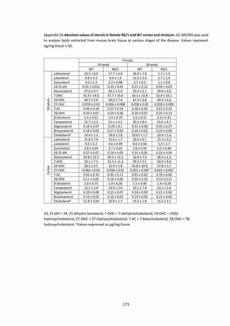
171
Appendix 2b Absolute values of sterols in female R6/1 and WT cortex and striatum. GC-MS/MS was used
to analyse lipids extracted from mouse brain tissue at various stages of the disease. Values represent
ng/mg tissue ± SD.
24, 25 diH = 24, 25 dihydro lanosterol; 7-DHC = 7-dehydrocholesterol; 24-OHC = 24(S)-
hydroxycholesterol; 27-OHC = 27-hydroxycholesterol; 7-KC = 7-ketocholesterol; 7β-OHC = 7β-
hydroxycholesterol. aValues expressed as µg/mg tissue.
Female
24 week 28 week
WT R6/1 WT R6/1
Stri
atu
m
Lathosterol 33.5 ± 8.9 17.7 ± 6.8 36.4 ± 7.8 5.7 ± 1.9
Lanosterol 9.8 ± 4.3 6.4 ± 1.9 13.3 ± 3.0 2.7 ± 1.4
Zymosterol 3.0 ± 1.2 2.3 ± 0.98 2.7 ± 0.3 1.1 ± 0.8
24,25 diH 0.33 ± 0.012 0.23 ± 0.05 0.21 ± 0.12 0.04 ± 0.03
Desmosterol 37.0 ± 6.7 45.1 ± 5.4 33.4 ± 5.1 39.4 ± 4.0
7-DHC 42.9 ± 14.0 47.7 ± 16.6 56.5 ± 10.8 55.4 ± 16.1
24-OHC 46.5 ± 5.6 43.6 ± 7.4 61.9 ± 6.8 49.3 ± 4.6
27-OHC 0.078 ± 0.02 0.056 ± 0.008 0.058 ± 0.02 0.028 ± 0.009
7-KC 0.49 ± 0.18 0.57 ± 0.19 0.38 ± 0.05 0.57 ± 0.19
7β-OHC 0.20 ± 0.07 0.24 ± 0.06 0.20 ± 0.07 0.25 ± 0.13
β-Sitosterol 2.4 ± 0.62 3.4 ± 0.79 2.6 ± 0.31 3.3 ± 0.31
Campesterol 15.7 ± 2.3 21.1 ± 4.5 30.1 ± 8.4 31.6 ± 4.7
Stigmasterol 0.19 ± 0.07 0.29 ± 0.1 0.31 ± 0.09 0.55 ± 0.27
Brassicasterol 0.18 ± 0.03 0.27 ± 0.04 0.20 ± 0.02 0.23 ± 0.09
Cholesterola 16.4 ± 1.5 18.0 ± 2.8 20.67 ± 1.7 20.4 ± 1.6
Co
rtex
Lathosterol 31.8 ± 7.9 15.6 ± 1.7 30.6 ± 6.1 15.3 ± 4.0
Lanosterol 9.5 ± 2.2 4.6 ± 0.99 9.6 ± 0.94 5.3 ± 1.7
Zymosterol 3.8 ± 0.94 2.7 ± 0.65 2.8 ± 0.50 2.0 ± 0.48
24,25 diH 0.27 ± 0.07 0.14 ± 0.05 0.32 ± 0.05 0.23 ± 0.04
Desmosterol 34.8 ± 12.5 39.5 ± 12.2 32.8 ± 7.9 38.5 ± 4.3
7-DHC 24.1 ± 7.5 31.3 ± 11.3 49.2 ± 5.5 50.4 ± 9.6
24-OHC 38.5 ± 9.5 32.9 ± 5.8 45.8 ± 10.9 37.8 ± 3.7
27-OHC 0.060 ± 0.03 0.039 ± 0.01 0.055 ± 0.007 0.033 ± 0.007
7-KC 0.33 ± 0.10 0.35 ± 0.11 0.91 ± 0.62 0.78 ± 0.68
7β-OHC 0.11 ± 0.02 0.18 ± 0.06 0.35 ± 0.35 0.23 ± 0.11
β-Sitosterol 2.0 ± 0.75 1.9 ± 0.36 1.5 ± 0.40 1.4 ± 0.28
Campesterol 12.1 ± 3.6 13.9 ± 2.0 23.1 ± 7.8 23.1 ± 3.8
Stigmasterol 0.19 ± 0.08 0.13 ± 0.07 0.24 ± 0.09 0.21 ± 0.06
Brassicasterol 0.14 ± 0.05 0.16 ± 0.01 0.14 ± 0.05 0.14 ± 0.03
Cholesterola 11.8 ± 3.04 10.9 ± 1.7 15.5 ± 1.8 15.2 ± 3.1

172
Appendix 3a Absolute values of sterols measured in combined sexes R6/1 and WT cortex and striatum.
GC-MS/MS was used to analyse lipids extracted from mouse brain tissue at various stages of the disease.
Values represent ng/mg tissue ± SD.
24, 25 diH = 24, 25 dihydro lanosterol; 7-DHC = 7-dehydrocholesterol; 24-OHC = 24(S)-
hydroxycholesterol; 27-OHC = 27-hydroxycholesterol; 7-KC = 7-ketocholesterol; 7β-OHC = 7β-
hydroxycholesterol. aValues expressed as µg/mg tissue.
Combined sexes
6 week 12 week 20 week
WT R6/1 WT R6/1 WT R6/1
Stri
atu
m
Lathosterol 56.5 ± 9.6 38.8 ± 7.8 45.0 ± 11.3 19.8 ± 5.6 44.6 ± 6.0 18.3 ± 5.9
Lanosterol 15.2 ± 5.4 11.5 ± 3.6 12.9 ± 4.8 8.1 ± 2.5 14.3 ± 3.4 8.5 ± 3.2
Zymosterol 5.0 ± 1.2 3.8 ± 0.88 3.5 ± 0.9 2.2 ± 0.6 3.0 ± 0.65 2.0 ± 0.65
24,25 diH 0.38 ± 0.21 0.29 ± 0.19 0.44 ± 0.22 0.29 ± 0.19 0.28 ± 0.05 0.20 ± 0.05
Desmosterol 55.5 ± 8.9 49.6 ± 9.9 40.2 ± 12.4 40.1 ± 11.9 34.5 ± 5.0 41.5 ± 9.5
7-DHC 66.6 ± 14.7 52.2 ± 7.8 50.8 ± 21.6 50.5 ± 15.8 50.2 ± 16.6 45.2 ± 13.2
24-OHC 43.7 ± 9.5 38.2 ± 5.6 45.5 ± 4.1 41.0 ± 3.6 57.1 ± 8.5 49.1 ± 11.4
27-OHC 0.037 ± 0.02 0.033 ± 0.01 0.071 ± 0.02 0.047 ± 0.01 0.77 ± 0.02 0.51 ± 0.15
7-KC 0.74 ± 0.40 0.48 ± 0.26 0.59 ± 0.17 0.67 ± 0.15 0.032 ± 0.006 0.030 ± 0.008
7β-OHC 0.36 ± 0.19 0.27 ± 0.14 0.42 ± 0.15 0.54 ± 0.22 0.24 ± 0.005 0.025 ± 0.007
β-Sitosterol 2.6 ± 1.0 2.2 ± 0.39 2.2 ± 0.66 2.7 ± 0.62 1.9 ± 0.35 1.9 ± 0.37
Campesterol 17.2 ± 3.1 16.3 ± 3.2 16.9 ± 2.2 18.4 ± 2.3 18.3 ± 4.3 17.5 ± 4.4
Stigmasterol 0.38 ± 0.19 0.31 ± 0.09 0.12 ± 0.09 0.18 ± 0.11 0.23 ± 0.12 0.26 ± 0.12
Brassicasterol 0.11 ± 0.06 0.079 ± 0.05 0.14 ± 0.06 0.17 ± 0.05 0.15 ± 0.010 0.17 ± 0.007
Cholesterola 14.8 ± 1.3 13.3 ± 2.9 18.5 ± 3.4 19.9 ± 1.3 22.3 ± 3.5 23.3 ± 5.2
Co
rtex
Lathosterol 36.6 ± 9.7 27.0 ± 10.2 39.7 ± 9.3 18.3 ± 6.1 35.8 ± 9.5 17.5 ± 5.1
Lanosterol 8.6 ± 2.6 7.8 ± 2.0 9.2 ± 3.2 6.4 ± 1.5 7.7 ± 2.4 4.5 ± 1.3
Zymosterol 2.5 ± 0.55 2.8 ± 0.84 3.0 ± 0.6 2.7 ± 0.5 2.5 ± 0.4 1.9 ± 0.4
24,25 diH 0.39 ± 0.12 0.37 ± 0.12 0.34 ± 0.13 0.23 ± 0.09 0.32 ± 0.12 0.25 ± 0.15
Desmosterol 31.3 ± 11.2 36.6 ± 9.2 34.1 ± 15.7 45.3 ± 17.0 35.3 ± 14.0 45.7 ± 5.5
7-DHC 40.0 ± 12.6 40.1 ± 9.2 42.8 ± 6.4 45.3 ± 14.4 43.3 ± 6.5 45.3 ± 14.4
24-OHC 27.5 ± 7.5 29.3 ± 7.5 30.1 ± 6.0 33.0 ± 5.1 39.6 ± 13.0 37.3 ± 6.5
27-OHC 0.036 ± 0.01 0.032 ± 0.007 0.041 ± 0.01 0.043 ± 0.01 0.047 ± 0.04 0.044 ± 0.03
7-KC 0.37 ± 0.18 0.33 ± 0.11 0.50 ± 0.12 0.48 ± 0.14 0.41 ± 0.17 0.54 ± 0.20
7β-OHC 0.19 ± 0.11 0.20 ± 0.12 0.38 ± 0.14 0.36 ± 0.13 0.13 ± 0.045 0.16 ± 0.033
β-Sitosterol 1.2 ± 0.63 1.1 ± 0.45 1.85 ± 0.40 1.94 ± 0.49 1.5 ± 0.34 1.8 ± 0.57
Campesterol 13.7 ± 2.8 12.7 ± 3.3 14.3 ± 3.2 13.5 ± 2.3 23.6 ± 7.4 23.3 ± 4.3
Stigmasterol 0.19 ± 0.16 0.18 ± 0.11 0.14 ± 0.08 0.15 ± 0.08 0.30 ± 0.12 0.36 ± 0.14
Brassicasterol 0.063 ± 0.01 0.057 ± 0.02 0.14 ± 0.05 0.14 ± 0.04 0.14 ± 0.07 0.15 ± 0.05
Cholesterola 10.1 ± 2.4 9.2 ± 2.5 15.4 ± 2.7 15.0 ± 2.1 11.0 ± 2.5 11.9 ± 1.7
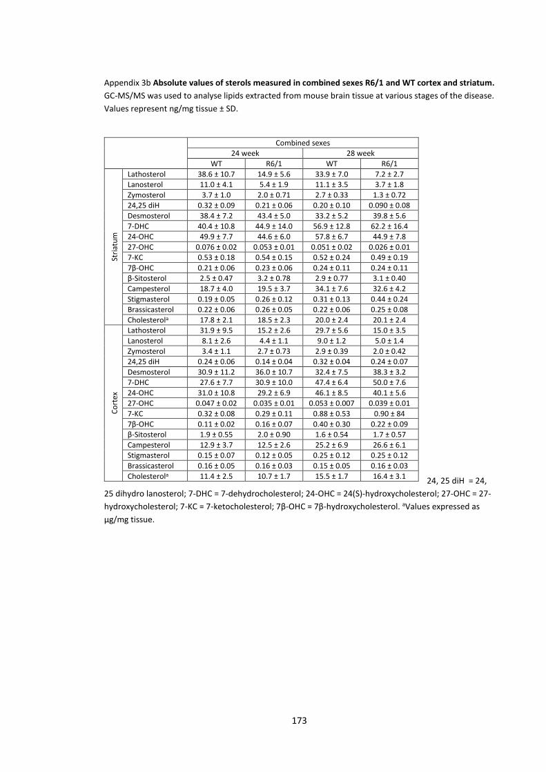
173
Appendix 3b Absolute values of sterols measured in combined sexes R6/1 and WT cortex and striatum.
GC-MS/MS was used to analyse lipids extracted from mouse brain tissue at various stages of the disease.
Values represent ng/mg tissue ± SD.
24, 25 diH = 24,
25 dihydro lanosterol; 7-DHC = 7-dehydrocholesterol; 24-OHC = 24(S)-hydroxycholesterol; 27-OHC = 27-
hydroxycholesterol; 7-KC = 7-ketocholesterol; 7β-OHC = 7β-hydroxycholesterol. aValues expressed as
µg/mg tissue.
Combined sexes
24 week 28 week
WT R6/1 WT R6/1
Stri
atu
m
Lathosterol 38.6 ± 10.7 14.9 ± 5.6 33.9 ± 7.0 7.2 ± 2.7
Lanosterol 11.0 ± 4.1 5.4 ± 1.9 11.1 ± 3.5 3.7 ± 1.8
Zymosterol 3.7 ± 1.0 2.0 ± 0.71 2.7 ± 0.33 1.3 ± 0.72
24,25 diH 0.32 ± 0.09 0.21 ± 0.06 0.20 ± 0.10 0.090 ± 0.08
Desmosterol 38.4 ± 7.2 43.4 ± 5.0 33.2 ± 5.2 39.8 ± 5.6
7-DHC 40.4 ± 10.8 44.9 ± 14.0 56.9 ± 12.8 62.2 ± 16.4
24-OHC 49.9 ± 7.7 44.6 ± 6.0 57.8 ± 6.7 44.9 ± 7.8
27-OHC 0.076 ± 0.02 0.053 ± 0.01 0.051 ± 0.02 0.026 ± 0.01
7-KC 0.53 ± 0.18 0.54 ± 0.15 0.52 ± 0.24 0.49 ± 0.19
7β-OHC 0.21 ± 0.06 0.23 ± 0.06 0.24 ± 0.11 0.24 ± 0.11
β-Sitosterol 2.5 ± 0.47 3.2 ± 0.78 2.9 ± 0.77 3.1 ± 0.40
Campesterol 18.7 ± 4.0 19.5 ± 3.7 34.1 ± 7.6 32.6 ± 4.2
Stigmasterol 0.19 ± 0.05 0.26 ± 0.12 0.31 ± 0.13 0.44 ± 0.24
Brassicasterol 0.22 ± 0.06 0.26 ± 0.05 0.22 ± 0.06 0.25 ± 0.08
Cholesterola 17.8 ± 2.1 18.5 ± 2.3 20.0 ± 2.4 20.1 ± 2.4
Co
rtex
Lathosterol 31.9 ± 9.5 15.2 ± 2.6 29.7 ± 5.6 15.0 ± 3.5
Lanosterol 8.1 ± 2.6 4.4 ± 1.1 9.0 ± 1.2 5.0 ± 1.4
Zymosterol 3.4 ± 1.1 2.7 ± 0.73 2.9 ± 0.39 2.0 ± 0.42
24,25 diH 0.24 ± 0.06 0.14 ± 0.04 0.32 ± 0.04 0.24 ± 0.07
Desmosterol 30.9 ± 11.2 36.0 ± 10.7 32.4 ± 7.5 38.3 ± 3.2
7-DHC 27.6 ± 7.7 30.9 ± 10.0 47.4 ± 6.4 50.0 ± 7.6
24-OHC 31.0 ± 10.8 29.2 ± 6.9 46.1 ± 8.5 40.1 ± 5.6
27-OHC 0.047 ± 0.02 0.035 ± 0.01 0.053 ± 0.007 0.039 ± 0.01
7-KC 0.32 ± 0.08 0.29 ± 0.11 0.88 ± 0.53 0.90 ± 84
7β-OHC 0.11 ± 0.02 0.16 ± 0.07 0.40 ± 0.30 0.22 ± 0.09
β-Sitosterol 1.9 ± 0.55 2.0 ± 0.90 1.6 ± 0.54 1.7 ± 0.57
Campesterol 12.9 ± 3.7 12.5 ± 2.6 25.2 ± 6.9 26.6 ± 6.1
Stigmasterol 0.15 ± 0.07 0.12 ± 0.05 0.25 ± 0.12 0.25 ± 0.12
Brassicasterol 0.16 ± 0.05 0.16 ± 0.03 0.15 ± 0.05 0.16 ± 0.03
Cholesterola 11.4 ± 2.5 10.7 ± 1.7 15.5 ± 1.7 16.4 ± 3.1
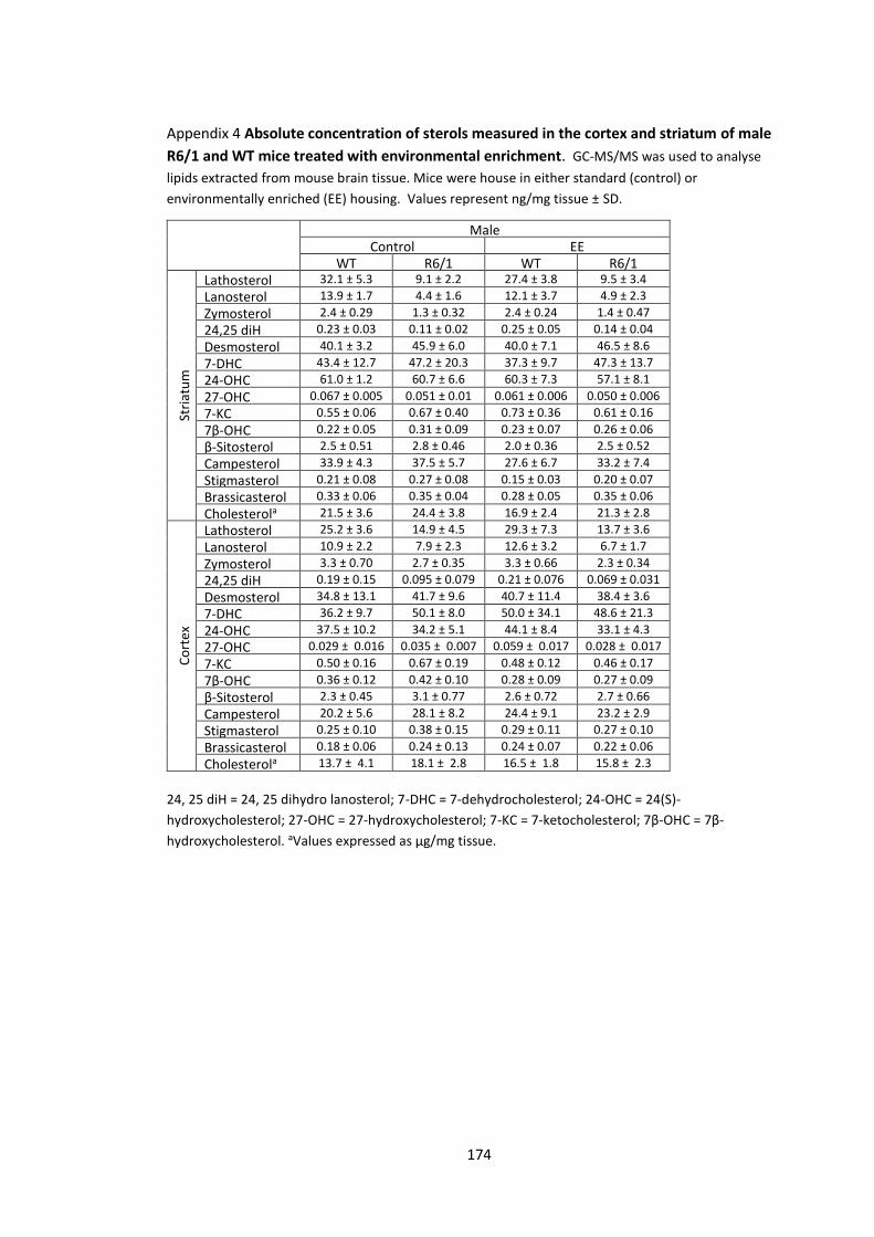
174
Appendix 4 Absolute concentration of sterols measured in the cortex and striatum of male
R6/1 and WT mice treated with environmental enrichment. GC-MS/MS was used to analyse
lipids extracted from mouse brain tissue. Mice were house in either standard (control) or
environmentally enriched (EE) housing. Values represent ng/mg tissue ± SD.
24, 25 diH = 24, 25 dihydro lanosterol; 7-DHC = 7-dehydrocholesterol; 24-OHC = 24(S)-
hydroxycholesterol; 27-OHC = 27-hydroxycholesterol; 7-KC = 7-ketocholesterol; 7β-OHC = 7β-
hydroxycholesterol. aValues expressed as µg/mg tissue.
Male Control EE
WT R6/1 WT R6/1
Stri
atu
m
Lathosterol 32.1 ± 5.3 9.1 ± 2.2 27.4 ± 3.8 9.5 ± 3.4
Lanosterol 13.9 ± 1.7 4.4 ± 1.6 12.1 ± 3.7 4.9 ± 2.3
Zymosterol 2.4 ± 0.29 1.3 ± 0.32 2.4 ± 0.24 1.4 ± 0.47
24,25 diH 0.23 ± 0.03 0.11 ± 0.02 0.25 ± 0.05 0.14 ± 0.04
Desmosterol 40.1 ± 3.2 45.9 ± 6.0 40.0 ± 7.1 46.5 ± 8.6
7-DHC 43.4 ± 12.7 47.2 ± 20.3 37.3 ± 9.7 47.3 ± 13.7
24-OHC 61.0 ± 1.2 60.7 ± 6.6 60.3 ± 7.3 57.1 ± 8.1
27-OHC 0.067 ± 0.005 0.051 ± 0.01 0.061 ± 0.006 0.050 ± 0.006
7-KC 0.55 ± 0.06 0.67 ± 0.40 0.73 ± 0.36 0.61 ± 0.16
7β-OHC 0.22 ± 0.05 0.31 ± 0.09 0.23 ± 0.07 0.26 ± 0.06
β-Sitosterol 2.5 ± 0.51 2.8 ± 0.46 2.0 ± 0.36 2.5 ± 0.52
Campesterol 33.9 ± 4.3 37.5 ± 5.7 27.6 ± 6.7 33.2 ± 7.4
Stigmasterol 0.21 ± 0.08 0.27 ± 0.08 0.15 ± 0.03 0.20 ± 0.07
Brassicasterol 0.33 ± 0.06 0.35 ± 0.04 0.28 ± 0.05 0.35 ± 0.06
Cholesterola 21.5 ± 3.6 24.4 ± 3.8 16.9 ± 2.4 21.3 ± 2.8
Co
rtex
Lathosterol 25.2 ± 3.6 14.9 ± 4.5 29.3 ± 7.3 13.7 ± 3.6
Lanosterol 10.9 ± 2.2 7.9 ± 2.3 12.6 ± 3.2 6.7 ± 1.7
Zymosterol 3.3 ± 0.70 2.7 ± 0.35 3.3 ± 0.66 2.3 ± 0.34
24,25 diH 0.19 ± 0.15 0.095 ± 0.079 0.21 ± 0.076 0.069 ± 0.031
Desmosterol 34.8 ± 13.1 41.7 ± 9.6 40.7 ± 11.4 38.4 ± 3.6
7-DHC 36.2 ± 9.7 50.1 ± 8.0 50.0 ± 34.1 48.6 ± 21.3
24-OHC 37.5 ± 10.2 34.2 ± 5.1 44.1 ± 8.4 33.1 ± 4.3
27-OHC 0.029 ± 0.016 0.035 ± 0.007 0.059 ± 0.017 0.028 ± 0.017
7-KC 0.50 ± 0.16 0.67 ± 0.19 0.48 ± 0.12 0.46 ± 0.17
7β-OHC 0.36 ± 0.12 0.42 ± 0.10 0.28 ± 0.09 0.27 ± 0.09
β-Sitosterol 2.3 ± 0.45 3.1 ± 0.77 2.6 ± 0.72 2.7 ± 0.66
Campesterol 20.2 ± 5.6 28.1 ± 8.2 24.4 ± 9.1 23.2 ± 2.9
Stigmasterol 0.25 ± 0.10 0.38 ± 0.15 0.29 ± 0.11 0.27 ± 0.10
Brassicasterol 0.18 ± 0.06 0.24 ± 0.13 0.24 ± 0.07 0.22 ± 0.06
Cholesterola 13.7 ± 4.1 18.1 ± 2.8 16.5 ± 1.8 15.8 ± 2.3
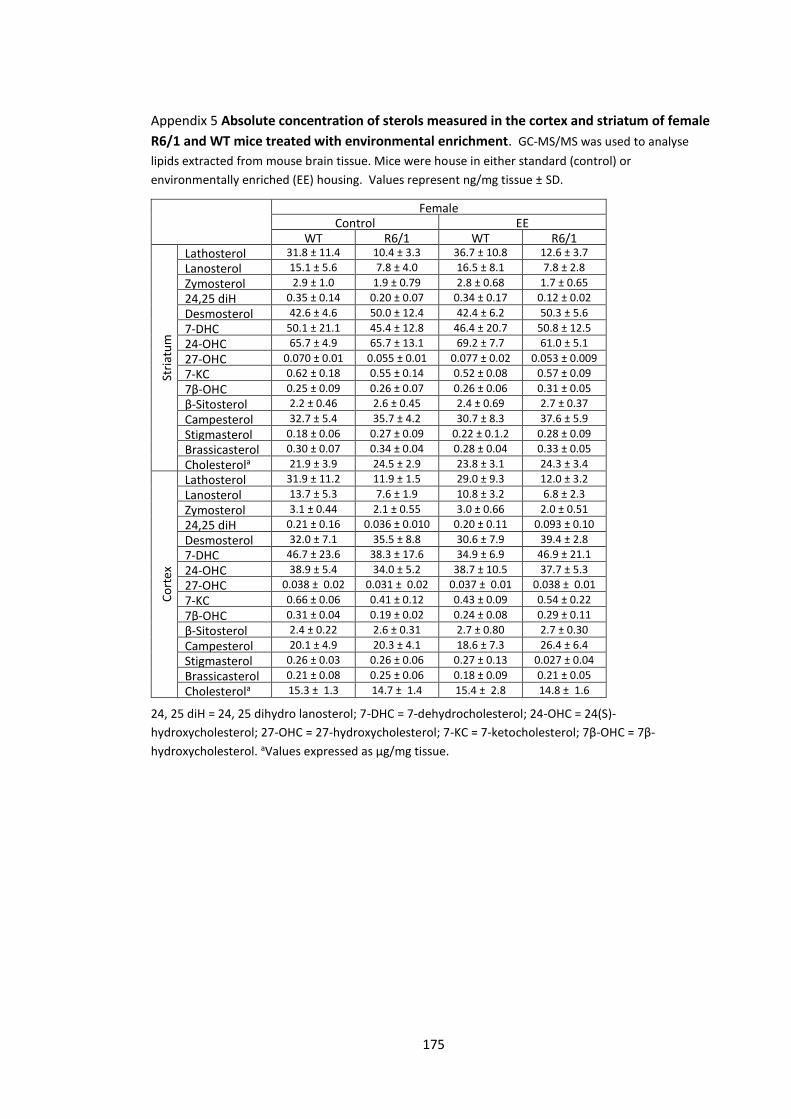
175
Appendix 5 Absolute concentration of sterols measured in the cortex and striatum of female
R6/1 and WT mice treated with environmental enrichment. GC-MS/MS was used to analyse
lipids extracted from mouse brain tissue. Mice were house in either standard (control) or
environmentally enriched (EE) housing. Values represent ng/mg tissue ± SD.
24, 25 diH = 24, 25 dihydro lanosterol; 7-DHC = 7-dehydrocholesterol; 24-OHC = 24(S)-
hydroxycholesterol; 27-OHC = 27-hydroxycholesterol; 7-KC = 7-ketocholesterol; 7β-OHC = 7β-
hydroxycholesterol. aValues expressed as µg/mg tissue.
Female Control EE
WT R6/1 WT R6/1
Stri
atu
m
Lathosterol 31.8 ± 11.4 10.4 ± 3.3 36.7 ± 10.8 12.6 ± 3.7
Lanosterol 15.1 ± 5.6 7.8 ± 4.0 16.5 ± 8.1 7.8 ± 2.8
Zymosterol 2.9 ± 1.0 1.9 ± 0.79 2.8 ± 0.68 1.7 ± 0.65
24,25 diH 0.35 ± 0.14 0.20 ± 0.07 0.34 ± 0.17 0.12 ± 0.02
Desmosterol 42.6 ± 4.6 50.0 ± 12.4 42.4 ± 6.2 50.3 ± 5.6
7-DHC 50.1 ± 21.1 45.4 ± 12.8 46.4 ± 20.7 50.8 ± 12.5
24-OHC 65.7 ± 4.9 65.7 ± 13.1 69.2 ± 7.7 61.0 ± 5.1
27-OHC 0.070 ± 0.01 0.055 ± 0.01 0.077 ± 0.02 0.053 ± 0.009
7-KC 0.62 ± 0.18 0.55 ± 0.14 0.52 ± 0.08 0.57 ± 0.09
7β-OHC 0.25 ± 0.09 0.26 ± 0.07 0.26 ± 0.06 0.31 ± 0.05
β-Sitosterol 2.2 ± 0.46 2.6 ± 0.45 2.4 ± 0.69 2.7 ± 0.37
Campesterol 32.7 ± 5.4 35.7 ± 4.2 30.7 ± 8.3 37.6 ± 5.9
Stigmasterol 0.18 ± 0.06 0.27 ± 0.09 0.22 ± 0.1.2 0.28 ± 0.09
Brassicasterol 0.30 ± 0.07 0.34 ± 0.04 0.28 ± 0.04 0.33 ± 0.05
Cholesterola 21.9 ± 3.9 24.5 ± 2.9 23.8 ± 3.1 24.3 ± 3.4
Co
rtex
Lathosterol 31.9 ± 11.2 11.9 ± 1.5 29.0 ± 9.3 12.0 ± 3.2
Lanosterol 13.7 ± 5.3 7.6 ± 1.9 10.8 ± 3.2 6.8 ± 2.3
Zymosterol 3.1 ± 0.44 2.1 ± 0.55 3.0 ± 0.66 2.0 ± 0.51
24,25 diH 0.21 ± 0.16 0.036 ± 0.010 0.20 ± 0.11 0.093 ± 0.10
Desmosterol 32.0 ± 7.1 35.5 ± 8.8 30.6 ± 7.9 39.4 ± 2.8
7-DHC 46.7 ± 23.6 38.3 ± 17.6 34.9 ± 6.9 46.9 ± 21.1
24-OHC 38.9 ± 5.4 34.0 ± 5.2 38.7 ± 10.5 37.7 ± 5.3
27-OHC 0.038 ± 0.02 0.031 ± 0.02 0.037 ± 0.01 0.038 ± 0.01
7-KC 0.66 ± 0.06 0.41 ± 0.12 0.43 ± 0.09 0.54 ± 0.22
7β-OHC 0.31 ± 0.04 0.19 ± 0.02 0.24 ± 0.08 0.29 ± 0.11
β-Sitosterol 2.4 ± 0.22 2.6 ± 0.31 2.7 ± 0.80 2.7 ± 0.30
Campesterol 20.1 ± 4.9 20.3 ± 4.1 18.6 ± 7.3 26.4 ± 6.4
Stigmasterol 0.26 ± 0.03 0.26 ± 0.06 0.27 ± 0.13 0.027 ± 0.04
Brassicasterol 0.21 ± 0.08 0.25 ± 0.06 0.18 ± 0.09 0.21 ± 0.05
Cholesterola 15.3 ± 1.3 14.7 ± 1.4 15.4 ± 2.8 14.8 ± 1.6
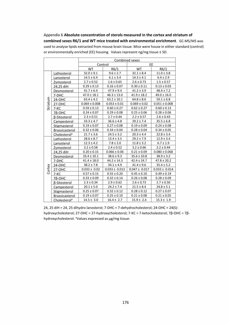
176
Appendix 6 Absolute concentration of sterols measured in the cortex and striatum of
combined sexes R6/1 and WT mice treated with environmental enrichment. GC-MS/MS was
used to analyse lipids extracted from mouse brain tissue. Mice were house in either standard (control)
or environmentally enriched (EE) housing. Values represent ng/mg tissue ± SD.
24, 25 diH = 24, 25 dihydro lanosterol; 7-DHC = 7-dehydrocholesterol; 24-OHC = 24(S)-
hydroxycholesterol; 27-OHC = 27-hydroxycholesterol; 7-KC = 7-ketocholesterol; 7β-OHC = 7β-
hydroxycholesterol. aValues expressed as µg/mg tissue.
Combined sexes Control EE
WT R6/1 WT R6/1
Stri
atu
m
Lathosterol 32.0 ± 9.1 9.6 ± 2.7 32.1 ± 8.4 11.0 ± 3.8
Lanosterol 14.5 ± 6.4 6.1 ± 3.4 14.3 ± 4.1 6.4 ± 2.9
Zymosterol 2.7 ± 0.52 1.6 ± 0.65 2.6 ± 0.73 1.5 ± 0.57
24,25 diH 0.29 ± 0.13 0.16 ± 0.07 0.30 ± 0.11 0.13 ± 0.03
Desmosterol 41.7 ± 6.4 47.9 ± 9.4 41.2 ± 3.9 48.4 ± 7.2
7-DHC 47.0 ± 18.1 46.3 ± 13.0 41.9 ± 18.2 49.0 ± 16.0
24-OHC 63.4 ± 4.2 63.2 ± 10.1 64.8 ± 8.6 59.1 ± 6.8
27-OHC 0.069 ± 0.008 0.053 ± 0.01 0.069 ± 0.02 0.051 ± 0.008
7-KC 0.59 ± 0.13 0.60 ± 0.27 0.62 ± 0.27 0.60 ± 0.13
7β-OHC 0.24 ± 0.07 0.29 ± 0.08 0.25 ± 0.06 0.28 ± 0.06
β-Sitosterol 2.3 ± 0.51 2.7 ± 0.44 2.2 ± 0.57 2.6 ± 0.43
Campesterol 33.3 ± 4.7 36.6 ± 4.8 29.2 ± 7.4 35.5 ± 6.8
Stigmasterol 0.19 ± 0.07 0.27 ± 0.08 0.19 ± 0.09 0.24 ± 0.08
Brassicasterol 0.32 ± 0.06 0.34 ± 0.04 0.28 ± 0.04 0.34 ± 0.05
Cholesterola 21.7 ± 3.6 24.5 ± 3.2 20.3 ± 4.4 22.8 ± 3.4
Co
rtex
Lathosterol 28.6 ± 8.7 13.4 ± 3.5 29.2 ± 7.9 12.9 ± 3.4
Lanosterol 12.3 ± 4.2 7.8 ± 2.0 11.8 ± 3.2 6.7 ± 1.9
Zymosterol 3.2 ± 0.58 2.4 ± 0.52 3.2 ± 0.66 2.2 ± 0.44
24,25 diH 0.20 ± 0.15 0.066 ± 0.06 0.21 ± 0.09 0.080 ± 0.068
Desmosterol 33.4 ± 10.1 38.6 ± 9.3 35.6 ± 10.8 38.9 ± 3.2
7-DHC 41.4 ± 18.0 44.2 ± 14.3 42.4 ± 24.7 47.8 ± 20.2
24-OHC 38.2 ± 7.8 34.1 ± 4.9 41.4 ± 9.6 35.4 ± 5.2
27-OHC 0.033 ± 0.02 0.033 ± 0.013 0.047 ± 0.017 0.033 ± 0.014
7-KC 0.57 ± 0.15 0.55 ± 0.20 0.45 ± 0.10 0.49 ± 0.19
7β-OHC 0.33 ± 0.09 0.32 ± 0.14 0.26 ± 0.08 0.28 ± 0.09
β-Sitosterol 2.3 ± 0.34 2.9 ± 0.62 2.6 ± 0.73 2.7 ± 0.50
Campesterol 20.1 ± 5.0 24.2 ± 7.4 21.5 ± 8.4 24.8 ± 5.1
Stigmasterol 0.25 ± 0.07 0.32 ± 0.12 0.28 ± 0.12 0.27 ± 0.07
Brassicasterol 0.19 ± 0.07 0.25 ± 0.10 0.21 ± 0.08 0.21 ± 0.05
Cholesterola 14.5 ± 3.0 16.4 ± 2.7 15.9 ± 2.4 15.3 ± 1.9

177
Appendix 7 Absolute concentration of sterols measured in the cortex and striatum of male
R6/1 and WT mice receiving dietary supplementation. GC-MS/MS was used to analyse lipids
extracted from mouse brain tissue. Mice received either control or berry extract (BE) supplemented
diets. Values represent ng/mg tissue ± SD.
24, 25 diH = 24, 25 dihydro lanosterol; 7-DHC = 7-dehydrocholesterol; 24-OHC = 24(S)-
hydroxycholesterol; 27-OHC = 27-hydroxycholesterol; 7-KC = 7-ketocholesterol; 7β-OHC = 7β-
hydroxycholesterol. aValues expressed as µg/mg tissue.
Male Control BE
WT R6/1 WT R6/1
Stri
atu
m
Lathosterol 33.9 ± 7.7 12.2 ± 5.2 34.1 ± 11.9 9.7 ± 2.7
Lanosterol 13.8 ± 4.6 7.1 ± 2.8 13.1 ± 5.9 4.5 ± 1.9
Zymosterol 2.2 ± 0.7 1.4 ± 0.6 2.5 ± 0.6 1.1 ± 0.6
24,25 diH 0.25 ± 0.11 0.11 ± 0.05 0.21 ± 0.01 0.13 ± 0.07
Desmosterol 41.6 ± 5.4 40.9 ± 8.9 43.4 ± 8.1 42.2 ± 5.9
7-DHC 49.5 ± 21.9 68.4 ± 31.4 65.5 ± 15.4 72.5 ± 16.1
24-OHC 64.5 ± 3.3 55.5 ± 13.8 67.5 ± 5.5 58.5 ± 3.8
27-OHC 0.078 ± 0.009 0.044 ± 0.017 0.078 ± 0.013 0.048 ± 0.010
7-KC 0.56 ± 0.31 1.36 ± 1.1 1.04 ± 0.57 1.60 ± 0.97 7β-OHC 0.32 ± 0.13 0.52 ± 0.20 0.59 ± 0.26 0.87 ± 0.45 β-Sitosterol 3.8 ± 0.7 4.5 ± 0.9 4.3 ± 0.8 4.5 ± 1.2 Campesterol 40.9 ± 7.7 39.4 ± 5.3 42.2 ± 11.9 41.5 ± 12.0 Stigmasterol 0.48 ± 0.13 0.59 ± 0.16 0.50 ± 0.08 0.59 ± 0.18 Brassicasterol 0.44 ± 0.09 0.42 ± 0.08 0.45 ± 0.11 0.44 ± 0.07 Cholesterola 15.5 ± 1.2 15.8 ± 1.8 16.6 ± 2.3 16.8 ± 1.6
Co
rtex
Lathosterol 36.6 ± 5.2 16.9 ± 3.5 27.4 ± 5.0 14.8 ± 4.4 Lanosterol 11.8 ± 1.5 7.8 ± 1.9 10.1 ± 2.7 7.1 ± 3.1 Zymosterol 2.0 ± 0.7 2.5 ± 1.4 1.7 ± 0.9 3.0 ± 1.3 24,25 diH 0.22 ± 0.12 0.10 ± 0.05 0.16 ± 0.04 0.09 ± 0.03 Desmosterol 32.0 ± 7.2 34.0 ± 5.7 39.2 ± 11.1 42.5 ± 11.2 7-DHC 49.8 ± 17.1 53.8 ± 15.7 46.4 ± 12.0 50.3 ± 9.7 24-OHC 53.3 ± 11.2 49.8 ± 10.9 57.7 ± 15.8 47.9 ± 8.1 27-OHC 0.051 ±
0.015 0.031 ± 0.010
0.047 ± 0.010
0.027 ± 0.008 7-KC 0.42 ± 0.11 0.36 ± 0.10 0.41 ± 0.14 0.37 ± 0.13
7β-OHC 0.21 ± 0.08 0.22 ± 0.08 0.21 ± 0.07 0.19 ± 0.04 β-Sitosterol 2.4 ± 0.58 2.4 ± 0.31 1.9 ± 0.38 2.0 ± 0.17 Campesterol 39.7 ± 5.2 36.9 ± 8.0 35.1 ± 7.4 37.0 ± 8.8 Stigmasterol 0.26 ± 0.13 0.26 ± 0.05 0.20 ± 0.08 0.23 ± 0.05 Brassicasterol 0.40 ± 0.06 0.39 ± 0.21 0.38 ± 0.12 0.40 ± 0.13 Cholesterola 12.7 ± 1.8 13.0 ± 2.0 13.6 ± 1.4 13.8 ± 1.8

178
Appendix 8 Absolute concentration of sterols measured in the cortex and striatum of female
R6/1 and WT mice receiving dietary supplementation. GC-MS/MS was used to analyse lipids
extracted from mouse brain tissue. Mice received either control or berry extract (BE) supplemented
diets. Values represent ng/mg tissue ± SD.
24, 25 diH = 24, 25 dihydro lanosterol; 7-DHC = 7-dehydrocholesterol; 24-OHC = 24(S)-
hydroxycholesterol; 27-OHC = 27-hydroxycholesterol; 7-KC = 7-ketocholesterol; 7β-OHC = 7β-
hydroxycholesterol. aValues expressed as µg/mg tissue.
Female Control BE
WT R6/1 WT R6/1
Stri
atu
m
Lathosterol 32.3 ± 9.2 11.3 ± 3.9 32.6 ± 8.9 11.6 ± 3.4
Lanosterol 11.8 ± 5.1 5.7 ± 2.7 11.5 ± 4.2 6.6 ± 2.7
Zymosterol 2.4 ± 0.8 1.3 ± 0.3 2.3 ± 0.8 1.4 ± 0.4
24,25 diH 0.23 ± 0.09 0.17 ± 0.08 0.24 ± 0.15 0.14 ± 0.01
Desmosterol 35.3 ± 6.7 45.1 ± 6.5 38.1 ± 7.2 47.2 ± 8.4
7-DHC 67.2 ± 29.p 80.3 ± 33.5 64.3 ± 34.1 72.3 ± 23.6
24-OHC 61.0 ± 8.6 55.6 ± 7.7 66.8 ± 3.2 54.6 ± 11.1
27-OHC 0.076 ± 0.014 0.055 ± 0.014 0.069 ± 0.013 0.052 ± 0.015
7-KC 0.63 ± 0.16 0.64 ± 0.18 0.98 ± 0.50 0.64 ± 0.15 7β-OHC 0.47 ± 0.24 0.41 ± 0.11 0.57 ± 0.42 0.38 ± 0.12 β-Sitosterol 3.6 ± 0.9 5.0 ± 1.7 4.5 ± 0.7 4.0 ± 0.6 Campesterol 26.0 ± 6.6 46.6 ± 7.9 36.5 ± 9.2 40.7 ± 7.6 Stigmasterol 0.49 ± 0.19 0.71 ± 0.24 0.69 ± 0.22 0.52 ± 0.14 Brassicasterol 0.25 ± 0.06 0.47 ± 0.12 0.37 ± 0.11 0.42 ± 0.06 Cholesterola 14.9 ± 2.2 16.6 ± 1.6 15.9 ± 2.3 16.3 ± 2.9
Co
rtex
Lathosterol 34.6 ± 9.7 15.6 ± 4.2 30.8 ± 13.9 14.3 ± 3.2 Lanosterol 12.0 ± 2.8 7.0 ± 1.7 10.1 ± 4.1 6.7 ± 1.8 Zymosterol 2.1 ± 1.3 2.6 ± 0.9 2.4 ± 1.2 2.2 ± 0.5 24,25 diH 0.14 ± 0.07 0.09 ± 0.05 0.15 ± 0.06 0.09 ± 0.03 Desmosterol 36.0 ± 6.3 43.4 ± 7.6 28.3 ± 8.2 45.7 ± 5.1 7-DHC 51.8 ± 12.2 55.5 ± 20.6 33.2 ± 9.4 54.1 ± 13.9 24-OHC 60.5 ± 9.9 50.0 ± 3.7 50.0 ± 13.1 52.9 ± 9.5 27-OHC 0.061 ±
0.015 0.044 ± 0.018
0.054 ± 0.023
0.047 ± 0.017 7-KC 0.29 ± 0.08 0.43 ± 0.12 0.47 ± 0.28 0.56 ± 0.35
7β-OHC 0.15 ± 0.04 0.19 ± 0.06 0.17 ± 0.02 0.21 ± 0.06 β-Sitosterol 2.5 ± 1.2 2.4 ± 0.64 1.8 ± 0.50 2.6 ± 0.39 Campesterol 23.5 ± 4.4 37.1 ± 11.1 27.9 ± 9.6 32.9 ± 9.4 Stigmasterol 0.24 ± 0.15 0.25 ± 0.12 0.22 ± 0.10 0.25 ± 0.06 Brassicasterol 0.22 ± 0.05 0.34 ± 0.10 0.26 ± 0.09 0.33 ± 1.5 Cholesterola 12.2 ± 1.0 13.6 ± 1.7 13.1 ± 1.5 13.4 ± 1.4
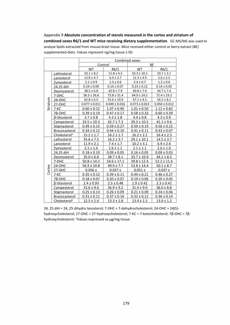
179
Appendix 9 Absolute concentration of sterols measured in the cortex and striatum of
combined sexes R6/1 and WT mice receiving dietary supplementation. GC-MS/MS was used to
analyse lipids extracted from mouse brain tissue. Mice received either control or berry extract (BE)
supplemented diets. Values represent ng/mg tissue ± SD.
24, 25 diH = 24, 25 dihydro lanosterol; 7-DHC = 7-dehydrocholesterol; 24-OHC = 24(S)-
hydroxycholesterol; 27-OHC = 27-hydroxycholesterol; 7-KC = 7-ketocholesterol; 7β-OHC = 7β-
hydroxycholesterol. aValues expressed as µg/mg tissue.
Combined sexes Control BE
WT R6/1 WT R6/1
Stri
atu
m
Lathosterol 33.1 ± 8.2 11.8 ± 4.5 33.3 ± 10.1 10.7 ± 3.1
Lanosterol 12.8 ± 4.7 6.4 ± 2.7 12.3 ± 4.9 5.6 ± 2.5
Zymosterol 2.1 ± 0.9 1.3 ± 0.6 2.4 ± 0.7 1.2 ± 0.6
24,25 diH 0.24 ± 0.09 0.14 ± 0.07 0.23 ± 0.12 0.14 ± 0.05
Desmosterol 38.5 ± 6.8 42.8 ± 7.9 40.8 ± 7.9 44.7 ± 7.4
7-DHC 58.3 ± 26.6 73.8 ± 31.4 64.9 ± 24.2 72.4 ± 19.2
24-OHC 62.8 ± 6.5 55.6 ± 10.9 67.1 ± 4.3 56.5 ± 8.2
27-OHC 0.077 ± 0.011 0.049 ± 0.016 0.073 ± 0.013 0.050 ± 0.012
7-KC 0.60 ± 0.22 1.07 ± 0.90 1.01 ± 0.50 1.12 ± 0.82 7β-OHC 0.39 ± 0.19 0.47 ± 0.17 0.58 ± 0.33 0.60 ± 0.39 β-Sitosterol 3.7 ± 0.8 4.3 ± 1.8 4.4 ± 0.8 4.3 ± 0.9 Campesterol 33.5 ± 10.3 42.7 ± 7.3 39.3 ± 10.5 41.1 ± 9.6 Stigmasterol 0.49 ± 0.15 0.59 ± 0.27 0.59 ± 0.19 0.56 ± 0.15 Brassicasterol 0.34 ± 0.12 0.44 ± 0.10 0.41 ± 0.11 0.43 ± 0.07 Cholesterola 15.2 ± 1.7 16.2 ± 1.7 16.2 ± 2.2 16.4 ± 2.3
Co
rtex
Lathosterol 35.6 ± 7.5 16.2 ± 3.7 29.1 ± 10.1 14.5 ± 3.7 Lanosterol 11.9 ± 2.1 7.4 ± 1.7 10.2 ± 3.1 6.9 ± 2.4 Zymosterol 2.1 ± 1.0 2.6 ± 1.1 2.1 ± 1.1 2.6 ± 1.0 24,25 diH 0.18 ± 0.10 0.09 ± 0.05 0.16 ± 0.05 0.09 ± 0.03 Desmosterol 35.0 ± 6.8 38.7 ± 8.1 33.7 ± 10.9 44.1 ± 8.5 7-DHC 50.8 ± 14.2 54.6 ± 17.1 39.8 ± 12.4 52.2 ± 11.6 24-OHC 56.9 ± 10.8 49.9 ± 7.7 53.8 ± 14.4 50.1 ± 8.7 27-OHC 0.056 ±
0.016 0.037 ± 0.016
0.051 ± 0.017
0.037 ± 0.016 7-KC 0.35 ± 0.12 0.39 ± 0.11 0.44 ± 0.21 0.46 ± 0.27
7β-OHC 0.18 ± 0.07 0.20 ± 0.07 0.19 ± 0.06 0.20 ± 0.05 β-Sitosterol 2.4 ± 0.93 2.5 ± 0.48 1.9 ± 0.42 2.3 ± 0.42 Campesterol 31.6 ± 9.6 36.9 ± 9.2 31.4 ± 9.0 36.0 ± 8.8 Stigmasterol 0.25 ± 0.13 0.26 ± 0.09 0.21 ± 0.09 0.24 ± 0.06 Brassicasterol 0.31 ± 0.11 0.37 ± 0.16 0.32 ± 0.12 0.36 ± 0.14 Cholesterola 12.5 ± 1.4 13.3 ± 1.8 13.4 ± 1.5 13.6 ± 1.2
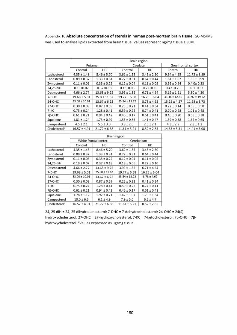
180
Appendix 10 Absolute concentration of sterols in human post-mortem brain tissue. GC-MS/MS
was used to analyse lipids extracted from brain tissue. Values represent ng/mg tissue ± SEM.
24, 25 diH = 24, 25 dihydro lanosterol; 7-DHC = 7-dehydrocholesterol; 24-OHC = 24(S)-
hydroxycholesterol; 27-OHC = 27-hydroxycholesterol; 7-KC = 7-ketocholesterol; 7β-OHC = 7β-
hydroxycholesterol. aValues expressed as µg/mg tissue.
Brain region
Putamen Caudate Grey frontal cortex
Control HD Control HD Control HD
Lathosterol 4.35 ± 1.48 8.46 ± 5.70 3.62 ± 1.55 3.45 ± 2.50 9.64 ± 4.65 11.72 ± 8.89
Lanosterol 0.89 ± 0.37 1.33 ± 0.81 0.72 ± 0.31 0.64 ± 0.44 1.81 ± 1.02 1.66 ± 0.99
Zymosterol 0.11 ± 0.06 0.35 ± 0.22 0.12 ± 0.04 0.11 ± 0.05 0.56 ± 0.24 0.4 0± 0.23
24,25 diH 0.19±0.07 0.37±0.18 0.18±0.06 0.22±0.10 0.42±0.25 0.61±0.33
Desmosterol 4.66 ± 2.77 13.68 ± 9.25 3.93 ± 1.82 6.71 ± 4.54 5.19 ± 1.61 5.80 ± 4.20
7-DHC 19.68 ± 5.01 25.8 ± 11.62 19.77 ± 6.68 16.26 ± 6.04 33.46 ± 12.31 39.97 ± 19.52
24-OHC 33.00 ± 10.01 13.67 ± 6.22 25.54 ± 13.72 8.78 ± 4.62 15.25 ± 4.27 11.98 ± 3.73
27-OHC 0.30 ± 0.09 0.87 ± 0.59 0.23 ± 0.21 0.41 ± 0.34 0.22 ± 0.14 0.65 ± 0.50
7-KC 0.75 ± 0.24 1.28 ± 0.41 0.59 ± 0.22 0.74 ± 0.41 0.70 ± 0.28 1.01 ± 0.48
7β-OHC 0.61 ± 0.21 0.94 ± 0.42 0.46 ± 0.17 0.61 ± 0.41 0.45 ± 0.20 0.68 ± 0.38
Squalene 1.81 ± 1.24 1.73 ± 0.99 1.53 ± 0.86 1.41 ± 0.47 1.39 ± 0.38 1.62 ± 0.65
Campesterol 4.5 ± 2.1 5.5 ± 3.0 3.8 ± 2.0 2.6 ± 2.1 4.3 ± 2.9 2.8 ± 1.2
Cholesterola 16.57 ± 4.91 21.72 ± 6.38 11.61 ± 5.21 8.52 ± 2.85 14.63 ± 5.31 14.41 ± 5.08
Brain region
White frontal cortex Cerebellum
Control HD Control HD
Lathosterol 4.35 ± 1.48 8.46 ± 5.70 3.62 ± 1.55 3.45 ± 2.50
Lanosterol 0.89 ± 0.37 1.33 ± 0.81 0.72 ± 0.31 0.64 ± 0.44
Zymosterol 0.11 ± 0.06 0.35 ± 0.22 0.12 ± 0.04 0.11 ± 0.05
24,25 diH 0.19 ± 0.07 0.37 ± 0.18 0.18 ± 0.06 0.22 ± 0.10
Desmosterol 4.66 ± 2.77 13.68 ± 9.25 3.93 ± 1.82 6.71 ± 4.54
7-DHC 19.68 ± 5.01 25.80 ± 11.62 19.77 ± 6.68 16.26 ± 6.04
24-OHC 33.00 ± 10.01 13.67 ± 6.22 25.54 ± 13.72 8.78 ± 4.62
27-OHC 0.30 ± 0.09 0.87 ± 0.59 0.23 ± 0.21 0.41 ± 0.34
7-KC 0.75 ± 0.24 1.28 ± 0.41 0.59 ± 0.22 0.74 ± 0.41
7β-OHC 0.61 ± 0.21 0.94 ± 0.42 0.46 ± 0.17 0.61 ± 0.41
Squalene 1.78 ± 1.12 1.92 ± 0.71 1.42 ± 1.07 1.79 ± 1.34
Campesterol 10.0 ± 6.6 6.1 ± 4.9 7.9 ± 5.0 6.5 ± 4.7
Cholesterola 16.57 ± 4.91 21.72 ± 6.38 11.61 ± 5.21 8.52 ± 2.85