Embed Size (px)
Citation preview
AlmaKnowledgeServerUncovers Hidden Knowledge from Medline
I NT H I S
I S S U E
2 NEW: Specifi c, Flexible Labeling of Proteins
4 Detect Protein Phosphorylation Directly Inside the Cell
5 NEW: Highly Specifi c Kinase Activity Assays
6 Application Note: Quantitation of Activated Transcription Factors
7 ChIP-IT™ Simplifi es Chromatin Immunoprecipitation
8 MethylDetector™ – Fast & Effi cient DNA Methylation Analysis
8 Co-Immunoprecipitation of Nuclear Protein Complexes
9 Deliver Functional Proteins Directly into Living Cells
10 NEW: Effective Fluorescent Labels for Bioanalysis
11 Tap into the Knowledge Stored in Medline with AlmaKnowledgeServer
12 Optimized Preparation of Cellular Samples
THE NEWSLETTER OF ACTIVE MOTIF — May 2006 • volume 7 • number 2
Toll Free — 1 877 222 95432
Improved co-localization studiesThe ability to label proteins in living cells is key to understanding the dynamics and functions of proteins. Classically, these studies have been performed by fusing a gene of interest to a fl uorescent protein, such as green fl uorescent pro-tein (GFP), and transfecting the construct into cells. These approaches have signifi -cantly expanded our understanding of protein function within the cell.
However, protein function is commonly regulated via protein-protein interac-tions, creating the need for multiple labeling systems. Using conventional approaches, this means you would need to clone a series of constructs con-taining your gene of interest fused to different fl uorescent proteins. This has made such studies time-consuming and labor intensive. In contrast, LigandLink is a novel tool that makes it possible to create a single clone that can be labeled in cells with different tags. This makes LigandLink an ideal tool for co-localiza-tion studies, where multiple labeling colors are required.
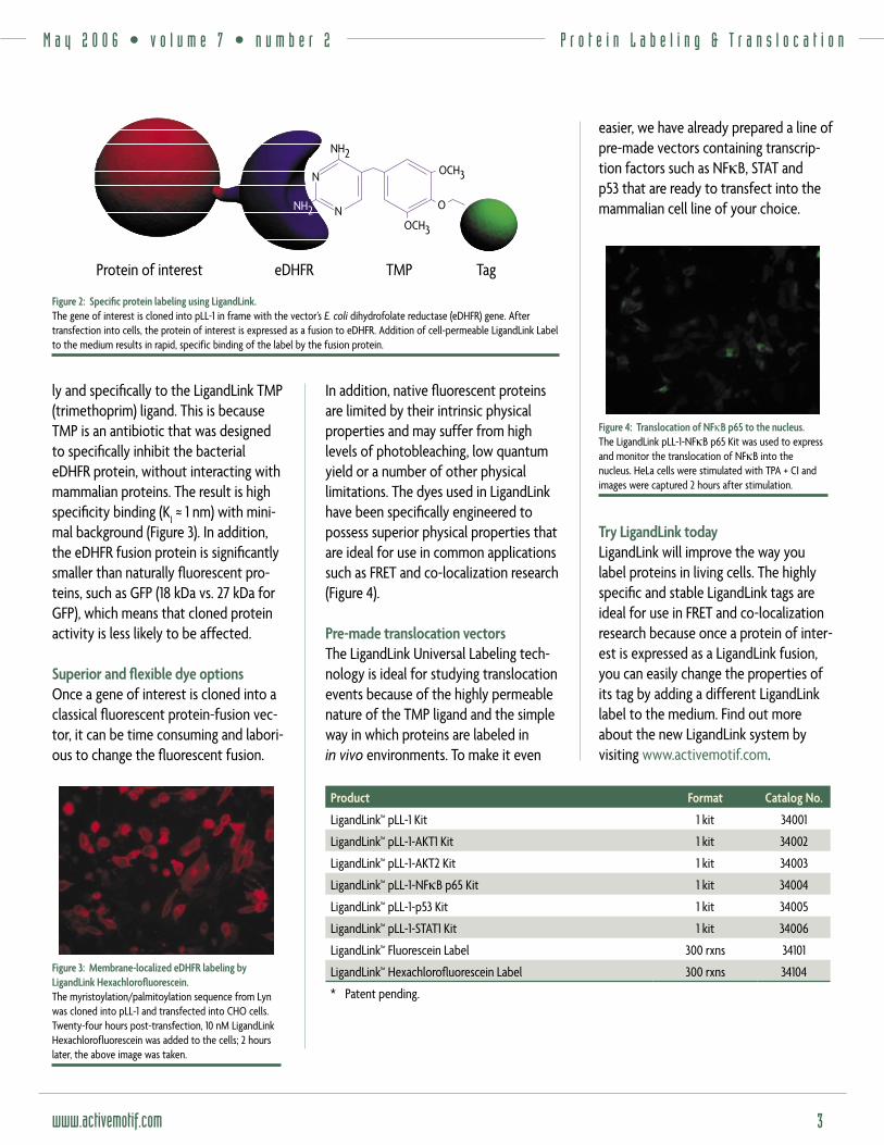
mammalian cells and used to express the fusion protein. Twenty-four hours post-transfection, the protein of interest can be labeled simply by adding the LigandLink ligand of choice to the cell medium (Figure 2). Depending on the cell type and the label used, cells can be imaged in as little as 10 minutes.
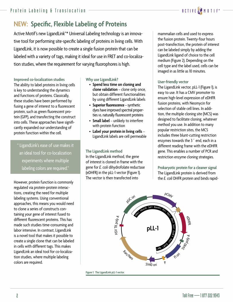
User-friendly vectorThe LigandLink vector, pLL-1 (Figure 1), is easy to use. It has a CMV promoter to ensure high-level expression of eDHFR fusion proteins, with Neomycin for selection of stable cell lines. In addi-tion, the multiple cloning site (MCS) was designed to facilitate cloning, whatever method you use. In addition to many popular restriction sites, the MCS includes three blunt-cutting restriction enzymes towards the 3´ end, each in a different reading frame with the eDHFR gene. This enables a number of PCR and restriction enzyme cloning strategies.
Prokaryotic protein for a cleaner signalThe LigandLink protein is derived from the E. coli DHFR protein and binds rapid-
Why use LigandLink?• Spend less time on cloning and
clone validation – clone only once, but obtain different functionalities by using different LigandLink labels
• Superior fl uorescence – synthetic dyes have improved spectral proper-ties vs. naturally fl uorescent proteins
• Small label – unlikely to interfere with protein function
• Label your protein in living cells – LigandLink labels are cell permeable
The LigandLink methodIn the LigandLink method, the gene of interest is cloned in frame with the gene for E. coli dihydrofolate reductase (eDHFR) in the pLL-1 vector (Figure 1). The vector is then transfected into
Active Motif’s new LigandLink™* Universal Labeling technology is an innova-
tive tool for performing site-specifi c labeling of proteins in living cells. With
LigandLink, it is now possible to create a single fusion protein that can be
labeled with a variety of tags, making it ideal for use in FRET and co-localiza-
tion studies, where the requirement for varying fl uorophores is high.
NEW: Specifi c, Flexible Labeling of Proteins
“ LigandLink’s ease of use makes it
an ideal tool for co-localization
experiments where multiple
labeling colors are required.”
P r o t e i n L a b e l i n g & T r a n s l o c a t i o n
Nh
e I Hind III Sal I BamH I Pst I Not I Xho I Sca I EcoR V Pml I EcoR I Age I
pCMV
eDHFR
SV40
pol
y A
f1 ori
PSV40 ori
Kan/Neo
HSV
TK p
oly A
pUC ori
pLL-1
Figure 1: The LigandLink pLL-1 vector.
M a y 2 0 0 6 • v o l u m e 7 • n u m b e r 2
www.activemotif.com 3
Product Format Catalog No.
LigandLink™ pLL-1 Kit 1 kit 34001
LigandLink™ pLL-1-AKT1 Kit 1 kit 34002
LigandLink™ pLL-1-AKT2 Kit 1 kit 34003
LigandLink™ pLL-1-NFκB p65 Kit 1 kit 34004
LigandLink™ pLL-1-p53 Kit 1 kit 34005
LigandLink™ pLL-1-STAT1 Kit 1 kit 34006
LigandLink™ Fluorescein Label 300 rxns 34101
LigandLink™ Hexachlorofl uorescein Label 300 rxns 34104
* Patent pending.
P r o t e i n L a b e l i n g & T r a n s l o c a t i o n
ly and specifi cally to the LigandLink TMP (trimethoprim) ligand. This is because TMP is an antibiotic that was designed to specifi cally inhibit the bacterial eDHFR protein, without interacting with mammalian proteins. The result is high specifi city binding (K
l ≈ 1 nm) with mini-
mal background (Figure 3). In addition, the eDHFR fusion protein is signifi cantly smaller than naturally fl uorescent pro-teins, such as GFP (18 kDa vs. 27 kDa for GFP), which means that cloned protein activity is less likely to be affected.
Superior and fl exible dye options Once a gene of interest is cloned into a classical fl uorescent protein-fusion vec-tor, it can be time consuming and labori-ous to change the fl uorescent fusion.
Figure 3: Membrane-localized eDHFR labeling by LigandLink Hexachlorofl uorescein.The myristoylation/palmitoylation sequence from Lyn was cloned into pLL-1 and transfected into CHO cells. Twenty-four hours post-transfection, 10 nM LigandLink Hexachlorofluorescein was added to the cells; 2 hours later, the above image was taken.
easier, we have already prepared a line of pre-made vectors containing transcrip-tion factors such as NFκB, STAT and p53 that are ready to transfect into the mammalian cell line of your choice.
Figure 4: Translocation of NFκB p65 to the nucleus.The LigandLink pLL-1-NFκB p65 Kit was used to express and monitor the translocation of NFκB into the nucleus. HeLa cells were stimulated with TPA + CI and images were captured 2 hours after stimulation.
Try LigandLink todayLigandLink will improve the way you label proteins in living cells. The highly specifi c and stable LigandLink tags are ideal for use in FRET and co-localization research because once a protein of inter-est is expressed as a LigandLink fusion, you can easily change the properties of its tag by adding a different LigandLink label to the medium. Find out more about the new LigandLink system by visiting www.activemotif.com.
In addition, native fl uorescent proteins are limited by their intrinsic physical properties and may suffer from high levels of photobleaching, low quantum yield or a number of other physical limitations. The dyes used in LigandLink have been specifi cally engineered to possess superior physical properties that are ideal for use in common applications such as FRET and co-localization research (Figure 4).
Pre-made translocation vectorsThe LigandLink Universal Labeling tech-nology is ideal for studying translocation events because of the highly permeable nature of the TMP ligand and the simple way in which proteins are labeled in in vivo environments. To make it even
���
����
����
������������������� ����� ������
���
�
�
�
Figure 2: Specifi c protein labeling using LigandLink.The gene of interest is cloned into pLL-1 in frame with the vector’s E. coli dihydrofolate reductase (eDHFR) gene. After transfection into cells, the protein of interest is expressed as a fusion to eDHFR. Addition of cell-permeable LigandLink Label to the medium results in rapid, specific binding of the label by the fusion protein.
Toll Free — 1 877 222 95434
P r o t e i n P h o s p h o r y l a t i o n
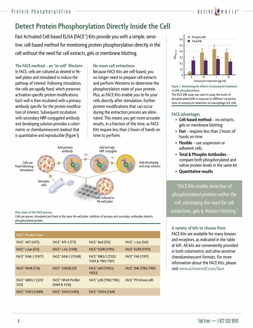
The FACE method – an “in-cell” WesternIn FACE, cells are cultured as desired in 96-well plates and stimulated to induce the pathway of interest. Following stimulation, the cells are rapidly fi xed, which preserves activation-specifi c protein modifi cations. Each well is then incubated with a primary antibody specifi c for the protein modifi ca-tion of interest. Subsequent incubation with secondary HRP-conjugated antibody and developing solution provides a colori-metric or chemiluminescent readout that is quantitative and reproducible (Figure 1).
0.4
0.5
0.3
0.2
0.1
1 100 0.1
OD 4
50 n
m
0.6
0
Anisomycin induction (µg/ml)
Phospho JNKTotal JNK
Figure 1: Monitoring the effects of anisomycin treatment on JNK phosphorylation.The FACE JNK assay was used to assay the levels of phosphorylated JNK in response to different concentra-tions of anisomycin treatment on macrophage 4/4 cells.
FACE advantages• Cell-based method – no extracts,
gels or membrane blotting• Fast – requires less than 2 hours of
hands on time• Flexible – use suspension or
adherent cells• Total & Phospho Antibodies –
compare both phosphorylated and native protein levels in the same kit
• Quantitative results
A variety of kits to choose fromFACE Kits are available for many kinases and receptors, as indicated in the table at left. All kits are conveniently provided in both colorimetric and ultra-sensitive chemiluminescent formats. For more information about the FACE Kits, please visit www.activemotif.com/face.
No more cell extractions Because FACE Kits are cell-based, you no longer need to prepare cell extracts and perform Westerns to determine the phosphorylation state of your protein. Plus, as FACE Kits enable you to fi x your cells directly after stimulation, further protein modifi cations that can occur during the extraction process are elimi-nated. This means you get more accurate results, in a fraction of the time, as FACE Kits require less than 2 hours of hands on time to perform.
Fast Activated Cell-based ELISA (FACE™) Kits provide you with a simple, sensi-
tive, cell-based method for monitoring protein phosphorylation directly in the
cell without the need for cell extracts, gels or membrane blotting.
Detect Protein Phosphorylation Directly Inside the Cell
“FACE Kits enable detection of
phosphorylated proteins within the
cell, eliminating the need for cell
extractions, gels & Western blotting.”
FACE™ Product Line
FACE™ AKT (S473) FACE™ ATF-2 (T71) FACE™ Bad (S112) FACE™ c-Jun (S63)
FACE™ c-Jun (S73) FACE™ c-Src (Y418) FACE™ EGFR (Y992) FACE™ EGFR (Y1173)
FACE™ ErbB-2 (Y877) FACE™ ErbB-2 (Y1248) FACE™ ERK1/2 (T202/Y204 & T185/Y187)
FACE™ FAK (Y397)
FACE™ FKHR (T24) FACE™ GSK3β (S9) FACE™ JAK1 (Y1022/Y1023)
FACE™ JNK (T183/Y185)
FACE™ MEK1/2 (S217/S221)
FACE™ NFκB Profi ler (S468 & S536)
FACE™ p38 (T180/Y182) FACE™ PI3 Kinase p85
FACE™ STAT2 (Y689) FACE™ STAT4 (Y693) FACE™ STAT6 (Y641)
Stimulate cells
3 hrs.1 hr.
Cells are fixed following
stimulation
Cells cultured in 96-well plate
Add primaryantibody
Add anti-IgG HRP conjugate
Add developingand stop solutionP P P
Flow chart of the FACE process.Cells are grown, stimulated and fixed in the same 96-well plate. Addition of primary and secondary antibodies detects phosphorylated protein.
M a y 2 0 0 6 • v o l u m e 7 • n u m b e r 2
www.activemotif.com 5
K i n a s e A s s a y s
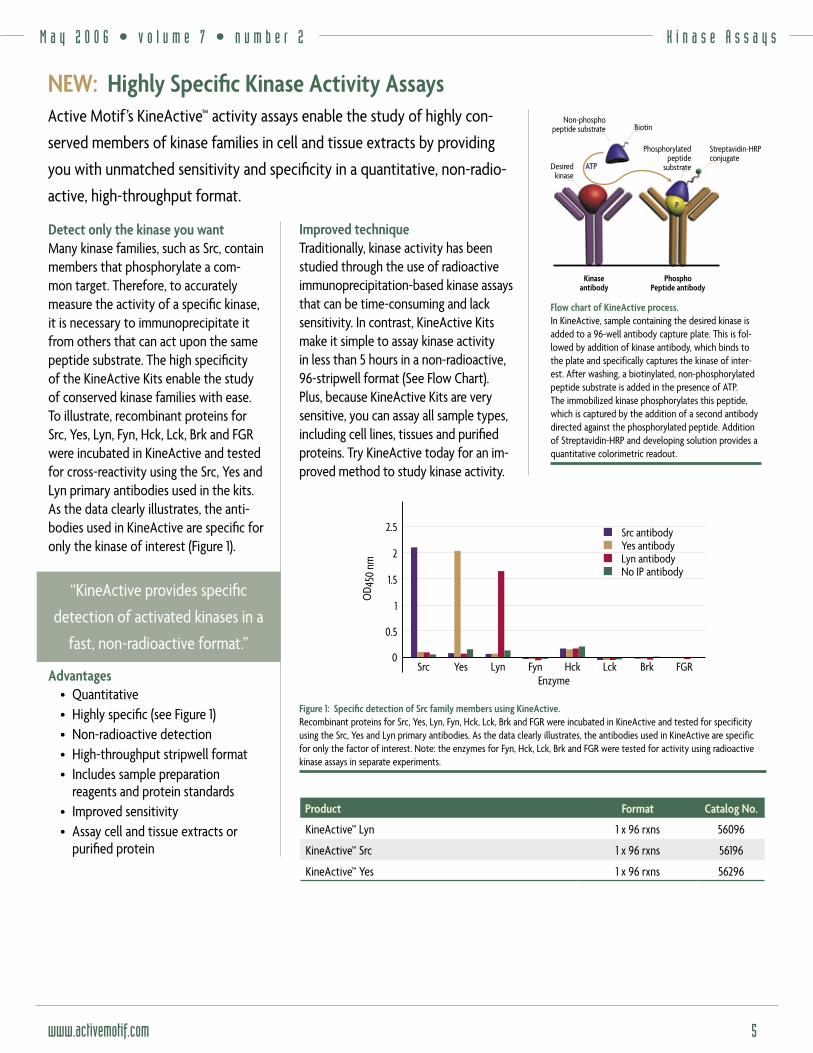
Detect only the kinase you wantMany kinase families, such as Src, contain members that phosphorylate a com-mon target. Therefore, to accurately measure the activity of a specifi c kinase, it is necessary to immunoprecipitate it from others that can act upon the same peptide substrate. The high specifi city of the KineActive Kits enable the study of conserved kinase families with ease. To illustrate, recombinant proteins for Src, Yes, Lyn, Fyn, Hck, Lck, Brk and FGR were incubated in KineActive and tested for cross-reactivity using the Src, Yes and Lyn primary antibodies used in the kits. As the data clearly illustrates, the anti-bodies used in KineActive are specifi c for only the kinase of interest (Figure 1).
Advantages• Quantitative• Highly specifi c (see Figure 1)• Non-radioactive detection• High-throughput stripwell format • Includes sample preparation
reagents and protein standards • Improved sensitivity• Assay cell and tissue extracts or
purifi ed protein
PhosphoPeptide antibody
Kinaseantibody
Desiredkinase
Phosphorylatedpeptide
substrate
Streptavidin-HRPconjugate
P
Non-phosphopeptide substrate Biotin
ATP
Flow chart of KineActive process.In KineActive, sample containing the desired kinase is added to a 96-well antibody capture plate. This is fol-lowed by addition of kinase antibody, which binds to the plate and specifically captures the kinase of inter-est. After washing, a biotinylated, non-phosphorylated peptide substrate is added in the presence of ATP. The immobilized kinase phosphorylates this peptide, which is captured by the addition of a second antibody directed against the phosphorylated peptide. Addition of Streptavidin-HRP and developing solution provides a quantitative colorimetric readout.
Improved techniqueTraditionally, kinase activity has been studied through the use of radioactive immunoprecipitation-based kinase assays that can be time-consuming and lack sensitivity. In contrast, KineActive Kits make it simple to assay kinase activity in less than 5 hours in a non-radioactive, 96-stripwell format (See Flow Chart). Plus, because KineActive Kits are very sensitive, you can assay all sample types, including cell lines, tissues and purifi ed proteins. Try KineActive today for an im-proved method to study kinase activity.
Active Motif’s KineActive™ activity assays enable the study of highly con-
served members of kinase families in cell and tissue extracts by providing
you with unmatched sensitivity and specifi city in a quantitative, non-radio-
active, high-throughput format.
NEW: Highly Specifi c Kinase Activity Assays
“KineActive provides specifi c
detection of activated kinases in a
fast, non-radioactive format.”
Product Format Catalog No.
KineActive™ Lyn 1 x 96 rxns 56096
KineActive™ Src 1 x 96 rxns 56196
KineActive™ Yes 1 x 96 rxns 56296
������
�� ������
���
�
�
���
���
���� ��� ������ ��� ��� ������
��������������������������������������������������
Figure 1: Specifi c detection of Src family members using KineActive.Recombinant proteins for Src, Yes, Lyn, Fyn, Hck, Lck, Brk and FGR were incubated in KineActive and tested for specificity using the Src, Yes and Lyn primary antibodies. As the data clearly illustrates, the antibodies used in KineActive are specific for only the factor of interest. Note: the enzymes for Fyn, Hck, Lck, Brk and FGR were tested for activity using radioactive kinase assays in separate experiments.
Toll Free — 1 877 222 95436
T r a n s c r i p t i o n F a c t o r E L I S A s
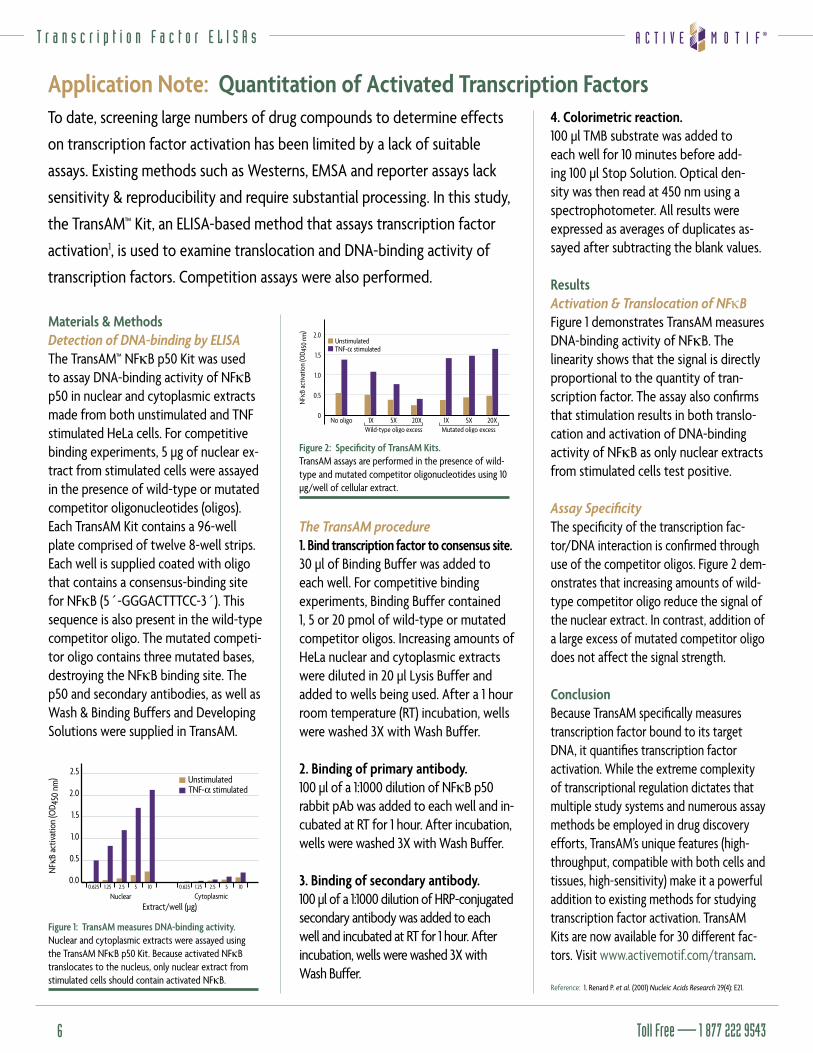
Materials & MethodsDetection of DNA-binding by ELISAThe TransAM™ NFκB p50 Kit was used to assay DNA-binding activity of NFκB p50 in nuclear and cytoplasmic extracts made from both unstimulated and TNF stimulated HeLa cells. For competitive binding experiments, 5 µg of nuclear ex-tract from stimulated cells were assayed in the presence of wild-type or mutated competitor oligonucleotides (oligos). Each TransAM Kit contains a 96-well plate comprised of twelve 8-well strips. Each well is supplied coated with oligo that contains a consensus-binding site for NFκB (5´-GGGACTTTCC-3´). This sequence is also present in the wild-type competitor oligo. The mutated competi-tor oligo contains three mutated bases, destroying the NFκB binding site. The p50 and secondary antibodies, as well as Wash & Binding Buffers and Developing Solutions were supplied in TransAM.
Figure 1: TransAM measures DNA-binding activity.Nuclear and cytoplasmic extracts were assayed using the TransAM NFκB p50 Kit. Because activated NFκB translocates to the nucleus, only nuclear extract from stimulated cells should contain activated NFκB.
4. Colorimetric reaction. 100 µl TMB substrate was added to each well for 10 minutes before add-ing 100 µl Stop Solution. Optical den-sity was then read at 450 nm using a spectrophotometer. All results were expressed as averages of duplicates as-sayed after subtracting the blank values.
ResultsActivation & Translocation of NFκBFigure 1 demonstrates TransAM measures DNA-binding activity of NFκB. The linearity shows that the signal is directly proportional to the quantity of tran-scription factor. The assay also confi rms that stimulation results in both translo-cation and activation of DNA-binding activity of NFκB as only nuclear extracts from stimulated cells test positive.
Assay Specifi cityThe specifi city of the transcription fac-tor/DNA interaction is confi rmed through use of the competitor oligos. Figure 2 dem-onstrates that increasing amounts of wild-type competitor oligo reduce the signal of the nuclear extract. In contrast, addition of a large excess of mutated competitor oligo does not affect the signal strength.
ConclusionBecause TransAM specifi cally measures transcription factor bound to its target DNA, it quantifi es transcription factor activation. While the extreme complexity of transcriptional regulation dictates that multiple study systems and numerous assay methods be employed in drug discovery efforts, TransAM’s unique features (high-throughput, compatible with both cells and tissues, high-sensitivity) make it a powerful addition to existing methods for studying transcription factor activation. TransAM Kits are now available for 30 different fac-tors. Visit www.activemotif.com/transam.
Reference: 1. Renard P. et al. (2001) Nucleic Acids Research 29(4): E21.
Figure 2: Specifi city of TransAM Kits.TransAM assays are performed in the presence of wild-type and mutated competitor oligonucleotides using 10 µg/well of cellular extract.
The TransAM procedure1. Bind transcription factor to consensus site. 30 µl of Binding Buffer was added to each well. For competitive binding experiments, Binding Buffer contained 1, 5 or 20 pmol of wild-type or mutated competitor oligos. Increasing amounts of HeLa nuclear and cytoplasmic extracts were diluted in 20 µl Lysis Buffer and added to wells being used. After a 1 hour room temperature (RT) incubation, wells were washed 3X with Wash Buffer.
2. Binding of primary antibody.100 µl of a 1:1000 dilution of NFκB p50 rabbit pAb was added to each well and in-cubated at RT for 1 hour. After incubation, wells were washed 3X with Wash Buffer.
3. Binding of secondary antibody. 100 µl of a 1:1000 dilution of HRP-conjugated secondary antibody was added to each well and incubated at RT for 1 hour. After incubation, wells were washed 3X with Wash Buffer.
To date, screening large numbers of drug compounds to determine effects
on transcription factor activation has been limited by a lack of suitable
assays. Existing methods such as Westerns, EMSA and reporter assays lack
sensitivity & reproducibility and require substantial processing. In this study,
the TransAM™ Kit, an ELISA-based method that assays transcription factor
activation1, is used to examine translocation and DNA-binding activity of
transcription factors. Competition assays were also performed.
Application Note: Quantitation of Activated Transcription Factors
M a y 2 0 0 6 • v o l u m e 7 • n u m b e r 2
www.activemotif.com 7
C h r o m a t i n I m m u n o p r e c i p i t a t i o n
How ChIP worksChromatin immunoprecipitation is a powerful tool for analyzing genome regulation. In ChIP-IT, intact cells are fi xed using formaldehyde, which cross-links and preserves protein/DNA interactions. The DNA is then sheared into small, uniform fragments and protein/DNA complexes are immunoprecipitated using antibodies directed against the protein(s) of interest. Next, the cross-links are reversed and DNA fragments are purifi ed and screened to determine which gene, or group of genes, were bound by the protein of interest.
1000 bp
500 bp
100 bp
1 2 3 4 5
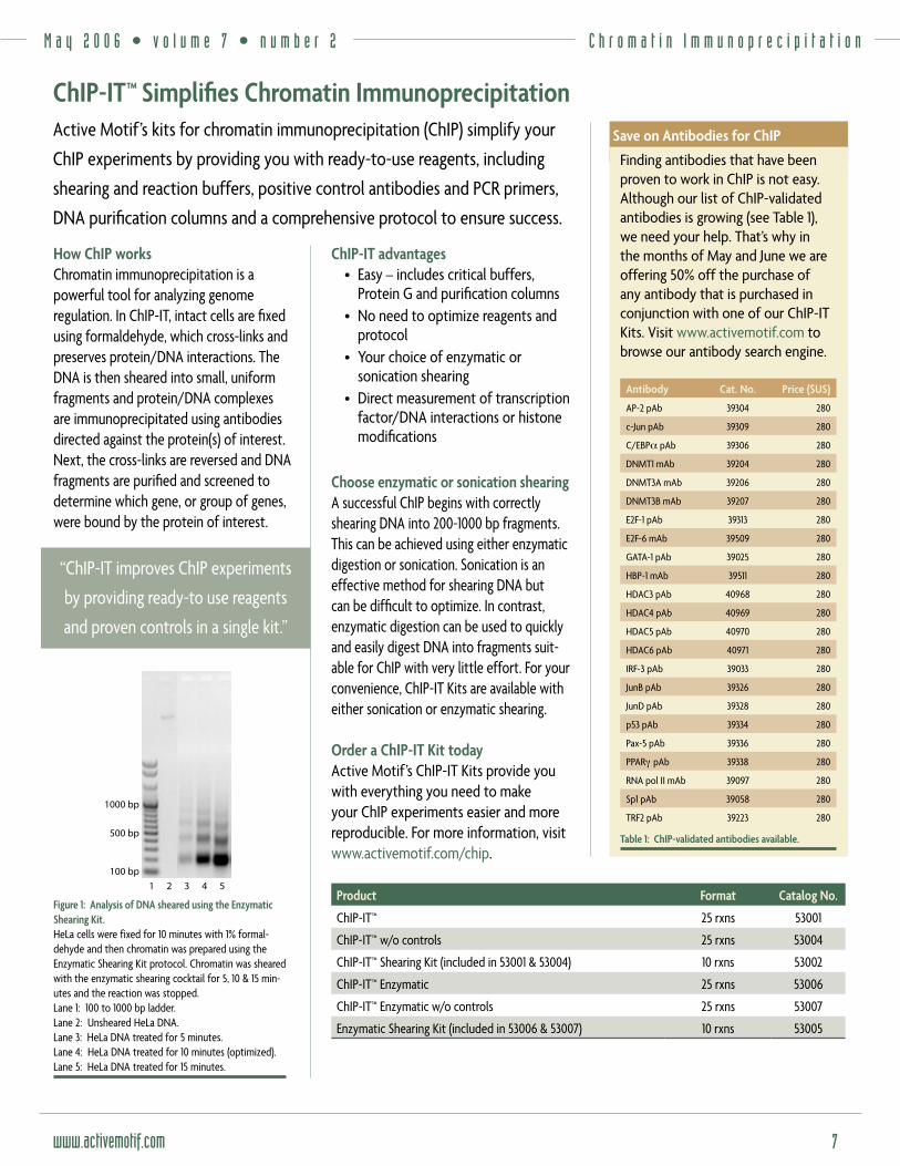
Figure 1: Analysis of DNA sheared using the Enzymatic Shearing Kit.HeLa cells were fixed for 10 minutes with 1% formal-dehyde and then chromatin was prepared using the Enzymatic Shearing Kit protocol. Chromatin was sheared with the enzymatic shearing cocktail for 5, 10 & 15 min-utes and the reaction was stopped.Lane 1: 100 to 1000 bp ladder.Lane 2: Unsheared HeLa DNA.Lane 3: HeLa DNA treated for 5 minutes.Lane 4: HeLa DNA treated for 10 minutes (optimized).Lane 5: HeLa DNA treated for 15 minutes.
ChIP-IT advantages• Easy – includes critical buffers,
Protein G and purifi cation columns • No need to optimize reagents and
protocol• Your choice of enzymatic or
sonication shearing• Direct measurement of transcription
factor/DNA interactions or histone modifi cations
Choose enzymatic or sonication shearingA successful ChIP begins with correctly shearing DNA into 200-1000 bp fragments. This can be achieved using either enzymatic digestion or sonication. Sonication is an effective method for shearing DNA but can be diffi cult to optimize. In contrast, enzymatic digestion can be used to quickly and easily digest DNA into fragments suit-able for ChIP with very little effort. For your convenience, ChIP-IT Kits are available with either sonication or enzymatic shearing.
Order a ChIP-IT Kit todayActive Motif’s ChIP-IT Kits provide you with everything you need to make your ChIP experiments easier and more reproducible. For more information, visit www.activemotif.com/chip.
Active Motif’s kits for chromatin immunoprecipitation (ChIP) simplify your
ChIP experiments by providing you with ready-to-use reagents, including
shearing and reaction buffers, positive control antibodies and PCR primers,
DNA purifi cation columns and a comprehensive protocol to ensure success.
ChIP-IT™ Simplifi es Chromatin Immunoprecipitation
Product Format Catalog No.
ChIP-IT™ 25 rxns 53001
ChIP-IT™ w/o controls 25 rxns 53004
ChIP-IT™ Shearing Kit (included in 53001 & 53004) 10 rxns 53002
ChIP-IT™ Enzymatic 25 rxns 53006
ChIP-IT™ Enzymatic w/o controls 25 rxns 53007
Enzymatic Shearing Kit (included in 53006 & 53007) 10 rxns 53005
“ChIP-IT improves ChIP experiments
by providing ready-to use reagents
and proven controls in a single kit.”
Save on Antibodies for ChIP
Finding antibodies that have been proven to work in ChIP is not easy. Although our list of ChIP-validated antibodies is growing (see Table 1), we need your help. That’s why in the months of May and June we are offering 50% off the purchase of any antibody that is purchased in conjunction with one of our ChIP-IT Kits. Visit www.activemotif.com to browse our antibody search engine.
Antibody Cat. No. Price ($US)
AP-2 pAb 39304 280
c-Jun pAb 39309 280
C/EBPα pAb 39306 280
DNMT1 mAb 39204 280
DNMT3A mAb 39206 280
DNMT3B mAb 39207 280
E2F-1 pAb 39313 280
E2F-6 mAb 39509 280
GATA-1 pAb 39025 280
HBP-1 mAb 39511 280
HDAC3 pAb 40968 280
HDAC4 pAb 40969 280
HDAC5 pAb 40970 280
HDAC6 pAb 40971 280
IRF-3 pAb 39033 280
JunB pAb 39326 280
JunD pAb 39328 280
p53 pAb 39334 280
Pax-5 pAb 39336 280
PPARγ pAb 39338 280
RNA pol II mAb 39097 280
Sp1 pAb 39058 280
TRF2 pAb 39223 280
Table 1: ChIP-validated antibodies available.
Toll Free — 1 877 222 95438
D N A M e t h y l a t i o n & C o - I P
Co-Immunoprecipitation of Nuclear Protein Complexes
MethylDetector™ – Fast & Effi cient DNA Methylation Analysis
Performing co-immunoprecipitation of nuclear protein complexes has just
gotten easier as Active Motif’s Nuclear Complex Co-IP Kit provides optimized
reagents that maintain DNA-binding protein complexes and improve results.
Optimized method and reagentsCo-immunoprecipitation (Co-IP) is often used to analyze protein/protein interactions. However, traditional Co-IP methods are not optimal for studying DNA-binding protein complexes as these complexes are unstable. The Nuclear Complex Co-IP Kit helps maintain these complexes by providing optimized re-agents for both nuclear extract prepara-tion and immunoprecipitation, including both high and low stringency buffers, as well as additional salt and detergent to modify stringency as required.
Advantages• Simple and effi cient• Optimized extraction procedure
preserves nuclear protein complexes • Easily alter IP stringency to detect
interactions of varying strengths
Order the new Co-IP Kit todayThe Nuclear Complex Co-IP Kit enables you to study tightly bound or weak protein complexes with equal ease. Visit www.activemotif.com.
Active Motif’s MethylDetector™ Bisulfi te Modifi cation Kit makes DNA
methylation analysis fast and effi cient by combining optimized reagents for
performing DNA conversion with time-saving DNA purifi cation columns and
positive control PCR primers to help you validate your results.
Proven controls ensure successDNA methylation analysis typically involves using bisulfi te to convert unmethylated cytosines to uracils, while leaving methylated cytosines unchanged. The DNA is then amplifi ed by PCR and analyzed by sequencing or restriction digest. However, bisulfi te conversion can be technically challenging, and confi rming the process was successful before sample analysis is preferred. MethylDetector provides positive control PCR primers that are specifi c for bisulfi te-converted DNA, so you can confi rm the procedure worked before starting further costly analysis (Figure 1). Product Format Catalog No.
MethylDetector™ 50 rxns 55001
Product Format Catalog No.
Nuclear Complex Co-IP Kit 50 rxns 54001
1
250
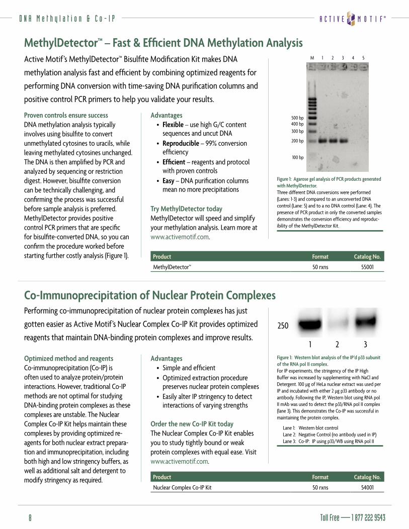
2 3Figure 1: Western blot analysis of the IP’d p33 subunit of the RNA pol II complex. For IP experiments, the stringency of the IP High Buffer was increased by supplementing with NaCl and Detergent. 100 µg of HeLa nuclear extract was used per IP and incubated with either 2 µg p33 antibody or no antibody. Following the IP, Western blot using RNA pol II mAb was used to detect the p33/RNA pol II complex (lane 3). This demonstrates the Co-IP was successful in maintaining the protein complex.
Lane 1: Western blot control Lane 2: Negative Control (no antibody used in IP)Lane 3: Co-IP: IP using p33/WB using RNA pol II
Advantages• Flexible – use high G/C content
sequences and uncut DNA• Reproducible – 99% conversion
effi ciency • Effi cient – reagents and protocol
with proven controls• Easy – DNA purifi cation columns
mean no more precipitations
Try MethylDetector todayMethylDetector will speed and simplify your methylation analysis. Learn more at www.activemotif.com.
M
500 bp400 bp300 bp
200 bp
100 bp
1 2 3 4 5
Figure 1: Agarose gel analysis of PCR products generated with MethylDetector.Three different DNA conversions were performed (Lanes: 1-3) and compared to an unconverted DNA control (Lane: 5) and to a no DNA control (Lane: 4). The presence of PCR product in only the converted samples demonstrates the conversion efficiency and reproduc-ibility of the MethylDetector Kit.
M a y 2 0 0 6 • v o l u m e 7 • n u m b e r 2
www.activemotif.com 9
P r o t e i n T r a n s f e c t i o n
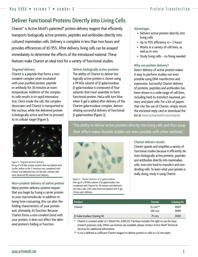
Targeted deliveryChariot is a peptide that forms a non-covalent complex when incubated with your purifi ed protein, peptide or antibody for 30 minutes at room temperature. Addition of the complex to cells results in its rapid internaliza-tion. Once inside the cell, the complex dissociates and Chariot is transported to the nucleus, while the delivered protein is biologically active and free to proceed to its cellular target (Figure 1).
Figure 1: Targeted protein delivery.50 ng of a 10 kDa nuclear protein that was labeled with Lucifer yellow at the C-terminus was complexed with Chariot and delivered into HS-68 cells. Unfixed cells were observed 90 minutes post-delivery.
Non-covalent delivery of native proteinMany protein delivery systems require that you begin by fusing a carrier protein to your macromolecule. In addition to being time-consuming, this can alter the folding characteristics of your protein and, ultimately, its function. Because Chariot forms a non-covalent bond with your protein, it does not affect the deliv-ered protein’s folding or function.
Advantages• Delivers active protein directly into
living cells• Up to 95% effi ciency in < 2 hours• Works in a variety of cell lines, as
well as in vivo• Study living cells – no fi xing needed
Why use protein delivery?Direct delivery of active protein makes it easy to perform studies not even possible using DNA transfection and expression. Successful Chariot delivery of proteins, peptides and antibodies has been shown in a wide range of cell lines, including hard-to-transfect neuronal, pri-mary and plant cells. For a list of papers that cite the use of Chariot, simply return the enclosed reply card or download the list at www.activemotif.com/chariot.
Chariot delivers resultsChariot speeds and simplifi es a variety of functional studies because it effi ciently de-livers biologically active proteins, peptides and antibodies directly into mammalian cells, even into hard-to-transfect and non-dividing cells. To learn what your protein is really doing, study it using Chariot.
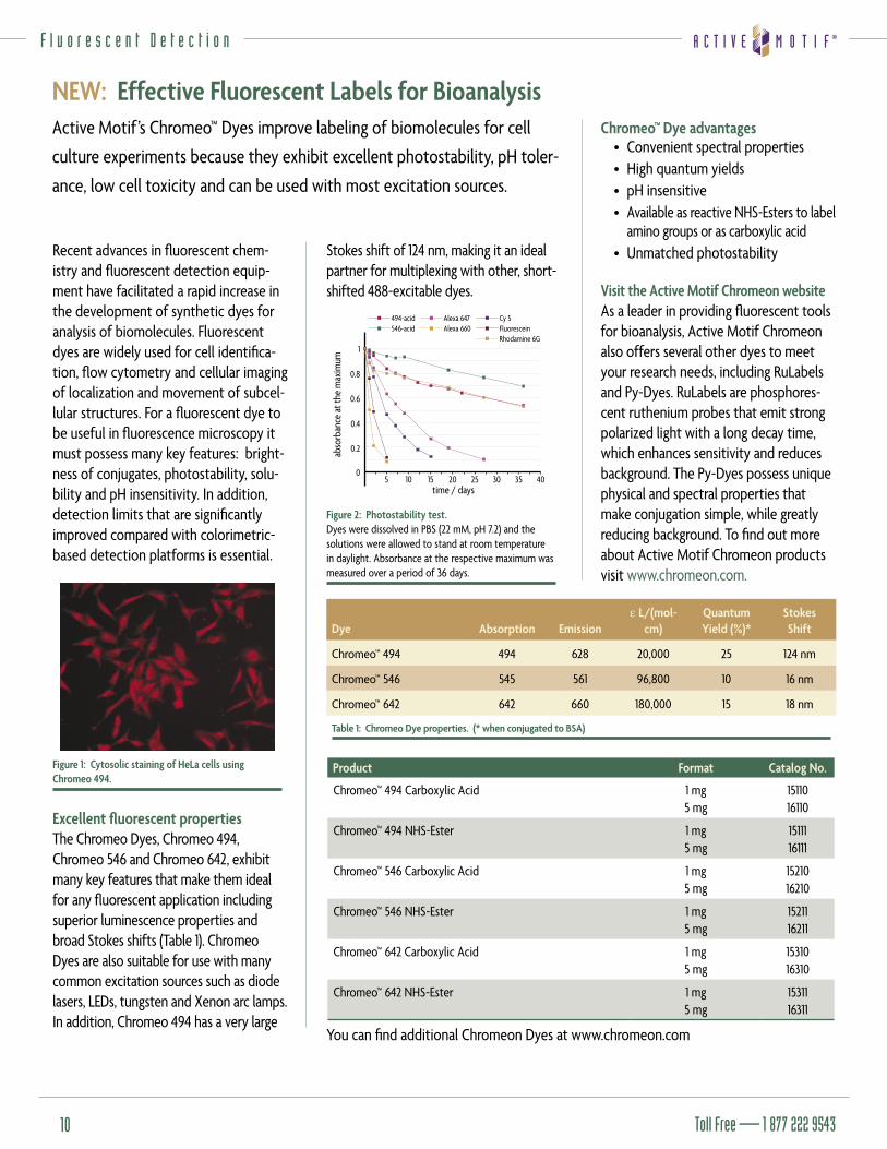
Deliver biologically active proteinsThe ability of Chariot to deliver bio-logically active protein is shown using a 119 kDa subunit of β-galactosidase. β-galactosidase is composed of four subunits that must assemble to form functional protein. HeLa cells turn blue when X-gal is added after delivery of the Chariot-galactosidase complex, demon-strating successful delivery of functional β-galactosidase (Figure 2).
Figure 2: Chariot delivery of β-galactosidase.One µg of a 119 kDa subunit of β-galactosidase was complexed with Chariot for 30 minutes and delivered into HeLa cells. Cells were fixed and stained with X-gal 2 hours post-delivery.
Chariot™ is Active Motif’s patented* protein delivery reagent that effi ciently
transports biologically active proteins, peptides and antibodies directly into
cultured mammalian cells. Delivery is complete in less than two hours and
provides effi ciencies of 65-95%. After delivery, living cells can be assayed
immediately to determine the effects of the introduced material. These
features make Chariot an ideal tool for a variety of functional studies.
Deliver Functional Proteins Directly into Living Cells
Product Format Catalog No.
Chariot™ 25 rxns**100 rxns
3002530100
β-Galactosidase Staining Kit 75 rxns 35001
* Chariot is covered under U.S. Patent No. 6,841,535. Purchase includes the right to use for basic research purposes only. Other-use licenses are available; please contact Active Motif Technical Services for additional information.
** A rxn is defi ned as suffi cient Chariot reagent to deliver protein to cells in a 35 mm plate.
“The ability to deliver active proteins directly into living cells and then assay
their effect makes feasible studies not even possible with other methods.”
Toll Free — 1 877 222 954310
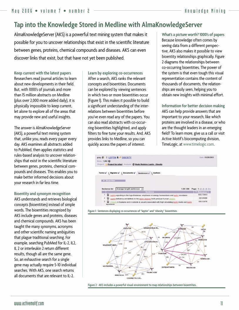
Recent advances in fl uorescent chem-istry and fl uorescent detection equip-ment have facilitated a rapid increase in the development of synthetic dyes for analysis of biomolecules. Fluorescent dyes are widely used for cell identifi ca-tion, fl ow cytometry and cellular imaging of localization and movement of subcel-lular structures. For a fl uorescent dye to be useful in fl uorescence microscopy it must possess many key features: bright-ness of conjugates, photostability, solu-bility and pH insensitivity. In addition, detection limits that are signifi cantly improved compared with colorimetric-based detection platforms is essential.
Figure 1: Cytosolic staining of HeLa cells using Chromeo 494.
Excellent fl uorescent propertiesThe Chromeo Dyes, Chromeo 494, Chromeo 546 and Chromeo 642, exhibit many key features that make them ideal for any fl uorescent application including superior luminescence properties and broad Stokes shifts (Table 1). Chromeo Dyes are also suitable for use with many common excitation sources such as diode lasers, LEDs, tungsten and Xenon arc lamps. In addition, Chromeo 494 has a very large
Stokes shift of 124 nm, making it an ideal partner for multiplexing with other, short-shifted 488-excitable dyes.
�
���
���
���
���
��� �� �� �� �� ����������������
�������������������������
������������������
��������������������
������������������������������
Figure 2: Photostability test.Dyes were dissolved in PBS (22 mM, pH 7.2) and the solutions were allowed to stand at room temperature in daylight. Absorbance at the respective maximum was measured over a period of 36 days.
Active Motif’s Chromeo™ Dyes improve labeling of biomolecules for cell
culture experiments because they exhibit excellent photostability, pH toler-
ance, low cell toxicity and can be used with most excitation sources.
NEW: Effective Fluorescent Labels for Bioanalysis
Product Format Catalog No.
Chromeo™ 494 Carboxylic Acid 1 mg5 mg
1511016110
Chromeo™ 494 NHS-Ester 1 mg5 mg
1511116111
Chromeo™ 546 Carboxylic Acid 1 mg5 mg
1521016210
Chromeo™ 546 NHS-Ester 1 mg5 mg
1521116211
Chromeo™ 642 Carboxylic Acid 1 mg5 mg
1531016310
Chromeo™ 642 NHS-Ester 1 mg5 mg
1531116311
Chromeo™ Dye advantages• Convenient spectral properties • High quantum yields • pH insensitive • Available as reactive NHS-Esters to label
amino groups or as carboxylic acid • Unmatched photostability
Visit the Active Motif Chromeon websiteAs a leader in providing fl uorescent tools for bioanalysis, Active Motif Chromeon also offers several other dyes to meet your research needs, including RuLabels and Py-Dyes. RuLabels are phosphores-cent ruthenium probes that emit strong polarized light with a long decay time, which enhances sensitivity and reduces background. The Py-Dyes possess unique physical and spectral properties that make conjugation simple, while greatly reducing background. To fi nd out more about Active Motif Chromeon products visit www.chromeon.com.
You can fi nd additional Chromeon Dyes at www.chromeon.com
Dye Absorption Emissionε L/(mol-
cm)QuantumYield (%)*
StokesShift
Chromeo™ 494 494 628 20,000 25 124 nm
Chromeo™ 546 545 561 96,800 10 16 nm
Chromeo™ 642 642 660 180,000 15 18 nm
Table 1: Chromeo Dye properties. (* when conjugated to BSA)
F l u o r e s c e n t D e t e c t i o n
M a y 2 0 0 6 • v o l u m e 7 • n u m b e r 2
www.activemotif.com 11
K n o w l e d g e M i n i n g
Keep current with the latest papersResearchers read journal articles to learn about new developments in their fi eld. But, with 1000’s of journals and more than 15 million abstracts on Medline (plus over 2,000 more added daily), it is physically impossible to keep current, let alone to explore all of the areas that may provide new and useful insights.
The answer is AlmaKnowledgeServer (AKS), a powerful text mining system that, unlike you, reads every paper every day. AKS examines all abstracts added to PubMed, then applies statistics and rules-based analysis to uncover relation-ships that exist in the scientifi c literature between genes, proteins, chemical com-pounds and diseases. This enables you to make better informed decisions about your research in far less time.
Bioentity and synonym recognitionAKS understands and retrieves biological concepts (bioentities) instead of simple words. The bioentities recognized by AKS include genes and proteins, diseases and chemical compounds. AKS has been taught the many synonyms, acronyms and other scientifi c naming ambiguities that plague traditional searching. For example, searching PubMed for IL-2, IL2, IL 2 or interleukin 2 return different results, though all are the same gene. So, an exhaustive search for a single gene may actually require 5-10 individual searches. With AKS, one search returns all documents that are relevant to IL-2.
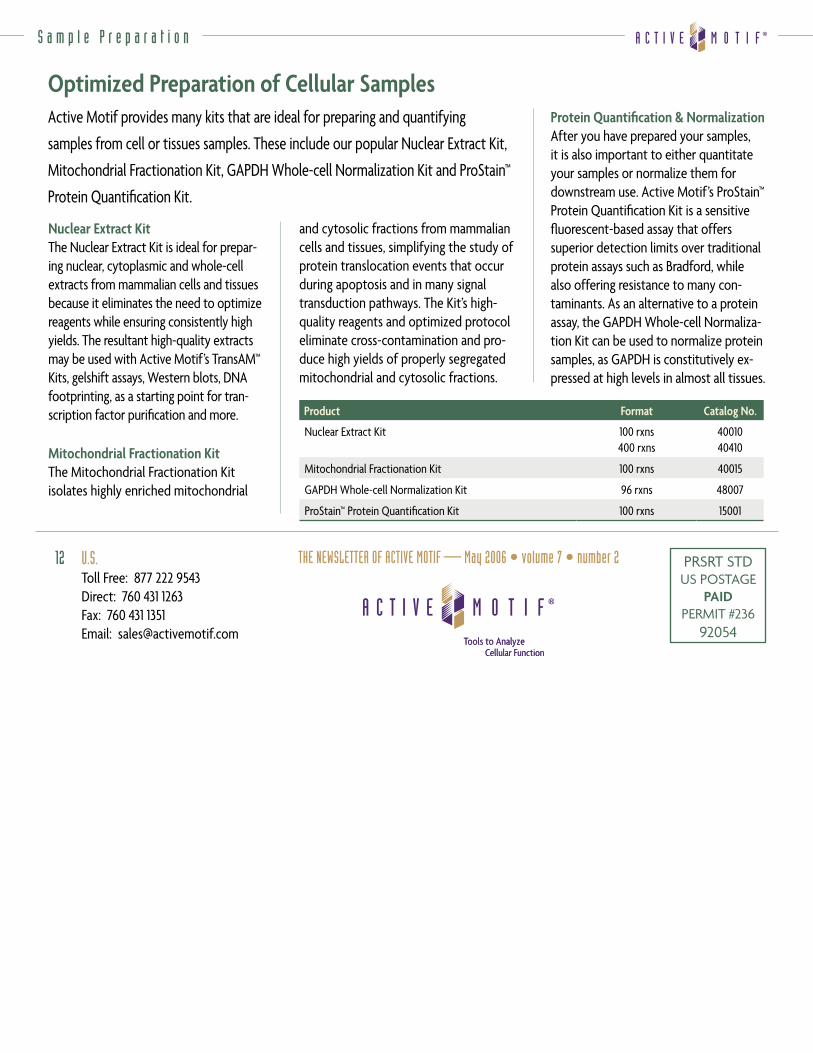
What’s a picture worth? 1000’s of papersBecause knowledge often comes by seeing data from a different perspec-tive, AKS also makes it possible to view bioentity relationships graphically. Figure 2 diagrams the relationships between co-occurring bioentities. The power of the system is that even tough this visual representation contains the content of thousands of documents, the relation-ships are easily seen, helping you to obtain new insights with minimal effort.
Information for better decision making AKS can help provide answers that are important to your research, like which proteins are involved in a disease, or who are the thought leaders in an emerging fi eld? To learn more, give us a call or visit Active Motif’s biocomputing division, TimeLogic, at www.timelogic.com.
Learn by exploring co-occurrencesAfter a search, AKS ranks the relevant concepts and bioentities. Documents can be explored by viewing sentences in which two or more bioentities occur (Figure 1). This makes it possible to build a signifi cant understanding of the inter-relations between bioentities before you’ve even read any of the papers. You can also read abstracts with co-occur-ring bioentities highlighted, and apply fi lters to fi ne tune your results. And, AKS provides links to Medline, so you can quickly access the papers of interest.
AlmaKnowledgeServer (AKS) is a powerful text mining system that makes it
possible for you to uncover relationships that exist in the scientifi c literature
between genes, proteins, chemical compounds and diseases. AKS can even
discover links that exist, but that have not yet been published.
Tap into the Knowledge Stored in Medline with AlmaKnowledgeServer
Figure 2: AKS includes a powerful visual environment to map relationships between bioentities.
Figure 1: Sentences displaying co-occurrences of “leptin” and “obesity” bioentities.
PRSRT STDUS POSTAGE
PAIDPERMIT #236
92054
12
S a m p l e P r e p a r a t i o n
Active Motif provides many kits that are ideal for preparing and quantifying
samples from cell or tissues samples. These include our popular Nuclear Extract Kit,
Mitochondrial Fractionation Kit, GAPDH Whole-cell Normalization Kit and ProStain™
Protein Quantifi cation Kit.
Optimized Preparation of Cellular Samples
Product Format Catalog No.
Nuclear Extract Kit 100 rxns400 rxns
4001040410
Mitochondrial Fractionation Kit 100 rxns 40015
GAPDH Whole-cell Normalization Kit 96 rxns 48007
ProStain™ Protein Quantifi cation Kit 100 rxns 15001
Nuclear Extract KitThe Nuclear Extract Kit is ideal for prepar-ing nuclear, cytoplasmic and whole-cell extracts from mammalian cells and tissues because it eliminates the need to optimize reagents while ensuring consistently high yields. The resultant high-quality extracts may be used with Active Motif’s TransAM™ Kits, gelshift assays, Western blots, DNA footprinting, as a starting point for tran-scription factor purifi cation and more.
Mitochondrial Fractionation KitThe Mitochondrial Fractionation Kit isolates highly enriched mitochondrial
Protein Quantifi cation & NormalizationAfter you have prepared your samples, it is also important to either quantitate your samples or normalize them for downstream use. Active Motif’s ProStain™ Protein Quantifi cation Kit is a sensitive fl uorescent-based assay that offers superior detection limits over traditional protein assays such as Bradford, while also offering resistance to many con-taminants. As an alternative to a protein assay, the GAPDH Whole-cell Normaliza-tion Kit can be used to normalize protein samples, as GAPDH is constitutively ex-pressed at high levels in almost all tissues.
and cytosolic fractions from mammalian cells and tissues, simplifying the study of protein translocation events that occur during apoptosis and in many signal transduction pathways. The Kit’s high-quality reagents and optimized protocol eliminate cross-contamination and pro-duce high yields of properly segregated mitochondrial and cytosolic fractions.
U.S.Toll Free: 877 222 9543Direct: 760 431 1263Fax: 760 431 1351Email: [email protected]
THE NEWSLETTER OF ACTIVE MOTIF — May 2006 • volume 7 • number 2