Embed Size (px)
Citation preview

Allie Harbert, MS4
Journal Club 6/16/20

Learning Objectives
By the end of this journal club, participants will be able to:
1. Know the epidemiology, presentation, and treatment
of pituitary adenomas.
2. Understand differences in symptoms and treatment for
functional pituitary adenomas.
3. Identify important imaging features of pituitary
adenomas and their clinical significance.

Module Outline
I. Case
II. Background
III. Article Overview
IV. Clinical Questions
V. Key Points

UNC Case: 46 yo female with a history of DM, HTN, and OSA presents with several years of worsening weight gain, knee pain, carpal tunnel syndrome, widening of spaces between her teeth, increased size of her hands and feet, and coarse facial features. More recently she developed bilateral temporal vision loss and headaches.
Labs revealed IGF-1: 1213 and GH: 72 She underwent head CT followed by head MRI…
1. What is your differential diagnosis for these symptoms? For the lesion seen on
imaging?
2. What is your impression of the CT? MRI?
3. Possible next steps?

Case: Pathology Report
• Underwent endonasal endoscopic transellar resection
• Diagnosis: Pituitary adenoma
• Microscopic examination demonstrates a densely granulated hypercellularpituitary neoplasm consisting of deeply eosinophilic tumor cells with nuclei showing mild to moderate nuclear atypia with granular chromatin and frequent prominent nucleoli. Tumor cells are arranged in loose nests and form frequent rosette–-like structures.
• Immunohistochemical stains demonstrate the tumor cells are diffusely positive for CAM 5.2 in a perinuclear pattern, synaptophysin and human growth hormone. The tumor cells are negative for prolactin and ACTH. Overall the findings are consistent with a diagnosis of pituitary adenoma.
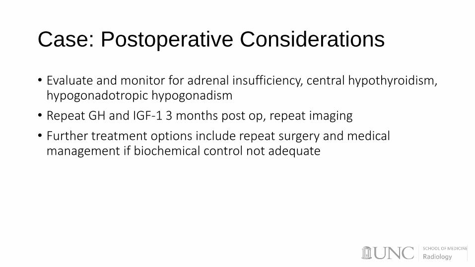
Case: Postoperative Considerations
• Evaluate and monitor for adrenal insufficiency, central hypothyroidism, hypogonadotropic hypogonadism
• Repeat GH and IGF-1 3 months post op, repeat imaging
• Further treatment options include repeat surgery and medical management if biochemical control not adequate
Case: Questions to Consider
• What are the important imaging characteristics to consider?
• What other treatment options could have been considered in this case?
• What will management look like after surgery?

Module Outline
I. Case
II. Background
III. Article Overview
IV. Clinical Questions
V. Key Points

Background on Pituitary Adenomas
• Prevalence – estimated 10% of population based on autopsy and MRI findings of “normal” pituitaries
• Size Classification: microadenomas (<10mm), macroadenomas (>10mm), giant adenomas (>40mm)
• Pathogenesis largely unknown• <5% have pathogenic mutations identified
• Symptoms from mass itself include: • Hypopituitarism
• Bitemporal hemianopsia

Types of Pituitary Adenomas
• Growth-hormone secreting tumor (acromegaly)• 8-16% pituitary adenomas, causes >95% of acromegaly
• Presentation: DM, HTN, OSA, arthritis, carpal tunnel syndrome, enlargement of hands and feet, changes in facial features
• Workup: history, physical, IGF-1 and GH level, MRI
• Prolactinomas• 32-66% pituitary adenomas, most common in women age 20-50
• Presentation: loss of libido, infertility, and osteoporosis in both sexes, oligomenorrhea or amenorrhea and galactorrhea in women and erectile dysfunction in men.
• Workup: history, physical, MRI, and labs (prolactin level, Cr, thyropropin)

Types of Pituitary Adenomas Cont.
• ACTH-secreting tumor (Cushing’s disease)• 2-6% of pituitary adenomas, 65-70% of Cushing’s disease
• Presentation: weight gain, redistribution of fat, facial rounding, violaceous skin striae and ecchymoses, DM, HTN, mood disorders, and osteoporosis
• Workup: late night salivary cortisol, overnight dexamethasone suppression test, ACTH, MRI
• Non-functioning Adenomas• 14-54% of pituitary adenomas
• Presentation: either found incidentally or patient has mass effect symptoms
• Workup: lab work to evaluate for hormone over secretion or hypopituitarism and visual field testing if near the optic chiasm

Treatment
• Transsphenoidal resection• First line treatment other than
for prolactinomas (dopamine agonist)
• Indicated for nonfunctioning adenomas that are symptomatic from size or are growing
• Recommended for GH-secreting and ACTH-secreting tumors
• Primary or preoperative medical treatment indicated in certain scenarios for biochemical control


T1 vs T2 MRI
• T2 reflects the length of time it takes for the MR signal to decay in the transverse plane. A short T2 means that the signal decays rapidly. So substances with short T2’s have smaller signals and appear darker than substances with longer T2 values.

Module Outline
I. Case
II. Background
III. Article Overview
IV. Clinical Questions
V. Key Points

Article Nuts and Bolts
Purpose: assess whether T2 signal intensity could determine long-term response to first-line SA treatment and to assess clinical and biochemical baseline characteristics, as well as histological subtype in relation to the MRI
Journal: Clinical Endocrinology. 2012
Study Type: Retrospective observational study of cases at the Oslo Hospital between 2003 – 2010
Number of Cases: 45 patients for analysis of biochemical and clinical baseline variables, 25/45 patients treated with long acting SA, 34/45 cases for immunohistochemical granulation pattern assessment
Data: GH and IGF-1 levels, MRIs reviewed by 2 neuroradiologists, granulation status determined by pathologist

Patients with new treatment naïve
acromegaly identified (45)
40/45 underwent transsphenoidal
resection
Histological specimens for granulation pattern
on 34/40 cases (19 from SA group, 15 others)
25/45 underwent primary treatment with
SA analogue for ≈6 months
Had repeat GH and IGF-1, 19/25 underwent
surgery
All patients received baseline MRI and labs
(GH and IGF-1)

Material and Methods Cont.
• Prior to treatment, patients GH response to octreotide was measured
• IGF-1 expressed as measured value/age adjusted ULN
• All MRIs evaluated by 2 blinded neuroradiologists
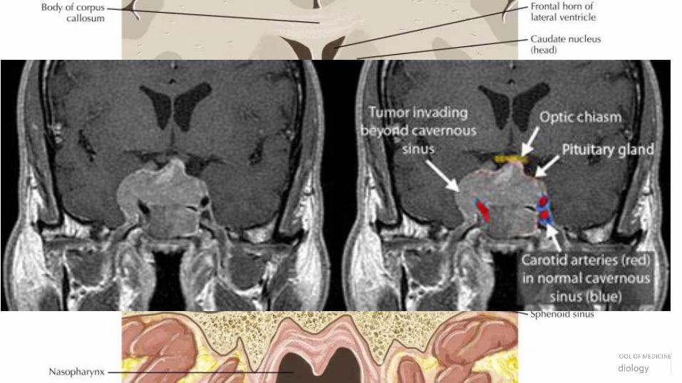
• T2 signal of solid portion of mass compared to adjacent temporal lobe grey and white matter• Equal or less than white matter = hypointense
• Equal or greater than grey matter = hyperintense
• Between white and grey matter = isointense
• Tumors classified histologically as densely, transitional, or sparsely granulated based on CAM5.2 staining
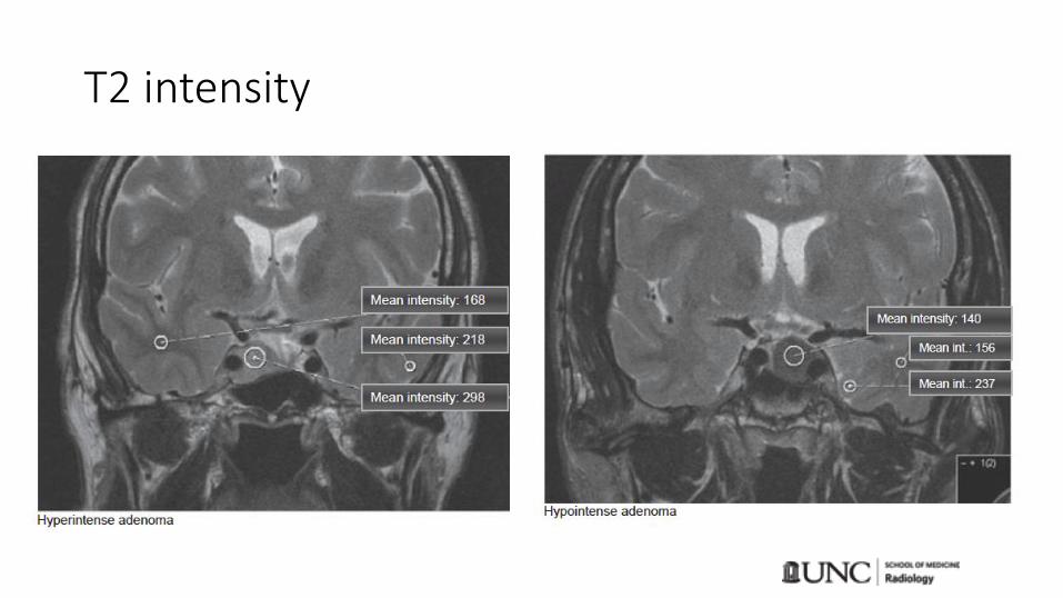
T2 intensity

Results
*
*
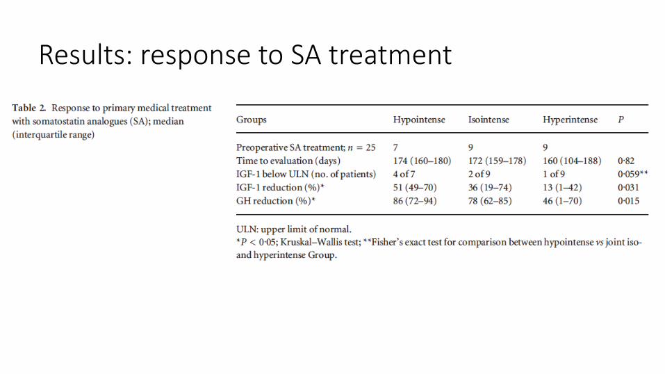
Results: response to SA treatment

Results: granulation pattern

Discussion
• At baseline, hyperintense adenomas had lower GH and IGF-1 levels despite younger age and larger tumors• Possible that different subgroups of GH secreting pituitary adenomas exist or
that the hyperintense adenomas have some “silent” features
• T2 intensity predictive of SA response• model performed best when combined with octreotide test
• Increased SA response rate in T2 hypointense adenomas• Less than other studies

Discussion Cont.
• Sparsely granulated adenomas associated with T2 hyperintensity• Has been shown that sparsely granulated adenomas respond poorly
to octreotide treatment
• This study links all three variables together in analysis

Hold On!
• Retrospective• Varying lengths of SA treatment (not significant)
• GH and IGF-1 measurement technique varied over time
• Octreotide dosage not standard, although most patients received the same dose
• MRI scanner and protocol not consistent
• Non randomized, patients chosen for SA treatment based on clinical judgement• Not a representative sample, limited external validity
• Histological specimens after SA treatment

Module Outline
I. Case
II. Background
III. Article Overview
IV. Clinical Questions
V. Key Points

Clinical Questions
• At UNC – is preoperative/primary medical treatment ever used in acromegaly patients? If so how is it decided whether a patient is a good candidate?
• Is T2 intensity standardly evaluated in pituitary adenomas?

Module Outline
I. Case
II. Background
III. Article Overview
IV. Clinical Questions
V. Key Points

Key Points• T2 intensity is correlated with GH and IGF-1 level,
granulation pattern, and response to SA treatment
• The goal of GH-secreting pituitary adenoma treatment is biochemical control
• Transphenoidal surgery is the primary treatment modality, however medical management is indicated in certain cases
• T2 intensity is a useful factor in evaluating someone as a candidate for SA trial in treating acromegaly

References
• Heck, Ansgar, et al. "Intensity of pituitary adenoma on T2‐weighted magnetic resonance imaging predicts the response to octreotide treatment in newly diagnosed acromegaly." Clinical endocrinology 77.1 (2012): 72-78.
• Molitch, Mark E. "Diagnosis and treatment of pituitary adenomas: a review." Jama 317.5 (2017): 516-524.
• Zahr, Roula, and Maria Fleseriu. "Updates in diagnosis and treatment of acromegaly." European endocrinology 14.2 (2018): 57.
• Katznelson, Laurence, et al. "Acromegaly: an endocrine society clinical practice guideline." The Journal of Clinical Endocrinology & Metabolism 99.11 (2014): 3933-3951.
• Buchfelder, Michael, and Sven-Martin Schlaffer. "The surgical treatment of acromegaly." Pituitary 20.1 (2017): 76-83.