Embed Size (px)
Citation preview
ANNALS OF ALLERGY, ASTHMA, & IMMUNOLOGY March 2008; Volume 100, Number 3, Supplement 3
Allergy Diagnostic Testing: An Updated Practice Parameter
Practice Parameter
Allergy Diagnostic Testing: An Updated PracticeParameterI. Leonard Bernstein, MD; James T. Li, MD, PhD; David I. Bernstein, MD;Robert Hamilton, PhD, DABMLI; Sheldon L. Spector, MD; Ricardo Tan, MD; Scott Sicherer, MD;David B. K. Golden, MD; David A. Khan, MD; Richard A. Nicklas, MD; Jay M. Portnoy, MD;Joann Blessing-Moore, MD; Linda Cox, MD; David M. Lang, MD; John Oppenheimer, MD;Christopher C. Randolph, MD; Diane E. Schuller, MD; Stephen A. Tilles, MD; Dana V. Wallace, MD;Estelle Levetin, PhD; and Richard Weber, MD
TABLE OF CONTENTSI. Preface . . . . . . . . . . . . . . . . . . . . . . . . . . . . . . . . . . . . . . . . . . . . . . . . . . . . . . . . . . . . . . . . . . . . . . . . . . . . . . . . . . . . S2
II. Executive Summary . . . . . . . . . . . . . . . . . . . . . . . . . . . . . . . . . . . . . . . . . . . . . . . . . . . . . . . . . . . . . . . . . . . . . . . . . S3III. Collation of Summary Statements . . . . . . . . . . . . . . . . . . . . . . . . . . . . . . . . . . . . . . . . . . . . . . . . . . . . . . . . . . . . . . S5IV. Part 1 . . . . . . . . . . . . . . . . . . . . . . . . . . . . . . . . . . . . . . . . . . . . . . . . . . . . . . . . . . . . . . . . . . . . . . . . . . . . . . . . . . . . S15V. In Vivo Diagnostic Tests of Immediate Hypersensitivity Reactions . . . . . . . . . . . . . . . . . . . . . . . . . . . . . . . . . . S15
VI. Organ Challenge Tests . . . . . . . . . . . . . . . . . . . . . . . . . . . . . . . . . . . . . . . . . . . . . . . . . . . . . . . . . . . . . . . . . . . . . . S29VII. Tests to Distinguish Clinical Obstructive Diseases Resembling Asthma . . . . . . . . . . . . . . . . . . . . . . . . . . . . . . . S33
VIII. In Vivo Diagnostic Tests of Cell-Mediated Immunity . . . . . . . . . . . . . . . . . . . . . . . . . . . . . . . . . . . . . . . . . . . . . S34IX. In Vitro Diagnostic Tests of Immediate Hypersensitivity . . . . . . . . . . . . . . . . . . . . . . . . . . . . . . . . . . . . . . . . . . . S43X. In Vitro Diagnostic Tests of Cell-Mediated Immunity . . . . . . . . . . . . . . . . . . . . . . . . . . . . . . . . . . . . . . . . . . . . . S59
XI. Other Diagnostic Immunologic Tests . . . . . . . . . . . . . . . . . . . . . . . . . . . . . . . . . . . . . . . . . . . . . . . . . . . . . . . . . . . S64XII. Unproven Tests . . . . . . . . . . . . . . . . . . . . . . . . . . . . . . . . . . . . . . . . . . . . . . . . . . . . . . . . . . . . . . . . . . . . . . . . . . . . S65
XIII. Part 2 . . . . . . . . . . . . . . . . . . . . . . . . . . . . . . . . . . . . . . . . . . . . . . . . . . . . . . . . . . . . . . . . . . . . . . . . . . . . . . . . . . . . S66XIV. Allergens . . . . . . . . . . . . . . . . . . . . . . . . . . . . . . . . . . . . . . . . . . . . . . . . . . . . . . . . . . . . . . . . . . . . . . . . . . . . . . . . . S67XV. Assessment of Inhalant Allergy . . . . . . . . . . . . . . . . . . . . . . . . . . . . . . . . . . . . . . . . . . . . . . . . . . . . . . . . . . . . . . . S74
XVI. Assessment of Food Allergy. . . . . . . . . . . . . . . . . . . . . . . . . . . . . . . . . . . . . . . . . . . . . . . . . . . . . . . . . . . . . . . . . S102XVII. Assessment of Stinging Insect Allergy . . . . . . . . . . . . . . . . . . . . . . . . . . . . . . . . . . . . . . . . . . . . . . . . . . . . . . . . S106
XVIII. Assessment of Drug Allergy. . . . . . . . . . . . . . . . . . . . . . . . . . . . . . . . . . . . . . . . . . . . . . . . . . . . . . . . . . . . . . . . . S109XIX. Assessment of Allergic Contact Dermatitis . . . . . . . . . . . . . . . . . . . . . . . . . . . . . . . . . . . . . . . . . . . . . . . . . . . . . S115XX. Acknowledgments . . . . . . . . . . . . . . . . . . . . . . . . . . . . . . . . . . . . . . . . . . . . . . . . . . . . . . . . . . . . . . . . . . . . . . . . . S121
XXI. References . . . . . . . . . . . . . . . . . . . . . . . . . . . . . . . . . . . . . . . . . . . . . . . . . . . . . . . . . . . . . . . . . . . . . . . . . . . . . . . S122
The American Academy of Allergy, Asthma and Immunology (AAAAI)and the American College of Allergy, Asthma and Immunology(ACAAI) have jointly accepted responsibility for establishing theAllergy Diagnostic Testing: An Updated Practice Parameter. This is acomplete and comprehensive document at the current time. The medicalenvironment is a changing environment and not all recommendationswill be appropriate for all patients. Because this document incorporatedthe efforts of many participants, no single individual, including thosewho served on the Joint Task Force, is authorized to provide an official
AAAAI or ACAAI interpretation of these practice parameters. Anyrequest for information about or an interpretation of these practiceparameters by the AAAAI or ACAAI should be directed to theExecutive Offices of the AAAAI, the ACAAI, and the Joint Council ofAllergy, Asthma and Immunology. These parameters are not designedfor use by pharmaceutical companies in drug promotion.
Received for publication October 27, 2007; Accepted for publicationNovember 16, 2007.
VOLUME 100, MARCH, 2008 S1
PREFACEThe major emphasis of this updated version of PracticeParameters for Allergy Diagnostic Testing is focused on howtechnological refinements and their validations during thepast decade are being incorporated into the diagnostic arma-mentarium of allergists/clinical immunologists and how theiroptimal use enables confirmation of human clinical sensitiv-ity. The term allergy in this Practice Parameter denotes majorcategories of human hypersensitivity. Pertinent clinical im-munologic techniques are oriented to this category of adap-tive immunity but not to infection, cancer, or transplantationimmunology.
The impetus for Practice Parameters for Allergy Diagnos-tic Testing originally stemmed from a consensus conferencesponsored by the National Institute of Allergy and InfectiousDiseases and published as a supplement to the Journal ofAllergy and Clinical Immunology in September 1988. One ofthe major conclusions of that workshop was that periodicreassessment of diagnostic techniques should be mandatory,and in keeping with that recommendation, the 1995 PracticeParameters for Allergy Diagnostic Tests further reviewed andconsidered new developments up to that time. In the 13-yearinterval since that publication, there has been an exponentialprogression of basic and translational immunologic research,some of which produced novel and practical diagnostic pos-sibilities. Obviously, these advancements necessitated anoverhaul of the 1995 Allergy Diagnostic Parameter commen-surate with the extensive database currently available. Theultimate goals were to formulate recommendations based onevidence-based literature and to achieve balanced use ofclassic and new diagnostic methods.
The working draft of the Parameter on Allergy DiagnosticTests update was based on an outline jointly conceived byJames T. Li and I. Leonard Bernstein and realized by a workgroup (Robert Hamilton, Sheldon Spector, Ricardo Tan,David I. Bernstein, Scott Sicherer, David B. K. Golden, andDavid Khan) chaired by I. Leonard Bernstein. As with pre-vious parameters, the draft was based on a review of themedical literature using a variety of search engines, such asPubMed. Published clinical and basic studies were rated bycategories of evidence and used to establish the strength ofrecommendations (Table 1). The initial draft was then re-viewed by all members of the Joint Task Force and subse-quently by the American Academy of Allergy, Asthma andImmunology (AAAAI), the American College of Allergy,Asthma and Immunology (ACAAI), and the Joint Council ofAllergy, Asthma and Immunology and a number of expertson in vivo and in vitro diagnostic immunology selected by thesupporting organizations. Comments were also solicited fromthe general membership of these societies via their Web sites.This document therefore represents an evidence-based,broadly accepted consensus opinion. The peer review processand general format of the Practice Parameter are consistentwith recommendations of the American College of MedicalQuality, which defines practice guidelines. As such, it is
anticipated to serve as a reference source for current utilityand validity of allergy diagnostic tests.
The organization of Practice Parameters on Allergy Diag-nostic Tests is similar to previous Joint Task Force parame-ters except that a single algorithm with annotations was notappropriate to the mission of the parameter. The broad rangeof diagnostic techniques for varying purposes could not pos-sibly be stratified into a uniform paradigm encompassingdiverse clinical sensitivity disorders that require objectiveconfirmatory tests. An Executive Summary is followed by acollation of Summary Statements, which also precede refer-enced narrative discussions on each subject. The PracticeParameter is divided into 2 parts: part 1 is a detailed descrip-tion of diagnostic modalities currently available to allergists/clinical immunologists. It encompasses both IgE and cell-mediated in vivo (skin and patch) and in vitro tests for a widespectrum of inhalant, food, and contactant allergens. Organchallenge tests are discussed in greater detail in this revisedPractice Parameter because controlled challenges or super-vised exposure ultimately serve as the appropriate gold stan-dard for assessing whether clinical sensitivity is present.Consonant with their recent emergence as diagnostic ad-juncts, the section concerning current status of cytokines andchemokines has been expanded. A new section on “OtherImmunologic Tests” has been added in recognition that manyallergists/clinical immunologists have considerable interestsand expertise in a variety of laboratory immunologic tech-niques commonly used to corroborate the diagnosis of non-IgE, non–cell-mediated clinical immunologic diseases. Adiscussion about unproven techniques is relevant becausethese methods still have advocates who promote them topatients desperately seeking alternative approaches for theirparticular problems.
Part 2 considers optimal utilization and integration of ev-idence-based diagnostic methods for various clinical situa-tions, which include inhalant, food, insect venom, drug andcontact sensitivities. Practice parameters of diagnosis andmanagement for each of these clinical entities have beenpreviously published with algorithms tailored to fit the spe-cific clinical situation. Many of the diagnostic recommenda-tions of part 2 were extracted or in some cases quotedverbatim from each of these published guidelines.
The Joint Task Force acknowledges that rapid advance-ments in diagnostic technology could render specific past andcurrent recommendations obsolete at any time and that at-tempts to revise will have to be undertaken at appropriateintervals. Nevertheless, whatever the update interim periodmay be, the allergy/clinical immunology community shouldbe prepared to accept novel new diagnostic techniques, pro-vided that they are validated by scientifically accepted ap-proaches.
The overall objectives of this Parameter on Allergy Diag-nostic Tests are tripartite: (1) to develop a reliable referenceresource for selecting appropriate diagnostic tests; (2) toprovide guidelines and support for the practicing physician onhow diagnostic tests should be used in an appropriate and
S2 ANNALS OF ALLERGY, ASTHMA & IMMUNOLOGY
cost-effective manner; and (3) to improve the quality of careof patients by facilitating prompt and accurate diagnosis oftheir hypersensitivity disorders.
EXECUTIVE SUMMARYThere is a wide array of diagnostic modalities for humanhypersensitivity diseases. Among these, skin tests for imme-diate hypersensitivity and delayed hypersensitivity are ofparamount importance. As immunologic diagnostic technol-ogy advances, in vitro tests for both IgE- and cell-mediatedimmunity have also assumed greater significance. In someinstances, lymphocyte functional assays may be applicablefor confirmation of humoral or cell-mediated immunity cy-totoxicity syndromes, as well as classic delayed hypersensi-tivity reactions.
Specific cellular components of both immediate hypersen-sitivity– and cell-mediated immunity induced inflammationcan be identified by their unique transcription markers, pro-tein products, or cell surface differentiation markers. Anincrease in eosinophils and their products often occurs in bothimmediate- and late-phase responses of IgE-mediated reac-tions. The role of the basophil in such reactions can also beevaluated by basophil histamine release tests and, more re-cently, the basophil activation test. When tests for IgE-me-diated immunity are equivocal, organ challenge testing is themost direct way of ascertaining whether bona fide clinicalsensitivity exists.
Mononuclear cells (monocytes, macrophages, and lympho-cytes) are essential constituents of adaptive immunity. Inparticular, their role in cell-mediated immunity has long beenrecognized. Lymphocyte subsets, their cytokines, and theirchemokines may be readily identified and measurable in bodyfluids and tissue sites. Several applications of this technologyhave become standard clinical tests (eg, CD4� cells in ac-quired immunodeficiency); others are being vigorously pur-sued (eg, interleukin [IL] 6, IL-8, IL-10, and transforminggrowth factor �). Increases in specific cytokines such asmacrophage inhibitory factor (MIF) and IL-16 are associatedwith active cell-mediated immunity processes.
Well-established techniques to detect IgG/IgG subclassantibodies by enzyme-linked immunosorbent assay (ELISA),immunodiffusion, and immunoprecipitation are available forspecific antigens and autoantibodies. Antigen antibody com-plexes may be associated with increased C1q binding andcryoglobulins.
Prick/puncture tests or intracutaneous tests are the pre-ferred techniques for IgE-mediated hypersensitivity. It is ad-visable to use prick/puncture devices, which are relativelynontraumatic and elicit reproducible results when placed onspecific areas of the body (ie, arms or back). Optimal resultsdepend on use of potent test extracts and proficiency of theskin tester (ie, demonstration of coefficient of variation�30% at different periods). It is essential that objectivewheal-and-flare responses be recorded in millimeters (diam-eter or area) because cutoff levels (in millimeters) may ob-viate the necessity for confirmatory respiratory and food
allergen challenge tests. This interpretation system also en-ables easier comparison among physicians. Intracutaneoustests are generally used for specific allergens (ie, Hymenop-tera venoms and penicillin), but they may also be applied ifprick/puncture test results are negative and there is a stronghistorical likelihood of clinical allergy to specific allergens.Some clinicians prefer intracutaneous tests without precedingprick/puncture tests, but when this alternative is elected,special care must be taken to ensure that intracutaneousallergen concentrations are nonirritant and correlative withend organ sensitivity. However, there are safety concernswhen intracutaneous tests are performed without precedingprick/puncture tests. A suggested way of determining appro-priate intracutaneous test concentrations is a serial end pointtitration regimen, one of which reported that intracutaneousdilutions between 1:12,500 and 1:312,000 (wt/vol) were non-irritant. Late-phase cutaneous responses, which reflect thepersistent IgE allergic inflammatory milieu, may occur aftereither prick/puncture or intracutaneous tests but are morelikely to do so after the latter. Preliminary data suggest thatdecrease of late-phase cutaneous response may occur aftersuccessful allergen immunotherapy.
The prototypic skin test for delayed hypersensitivity is thetuberculin skin test, which is evaluated by degree of indura-tion in millimeters 48 hours after application. Similar tests areno longer commercially available for pathogenic fungi (eg,Histoplasma capsulatum). A positive tuberculin reading var-ies from 10 to 15 mm in induration, depending on the inci-dence of active tuberculosis within the indigenous populationof the patient. Decreased cell-mediated immunity response oranergy may be evaluated by delayed hypersensitivity antigens(ie, tetanus toxoid, Candida, and Trichophyton) to whichmost members of a population have been exposed. Formerlythe validity of anergy testing was compared with the meannumber of positive reactions elicited by 4 to 5 delayed hy-persensitivity antigens in a large normal control population.Absence of reactivity to all or all except 1 was equated withcomplete or relative anergy, respectively. Currently, there areonly 3 delayed hypersensitivity antigens for testing (tetanustoxoid, Candida, and Trichophyton), and these have not beenevaluated in a large population as described above. Therefore,interpretation of anergy using these 3 antigens is circumspect.Concurrent anergy and tuberculin skin testing is no longerrecommended in patients with human immunodeficiency vi-rus (HIV) suspected of having mycobacterial infections.
Allergic contact dermatitis (ACD) is a special form ofdelayed hypersensitivity evaluated by epicutaneous or patchtests. More than 3,700 substances have been reported toinduce contactant sensitivity. Direct irritants may cause irri-tant contact dermatitis (ICD), which often is morphologicallyindistinguishable from ACD. The irritancy threshold of eachtest agent must be predetermined to exclude the possibility ofICD. Patch testing should be considered for any dermatitis forwhich contactant exposure, either natural or secondary totopical agents, might be implicated. Most ACD can be de-tected by 65 substances recommended by the North American
VOLUME 100, MARCH, 2008 S3
Contact Dermatitis Research Group. The only available Foodand Drug Administration (FDA)–cleared patch test kit is theT.R.U.E. test, which covers a range of approximately 25% to30% of the most common ACD contactant allergens. There-fore, customized patch testing is often necessitated. Patchtests are read at least twice (48 and 72 to 96 hours afterapplication) and occasionally 7 days later in the case of weakACD allergens. Such allergens can also be detected by arepeat open application test protocol. Atopy patch tests tofoods and drugs are being investigated as a complementaryaid in the diagnosis of food and drug allergies. These testshave not yet been validated by a sufficient number of con-trolled studies.
Laboratory tests may also provide useful information toevaluate either immediate hypersensitivity or cell-mediatedimmune reactions. Currently, commercial availability consid-erations are such that specific IgE tests are used more fre-quently than is the case for functional in vitro cell-mediatedimmunity assays. Within the past decade, however, immuno-assays of certain cell-mediated immunity products (ie, cyto-kines or chemokines) may be demonstrating sufficient pre-dictability to be considered as surrogates of cell-mediatedimmunity.
The discovery of IgE and availability of IgE myelomasenabled the production of large quantities of IgE. This per-mitted the production of highly specific anti-human IgE an-tibodies, which led to immunoassays capable of measuringboth total IgE and allergen specific IgE concentrations inserum and body fluids. A succession of modified assaysensued. Subsequent modifications are calibrated using heter-ologous interpolation against the World Health Organization(WHO) 75/502 international human serum IgE referencepreparation, thereby establishing a uniform system of specificIgE antibody in quantitative kilo international units (kIU) perliter (ie, 1 kIU � 2.4 ng IgE). The method of total andspecific IgE assays are discussed in detail, including theindications, advantages, and limitations of these assays. TheFDA guideline regulations now stipulate guidance regula-tions for all IgE methods, including semiautomatic, auto-matic, and multiplexed systems. According to these qualityassurance suggestions, each allergen assay should include itsspecific homologous reference serum (ragweed vs ragweedreference serum) as an additional internal control wheneversufficient quantities of specific reference sera can be ob-tained. It is anticipated that multiplexed arrays for assays ofIgE will soon be generally available. Secondary antibodydetector systems for these modified techniques includechemiluminescence and fluorescence. Allergen specificityand cross-allergenicity may be determined by an inhibitiontechnique. Although correlation of higher kIU levels of spe-cific IgE to clinical sensitivity for some allergens is equiva-lent to prick/puncture tests, skin prick/puncture tests gener-ally have better overall predictability and are the preferredinitial diagnostic approach.
Interpretation of both skin and serum specific IgE tests ishighly dependent on the constitutive allergenicity, potency,
and stability of the allergen extract being used. For thesereasons, sensitivity tends to be higher among pollens, certainfoods, dust mite, fungi, and certain epidermals compared withvenoms, drugs, and chemicals. Recommendations for aller-gen immunotherapy based solely on results of skin or specificIgE tests without appropriate clinical correlation are not ap-propriate.
IgG and IgG subclasses can be measured using immuno-assays similar to those used for allergen specific IgE. Con-troversy exists regarding whether increases of IgG4 are validharbingers of either diagnosis or clinical efficacy after im-munotherapy. Specific IgG/IgG4 results do not correlate withoral food challenges and are not recommended for the diag-nosis of food allergy.
Other less frequently used assays for IgE-mediated reac-tions include histamine release from basophils and plasmatryptase secondary to mast cell degranulation. The latter testmay be useful in the detection of anaphylaxis and mastocy-tosis.
Eosinophils and their generated products, such as eosino-philic cationic protein (ECP), are key cells in allergic inflam-mation, particularly late-phase responses. Increased numbersof these cells in nasal smears and induced sputum may beuseful indicators of the existence and extent of allergic in-flammation. In the case of sputum, they may also be indica-tive of asthma exacerbation or the presence of chronic eosin-ophilic bronchitis or esophagogastritis.
The basophil activation test, as detected by the expressionof CD63 and/or CD203C surface markers by flow cytometry,is being vigorously investigated for both diagnosis and serialmonitoring of therapeutic efficacy. This test has not yet beencleared in the United States by the FDA.
Cell types that contribute to cell-mediated immunity reac-tions include lymphocytes, monocytes, macrophages, den-dritic cells, Langerhans cells, and granulocytes. Most labora-tory tests of cell-mediated immunity quantify lymphocytefunction with respect to (1) proliferation; (2) production ofinflammatory mediators, cytokines, and chemokines; (3)monitoring of cytotoxic reactions; and (4) regulation of im-mune responses. Techniques to measure each of these func-tions are discussed in the context of advantages and disad-vantages of each method. Several nonradioactive assays oflymphocyte proliferation and cytotoxicity are now available.Although a functional assay of macrophage inhibition is notcommercially available, the cytokine responsible for this test,MIF, can be measured by immunoassay. Other cytokines orchemokines of special importance to cell-mediated immunity,such as IL-12, IL-16, and monocyte chemoattractant proteins(MCPs) 1, 2, and 3, can also be measured by ELISA immu-noassays.
Evaluation of non-IgE and non–cell-mediated immunityclinical immunologic diseases may include laboratory screen-ing for (1) primary and acquired immunodeficiency, (2) im-mune-mediated gammopathies, (3) complement activationdisorders, and (4) a diverse spectrum of autoimmune andvasculitic diseases. Brief summaries of diagnostic techniques
S4 ANNALS OF ALLERGY, ASTHMA & IMMUNOLOGY
available for these entities are discussed in part 1. Many ofthem have evolved to ELISA and Western and immunoblotassays, although indirect immunofluorescence tests are stillrequired for confirmation in certain autoimmune diseases.Tests of complement activation are especially important inpatients who present with signs of leukocytoclastic vasculit-ides.
Specific organ challenge tests may facilitate or confirmclinical diagnosis under certain circumstances: (1) investiga-tion of potential “new” allergens, (2) confirmation of clinicaldiagnosis when the history is suggestive but skin and/or invitro test results are negative, (3) confirming food allergy, (4)monitoring of therapy, and (5) substantiating occupationalsensitivity. This section has been expanded substantially toinclude detailed descriptions of the indications and objectivetechniques for evaluating allergen-specific conjunctival, na-sal, and bronchial challenges. Protocols for food challengesare discussed in the part 2 section on “Evaluation of FoodAllergy.” Details of laboratory supervised and workplacechallenges for confirmation of occupational asthma (OA) arealso included.
A new section, “Inflammatory Biomarkers of Upper andLower Airway Fluids,” has been added because such tech-niques often provide confirmatory evidence of suspectedclinical diseases (eg, eosinophilic vs neutrophilic asthma;bronchoalveolar lavage (BAL) CD8� lymphocytic alveolitisas an indicator of hypersensitivity pneumonitis). In addition,
current diagnostic roles of 2 new noninvasive methods (ex-haled nitric oxide and exhaled breath condensate) are sum-marized.
A brief review of unproven tests is included near the end ofpart 1. The unproven nature of these tests is supported byplacebo-controlled studies in some instances. In other situa-tions, clinical samples submitted for diagnostic evaluationyielded completely false results.
The section on allergens has been retained because it is oneof the most reliable sources of plant, animal, and chemicals towhich North American patients are exposed. As cited previ-ously, the number of positive allergenic contactants exceeds3,700. A reliable reference source for such contact substancesmay be found in the patch test discussion of part 1. Theallergens section also reviews essential information aboutcross-allergenicity, which should aid the clinician in specificdecisions about skin tests and allergen immunotherapy. (Alsosee Allergome – a database of allergenic molecules – http://www.allergome.org.)
Part 2 of this parameter provides evidence-based likelihooddecisions on selecting confirmatory laboratory diagnostictests for inhalant, food, insect venom, drug, and contactantallergies. When the data are not sufficiently evidence basedfor such choices, alternative pathways are suggested. In eachof these clinical subsections, discussions about use of in vivovs in vitro tests are commensurate with Category I evidencecriteria. All clinical topics in part 2 provide a basis forintegrating historical features, physical signs, and diagnosticrecommendations of previously published Practice Parame-ters (Disease Management of Drug Hypersensitivity: A Prac-tice Parameter; Allergen Immunotherapy: A Practice Param-eter; Stinging Insect Hypersensitivity: A Practice Parameter;Food Allergy: A Practice Parameter; and Contact Dermatitis:A Practice Parameter), with the current updated diagnostictechniques presented in part 1.
COLLATION OF SUMMARY STATEMENTSSummary Statement 1. First described in 1867 by Dr CharlesBlackley, skin tests (prick/puncture and intracutaneous) haveevolved as reliable, cost effective techniques for the diagnosisof IgE-mediated diseases. (B)
Summary Statement 2. Prick/puncture tests are used toconfirm clinical sensitivity induced by aeroallergens, foods,some drugs, and a few chemicals. (B)
Summary Statement 3. A number of sharp instruments(hypodermic needle, solid bore needle, lancet with or withoutbifurcated tip, and multiple-head devices) may be used forprick/puncture tests. (C)
Summary Statement 4. Although a number of individualprick/puncture comparative studies have championed a par-ticular instrument, an objective comparison has not shown aclear-cut advantage for any single or multitest device. Fur-thermore, interdevice wheal size variability at both positiveand negative sites is highly significant. (C)
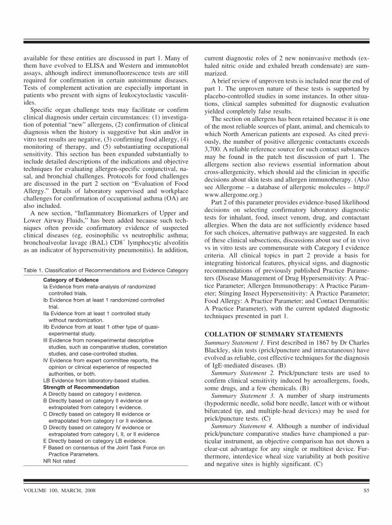
Table 1. Classification of Recommendations and Evidence Category
Category of EvidenceIa Evidence from meta-analysis of randomized
controlled trials.Ib Evidence from at least 1 randomized controlled
trial.IIa Evidence from at least 1 controlled study
without randomization.IIb Evidence from at least 1 other type of quasi-
experimental study.III Evidence from nonexperimental descriptive
studies, such as comparative studies, correlationstudies, and case-controlled studies.
IV Evidence from expert committee reports, theopinion or clinical experience of respectedauthorities, or both.
LB Evidence from laboratory-based studies.Strength of RecommendationA Directly based on category I evidence.B Directly based on category II evidence or
extrapolated from category I evidence.C Directly based on category III evidence or
extrapolated from category I or II evidence.D Directly based on category IV evidence or
extrapolated from category I, II, or II evidenceE Directly based on category LB evidence.F Based on consensus of the Joint Task Force on
Practice Parameters.NR Not rated
VOLUME 100, MARCH, 2008 S5
Summary Statement 5. Optimal results can be expected bychoosing a single prick/puncture device and properly trainingskin technicians in its use. (C)
Summary Statement 6. Although prick/puncture tests aregenerally age, sex, and race independent, certain age (chil-dren younger than 2 years and adults older than 65 years) andracial (African American children) factors may affect theirinterpretation. (C)
Summary Statement 7. Skin test allergens used for prick/puncture tests should be potent and stable. (B)
Summary Statement 8. To ensure proper interpretation,positive (histamine) and negative (saline or 50% glycerinatedhuman serum albumin [HSA]–saline) should be performed atthe same time as allergen tests. (B)
Summary Statement 9. The peak reactivity of prick/punc-ture tests is 15 to 20 minutes at which time both wheal anderythema diameters (or areas) should be recorded in millime-ters and compared with positive and negative controls. (B)
Summary Statement 10. Qualitative scoring (0 to 4�; pos-itive or negative) is no longer used by many clinicians be-cause of interphysician variability in this method of scoringand interpretation. (B)
Summary Statement 11. The diagnostic validity of prick/puncture tests has been confirmed not only in patients ex-posed to allergens under natural conditions but also in pa-tients undergoing controlled organ challenge tests. (B)
Summary Statement 12. Although prick/puncture testingoften correlates with exposure history, there are significantexceptions to this observation. (B)
Summary Statement 13. Many studies have verified thesensitivity and specificity of prick/puncture tests for bothinhalant and food allergens when correlated with nasal andoral challenge tests. (B)
Summary Statement 14. Compared with clinical historyalone, the diagnostic accuracy of prick/puncture tests showedmore limited capacity to predict clinical sensitivity for bothinhalant and food allergens. (C)
Summary Statement 15. The reliability of prick/puncturetests depends on the skill of the tester, the test instrument,color of the skin, skin reactivity on the day of the test, age,and potency and stability of test reagents. (C)
Summary Statement 16. False-positive prick/puncture testresults may occur (1) to tree pollens in honey bee–sensitivepatients due to cross-reactive carbohydrate determinantspresent in honey bee venom and (2) in tree-sensitive patientsbeing tested to tree pollens no longer indigenous to the area.(C)
Summary Statement 17. The rare occurrence of specificpositive organ challenge test results in patients with bothnegative prick/puncture and intracutaneous test results sug-gests that alternative pathways, including locally secretedIgE, IgE-independent, or nonimmune stimuli may activatemediator release in the end organ. (C)
Summary Statement 18. Life-threatening generalized sys-temic reactions are rarely caused by prick/puncture tests. In a
recent retrospective survey, 1 death was reported in a patientwho received 90 food prick/puncture tests at one time. (C)
Summary Statement 19. Intracutaneous tests will identify alarger number of patients with lower skin test sensitivity andare used when increased sensitivity is the main goal oftesting. (B)
Summary Statement 20. Intracutaneous tests are useful forevaluation of anaphylaxis, particularly drug (ie, penicillin)and Hymenoptera venom anaphylaxis. (A)
Summary Statement 21. When compared with specific na-sal challenge, skin end point titration (SET) is equivalent toprick/puncture skin tests. (B)
Summary Statement 22. Intracutaneous tests should beperformed with small volumes (approximately 0.02 to 0.05mL) of allergens injected intracutaneously with a disposable0.5- or 1.0-mL syringe. (C)
Summary Statement 23. As a general rule, the starting doseof an intracutaneous allergen test ranges from 100- to 1,000-fold more dilute than the allergen concentration used forprick/puncture tests. (C)
Summary Statement 24. Intracutaneous tests are read 10 to15 minutes after injection and both wheal and erythema (inmillimeters) should be recorded. (B)
Summary Statement 25. The diagnostic sensitivity of intra-cutaneous tests is probably greater than prick/puncture testswhen testing for penicillin, insect venom, or certain drugclass (eg, insulin, heparin, muscle relaxants) hypersensitivity.(C)
Summary Statement 26. The greater sensitivity of titratedintracutaneous tests, especially in the erythema component, isan advantage for determining biologic potency of allergenextracts and biologic allergy units (BAU) as based on intra-cutaneous erythema assays in sensitive human volunteers. (B)
Summary Statement 27. At dilutions between 10�2 and 10�3
(wt/vol), intracutaneous tests for most allergens exhibit poorefficiency in predicting organ challenge responses and cor-relating with the presence of detectable serum specific IgE.(C)
Summary Statement 28. There are limited data about equiv-alency of sensitivity, specificity, and predictive indices be-tween intracutaneous and prick/puncture tests when com-pared with organ challenge tests. One study demonstratedthat more dilute intracutaneous concentrations were compa-rable to prick/puncture tests in predicting positive nasal chal-lenges. (C)
Summary Statement 29. Similar comparative equivalencystudies based on history and symptoms alone revealed thatintracutaneous tests were comparable to prick/puncture testsonly at intracutaneous titration end points between 10�5 and10�6 g/mL (wt/vol). (B)
Summary Statement 30. Because clinical use of intracuta-neous tests is usually restricted to a single dose (ie, 1:1,000wt/vol), which may be irritant, predictive accuracy of thesetests at this concentration is often confounded by false-posi-tive results. (C)
S6 ANNALS OF ALLERGY, ASTHMA & IMMUNOLOGY
Summary Statement 31. For most allergens, a fixed dilution(1:1,000 wt/vol) of intracutaneous tests has poor efficiency inpredicting organ challenge responses. (A)
Summary Statement 32. Intracutaneous tests are occasion-ally negative in venom-sensitive patients who experiencelife-threatening reactions. (C)
Summary Statement 33. Repetitive (�2) intracutaneouspenicillin testing may sensitize a small number of individualsto penicillin. (C)
Summary Statement 34. Immediate systemic reactions aremore common with intracutaneous tests; 6 fatalities werereported in a recent retrospective survey. (C)
Summary Statement 35. Prescreening with prick/puncturetests is a practical way to avoid life-threatening reactions tointracutaneous tests. (C)
Summary Statement 36. If prick/puncture prescreening isnot used, preliminary intracutaneous serial threshold titra-tions should be considered, starting at high dilutions (eg, 10�5
to 10�8 g/mL [wt/vol]). This is of particular importance ifexquisite sensitivity (eg, anaphylaxis to foods and drugs) issuspected. (D)
Summary Statement 37. The late-phase cutaneous responseis a continuation of either prick/puncture or intracutaneoustesting, generally the latter, and is characterized by erythema,induration or edema, and dysesthesia. (B)
Summary Statement 38. The late-phase cutaneous responsemay occur after both immune and nonimmune activation.Many allergens have been implicated. (B)
Summary Statement 39. The late-phase cutaneous responseshould be read between the 6th and 12th hours after the skintests are applied; measurements of mean diameter and/or areaof induration or edema should be recorded. (B)
Summary Statement 40. Although the clinical relevance oflate-phase cutaneous response is not as yet fully established,several randomized, controlled studies suggest that reductionin sizes of late-phase cutaneous response may parallel clinicalresponse to immunotherapy. (B)
Summary Statement 41. The same principles that pertain tosafety of skin tests apply to late-phase cutaneous responses.(C)
Summary Statement 42. Preadministration of drugs, such ascalcineurin inhibitors, misoprostol, prednisone, and azelas-tine, before application of skin tests partially or completelyinhibit the late-phase cutaneous response. (B)
Summary Statement 43. The number of skin tests and theallergens selected for skin testing should be determined basedon the patient’s age, history, environment and living condi-tions (eg, region of the country), occupation, and activities.Routine use of a large number of skin tests or routine annualtests without a definite clinical indication are clearly notjustified. (D)
Summary Statement 44. Respiratory challenge tests areused when an objective gold standard for establishing clinicalsensitivity is indicated. (B)
Summary Statement 45. Conjunctival challenge tests areusually conducted for suspected localized eye allergy but in
some cases they may also be helpful in investigating nasalallergy. (B)
Summary Statement 46. Conjunctival challenge tests areevaluated by symptoms of itching and objective indices,including tear volume, amount of mucus, and palpebral orbulbar erythema. (B)
Summary Statement 47. Nasal challenges provide objectiveevidence of clinical sensitivity when the diagnosis is in ques-tion or in situations when it is desirable to evaluate efficacyof therapeutic management. (B)
Summary Statement 48. Nasal challenge responses areevaluated by subjective symptoms and objective measure-ments of nasal airway resistance, the number of sneezes, andthe measurement of inflammatory mediators in nasal secre-tions. (B)
Summary Statement 49. Specific (allergic) bronchial chal-lenge provides a measure of lower airway clinical sensitivitywhen there is uncertainty or dispute. (B)
Summary Statement 50. Guidelines for the performance ofspecific bronchial challenge include factors such as withhold-ing certain medications before the test, determining the initialallergen dose by preliminary skin or methacholine challengetesting, a beginning forced expiratory volume in 1 second(FEV1) baseline of 70% or better, the amount or duration ofexposure to allergen, measurement of FEV1 at intervals afterthe exposure, careful observation for late-phase responses,comparison to a placebo-controlled challenge usually per-formed the day before the specific challenge, and, optionally,repetition of methacholine challenge 24 to 48 hours afterspecific challenge for evaluation of induced bronchial hyper-responsiveness. (B)
Summary Statement 51. Occupational challenge testingrequires special precautions with respect to the innate toxicityof the suspected allergen and special apparatuses used tomeasure and control the quantity of challenge substances,such as potentially irritating volatile agents and dust. (B)
Summary Statement 52. A practical clinical method ofassessing OA is prospective monitoring of the worker at andaway from work by serial peak expiratory flow rates (PEFRs)or FEV1 values if this can be arranged by mutual agreementof employee and employer. (B)
Summary Statement 53. Many inflammatory correlates canbe evaluated and studied serially in respiratory and otherbody fluids, such as nasal smears or lavage, induced sputum,or BAL. These may define specific phenotypes or in somecases predict severity. (B)
Summary Statement 54. Exhaled nitric oxide is a noninva-sive measure of airway inflammation and is useful for mon-itoring objective responses to topically administered cortico-steroids. (B)
Summary Statement 55. Although breath condensate anal-ysis is an evolving noninvasive method for evaluation ofasthma, results are still variable and further refinements arerequired before it can be accepted as a valid diagnosticmethod. (C)
VOLUME 100, MARCH, 2008 S7
Summary Statement 56. Bronchoalveolar lavage obtainedthrough flexible bronchoscopy is useful in phenotypingasthma. The finding of lymphocytic alveolitis may suggest adiagnosis of hypersensitivity pneumonitis. (B)
Summary Statement 57. Cystic fibrosis may not only beconfused with asthma but certain genetic variants may beassociated with increased asthma risks. (B)
Summary Statement 58. Although major phenotypes of�1-antitrypsin deficiency do not occur in asthma, recent sur-veys demonstrated a high prevalence of asthma in young ZZhomozygous antitrypsin deficiency patients. (B)
Summary Statement 59. Purified protein derivative (PPD)of tuberculin is the prototype antigen recall test and providesdirect evidence that hypersensitivity, as opposed to toxicity,is elicited by the antigens in Mycobacterium hominis orrelated mycobacterial species. (B)
Summary Statement 60. The tuberculin skin test is elicitedby the intracutaneous injection of 0.1 mL of standardizedPPD starting with the intermediate strength of 5 tuberculinunits. (C)
Summary Statement 61. Recall antigen skin tests are usedto evaluate cellular immunity in patients with infection (eg,life-threatening sepsis), cancer, pretransplantation screening,endstage debilitating diseases, and the effect of aging. (C)
Summary Statement 62. Reduced or absent recall antigentests are termed anergy, which develops frequently in certaindiseases, such as hematogenous tuberculosis, sarcoidosis, andatopic dermatitis. (C)
Summary Statement 63. Candida albicans, Trichophytonmentagrophytes, and Tetanus toxoid, the currently availablerecall antigens, are injected intracutaneously in the same wayas the PPD test. (C)
Summary Statement 64. The size of the delayed skin testreaction is measured 48 hours after antigen challenge, and thelargest diameter of the palpable firm area that outlines theinduration response should be measured to the nearest milli-meter. (C)
Summary Statement 65. When a single intracutaneous an-tigen (other than PPD) is used to evaluate prior sensitizationto a potential pathogen, a reaction of 5 mm or greater maysuffice as the cutoff point for positive tests, but smallerreactions (2 to 4 mm) may be clinically important. (C)
Summary Statement 66. The absence of delayed-type hy-persensitivity to all the test antigens would suggest an anergicstate. (C)
Summary Statement 67. The most important use of de-layed-type hypersensitivity skin testing is epidemiologicscreening of susceptible populations exposed to bacterial andfungal pathogens. (C)
Summary Statement 68. The widest application of recallantigen testing is the detection of anergy and as an in vivoclinical correlate of cell-mediated immunoincompetency. (C)
Summary Statement 69. Although anergy testing was for-merly conducted frequently in HIV patients to determinewhether a concurrent negative tuberculin skin test result rulesout active tuberculosis, recent evidence mitigates against this
approach. Recall antigen anergy in HIV patients has alsobeen investigated as an indicator of staging, progression ofdisease, and response to therapy. (C)
Summary Statement 70. Although the standardized PPDantigen has been used for many years as a predictor of activeor latent tuberculosis infection, confounders, such as suscep-tible populations, BCG vaccination, and cross-sensitizationwith other atypical mycobacterial species have all affectedthe diagnostic accuracy of the tuberculin skin test and, byextrapolation, other delayed-type hypersensitivity tests. (C)
Summary Statement 71. The gross appearance of a late-phase cutaneous response and delayed-type hypersensitivityreactions may not be completely distinguishable except thatthe latter are more characterized by prolonged induration. (B)
Summary Statement 72. Although systemic corticosteroidswill render delayed-type hypersensitivity skin test resultsuninterpretable, 28 days of treatment with high-dose inhaledfluticasone (220 �g, 2 puffs twice a day) did not suppressdelayed-type hypersensitivity to PPD in healthy volunteers.(B)
Summary Statement 73. Neither anergy nor tuberculin test-ing obviates the need for microbiologic evaluation when thereis a suspicion of active tuberculosis or fungal infections. (F)
Summary Statement 74. Several new in vitro assays (ie,interferon-� and polymerase chain reaction) appear to bemore reliable in predicting active tuberculosis in BCG-vac-cinated persons or when cross-sensitivity to atypical myco-bacteria may coexist. (C)
Summary Statement 75. Immediate hypersensitivity reac-tions, including anaphylaxis, have been reported after tuber-culin skin tests. (D)
Summary Statement 76. The number of skin tests for de-layed, cell-mediated hypersensitivity reactions is limited. (C)
Summary Statement 77. First introduced by Jadassohn in1896, the epicutaneous patch test has evolved as the defini-tive diagnostic technique for the diagnosis of allergic contactdermatitis (ACD). (A)
Summary Statement 78. When clinical evaluations suggestthat exposure to a specific contactant has occurred either in anoccupational or nonoccupational clinical setting, patch testingcan be used to confirm the diagnosis. (C)
Summary Statement 79. From a public health perspective,patch testing is useful to identify potential health hazards ofunknown and newly introduced contact allergens for themedical community and industrial hygienists. (C)
Summary Statement 80. The most common patch test tech-niques are the individual Finn Chamber and the T.R.U.E.TEST, an FDA-approved screening method for screeningcontactant allergens. The T.R.U.E. TEST is preloaded with23 common contactants and vehicle control that have beenpreviously incorporated into a dried-in-gel delivery system,which is coated onto a polyester backing to form a patchtemplate. (B)
Summary Statement 81. If photocontact sensitivity is sus-pected, the appropriate allergens should be subjected to pho-
S8 ANNALS OF ALLERGY, ASTHMA & IMMUNOLOGY
topatch tests primarily in the UV-A range of 320 to 400 nm.(C)
Summary Statement 82. Traditionally, patch tests remain inplace for 48 hours. After the 48-hour patch test reading,additional readings at 3 to 4 days and in some cases 7 daysafter the original application of the patch yield the bestoverall reading reliability. (C)
Summary Statement 83. A descriptive reading scale devel-oped by 2 major international ACD research groups is thecurrent standard for interpreting patch test results. (C)
Summary Statement 84. Although patch tests are indicatedin any patient with a chronic eczematous dermatitis if ACD issuspected, patch tests are especially important in identifyingboth ICD and ACD in the occupational setting. (C)
Summary Statement 85. Other important exposures associ-ated with ACD include the use of topical medication, includ-ing corticosteroids, plant-induced ACD, and dermatitis oc-curring after the use of cosmetics and personal hygieneproducts. (C)
Summary Statement 86. Unprotected work and repetitiveexposure to surfactants may predispose patients to occupa-tional dermatitis, including ICD and ACD. (C)
Summary Statement 87. Certain contactant allergens in theT.R.U.E. TEST panel, such as nickel and some rubber chem-icals, have a high degree of relevant (approximately 75%)correlation with clinical sensitivity but others do not (eg,hydroxycitronellal, thimerosal). (B)
Summary Statement 88. Patch tests are most effective whenthe patients are selected on the basis of a clear-cut clinicalsuspicion of contact allergy and they are tested with thechemicals relevant to the problem; these conditions satisfythe prerequisites of high pretest probability. (C)
Summary Statement 89. Although the diagnostic accuracyof contactants cannot be compared with other in vivo or invitro tests, diagnostic concordance between patch test sensi-tivity and the outcomes of repeated open provocation testshas been demonstrated for some contactants. (B)
Summary Statement 90. The chief limitation to traditionalpatch testing for the diagnosis of ACD is the lack of a suitablegold standard by which it can be evaluated in terms ofdiagnostic accuracy predictors and likelihood ratios. (C)
Summary Statement 91. Other technical limitations ofpatch tests include the inclusion of relevant contact allergens,use of the proper vehicle, application to the proper skin area,proper reading and interpretation, and the ability to correlatethe tests with the patient’s specific exposure. (A)
Summary Statement 92. Other limiting factors concernreproducibility, lack of information about irritant thresholds,and minimal elicitation concentrations (MECs) for manycommon chemicals in the human environment. (C)
Summary Statement 93. The inability to separate irritantsfrom allergic responses is often encountered in the angry backsyndrome, which occurs in approximately 6% of cases and islikely to develop in patients with a longer duration of theprimary dermatitis. (C)
Summary Statement 94. Negative patch test reactions mayoccur even when the tests are performed with the correctsensitizing materials because the test fails to duplicate theconditions under which the dermatitis developed (eg, abra-sions, frequent use of irritating soaps, washing the hands withsolvents). (C)
Summary Statement 95. Systemic ACD after patch testingis rare, as is reactivation of patch test reactions after oralingestion of related allergens or even by inhalation of budes-onide in patients with sensitization to topical corticosteroids.(B)
Summary Statement 96. It is possible to sensitize a patientwho had not been previously sensitized to the allergen beingtested. This is particularly true of plant contactants, such aspoison ivy or oak and aniline dyes. (B)
Summary Statement 97. Two major variants of traditionalpatch tests are available: the atopy patch test (ATP) andrepeated use test (RUT). (B)
Summary Statement 98. Atopy patch tests have been eval-uated in patients with atopic dermatitis and eosinophilicesophagitis as an adjunct for the diagnosis of inhalant andfood allergy. (B)
Summary Statement 99. Atopy patch tests for foods areprepared with dried or desiccated foods mixed into an aque-ous solution and placed in 12-mm Finn Chambers beforepositioning on the patient’s back. (B)
Summary Statement 100. Atopy patch tests for the diagno-sis of drug allergy are performed by incorporating liquid orpowdered drugs into petrolatum or aqueous solvents, whichare added to 12-mm Finn Chambers and placed on the back.(B)
Summary Statement 101. Use tests have been developedfor weak sensitizers (repeated open application test [ROAT]),substances with poor percutaneous absorption (strip patchtest), and several premarketing dose response provocationtests for determining the minimal sensitizing dose of potentialcontactants in human volunteers. (B)
Summary Statement 102. In the strip patch test penetrationof substances is enhanced by repeated adhesive tape strippingbefore application of the contactant patch to the stripped area.(B)
Summary Statement 103. The ROAT is an exaggerated usetest designed to determine a patient’s biologic threshold orresponse to a suspected contactant, especially if this has notbeen achieved with prior open or closed patch testing. (B)
Summary Statement 104. Although clinical relevance isstill evolving with regard to the APT, several investigativegroups have reported that this test may be an adjunct indetection of specific allergens in atopic dermatitis and eosin-ophilic esophagitis. (B)
Summary Statement 105. The role of the atopy patch indetermining clinical allergy to food is indeterminate. (B)
Summary Statement 106. The lack of standardization ofAPTs for diagnosis of both food and drug allergy is the chieflimitation. (C)
VOLUME 100, MARCH, 2008 S9
Summary Statement 107. Although the purpose of APTs isto test for food and drug nonimmediate reactions, the possi-bility of anaphylaxis must be considered because there couldbe significant percutaneous absorption of proteins and/orsimple chemicals with high anaphylactogenic potential. (B)
Summary Statement 108. The appropriate number of atopicpatch tests is indeterminate because they are not routinelyperformed. (D)
Summary Statement 109. Because ACD is frequentlycaused by unsuspected substances, up to 65 patch tests maybe required for diagnosis. (B)
Summary Statement 110. Total serum IgE concentrationsare reported in international units or nanograms per milliliter(1 IU/mL � 2.44 ng/mL). (A)
Summary Statement 111. Total IgE is cross-standardizedwith the WHO 75/502 human reference IgE serum verified byperiodic proficiency surveys. (B)
Summary Statement 112. The clinical applications of totalserum IgE are of modest value. High serum IgE concentra-tions occur in allergic bronchopulmonary Aspergillosis(ABPA), the therapeutic response of which is evaluated byserial IgE values. (B)
Summary Statement 113. Total serum IgE is required forassessing the suitability of a patient for omalizumab therapyand determining the initial dose. (B)
Summary Statement 114. As with total IgE, commercialspecific IgE antibody assays are calibrated using heterolo-gous interpolation against the WHO 75/502 human IgE ref-erence serum, thereby enabling a uniform system of report-ing. (E)
Summary Statement 115. In addition to WHO 75/502 cal-ibration, an earlier specific IgE classification system wasbased on internal positive calibration curves from a positivecontrol heterologous serum containing specific IgE antibod-ies, which in the original RAST was white birch specific.However, FDA clearance for modified specific IgE testsrequires use of homologous internal control allergic serawhenever this is possible to obtain. (E)
Summary Statement 116. The precise sensitivity of theseimmunoassays compared with prick/puncture skin tests hasbeen reported to range from less than 50% to more than 90%,with the average being approximately 70% to 75% for moststudies; similar sensitivity ranges pertain when immunoas-says are compared with symptoms induced after natural orcontrolled organ challenge tests. (C)
Summary Statement 117. As with skin tests, the interpre-tation of specific IgE results requires correlation with thehistory, physical examination, and, in some cases, symptomsdirectly observed after natural or laboratory exposure to al-lergens. This cannot be accomplished by commercial remotepractice laboratories, which base recommendations for im-munotherapy on a history form submitted by the patient andspecific IgE results. (B)
Summary Statement 118. Because the constitutive allerge-nicity, potency, and stability are variable among commercialallergen extract reagents, sensitivity and the positive predic-
tive value of both prick/puncture and specific IgE tests gen-erally tend to be higher among pollens, stable anaphylacto-genic foods, house dust mite, certain epidermals, and fungicompared with venoms, drugs, and chemicals. (C)
Summary Statement 119. Proper interpretation of specificIgE test results needs to take into consideration variables suchas the binding affinity or avidity of allergens, solid-phasesystems, cross-reactive proteins and glycoepitopes, specificIgG antibodies in the test system, and high total serum IgE(�20,000 IU). (E)
Summary Statement 120. A multiallergen (up to 15 aller-gens bound to a linear solid-phase system) test can screen foratopic status, following which allergen specific tests are re-quired for more definitive evaluation. (C)
Summary Statement 121. Specific IgE immunoassays arenot recommended as a definitive confirmatory test for severalspecific clinical conditions. They provide neither diagnosticnor prognostic information when measured in the cord bloodof newborn infants. They do not have sufficient sensitivity forfoolproof prediction of anaphylactic sensitivity to venoms orpenicillins. (B)
Summary Statement 122. Specific IgE immunoassays maybe preferable to skin testing under special clinical conditions,such as widespread skin disease, patients receiving skin testsuppressive therapy, uncooperative patients, or when the his-tory suggests an unusually greater risk of anaphylaxis fromskin testing. (B)
Summary Statement 123. Determination of allergen speci-ficity by inhibition of specific IgE binding is a unique at-tribute of specific IgE testing. (E)
Summary Statement 124. Automated systems using multi-plexed allergen assays are being rapidly developed. One ofthese is cleared by the FDA for the simultaneous measure-ment of 10 allergens. (E)
Summary Statement 125. Allergen specific IgG may bemeasured by immunodiffusion or immunoabsorption. (E)
Summary Statement 126. Immunodiffusion antibodies tocow’s milk are associated with Heiner’s disease, a non-IgEdisorder that presents in infants with pulmonary infiltrates.(B)
Summary Statement 127. IgG and IgG subclass antibodytests for food allergy do not have clinical relevance, are notvalidated, lack sufficient quality control, and should not beperformed. (B)
Summary Statement 128. Although a number of investiga-tors have reported modest increases of IgG4 during venomimmunotherapy, confirmation and validation of the predictivevalue of IgG4 for therapeutic efficacy of venom immunother-apy are not yet proven. (C)
Summary Statement 129. The probability distribution ofspecific IgE for several anaphylactogenic foods (peanuts, eggwhite, cow’s milk, and codfish) can define clinical sensitivityas verified by double-blind oral challenge tests; similar rela-tionships have been defined for several respiratory allergens.(A)
S10 ANNALS OF ALLERGY, ASTHMA & IMMUNOLOGY
Summary Statement 130. Although allergens can be stan-dardized either by radioimmunodiffusion or immunoassayinhibition based on major allergenic epitopes, the FDA se-lected BAU instead because in vitro analytic techniqueswould have been variable from allergen to allergen and wouldhave caused great confusion. (C)
Summary Statement 131. Histamine and leukotriene re-lease measurements from human basophils after incubationwith allergen are valuable research tools for in vitro investi-gations of allergy. (B)
Summary Statement 132. The recent availability of severalsensitive immunoassays for histamine and leukotriene C4 is asignificant technological advance for measuring these medi-ators in various biologic fluids or release from whole blood,isolated basophils, mast cells, or other cultured cells. (B)
Summary Statement 133. Histamine and its N-methyl his-tamine metabolite may be measured in 24-hour urine samplesafter suspected anaphylactic episodes. (B)
Summary Statement 134. Plasma tryptase, particularly the� form, should be obtained within 4 hours after an anaphy-lactic episode. (B)
Summary Statement 135. Combined � and � species ofplasma tryptase are elevated in patients with systemic mas-tocytosis. (A)
Summary Statement 136. Eosinophils in body fluids corre-late highly with the diagnosis of allergic rhinitis, allergicasthma, and eosinophilic bronchitis. (B)
Summary Statement 137. Elevated eosinophil derived sub-stances (ie, ECP) and chemoattractants (ie, eotaxin) in bodyfluids are indicators of allergic inflammatory disease. (B)
Summary Statement 138. A basophil activation test mea-sured by expression of CD63 and CD203c and detected byflow cytometry is being evaluated for many IgE-mediateddisorders. (C)
Summary Statement 139. Tests that quantify lymphocytefunction measure the ability of lymphocytes to (1) proliferate,(2) produce inflammatory mediators and cytokines or chemo-kines, (3) mount cytotoxic responses, and (4) regulate im-mune responses. (B)
Summary Statement 140. Lymphocyte proliferative re-sponses may be evaluated by either nonspecific mitogens (eg,phytohemagglutinin, concanavalin A, or pokeweed) or spe-cific soluble and cell-bound antigens. (B)
Summary Statement 141. In vitro proliferative responses tosome soluble antigens, but not mitogens, have been shown tocorrelate with in vivo delayed hypersensitivity. The role,however, of lymphocyte proliferation as measured in vitro inthe pathogenesis of the delayed-type hypersensitivity tissuereaction is unclear. (B)
Summary Statement 142. Cytokines (IL-1 through IL-33)and growth factors are glycoproteins produced by a variety ofcells that are capable of altering activities of other cellsthrough interaction with specific surface receptors. (E)
Summary Statement 143. Chemokines are small (8 to 10kDa) proteins secreted by many immune and nonimmune
cells with essential roles in inflammatory and immune reac-tions, including the late-phase cutaneous response. (E)
Summary Statement 144. Cytokine and chemokine profilesplay essential roles in allergic inflammation and are beingincreasingly evaluated as phenotypic markers and in thedifferential diagnosis of human hypersensitivity disorders.(B)
Summary Statement 145. Other bioactive indices of cell-mediated immunity include cytotoxic assays, cultures ofmixed lymphocytes, and macrophage inhibition. (E)
Summary Statement 146. Most cytokines and chemokinescan be measured by commercial ELISA and ELISpot immu-noassays. (E)
Summary Statement 147. Proinflammatory cytokines orchemokines, which are particularly associated with cell-me-diated immunity, include interferon-�, IL-12, tumor necrosisfactor � (TNF-�), IL-16, MIF, macrophage inflammatoryprotein 1 (MIP-1), and MCP 1, 2, and 3. (B)
Summary Statement 148. Simple, cost-effective tests in-clude (1) an absolute lymphocyte count, (2) the absolutenumber of CD4� T cells, and (3) the CD4�/CD8� ratio. (B)
Summary Statement 149. Investigation of non-IgE andnon–cell-mediated clinical immunologic disorders may re-quire tests that indicate abnormal adaptive and innate immunereactions. (B)
Summary Statement 150. Abnormal serum and urine pro-teins, including cryoglobulins, may be associated with severalabnormal immune syndromes. (B)
Summary Statement 151. The inflammatory consequencesinduced by immune functions may be detected by nonspecifictests, such as a complete blood cell count with differential,sedimentation rate, C-reactive protein, and other acute-phasereactants. In some instances, functional assays of neutrophilsand macrophages may be necessary to pinpoint inflammatoryresponses. (B)
Summary Statement 152. Evaluation of complement acti-vation with a decrease of C3 and C4 may indicate comple-ment deficiency, drug reactions, or the presence of immunecomplexes, which often are associated with increases in se-rum cryoglobulins and C1q binding. (B)
Summary Statement 153. Autoantibody profiles offer im-portant diagnostic adjuncts in the diagnosis of collagen vas-cular diseases, vasculitides, and cytotoxicity disorders. (B)
Summary Statement 154. Procedures for which there is noevidence of diagnostic validity include cytotoxic tests, prov-ocation-neutralization, electrodermal testing, applied kinesi-ology, iridology, hair analysis, or food specific IgG, IgG4,and IgG/IgG4 antibody tests. (B)
Summary Statement 155. Although North American inhal-ant allergens are botanically and ecologically diverse, severalexpert committees consisting of members with botanic andmycologic expertise have compiled and selected 36 key al-lergens in North America, based on Thommen’s postulates.(D)
Summary Statement 156. For individual patients, thechoice of test allergens is guided by the history and physical
VOLUME 100, MARCH, 2008 S11
examination and the physician’s knowledge, training, andexperience. (B)
Summary Statement 157. A well-designed skin test andlaboratory ordering form should provide useful informationto the ordering physician, his/her staff, health care providers,and other physicians who may be consulted in the future. (B)
Summary Statement 158. The best indicators in the selec-tion of appropriate pollens for clinical use are extensiveprevalence in the air and concurrent allergy symptoms duringannually recurrent seasons when such pollens are expected tobe present in the ambient air. (B)
Summary Statement 159. The clinical significance of asingle fungus test reagent may be difficult to ascertain be-cause of important confounders, such as sampling method,culture conditions, nonculturable species, allergenic differ-ences between spores, and hyphae and preferential ecologicniches. (A)
Summary Statement 160. For clinical purposes, molds areoften characterized as outdoor (Alternaria and Cladosporiumspecies), indoor (Aspergillus and Penicillium species), orboth (Alternaria, Aspergillus, and Penicillium species). (B)
Summary Statement 161. Five Hymenoptera venom ex-tracts are available for evaluation of anaphylactic reactions tohoneybee, yellow jacket, yellow hornet, white faced hornet,and Polistes wasp. A whole-body extract is the only currentlyavailable diagnostic reagent for fire ant sting allergy. (A)
Summary Statement 162. Major inhalant acarid and insectallergens include several species of house dust mite andcockroach. (A)
Summary Statement 163. Animal clinical sensitivity ismost often associated with domestic pets (cats, dogs, birds)and laboratory animals (rodents, rabbits). Specific testing isguided by history of appropriate animal exposure. (A)
Summary Statement 164. Selection of food tests for IgE-mediated clinical sensitivity is usually tailored to the patient’stemporal history, which may be supplemented by a fooddiary. (A)
Summary Statement 165. Although commercial skin testsfor drugs, biologics, and chemicals are not available, special-ized medical centers prepare and use such tests under appro-priate clinical situations. The validity of such tests is ad-judged on a case by case basis. (C)
Summary Statement 166. More than 300 low- and high-molecular-weight occupational allergens have been identi-fied. Test reagents for these agents are generally available inspecialized occupational allergy centers. (A)
Summary Statement 167. A variety of plant or plant-de-rived proteins or glycoproteins may be associated with sys-temic allergic symptoms. (A)
Summary Statement 168. Chemicals, plant resins, and lipidconstituents are the chief causes of ACD, which requirespatch testing for confirmation. (A)
Summary Statement 169. As previously emphasized,knowledge of specific patterns of cross-reactivity among tree,grass, and weed pollens is essential in preparing an efficientpanel of test reagents. (A)
Summary Statement 170. Although cross-reactivity amongrelated pollen families can usually be ascribed to specificepitopic determinants, more diffuse cross-reactivity due toplant profilins and cross-reactive carbohydrate determinantsmay also be present. (A)
Summary Statement 171. Cross-reactivity data on fungi areextremely sparse. (C)
Summary Statement 172. The skin prick/puncture test issuperior to intracutaneous testing for predicting nasal allergicsymptoms triggered by exposure to pollen. (B)
Summary Statement 173. A skin prick/puncture test issuperior to intracutaneous testing for predicting allergic rhi-nitis and allergic asthma triggered by cat allergen exposure.(B)
Summary Statement 174. The skin prick/puncture can beused to rule out allergic rhinitis and allergic asthma triggeredby cat allergen exposure. (B)
Summary Statement 175. Knowledge of allergen cross-reactivity and local aerobiology is important in selectingappropriate allergens and in minimizing the number of aller-gens required for skin and specific IgE tests. (D)
Summary Statement 176. In general, skin prick/puncturetesting is more sensitive for identifying sensitization to in-halant allergens and confirming clinical allergy. However,specific IgE assays with defined quantifiable threshold levelscan also predict positive respiratory responses after allergenexposure. (B)
Summary Statement 177. Demonstration of sensitization toan occupational agent by specific IgE and/or skin testingalone is insufficient to establish a diagnosis of OA. (B)
Summary Statement 178. Skin prick testing with certainwell-characterized occupational protein allergens possessesadequate sensitivity such that a negative skin test result(�3-mm wheal diameter) can be used to rule out clinicalallergy. (B)
Summary Statement 179. Test performance characteristicsof specific IgE assays and skin testing for detection of chem-ical IgE-mediated sensitization must undergo validation andreproducibility in controlled studies using standardized anti-gens and assay protocols before these can be consideredreliable for routine evaluation of workers suspected of OA.(B)
Summary Statement 180. In patients undergoing evaluationfor suspected work-related natural rubber latex (NRL) al-lergy, a positive skin prick test result with a NRL extract (ifavailable) is preferred to demonstration of elevated specificIgE with an FDA-cleared assay due to higher sensitivity ofthe former. Current IgE-mediated allergy and asthma causedby NRL allergens is highly unlikely in the presence of anegative skin prick test result with a reliable crude NRLallergen extract. Elevated in vitro specific IgE levels can beused to confirm NRL allergy, but a negative result does notexclude NRL allergen sensitization. (B)
Summary Statement 181. The primary tools available toevaluate patients’ adverse reactions to foods include history(including diet records), physical examination, prick/puncture
S12 ANNALS OF ALLERGY, ASTHMA & IMMUNOLOGY
skin tests, serum tests for food specific IgE antibodies, trialelimination diets, and oral food challenges. (B)
Summary Statement 182. A detailed dietary history, attimes augmented with written diet records, is necessary todetermine the likelihood that food is causing the disorder,identify the specific food, and determine the potential immu-nopathophysiology. (D)
Summary Statement 183. With regard to evaluations forIgE antibody–associated food allergies, tests for food specificIgE antibody include percutaneous skin tests (prick/puncturetests) and serum assays. In general, these tests are highlysensitive (generally �85%) but only modestly specific (ap-proximately 40% to 80%) and therefore are well suited foruse when suspicion of a particular food or foods is high. Theyare not effective for indiscriminate screening (eg, using pan-els of tests without consideration of likely causes) and there-fore generally should not be used for that purpose. (B)
Summary Statement 184. Intracutaneous (intradermal) skintests for foods are potentially dangerous, are overly sensitive,increase the chance of a false-positive test result, and are notrecommended. (D)
Summary Statement 185. Based on studies in infants andchildren, increasingly higher concentrations of food specificIgE antibodies (reflected by increasingly larger percutaneousskin test size and/or higher concentrations of food specificserum IgE antibody) correlate with an increasing risk for aclinical reaction. (B)
Summary Statement 186. A trial elimination diet may behelpful to determine if a disorder with frequent or chronicsymptoms is responsive to dietary manipulation. (D)
Summary Statement 187. Graded oral food challenge is auseful means to diagnose an adverse reaction to food. (B)
Summary Statement 188. A number of additional diagnos-tic tests are under investigation, including APTs and tests forIgE binding to specific epitopes. (B)
Summary Statement 189. The rational selection, applica-tion, and interpretation of tests for food specific IgE antibod-ies require consideration of the epidemiology and underlyingimmunopathophysiology of the disorder under investigation,estimation of prior probability that a disorder or reaction isattributable to particular foods, and an understanding of thetest utility and limitations. (D)
Summary Statement 190. Diagnostic skin and/or specificIgE tests are used to confirm clinical sensitivity to venoms ina patient with a history of a prior systemic reaction. (B)
Summary Statement 191. Although diagnostic tests identifyspecies specificity of venom sensitization, they do not reli-ably predict severity of the sting reaction. (B)
Summary Statement 192. Standardized honeybee, Polistes,and Vespula antigens are commercially available as skin testreagents. (A)
Summary Statement 193. The skin test reagent available forevaluation of imported fire sting allergy is a nonstandardizedwhole-body extract. (C)
Summary Statement 194. In the case of a history of ana-phylaxis to Hymenoptera venoms, intracutaneous skin tests
are generally performed to 5 of the available venoms in adose response protocol (up to 1 �g/mL [wt/vol]) when pre-liminary prick/puncture test results are negative. (B)
Summary Statement 195. The FDA-cleared specific IgEassays have comparable specificity but decreased sensitivitycompared with venom skin tests. (B)
Summary Statement 196. Paradoxically, as many as 16% ofinsect-allergic patients with negative venom skin test resultshave positive results on currently available specific IgE invitro tests. (B)
Summary Statement 197. A small percentage of patients(1%) with negative results to both skin and in vitro tests mayexperience anaphylaxis after a field sting. (B)
Summary Statement 198. A skin test refractory periodlasting up to 6 weeks after a venom sting has been demon-strated by recent data. (B)
Summary Statement 199. Because of the predictive incon-sistencies of both skin and serum specific IgE tests, patientswith a convincing history of venom-induced systemic reac-tions should be evaluated by both methods. (D)
Summary Statement 200. Cross-allergenicity among insectvenoms is (1) extensive among vespid venoms, (2) consider-able between vespids and Polistes, (3) infrequent betweenbees and vespids, and (4) very limited between yellow jacketand imported fire ants. (B)
Summary Statement 201. If Hymenoptera venom sensitiv-ity is suspected, initial prick/puncture tests followed by serialendpoint titration with intracutaneous tests may be required.(B)
Summary Statement 202. Venom skin test may be repeatedonce or twice at 3- to 6-month intervals to confirm thediagnosis in a patient who initially had negative test results.(D)
Summary Statement 203. When the diagnosis is highlysuspected but not proved by skin and specific IgE tests,supervised live insect challenge sting may confirm clinicalsensitivity. Nevertheless, most patients with suspected venomallergy do not require live stings. (D)
Summary Statement 204. Evaluation of drug-specific IgEantibodies induced by many high-molecular-weight and sev-eral low-molecular-weight agents is often highly useful forconfirming the diagnosis and prediction of future IgE-medi-ated reactions, such as anaphylaxis and urticaria. (B)
Summary Statement 205. Neither immediate skin nor testsfor specific IgE antibodies are diagnostic of cytotoxic, im-mune complex, or cell-mediated drug-induced allergic reac-tions. (B)
Summary Statement 206. The availability of specific lab-oratory tests for non–IgE-mediated drug allergies is limited.(C)
Summary Statement 207. Atopy patch tests, lymphocyteproliferation tests, and basophil activation tests are additionaldiagnostic tests for drug allergy. Further studies are requiredto confirm their clinical utility in the evaluation of drugallergic patients. (B)
VOLUME 100, MARCH, 2008 S13
Summary Statement 208. A graded challenge (test dose) isa procedure to determine if a drug is safe to administer and isintended for patients who are unlikely to be allergic to thegiven drug. In contrast to desensitization, a graded challengedoes not modify the immune response to a drug. (B)
Summary Statement 209. Atopy patch tests, lymphocyteproliferation tests, and basophil activation tests are additionaldiagnostic tests for drug allergy. Further studies are requiredto confirm their clinical utility in the evaluation of drugallergic patients. (B)
Summary Statement 210. Penicillin skin testing is the mostreliable method for evaluating IgE-mediated penicillin al-lergy provided that the necessary reagents are available.When performed with both major and minor determinants,the negative predictive value of penicillin skin testing forimmediate reactions approaches 100%, whereas the positivepredictive value is between 40% and 100%. (B)
Summary Statement 211. Skin testing with penicilloyl-polylysine and penicillin G appears to have adequate negativepredictive value in the evaluation of penicillin allergy. (C)
Summary Statement 212. Penicillin skin test–negative pa-tients (as determined by testing with major and minor deter-minants) may receive penicillin, and depending on whichskin test reagents are used and the reaction history, the firstdose may need to be given via a test challenge with a lowerdose under observation. (D)
Summary Statement 213. In the absence of validated skintest reagents, the approach to patients with a history ofpenicillin allergy is similar to that of other antibiotics forwhich no validated in vivo or in vitro diagnostic tests areavailable. Therapeutic options include (1) prescribing an al-ternative antibiotic, (2) performing a graded challenge, and(3) performing penicillin desensitization. (D)
Summary Statement 214. In patients who have reacted tosemisynthetic penicillins, consideration should be given toskin test the implicated antibiotic and penicillin determinants.(B)
Summary Statement 215. There are no validated diagnostictests of sufficient sensitivity for evaluation of IgE-mediatedallergy to antibiotics other than penicillin. (C)
Summary Statement 216. Skin testing with nonirritatingconcentrations of other antibiotics is not standardized. Anegative skin test result does not rule out the possibility of animmediate-type allergy. A positive skin test result suggeststhe presence of drug-specific IgE antibodies, but the predic-tive value is unknown. (C)
Summary Statement 217. A presumptive diagnosis of as-pirin-exacerbated respiratory disease (AERD) can often bemade by history; however, in some cases, aspirin provocationtests might be considered for a definitive diagnosis. (B)
Summary Statement 218. Urticaria, angioedema, and ana-phylactic reactions to nonsteroidal anti-inflammatory drugs(NSAIDs) are distinctly different drug reactions from AERDreactions. In contrast to AERD reactions, anaphylactic reac-tions to NSAIDs are usually drug specific, and patients typ-ically tolerate other structurally dissimilar NSAIDs. (B)
Summary Statement 219. Skin testing is a useful diagnostictool in cases of perioperative anaphylaxis, and when skintesting is used to guide subsequent anesthetic agents, the riskof recurrent anaphylaxis to anesthesia is low. (C)
Summary Statement 220. Skin testing is not helpful incases of taxane-induced anaphylactoid reactions. (C)
Summary Statement 221. Skin testing to carboplatin yieldsfavorable predictive values. (C)
Summary Statement 222. Skin testing with asparaginasebefore treatment is recommended but does not identify allpatients at risk of reactions. (C)
Summary Statement 223. Skin testing for diagnosis of localanesthetic allergy is limited by false-positive reactions. Thegold standard for establishing a diagnosis of local anestheticallergy is the provocative challenge. (C)
Summary Statement 224. The specificity and sensitivity ofskin tests for systemic corticosteroid allergy are unknown,and cases of corticosteroid allergy with negative skin testresults to the implicated corticosteroid have been reported.(D)
Summary Statement 225. For most allergic reactions toadditives, skin tests are of no diagnostic value, and placebo-controlled oral challenges are required. (D)
Summary Statement 226. Contact dermatitis is a commonskin disorder seen by allergists and dermatologists and canpresent with a spectrum of morphologic cutaneous reactions.(C)
Summary Statement 227. The initial approach to clinicaldiagnosis of CD is to distinguish between ACD and ICD. (C)
Summary Statement 228. The inflammatory lesions of CDmay result from either ACD or ICD mechanisms. Factors thataffect response to the contact agent include the agent itself,the patient, the type and degree of exposure, and the envi-ronment. (A)
Summary Statement 229. Tissue reactions to contactantsare attributable primarily to cellular immune mechanismsexcept for contact urticaria. (A)
Summary Statement 230. Irritant contact dermatitis is usu-ally the result of nonimmunologic, direct tissue reaction andmust be clearly differentiated from ACD. (A)
Summary Statement 231. The diagnosis of ACD is sus-pected from the clinical presentation of the rash, which thenmust be supported by a history of exposure to a putative agentand subsequently confirmed by patch testing whenever this ispossible. (C)
Summary Statement 232. The skin site of the dermatitis isimportant in the diagnosis of ACD because the area of pre-dominant involvement and the regional distribution of thelesions often reflect the area of contact with the allergen. (A)
Summary Statement 233. Epicutaneously applied patchtests are the standardized diagnostic procedures to confirmACD. (A)
Summary Statement 234. Patch tests are indicated in anypatient with a chronic, pruritic, eczematous, or lichenifieddermatitis if underlying or secondary ACD is suspected. (C)
S14 ANNALS OF ALLERGY, ASTHMA & IMMUNOLOGY
Summary Statement 235. Patch test results are affected byoral corticosteroids but not by antihistamines. (A)
Summary Statement 236. Reading and interpretation ofpatch test results should conform to principles developed bythe International Contact Dermatitis Research Group and theNorth American Contact Dermatitis Research Group. (A)
Summary Statement 237. A 96-hour reading may be nec-essary because 30% of relevant allergens that are negative atthe 48-hour reading become positive in 96 hours. (A)
Summary Statement 238. Nonstandardized and customizedpatch testing is often required, depending on the patient’sexposure history. (C)
Summary Statement 239. A problem-oriented approach todiagnostic patch testing using evidence-based principles oflikelihood ratios and posttest probability is more likely toconfirm clinical ACD than a randomly selected patch testapproach. (B)
Summary Statement 240. Several in vitro procedures arebeing investigated for the diagnosis of ACD. (A)
Summary Statement 241. The differential diagnosis for CDis influenced by many factors, such as the clinical appearanceof the lesions, distribution of the dermatitis, and associatedsystemic manifestations. (B)
Summary Statement 242. Occupational contact dermatitis(OCD) is an inflammatory cutaneous disease caused or ag-gravated by workplace exposure. (B)
Summary Statement 243. There are 7 generally acceptablecriteria for establishing causation and aggravation of OCD.(C)
Summary Statement 244. Among health care professionals,ACD may occur as part of the spectrum of immunoreactivityto NRL in latex gloves. (A)
Summary Statement 245. Allergic contact dermatitis fromexposure to plants is the result of specific cell-mediatedhypersensitivity induced by previous contact with that familyof plants. (A)
Summary Statement 246. Contact dermatitis is commonlyimplicated after exposure to topical medications, includinglanolin, para-aminobenzoic acid (PABA), caine derivatives,antihistamines, iodochlorhydroxyquin, NSAIDs, and cortico-steroids. (A)
Summary Statement 247. Allergic contact dermatitis due totopical corticosteroids may occur in up to 5% of patients withsuspected CD. (A)
Summary Statement 248. Simultaneous exposure to aller-gens and irritants may produce both additive and synergisticACD responses due to their interaction. (A)
Summary Statement 249. The role of detergents in handdermatitis is a reflection of their ability to disrupt the skinbarrier. (A)
Summary Statement 250. Allergic contact dermatitis is asignificant clinical problem in children. (A)
PART 1Part 1 is an update of in vivo and in vitro techniques that areavailable as adjunctive diagnostic instruments for confirma-
tion of common allergic problems. These problems includeboth IgE and delayed hypersensitivity (ie, tuberculin-like andcontactant allergy) adaptive immune responses. Emphasis isplaced on reliability of reagents and devices. Quality assur-ance is also discussed in the context of reproducibility and theneed to minimize intertechnician and interlaboratory variabil-ity.
IN VIVO DIAGNOSTIC TESTS OF IMMEDIATEHYPERSENSITIVITY REACTIONS
Percutaneous and Intracutaneous In Vivo Diagnostic SkinTestsSummary Statement 1. First described in 1867 by Dr CharlesBlackley, skin tests (prick/puncture and intracutaneous) haveevolved as reliable, cost-effective techniques for the diagno-sis of IgE-mediated diseases. (B)
History and BackgroundAlthough Blackley first documented the diagnostic potentialof skin testing by placement of an allergen on abraded skin,the introduction of the cutaneous test for tuberculosis by vonPirquet became the chief impetus for subsequent allergy skintesting.1,2 The first of these was the scratch test made byrubbing the allergen into a small, blood-free scratched area ofthe forearm and introduced by Schloss for the diagnosis offood allergy in children.2 A few years later, Schick and Cookeindependently introduced the intracutaneous test as a diag-nostic method.2 Although the scratch method was used ex-tensively in the past, it has fallen out of general use becauseof greater patient discomfort, poor reproducibility, and thepossibility of residual linear pigmented or depigmented ar-eas.3 As a way to avoid these problems, prick/puncture testswere introduced in the early 1950s. Sir Thomas Lewis hadfirst suggested the puncture technique as an alternative skintest.4 However, Squire first called attention to the quantitativeaspects of the prick/puncture technique as a method of de-tecting sensitization to various proteins.5 He estimated that asmall amount (3 � 10�6 mL) of the test solution was intro-duced through the puncture site. Prick/puncture tests havebeen widely adapted throughout the world, although somepractitioners prefer exclusive use of intracutaneous tests.
Prick/Puncture TestsSummary Statement 2. Prick/puncture tests are used to con-firm clinical sensitivity induced by aeroallergens, foods,some drugs, and a few chemicals. (B)
Present applicationPrick/puncture tests are widely used for confirmation of clin-ical immediate hypersensitivity induced by a wide variety ofnaturally occurring allergens such as inhalants and foods.Under carefully defined circumstances, these tests are alsouseful in the diagnosis of drug and chemical hypersensitivity(platinum salts, acid anhydrides, polyisocyanates, sulfone-chloramide, and succinylcholine analogs) reactions. They arefrequently used as reference standards for evaluating speci-ficity and sensitivity of specific in vitro tests for IgE, and they
VOLUME 100, MARCH, 2008 S15
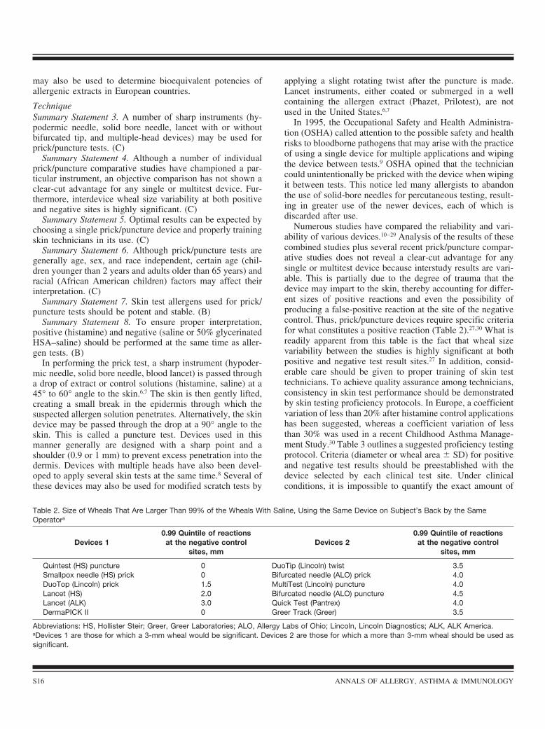
may also be used to determine bioequivalent potencies ofallergenic extracts in European countries.
TechniqueSummary Statement 3. A number of sharp instruments (hy-podermic needle, solid bore needle, lancet with or withoutbifurcated tip, and multiple-head devices) may be used forprick/puncture tests. (C)
Summary Statement 4. Although a number of individualprick/puncture comparative studies have championed a par-ticular instrument, an objective comparison has not shown aclear-cut advantage for any single or multitest device. Fur-thermore, interdevice wheal size variability at both positiveand negative sites is highly significant. (C)
Summary Statement 5. Optimal results can be expected bychoosing a single prick/puncture device and properly trainingskin technicians in its use. (C)
Summary Statement 6. Although prick/puncture tests aregenerally age, sex, and race independent, certain age (chil-dren younger than 2 years and adults older than 65 years) andracial (African American children) factors may affect theirinterpretation. (C)
Summary Statement 7. Skin test allergens used for prick/puncture tests should be potent and stable. (B)
Summary Statement 8. To ensure proper interpretation,positive (histamine) and negative (saline or 50% glycerinatedHSA–saline) should be performed at the same time as aller-gen tests. (B)
In performing the prick test, a sharp instrument (hypoder-mic needle, solid bore needle, blood lancet) is passed througha drop of extract or control solutions (histamine, saline) at a45° to 60° angle to the skin.6,7 The skin is then gently lifted,creating a small break in the epidermis through which thesuspected allergen solution penetrates. Alternatively, the skindevice may be passed through the drop at a 90° angle to theskin. This is called a puncture test. Devices used in thismanner generally are designed with a sharp point and ashoulder (0.9 or 1 mm) to prevent excess penetration into thedermis. Devices with multiple heads have also been devel-oped to apply several skin tests at the same time.8 Several ofthese devices may also be used for modified scratch tests by
applying a slight rotating twist after the puncture is made.Lancet instruments, either coated or submerged in a wellcontaining the allergen extract (Phazet, Prilotest), are notused in the United States.6,7
In 1995, the Occupational Safety and Health Administra-tion (OSHA) called attention to the possible safety and healthrisks to bloodborne pathogens that may arise with the practiceof using a single device for multiple applications and wipingthe device between tests.9 OSHA opined that the techniciancould unintentionally be pricked with the device when wipingit between tests. This notice led many allergists to abandonthe use of solid-bore needles for percutaneous testing, result-ing in greater use of the newer devices, each of which isdiscarded after use.
Numerous studies have compared the reliability and vari-ability of various devices.10–29 Analysis of the results of thesecombined studies plus several recent prick/puncture compar-ative studies does not reveal a clear-cut advantage for anysingle or multitest device because interstudy results are vari-able. This is partially due to the degree of trauma that thedevice may impart to the skin, thereby accounting for differ-ent sizes of positive reactions and even the possibility ofproducing a false-positive reaction at the site of the negativecontrol. Thus, prick/puncture devices require specific criteriafor what constitutes a positive reaction (Table 2).27,30 What isreadily apparent from this table is the fact that wheal sizevariability between the studies is highly significant at bothpositive and negative test result sites.27 In addition, consid-erable care should be given to proper training of skin testtechnicians. To achieve quality assurance among technicians,consistency in skin test performance should be demonstratedby skin testing proficiency protocols. In Europe, a coefficientvariation of less than 20% after histamine control applicationshas been suggested, whereas a coefficient variation of lessthan 30% was used in a recent Childhood Asthma Manage-ment Study.30 Table 3 outlines a suggested proficiency testingprotocol. Criteria (diameter or wheal area � SD) for positiveand negative test results should be preestablished with thedevice selected by each clinical test site. Under clinicalconditions, it is impossible to quantify the exact amount of
Table 2. Size of Wheals That Are Larger Than 99% of the Wheals With Saline, Using the Same Device on Subject’s Back by the SameOperatora
Devices 10.99 Quintile of reactions
at the negative controlsites, mm
Devices 20.99 Quintile of reactions
at the negative controlsites, mm
Quintest (HS) puncture 0 DuoTip (Lincoln) twist 3.5Smallpox needle (HS) prick 0 Bifurcated needle (ALO) prick 4.0DuoTop (Lincoln) prick 1.5 MultiTest (Lincoln) puncture 4.0Lancet (HS) 2.0 Bifurcated needle (ALO) puncture 4.5Lancet (ALK) 3.0 Quick Test (Pantrex) 4.0DermaPICK II 0 Greer Track (Greer) 3.5
Abbreviations: HS, Hollister Steir; Greer, Greer Laboratories; ALO, Allergy Labs of Ohio; Lincoln, Lincoln Diagnostics; ALK, ALK America.aDevices 1 are those for which a 3-mm wheal would be significant. Devices 2 are those for which a more than 3-mm wheal should be used assignificant.
S16 ANNALS OF ALLERGY, ASTHMA & IMMUNOLOGY
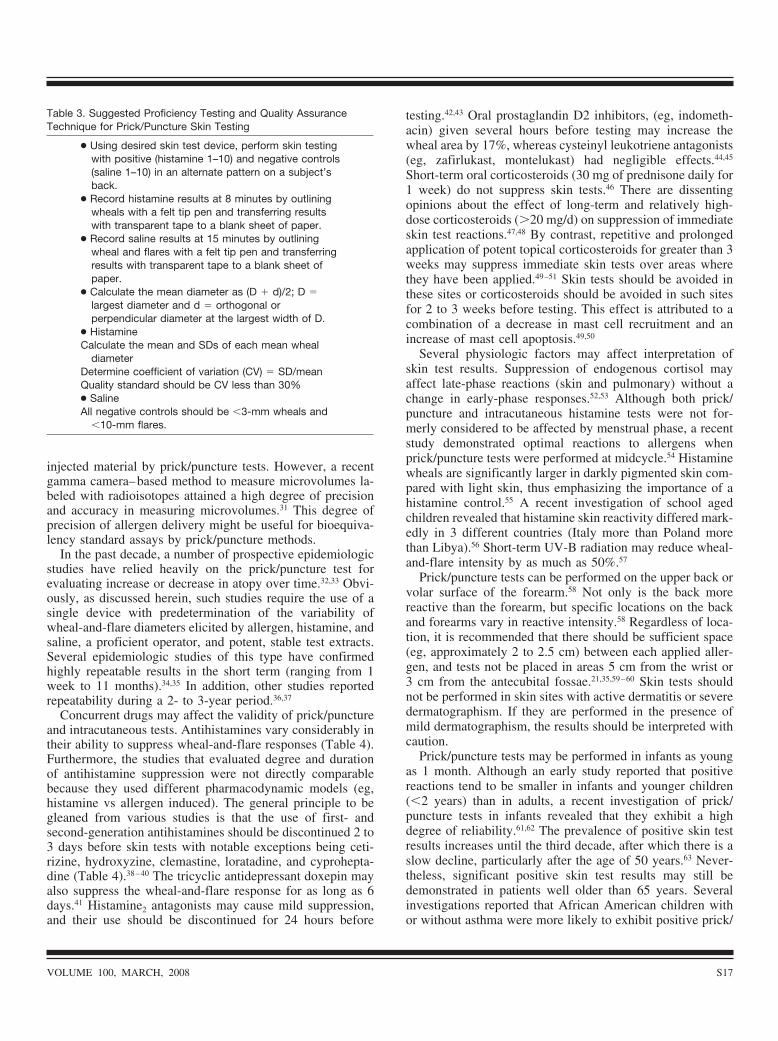
injected material by prick/puncture tests. However, a recentgamma camera–based method to measure microvolumes la-beled with radioisotopes attained a high degree of precisionand accuracy in measuring microvolumes.31 This degree ofprecision of allergen delivery might be useful for bioequiva-lency standard assays by prick/puncture methods.
In the past decade, a number of prospective epidemiologicstudies have relied heavily on the prick/puncture test forevaluating increase or decrease in atopy over time.32,33 Obvi-ously, as discussed herein, such studies require the use of asingle device with predetermination of the variability ofwheal-and-flare diameters elicited by allergen, histamine, andsaline, a proficient operator, and potent, stable test extracts.Several epidemiologic studies of this type have confirmedhighly repeatable results in the short term (ranging from 1week to 11 months).34,35 In addition, other studies reportedrepeatability during a 2- to 3-year period.36,37
Concurrent drugs may affect the validity of prick/punctureand intracutaneous tests. Antihistamines vary considerably intheir ability to suppress wheal-and-flare responses (Table 4).Furthermore, the studies that evaluated degree and durationof antihistamine suppression were not directly comparablebecause they used different pharmacodynamic models (eg,histamine vs allergen induced). The general principle to begleaned from various studies is that the use of first- andsecond-generation antihistamines should be discontinued 2 to3 days before skin tests with notable exceptions being ceti-rizine, hydroxyzine, clemastine, loratadine, and cyprohepta-dine (Table 4).38–40 The tricyclic antidepressant doxepin mayalso suppress the wheal-and-flare response for as long as 6days.41 Histamine2 antagonists may cause mild suppression,and their use should be discontinued for 24 hours before
testing.42,43 Oral prostaglandin D2 inhibitors, (eg, indometh-acin) given several hours before testing may increase thewheal area by 17%, whereas cysteinyl leukotriene antagonists(eg, zafirlukast, montelukast) had negligible effects.44,45
Short-term oral corticosteroids (30 mg of prednisone daily for1 week) do not suppress skin tests.46 There are dissentingopinions about the effect of long-term and relatively high-dose corticosteroids (�20 mg/d) on suppression of immediateskin test reactions.47,48 By contrast, repetitive and prolongedapplication of potent topical corticosteroids for greater than 3weeks may suppress immediate skin tests over areas wherethey have been applied.49–51 Skin tests should be avoided inthese sites or corticosteroids should be avoided in such sitesfor 2 to 3 weeks before testing. This effect is attributed to acombination of a decrease in mast cell recruitment and anincrease of mast cell apoptosis.49,50
Several physiologic factors may affect interpretation ofskin test results. Suppression of endogenous cortisol mayaffect late-phase reactions (skin and pulmonary) without achange in early-phase responses.52,53 Although both prick/puncture and intracutaneous histamine tests were not for-merly considered to be affected by menstrual phase, a recentstudy demonstrated optimal reactions to allergens whenprick/puncture tests were performed at midcycle.54 Histaminewheals are significantly larger in darkly pigmented skin com-pared with light skin, thus emphasizing the importance of ahistamine control.55 A recent investigation of school agedchildren revealed that histamine skin reactivity differed mark-edly in 3 different countries (Italy more than Poland morethan Libya).56 Short-term UV-B radiation may reduce wheal-and-flare intensity by as much as 50%.57
Prick/puncture tests can be performed on the upper back orvolar surface of the forearm.58 Not only is the back morereactive than the forearm, but specific locations on the backand forearms vary in reactive intensity.58 Regardless of loca-tion, it is recommended that there should be sufficient space(eg, approximately 2 to 2.5 cm) between each applied aller-gen, and tests not be placed in areas 5 cm from the wrist or3 cm from the antecubital fossae.21,35,59–60 Skin tests shouldnot be performed in skin sites with active dermatitis or severedermatographism. If they are performed in the presence ofmild dermatographism, the results should be interpreted withcaution.
Prick/puncture tests may be performed in infants as youngas 1 month. Although an early study reported that positivereactions tend to be smaller in infants and younger children(�2 years) than in adults, a recent investigation of prick/puncture tests in infants revealed that they exhibit a highdegree of reliability.61,62 The prevalence of positive skin testresults increases until the third decade, after which there is aslow decline, particularly after the age of 50 years.63 Never-theless, significant positive skin test results may still bedemonstrated in patients well older than 65 years. Severalinvestigations reported that African American children withor without asthma were more likely to exhibit positive prick/
Table 3. Suggested Proficiency Testing and Quality AssuranceTechnique for Prick/Puncture Skin Testing
● Using desired skin test device, perform skin testingwith positive (histamine 1–10) and negative controls(saline 1–10) in an alternate pattern on a subject’sback.
● Record histamine results at 8 minutes by outliningwheals with a felt tip pen and transferring resultswith transparent tape to a blank sheet of paper.
● Record saline results at 15 minutes by outliningwheal and flares with a felt tip pen and transferringresults with transparent tape to a blank sheet ofpaper.
● Calculate the mean diameter as (D � d)/2; D �largest diameter and d � orthogonal orperpendicular diameter at the largest width of D.
● HistamineCalculate the mean and SDs of each mean wheal
diameterDetermine coefficient of variation (CV) � SD/meanQuality standard should be CV less than 30%● SalineAll negative controls should be �3-mm wheals and
�10-mm flares.
VOLUME 100, MARCH, 2008 S17
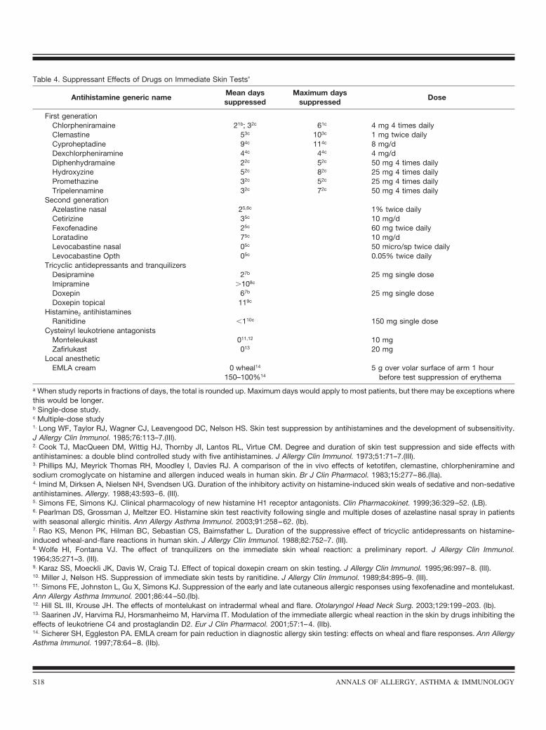
Table 4. Suppressant Effects of Drugs on Immediate Skin Testsa
Antihistamine generic nameMean dayssuppressed
Maximum dayssuppressed
Dose
First generationChlorpheniramaine 21b; 32c 61c 4 mg 4 times dailyClemastine 53c 103c 1 mg twice dailyCyproheptadine 94c 114c 8 mg/dDexchlorpheniramine 44c 44c 4 mg/dDiphenhydramaine 22c 52c 50 mg 4 times dailyHydroxyzine 52c 82c 25 mg 4 times dailyPromethazine 32c 52c 25 mg 4 times dailyTripelennamine 32c 72c 50 mg 4 times daily
Second generationAzelastine nasal 25,6c 1% twice dailyCetirizine 35c 10 mg/dFexofenadine 25c 60 mg twice dailyLoratadine 75c 10 mg/dLevocabastine nasal 05c 50 micro/sp twice dailyLevocabastine Opth 05c 0.05% twice daily
Tricyclic antidepressants and tranquilizersDesipramine 27b 25 mg single doseImipramine �108c
Doxepin 67b 25 mg single doseDoxepin topical 119c
Histamine2 antihistaminesRanitidine �110c 150 mg single dose
Cysteinyl leukotriene antagonistsMonteleukast 011,12 10 mgZafirlukast 013 20 mg
Local anestheticEMLA cream 0 wheal14
150–100%14
5 g over volar surface of arm 1 hourbefore test suppression of erythema
a When study reports in fractions of days, the total is rounded up. Maximum days would apply to most patients, but there may be exceptions wherethis would be longer.b Single-dose study.c Multiple-dose study1. Long WF, Taylor RJ, Wagner CJ, Leavengood DC, Nelson HS. Skin test suppression by antihistamines and the development of subsensitivity.J Allergy Clin Immunol. 1985;76:113–7.(III).2. Cook TJ, MacQueen DM, Wittig HJ, Thornby JI, Lantos RL, Virtue CM. Degree and duration of skin test suppression and side effects withantihistamines: a double blind controlled study with five antihistamines. J Allergy Clin Immunol. 1973;51:71–7.(III).3. Phillips MJ, Meyrick Thomas RH, Moodley I, Davies RJ. A comparison of the in vivo effects of ketotifen, clemastine, chlorpheniramine andsodium cromoglycate on histamine and allergen induced weals in human skin. Br J Clin Pharmacol. 1983;15:277–86.(IIa).4. lmind M, Dirksen A, Nielsen NH, Svendsen UG. Duration of the inhibitory activity on histamine-induced skin weals of sedative and non-sedativeantihistamines. Allergy. 1988;43:593–6. (III).5. Simons FE, Simons KJ. Clinical pharmacology of new histamine H1 receptor antagonists. Clin Pharmacokinet. 1999;36:329–52. (LB).6. Pearlman DS, Grossman J, Meltzer EO. Histamine skin test reactivity following single and multiple doses of azelastine nasal spray in patientswith seasonal allergic rhinitis. Ann Allergy Asthma Immunol. 2003;91:258–62. (Ib).7. Rao KS, Menon PK, Hilman BC, Sebastian CS, Bairnsfather L. Duration of the suppressive effect of tricyclic antidepressants on histamine-induced wheal-and-flare reactions in human skin. J Allergy Clin Immunol. 1988;82:752–7. (III).8. Wolfe HI, Fontana VJ. The effect of tranquilizers on the immediate skin wheal reaction: a preliminary report. J Allergy Clin Immunol.1964;35:271–3. (III).9. Karaz SS, Moeckli JK, Davis W, Craig TJ. Effect of topical doxepin cream on skin testing. J Allergy Clin Immunol. 1995;96:997–8. (III).10. Miller J, Nelson HS. Suppression of immediate skin tests by ranitidine. J Allergy Clin Immunol. 1989;84:895–9. (III).11. Simons FE, Johnston L, Gu X, Simons KJ. Suppression of the early and late cutaneous allergic responses using fexofenadine and montelukast.Ann Allergy Asthma Immunol. 2001;86:44–50.(Ib).12. Hill SL III, Krouse JH. The effects of montelukast on intradermal wheal and flare. Otolaryngol Head Neck Surg. 2003;129:199–203. (Ib).13. Saarinen JV, Harvima RJ, Horsmanheimo M, Harvima IT. Modulation of the immediate allergic wheal reaction in the skin by drugs inhibiting theeffects of leukotriene C4 and prostaglandin D2. Eur J Clin Pharmacol. 2001;57:1–4. (IIb).14. Sicherer SH, Eggleston PA. EMLA cream for pain reduction in diagnostic allergy skin testing: effects on wheal and flare responses. Ann AllergyAsthma Immunol. 1997;78:64–8. (IIb).
S18 ANNALS OF ALLERGY, ASTHMA & IMMUNOLOGY
puncture test results to outdoor aeroallergens than their coun-terpart white cohorts.64,65
Allergen extracts used for percutaneous and intracutaneoustesting ideally should be of known composition and potency.Although a limited number of standardized extracts are com-mercially available, most inhalant and food extracts are notstandardized. Before the recent availability of standardizedextracts, the composition of nonstandardized, commerciallyavailable extracts varied greatly between the manufacturers.66
This situation is slowly improving with the introduction ofbioequivalent extracts.67,68 In Nordic countries, the wheal andflare of a positive control histamine test are used to assignbiologic equivalency to allergen materials.69,70 Bioequiva-lency by this system is defined as histamine equivalent prick(HEP) units. Although relatively few commercialized ex-tracts are yet designated in bioequivalent allergy units (eg,grass, cat), the trend toward universal bioequivalency is wellunder way, as evidenced by more recent attempts to standard-ize commercial food antigen extracts not only by wheal areabut also by objective organ challenges.71,72
Stability and potency of allergenic test extracts are alsoimportant issues. Since it is known that allergen extractsdeteriorate with time, accelerated by dilution and highertemperatures, allergen skin test extracts are usually preservedwith 50% glycerin.73,74 If dilutions are required for skin testthreshold testing, the diluent should be HSA (0.03%)–sa-line.73 All extracts should be stored under cold (4°C) toensure stability.74 In vivo biologic activity of geneticallyengineered recombinant allergens has been evaluated andcompared with specific allergens from which they were de-rived.75,76 In general, they appear to be highly specific andsafe. However, the sensitivity of single recombinant allergensis usually lower than those obtained with natural allergenextracts.75 The precise role of recombinant allergens as invivo diagnostic tools remains to be determined.
Positive and negative controls should be performed with alltests. In the United States for many years the only availablepositive control was histamine phosphate (2.7 mg/mL equiv-alent to a 1.0-mg/mL histamine base). Wheal diameters withthis preparation range from 2 to 7 mm.59 Currently, a 10-mg/mL histamine dehydrochloride control is available, andthis is the preferred positive control for prick/puncture skintests. The negative control consists of 50% glycerinatedHSA–saline if concentrated extracts are used.
Reading the test resultsSummary Statement 9. The peak reactivity of prick/puncturetests is 15 to 20 minutes at which time both wheal anderythema diameters (or areas) should be recorded in millime-ters and compared with positive and negative controls. (B)
Summary Statement 10. Qualitative scoring (0 to 4�; pos-itive or negative) is no longer used by many clinicians be-cause of interphysician variability in this method of scoringand interpretation. (B)
A standardized approach to reading the tests has not yetbeen achieved. For example, some clinicians advocate imme-
diate blotting of the allergen after the prick/puncture test toreduce the risk of an adverse reaction, whereas others leavethe allergen in place for 20 minutes.77 No essential differencehas been found between these techniques. Histamine controltests should be read 15 minutes after application at the peakof reactivity.69 The peak of allergen prick/puncture tests isusually 15 to 20 minutes after application. Although someinvestigators have advocated the primary importance of thewheal diameter,70 both erythema and wheal should be mea-sured and recorded in millimeters for appropriate compari-sons with positive (ie, histamine) and negative controls (ie,buffered diluent or 50% glycerinated extracts). Since traumamay affect wheal size (Table 2), an allergen response lessthan 3 mm generally should not be regarded as positive.27,30,70
Devices that produce wheals that exceed 3 mm at negativecontrol sites should be avoided.11 Unfortunately, there isvariability among physicians and investigators in recordingthe dimensions of flare, wheal, or both. The size of thereaction may be recorded as a mean wheal diameter, D � d/2(with D indicating the largest diameter of the wheal and dindicating the largest diameter orthogonal to D), planimetry(either direct or from a traced copy), minimal diameter of asignificant wheal �3 mm, comparison to an HEP test causedby 1 or 10 mg/mL of histamine dihydrochloride (defined as 1HEP with 10 mg/mL being the preferred reference standard),or a score related to a codeine phosphate control defined as awheal of 75% or greater of a control codeine phosphatesolution (25 mg/mL).17,24,78 Qualitative scoring (0 to 4�; 0 or�) is no longer used by many clinicians because of markedinterphysician variability in scoring and interpretation of thismethod.79
To summarize, a prick/puncture test with a response of atleast 3-mm diameter (with equivalent erythema) more thandiluent control done at the same time is required as proof ofthe presence of cutaneous allergen specific IgE. There is arecent trend to develop more precise methods of measuringwheal area, such as handheld scanners with appropriate com-puter software, end point titration, and morphometry becausewheal size (area or diameter) has assumed greater diagnosticsignificance.80–84 Several investigators have determined thatspecific cutoff values (eg, �8 mm for peanut) obviate theneed to confirm clinical sensitivity by organ challengetests.85,86 If similar pretest probabilities for clinical sensitivitycan be developed for more allergens by defining precisecutoff prick/puncture measurement results, the clinical utilityof prick/puncture tests will be greatly enhanced.
Clinical relevanceSummary Statement 11. The diagnostic validity of prick/puncture tests has been confirmed not only in patients ex-posed to allergens under natural conditions but also in pa-tients undergoing controlled organ challenge tests. (B)
Summary Statement 12. Although prick/puncture testingoften correlates with exposure history, there are significantexceptions to this observation. (B)
VOLUME 100, MARCH, 2008 S19
The diagnostic value of prick/puncture tests has been de-termined chiefly by comparing the test to history of symp-toms associated with exposure. They have been used mostfrequently to evaluate individual cases and populations ofallergic patients. The diagnostic validity of prick/puncturetests has been confirmed as a correlate of clinical sensitivityin double-blind, randomized control studies under outdoor(parks) and indoor (controlled environmental exposure units)exposure conditions.87 The diagnostic accuracy in prick/punc-ture tests has also been confirmed in groups of clinicallyallergic patients undergoing specific nasal bronchoprovoca-tion challenge measured by nasal resistance or acoustic rhi-nometry under controlled laboratory conditions.88–92 Like-wise, in the case of foods, prick/puncture tests have beendemonstrated to correlate with clinical symptoms that occurafter either open or double-blind food challenges.93–97 How-ever, 1 of these studies revealed that this correlation did notnecessarily apply to all foods.93 When used as a diagnostictest for potential symptoms based on exposure and/or historyalone, the utility of prick/puncture tests is highly allergendependent, giving concordant results with certain allergens,such as cat dander, but not with others.97–99 Nevertheless,given their generally favorable diagnostic characteristics,other tests (intracutaneous, atopy patch, various specific IgEtests) are often compared with prick/puncture tests as a ref-erence.100–108 Interestingly, a recent prospective study re-ported that 60% of skin sensitive (wheal �4 mm) asymptom-atic subjects developed clinical allergy. These resultssuggested that a positive prick/puncture test result in anasymptomatic person may predict subsequent clinical aller-gy.108
Sensitivity, specificity, and positive and negative predictiveindicesSummary Statement 13. Many studies have verified the sen-sitivity and specificity of prick/puncture tests for both inhal-ant and food allergens when correlated with nasal and oralchallenge tests. (B)
Summary Statement 14. Compared with clinical historyalone, the diagnostic accuracy of prick/puncture tests showedmore limited capacity to predict clinical sensitivity for bothinhalant and food allergens. (C)
It is generally accepted that prick/puncture tests are lesssensitive than intracutaneous tests. This is partially explainedby the larger volumes of test solutions administered by theintracutaneous route. To compensate for this, positive prick/puncture tests require that the test extracts be 50 to 100 timesmore concentrated than intracutaneous test solutions. Thisrelative lack of sensitivity to prick/puncture tests can bepartially compensated for by avoidance of glycerinated ex-tracts or by adding small amounts of Tween 80 (0.0005%).15
On the other hand, prick tests are more specific than intracu-taneous tests because the increased sensitivity at a fixedconcentration of the intracutaneous test (1 in 1,000 wt/vol)may be responsible for a small but reproducible number offalse-positive reactions, presumably because of an irritant
effect. Because of the uncertainty created by this relationshipbetween prick/puncture and intracutaneous tests, comparativeinvestigations have been conducted to establish cutoff values,sensitivity, specificity and predictive indices of these testswith respect to inhalants and selected food allergens. Inter-pretation of these results varies, depending on whether thecomparative gold standard is clinical history or controlledprovocation challenges. With respect to inhalant allergens,several investigations have demonstrated that it is possible toestablish more scientific guidelines for interpreting the testsand what they predict.90,91,97,109 Using positive nasal provoca-tion challenges as a standard, the sensitivity of prick/puncturetests ranges from 85% to 87%, whereas the specificity ofthese tests is between 79% and 86%.90,91 A recent meta-analysis comparing prick/puncture tests to nasal challengerevealed positive likelihood ratios of 4.93, 16.17, 3.23, and4.06 for cat, tree pollen, grass pollen, and house dust allergen,respectively, whereas the corresponding negative likelihoodratios were 0.08, 0.03, 0.04, and 0.03.109–113 In a single moldinvestigation (Alternaria sp) that compared skin testing tochallenge tests, positive and negative likelihood ratios weresimilar for both prick/puncture and intracutaneous tests(prick: positive likelihood ratio of 11.75, negative likelihoodratio of 0.05; intracutaneous: positive likelihood ratio of 8.80,negative likelihood ratio of 0.05).114 (Refer to Evaluation ofInhalant Allergy, Part 2 for clinical significance.)
A comparative study of allergic asthmatic patients under-going nonspecific methacholine challenge causing a 20% fallin FEV1 of 4 mg/mL or less (wt/vol) or 8 mg/mL or less(wt/vol) revealed that the sensitivity, specificity, and negativepredictive value of prick/puncture tests were 91%, 52%, and85%, respectively, with the cutoff value of provocation con-centration that caused a decrease in FEV1 of 20% (PC20) 8mg/mL or less (wt/vol).108 The lower cutoff PC20 of 4 mg/mLor less (wt/vol) increased the sensitivity and negative predic-tive value to 98.2% and 97.8%, respectively. This suggeststhat positive prick/puncture skin test results are more likely tobe associated with asthma of greater severity, as indexed bythe lower cutoff methacholine PC20 value of less than 4mg/mL (wt/vol). A negative prick/puncture test result de-creased the probability of having asthma by 10- to 20-fold insubjects whose pretest probability was low to moderate.109
The diagnostic accuracy of prick/puncture tests in food al-lergy has been compared with patients (mostly children) whohave positive open or double-blinded controlled positive re-actions to specific foods.85,93–97,115–118 In several of these stud-ies, it was possible to determine cutoff levels of skin prick/puncture tests wheal diameters that were 100% diagnostic forseveral foods (eg, �8 mm for milk; �7 mm for egg; �8 mmfor peanuts).94–99,115 These specific food cutoff values alsoindicate the probability of more severe food allergy becausethe controlled oral food challenges to which these werecompared reproduced clinical anaphylactic events, whichcould be carefully monitored and treated. However, cutoffwheal sizes associated with high likelihood of allergy arevariable, depending on the age (older children and infants),
S20 ANNALS OF ALLERGY, ASTHMA & IMMUNOLOGY
device, and reagents. Once cutoff values are ascertained andvalidated, both likelihood ratios and the area under the re-ceiver operating characteristic curve can be calculated withthe goal of eliminating the need for confirmation by provoc-ative challenges.85,86,115
Sensitivity, specificity, and the predictive indices have alsobeen compared with clinical history, both for inhalant andfood allergens.97,98,119–122 Several of these investigations usedthe area under the receiver operating characteristic curves todetermine optimal cutoff values and to evaluate the ability ofvarious allergens to predict symptom histories of hay feverand asthma.120,121 Analysis of these studies revealed no uni-fying principle about the accuracy of prick/puncture skin testsas predictors for hay fever and asthma. Thus, 1 study con-cluded that even the combination of history to commonallergens and physical examination is not diagnostic withrespect to skin prick/puncture and specific IgE tests.98 Thereare exceptions, one of which concluded that a skin prick/puncture wheal size of 3 mm or larger to cat elicited asensitivity of 0.9, a specificity of 0.9, and a diagnostic accu-racy of 0.9.97 The limited capacity of skin prick/puncture testsfor predicting clinical symptoms was also tested by structuredinterviews with patients undergoing aeroallergen skin tests.123
Patients were found to have limited ability to correctly predictpositive skin test results to aeroallergens based on their ownclinical symptom experiences.
LimitationsSummary Statement 15. The reliability of prick/puncture testsdepends on the skill of the tester, the test instrument, color ofthe skin, skin reactivity on the day of the test, potency, andstability of test reagents. (C)
Summary Statement 16. False-positive prick/puncture testsmay occur (1) to tree pollens in honey bee–sensitive patientsdue to cross-reactive carbohydrate determinants present inhoney bee venom and (2) in tree-sensitive patients beingtested to tree pollens no longer indigenous to the area. (C)
Summary Statement 17. The rare occurrence of specificpositive organ challenge test results in patients with bothnegative prick/puncture and intracutaneous tests suggests thatalternative pathways, including locally secreted IgE, IgE-independent, or nonimmune stimuli may activate mediatorrelease in the end organ. (C)
The reliability and interpretation of the prick/puncture testis heavily dependent on the skill and interpretation of theindividual tester, the reliability of the test instrument, thecolor of the skin, the status of skin reactivity on the day of thetest, potency and stability of test extracts, especially theoptimum concentrations used for the test, and experimentaldifferences between duplicate prick tests.13,19,30 Appropriateproficiency test methods for evaluating accuracy, precision,and reproducibility of skin testing are encouraged in thetraining of personnel (Table 3).30,124
If quality controls are not used, interpretation of the testresults varies from one technician to another. The hazards ofblood contamination with the use of all instruments must be
given appropriate attention, and all technicians must be care-fully trained in appropriate barrier techniques, as well asavoidance of accidental needle punctures. Reliability ofprick/puncture tests requires that allergen extracts be potentand of known composition. Whenever possible, extracts withknown biologic potency should be used.66,124,125 For example,commercial extracts of fruits and vegetables are likely to losepotency over relatively short periods. Therefore, prick/punc-ture tests for these potential allergens should be performedeither with freshly made food extracts or by the prick-prickmethod in which the tester first pricks the fresh food and thenthe skin. This method may be particularly helpful when thereare differences in the allergenicity of different cultivar strains(eg, apples).126,127
If interpretation of allergen prick/puncture tests are ex-pressed as a ratio of equivalency to a positive control (eg,HEP), selection of the positive control may affect the diag-nostic accuracy of the test. It has been shown that using aratio of allergen to positive histamine control for gradingragweed reactivity elicited better diagnostic accuracy than theratio of allergen to a codeine phosphate control.128 By con-trast, relating allergen-induced and control histamine whealsreduced intertechnician reproducibility.129
Several confounding issues concerning test extracts couldlimit diagnostic accuracy of prick/puncture tests. A recentreport demonstrated that approximately 16% of honeybeevenom allergic patients may be misdiagnosed as having mul-tivalent pollen sensitization because they reacted to nonspe-cific cross carbohydrate determinants in venom extracts.130 Arecent study of patients sensitive to multiple tree pollensrevealed a lack of correlation between prick/puncture testsusing commercial extracts of 15 previously reported indige-nous tree species compared with actual mean tree speciespollen counts samples in the local aerobiology system.99 Thisstudy indicated that prick tests to tree pollens should only beperformed with those species that have been confirmed asbeing current airborne aeroallergens by aerobiologic sam-pling (see nationalallergybureauwww.aaaai.org/nab/).
For unknown reasons, the diagnostic accuracy of intracu-taneous testing is superior to that of prick/puncture tests inseveral well-established IgE-mediated anaphylactic reactions(eg, penicillin, muscle relaxant, and venom hypersensitivity).In recent years, however, even intracutaneous negative Hy-menoptera allergic patients have been reported to experienceanaphylaxis.131,132
Although the accuracy of prick/puncture tests in predict-ing the presence or absence of clinical allergy has beengenerally confirmed by previously cited studies, providedthe proper cutoff levels of interpretation are used, there arespecific reports of proven end-organ sensitivity in theabsence of positive prick/puncture or intracutaneous testresults.133–143 This occurrence has not been explained, al-though pathways such as locally secreted IgE, IgE-inde-pendent, or non-IgE stimuli have been suggested.139,144,145
In the case of reactions to foods despite negative testresults, the trigger protein in the test reagent may not have
VOLUME 100, MARCH, 2008 S21
been fully extracted (with respect to the appropriateepitope) or the causal protein was rapidly degraded.140 –143
SafetySummary Statement 18. Life-threatening generalized sys-temic reactions are rarely caused by prick/puncture tests. In arecent retrospective survey, 1 death was reported in a patientwho received 90 food prick/puncture tests at one time. (C)
In a retrospective analysis of children being tested foratopy, 6 cases of generalized reactions occurred in infantsyounger than 6 months who showed positive skin prick testresults to fresh food specimens. Other common features inthis group of patients were active eczema and a familyhistory of allergic diseases. All infants received prompttreatment and recovered well.146 The overall rate of gen-eralized reactions was 521 per 100,000 tested children. Ina 12-year survey of fatal reactions to allergen injectionsand skin testing in both adults and children from 1990 to2001, one fatality was confirmed after skin prick testingwith multiple food allergens.147 This patient also had mod-erately persistent asthma, and 90 food prick tests wereapplied at one time. Analysis of near or life-threateningreactions in the same survey revealed no instances ofreactions attributed to inhalant prick/puncture tests. In therecently published Practice Parameter, The Diagnosis andManagement of Anaphylaxis, the concurrent use of�-blockers and angiotensin-converting enzyme inhibitorsis cited as a relative contraindication to skin testing.148 –151
Intracutaneous Tests
Present applicationsSummary Statement 19. Intracutaneous tests will identify alarger number of patients with lower skin test sensitivity andare used when increased sensitivity is the main goal oftesting. (B)
Summary Statement 20. Intracutaneous tests are useful forevaluation of anaphylaxis, particularly drug (ie, penicillin)and Hymenoptera venom anaphylaxis. (A)
Summary Statement 21. When compared with specific na-sal challenge, skin end point titration (SET) is equivalent toprick/puncture skin tests. (B)
Intracutaneous tests are generally used when increasedsensitivity is the main goal of testing (ie, when prick/puncturetest results are negative despite very convincing history ofexposure).152 They permit identification of a larger number ofclinically reactive patients, especially those with lower skintest sensitivity. In addition, skin sensitivity to low potencyallergenic extracts may best be evaluated by this method.
As previously discussed, intracutaneous tests are prefera-ble for diagnosis of drug and venom anaphylaxis.153–161 Theutility of intracutaneous tests for diagnosis of drug-inducedpenicillin anaphylaxis has been extended to a variety of drugclasses, including cancer chemotherapeutic agents, musclerelaxants, insulin, and heparin.162–166 Although experience andstandardization of these drug categories are limited compared
with penicillin and venoms, their negative and positive pre-dictive values appear to be comparable.163
Although intracutaneous tests at strengths customarily per-formed (1:100 to 1:1,000 [wt/vol] from manufacturer’s con-centrate) are more sensitive, there are conflicting resultsabout their ability to predict clinical allergy. Several studiesin the previously cited meta-analysis investigated how wellintracutaneous tests predict symptoms after natural or labo-ratory allergen challenges.110 Two high-quality studies con-ducted in cat- and grass-sensitive patients concluded thatpositive likelihood ratios were poor (0.89 and 1.05 for cat andgrass, respectively) as were negative likelihood ratios (1.24and 0.98 for cat and grass, respectively).111,167 By contrast, theaccuracy of intracutaneous tests was excellent for Alternariaspecies, as evidenced by positive and negative likelihoodratios of 8.80 and 0.05, respectively.114 These disparate re-sults probably reflect the intrinsic variability of individualallergens among investigators and their abilities to predictclinical allergy.113
One recent investigation demonstrated that SET, which isa modified quantitative testing method, is equivalent to prick/puncture testing for both positive and negative predictabilityof clinical allergy when both are compared with nasal chal-lenge.90 The end point response in SET is the lowest concen-tration of allergen that produces a wheal: (1) that is the firstwheal 2 mm larger than the negative control wheal and (2) isfollowed by a second wheal that is at least 2 mm larger thanthe preceding one.90 It should be stressed, however, that SETis roughly equivalent to new skin prick tests only at dilutionsranging from 1:12,500 (wt/vol) to 1:312,000 (wt/vol). Bycomparison, most physicians who perform intracutaneoustesting use dilutions ranging from 1:100 (wt/vol) to 1:1,000(wt/vol).90 Indeed, a study designed to test the predictiveresponse of timothy prick/puncture and intracutaneous teststo nasal provocation revealed that the addition of a singleintracutaneous test at a dilution of 1:500 (wt/vol) (No. 2 in theRinkel nomenclature) adds no additional predictability whenthe prick test result is negative and therefore appears to beunwarranted.91 Similar disappointing results were obtainedwhen Alternaria intracutaneous tests at a dose of 1:500 (wt/vol) were compared with specific nasal challenge93 and con-trasted sharply with a previous Alternaria study.114
TechniquesSummary Statement 22. Intracutaneous tests should be per-formed with small volumes (approximately 0.02 to 0.05 mL)of allergens injected intracutaneously with a disposable 0.5-or 1.0-mL syringe. (C)
Summary Statement 23. As a general rule, the starting doseof an intracutaneous allergen test ranges from 100- to 1,000-fold more dilute than the allergen concentration used forprick/puncture tests. (C)
A single-unit, 0.5- or 1.0-mL disposable syringe with anattached hypodermic needle is preferred. The gauge of theattached hypodermic needle may vary from 26 to 30.168 Theuse of a Hamilton calibrated syringe ensures a reproducible
S22 ANNALS OF ALLERGY, ASTHMA & IMMUNOLOGY
injected volume, but this appears to offer little advantage tocareful injections that produce wheals of similar size.168,169
The reproducibility of intracutaneous tests is affected by thesame variables as those described for prick/puncture tests.57
These include the age of the skin, the area of the body wherethe tests are applied, skin pigmentation, interference by con-current medications, and potency and biologic stability of theallergen test extracts. Intracutaneous tests are usually placedon the upper arm or volar surface of the forearm rather thanthe back to allow for application of a tourniquet shouldsystemic symptoms occur. The back also reveals considerabledifferences in skin reactivity between different areas of theback of individual patients.168 There may be leakage of theallergen at the injection site because of improper technique,but this can be prevented by the use of unitized syringes andneedles. Concurrent tests with diluent control solutions alsoshould be performed. In addition, a positive histamine control(equivalent to 0.10 mg/mL [wt/vol] of histamine base) shouldbe included to evaluate the degree of skin response at the timeof the test. The volumes of intracutaneous test solutions mayvary from 0.02 to 0.05 mL, depending on the purpose of thetest. Delivery of small volumes (�0.03 mL) is difficult toattain with regularity. Because of the greater possibility ofsystemic reactions after intracutaneous testing, special careshould be given to preparing less potent test dilutions. As ageneral rule, the starting dose of intracutaneous extract solu-tions in patients with a preceding negative prick test resultshould range from 100- to 1,000-fold dilutions of the con-centrated extracts used for prick/puncture tests.58 In the caseof standardized allergens, such as ragweed, grass, dust mite,and cat, the range of starting intracutaneous test solutions inpatients with preceding negative prick/puncture test results isbetween 10 and 100 BAU.58,170
Most of the factors that affect the reliability of prick/puncture tests also apply to intracutaneous tests. Several ofthese have already been discussed (ie, smaller dose of thepositive histamine phosphate control and the unsuitability ofthe back for intracutaneous tests). Technical training forprecision and reproducibility of intracutaneous tests shouldalso be emphasized, especially for those persons performingbiologic equivalency tests. A recent investigation of intracu-taneous skin tests noted that intracutaneous testing had poorreproducibility, appearing to confirm a much earlierstudy.171,172 The effects of drugs on intracutaneous testing aresimilar to the agents discussed under prick/puncture tests.Although immediate-phase reactions are not affected by cys-teinyl leukotriene modifiers, the late-phase cutaneous reac-tion is reduced.45
Reading the test resultsSummary Statement 24. Intracutaneous tests are read 10 to 15minutes after injection, and both wheal and erythema (inmillimeters) should be recorded. (B)
For intracutaneous tests, histamine controls and allergensites are usually read 10 to 15 minutes, respectively, after theinjections. Similar to prick/puncture tests, various indices,
such as the longest diameter, the sum of the largest diameterand its orthogonal diameter divided by 2, products of thediameters, planimetry, and measurement of paper traced fromskin responses, have been used to interpret intracutaneousresults. Both erythema and wheal diameters should be mea-sured and recorded. Erythema can be measured as reliably aswheal reactions and is the sole criterion for bioequivalencytests in the United States.59,124 Any reaction larger than thenegative control may indicate the presence of specific IgEantibody. Given the greater sensitivity and equivocal repro-ducibility of intracutaneous testing, however, small positivereactions may not be clinically significant.173 There are noevidence-based studies on standardized intracutaneous testgrading. Eighty-five percent of board-certified allergists re-cently surveyed reported that they used the criterion of 3 mmabove the negative control as a threshold for a positiveintracutaneous test result.174 The criteria for determining theSET titration threshold stipulate a measurement of 4 mmabove the negative control.90
Clinical relevanceSummary Statement 25. The diagnostic sensitivity of intracu-taneous tests is probably greater than prick/puncture testswhen testing for penicillin, insect venom, or certain drugclass (eg, insulin, heparin, muscle relaxants) hypersensitivity.(C)
Summary Statement 26. The greater sensitivity of titratedintracutaneous tests, especially in the erythema component, isan advantage for determining biologic potency of allergenextracts and biologic allergy units (BAU) as based on intra-cutaneous erythema assays in sensitive human volunteers. (B)
In general, intracutaneous tests are useful in detectingpatients with lower levels of clinical sensitivity when evalu-ating allergens (both natural and recombinant) of low skinreactive potency (eg, Hymenoptera). They have been evalu-ated and validated in diagnosis of several important IgE-mediated drug reactions, including anaphylactic reactionsinduced by penicillin, succinylcholine analogs, and cancerchemotherapeutic agents. In the case of penicillin anaphylac-tic hypersensitivity, intracutaneous testing (after initial pricktesting) is a first-line approach. Under the proper test condi-tions1 (use of both major and minor penicillin determinants),these tests were found to have a negative predictive value ofalmost 99% in a large, multicentered clinical trial.153 Recentreports suggested that intracutaneous tests might also beuseful adjuncts for the diagnosis of nonimmediate allergicreactions to aminopenicillins.175 The diagnostic accuracy ofintracutaneous tests for predicting anaphylaxis associatedwith cephalosporins and other non–�-lactam antibiotics islimited because standardized reagents are not available formost of these antibiotics.176 Intracutaneous tests are usedmostly as a complement to prick/puncture tests in the evalu-ation of anaphylaxis to muscle relaxants.163,166 Test concen-trations between 10 and 1,000 �g/mL (wt/vol) have shown97% concordance with prick/puncture tests.177 Although sys-temic IgE-mediated reactions are much less frequent with
VOLUME 100, MARCH, 2008 S23
commonly used biologics (eg, protamine, human insulin,heparin), a number of case reports suggest that they may beuseful for confirming the immunologic nature of these reac-tions.166,178–180 Venom intracutaneous skin testing is the mostuseful in vivo immunologic procedure for confirming imme-diate hypersensitivity to venoms.181
The greater sensitivity of intracutaneous tests offers anadvantage for determination of biologic potency of allergenicextracts and their respective recombinant allergens.75,182 Di-agnostic markers for ABPA were identified by intracutaneoustesting of a panel of recombinant antigens derived fromAspergillus fumigatus.182 Variability among commercialvenom extracts may also be evaluated by intracutaneoustesting.183
Dose response assays of erythema in response to intracu-taneous testing in sensitive human volunteers are the basis ofBAU in the United States.124 In Europe, it has been suggestedthat bioequivalency could be based on prick/puncturewheals125
Sensitivity, specificity, and positive and negative predictiveindicesSummary Statement 27. At dilutions between 10�2 and 10�3
(wt/vol), intracutaneous tests for most allergens exhibit poorefficiency in predicting organ challenge responses and cor-relating with the presence of detectable serum specific IgE.(C)
Summary Statement 28. There are limited data about equiv-alency of sensitivity, specificity, and predictive indices be-tween intracutaneous and prick/puncture tests when com-pared with organ challenge tests. One study demonstratedthat more dilute intracutaneous concentrations were compa-rable to prick/puncture tests in predicting positive nasal chal-lenges. (C)
Summary Statement 29. Similar comparative equivalencystudies based on history and symptoms alone revealed thatintracutaneous tests were comparable to prick/puncture testsonly at intracutaneous titration end points between 10�5 and10�6 g/mL (wt/vol). (B)
Summary Statement 30. Because clinical use of intracuta-neous tests is usually restricted to a single dose (ie, 1:1,000wt/vol), which may be irritant, predictive accuracy of thesetests at this concentration is often confounded by false-posi-tive results. (C)
Quantitative estimates of sensitivity, specificity, and thepredictive indices are difficult to evaluate because most of theclinical experience with allergen intracutaneous testing hasbeen performed at a single dilution (1:1,000 wt/vol). Forexample, a recent investigation of potential clinical moldallergy could not distinguish between atopic and nonatopicphenotypes in patients being tested to molds at this dilu-tion.171 However, a study comparing intracutaneous titrationend point with prick testing showed a modest correlation fora panel of 8 allergens.106 Although these data suggest thatintracutaneous end point titration might achieve sensitivityand specificity values equivalent to the prick test, there is
only one head-to-head comparison of the 2 methods withclinical history and/or provocation testing.91 By comparingintracutaneous end point titration, skin prick/puncture tests,and nasal provocation determined by acoustic rhinometry,this investigation revealed that skin prick/puncture tests weremore sensitive (85.3% vs 79.4%) and more specific (78.6%vs 67.9%) than intracutaneous end point titration as a screen-ing procedure. The positive and negative predictive values ofintracutaneous end point titration were 75% and 73%, respec-tively.91 Another study comparing intracutaneous tests to skinprick/puncture tests at 30 and 3,000 biologic units/mL, re-spectively, found positive predictive values of 87.1% and79.1% for intracutaneous and prick/puncture tests, respective-ly.170 The same investigators also established optimum intra-cutaneous and prick/puncture cutoff values of 0.7 and 0.4HEP equivalents, respectively.184 Compared with clinical his-tory, the positive predictive value for detection of allergicsensitization was 77% for intracutaneous tests and 86% forprick/puncture tests.184 End point intracutaneous titrations toa single allergen (ragweed) were compared with history andspecific in vitro IgE (RAST) in a group of patients beingevaluated for possible clinical allergy.185 At intracutaneoustitration end points between 10�6 and 10�8 g/mL (wt/vol),70% of the patients had a positive history and approximately50% of the patients had a positive RAST result. At intracu-taneous titration end points between 10�5 and 10�3 g/mL(wt/vol), only 60% of patients gave positive histories and15% exhibited specific IgE.185 This study indicated that moredilute end point threshold levels of intracutaneous tests couldapproach the diagnostic accuracy of prick/puncture tests. In amore recent investigation using recombinant birch pollen Betv 1 as the allergen, the endpoint intracutaneous titrationmethod correlated modestly with basophil histamine releasebut not with specific serum IgE.186 Thus, in this study, thebiologic sensitivity of the intracutaneous end point titrationthreshold appeared to outperform both basophil histaminerelease and serum specific IgE.
LimitationsSummary Statement 31. For most allergens, a fixed dilution(1:1,000 [wt/vol]) of intracutaneous tests has poor efficiencyin predicting organ challenge responses. (A)
Summary Statement 32. Intracutaneous tests are occasion-ally negative in venom-sensitive patients who experiencelife-threatening reactions. (C)
Summary Statement 33. Repetitive (�2) intracutaneouspenicillin testing may sensitize a small number of individualsto penicillin. (C)
The chief problem with intracutaneous tests performed at afixed dilution (1:1,000 [wt/vol]) for most allergens is rela-tively poor efficiency in predicting organ challenge re-sponses, the most reliable predictors of clinical sensitivity. Ina recent study that specifically evaluated this relationship,this limitation appeared to apply to most of the commonindoor and outdoor allergens.171 Similar findings were re-ported in an exposed population being evaluated for mold-
S24 ANNALS OF ALLERGY, ASTHMA & IMMUNOLOGY
related health effects.187 In venom allergy, intracutaneousskin test results are occasionally negative, even in patientswith near life-threatening reactions to the specific venom.188
A deliberate sting challenge under controlled conditionsshould be considered in such an unusual circumstance.
There is some evidence that anaphylactoid reactions tovenom occur in a substantial number of patients with masto-cytosis or urticaria pigmentosa having relatively high consti-tutive levels of serum tryptase.189,190 Also, as discussed underSummary Statement 17, skin test results to inhalants andfoods may rarely be negative despite positive end organchallenge test results. Repetitive (�2) intracutaneous penicil-lin testing may sensitize a small number of individuals topenicillin.191 Six of 239 (2.5%) volunteers who were skintested to penicillin on 2 occasions converted to a positive skintest result. Intracutaneous tests often do not correlate wellwith serum specific IgE levels. One possible explanation forthis disparity was a recent study in which binding of allergenspecific IgE antibodies to the � chain of Fc�I receptor wassuboptimal and did not correlate with either intracutaneoustests or specific basophil sensitivity.186,192
SafetySummary Statement 34. Immediate systemic reactions aremore common with intracutaneous tests; 6 fatalities werereported in a recent retrospective survey. (C)
Summary Statement 35. Prescreening with prick/puncturetests is a practical way to avoid life-threatening reactions tointracutaneous tests. (C)
Summary Statement 36. If prick/puncture prescreening isnot used, preliminary serial threshold titrations should beconsidered, starting at high dilutions (10�5 to 10�8 g/mL[wt/vol]). This is of particular importance if exquisite sensi-tivity (eg, anaphylaxis to foods and drugs) is suspected. (D)
Although adverse events occurring after intracutaneoustests are rare, they can occur.193,194 Large local reactions, bothimmediate and late, may cause discomfort and occasionallymild, nonprogressive systemic reactions may be associatedwith the latter. Immediate systemic reactions are more com-mon with intracutaneous tests because larger volumes areinjected. Six fatalities attributed to intracutaneous skin testswere reported by the Committee on Allergen Standardizationof the AAAAI.195 Five of these patients had asthma and weretested without preceding prick/puncture tests. No fatalitieswere associated with intracutaneous testing in the most recent12-year survey of fatal reactions from 1990 to 2001.147
To reduce the likelihood of adverse reactions during skintesting, several precautions may be taken. Prescreening withprick/puncture test is a practical way to avert an untowardnumber of adverse local and/or systemic responses in routineskin testing of patients. If prick/puncture tests are not per-formed routinely, preliminary threshold intracutaneous test-ing should be considered, beginning at higher dilutions (ie,10�5 to 10�8 g/mL [wt/vol]). Even greater precautions shouldbe observed if patients are suspected of having exquisitesensitivity, such as anaphylaxis, to certain foods and drugs. In
such cases, even prick/puncture tests should be initiated withseveral serial 10-fold dilutions of the usual test concentration.Patients receiving �-adrenergic blocking agents and mono-amine oxidase inhibitors may present special risk-benefitproblems. If a systemic reaction should occur, epinephrinemay not be totally effective in patients taking �-blockers, andepinephrine may adversely affect patients taking monoamineoxidase inhibitors.59
Late-Phase Cutaneous Reactions
Definition and descriptionSummary Statement 37. The late-phase cutaneous response isa continuation of either prick/puncture or intracutaneous test-ing, generally the latter, and is characterized by erythema,induration or edema, and dysesthesia. (B)
The late-phase cutaneous reaction develops progressivelyat sites of immediate wheal-and-flare reactions and is char-acterized by erythema, induration or edema, and dysesthe-sia.196–201 Histopathologically, it is characterized by the pres-ence of edema, mixed cellular infiltrates, and sometimesfibrin deposition scattered throughout the dermis without thedeposition of complement, IgG, IgA, IgM, or vascular dam-age. Less frequently, the late-phase cutaneous response mayoccur in the absence of an immediate skin test response andmay be confused with cell-mediated, delayed hypersensitiv-ity.202,203 Isolated late cutaneous reactions were observed inapproximately 36% of children undergoing skin tests forsuspected allergies. Most of these isolated late-phase cutane-ous responses were due to inhalant allergens, such as cock-roach and various mold spores.204 The clinical significance ofthis is as yet unknown.
CausesSummary Statement 38. The late-phase cutaneous responsemay occur after both immune and nonimmune activation.Many allergens have been implicated. (B)
Late-phase cutaneous reactions occur after both immuneand nonimmune (eg, 48/80, kallikrein) mast cell activation.Agents stimulating immunologic activation of the mast cellsthat have induced the late-phase cutaneous response includeanti-IgE antibodies and the following allergens or antigens:aeroallergens (molds, pollens, danders, mites, and enzymes),penicillin, heparin, insulin, and possibly some foods.205,206
The propensity to develop the late-phase cutaneous responsemay be dependent on the type of antigen, host sensitivity, andthe concentration of injected antigen or allergen.207
Reading the test resultsSummary Statement 39. The late-phase cutaneous responseshould be read between the 6th and 12th hours after the skintests are applied; measurements of mean diameter and/or areaof induration or edema should be recorded. (B)
After challenge with diverse stimuli causing immediatewheal-and-flare responses, the intensity of the late-phasecutaneous response increases rapidly (doubling or tripling insize) during the first 2 hours.198–201,208 The response plateausbetween the 6th and 12th hours, is present at 24 hours, and
VOLUME 100, MARCH, 2008 S25
usually disappears by 48 hours after challenge. Accordingly,these reactions should be quantified between the 6th and 12thhours (most commonly at the 6th or 8th hour) by measure-ments of mean diameter and/or area of induration or edema.
Although the minimum size of induration or erythema ofthe late-phase cutaneous response has not yet been standard-ized, the extent of these measurements should be compareddirectly with previously applied diluent or histamine sites,which typically demonstrate neither induration nor erythema6 to 8 hours later.197,207 One investigator suggests a minimumof 5 mm of induration and/or erythema be considered.204 Thelate-phase cutaneous response is in part mediated by antigen-specific major histocompatibility complex restricted T cells,which in the past were thought to be prototypic of tuberculin-induced delayed-type hypersensitivity. However, it has beendemonstrated that both characteristic histologic features andthe occurrence of isolated late-phase cutaneous response afterimmunization with T-cell–specific small overlapping aller-genic (eg, from Fel d 1) peptides can distinguish between alate-phase cutaneous response and delayed-type hypersensi-tivity.202,209 Immunochemical histologic analysis at variousstages of the inflammatory milieu of a late-phase cutaneousresponse reveals a diversity of cells, including macrophages,eosinophils, neutrophils, tryptase positive mast cells, Lang-erhans cells, and, interestingly, large numbers of ba-sophils.210,211 T cells are also present and the late-phase cu-taneous response is thought to be partially dependent onthem, possibly through effects of cytokines, particularly IL-4,IL-5, and IL-10.210,212–214 Also noteworthy is up-regulation ofthe CCR3L (eotaxin) and CCR4L chemokines in T cells(skin, lung, and blood) after allergen-induced late-phase cu-taneous response.215,216 Not surprisingly, a variety of othermediators and proinflammatory cytokines have also beendescribed in association with the late-phase cutaneous re-sponse.217–219
Clinical relevanceSummary Statement 40. Although the clinical relevance oflate-phase cutaneous response is not as yet fully established,several randomized, controlled studies suggest that reductionin sizes of late-phase cutaneous response may parallel clinicalresponse to immunotherapy. (B)
Although the clinical relevance of late-phase cutaneousresponse cannot yet be delineated with certainty, there hasbeen preliminary progress about some potential clinical ap-plications. At least 4 randomized, controlled clinical trials ofimmunotherapy in patients with allergic rhinitis have shownmarked reductions in late-phase cutaneous response in pa-tients who experience successful reduction of clinical symp-toms.212,220–222 Furthermore, reduction in size of the late-phasecutaneous response was also associated with recruitment ofCD4� CD25� regulatory T cells and CD4� interferon-�� TH1cells to sites of allergen-induced late-phase cutaneous re-sponse in cat-allergic subjects.223 It has been suggested thatpatients with atopic dermatitis may be classified phenotypi-cally into either positive or negative late-phase cutaneous
response reactors.208 Atopic dermatitis patients with signifi-cant late-phase cutaneous response reactions were morelikely to demonstrate higher levels of IL-5 and specific IgE tohouse dust mite antigens.214,224 Of related interest was a studyin which birch pollen–sensitive patients with atopic dermati-tis and isolated late eczematous reactions to birch pollen–related foods demonstrated up-regulation of specific T cellsin biopsy specimens of delayed skin lesions.225 Recently,there have been numerous anecdotal case reports that suggestdelayed-type intracutaneous tests are useful for the diagnosisof various drug allergies, including nonimmediate allergicreactions to muscle relaxants, penicillins, non–�-lactam an-tibiotics, antiepileptics, and heparins.177,226–230 However,many of these delayed intracutaneous tests were not inter-preted within the 6- to 12-hour range of the late-phase cuta-neous reaction, so it is not clear whether such testing repre-sents cell-mediated hypersensitivity reactions or variants ofthe late-phase cutaneous response. Further research is neededto clarify this issue.
Sensitivity, specificity, and positive and negative predictiveindicesNone of these indices are available for late-phase cutaneousresponse because there are too few clinical trials to provide apractical basis for determining sensitivity, specificity, pre-dictability, or likelihood ratios.
SafetySummary Statement 41. The same principles that pertain tosafety of skin tests apply to late-phase cutaneous responses.(C)
The same principles that pertain to safety of prick andintracutaneous tests used to detect immediate hypersensitivityapply to late-phase cutaneous responses. Possible severe im-mediate reactions would only occur during the initial imme-diate phase and not during the late-phase cutaneous response–evolving reaction in the 6- to 12-hour period after applicationof the test. However, systemic reactions that occur during thereading period of intracutaneous testing could possibly persistor worsen and present a clinical problem if the mediatorrelease was intense enough. This could occur at the same timeas the late-phase cutaneous response might be expected topeak. In both safety surveys previously discussed, no evi-dence of life-threatening events or fatalities to late-phasecutaneous responses has been reported. Antihistamines mayoffer symptomatic relief for persistent erythema and pruritus,presumably due to histamine newly released from previouslyunstimulated mast cells recruited to the lesion.
Inhibitors of the late-phase cutaneous responseSummary Statement 42. Preadministration of drugs, such ascalcineurin inhibitors, misoprostol, prednisone, and azelas-tine, before application of skin tests partially or completelyinhibit the late-phase cutaneous response. (B)
Preadministration of calcineurin inhibitors and misoprostolresults in complete inhibition of late-phase cutaneous re-sponse, whereas prednisone and azelastine are partial inhib-
S26 ANNALS OF ALLERGY, ASTHMA & IMMUNOLOGY
itors.231–234 However, none of these agents has been proven tobe inhibitory once late-phase cutaneous response is fullyestablished.
Number of Skin TestsSummary Statement 43. The number of skin tests and theallergens selected for skin testing should be determined basedon the patient’s age, history, environment and living condi-tions (eg, region of the country), occupation, and activities.Routine use of large numbers of skin tests or routine annualtests without a definite clinical indication are clearly notjustified. (D)
Although recommending a standard panel of skin tests thatwould encompass all possible clinical situations in NorthAmerica may prove to be unattainable, expert consensuspanels have ventured opinions with the expectation that rel-ative consistency of skin testing, including number of tests, isa desirable goal for both clinical practice and research.235,236
These opinions are based on current principles regardingconstitutive allergenicity, cross-allergenicity, aerobiologicmonitoring, and correlation with organ challenge testing orsupervised natural exposure (ie, a park study or environmen-tal exposure unit). Wherever possible, evidence-basedsources should be used to determine whether specific allergentests based on pretest probability are likely to confirm asuspected clinical diagnosis.
Special clinical situations and exposures must be consid-ered in selecting skin test reagents. Prick/puncture or intra-cutaneous skin tests are important for diagnosis of inhalantallergy. Some clinicians prefer to initially screen with prick/puncture followed by intracutaneous tests if the results of theformer are negative, whereas others exclusively use intracu-taneous tests. Initial prick/puncture screening followed byend point intracutaneous serial titration is an accepted regi-men for evaluation of Hymenoptera and several clinical drugsensitivities. Only prick/puncture tests should be performedto define food sensitivity. Each of these situations involvesspecial approaches, which will be addressed in the followingdiscussion.
Restricted allergen panels may be adequate for epidemio-logic cross-sectional or prospective population studies.236,237
Similarly, baseline atopic phenotype, as determined by se-lected skin test allergen panels, is a necessary prerequisite forevaluation of genetic or environmental interactions. For in-dividual patient evaluations, a larger number of skin tests isusually necessary in the rational planning of avoidance mea-sures and immunotherapy if that should be required.
Although recognizing that the history may be a relativelyinsensitive predictor of clinical sensitivity in some situations,certain historical features serve as important pretest probabil-ity guides to the numerical extent of skin tests. Generally,fewer prick/puncture tests need to be performed in infants andvery young children (�2 years of age) because these childrenare not likely to be sensitized to as many allergens as olderchildren and adults. In toddlers, sensitization is more apt toreflect intense and prolonged exposure to allergens encoun-
tered earliest in life, such as foods, house dust mites, indoormolds, and animal danders rather than pollen.
If inhalation allergy is narrowly confined to a single season(eg, ragweed in North America or birch in European northerncountries), a limited number of relevant skin tests wouldsuffice for confirmation of the clinical diagnosis and testingto irrelevant inhalant and food allergens would be inappro-priate. By contrast, perennial symptoms would require a moreextended skin test panel of both indigenous outdoor andindoor inhalants but not foods unless a history of food allergyhappened to be a concurrent problem of the patient.
Occupationally related clinical allergy (eg, latex, food in-halants, chemicals) is a special circumstance for which lim-ited skin test reagents would be satisfactory. Similarly, skintests for a few drugs that cause anaphylaxis (eg, penicillin,succinylcholine analogs) reliably predict life-threatening ana-phylactic reactions. A history of anaphylactic reactions toinsect venom stings requires skin test confirmation. There are6 commercially available skin test preparations for stingingand biting insects (eg, honey bee, wasp, yellow jacket, yellowfaced hornet, white faced hornet, and imported fire ant).
The most controversial aspect of defining a standardizedskin test panel relates to inhalant allergens. Of these, there isgeneral agreement that significant indoor allergens such ashouse dust mite, prevailing indoor fungal allergens (Penicil-lium species, Aspergillus species, Alternaria alternata), cock-roach, and epidermals (cat, dog, feathers), should be tested inpatients with perennial respiratory symptoms. Pollens mayalso be found indoors when windows are kept open.238 Thegeographic variability of airborne-pollinating plants through-out the floristic zones of the world, particularly in NorthAmerica, raises a cogent concern about how to select thenumber of skin tests and treatment reagents for this class ofallergens.
Certain key botanical and aerobiologic considerations areapplicable to the selection process. First and foremost, all 5 ofthe postulates regarding clinically significant pollen allergensoriginally proposed by Thommen should be satisfied: (1)constitutive allergenicity of the pollen, as determined bysymptoms occurring during its exposure in addition to thepresence of a positive skin test result; (2) the pollen isanemophilous; (3) the pollen is produced in sufficiently largequantities; (4) the pollen is sufficiently buoyant to be carriedconsiderable distances; and (5) the plant producing the pollenis widely and abundantly distributed.239 Thus, pollen of waterand insect pollinated plants are automatically eliminated fromfurther consideration. This applies to such plants as golden-rod, daisy, sunflower, dahlia, and rhododendron. Althoughpine pollen satisfies postulates 2 to 5, it is not a clinicallyimportant allergen because its constitutive allergenicity isweak. A cardinal principle of Thommen’s postulates is thatskin test reactivity alone does not define clinical sensitivity.Skin test positivity must be combined with observable symp-toms, increased symptom scores or physical signs during aknown pollen season, controlled laboratory, or environmentalexposure unit challenges.240,241 Pollen quantitative sampling
VOLUME 100, MARCH, 2008 S27
by Burkard traps, Hirst traps, Rotorods, or personal samplingmust be sufficiently high to fulfill the other Thommen pos-tulates. Annual pollen sampling data in various regions of thecountry are available at the National Allergy Bureau web site(www.aaaai.org).
Aerobiologic variability also affects pollen distribution.Masting, the simultaneous production of large numbers ofpollens by a plant population, is a common feature amongtrees in temperate forests.242 Allergenicity is enhanced withhigher daily mean temperatures.243 Successive wetting anddrying cycles release not only pollen but also very smallcytoplasmic fragments (30 nm to 4 �m) that retain allergenicactivity.244,245 The latter are detected by immunostaining ofpersonal cascade impactors.244–246 Pollen distribution alsovaries with altitude.247,248
The difference between cosensitization and cross-sensiti-zation is often misunderstood in the selection of relevant skintest and extract reagents. The Allergome database revealedthat pollen allergens can be classified into 29 of 7,868 proteinfamilies.249 Panallergens such as profilin, polcalcin, 1,3-�-gluconase, and cross-reactive carbohydrate determinants ac-count for extensive cross-reactivity among pollen-sensitizedpatients.249–254 Thus, the relevant allergen profile of ash sharesepitopes with pollen allergens not only from other tree pol-lens but also from grass and weed pollen species.255 Similarly,pollen allergy to white birch, a member of the order Fagales,can be found in birch-free geographic areas that have othernon–birch tree species of the Fagales order.256 This is partic-ularly germane to the decision about number of tests becauseit demonstrates clearly that skin test reactivity alone cannotdecide the clinical significance of an allergen. A high per-centage of reactions to the ornamental black locust in polli-nosis patients is ascribed to cross-sensitization to panaller-gens in other common pollens.257 This is termed an allergymirage.257 Cross-sensitivity to pollen profilins has been dem-onstrated in CD4� TH2 clones, which promiscuously recog-nize homologously conserved regions on birch and grassprofilins.258 This may be in part due to conserved allergen-specific motifs.259 Cross-sensitization to profilin and/or bro-melain-type cross-reactive carbohydrate determinants causedby timothy grass or mugwort pollen has also been reported invenom sensitization.130,260 These recent demonstrations of ex-tensive cross-sensitivity among all pollens must be criticallyreviewed either when selecting a skin test panel or wheninterpreting the results.
The reported prevalence of outdoor airborne fungi dependson sampling technique (viable vs nonviable; bioaerosol vssurface) and the collecting device.261–266 Viable cultures alsovary depending on media and duration of culture. In general,Cladosporium and Alternaria species are predominant in thesummer months. Indoor mold sampling almost always detectsspecies of Aspergillus and Penicillium.267,268 Recent molecu-lar cloning of airborne fungal spores can also identify manyBasidiomycetes and Ascomycetes.269 Although many otherspecies have been identified, their comparative significance is
difficult to ascertain. Cross-allergenicity among major classesof airborne fungi has not been well delineated.
These facts about cross-allergenicity are particularly ger-mane to formulation of treatment extracts for a particularfloristic region, which should reflect the validity of aerobio-logic sampling, the constitutive allergenicity of pollens, asevaluated by direct skin, exposure, and/or challenge tests, andhow well positive test results correlate with the patient’sclinical symptoms. Other factors that may need to be incor-porated into the final formulation decision include unex-pected allergen exposure because of frequent travel to otherfloristic zones and commercial availability of appropriateallergen extracts.
Food prick and puncture skin tests are excellent diagnosticmodalities for the diagnosis of IgE-mediated clinical entities,which include anaphylaxis, food-dependent exercise-inducedanaphylaxis, acute urticaria, atopic dermatitis, and the oralallergy syndrome. The last is often associated with cross-sensitivity to panallergens in pollens.270 In many instances,the history suggests appropriate allergen testing; in othersituations, a preliminary diet history and diaries provide ad-ditional clues. At times, reconstruction of a suspected etio-logic meal may direct suspicion to specific food componentsin that meal. Relatively few foods account for most IgE-mediated allergic reactions in both children and adults. Themore common food allergens in infants and young childrenare cow’s milk, hen’s egg, peanuts, tree nuts, soybeans, andwheat, whereas the adult counterparts are peanuts, tree nuts,fish, crustaceans, mollusks, fruits, and vegetables. Commer-cial fruit and vegetable extracts rapidly lose potency so manyclinicians either prepare fresh extracts of these classes offoods or test by the prick-prick method. This method ispreferred to detect strain differences in fruit allergens (eg,apple).126 Because of many false-positive test results andpotential risks, intracutaneous tests to foods are not recom-mended. Food tests are inappropriate for investigation ofchronic idiopathic urticaria (CIU) or angioedema.
The scope and number of skin tests for allergy diagnosisreflect the clinician’s scientific knowledge and clinical expe-rience. The choice and number of test allergens should becontinuously refined in accord with scientific advances, bo-tanic and aerobiologic surveys, demographic trends, andavailability of relevant, defined reagents. Practice must bedirected to the best documented concepts of allergen preva-lence, geographic distribution, and immunochemical relation-ships.
Although no prospective studies provide direct evidencefor these issues, the literature concerning clinically relevantallergens suggests a rationale and evidence-based process fordetermining the number of skin test reagents. This issue hadreceived serious consideration by the Joint Task Force onPractice Parameters in conjunction with expert consultantsduring the preparation of Allergen Immunotherapy: A Prac-tice Parameter, at which time the Joint Task Force suggesteda core panel of indoor and outdoor inhalant allergens. Thislist includes representative species of the major classes of
S28 ANNALS OF ALLERGY, ASTHMA & IMMUNOLOGY
trees, grasses, and weeds, commonly sampled species of theDeuteromycetes fungal class, and a group of well-recognizedindoor allergens.
After full consideration of the previously discussed vari-ables and confounders that may affect clinical sensitization toinhalants, the Joint Task Force on Practice Parameters con-cludes the number of skin tests (eg, �70 prick/puncture and40 intracutaneous tests) for inhalant allergens, as published inthe Practice Parameters on Allergy Diagnostic Testing in1995, is justified as an initial diagnostic evaluation. However,routine annual tests without a definite clinical indication areclearly not indicated.
Based on recent extensive food allergy research reviewedin Food Allergy: A Practice Parameter, relatively few foodsare responsible for most clinical food allergy suggested by thepatient’s history and pretest probability. However, this gen-erality does not exclude the possibility that larger numbers oftests may be required in certain disease states in whichmultiple or hidden food allergy is possible (eg, eosinophilicesophagitis; anaphylactic reactions after a restaurant meal;food-dependent, exercise-induced anaphylaxis) or for evalu-ating the potential that allergy to additional allergenic foodsmay exist or occur once a diagnosis of food allergy is con-firmed to be likely. Tests for venom and drug sensitivities arenot included in this calculation because these tests are per-formed only in patients with a strongly suggestive history ofanaphylaxis and not routinely in patients who present withinhalant or food allergy.
Exceptions to these recommendations may occur based oncausal factors suggested by the patient’s history. Additionaltest allergens may be required for exposures to occupationalallergens, in patients with unusual hobbies or personal con-tact with less common pets (eg, rodents) or livestock. In somecases, the history may be misleading. For example, somepatients with predominantly seasonal symptoms and an in-definite history after exposure to house dust may exhibitpositive skin test results to house dust mites, subsequentavoidance of which may decrease both seasonal and nonsea-sonal symptoms.
From time to time, patients may present with symptomscaused by previously unidentified substances that potentiallyare new allergens. There is a role for testing such patientswith properly prepared extracts of a new allergen. There isinsufficient evidence, however, to justify tests for nonprovenagents, such as newsprint, sugar, cornstarch, orris root, to-bacco smoke, cotton, formaldehyde, and smog.
If a patient presents with idiopathic anaphylaxis, up to 30screening prick/puncture tests have been reported to identifycausal foods in a small percentage of such patients.271 Asubsequent overview of this study questioned whether thediagnostic yield of such a strategy was worthwhile.272 Nev-ertheless, in the diagnostic evaluation of suspected anaphy-laxis, it would be prudent to distribute the total number ofscreening tests over several clinic visits to avoid the possi-bility of severe anaphylaxis if multiple reactions occurred.
Apart from the exceptions noted herein, the consensus ofthe Joint Task Force is that it is rarely necessary to exceed thenumber of tests cited in the previously published statement.
ORGAN CHALLENGE TESTS
IntroductionSummary Statement 44. Respiratory challenge tests are usedwhen an objective gold standard for establishing clinicalsensitivity is indicated. (B)
Historically, provocation challenge tests with inhalant al-lergens have been used to clarify the role of allergens inspecific organs. They may occasionally facilitate or confirmthe diagnosis of clinical sensitivity when the history is sug-gestive but skin and/or specific IgE test results are nega-tive.133–138,145,185,188 If negative, a nonallergic trigger is likely.They also are used to evaluate response to therapy, eitherpharmacologic or immunologic.273 In general, these tests re-quire cooperative patients with respect to both age and mentalstatus. The site of specific organ challenge is history depen-dent (ie, conjunctival, nasal, bronchial, or skin) (eg, patchtests for ACD; supervised insect stings).189,274,275 These testsare often the tools of research protocols that require anobjective gold standard for establishing clinical sensitivity.They are often required to substantiate clinical sensitivity ofoccupationally induced diseases after cutaneous and respira-tory exposure to proven and possible new workplace aller-gens.276 Since occupational exposures may occur via fluids,aerosols, vapors, or dust, special exposure apparatuses forsuch tests are necessary and may only be available in tertiarymedical centers. New techniques for assessing local andsystemic inflammatory biomarkers are emerging as usefulclinical diagnostic adjuncts for both immediate and delayedhypersensitivity diseases. In this regard, components of ex-ternal secretions (ie, tears, nasal lavage, induced sputum,BAL), exhaled nitric oxide, and breath condensates are cur-rently being used independently or in conjunction with chal-lenge regimens.
Conjunctival ChallengeSummary Statement 45. Conjunctival challenge tests are usu-ally conducted for suspected localized eye allergy but in somecases they may also be helpful in investigating nasal allergy.(B)
Summary Statement 46. Conjunctival challenge tests areevaluated by symptoms of itching and objective indices,including tear volume, amount of mucus, and palpebral orbulbar erythema. (B)
Although conjunctival tests are used primarily for sus-pected localized eye allergy, some clinicians also considerthem to be confirmatory for nasal allergy.274,277,278 The inferiorconjunctival fornix is a point where the inferior palpebral andbulbar conjunctivae meet and is the most convenient area toapply either dry or fluid challenge allergens.279,280 Dry mate-rials may be placed directly with an applicator (eg, tooth-pick), whereas fluid materials may be applied with an eyedropper, with a pipette, or through a high-water-content con-
VOLUME 100, MARCH, 2008 S29
tact lens. For solutions, a starting concentration is usually 3-to 4-log fold less than prick/puncture allergen concentrations(ie, 1:10 [wt/vol]). If test results are negative at these dilu-tions, serial log fold increasing concentrations are tested up toa final concentration of 1:1,000 (wt/vol). Before beginningthe challenge, placebo tests (isotonic sodium chloride solu-tion or an inert, nonirritant dust particle) are applied to theopposite eye. Subjective and objective responses can be mea-sured before and 5, 10, and 15 minutes after the challenge.Subjective symptom scores for erythema, edema, and sensa-tion may be obtained for the subject. These 3 features areusually graded on a scale of 0 to 4. Sensation (usually itching)is usually the first to occur followed by erythema and edemawithin 10 minutes of the challenge. Recording the duration ofitching may add more objectivity to this measurement.281
Quantitative measures include tear volume, amount of mucus,palpebral and/or bulbar conjunctival hyperemia or erythema,edema, or surface sensation of itching. Objective conjunctivalchanges can be examined with slit-lamp magnification. Amore precise technique is spectroradiometry, which uses thechromaticity of light reflected from the conjunctivae to quan-tify erythema.281 Edema can be measured with the fractionalmillimeter reticule of the slit-lamp microscope. Measure-ments are made of the lower lid and bulbar conjunctivae.281
Tears and secretions can be further evaluated according tocomposition and cytology (ie, inflammatory cells, mediators,cytokines, specific IgE antibodies).282,283
Nasal ChallengeSummary Statement 47. Nasal challenges provide objectiveevidence of clinical sensitivity when the diagnosis is in ques-tion or in situations when it is desirable to evaluate efficacyof therapeutic management. (B)
Summary Statement 48. Nasal challenge responses areevaluated by subjective symptoms and objective measure-ments of nasal airway resistance, the number of sneezes, andthe measurement of inflammatory mediators in nasal secre-tions. (B)
Blackley first reported the effects of applying allergen inthe nose as a diagnostic and research tool.1 In clinical practicetoday, nasal challenge testing is infrequent but may be usedas an objective test of clinical sensitivity when the diagnosisis in question or to evaluate efficacy of therapeutic manage-ment.90,185,133–138 Clinical investigators consider it to be anespecially valuable technique of evaluating new therapeuticagents.284,285
Almost any allergen may be used for a nasal challenge.Over the years, many procedures for delivering allergen havebeen used. The allergen can be applied as a dry or fluidpreparation. Dry grains of pollen and other allergens havebeen placed or inhaled directly in the nasal mucosa but can bedifficult to distribute evenly and prevent inhalation into thelower airways.286 Allergen extracts can be directly applied tothe nasal mucosa with paper disks, pipettes, syringes, orspraying with an atomizer. The particles should be largeenough to permit trapping in the nose because fine particles
may tend to go beyond the nasal passages into the lowerairways and produce undesirable effects.287,288 Paper diskssoaked with fluid appear to provide the most localized deliv-ery and avoid the spread of fluid droplets to other areas,especially the lower airways.279 Dose responses using nasalsolutions are similar to those described for conjunctionaltests. Fluid allergen preparations can also be sprayed intra-nasally by aerosol. Spraying aerosol particles (0.1 to 0.4 mL)with an atomizer reaches a wider area of the nasal passagesand has been referred to as a whole-nose challenge.280 Thediluent often used is 0.9% saline. For research purposes,pollen exposure simulating natural exposures has been con-ducted in large exposure chambers or rooms.281
ProcedureNasal challenges should be conducted in a quiet room withtemperature and humidity being recorded. The subject shouldbe allowed to accommodate to the environment for at least 30minutes before testing is started. Inasmuch as nasal conges-tion is the primary response, a baseline measurement of nasalairway resistance is first performed (eg, anterior [ie, inspira-tory and expiratory nasal PEFR], posterior, or acoustic rhi-nometry).286,288–300 This is followed by a control challengemost often with the saline diluent. If the nasal airway resis-tance increases by more than 30% from baseline, the testingis deferred. Otherwise, testing continues with increasing con-centrations of the allergen challenge material and measure-ment of nasal airway resistance or ancillary tests at regularintervals (eg, every 1 minute for the first 5 minutes, every 2minutes for the next 10 to 15 minutes, every 5 minutes iftesting is continued beyond 15 minutes).287,288,294–297
Supplementary measurementsSubjective responses may be obtained by symptom scores orvisual analog scales.298,299 Objective ancillary measures in-clude counting of sneezes and measurement of secretions.The volume of secretions can be measured by collecting allsecretions within a specified period by suction, lavage, hand-kerchiefs, filter paper, or simply gravity drainage from thesubject’s nose into a container.290,300 Nasal secretions, as wellas specimens obtained by lavage, biopsies, and brushingsafter a challenge can be studied and analyzed for inflamma-tory mediators, cytokines, cells, and other components.301–304
Nasal specific IgE may suggest local production of IgEantibody.301
Specific Bronchial ChallengeSummary Statement 49. Specific (allergic) bronchial chal-lenge provides a measure of lower airway clinical sensitivitywhen there is uncertainty or dispute. (B)
Summary Statement 50. Guidelines for the performance ofspecific bronchial challenge include factors such as withhold-ing certain medications before the test, determining the initialallergen dose by preliminary skin or methacholine challengetesting, a beginning forced expiratory volume in 1 second(FEV1) baseline of 70% or better, the amount or duration ofexposure to allergen, measurement of FEV1 at intervals after
S30 ANNALS OF ALLERGY, ASTHMA & IMMUNOLOGY
the exposure, careful observation for late-phase responses,comparison to a placebo-controlled challenge usually per-formed the day before the specific challenge, and, optionally,repetition of methacholine challenge 24 to 48 hours afterspecific challenge for evaluation of induced bronchial hyper-responsiveness. (B)
General considerationsIn general, specific bronchial challenge testing is most oftenperformed for research or when there is diagnostic uncer-tainty or dispute.305 Additionally, possible new asthma trig-gers can be investigated and confirmed with specific bron-chial challenge. Before implementation of a specificbronchial challenge, many centers elect to determine thedegree of nonspecific bronchial hyperresponsiveness as aguide to allergen dosage and duration of allergen challenge.This is usually scheduled 1 day before specific bronchialchallenge. After baseline and control FEV1 tests are mea-sured, dose increments of methacholine or histamine areinhaled every 10 minutes and followed by FEV1 tests. Theend point PC of either methacholine or histamine is extrap-olated from the respective dose response curves at the pointwhere the FEV1 decreases 20% from the control (saline) test,and these are designated as PC20 METH or PC20 HIST. Bronchialhyperresponsiveness is defined as PC20 METH of 10 mg/mL orless or PC20 HIST of 8 mg/mL or less. The precautions andpreparations recommended for specific new challenges areidentical to those for nonspecific testing (eg, methacholine,histamine).273,306 Use of short-acting �2-agonists should bestopped 8 hours before the challenge, whereas long-acting�2-agonists, leukotriene antagonists, and sustained-releasetheophylline should be withheld 48 hours before the test. Useof inhaled cromolyn and steroids is preferably discontinued 1month before the challenge if the purpose is to identify orconfirm a specific allergenic trigger. Antihistamines shouldbe withheld for at least 72 hours. Systemic steroids inhibitmainly the late response and should be withheld for at least24 to 48 hours if the presence of a late response needs to beobserved. If medications cannot be withheld without wors-ening of symptoms and maintaining the FEV1 at 70% or moreof the normal predicted value, the test should be postponedbecause symptomatic, unstable asthma may lead to false-positive results. Ideally, if testing is being performed foroccupational allergens, the subject should have been awayfrom work for at least a week or until the asthma disappears.Specific bronchial challenge should be performed only in amedically supervised setting, usually a hospital or researchfacility, with resuscitation equipment readily available in theevent of life-threatening anaphylactic or asthmatic reac-tions.307
In the case of natural allergens (eg, pollen, molds, housedust mite), there are no clear guidelines for the initial con-centration of allergen or exposure time to be used for test-ing.307,308 Standardized (AU or BAU) or conventional (wt/volor protein nitrogen units [PNU]) allergenic extracts can beused. A prior intracutaneous skin test SET may be performed
to estimate the initial challenge dose. In general, the initialconcentration can be 10 to 100-fold more concentrated thanthe concentration that produced a 2� reaction with a whealgreater than 5 mm (eg, an initial concentration of 0.05 �g/mLif the SET was 0.0005 �g/mL).45 Exposure to ambient andparticulate allergens (eg, epidermals, pollens) is a more dif-ficult procedure because the combined logistics of locale,ambient measurements and exposure time require specialattention. Preliminary nonspecific bronchial challenge resultswith methacholine or histamine (ie, PC20METH or PC20HIST)may be useful for planning the duration of exposure.309
ProceduresBecause suspected allergens or agents in the home or work-place have different physical configurations, protocols forexposing a patient during a challenge are variable, and thereare currently no standardized or universally accepted proto-cols for specific bronchial challenge testing. For solubleallergens, aerosolization is the preferred technique. The di-luent used in the allergen extract should be used as the controlaerosol at the beginning of the specific bronchial challenge.Various types of nebulizers may be used, including the De-Vilbiss jet nebulizer, Wright nebulizer, Rosenthal dosimeter,or an ultrasonic nebulizer.310 The fall in FEV1 after controlexposure, if any, should be less than 10% from the baseline.A greater fall indicates bronchial lability that can affect testresults, and further testing should be postponed until theunderlying asthma is stabilized.
Since the early airway response usually occurs within 10 to12 minutes after challenge, the subject is dosed with increas-ing concentrations (2- to 5-fold) of allergen every 15 to 20minutes. Pulmonary function tests are best performed 10 to15 minutes after aerosol challenge.311 A sustained fall inFEV1 of 20% or more from the baseline at any time isconsidered a positive response, and the testing is stopped ifthis occurs. The results of the challenge can be expressed asPC20, which is derived from a log dose-response curve. In-haled short-acting �2-agonists should be given to restoreFEV1 to within 10% of baseline. Since late-phase asthmaticresponses may occur, arrangements should be made for peakflow monitoring or direct observation of such reactions,which usually appear 6 to 12 hours later.312 Several doses ofsystemic steroids may be required if the FEV1 does notreverse after inhaled �2-agonist treatment of the late-phaseresponse. Some clinicians repeat methacholine challenge 24to 48 hours after specific challenge for evaluation of inducedbronchial hyperresponsiveness.
Allergen exposure unitsAllergen exposure units, also known as challenge chambers,enable a controlled environment where the delivery of theallergen into the atmosphere can closely approximate naturalexposure and where the concentration can be rigorously con-trolled. Such units range from a simple enclosed space to aspecially constructed chamber for precisely monitoring vari-ables such as humidity and temperature. The Vienna chal-
VOLUME 100, MARCH, 2008 S31
lenge chamber was the first chamber developed for controlledallergen exposure of several subjects at one time.303 Mostchallenge chambers are currently located only in academicmedical centers and research facilities.313
Occupational challenge testingSummary Statement 51. Occupational challenge testing re-quires special precautions with respect to the innate toxicityof the suspected allergen and special apparatuses used tomeasure and control the quantity of challenge substances,such as potentially irritating volatile agents and dust. (B)
The American Conference for Governmental IndustrialHygienists sets the threshold limit value and short-term ex-posure limit for many occupational agents.314 Ideally, theselimits should not be exceeded in any specific bronchial chal-lenge testing. If possible, the level of the suspected agent ismeasured in the workplace, and this level is used to guide thedose for testing so that unrealistically high concentrations arenot inappropriately used. The duration and concentration inthe challenge are determined by the investigator based on thesubject’s clinical history, airway hyperresponsiveness onprior nonspecific bronchial challenge testing, and nature ofthe test agent. If the subject has a history of a severe,immediate reaction, exposure should be shorter and moreincremental. The lower the PC20, the greater the baselineairway hyperresponsiveness and the greater likelihood of animmediate significant reaction. A shorter or longer startingduration of exposure is used if the PC20 is 0.25 mg/mL or lessor more than 0.25 mg/mL, respectively.309,315 High-molecu-lar-weight allergens (eg, animal or vegetable proteins) usu-ally cause immediate reactions with an isolated early orbiphasic reaction (early and late) and can often adequately betested in 1 active challenge day.316 On the other hand, low-molecular-weight agents (eg, polyisocyanates, plicatic acid)induce non–IgE-mediated isolated late or biphasic reactions,necessitating progressive incremental testing over severaldays.317 Many OA laboratories conduct follow-up nonspecificbronchial challenge tests to determine if the challenge testitself has caused an increase in bronchial hyperresponsive-ness.316,317
In 1989, the AAAAI Subcommittee on Bronchoprovoca-tion for Occupational Asthma released the Guidelines forBronchoprovocation on the Investigation of OccupationalAsthma, which reviewed general principles for specific bron-chial challenge testing.318 The Canadian Thoracic Society hasalso released guidelines on the diagnosis and management ofOA.317 The nature of workplace exposure should be simulatedas closely as possible. Special protocols and closed circuitapparatuses for specific types of agents, including dust andvapor challenges, have been developed in OA research cen-ters.316,317,319
Evaluation at and away from workSummary Statement 52. A practical clinical method of assess-ing OA is prospective monitoring of the worker at and awayfrom work by serial peak expiratory flow rates (PEFRs) or
FEV1 values if this can be arranged by mutual agreement ofemployee and employer. (B)
A practical clinical method of assessing OA is prospectivemonitoring of the worker at and away from work if this canbe arranged with mutual agreement of employee and em-ployer. After symptomatic asthma has disappeared duringabsence from work, the worker returns to his/her job for aperiod of 1 to 2 weeks. During this time, a symptom log iskept and supervised PEFR tests 4 times a day are obtained.Similar data are collected for 1 to 2 weeks away from work.The PEFR records are plotted serially to determine changesover time. This is accomplished by visual inspection, but acomputer-based pattern recognition system having the advan-tage of complete repeatability is available.320
Animal exposure challengesAnimal exposure challenges are used primarily in researchsettings to determine the efficacy of medication regimens orenvironmental interventions. Exposure challenges using livecats in enclosed rooms, commonly known as cat rooms, arebeing used more frequently to evaluate medication efficacy incat-allergic patients.321 Although levels of cat (Fel d 1) anti-gens vary widely, the cat room is still considered a conve-nient and valid challenge technique that closely approximatesnatural cat exposure.
Workplace challenge is a direct approach to determineanimal allergy in the workplace (eg, laboratory workerswhose primary research requires exposure to mice, rats,guinea pigs, and rabbits).322,323 It is estimated that a third oflaboratory animal workers have allergy to animals and a thirdof allergic workers have asthma.324 The diagnosis is oftenmade by a suggestive history, positive skin test responses tothe relevant allergens, and PEFR monitoring inside and out-side the workplace. Although specific challenge testing israrely necessary in the laboratory, as with other occupationalallergens specific bronchial challenge may be useful underspecial circumstances.
Inflammatory Biomarkers of Upper and Lower AirwayFluidsSummary Statement 53. Many inflammatory correlates can beevaluated and studied serially in respiratory and other bodyfluids, such as nasal smears or lavage, induced sputum, andBAL. These may define specific phenotypes or in some casespredict severity. (B)
Respiratory fluids (in some cases blood and urine) mayreflect the presence of both specific sensitivity and non-specific inflammatory events. Measurement of inflamma-tory markers is emerging as a common clinical paradigm.Noninvasive techniques, such as nasal or sputum eosino-phils, have been previously discussed. However, improvednasal lavage and induced sputum techniques have substan-tially expanded our ability to measure various inflamma-tory indices.303,304,325–332 For example, the number of CD4�
and CD8� cells, macrophages bearing IL-10 or IL-12,ECP, myeloperoxidase, and cytokines or chemokines may
S32 ANNALS OF ALLERGY, ASTHMA & IMMUNOLOGY
identify specific asthma phenotypes or differentiate asthmaseverity.326,332 In some instances, inflammatory mediatorsmay be serially assayed in blood and urine.333,334 Variouscell populations and inflammatory proteins have also beenidentified in BAL.335–341 Soluble factors can be identifiedby proteonomic analysis.342,343
Summary Statement 54. Exhaled nitric oxide is a noninva-sive measure of airway inflammation and is useful for mon-itoring objective responses to topically administered cortico-steroids. (B)
Many recent clinical studies have demonstrated thatexhaled nitric oxide is a suitable, noninvasive measure ofairway inflammation, particularly in atopic subjects.338 –348
Several reports indicate that it is as good a predictor ofasthma as nonspecific bronchoprovocation tests.344,345,347,349
It is particularly useful for monitoring objective responsesto topically administered corticosteroids.338 A recent lon-gitudinal monitoring study of lung injury and reactiveairways dysfunction syndrome after short-term chlorineexposure revealed marked reduction of exhaled nitric ox-ide during the acute toxic phase with gradual return tonormal during the next 15 months.350 This was interpretedas a reflection of acute damage to epithelial cells, whichare the chief sources of nitric oxide synthesis.
Summary Statement 55. Although breath condensate anal-ysis is an evolving noninvasive method for evaluation ofasthma, results are still variable and further refinements arerequired before it can be accepted as a valid diagnosticmethod. (C)
Exhaled breath condensate analysis is an evolving nonin-vasive method for evaluation of asthma.351 A number ofinflammatory and oxidative stress proteins associated withasthma have been demonstrated by this method.351–354 How-ever, results in several studies were variable, indicating thatfurther sensitivity adjustments in the technique wouldbroaden its applicability.355
Summary Statement 56. Bronchoalveolar lavage obtainedthrough flexible bronchoscopy is useful in phenotypingasthma. The finding of lymphocytic alveolitis may suggest adiagnosis of hypersensitivity pneumonitis. (B)
Flexible bronchoscopy may occasionally be necessary fordifferential diagnosis of nonasthmatic endobronchial obstruc-tion–induced wheezing in adults.356–359 Several indications inchildren include suspected tracheomalacia, persistent middlelobe syndrome, and recurrent wheeze with cough.360–364 Bron-choscopy is used primarily to obtain BAL. All inflammatorymarkers previously discussed in the induced sputum sectionmay be readily evaluated in BAL.335–343 Higher levels ofimmunoglobulins in BAL may indicate increased permeabil-ity of respiratory membranes.365 Bronchoalveolar lavage isroutinely evaluated before and after segmental bronchialchallenge, a research procedure.340 As asthma phenotypingbecomes more of a clinical reality, the cellular components ofBAL assume paramount importance in distinguishing be-tween eosinophilic and neutrophilic asthma. Atopic asthma isalso associated with specific cytokines and chemo-
kines.333,339,366,367 The finding of lymphocyte alveolitis withincreased CD8� lymphocyte counts may contribute to thediagnosis of hypersensitivity pneumonitis.368–370
TESTS TO DISTINGUISH CLINICALOBSTRUCTIVE DISEASES RESEMBLING ASTHMA
Cystic FibrosisSummary Statement 57. Cystic fibrosis may not only beconfused with asthma, but certain genetic variants may beassociated with increased asthma risks. (B)
Patients with well-defined genetic diseases, such as cys-tic fibrosis and �1-antitrypsin deficiency, may require con-firmatory tests if a differential diagnosis suggests a rea-sonable suspicion. In addition, specific allelic inheritancepatterns in these patients may predict a higher risk fordeveloping asthma in addition to the underlying disease.Whenever doubt exists, a sweat chloride sample should beobtained, especially in children and young adults. Com-mercial test kits are now widely available for cystic fibro-sis mutation testing.371 A recent large population survey inDenmark revealed that 5T homozygosity or F508 del het-erozygosity of the CF transmembrane conductance regu-lator gene was associated with increased asthma risk.372
Cytokine levels (ie, IL-8 and TNF receptor) were higher incystic fibrosis than asthma patients.373
�1-Trypsin DeficiencySummary Statement 58. Although major phenotypes of�1-antitrypsin deficiency do not occur in asthma, recentsurveys demonstrated a high prevalence of asthma inyoung ZZ homozygous �1-antitrypsin deficiency patients.(B)
The frequent occurrence of asthma symptoms amongpatients with �1-antitrypsin deficiency led to a brisk andpersistent controversy, with conflicting reports about thescientific advisability of checking for �1-antitrypsin defi-ciency in children and young adults.374 –382 For many years,the debate focused on the major phenotypes of �1-anti-trypsin deficiency (MM, MX, MS, MZ), but the distribu-tion of these phenotypes in asthmatic patients does notdiffer from that found in the general population.375,378 Nev-ertheless, recent surveys demonstrated a high prevalenceof asthma symptoms in young ZZ homozygous �1-antit-rypsin deficiency patients. Furthermore, a gene-environ-ment interaction may predispose farmers with rare pheno-types (SZ, SS, and ZZ) to develop house mite sensitizationand asthma in contrast to what is found in other youngpeople living in rural areas.380,382 Despite these inconsis-tencies, �1-antitrypsin deficiency tests and even �1-anti-trypsin phenotyping may be indicated under special cir-cumstances. Mutational screening for �1-antitrypsindeficiency may be obtained through pro bono commercialprograms.
VOLUME 100, MARCH, 2008 S33
Specific tests are available to distinguish other wheezingdisorders, such as carcinoid (urine 5-hydroxyindole aceticacid) and mastocytosis (serum tryptase).383–385
IN VIVO DIAGNOSTIC TESTS OF CELL-MEDIATED IMMUNITY
Intracutaneous Tests
Tuberculin and Recall Intracutaneous TestsSummary Statement 59. Purified protein derivative (PPD) oftuberculin is the prototype antigen recall test and providesdirect evidence that hypersensitivity, as opposed to toxicity,is elicited by the antigens in Mycobacterium hominis orrelated mycobacterial species. (B)
Summary Statement 60. The tuberculin skin test is elicitedby the intracutaneous injection of 0.1 mL of standardizedPPD starting with the intermediate strength of 5 tuberculinunits. (C)
Summary Statement 61. Recall antigen skin tests are usedto evaluate cellular immunity in patients with infection (eg,life-threatening sepsis), cancer, pretransplantation screening,end-stage debilitating diseases, and the effect of aging. (C)
Summary Statement 62. Reduced or absent recall antigentests are termed anergy, which develops frequently in certaindiseases, such as hematogenous tuberculosis, sarcoidosis, andatopic dermatitis. (C)
Present applicationsPurified protein derivative of tuberculin is the prototyperecall test antigen.386 Purified protein derivative provokes adelayed cutaneous reaction in most (but not all) immunocom-petent subjects who have had past or present infection with Mhominis. This test provides direct evidence that hypersensi-tivity, as opposed to toxicity, is elicited by the antigen.Purified protein derivative–tuberculin is an ammonium sul-fate precipitate of the heated aqueous ultrafiltrate from abroth culture of M hominis. The skin test is elicited by theintracutaneous injection of 0.1 mL of standardized PPD. Thereaction begins within hours and reaches maximum size in 48hours. The involved skin feels firm or indurated to the touch.Erythema and edema are not necessary components of thetuberculin reaction but are usually present.387 The reactioncan persist for a week or longer. Rarely, vesiculation andblistering indicative of exquisite delayed cutaneous hypersen-sitivity may occur. A positive tuberculin skin test resultidentifies prior exposure and sensitization to the tuberclebacillus and/or possible active infection. Prior cross-sensiti-zation to nonpathogenic soil or atypical mycobacteria canproduce small or modest size positive tuberculin test re-sults.388 Although earlier studies recommended that a tuber-culin skin test of more than 10 mm in diameter identifies 90%of healthy persons who have been sensitized to the tuberculebacillus, a recent study in 2,848 healthy non–BCG-vaccinatedpersons revealed that this cutoff value was valid only if theinfection prevalence in the tested population was at least10%, but it lost predictive accuracy at low infection preva-lences. Thus, in populations with lower prevalences of latent
tuberculosis, a cutoff value of more than 15-mm diameter isproposed.389–391 When the skin test is applied in immunoin-competent sick patients, smaller reactions, which may beindicative of prior sensitization to M hominis, are often ob-served.392,393 One of the most important uses of the tuberculinskin tests is to evaluate successful skin conversion after BCGvaccination.394
Delayed-type recall antigen skin tests are used to evaluatecellular immunity in patients with infection (eg, life-threat-ening sepsis), cancer, pretransplantation screening, end-stagedebilitating diseases, and the effect of aging.395–397 Recallantigen skin tests can also be used to predict survival ofpatients, to detect disease-related changes in immunity, andas a guide to therapy outcome.398–411 A recent study suggestedthat anergy appears to be a simple and reliable biomarker ofinflammatory activity in sarcoidosis patients.412 Impaired invivo reactions to recall antigens occur in atopic dermatitisdespite normal in vitro lymphocyte transformation respons-es.413 It is postulated that this in vivo defect in cell-mediatedimmunity may also impair host defense for certain infections,such as chronic Molluscum contagiosum.414 A normal delayedhypersensitivity response provides evidence of intact cell-mediated immunity and predicts the host’s ability to eliminateobligate intracellular pathogens and parasites. In contrast,anergy provides evidence of impaired cellular immunityand/or absence of prior sensitization such as occurs in hema-togenous tuberculosis. Discrepancies in interphysician eval-uation of delayed-type hypersensitivity skin tests occur be-cause of the use of different antigens, variability of readingtimes, and lack of standardization of test methods.414 Despitethese differences, recall antigen skin tests are in vivo corre-lates of lymphocyte- and macrophage-dependent delayed-type hypersensitivity responses and may be used to avoidcostly and more labor-intensive laboratory tests of cell-me-diated immunity.415
TechniqueSummary Statement 63. Candida albicans, Trichophytonmentagrophytes, and Tetanus toxoid, the currently availablerecall antigens, are injected intracutaneously in the same wayas the PPD test. (C)
Currently available recall antigens other than PPD includeC albicans (Candin Allermed Laboratories Inc; CASTAGreer Laboratories), Tetanus toxoid vaccine (Aventis Pas-teur), and Trichophytin (Allermed Laboratories; Greer Lab-oratories). The MULTI TEST cell-mediated immunity, whichhad included 7 recall antigens, mumps skin test antigen, anda number of other bacterial and fungal recall antigens, is nolonger commercially available.
The standard Mantoux method for performing recall anti-gen skin tests consists of the intracutaneous injection of 0.1mL of antigen solution. The skin test is initiated with thebevel of a No. 27 gauge, 0.5-in needle directed upward andthe needle held at a 15° to 20° angle to the skin. The needleshould be inserted into the skin and channeled several milli-meters through the dermis. When correctly done, the skin will
S34 ANNALS OF ALLERGY, ASTHMA & IMMUNOLOGY
dimple with slight pressure or downward movement at the tipof the needle. Injection of a 0.1-mL volume of antigen solu-tion usually provokes a transient, mild burning discomfortand a 5- to 10-mm wheal in the skin. Prior high level ofnatural exposure is the criterion used to select potentiallyuseful delayed-type hypersensitivity antigens. Appropriatedelayed-type hypersensitivity skin test reagents include tu-berculin, trichophytin, oidiomycin (C albicans), and Tetanustoxoid. In the case of tuberculin tests, several disposablevarieties (tine test and Heaf) are available.416–418
Reading the test resultsSummary Statement 64. The size of the delayed skin testreaction is measured 48 hours after antigen challenge, and thelargest diameter of the palpable firm area that outlines theinduration response should be measured to the nearest milli-meter. (C)
The size of the delayed skin test reaction is measured 48hours after antigen challenge. The diameter of the palpablefirm area of the induration response should be estimated asthe average of orthogonal diameters measured to the nearestmillimeter. Gentle pressure with a ballpoint pen can be usedto dimple the skin and define the homogeneous area ofinduration.419 Although measurement of accompanying ery-thema was not formerly considered to be essential, recentinvestigations showed that both erythema and indurationmeasurements were equally effective for identifying tubercu-losis hypersensitivity in schoolchildren vaccinated with BCGand active tuberculosis patients.387,394,420 A reaction diameterof 2 mm or greater should be used as the threshold for aminimal but measurable reaction. The size of all measurablereactions, including immediate ones, which can occur in up to90% of normal subjects, should be recorded.420 If immediatereactions to delayed-type hypersensitivity skin tests occur,the diameters of erythema and wheal reactions at 15 to 20minutes, the erythema, edema, and induration at 6 and 24hours, and induration at 24, 48, and 72 hours should bemeasured. Notation of changes in the skin test reactions overtime should be used to differentiate immediate, late-phasecutaneous response, and delayed-type hypersensitivity reac-tions and detect adverse (�40 mm) skin test reactions underthese circumstances. Although rare, severe local reactions caninclude blisters, necrosis, scar formation, changes in pigmen-tation, local lymphadenopathy, and systemic symptoms, suchas fever.421,422
Summary Statement 65. When a single intracutaneous an-tigen (other than PPD) is used to evaluate prior sensitizationto a potential pathogen, a reaction of 5 mm or greater maysuffice as the cutoff point for positive tests, but smallerreactions (2 to 4 mm) may be clinically important. (C)
Tuberculin reagents and reading criteria have been sub-jected to extensive prospective investigations. The PPD vac-cine is available in 3 strengths (first, intermediate, and sec-ond). Widespread use of the intermediate strength of PPD (5tuberculin units) has demonstrated that reactors can be sep-arated from nonreactors by diameters of 10 mm or greater of
induration if the prevalence of active infection in the testedpopulation at large is 10% or more. At this level, the presenceof turgidity was associated with a higher occurrence of activetuberculosis.423 The cutoff value is 15 mm or more when thereis a lower prevalence of latent tuberculosis in the generalpopulation.424 Tuberculin skin test readings up to 168 hoursafter application may still be reliable.425 Nevertheless, thenumber of mitigating circumstances to be discussed under“Limitations” may affect the interpretation of the tuberculinskin test.
Most recall antigen tests for evaluation of delayed-typehypersensitivity have not been standardized to the same ex-tent as the tuberculin skin test. However, the potency ofseveral C albicans commercial antigens has been determinedby delayed-type hypersensitivity skin tests in immunocom-petent human volunteers426 (Candin package insert). A posi-tive response at 48 hours is 5-mm induration or more. Whenmultiple antigens are collectively interpreted, the identifica-tion of 2 or more reactions of 2-mm diameter or more can beaccepted as reliable evidence of intact delayed cutaneoushypersensitivity. When a single intracutaneous antigen (otherthan PPD) is used to evaluate prior sensitization to a potentialpathogen, a reaction of 5 mm or more may suffice as a cutofffor a positive test result, but smaller reactions (2 to 4 mm)should also be clinically correlated.
Clinical relevanceSummary Statement 66. The absence of delayed-type hyper-sensitivity to all the test antigens would suggest an anergicstate. (C)
The absence of delayed-type hypersensitivity to all of thetest antigens suggests an anergic state. Above and beyond thenull reactive state, differences in relative levels of delayed-type hypersensitivity in diameters and the ratio of the numberof positive to the total number of tests must be compared inan appropriately matched control population within that pop-ulation’s reference range of estimated exposures to the par-ticular test antigen(s). This has not been accomplished formost nontuberculin antigens. Thus, if all test results in theanergy panel are negative, the significance of this findingimplies that 95% or more of an appropriate reference popu-lation has reacted to 2 or more of the antigens on the samerecall test panel. Apart from the tuberculin skin test, qualityperformance of this type has only been established in the caseof C albicans delayed-type hypersensitivity.424,426 The recallpanel on which this criterion is based contained Varidase, atest antigen that is no longer available. Since the 3 currentlyavailable recall antigens have not been compared en bloc ina large panel of immunocompetent volunteers, anergy mayonly be inferred if all 3 tested antigens are negative. Never-theless, in a relatively small study of immunocompetent chil-dren ages 6 weeks to 12 years, 73% of subjects tested to 2recall antigens (C albicans and Tetanus toxoid) had at least 1positive response.427
Summary Statement 67. The most important use of de-layed-type hypersensitivity skin testing is epidemiologic
VOLUME 100, MARCH, 2008 S35
screening of susceptible populations exposed to bacterial andfungal pathogens. (C)
Summary Statement 68. The widest application of recallantigen testing is the detection of anergy and as an in vivoclinical correlate of cell-mediated immunoincompetency. (C)
Summary Statement 69. Although anergy testing was for-merly conducted frequently in HIV patients to determinewhether a concurrent negative tuberculin skin test result rulesout active tuberculosis, recent evidence mitigates against thisapproach. Recall antigen anergy in HIV patients has alsobeen investigated as an indicator of staging, progression ofdisease, and response to therapy. (C)
The most important use of delayed-type hypersensitivityskin testing is to confirm exposure to many bacterial andfungal pathogens such as tuberculosis and histoplasmosisin susceptible populations. Detection of positive reactionsto these and other organisms that induce delayed-typehypersensitivity may then lead to the proper diagnosis ofactive infection (if this is present) or a state of latent orherd infection that may or may not require appropriatetherapy.428 – 433 The use of delayed-type hypersensitivityskin tests for diagnosis of blastomycosis and coccidiomy-cosis is no longer considered to be reliable.434 Althoughhistoplasmin has been used for diagnosis of histoplasmosisin the past, a commercial histoplasmin reagent is no longeravailable. Although tuberculin skin testing has been usedextensively to evaluate the efficacy of BCG vaccination,recent doubts have been raised about the reliability of suchtests, either for the protective capacity of BCG or theconfounding effect of BCG vaccination in detecting activeinfection in large populations.435,436 The widest applicationof recall antigen testing is the detection of anergy, an invivo clinical correlate of cell-mediated immunity incom-petency. In the case of tuberculosis detection, anergy ispurported to obfuscate tuberculin skin sensitivity in ap-proximately 8% of patients with active tuberculosis, par-ticularly those patients with meningitis and miliary tuber-culosis.397,398,437 Because active tuberculosis is so commonin HIV-infected individuals, anergy skin tests were oftenperformed at the same time as the tuberculin skin test todetermine whether a negative tuberculin skin test resultcould reliably rule out tuberculosis.438 However, a numberof recent studies have concluded that concurrent anergy isnot a reliable way of deciding whether HIV-infected indi-viduals have active tuberculosis.439 – 441 Apart from thequestion of HIV patients being coinfected with tuberculo-sis, recall antigen anergy has been investigated in AIDS asan indicator of staging, progression of disease, and re-sponse to therapy.442– 446 As alluded to in the previoussection, anergy investigations have been performed in dis-eases in which delayed-type hypersensitivity immunoregu-latory mechanisms are affected.
Intracutaneous tests are currently being evaluated as diag-nostic adjuncts for nonimmediate allergic reactions to variousdrugs.175–179 There is no precedent for such testing becausemetabolites or allergenic determinants have not yet been
found for many of these drugs. Nevertheless, anecdotal re-ports are appearing more frequently with respect to drugssuch as lidocaine, heparins, semisynthetic heparinoids, andeven iodinated contrast media.447–450 In a larger prospectivestudy of 947 patients with cutaneous adverse drug reactions,intracutaneous tests were not useful.228
Sensitivity, specificity, and positive and negative predictiveindicesSummary Statement 70. Although the standardized PPD an-tigen has been used for many years as a predictor of active orlatent tuberculosis infection, confounders, such as susceptiblepopulations, BCG vaccination, and cross-sensitization withother atypical mycobacterial species, have all affected thediagnostic accuracy of the tuberculin skin test and, by extrap-olation, other delayed-type hypersensitivity tests. (C)
It would appear that these indices should be readily avail-able in the case of PPD, a standardized antigen used for manyyears as a predictor of active or latent infection. However,establishing the cutoff value for mean wheal diameter oftuberculin reactions has not been universally accepted be-cause of several confounding factors. First, part of the whealdiameter may be due to cross-sensitization with atypicalmycobacterial species, and if these are strongly suspected,specific delayed-type hypersensitivity skin tests for theseantigens may need to be evaluated.387 Other situations thatinfluence and give rise to positive tuberculin reactions areprior BCG vaccination and a delayed boosting effect in healthcare workers.451 By contrast, tuberculin skin test reactionsmay be either reduced or abolished if concurrent anergyexists. Thus, sensitivity, specificity, and predictive indiceswould not be applicable to large population groups unlessthese confounders could be eliminated. Standardization ofother recall antigens present in anergy panels is incomplete,in respect to the antigens themselves, the diameters of whealand induration reactions, and the extent of exposure of testpopulations to various antigens in the panel with the possibleexception of C albicans.
LimitationsSummary Statement 71. The gross appearance of a late-phasecutaneous response and delayed-type hypersensitivity reac-tions may not be completely distinguishable except that thelatter are more characterized by prolonged induration. (B)
Summary Statement 72. Although systemic corticosteroidswill render delayed-type hypersensitivity skin tests uninter-pretable, 28 days of treatment with high-dose inhaled fluti-casone (220 �g, 2 puffs twice a day) did not suppress de-layed-type hypersensitivity to PPD in healthy volunteers. (B)
Summary Statement 73. Neither anergy nor tuberculin test-ing obviates the need for microbiologic evaluation when thereis a suspicion of active tuberculosis or fungal infections. (F)
Summary Statement 74. Several new in vitro assays (ie,interferon-� and polymerase chain reaction) appear to bemore reliable in predicting active tuberculosis in BCG-vac-
S36 ANNALS OF ALLERGY, ASTHMA & IMMUNOLOGY
cinated persons or when cross-sensitivity to atypical myco-bacteria may coexist. (C)
The gross appearance of late-phase cutaneous and delayed-type hypersensitivity reactions may not be completely distin-guishable except that the latter reactions are more character-ized by prolonged induration. If this should occur withantigens that have both IgE-mediated- and delayed-type hy-persensitivity characteristics (eg, trichophytin), histologicstudies might be required.452–455 Systemic corticosteroids willrender delayed-type hypersensitivity skin test results uninter-pretable.456 Interestingly, 28 days of treatment with high-doseinhaled fluticasone (220 �g, 2 puffs twice a day) did notsuppress delayed-type hypersensitivity to PPD in healthyvolunteers.557 The validity of anergy testing as a guide tointerpretation of tuberculin skin testing has been questionedby many experts.458 Neither anergy testing nor tuberculintesting obviates the need for microbiologic evaluation whenthere is a suspicion of active tuberculosis infection. In recentyears, reliability of delayed-type hypersensitivity skin testshave been compared with polymerase chain reaction assaysand a whole-blood interferon-� assay based on stimulationwith M hominis–specific antigens. Thus far, these studiesappear to be more reliable in predicting active tuberculosis inBCG-vaccinated individuals and situations where cross-sen-sitivity to atypical mycobacteria may coexist.459–464 The Cen-ters for Disease Control and Prevention has recommendedthat one of the recently FDA-approved interferon-� tests, theQuantiferon (QFT) – TB Gold (Cellestis, Victoria, Australia),replace the tuberculin skin test.465
SafetySummary Statement 75. Immediate hypersensitivity reactions,including anaphylaxis, have been reported after tuberculinskin tests. (D)
Immediate hypersensitivity reactions after tuberculinskin testing have occurred. Within an 11-year period from1989 to 2000, there were 24 reports that were classified asserious.421,422 Of these, 9 are identified as being due toanaphylaxis. There were no deaths in this group. Otherreactions included paresthesias, seizures, chest pain, syn-cope, Guillain-Barre syndrome, and vasovagal reaction. In1% to 2% of positive test results, blistering or even localnecrosis may occur, but this is usually self-limited. Localreactions such as regional lymphangitis and adenitis mayalso occur on rare occasions. There is 1 reported case ofacute transverse myelitis associated with tuberculin skintesting.466
Number of Cell-Mediated Hypersensitivity TestsSummary Statement 76. The number of skin tests for delayed,cell-mediated hypersensitivity reactions is relatively limited.(C)
As previously discussed, delayed hypersensitivity skintesting is now limited to tuberculin testing and anergy testingis available for 3 recall antigens (Candida, Trichophyton, andTetanus toxoid).
Epicutaneous Tests
History and backgroundSummary Statement 77. First introduced by Jadassohn in1896, the epicutaneous patch test has evolved as the defini-tive diagnostic technique for the diagnosis of ACD. (A)
The patch test was introduced by Jadassohn in 1896 as thedefinitive method for verifying the presence of ACD.467 Asmall area of skin was covered with a semiocclusive bandagethat contained the reputed causative agent. A positive testresult was declared when the clinical disease state was repro-duced. Procedures for performing this deceptively simple testhave evolved to provide adequate, nonirritating controlledexposure to a defined amount of substance in a nonsensitizingand nonsensitizer-containing patch test system.
Patch Tests
Present applicationsSummary Statement 78. When clinical evaluations suggestthat exposure to a specific contactant has occurred either in anoccupational or nonoccupational clinical setting, patch testingcan be used to confirm the diagnosis. (C)
Summary Statement 79. From a public health perspective,patch testing is useful to identify potential health hazards ofunknown and newly introduced contact allergens for themedical community and industrial hygienists. (C)
Patch testing is used to determine the causative agent inany chronic eczematous dermatitis if underlying or secondaryACD is suspected. Dermatitis of the hands, feet, lips, ano-genital region, and multiple areas of the body is a clinicalsituation in which patch testing is useful. Additional indica-tions include chronic occupational dermatitis to differentiateICD and also when a change of job is being considered.Contact dermatitis due to topical medications may be super-imposed on all dermatologic conditions, including atopicdermatitis. When clinical evaluation suggests exposure to aspecific contactant has occurred in a clinical setting, patchtesting can be used to confirm the diagnosis. Patch testing isalso used when ACD is suspected but unproved and theallergen is unknown. Patch testing may also be important toinform a patient that sensitivity to a specific substance is notpresent. For medicolegal adjudication purposes, it is essentialto include or exclude the diagnosis of ACD. From a publichealth perspective, patch testing is useful to identify potentialhealth hazards of known and newly introduced contact aller-gens for the medical community and industrial hygienist.59
This is of particular importance considering that there aremore than 85,000 chemicals in the world environment today,and of these, more than 3,700 substances have been identifiedas contact allergens.468,469
TechniqueSummary Statement 80. The most common patch test tech-niques are the individual Finn Chamber and the T.R.U.E.TEST, an FDA-approved screening method for screeningcontactant allergens. The T.R.U.E. TEST is preloaded with23 common contactants and vehicle control that have been
VOLUME 100, MARCH, 2008 S37
previously incorporated into a dried-in-gel delivery system,which is coated onto a polyester backing to form a patchtemplate. (B)
Summary Statement 81. If photocontact sensitivity is sus-pected, the appropriate allergens should be subjected to pho-topatch tests primarily in the UV-A range of 320 to 400 nm.(C)
Aluminum is the major substrate for current patch testsbecause of its low allergenicity.470 The Finn Chamber is themost popular system and uses small 8-mm (inner diameter)aluminum chambers that are occlusive and permit more ac-curate quantification of the dose of allergen per unit area ofskin. Individual Finn Chambers are filled with contactantsand applied at the time of testing. The chamber is applied tothe skin and held in place by hypoallergenic tape. TheT.R.U.E. (thin layer rapid use epicutaneous) TEST is anFDA-approved method for screening contactant allergens.The T.R.U.E. TEST is preloaded with 23 common contac-tants and vehicle control that have been previously incorpo-rated into a dried-in-gel delivery system, which is coated ontoa polyester backing to form a patch template. When thetemplate is applied to the skin, the allergens are released asthe gel becomes moisturized by transepidermal water.
Although many contact allergens have been identified andreported, most cases of ACD are due to relatively few sub-stances. Fewer than 40 allergens produce most cases of ACD.Identification of the actual sensitizer in a complex productcan at times be daunting. Contaminating chemicals and minoringredients may be the actual allergen(s), whereas the parentcompound or major component(s) was originally consideredto be the sensitizer. In some contactant tests, mixtures such asbalsam of Peru, a mixture of various fragrances, the precisechemical antigen is yet to be determined. In other cases, thehapten may be an altered product metabolized after contact ofthe substance to skin has occurred. The allergens formulatedin the T.R.U.E. TEST panel can be estimated to identifyapproximately 25% to 30% of clinically relevant causes ofACD.469–471 Selected panels of contactant allergen based onthe patient’s history may be required to supplement thescreening panel of allergens to cover as completely as pos-sible the range of exposures of the patient.472 Kits for specificoccupations (eg, hairdressers, machinists) and exposures (eg,shoes, plants, photoallergens) permit identification of othersignificant contactant allergens. Each new antigen that isevaluated requires identification, validation, and determina-tion of the MEC and the zero-level irritant concentration forappropriate patch testing.
Petrolatum is the most widely used vehicle for dispersionof allergens. Although it has good stability and simplicity formany antigens, some substances do not disperse well in thismedium. The quality of dispersion can be evaluated by lightmicroscopy of a test substance in petrolatum. Substancesadded to enhance antigen dispersion in petrolatum introducean additional variable into patch testing and require patch testcontrols of the additive substance in petrolatum without an-tigen. Some materials penetrate the stratum corneum to a
greater degree in aqueous (hydrogel) or propylene glycol–containing vehicles and will give consistently negative resultsif tested in petrolatum (eg, NSAIDs).473 Failure to appreciatethe importance of vehicle-dependent delivery of a contactallergen may lead to errors in diagnosis. Therefore, an ap-propriate vehicle control must always be used.474 IndividualFinn Chambers or the T.R.U.E. TEST template en bloc areplaced on the upper or middle back areas (2.5 cm lateral to amidspinal reference point), which must be free of dermatitisand hair. If shaving is required, an electric razor is preferable.
Certain contactants (eg, antibiotics, PABA) may inducephotocontact ACD or phototoxic CD (eg, carrot, celery, fen-nel, lemon-lime, grapefruit). When these are suspected, photopatch tests, primarily in the UV-A range of 320 to 400 nm,are recommended.475–478
Reading the test resultsSummary Statement 82. Traditionally, patch tests remain inplace for 48 hours. After the 48-hour patch test reading,additional readings at 3 to 4 days and, in some cases, 7 daysafter the original application of the patch yield the bestoverall reading reliability. (C)
Summary Statement 83. A descriptive reading scale devel-oped by 2 major international ACD research groups is thecurrent standard for interpreting patch test results. (C)
Traditionally, patch tests remain in place for 48 hours.479 A24-hour reading time has also been used, but the 48-hourpatch test reading will detect a greater number of sensitizedpersons.480 Additional reading schedules have also been rec-ommended by 2 collaborative group studies (The Interna-tional Contact Dermatitis Research Group and the NorthAmerican Contact Dermatitis Group).481,482 These large-scaleinvestigations documented that approximately 30% of rele-vant allergens that are negative at a 48 hour reading becomepositive at 96 hours.481,482 If positive reactions at 48 hoursdisappear by 96 hours, they may be due to irritants. Readingsat 96 hours are conducted 48 hours after removal of theoriginal 48-hour occlusive patch. For weak sensitizers, a7-day reading time may be necessary.
Consensus of the 2 major ACD research groups has led tothe development and refinement of the currently used non-linear descriptive scale, which has been accepted almostuniversally.483,484 This reading scale is described in moredetail in Contact Dermatitis: A Practice Parameter. Withsome experience in grading, most observers can replicate thescores of more experienced graders. Although novel bioengi-neered techniques (laser Doppler or reflectance measure-ments) are objective and offer the advantage of metric results,they have not supplanted the descriptive scale for routineclinical observations.485
Clinical relevanceSummary Statement 84. Although patch tests are indicated inany patient with a chronic eczematous dermatitis if ACD issuspected, patch tests are especially important in identifyingboth ICD and ACD in the occupational setting. (C)
S38 ANNALS OF ALLERGY, ASTHMA & IMMUNOLOGY
Summary Statement 85. Other important exposures associ-ated with ACD include the use of topical medication, includ-ing corticosteroids, plant-induced ACD, and dermatitis oc-curring after use of cosmetics and personal hygiene products.(C)
Summary Statement 86. Unprotected work and repetitiveexposure to surfactants may predispose patients to occupa-tional dermatitis, including ICD and ACD. (C)
Summary Statement 87. Certain contactant allergens in theT.R.U.E. TEST panel, such as nickel and some rubber chem-icals, have a high degree of relevant (approximately 75%)correlation with clinical sensitivity but others do not (eg,hydroxycitronellal, thimerosal). (B)
Patch testing is the gold standard for identification of asuspected contact allergen. It is indicated in any patient witha chronic, pruritic, eczematous, or lichenified dermatitis ifunderlying or secondary ACD is suspected. Patch tests areespecially important in identifying occupational dermatitis.The most common occupations associated with occupationaldermatitis (both ICD and ACD) are the health professions(especially nurses), food processors, beauticians/hairdressers,machinists, and construction workers. Medicinal-induced CDis a common cause of ACD. It is estimated that up to 5% ofpatients using topical corticosteroids may develop ACD486–489
(see Contact Dermatitis: A Practice Parameter). Other impli-cated agents include lanolin, PABA, caine derivatives, neo-mycin, bacitracin, and NSAIDs. Allergic contact dermatitis tocosmetics and personal hygiene products are common be-cause these agents are ubiquitous in today’s society. At times,inert formulation excipients in commercial formulations arethe sensitizers rather than the main ingredient. Plant-inducedACD is the most common form of ACD, but patch tests forthe various varieties of culprit plants are usually not appro-priate because of their high sensitization potentials. However,open patch tests are valuable to demonstrate ACD to sesquit-erpene lactones and tuliposides in florists, bulb growers, andother workers in the bulb industry.490
Unprotected wet work and repetitive exposure to surfac-tants may predispose patients to occupational dermatitis.Lower irritant thresholds, initially determined by dose re-sponse reactions to a common detergent (ie, sodium laurylsulfate), were associated with subsequent development ofhand ACD in a prospective study conducted in apprenticehairdressers.491
Certain contactant allergens in the T.R.U.E. TEST panel,such as nickel and some rubber chemicals, have high clinicalrelevance (approximately 75%) to clinical sensitivity,whereas others such as thimerosal and hydroxycitronellalappear to have decreasing clinical relevance in recentyears.492,493 Correlations between initial patch test reactivityand subsequent ROATs revealed a correlation between MECof various allergens on patch testing and positive reactionsafter use testing.494
Because sensitivity to multiple allergens in test panels suchas the T.R.U.E. TEST occurs frequently, it has been proposedthat a susceptibility factor may determine the occurrence of
multiple ACD sensitivity in patients. One recent study pro-posed a “multiple sensitivity index” in patients exhibitingmultiple reactions.495 For 17 allergens examined, 131,072possible combinations were evaluated in a total of 2,881patients. A total of 12.4% of these patients had multiplepositive patch test reactions ranging from 2 to 7 allergens. Nocluster patterns were evident in patients exhibiting 3 to 7positive combinations. However, dual combinations weremost frequently observed with nickel sulfate and potassiumdichromate; formaldehyde and quaternium-15; and nickelsulfate and formaldehyde. In this study, nickel sulfate onceagain was the most frequent sensitizer.
Sensitivity, specificity, and positive and negative predictiveindicesSummary Statement 88. Patch tests are most effective whenthe patients are selected on the basis of a clear-cut clinicalsuspicion of contact allergy, and they are tested with thechemicals relevant to the problem; these conditions satisfythe prerequisites of high pretest probability. (C)
Summary Statement 89. Although the diagnostic accuracyof contactants cannot be compared with other in vivo or invitro tests, diagnostic concordance between patch test sensi-tivity and the outcomes of repeated open provocation testshas been demonstrated for some contactants. (B)
If patch tests are to be considered as hallmarks of theevidence-based diagnosis of ACD, the sensitivity, specificity,predictive indices, and likelihood ratios must ultimately beascertained in cohorts of patients who have ACD and controlpopulations who do not. To some extent, these data areavailable for ACD of workers in certain industries. For ex-ample, a large industrial investigation revealed significantlyhigher sensitization rates of employees in the food processingindustry compared with the total test population for nickelsulfate (22% vs 17.2%, P � .0005), thiuram mix (4.9% vs2.6%, P � .0005), and formaldehyde (3.5% vs 2.1%, P �.005).496 These results were predicated on a final physiciandiagnosis of ACD, which integrated the history of exposureand clinical appearance of the lesions with patch test results.
In lieu of a valid clinical surrogate of ACD, the clinicalrelevance of patch tests has been investigated by correlatingthem with repeated open provocation tests. Of necessity,these are dose response studies and must establish the MECof contactants before the onset of use testing. Of the reportedassays of this type, colophony, cinnamic aldehyde, isoeuge-nol, and methyldibromoglutaronitrile showed concordancebetween thresholds of patch test sensitivity and outcomes ofuse tests with these chemicals.494,497,498 On the other hand,several chemicals such as hydroxycitronellal and formalde-hyde did not show good concordance with use tests, therebyposing the question of what constitutes a suitable gold stan-dard for predictors of clinical diagnosis. Given the currentuncertainty with regard to predictors, the opinion of 1 inves-tigator that “patch testing is cost effective only if patients areselected on the basis of a clear-cut clinical suspicion ofcontact allergy and only if patients are tested with chemicals
VOLUME 100, MARCH, 2008 S39
relevant to the problem (ie, high pretest probability)” appearsto be a reasonable summation of the utility of patch tests inACD.499
LimitationsSummary Statement 90. The chief limitation to traditionalpatch testing for the diagnosis of ACD is the lack of a suitablegold standard by which it can be evaluated in terms ofdiagnostic accuracy predictors and likelihood ratios. (C)
Summary Statement 91. Other technical limitations ofpatch tests include the inclusion of relevant contact allergens,use of the proper vehicle, application to the proper skin area,proper reading and interpretation, and the ability to correlatethe tests with the patient’s specific exposure. (B)
Summary Statement 92. Other limiting factors concernreproducibility, lack of information about irritant thresholds,and minimal elicitation concentrations (MECs) for manycommon chemicals in the human environment. (C)
Summary Statement 93. The inability to separate irritantsfrom allergic responses is often encountered in the angry backsyndrome, which occurs in approximately 6% of cases and islikely to develop in patients with a longer duration of theprimary dermatitis. (C)
Summary Statement 94 Negative patch test reactions mayoccur even when the tests are performed with the correctsensitizing materials because the test fails to duplicate theconditions under which the dermatitis developed (eg, abra-sions, frequent use of irritating soaps, washing the hands withsolvents). (C)
The chief limitation to traditional patch testing for diagno-sis of ACD is the lack of a suitable gold standard by which itcan be evaluated in terms of diagnostic accuracy predictorsand likelihood ratios. It itself constitutes a direct organ chal-lenge with the suspected agent, and if irritancy effects can beexcluded, it could have the same challenge significance asother double-blinded organ challenge tests. The issue is fur-ther clouded by the fact that each of the 3,700 contactantsubstances has its own unique MEC, which may vary de-pending on whether it is incorporated into petrolatum oraqueous solvents. Attempts to relate positive reactions toclinical history are not feasible because specific agents areoften not suspected by the patient. One clinical approachsuggested many years ago was to correlate the test resultswith clinical response after elimination of the patch test–positive contactant. This can be accomplished by having thepatient take a vacation, a change in the nature of his/her work,a change in the home environment, or the use of protectivegloves. Even so, this is a painstaking process and does notlend itself to prospective scientific investigations with appro-priate cohort controls. Alternatively, after symptoms andsigns of dermatitis have subsided by an elimination trial,modified use tests conducted by single- or double-blindedprotocols could serve as a challenge gold standard regimenfor specified contactants. One of these tests is the ROAT,which is described in greater detail later in this section.
Technical problems of selecting relevant contact allergensusing the proper vehicle, applying them to the proper skinarea, reading and interpreting them properly, and correlatingthe tests with the patient’s specific exposure constitute theother limitations of patch testing. Spurious outcomes mayalso be due to the difficulty of identifying a uniform testprocedure that reliably separates irritants from allergic re-sponses. The latter problem is especially prevalent in personswith the angry back or excited skin syndrome.500 Patch testreactions are not uniformly reproducible. The greatestsources of irreproducible reactions are apparently weak 1� or�/� reactions. The accuracy of 1� reactions has been esti-mated to be 20% to 50%, depending on the allergen andvehicle, whereas 2� and 3� reactions are accurate 80% to100% of the time.501 Data are sparse about irritancy and theMECs for many common chemicals in the human environ-ment. Danish workers, however, have established that nickel-containing objects that release no more than 0.5 �g/cm2 perweek of nickel pose a minimal risk of sensitization andelicitation of CD to nickel, which is one of the most prevalentsensitizers.502
The diagnostic value of patch tests hinges on reproducibil-ity. Although an earlier study found 40% of patch tests to benonreproducible, recent studies have shown excellent repro-ducibility and reliability for a test panel of 30 allergens fromdifferent commercial sources, with 97.2% concordant nega-tive and 95% concordant positive results.503 This degree ofreproducibility also applies to the T.R.U.E. TEST.504
The interpretation of a single test result is susceptible toboth interobserver and intraobserver variation. Several largestudies have compared the results of simultaneous applica-tions of several allergens tested by different patch test tech-niques and interpreted by the same observers. Both Europeanand Asian study centers revealed a 64% concordance betweenFinn Chamber and T.R.U.E. TEST patch test methods. Irri-tant or questionable reactions occurred in less than 1% of allapplied patches, but false-negative and false-positive testresults can occur with either technique.505,506 The ability toseparate irritant from allergic responses is often encounteredin the angry back or excited skin syndromes. A recent pro-spective study revealed that this occurred with a frequency of6.2% and was more likely to develop in patients with a longerduration of the primary dermatitis.507 The position of contac-tants in the testing template should be considered, especiallyif cross-reacting or cosensitizing substances are tested adja-cent to a relatively potent sensitizing agent.508 Not infre-quently, negative reactions occur even when the tests areperformed with the correct sensitizing materials because thetest fails to duplicate the conditions under which the derma-titis developed. Abrasions of the skin, frequent use of irritat-ing soaps, washing the hands with grease solvent, and ac-companying infection of the skin are all factors that could actas cofactors for either induction or elicitation of patch testsensitivity.
S40 ANNALS OF ALLERGY, ASTHMA & IMMUNOLOGY
SafetySummary Statement 95. Systemic ACD after patch testing israre, as is reactivation of patch test reactions after oral inges-tion of related allergens or even by inhalation of budesonidein patients with sensitization to topical corticosteroids. (B)
Summary Statement 96. It is possible to sensitize a patientwho had not been previously sensitized to the allergen beingtested. This is particularly true of plant contactants, such aspoison ivy or oak and aniline dyes. (B)
Systemic ACD occurring after patch testing is rare.509,510
However, it is not uncommon for patients to experience localflares over patch test sites after peroral challenges with fra-grance-containing foods, Chinese herbs, contactant chemicals(nickel, gold), or drugs.511–516 Reactivation of patch test reac-tions caused by budesonide have also been reported afterinhalation of the same drug weeks after the positive patch testresult.517 Exaggerated local reactivities may also be encoun-tered if the concentration of the patch test substance is toostrong, thereby causing both an irritant and increased allergicreaction. There is also the possibility of sensitizing a patientwho has not previously been sensitive to the allergen beingtested. This is particularly true of plant allergens, such aspoison ivy or oak, and aniline dyes. The possibility of activesensitization can be minimized by testing with dilute concen-trations of various materials.
Foods that are prone to cause ACD and also have theability to cause systemic CD include flavoring agents (eg, oilof cinnamon, vanilla, balsam of Peru), various spices, garlic,and raw cashew nuts.512
Modified Epicutaneous APT and RUTSummary Statement 97. Two major variants of traditionalpatch tests are available: the atopy patch test (ATP) andrepeated use test (RUT). (B)
Present applicationsTwo major variants of traditional patch tests are available:APTs and RUTs. Evaluation of APTs as a diagnostic adjunctfor IgE-mediated inhalant and food allergy105,518–527 in pa-tients with atopic dermatitis has occurred chiefly in non–North American international centers. The diagnostic valueof ATPs has also been investigated in eosinophilic esophagi-tis.528 Use tests have been developed for weak sensitizers(ROAT), substances with poor percutaneous absorption (thestrip patch test), and several premarketing skin dose responseprovocation assays for determining the minimal sensitizingdose in human volunteers.
Technique and reading the test resultsSummary Statement 98. Atopy patch tests have been evalu-ated in patients with atopic dermatitis and eosinophilic esoph-agitis as an adjunct for the diagnosis of inhalant and foodallergy. (B)
Summary Statement 99. Atopy patch tests for foods areprepared with dried or desiccated foods mixed into an aque-ous solution and placed in 12 mm Finn Chambers beforepositioning on the patient’s back. (B)
Summary Statement 100. Atopy patch tests for the diagno-sis of drug allergy are performed by incorporating liquid orpowdered drugs into petrolatum or aqueous solvents, whichare added to 12-mm Finn Chambers and placed on the back.(B)
Summary Statement 101. Use tests have been developedfor weak sensitizers (repeated open application test [ROAT]),substances with poor percutaneous absorption (strip patchtest), and several premarketing dose response provocationtests for determining the minimal sensitizing dose of potentialcontactants in human volunteers. (B)
Summary Statement 102. In the strip patch test penetrationof substances is enhanced by repeated adhesive tape strippingbefore application of the contactant patch to the stripped area.(B)
Summary Statement 103. The ROAT is an exaggerated usetest designed to determine a patient’s biologic threshold orresponse to a suspected contactant, especially if this has notbeen achieved with prior open or closed patch testing. (B)
Evaluation of APTs has occurred, particularly in Eu-rope.517–528 They have been used as adjuncts for the diagnosisof inhalant and food allergy in atopic dermatitis and eosino-philic esophagitis (only in the United States) and identifica-tion of drugs that induce mixed cutaneous reactions.528–531 Foridentification of food allergy, on the test day 2 g of dried ordesiccated foods is mixed with 2 mL of an isotonic salinesolution. The mixtures are placed in 12-mm (internal diam-eter) Finn Chambers on Scanpore (Allerderm LaboratoriesInc, Petaluma, California) and placed on the patient’s back.Undiluted samples of commercially prepared single-ingredi-ent foods (foods, vegetables, and meats) are placed directly inthe Finn Chambers. The patches are removed at 48 hours andthe results read at 72 hours. Patch readings are the same asclassic patch test interpretation previously discussed. Al-though 6-mm chambers might be preferable on small backs ofyoung children, the 12-mm chamber size for APTs yieldsmuch better results than the 6-mm chamber size.532 A ready-to-use food APT (Diallertest) was recently compared with aFinn Chamber APT and was found to have good sensitivityand specificity.533 However, intercenter APTs are often dif-ficult to compare directly because of the variability of testpreparations.526,528
For determination of possible drug allergy, drug patch testsare performed with high concentrations of the commercialform of the drug.531 It has been determined that 30% is thehighest concentration possible for preparation of a homoge-nous dispersion in petrolatum, water, or alcohol. To avoidserious reactions, dilutions ranging from 1% to 10% may bepreferable for specific drugs. If a commercial tablet is used,the coating must first be removed before the substance withinthe tablet is pulverized to a very fine powder. The powder isthen incorporated into white petrolatum at a concentration of30% and also diluted at the same concentration in aqueoussolvents. Powder contained in capsules is also tested at 30%in petrolatum or solvent. The jacket portion of the capsule ismoistened and tested as is. Liquid formulations are tested
VOLUME 100, MARCH, 2008 S41
both as is and diluted 30% in solvent. Various formulationsare loaded onto Finn Chambers and placed on the upper back.Because some drugs can cause immediate positive reactions,drug patch test results should be read in 20 minutes. Fordelayed hypersensitivity readings, it is necessary to read thepatches at 48 and 96 hours and, if the results are negative, onday 7.531
An open topical provocation is also used for the diagnosisof mixed cutaneous drug eruptions. One modification of theopen technique is to incorporate drug preparations into di-methyl sulfoxide, which enhances absorption.531 This methodhas been successful for the diagnosis of metamizol- andnaproxen-induced fixed drug eruptions.534
The traditional patch test has been modified in other ways,depending on the purposes for which they are intended. Testchambers of various sizes are commercially available. Theseinclude 8-, 12-, and 18-mm Finn chambers and 19- and25-mm Hilltop chambers.535 For relatively weak-sensitizingsubstances, the larger test chambers (12-mm Finn Chamber)may be more useful for detection of ACD.536 This is also thecase in which irritant testing with such substances as sodiumlauryl sulfate is required.535–537
The strip patch test is a variant of patch testing used forsubstances with poor percutaneous penetration. Penetrationof substances is enhanced by repeated application of adhesivetape before applications of the contactant patch to the skin.Thus, for sequential strips, a 25-mm-diameter Blenderm sur-gical tape is vertically applied and gently pressed downwardwith the fingertips for approximately 2 seconds. The tape isthen removed in one quick movement at an angle of 45° in thedirection of adherence. Each strip is performed with a newpiece of tape on exactly the same skin area until the surfacestarts to glisten.538 The older Al-Test consists of larger alu-minum strips with Webril pads affixed by heat. These havebeen found to be more useful for retaining substances of highvolatility and leachability (eg, ethylene oxide).539 For premar-keting research purposes, several tests, including the humanrepeat insult patch test (RUT), the 4-day semiocclusive patchtest, and an occlusive patch test to the popliteal fossae for 6hours daily for 4 consecutive days, are available.540 TheROAT is an exaggerated use test designed to determine apatient’s biologic threshold of response to a suspected con-tactant, especially if this had not been achieved with prioropen or closed patch testing. It is often used as a special testfor leave-on products (eg, mascara, lotions, henna tattoos)intended for use on the skin.541 The ROAT is performed byapplying a suspected contactant to the antecubital fossaetwice daily up to 1 week and observing for dermatitis.540 Attimes, ROAT can be performed by applying the patch test tothe popliteal areas or on the back of the ear.
Clinical relevanceSummary Statement 104. Although clinical relevance is stillevolving with regard to the APT, several investigative groupshave reported that this test may be an adjunct in detection of
specific allergens in atopic dermatitis and eosinophilic esoph-agitis. (B)
Because APTs are as yet not standardized, there are ongo-ing attempts to establish reliable systems for evaluation ofclinical relevance. In patients being tested for aeroallergenreactivity, allergen-specific concordance of APTs was com-pared with prick/puncture tests and Pharmacia CAP testsusing 2 different concentrations and 2 different vehicles. In alimited US investigation, optimal concordance was obtainedwhen petrolatum was the vehicle and allergen concentrationwas [more than 1,000 PNU/g.542 Reproducibility was alsotested with allergens from different commercial sources. Re-producibility was 56% using the same manufacturer’s ex-tracts but much less when 2 different commercial extractswere compared.543 An interesting insight into APTs was pro-vided by a recent report that compared routine histologicanalysis and in situ hybridization between involved and non-involved skin of atopic dermatitis patients who exhibitedpositive ATP results. Interestingly, a positive APT reactionrequired the presence of epidermal IgE on the surface ofCD1a� cells in both clinically involved and noninvolvedskin.544
Although single-center studies have disagreed about theoverall reliability of APT for the diagnosis of inhalant allergyin atopic dermatitis patients,518–520 a large, multicenter Euro-pean study concluded that APTs had a higher specificity(64% to 91%), depending on the allergen, than skin prick/puncture (50% to 85%) or specific IgE tests (52% to 85%).Positive APT reactions were not seen in 10 nonatopic con-trols. The conclusion of this study was that the potential foraeroallergens and food as causes of atopic dermatitis flaresmay be evaluated by APTs in addition to prick/puncture andspecific IgE tests.530
With respect to the diagnosis of food allergy by APT inatopic dermatitis patients, several European investigativegroups show data to support that APTs may be adjunctivediagnostic methods of evaluating food allergy in atopic der-matitis patients, especially those patients having nonimmedi-ate or delayed reactions and in patients younger than 6years.522,523 In studies of eosinophilic esophagitis limited to acenter in the United States, the combination of prick/puncturetests and APTs led to the discovery of causative foods in 18of 26 cases.528 However, it is unlikely that APTs will havewide applicability in North America until issues of standard-ization and reproducibility of these tests are more fully re-solved.
Sensitivity, specificity, and positive and negative predictiveindicesSummary Statement 105. The role of the atopy patch inpredicting clinical allergy to food is indeterminate. (B)
In contrast to traditional ACD patch tests, both negativeand positive predictive indices of APTs can be determined bycorrelation with gold standard inhalation or oral food chal-lenge tests. When APTs were evaluated in 173 patients re-ceiving double-blinded, placebo-controlled food challenges,
S42 ANNALS OF ALLERGY, ASTHMA & IMMUNOLOGY
APT was the best single predictive test (positive predictivevalue of 95%); the combination of positive APT and positiveprick/puncture test results optimized the positive predictivevalue to 100%. For hen’s egg allergy, the APT was also thebest single predictive test (positive predictive value of94%).525 The conclusion of this study was that the combina-tion of positive APT results with high levels of specific IgEfor cow’s milk or hen’s egg, respectively, makes double-blinded, placebo-controlled food challenges unnecessary forthese respective food allergies in patients with atopic derma-titis.525 A recent larger study of suspected food allergy in 437children (90% with atopic dermatitis) revealed that APT wasmore specific than a prick/puncture or specific IgE tests butless sensitive, so that oral food challenge was only unneces-sary for 0.5% to 14% of the subjects.526
LimitationsSummary Statement 106. The lack of standardization of APTsfor diagnosis of both food and drug allergy is the chieflimitation. (C)
Although progress is being made, the lack of standardiza-tion is still the major limitation of APTs. As previouslydiscussed, there is also a lack of uniformity in preparingreagents, vehicles, and how the test should be read in auniform way. The diagnostic value and reliability of tests areat present restricted to several clinical entities, so it is notpossible to extrapolate to allergic conditions other than atopicdermatitis or eosinophilic esophagitis. There have been nocollaborative attempts to standardize ATPs for the diagnosisof drug allergy. Results are highly variable at present, and itis impossible to predict whether such testing will ultimatelybe generally useful in the diagnosis of cutaneous drug reac-tions.
SafetySummary Statement 107. Although the purpose of APTs is totest for food and drug nonimmediate reactions, the possibilityof anaphylaxis must be considered because there could besignificant percutaneous absorption of proteins and/or simplechemicals with high anaphylactogenic potential. (B)
Although the chief purpose of APTs is to test for food anddrug nonimmediate reactions, occurrence of anaphylaxismust be considered. The possibility that there could be sig-nificant percutaneous absorption of proteins and/or simplechemicals cannot be ignored, particularly in patients with ahistory of exquisite anaphylactic sensitivity in addition totheir nonimmediate reactions.
Number of Epicutaneous Skin TestsSummary Statement 108. The appropriate number of APTs isindeterminate because they are not routinely performed. (B)
Atopy patch tests are being evaluated as diagnostic ad-juncts chiefly to evaluate the role of inhalant and food aller-gens in atopic dermatitis and less often for the diagnosis ofdrug hypersensitivity. The use of APTs in the United States iscontroversial because there is no consensus about their rele-vance or number. The decision to use them is made on a
case-by-case basis, but previously discussed criteria for per-forming such tests should be reevaluated periodically as theirfuture use increases in the United States.
Summary Statement 109. Because ACD is frequentlycaused by unsuspected substances, up to 65 patch tests maybe required for diagnosis. (D)
The number of patch tests is highly variable and casedependent. The only FDA-approved test device is theT.R.U.E. TEST, which consists of 23 common contactants,but it is only diagnostic in approximately 25% to 30% ofcases. Supplementary patch tests are often required as sug-gested by the patient’s exposure history, and up to 65 con-tactant tests are recommended by the North American Con-tact Dermatitis Research Group.
IN VITRO DIAGNOSTIC TESTS OF IMMEDIATEHYPERSENSITIVITY
Measurements of IgE Antibodies
Historical PerspectiveOne of the most important advances in allergy research wasthe 1966 discovery that reaginic activity resided in a previ-ously unidentified immunoglobulin class.545–548 After consol-idation of the available data, the WHO named this class ofimmunoglobulin IgE. The availability of an IgE myelomaprovided relatively large quantities of IgE and allowed theproduction of human anti-IgE antibodies, which led to im-munoassays capable of measuring both total and allergenspecific IgE concentrations in serum and other body fluids.The first assay for allergen specific IgE was reported in 1967and was termed the RAST.549 Since its initial description andcommercialization, a number of technical improvements havebeen made in assay technology, including better character-ized allergen solid phases, monoclonal antibodies, decreasedassay time, less expensive instrumentation, automation, andthe substitution of enzyme labels for radioactive labels. Morerecent, modified allergen specific IgE antibody assays arecalibrated using heterologous interpolation against the WHO75/502 international human serum IgE reference preparation.This common calibration strategy among assay methods per-mits a uniform system of reporting IgE antibody results inquantitative kIUA/L units traceable to a common IgE stan-dard.
One of the major controversies in allergy has been thecomparison of immunoassays for allergen specific IgE withbiologic tests of allergic sensitivity.186 Much of the contro-versy results from failing to make a clear distinction aboutprecise questions to be answered by these studies. As is thecase with skin tests, a direct correlation cannot be assumedbetween the presence of specific IgE antibodies and clinicaldisease. Accepting the skin test as the equivalent of clinicalhypersensitivity creates problems because of factors dis-cussed in the previous section: (1) the lack of uniform pro-cedures for performing skin tests, (2) the lack of uniformcriteria for grading skin test results as positive or negative, (3)the difference among natural, purified, and recombinant test
VOLUME 100, MARCH, 2008 S43
allergens, and (4) the differential sensitivity of individualssensitized to the same allergen. In addition, there has beenlimited effort to ensure the quality of skin tests, since skintests are typically performed as a single determination, andthe skin test result may be falsely negative or positive. At-tempts have been made to resolve the question of false-positive skin test results by performing allergen challenges,but this may not always be relevant since there is generalagreement that some patients with allergen specific IgE donot respond to an allergen challenge, an inherent limitation ofthese procedures.550 This suggests that a positive skin testresult does not necessarily mean clinical allergy.550 Con-versely, some patients who respond to end organ allergenchallenge do not have positive specific IgE in vivo or in vitrotest results.90,133,136,185,527,551 Notwithstanding these controver-sies, there is general agreement that, for most allergens,allergen-specific immunoassays detect IgE antibody in theserum of most but not all patients who are clinically allergic.The precise sensitivity of these immunoassays compared withprick/puncture skin tests has been reported to range from lessthan 50% to greater than 90%, with the average being ap-proximately 70% to 75% for most studies.112,13,133,185,186,552–559 Inmost situations, skin tests are therefore the most clinically usefultests for the diagnosis of IgE-mediated sensitivity.
Total Serum IgE AssaysSummary Statement 110. Total serum IgE concentrations arereported in international units or nanograms per milliliter (1IU/mL � 2.44 ng/mL). (A)
Summary Statement 111. Total IgE is cross-standardizedwith the WHO 75/502 human reference IgE serum verified byperiodic proficiency surveys. (B)
Summary Statement 112. The clinical applications of totalserum IgE are of modest value. High serum IgE concentra-tions occur in allergic bronchopulmonary Aspergillosis(ABPA), the therapeutic response of which is evaluated byserial IgE values. (B)
Summary Statement 113. Total serum IgE is required forassessing the suitability of a patient for omalizumab therapyand determining the initial dose. (B)
The most frequently used method for measuring total IgEconcentrations is a sandwich-type assay. In this assay ananti-IgE antibody bound to a solid support is used to bind allIgE from the test sample. Serum proteins other than IgE arewashed away from the support, and the IgE remaining boundto the support is quantified by means of a second, labeled,anti-IgE antibody.59,549,560
Total serum IgE concentrations are most frequently re-ported as international units or nanograms per milliliter (1IU/mL � 2.44 ng/mL of IgE). Although the Systeme Inter-national (SI) specifies that IgE be reported as micrograms perliter with 2 significant digits (XX � 10n), it is still not widelyused.561 There are now a number of national and internationalreference preparations for total serum IgE.562–564 However, theWHO 75/502 is the principal human IgE reference serumpreparation to which all clinically used total serum IgE assays
are currently cross-standardized. The availability of these IgEreference preparations has led to improved interlaboratoryconcordance of clinical total IgE assay results. For mostcommercial methods, total serum IgE determinations shouldbe accurate to 2 significant digits, and the coefficient ofvariation for repeated assays should be less than 10%.59,564
This level of proficiency has been recently confirmed withdata from the College of American Pathologists DiagnosticAllergy External Proficiency Survey.565 The routine qualitycontrol for total serum IgE assays is primarily directed towardassessing accuracy and precision. Previous problems withinterference by other serum proteins have been largely elim-inated by the availability of commercial antibodies specificfor human IgE with high specificity and avidity. Accuracyand precision are evaluated by the inclusion of both internaland external standards in these assays.
One technical problem reported with some sandwich-typeimmunoassays for total serum IgE has been termed the hookeffect. This term describes the problem of samples with veryhigh total IgE concentrations that produce results identical tothose of samples with much lower IgE concentrations. Ifincreasing quantities of IgE are added in an assay, thereshould be a linear rise to a plateau. In some assays, however,the plateau may begin to fall to lower levels as increasingamounts of IgE are added. To avoid this problem, somelaboratories assay samples at 2 dilutions with the expectationthat the more dilute sample will produce a quantitativelylower result. If the more dilute sample does not produce alower result, the sample needs to be rediluted and reassayeduntil it is clear that the sample is appropriately diluted. At thispoint the concentration of IgE in the unknown can be extrap-olated from the linear rising portion of the standard calibra-tion curve. Interdilutional coefficients of variation shouldremain less than 20% for an assay that maintains parallelismbetween the reference curve and dilutions of test specimens.
Allergen Specific IgE AssaysSummary Statement 114. As with total IgE, commercial spe-cific IgE antibody assays are calibrated using heterologousinterpolation against the WHO 75/502 human IgE referenceserum, thereby enabling a uniform system of reporting. (E)
Summary Statement 115. In addition to WHO 75/502 cal-ibration, an earlier specific IgE classification system wasbased on internal positive calibration curves from a positivecontrol heterologous serum containing specific IgE antibod-ies, which in the original RAST was white birch specific.However, FDA clearance for modified specific IgE testsrequires use of homologous internal control allergic serawhenever this is possible to obtain. (E)
Summary Statement 116. The precise sensitivity of theseimmunoassays compared with prick/puncture skin tests hasbeen reported to range from less than 50% to more than 90%,with the average being approximately 70% to 75% for moststudies; similar sensitivity ranges pertain when immunoas-says are compared with symptoms induced after natural orcontrolled organ challenge tests. (C)
S44 ANNALS OF ALLERGY, ASTHMA & IMMUNOLOGY
Summary Statement 117. As with skin tests, the interpre-tation of specific IgE results requires correlation with thehistory, physical examination, and, in some cases, symptomsdirectly observed after natural or laboratory exposure to al-lergens. This cannot be accomplished by commercial remotepractice laboratories, which base recommendations for im-munotherapy on a history form submitted by the patient andspecific IgE results. (B)
Summary Statement 118. Because the constitutive allerge-nicity, potency, and stability are variable among commercialallergen extract reagents, sensitivity and the positive predic-tive value of both prick/puncture and specific IgE tests gen-erally tend to be higher among pollens, stable anaphylacto-genic foods, house dust mite, certain epidermals, and fungicompared with venoms, drugs, and chemicals. (C)
Summary Statement 119. Proper interpretation of specificIgE tests needs to take into consideration variables such as thebinding affinity or avidity of allergens, solid-phase systems,cross-reactive proteins and glycoepitopes, specific IgG anti-bodies in the test system, and high total serum IgE (�20,000IU). (E)
Summary Statement 120. A multiallergen (up to 15 aller-gens bound to a linear solid-phase system) test can screen foratopic status, following which allergen specific tests are re-quired for more definitive evaluation.
Summary Statement 121. Specific IgE immunoassays arenot recommended as a definitive confirmatory test for severalspecific clinical conditions. They provide neither diagnosticnor prognostic information when measured in the cord bloodof newborn infants. They do not have sufficient sensitivity forfoolproof prediction of anaphylactic sensitivity to venoms orpenicillins. (B)
Summary Statement 122. Specific IgE immunoassays maybe preferable to skin testing under special clinical conditionssuch as widespread skin disease, patients receiving skin testsuppressive therapy, uncooperative patients, or when the his-tory suggests an unusually greater risk of anaphylaxis fromskin testing. (B)
Summary Statement 123. Determination of allergen speci-ficity by inhibition of specific IgE binding is a unique at-tribute of specific IgE testing. (E)
Summary Statement 124. Automated systems using multi-plexed allergen assays are being rapidly developed. One ofthese is cleared by the FDA for the simultaneous measure-ment of 10 allergens. (E)
Commercially available assays for allergen specific IgE arebased on the principle of immunoadsorption.557,560,562,566 Theallergen specific IgE of interest binds to the allergen, whichhas either been previously bound to a solid phase or becomesbound to a solid phase after the IgE has been bound. IgE thatdoes not bind to the allergen, together with other irrelevantproteins, are then washed away from the solid phase. Theamount of the IgE bound to the allergen is quantitated usinga labeled anti-human IgE (monoclonal or mixture of mono-clonal) antibodies. The label can be a radioactive isotope an
enzyme, or a ligand to which an enzyme or antiligand con-jugate is bound.
A number of methods have been used throughout the yearsto report allergen specific IgE results.562,566–569 First, a quali-tative reporting scheme was used in which assay responseproduced by the test serum was compared with the resultsobtained with sera from nonatopic individuals who are knownto be free of allergen specific IgE antibody. The mean and SDare computed for the IgE antibody–negative sera. Only testsera that produces results greater than the mean � 2 or 3 SDsare called positive. The results of a test serum also can beexpressed as a ratio or a percentage of the mean of thenegative sera. In original RAST-type assays, a ratio of morethan 3 was considered positive. This qualitative ratio methodis presently used only in research IgE antibody assays and isno longer used by clinical laboratories certified by the Clin-ical Laboratory Improvement Amendments of 1988 reportingpatient data.
A second method for classifying IgE antibody results hasbeen to compare the results of a test serum to a calibrationcurve derived from a serum with a known amount of the samespecific IgE. This method is called “homologous interpola-tion” because the IgE antibody specificity being measured inthe test and reference serum are the same. Although a ho-mologous interpolation scheme is considered by some inves-tigators as the most attractive calibration approach, it is notused in many FDA-cleared IgE antibody clinical assays be-cause it was not always possible to find sufficient quantitiesof serum containing IgE antibody (ie, 35 to 100 serum sam-ples) from patients with relatively rare clinical allergies.However, the FDA Final Guidance for Industry documentstipulates specific provisions for using “allergen specific con-trol sera.”570
The first clinically used RAST incorporated a heterologousinterpolation scheme that related all allergen-specific IgEvalues to a standard curve derived from sera containing IgEanti–birch pollen.549 To provide a grading scheme, the cali-bration curve was divided into arbitrary classes from 0 to 4.In an attempt to improve the sensitivity of the RAST, themodified RAST scheme was developed. The modified scor-ing system relates the number of radioactive counts in eachunknown to class scores using a single control point (ie, 750normalized counts).571 Although the modified scoring systemartificially increases diagnostic sensitivity by lowering theassay threshold, it also reduces diagnostic specificity of theassay (ie, increase of false-positive results). This limitationconstitutes a major problem for those who continue to use thissystem.59
The major FDA-cleared semiautomated and automatedassays (Table 5) for allergen-specific IgE antibody use asecond, heterologous interpolation scheme in which a totalserum IgE calibration curve is used to report results as inter-national units of IgE per milliliter. This value can be con-verted into mass units of IgE per volume (eg, nanograms ofIgE per milliliter) because the IgE calibration curve is stan-dardized against the WHO Human IgE International Refer-
VOLUME 100, MARCH, 2008 S45
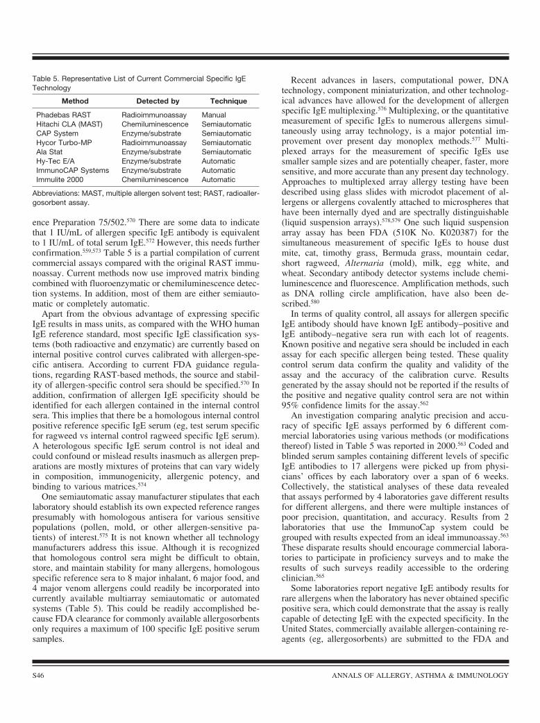
ence Preparation 75/502.570 There are some data to indicatethat 1 IU/mL of allergen specific IgE antibody is equivalentto 1 IU/mL of total serum IgE.572 However, this needs furtherconfirmation.559,573 Table 5 is a partial compilation of currentcommercial assays compared with the original RAST immu-noassay. Current methods now use improved matrix bindingcombined with fluoroenzymatic or chemiluminescence detec-tion systems. In addition, most of them are either semiauto-matic or completely automatic.
Apart from the obvious advantage of expressing specificIgE results in mass units, as compared with the WHO humanIgE reference standard, most specific IgE classification sys-tems (both radioactive and enzymatic) are currently based oninternal positive control curves calibrated with allergen-spe-cific antisera. According to current FDA guidance regula-tions, regarding RAST-based methods, the source and stabil-ity of allergen-specific control sera should be specified.570 Inaddition, confirmation of allergen IgE specificity should beidentified for each allergen contained in the internal controlsera. This implies that there be a homologous internal controlpositive reference specific IgE serum (eg, test serum specificfor ragweed vs internal control ragweed specific IgE serum).A heterologous specific IgE serum control is not ideal andcould confound or mislead results inasmuch as allergen prep-arations are mostly mixtures of proteins that can vary widelyin composition, immunogenicity, allergenic potency, andbinding to various matrices.574
One semiautomatic assay manufacturer stipulates that eachlaboratory should establish its own expected reference rangespresumably with homologous antisera for various sensitivepopulations (pollen, mold, or other allergen-sensitive pa-tients) of interest.575 It is not known whether all technologymanufacturers address this issue. Although it is recognizedthat homologous control sera might be difficult to obtain,store, and maintain stability for many allergens, homologousspecific reference sera to 8 major inhalant, 6 major food, and4 major venom allergens could readily be incorporated intocurrently available multiarray semiautomatic or automatedsystems (Table 5). This could be readily accomplished be-cause FDA clearance for commonly available allergosorbentsonly requires a maximum of 100 specific IgE positive serumsamples.
Recent advances in lasers, computational power, DNAtechnology, component miniaturization, and other technolog-ical advances have allowed for the development of allergenspecific IgE multiplexing.576 Multiplexing, or the quantitativemeasurement of specific IgEs to numerous allergens simul-taneously using array technology, is a major potential im-provement over present day monoplex methods.577 Multi-plexed arrays for the measurement of specific IgEs usesmaller sample sizes and are potentially cheaper, faster, moresensitive, and more accurate than any present day technology.Approaches to multiplexed array allergy testing have beendescribed using glass slides with microdot placement of al-lergens or allergens covalently attached to microspheres thathave been internally dyed and are spectrally distinguishable(liquid suspension arrays).578,579 One such liquid suspensionarray assay has been FDA (510K No. K020387) for thesimultaneous measurement of specific IgEs to house dustmite, cat, timothy grass, Bermuda grass, mountain cedar,short ragweed, Alternaria (mold), milk, egg white, andwheat. Secondary antibody detector systems include chemi-luminescence and fluorescence. Amplification methods, suchas DNA rolling circle amplification, have also been de-scribed.580
In terms of quality control, all assays for allergen specificIgE antibody should have known IgE antibody–positive andIgE antibody–negative sera run with each lot of reagents.Known positive and negative sera should be included in eachassay for each specific allergen being tested. These qualitycontrol serum data confirm the quality and validity of theassay and the accuracy of the calibration curve. Resultsgenerated by the assay should not be reported if the results ofthe positive and negative quality control sera are not within95% confidence limits for the assay.562
An investigation comparing analytic precision and accu-racy of specific IgE assays performed by 6 different com-mercial laboratories using various methods (or modificationsthereof) listed in Table 5 was reported in 2000.563 Coded andblinded serum samples containing different levels of specificIgE antibodies to 17 allergens were picked up from physi-cians’ offices by each laboratory over a span of 6 weeks.Collectively, the statistical analyses of these data revealedthat assays performed by 4 laboratories gave different resultsfor different allergens, and there were multiple instances ofpoor precision, quantitation, and accuracy. Results from 2laboratories that use the ImmunoCap system could begrouped with results expected from an ideal immunoassay.563
These disparate results should encourage commercial labora-tories to participate in proficiency surveys and to make theresults of such surveys readily accessible to the orderingclinician.565
Some laboratories report negative IgE antibody results forrare allergens when the laboratory has never obtained specificpositive sera, which could demonstrate that the assay is reallycapable of detecting IgE with the expected specificity. In theUnited States, commercially available allergen-containing re-agents (eg, allergosorbents) are submitted to the FDA and
Table 5. Representative List of Current Commercial Specific IgETechnology
Method Detected by Technique
Phadebas RAST Radioimmunoassay ManualHitachi CLA (MAST) Chemiluminescence SemiautomaticCAP System Enzyme/substrate SemiautomaticHycor Turbo-MP Radioimmunoassay SemiautomaticAla Stat Enzyme/substrate SemiautomaticHy-Tec E/A Enzyme/substrate AutomaticImmunoCAP Systems Enzyme/substrate AutomaticImmulite 2000 Chemiluminescence Automatic
Abbreviations: MAST, multiple allergen solvent test; RAST, radioaller-gosorbent assay.
S46 ANNALS OF ALLERGY, ASTHMA & IMMUNOLOGY
given FDA clearance when data are supplied by the manu-facturer that has analyzed at least specific 35 to 100 IgEantibody–positive serum samples from clinically allergic in-dividuals. When serum samples from 35 different sensitizedindividuals cannot be identified in the world for a particularallergen specificity, the FDA gives the allergen-containingreagent the designation of an analyte-specific reagent (ASR),which indicates that less than 35 serum samples have beenused to quality control the allergen-containing reagent. TheASR reagents are not fully FDA cleared, but they have beenquality controlled sufficiently by the manufacturer to validateallergen specificity and permit their use in clinical laboratorytesting with the caveat that they are “for research purposesonly.”
Ideally, a total serum IgE should be performed on all serumsamples that are assayed for allergen specific IgE antibody. Ifthe total serum IgE level is high (eg, 20,000 IU/mL for someassays), steps such as automatic dilution should be taken bythe laboratory to ensure that the assay results for specific IgEare true positives and not the result of nonspecific binding inthe assay. The total serum IgE level that produces a false-positive result due to nonspecific binding is presumably iden-tified by the manufacturer of all commercially available al-lergen specific IgE assays563,566,567 and should be madeavailable to the ordering clinician. A clue to possible non-specific binding is a report of weakly positive IgE antibodyresults with multiple allergens.581 Nonspecific binding byglycoepitopes (ie, cross-reactive carbohydrate determinants)is a potential source of a positive test result without clinicalsignificance.582–584 To check this, IgE reactivity of a glyco-protein to which the patient had not been sensitized (eg,bromelain) should be tested against the patient’s serum.582
An adequate presentation of the allergen in the assay isessential for optimal sensitivity of an assay.585 Assay inaccu-racies can result from a number of conditions: (1) the proteinrecognized by IgE may be a minor constituent of the totalprotein in the allergen preparation and hence it becomes aminor fraction of the protein bound to the solid phase, whichleads to insufficient protein for adequate IgE binding; (2) theprotein recognized by IgE may be labile because of either itsmolecular structure or the presence of proteolytic enzymes inthe allergen preparation; and (3) the chemical linkage used tobind the protein to the solid phase may destroy the epitoperecognized by IgE or the linkage may occur at a site so closeto the epitope that steric hindrance occurs.
IgG antibody specific for allergens may occur as a result ofnatural allergen exposure or active allergen immunotherapy.Since IgG antibody is often present in quantities greatlyexceeding the quantity of IgE antibody, specific IgG antibodymay bind to available sites of the allergosorbent, therebypreventing subsequent IgE binding and leading potentially tofalsely low or negative test results.586
Different allergen extracts may have identical proteins orpeptide epitopes recognized by IgE antibodies. In this case, apatient who is sensitized to an allergen may have a positivetest result to both the original allergen and other allergens that
cross-react with the original allergen.587 The relationship ofcross-reactive IgE antibodies evaluated by either skin orspecific IgE tests to clinical disease is known for some but notall allergens. Exposure to cross-reactive allergens may ormay not provoke symptoms (eg, most grass-sensitive patientstolerate wheat, a potent cross-reactant in grass pollen ex-tracts). This problem of allergen cross-reactivity may alsocomplicate interpretation of skin tests.
The detection of allergen specific IgE antibody in serumwith an FDA-cleared assay may be viewed as a risk factorthat supports a positive clinical history in making the diag-nosis of allergic disease. As with skin testing, IgE antibodyspecificities involving extracts that contain potent allergeniccomponents such as ragweed, house dust mite, and cat epi-dermals tend to correlate much better with clinical sensitivityand provocation tests. The use of purified fractions (ie, Amba 1, Der p I, Der f 1, Fel d 1, Alt a 29, Hev b 5) often fortifiessensitivity and the test’s correlation with clinical diseasecompared with unfortified immunosorbent.588 On the otherhand, extracts with weaker allergenic epitopes may demon-strate substantially less correlation with various indices ofclinical sensitization. This situation may be compoundedfurther in the case of foods in which multiple allergenicepitopes are often contained in the crude extract mixture andminor components may actually dilute the major allergenresponsible for clinical sensitization. Furthermore, as dis-cussed herein, certain allergenic epitopes in foods (ie, wheat)may strongly cross-react with allergens in 1 of the potentclasses of inhalant aeroallergens (ie, grass), leading to spuri-ously false-positive results. However, the predictive value ofanaphylactogenic food specific IgE for outcome of oral foodchallenge has received considerable attention and is discussedbelow and further in part 2.140,589–591 Because the constitutiveallergenicity, potency, and stability are variable among com-mercial allergen extract reagents, sensitivity and the positivepredictive value of both prick/puncture and specific IgE testsgenerally tend to be higher among pollens, anaphylactogenic,stable food proteins, house dust mite, certain epidermals, andfungi compared with venoms, drugs, and chemicals.
Inhibition of Specific IgE Antibody BindingThe most expedient method for determining the specificity ofIgE binding is to determine whether the addition of a smallquantity of a homologous allergen in the fluid phase willinhibit most IgE binding. Inhibition usually indicates that IgEbinding in the assay is a result of the IgE antibody specificallyrecognizing the allergenic protein.564 Theoretically, some al-lergen preparations may contain substances such as lectins,which could nonspecifically bind IgE. If IgE is being non-specifically bound, either most serum samples are positive inthe assay or there is a relationship between the total serumIgE concentration and an increase of assay positivity tomultiple lectin-containing allergens. In demonstrating spe-cific inhibition, it should be possible to inhibit at least 80% ofthe specific IgE binding in a dose response manner.564
VOLUME 100, MARCH, 2008 S47
The degree of binding inhibition produced when a fluidphase allergen is added to a serum containing allergen spe-cific IgE depends on the ratio between the quantity of specificIgE and the quantity of allergen added. When the quantity ofspecific IgE is kept constant, the percentage of inhibitionproduced can be used to estimate the quantity of allergen inthe fluid phase. Under appropriate experimental conditions,including an adequate supply of potent allergen specific IgE,inhibition can be used to standardize allergen extracts, esti-mate the quantities of allergens, and evaluate cross-reactivitybetween allergens.581
A specific allergen may be detected in a crude extract ofmultiple allergens using the inhibition technique.581 For ex-ample, if a patient is known to be allergic to peanuts, and thepatient has symptoms of an allergic reaction after eating apiece of candy, the question could be whether the candycontained peanut allergen. If an extract of the candy inhibitsthe binding of the patient’s IgE to a solid-phase peanutallergen preparation, there would be good reason to suspectthat the candy contained either peanut or an allergen thatcross-reacted with peanut.
Allergen Specific IgG and IgG Subclass AssaysSummary Statement 125. Allergen specific IgG may be mea-sured by immunodiffusion or immunoabsorption. (E)
Summary Statement 126. Immunodiffusion antibodies tocow’s milk are associated with Heiner’s disease, a non-IgEdisorder that presents in infants with pulmonary infiltrates.(B)
Summary Statement 127. IgG and IgG subclass antibodytests for food allergy do not have clinical relevance, are notvalidated, lack sufficient quality control, and should not beperformed. (B)
Summary Statement 128. Although a number of investiga-tors have reported modest increases of IgG4 during venomimmunotherapy, confirmation and validation of the predictivevalue of IgG4 for therapeutic efficacy of venom immunother-apy are not yet proven. (C)
Allergen specific IgG can be measured using immunoas-says similar to those used to measure allergen specific IgE.586
Allergen specific IgG is often easier to detect than specificIgE because it is usually present in a higher concentration.The antibody used to measure the IgG bound in an assay canbe either an anti-human IgG or specific for 1 of the subclassesof IgG (IgG1, IgG2, IgG3, or IgG4). When subclass specificIgG antibodies are used, the quantity of the particular IgGantibody subclass can be determined. IgG and IgG subclassantibodies specific for allergens usually are measured inarbitrary units, although mass values may be extrapolatedfrom a total or subclass specific standard curve. An allergenspecific IgG assay is subject to the same technical problemsas specific IgE assays, and specific IgG assays should beevaluated using the same criteria and techniques as those usedfor IgE assays. The level of expected precision should be 2significant figures with variation less than 15%, or lower,since the quantity of IgG to be measured is often relatively
large, especially after immunotherapy.586 Currently, noblinded proficiency surveys are available for evaluating in-terlaboratory performance of allergen specific IgG or IgGsubclass assays.
Clinical Application and Interpretation
Total serum IgE concentrationSeveral studies have proposed using the total IgE concentra-tion in cord blood as a method for predicting an infant’s riskof developing allergic disease.592 Although the results of theearly reports were promising, subsequent studies have notfound the cord blood IgE concentration to be a reliablepredictor of risk.592 Even if it were possible to predict the riskof allergic disease, such knowledge would have little clinicalvalue because there are not as yet proven methods for pre-venting allergic disease in high-risk children.
Measurements of total serum IgE concentration are ofmodest clinical value when used as a screen for allergicdisease or for predicting the risk of allergic disease.592 Al-though epidemiologic studies have shown that the risk ofasthma is highly correlated with the total serum IgE concen-tration, the variation from individual to individual is too greatto provide much diagnostic value.592–595 Similarly, the broadrange of values and the variation among individuals meansthat total serum IgE concentrations provide only modestinformation about the risk of allergic disease on an individualbasis.590 Furthermore, a normal total IgE does not excludeclinical allergy. Evaluation of patients with suspected ABPAis one of the few clear indications for measuring serum IgEconcentrations.596 An extremely elevated total serum IgEconcentration is found in nearly all patients with ABPA andis one of the major diagnostic criteria. There is also a sug-gestion that the serum IgE concentration is an indicator ofdisease activity and that serial determinations should be usedto evaluate the adequacy of treatment.596
With the licensing of omalizumab (Xolair) for the treat-ment of allergic asthma, another application of total serumIgE is verification that the patient is a suitable candidate foranti-IgE therapy with total serum IgE levels between 70 and800 IU/mL. The total serum IgE level before taking omali-zumab combined with the patient’s weight will determine thecorrect dosing to ensure efficacious reduction of free IgEcirculating in blood. After 1 month of taking omalizumab, anew assay that measures the level of circulating IgE that isfree or unbound with omalizumab can confirm the effective-ness of the dosing regimen.597 This test is not yet commer-cially available. Although as much as 62% loss in accuracywas observed in FDA-cleared human IgE assays, the Immu-noCAP system was sufficiently robust to provide accurateand reproducible total and allergen-specific antibody resultsin the presence of therapeutic levels of serum omalizumab.598
Serum IgE concentrations are often abnormal in patientswith congenital immunodeficiencies, but these abnormalitiesare rarely diagnostic.599 The primary exception to this state-ment is the syndrome of hyper-IgE, eczematous dermatitis,and recurrent pyogenic infections. In this syndrome, the total
S48 ANNALS OF ALLERGY, ASTHMA & IMMUNOLOGY
serum IgE level is extremely elevated and is one of the majordiagnostic criteria for the disease.599 Nonspecific elevation ofIgE level is also observed in the Wiskott-Aldrich, ataxiatelangiectasia, DiGeorge, and Ommen syndromes.600
Patients with acquired forms of immunodeficiency mayhave altered levels of serum IgE, but these alterations are notdiagnostic.601 A few published reports have indicated thatserum IgE is elevated in patients with HIV infection and thatthere is a modest correlation between IgE elevation andclinical course or state of the disease.602
IgE myeloma is a rare form of multiple myeloma, withfewer than 40 cases reported worldwide.603 Some cases of IgEmyeloma may have been misdiagnosed as light chain diseasebecause of failure to measure serum IgE concentrations.Since the course of IgE myeloma is distinct from that of lightchain disease and other myelomas, IgE should be measured inpatients with clinical symptoms suggestive of myeloma andin whom myelomas of other isotypes have been ruled out.603
Total serum IgE concentrations have been reported to beabnormally high in a variety of diseases. In drug-inducedinterstitial nephritis or graft vs host disease, there may be arelationship among the course of the disease, response totherapy, and the IgE level, but none of these relationships arefirm enough to recommend total IgE as part of the clinicalevaluation of these diseases.592
Allergen specific IgE concentrationSummary Statement 129. The probability distribution of
specific IgE for several anaphylactogenic foods (peanuts, eggwhite, cow’s milk, and codfish) can define clinical sensitivityas verified by double-blind oral challenge tests; similar rela-tionships have been defined for several respiratory allergens.(A)
Multiple studies have shown that allergen specific IgE israrely detectable in cord blood.592 In the few cases in whichspecific IgE for common allergens was detectable, neitherdiagnostic nor prognostic significance was demonstrated.Based on current information, there is no clinical indicationfor attempting to measure allergen specific IgE in cord blood.However, several investigations have shown that elevatedfood specific IgE in early infancy may predict respiratorysensitization at a later age.604–607
A recent study claimed virtually equivalent specific IgEsensitivity results between a blood spot test and serum.608 Theblood spot test was performed using paper-absorbed or-eluted blood obtained by finger prick.608 Preliminary resultssuggested this was a successful method for determining IgEsensitization in preschool children. Prototypic, miniaturized,multiarray assays may offer a similar advantage in the fu-ture.577,609
Efforts to develop a screening procedure have led to testsin which multiple allergens are coupled to a single solid-phase substrate560,610,611 (Table 5). If the multiple allergen testresult is positive, there is a high probability that the patient isallergic to at least 1 of the allergens included in the test.Additional tests that use individual allergens then can be used
to determine other allergens to which the patient may besensitive. In general, these multiallergen screening tests haveshown acceptable diagnostic sensitivity and specificity whencompared with skin tests.560,610,611
The clinical value of multiple allergen screening testsdepends on the selection of patients. In a symptomatic self-selected population, a positive test result would significantlyincrease the probability that the patient was allergic. If mul-tiple allergen tests were used to screen an unselected popu-lation, there would be an unacceptable number of false-positive and false-negative results. By itself, a positivemultiple allergen test result does not provide sufficient infor-mation to make a specific clinical diagnosis or to initiatetherapy.59 In addition, a negative multiple allergen test resultdoes not exclude clinical sensitivity because the commercial-ly-available multiallergen screening tests only screen for ap-proximately 15 aeroallergens.
Recommendations concerning the number of specific IgEtests for confirmation of suspected clinical sensitivity corre-spond to those discussed for prick/puncture tests in SummaryStatement 43.
There are no clinical scenarios in which immunoassays forallergen specific IgE can be considered either absolutelyindicated or contraindicated. There are some situations inwhich immunoassays may be preferable to skin testing for thediagnostic evaluation of patients. If the patient has had anearly fatal reaction to an allergen, the immunoassay offersthe advantage of testing the patient for allergen specific IgEwithout the risk of inducing a severe reaction from a skintest.612,613 In this situation, an IgE antibody measurementusing immunoassay is less likely to provoke severe patientanxiety about the possible adverse consequences of a skintest. A positive IgE antibody test result strongly supports theclinical impression. A negative test result reduces the prob-ability that the suspected allergen is causally associated, butit is essential that the negative result be confirmed by skin testbefore the allergen can be excluded as a possible anaphylac-togen.612,613 If both test results are negative, a supervisedchallenge may still be necessary. If a patient does not have asufficient large area of normal skin to allow skin testing,immunoassays for specific IgE are useful for confirmingclinical impressions.550,564 Examples would include individu-als with severe dermatographism, ichthyosis, or generalizedatopic dermatitis. Theoretically, a third situation in whichimmunoassay may be preferable is during the refractoryperiod immediately after a severe allergic reaction. If it wereclinically necessary to determine the patient’s sensitivitywithin a few days after such a reaction, an immunoassaymight provide a better way to ascertain the necessary infor-mation.550 Antihistamines and drugs such as tricyclic antide-pressants reduce or block skin test reactivity550 (Table 4). If itis necessary to document allergic sensitization either beforethe drug has been cleared from the patient’s body or if it isinadvisable to stop taking the medication, an immunoassaymay provide needed information. When a patient is unable tocooperate with skin testing because of mental or physical
VOLUME 100, MARCH, 2008 S49
impairment, measurement of specific IgE by immunoassaywould be preferable because of reduced risk to an agitatedpatient or personnel who would normally perform the skintesting.
Quantitative results from clinical IgE antibody assays haveallowed investigators to study whether the quantity of serumIgE antibody has any predictive utility in defining clinicalsensitivity. In the area of food allergy, several groups haveshown that the quantity of specific IgE antibody in serum topeanut, egg white, cow’s milk, and fish may define currentclinical sensitivity in many patients.140,526,589–591,614 Probabili-ty-based risk of clinical food allergy increases as the quantityof serum food specific IgE increases. Probability curves candefine, for some foods, levels at which reactions are highlylikely (eg, 95%) and may dissuade the need for an oral foodchallenge. Thus, the higher the value, the more specific thetest becomes in terms of clinical food allergy. Over interpret-ing values in the class 1 and 2 categories may lead to falseassumptions. When levels are undetectable, 5% to 20% maystill have reactions, and so the clinical history is important ininterpretation of results.589
Probability-based risk evaluation has also been extended torespiratory allergy using quantitative allergen specific IgEantibody data previously reported from four European labo-ratories.573 A logistic regression model was used to comparethe relationship between the physician’s final diagnosis ofallergic respiratory disease (positive or negative) based on theclinical history, physical examination, and skin testing andserologic testing data and the quantitative level of serum IgEantibody alone. Probability curves were calculated in thisstudy to show the relationship between IgE antibody in bloodand the dichotomous clinical diagnosis of the absence orpresence of allergic respiratory disease. The probability ofobtaining a positive allergy diagnosis at a given serum IgEantibody level by the Pharmacia UniCAP System has beenevaluated for different allergens at 4 clinics. Differences inthe shape of the IgE antibody level vs probability of clinicaldisease curves was seen both between allergens within aclinic and between clinics for the same allergen specificity.573
This indicates that use of specific IgE antibody levels tosupport the clinical diagnosis of respiratory allergic disease isdifferent for the same allergist depending on the particularinhalant allergen and between allergists for the same allergenspecificity. Importantly, however, the authors make the casethat quantitation of serum IgE antibody improves the confi-dence of the clinical diagnosis of inhalant allergies better thansimply knowing if IgE antibody is present or absent.
Another group also studied the clinical utility of quantita-tive serum IgE antibody measurements in the diagnosis ofrespiratory allergy.574 They used purified recombinant timo-thy grass and birch pollen allergens to compare the relativeability of puncture skin testing, nasal provocation, and IgEantibody serology by the CAP System to reflect immediate-type respiratory sensitivity. Although the skin test and nasalprovocation results were significantly correlated, the intensityof these biological reactions did not correlate with the level of
allergen specific IgE antibody in serum. The authors con-cluded that factors in addition to IgE influence the extent ofallergic tissue reactions.
A recent probability risk evaluation comparing skin testsand serum specific IgE to a panel of saprophytic mold aeroal-lergens revealed relatively poor correlations.615 The results ofthis investigation confirmed the relatively low sensitivityrank order for fungi when evaluated by in vitro serologictests.
Predictability of both skin and in vitro tests for IgE-medi-ated anaphylaxis to Hymenoptera venoms may also requirereconsideration, especially if patients are tested at extendedtimes after the anaphylactic episode. A recent investigationdemonstrated relatively poor reproducibility of both venomskin tests and serum specific IgE when 35 patients, who hadexperienced systemic reactions, were tested on 2 occasions 2and 6 weeks apart.616
Immunoassays for allergen specific IgE offer a uniqueadvantage when compared with skin testing in their ability touse soluble allergen inhibition to examine specificity andcross-reactivity among allergens. Although these assays areused chiefly for research purposes, they may be clinicallyimportant in some situations. For example, if a patient has ahistory of anaphylaxis after an insect sting and the patient isfound to be skin test positive to yellow jacket venom at a lowconcentration and positive to Polistes wasp venom at a higherconcentration of venom, the question arises whether the pa-tient is sensitive to both insects or whether skin test reactivityto wasp venom is the result of cross-reactivity. An inhibitionassay showing that all the reactivity to Polistes wasp venomcould be inhibited by yellow jacket venom strongly suggeststhat the positive skin test result to Polistes wasp was the resultof cross-reactivity. Furthermore, the patient could be success-fully treated with yellow jacket venom alone, saving theadded expense of treating with Polistes wasp venom. Aller-gen cross-reactivity may also be clinically relevant whendeciding how many species of weeds, grasses, trees, andmites need to be included in an immunotherapy regimen.
Allergen specific IgE measurements may be useful inevaluating fatalities that may have resulted from allergicreactions by determining the allergen responsibility for thefatal reaction.612,613
Allergic bronchopulmonary aspergillosis is an inflamma-tory disease of the lungs characterized by severe asthma,sputum production, peripheral blood eosinophilia, and anincreased total serum IgE concentration. If untreated, it mayprogress to central bronchiectasis and, ultimately, pulmonaryfibrosis and death. After proper treatment with corticoste-roids, total serum IgE levels usually decrease. Total serumIgE should be followed during the disease since an increase inIgE may herald a relapse of disease. Aspergillus specific IgEand IgG are usually present in the sera of patients withABPA. Although the levels of these antibodies do not alwayscorrelate with disease activity, they tend to decrease as activedisease subsides.596,617,618
S50 ANNALS OF ALLERGY, ASTHMA & IMMUNOLOGY
The initial immune response to many endoparasitic andectoparasitic organisms is predominantly an IgE response. Inmany parasitic infections, an increase in both parasite specificIgE and total serum IgE concentrations occurs.592 Detectionand measurement of the parasite specific IgE (eg, Toxo-plasma gondii) may be useful for diagnosis and potentiallyfor following the course of the infection.619 The major limi-tation of using assays for parasite specific IgE has been theavailability of adequate allergosorbent preparations from therelevant parasite. Cell-mediated immunity may be an equallyimportant pathogenetic factor in some parasite infections (ie,leishmaniasis). A recent report suggests that antimalarial spe-cific IgE in asymptomatic individuals may be associated withreduced risk for subsequent clinical malaria.620 It has beenpostulated that serum IgE may be a prognostic marker forAIDS in HIV-infected adults and that a switch to the TH2profile might represent a turning point in HIV.621,622 Anti-HIVIgE found in the serum of certain long-term pediatric survi-vors is associated with inhibition of HIV-1 production, pos-sibly through cytotoxicity rather than virus neutralization.623
Remote Formulation of Allergen ExtractsSeveral clinical laboratories offer nonallergists a service ofpreparing extract mixtures for allergen immunotherapy basedon results of specific IgE tests. In some cases, the extractprescription is also based partially on a patient self-adminis-tered questionnaire. One study prospectively compared theresults of allergy evaluations of 118 patients performed by agroup of practicing board-certified allergists vs a laboratorythat offered allergy diagnosis and recommendations for im-munotherapy.624 Although this study demonstrated that aller-gists identified allergy more frequently (53% vs 47%), theyactually recommended immunotherapy less frequently thandid the laboratory (35% vs 59%). The recommendations ofthe laboratory were deficient in that they were solely based onthe history form and results. The laboratory was unable toclarify answers or to further explore areas that were suggestedby patient responses or allergy testing results. On criticalanalysis of the laboratory-based extract recipes, it was foundthat the laboratory ignored the history forms and developedan extract formulation based solely on the results of antibodyanalysis in several cases. Overall, approximately 50% of therecommendations by the laboratory were inappropriate orincomplete. Even more serious errors could occur if thelaboratory offering such a service had a record of poor orunsubstantiated quality control for performance of specificIgE tests.563
This form of allergen treatment is therefore not in thepatient’s best interest and should be discontinued.624
Allergen Specific IgGAllergen specific IgG can be produced by persons either as aresult of natural allergen exposure or as a result of immuno-therapy.625–629 Allergen specific IgG may have specificity fordifferent allergen proteins or different protein epitopes thanthose eliciting an IgE response in the same person. There has
been no convincing evidence that the quantity of allergenspecific IgG produced as a result of natural exposure isrelated to or predictive of disease.590,629
Food specific IgG has been found in many healthy nonal-lergic individuals, and the quantity detected seems to dependon the quantity of the food ingested.625,629 Precipitating anti-bodies to cow’s milk are associated with Heiner’s disease,hallmarks of which are pulmonary infiltrates that disappearafter elimination of cow’s milk.630 No studies have convinc-ingly demonstrated a relationship between the presence offood specific IgG antibodies and allergic disease (see “Un-proven Tests”). By substituting an antibody specific for anIgG subclass for the antihuman IgG in the allergen specificIgG assay, it is possible to measure allergen-specific subclassdistribution of IgG antibody responses.586 A single studyreported that persons having irritable bowel symptoms afteringestion of foods have increased levels of IgG4 antibodysubclass to the offending food.631 Unfortunately, high levelsof the IgG4 subclass of food specific IgG antibody have notbeen consistent among studies so that the clinical value ofmeasuring subclass specific IgG antibody remains to be de-termined.625,632 Thus, IgG and IgG subclass antibody tests forfood allergy have not been demonstrated to have clinicalrelevance, are not validated, lack sufficient quality control,and should not be performed.
Some investigators have shown that there is a modestassociation between the quantity of venom-specific IgGproduced in response to immunotherapy and protectionfrom allergic reactions induced by an insect sting.586,633,634
The value of measuring IgG antibody during or afterimmunotherapy with other allergens has not been demon-strated. Although allergen specificity may occur in each ofthe 4 IgG subclasses during allergen immunotherapy, thereis conflicting evidence concerning the value of equatingsuch antibodies with efficacy.586,633,634 In the case of im-munotherapy with insect venoms, there appears to be amodest relationship between the presence of elevated spe-cific IgG4 for the venom and protection from anaphylaxisafter an insect sting.586,633– 637 Other studies have not foundany relationship between the quantity or specificity ofallergen specific subclass IgG and the outcome of pollenimmunotherapy.564 The general predictive value of sub-class IgG4 for successful immunotherapy is not proven atpresent.
Allergen specific IgG has been reported to be a potentiallyimportant biomarker of exposure to specific chemical aller-gens in the workplace.638 Thus far, the predictive value forsuch antibodies and emergence of clinical disease in exposedworkers has not been demonstrated.638
Immunoprecipitin tests to various causal proteins of hyper-sensitivity pneumonitis (eg, Micromonospora faeni, pigeonserum), allergic bronchopulmonary or sinus mycosis may beuseful diagnostic adjuncts.638–641 Panels of the most commonetiologic agents of these diseases are validated and commer-cially available.642
VOLUME 100, MARCH, 2008 S51
In Vitro Methods of Allergen StandardizationSummary Statement 130. Although allergens can be standard-ized either by radioimmunodiffusion or immunoassay inhibi-tion based on major allergenic epitopes, the FDA selectedBAU instead because in vitro analytic techniques would havebeen variable from allergen to allergen and would havecaused great confusion. (C)
BackgroundAll medicines should have a label that describes quantity andpotency. Traditionally, allergenic extracts have been labeledin weight to volume units or PNU, the quantity of phospho-tungstic acid precipitable nitrogen. Since allergenic compo-nents are known to be a small percentage of the total protein,source material can be manipulated to maximize the contentof proteins that contribute to the PNU value without regard tothe allergenically active proteins. Consequently, these proce-dures yield extracts whose labeling cannot be relied on toexpress the allergenic activity of the contents. In 1970 aprogram was initiated in the FDA Laboratory of AllergenicProducts to develop procedures that would permit a descrip-tion of the allergenic activity of extracts as determined bycomparison of laboratory and skin test reactivities.59
Current methodsThe first extracts in which an attempt was made to providelot-to-lot consistency were the insect venoms. These productsare labeled in arbitrary units of hyaluronidase enzyme per 100�g of protein. The next extract was that of short ragweedpollen. This was labeled in units of antigen E (Amb a 1) permilliliter (a unit of antigen E is approximately 1 �g). Amb a1 was initially measured by a radial immunodiffusion test andcurrently is measured by an enzyme immunoassay inhibitiontest.
If this program had continued, there would have been asmany types of analytical methods and labeled designations asthere are extracts, a situation that would have resulted inconsiderable confusion in the use of these products. There-fore, a potency unit was developed, BAU, which is based onskin bioreactivity. After this is determined, each lot is eval-uated by 1 or more laboratory tests that can be compared andexpressed in BAU potency equivalents. In the future, allstandardized extracts used in the United States will be veri-fied by carrying out the bioequivalent FDA tests.
ProceduresStandardization methods of allergenic extracts for use as skinand specific IgE reagents involve a series of tests. All tests aredescribed in detail along with statistical methods and vari-ability in the FDA’s Manual of Methods of the Laboratory ofAllergenic Products.124
These methods include (1) identification of the sourcematerial used for extract production, (2) determination of asatisfactory procedure for preparing an extract, and (3) testsof the extract that include total protein, radial immunodiffu-sion test for a single allergen (eg, short ragweed antigen E[Amb a 1]) and cat allergen (Fel d 1), enzyme assay (eg,
hyaluronidase in venoms), or immunoinhibition (pollen, miteand mold extracts). Where applicable, immunoblotting frompolyacrylamide gel electrophoresis or isoelectric focusingtechniques are performed to evaluate the allergenic identity ofextracts and the number of IgE-binding proteins. Using anextract standardized by these methods, serum pools collectedfrom sensitive patients can be evaluated to determine thevalidity and reproducibility of specific IgE tests.
Test variabilityFor the radial immunodiffusion method, the correlation co-efficient of the reference dose response line should be at least0.9. A single concentration for a test extract is reproducible to�25% when estimated from the calculated regression line.642
Immunoinhibition (RAST or ELISA) provides relative po-tency and consequently requires a reference preparation. Thedata are analyzed by means of parallel line statistics. There-fore, the frequently used methods of comparing extracts atextrapolated 50% inhibition values is without special mean-ing when this procedure is used for standardization of ex-tracts. All validity assays must be included in the data statis-tical analysis protocol as described in detail in the FDA’sManual of Methods.124 Prospective evaluation of the allergenstandardization procedure was performed in the FDA labo-ratory and other laboratories by assessing more than 45 setsof data from 3 individual investigators. The variability isproportional to the number of test methods, all of whichshould be performed at least in duplicate. For 3 tests, thecalculated variability was 47% to 213%, and for 5 tests, it was56% to 180%.
Histamine and Leukotriene TestsSummary Statement 131. Histamine and leukotriene releasemeasurements from human basophils after incubation withallergen are valuable research tools for in vitro investigationsof allergy. (B)
Summary Statement 132. The recent availability of severalsensitive immunoassays for histamine and leukotriene C4 is asignificant technological advance for measuring these medi-ators in various biologic fluids or release from whole blood,isolated basophils, mast cells, or other cultured cells. (B)
Summary Statement 133. Histamine and its N-methyl his-tamine metabolite may be measured in 24-hour urine samplesafter suspected anaphylactic episodes. (B)
BackgroundMore than 75 years ago, Dale et al demonstrated the presenceand physiologic action of histamine in different tissues.643
Later work established that histamine in tissue is present ingranules of cells and that in human tissue these granules arepresent only in basophils and mast cells. Over the years it wasdemonstrated that histamine or histamine-like material wasreleased into the blood of experimental animals during ana-phylactic reactions. This approach led to the demonstrationthat the addition of specific antigen or allergen to the blood orwashed leukocytes of either experimentally sensitized ani-
S52 ANNALS OF ALLERGY, ASTHMA & IMMUNOLOGY
mals or allergic persons can result in the release of histaminefrom basophils.
Basophils are the only cells in human peripheral blood thatcontain histamine. The interaction of specific allergen withthe IgE antibody fixed to high-affinity Fc� receptors on thebasophil membrane initiates release of preformed histamineand other inflammatory mediators associated with immediatehypersensitivity. The release of histamine is modulated by theaddition of a number of pharmacologic agents. The additionof plasma or serum factors is not essential for the releasereaction, although normal serum will enhance the releasereaction when conditions are suboptimal. The serum of aller-gic persons also contains blocking IgG antibodies that alsoreact with the allergen. When histamine release measure-ments are done with washed leukocytes, however, these an-tibodies probably do not influence the results.
ApplicationsHistamine release from human basophils is primarily a valu-able research tool for in vitro investigations of allergy. Inmost studies of histamine release, allergen or antigen is addedto washed leukocytes from venous blood. This can be sim-plified by eliminating the leukocyte preparation step andadding the allergen to whole heparinized blood.644 The hista-mine released into the supernatant then can be determineddirectly. More recently, leukotriene C4 release has been mon-itored from basophils exposed to allergen as an indication ofthe presence of specific IgE antibody.645,646
In ragweed-allergic persons there is a good correlationbetween the severity of the clinical symptoms and the extentof in vitro histamine release.647 The histamine release alsocorrelates with the magnitude of the skin test and the level ofserum IgE specific for ragweed Amb a 1. Both the antigenconcentration at which 30% to 50% histamine release (cellsensitivity) occurs and the maximum percentage of histaminerelease (cell reactivity) correlate with the clinical severity ofallergic rhinitis and the skin test. Patients with high levels ofserum ragweed specific IgE release histamine with low con-centrations of antigen. Similarly, in Hymenoptera venom–sensitive patients, there is good correlation between positivehistamine release in vitro and the magnitude of the skin testwith venom antigen.648 This procedure is also being used toevaluate the functional characteristics of autoantibodies toIgE or the Fc�R1 receptor in patients with chronic idiopathicurticaria (CIU).
Current Methods for Measuring HistamineThe discovery of histamine and the demonstration of itsbiologic importance were accomplished through the use ofbiologic assay systems dependent on the contractility ofsmooth muscle after the addition of this biologically activeamine. This early technique has been superseded by chemical(fluorometric) and most recently, immunologic methods.
ChemicalA method for the chemical determination of histamine wasfirst described by Shore et al in 1959.649 Since then, this
method has been modified to increase both its specificity andsensitivity. It is based on the coupling of ophthalaldehyde tohistamine at alkaline pH to form a fluorescent product. Thefluorescence of the histamine-o-phthalaldehyde complex ismore intense and more stable at an acid pH, unlike thecomplex formed by some other amines. To remove interfer-ing compounds, the histamine is extracted before the conden-sation step. Protein is removed from the sample to be ana-lyzed by perchloric acid precipitation; the histamine isextracted into n-butanol from the alkalinized salt-saturatedsolution. The histamine is recovered in an aqueous solution indilute hydrochloric acid by adding heptane. This dilute hy-drochloric acid solution is then used for the condensation ofhistamine with o-phthalaldehyde. The extraction procedurewith organic solvents is essential to remove histidine andother interfering compounds before the condensation step. Acompletely automated fluorometric technique is capable ofanalyzing 30 samples per hour with a precision between 1%and 2%.650 The sensitivity of the method is such that 0.1 to 10ng/mL of histamine can be accurately determined. Thismethod is convenient in handling large numbers of sampleswith excellent precision. The methods for both the manualand automated histamine analysis method have been de-scribed in detail.549,650
ImmunoassayThe fluorometric assay has technical requirements that min-imize its use to research laboratories. Simpler assay methodshave recently been developed that use antibodies to histamineor histamine analogs, and the reagents are available in com-mercially available kits.651 A variety of immunoassay kits areavailable. Many of these are competitive inhibition assays,and most use monoclonal antibodies.652 As with other immu-noassays, the methods used are sensitive, reproducible, andeasy to perform.653
InterpretationHistamine release results are expressed as a percentage oftotal cellular histamine determined after incubation with acalcium ionophore or boiling an aliquot of cells. Controlmeasurements include the histamine released in the absenceof added antigen, and this value is subtracted to calculate thespecific release. In most experiments, the nonspecific “blank”release should be less than 10% of the total cellular hista-mine. High spontaneous release of histamine from washedleukocytes has been reported in a small percentage of patientswho are highly atopic or sensitive to food.654 The significanceof that finding is not clear.654 Appropriate controls should alsoinclude the testing of the allergen with the cells of nonallergicdonors to demonstrate that the allergen does not contain anycytotoxic materials or histamine itself. Similarly, allergens orpharmacologic agents should be tested to see whether theyinfluence the histamine assay procedure nonspecifically andcontribute to erroneous results.
Histamine release results can be conveniently expressed by2 parameters: (1) cell sensitivity: this is the concentration of
VOLUME 100, MARCH, 2008 S53
antigen or allergen expressed in units (preferably microgramsper milliliter) required to release either 30% or 50% of totalcellular histamine; and (2) cell reactivity: this is the maximalamount (percentage) of histamine release obtained with anyamount of the antigen.
A positive control in histamine experiments should be theaddition of different dilutions of an anti-IgE antiserum to thecells. In general, the cells of most persons release more than10% histamine after challenge with anti-IgE.
The number of false-positive reactions to allergens deter-mined by histamine release is low. These are defined assubjects having a negative skin test result and a positivehistamine release result with an allergen.
The incidence of false-negative reactions is a more criticalfactor in the interpretation of histamine release tests. Somepatients have little histamine release at any concentration ofallergen, but nevertheless are sensitive by skin tests. Evenunder the best of circumstances, for example, skin test studieswith pure venom antigens, there is a significant number ofpeople who have positive skin test results and appear to beallergic by a convincing history but fail to release histamineafter challenge with appropriate allergens. The percentage ofthese persons may be as high as 10% to 15%. This raises theissue of how to interpret a negative test result. Cells frompatients with CIU frequently do not release histamine. De-sensitization of patients may also result in changes in thedegree of histamine release from leukocytes. Changes attrib-utable to immunotherapy are variable and inconsistent.
SignificanceHistamine release from leukocytes of allergic persons is anexcellent in vitro correlate of allergy. At present, it is primar-ily considered a research test and is not widely available fromclinical immunology laboratories. However, in rare instancesit may have confirmative value in assessing the presence orabsence of allergy. In vitro histamine release can be a usefuladjunct by supplying quantitative data on the degree of aller-gen specificity. Therefore, it can be compared with in vitroserologic methods using direct and inhibition techniques.Both of these assays suffer from the occurrence of false-negative results, that is, patients who are clinically sensitivebut exhibit negative findings to these tests.655 The serologicmeasurement of IgE antibody has the following advantages:(1) it requires a small amount of serum, (2) samples can bestored and processed at a central laboratory, and (3) thetechniques involving immunoassay are well established andFDA cleared. In contrast, histamine release requires a largerblood sample, it must be performed within a relatively shorttime after the sample of blood is obtained, and the techniquesare more complicated and not FDA cleared. The advantagesof histamine assays are that they require smaller amounts ofallergen, unlike skin testing they do not involve injection ofallergen into the subject, and they are not dependent oncoupling antigens to immobilized support systems with theinherent problems of antigen modification or unavailability ofbinding sites. By contrast, using washed leukocyte experi-
ments, there is no competition between IgG and IgE forantigenic binding sites, and, therefore, IgG cannot interfere inthe release assays as sometimes is the case with IgE antibodyserology.
Histamine and its metabolite, N-methyl histamine, may bemeasured in urine samples (usually 24-hour collection) aftera suspected anaphylactic episode or evaluation of suspectedmastocytosis.636,637 Plasma histamine is more likely to beelevated in patients who present to the emergency departmentwith acute allergic syndromes than tryptase.656–658
Plasma TryptaseSummary Statement 134. Plasma tryptase, particularly the �form, should be obtained within 4 hours after an anaphylacticepisode. (B)
Summary Statement 135. Combined � and � species ofplasma tryptase are elevated in patients with systemic mas-tocytosis. (A)
Mast cells that have been activated during an IgE-mediatedhypersensitivity reaction release proteases and prestored his-tamine and newly generated vasoactive mediators into sur-rounding soft tissue. Tryptase (molecular weight, 134,000kDa) is a neutral serine esterase with trypsin-like substratespecificity that is found in relatively large quantities in mastcells (approximately 10 pg per lung mast cell and up to 135pg per skin mast cell). It is stored in the secretory granules asan active enzyme complexed to and stabilized by heparin.Although several forms have been described (I, II�, III�, T)and all are found in mast cells, only the � and, less frequently,� species are clinically relevant. Since modest amounts arefound in basophils (less than 1% than found in tissue mastcells), tryptase is considered to be a good clinical marker ofmast cell activation.657,659 When dissociated from heparin,tryptase rapidly degrades into its monomers and loses enzy-matic activity.
Immunoreactive tryptase levels in serum of healthy adultsare less than 5 �g/L. Elevated levels of tryptase (�10 �g/Las measured by immunoassay) can be detected in serum from1 to 4 hours after the onset of systemic anaphylaxis withhypotension.660 In some cases, tryptase may reach levels ashigh as 1 mg/L. Recommended serum collection times for aserum tryptase are 30 minutes to 4 hours after the onset of anacute event. Although postmortem specimens are difficult toanalyze for tryptase due to gross lysis of cells, levels ofapproximately 10 �g/L in these specimens have been con-sidered abnormal. Elevated tryptase can be detected usuallywithin 15 to 30 minutes after an allergen challenge, and itdeclines with an approximate half-life of 2 hours. This is incontrast to histamine, which peaks more quickly within 5 to10 minutes after an event and may return to baseline levels inless than 1 hour. Tryptase has also been detected in BALfluid, nasal lavage fluid, tears, and skin chamber fluid, butthere are currently no clinical indications for such measure-ments. Like histamine, �-tryptase is released from mast cellswithin 15 minutes after in vitro degranulation and is thepredominant form detected 1 to 2 hours in the serum after
S54 ANNALS OF ALLERGY, ASTHMA & IMMUNOLOGY
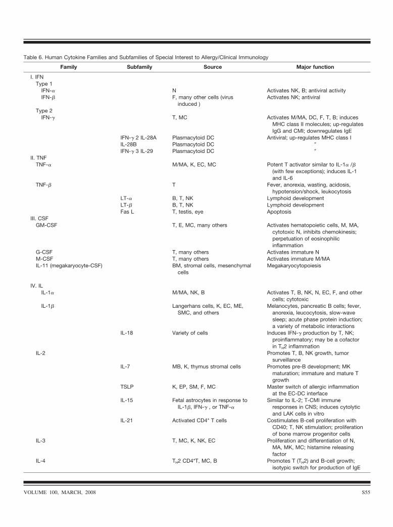
Table 6. Human Cytokine Families and Subfamilies of Special Interest to Allergy/Clinical Immunology
Family Subfamily Source Major function
I. IFNType 1
IFN-� N Activates NK, B; antiviral activityIFN-� F, many other cells (virus
induced )Activates NK; antiviral
Type 2IFN-� T, MC Activates M/MA, DC, F, T, B; induces
MHC class II molecules; up-regulatesIgG and CMI; downregulates IgE
IFN-� 2 IL-28A Plasmacytoid DC Antiviral; up-regulates MHC class IIL-28B Plasmacytoid DC �IFN-� 3 IL-29 Plasmacytoid DC �
II. TNFTNF-� M/MA, K, EC, MC Potent T activator similar to IL-1� /�
(with few exceptions); induces IL-1and IL-6
TNF-� T Fever, anorexia, wasting, acidosis,hypotension/shock, leukocytosis
LT-� B, T, NK Lymphoid developmentLT-� B, T, NK Lymphoid developmentFas L T, testis, eye Apoptosis
III. CSFGM-CSF T, E, MC, many others Activates hematopoietic cells, M, MA,
cytotoxic N, inhibits chemokinesis;perpetuation of eosinophilicinflammation
G-CSF T, many others Activates immature NM-CSF T, many others Activates immature M/MAIL-11 (megakaryocyte-CSF) BM, stromal cells, mesenchymal
cellsMegakaryocytopoiesis
IV. ILIL-1� M/MA, NK, B Activates T, B, NK, N, EC, F, and other
cells; cytotoxicIL-1� Langerhans cells, K, EC, ME,
SMC, and othersMelanocytes, pancreatic B cells; fever,
anorexia, leucocytosis, slow-wavesleep; acute phase protein induction;a variety of metabolic interactions
IL-18 Variety of cells Induces IFN-� production by T, NK;proinflammatory; may be a cofactorin TH2 inflammation
IL-2 Promotes T, B, NK growth, tumorsurveillance
IL-7 MB, K, thymus stromal cells Promotes pre-B development; MKmaturation; immature and mature Tgrowth
TSLP K, EP, SM, F, MC Master switch of allergic inflammationat the EC-DC interface
IL-15 Fetal astrocytes in response toIL-1�, IFN-� , or TNF-�
Similar to IL-2; T-CMI immuneresponses in CNS; induces cytolyticand LAK cells in vitro
IL-21 Activated CD4* T cells Costimulates B-cell proliferation withCD40; T, NK stimulation; proliferationof bone marrow progenitor cells
IL-3 T, MC, K, NK, EC Proliferation and differentiation of N,MA, MK, MC; histamine releasingfactor
IL-4 TH2 CD4*T, MC, B Promotes T (TH2) and B-cell growth;isotypic switch for production of IgE
VOLUME 100, MARCH, 2008 S55
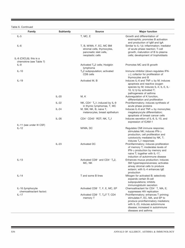
Table 6. Continued
Family Subfamily Source Major function
IL-5 T, MC, E Growth and differentiation ofeosinophils; promotes B activationand production of IgM and IgA
IL-6 T, B, M/MA, F, EC, MC BMstromal cells, thymocytes,pancreatic islet cells,neoplastic cells
Similar to IL-1�; inflammation; mediatorof acute phase reaction; T-cellgrowth, maturation of B to plasmacells; development of trophoblasts
IL-8 (CXCL8); this is achemokine (see Table 7)IL-9 Activated TH2 cells; Hodgkin
lymphomaPromotes MC and B growth
IL-10 TH2 subpopulation; activatedCD8 cells
Immune inhibitor (down-regulates IFN-� ); cofactor for proliferation ofthymocytes and B
IL-19 Activated M, B Induces IL-6 and TNF-� by M; inducesapoptosis and reactive oxygenspecies by M; induces IL-4; IL-5, IL-10, IL1� by activated T;pathogenesis of asthma
IL-20 M, K Autoregulation of K function,differentiation and proliferation
IL-22 NK, CD4� TH1; induced by IL-9in thymic lymphomas, T, MC
Proinflammatory; induces synthesis ofacute phase proteins
IL-24 M, SM, NK, B, naive T,melanocytes, breast epithelium
Induces IL-6 and TNF-� by monocytes;megakaryocyte differentiation;apoptosis of breast cancer cells
IL-26 CD4� CD45� ROT; NK, TH1 Induces secretion of IL-8, IL-10, andexpression of ICAM-1
IL-11 (see under III CSF)IL-12 M/MA, DC Regulates CMI immune response;
stimulates NK; induces IFN-�production, cell proliferation andcytotoxicity mediated by NK, T;induces TH1 responses
IL-23 Activated DC Proinflammatory; induces proliferationof memory T; moderates levels ofIFN-� production by memory andnaive T; together with IL-17,induction of autoimmune disease
IL-13 Activated CD8� and CD4� TH2,MC, NK
Enhances mucus production; inducesSM hyperresponsiveness; activatesairway stromal cells to produceeotaxin; with IL-4 enhances IgEproduction
IL-14 T and some B lines Mitogen for activated B; selectivelyexpands certain B-cellsubpopulations; inhibitsimmunoglobulin secretion
IL-16 (lymphocytechemoattractant factor)
Activated CD8� T, F, E, MC, EP Chemoattractant for CD4� T, MA, E;suppresses HIV replication
IL-17 Activated CD8� T, THI7 T; CD4memory T
Proinflammatory; enhances T priming;stimulates F, EC, MA, and EP toproduce proinflammatory mediators;with IL-23, induces autoimmunedisease; increased in autoimmunediseases and asthma
S56 ANNALS OF ALLERGY, ASTHMA & IMMUNOLOGY
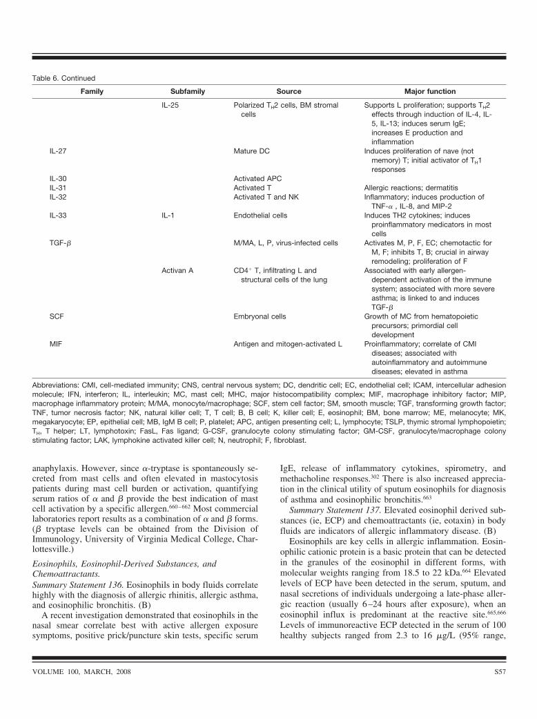
anaphylaxis. However, since �-tryptase is spontaneously se-creted from mast cells and often elevated in mastocytosispatients during mast cell burden or activation, quantifyingserum ratios of � and � provide the best indication of mastcell activation by a specific allergen.660–662 Most commerciallaboratories report results as a combination of � and � forms.(� tryptase levels can be obtained from the Division ofImmunology, University of Virginia Medical College, Char-lottesville.)
Eosinophils, Eosinophil-Derived Substances, andChemoattractants.Summary Statement 136. Eosinophils in body fluids correlatehighly with the diagnosis of allergic rhinitis, allergic asthma,and eosinophilic bronchitis. (B)
A recent investigation demonstrated that eosinophils in thenasal smear correlate best with active allergen exposuresymptoms, positive prick/puncture skin tests, specific serum
IgE, release of inflammatory cytokines, spirometry, andmethacholine responses.302 There is also increased apprecia-tion in the clinical utility of sputum eosinophils for diagnosisof asthma and eosinophilic bronchitis.663
Summary Statement 137. Elevated eosinophil derived sub-stances (ie, ECP) and chemoattractants (ie, eotaxin) in bodyfluids are indicators of allergic inflammatory disease. (B)
Eosinophils are key cells in allergic inflammation. Eosin-ophilic cationic protein is a basic protein that can be detectedin the granules of the eosinophil in different forms, withmolecular weights ranging from 18.5 to 22 kDa.664 Elevatedlevels of ECP have been detected in the serum, sputum, andnasal secretions of individuals undergoing a late-phase aller-gic reaction (usually 6–24 hours after exposure), when aneosinophil influx is predominant at the reactive site.665,666
Levels of immunoreactive ECP detected in the serum of 100healthy subjects ranged from 2.3 to 16 �g/L (95% range,
Table 6. Continued
Family Subfamily Source Major function
IL-25 Polarized TH2 cells, BM stromalcells
Supports L proliferation; supports TH2effects through induction of IL-4, IL-5, IL-13; induces serum IgE;increases E production andinflammation
IL-27 Mature DC Induces proliferation of nave (notmemory) T; initial activator of TH1responses
IL-30 Activated APCIL-31 Activated T Allergic reactions; dermatitisIL-32 Activated T and NK Inflammatory; induces production of
TNF-� , IL-8, and MIP-2IL-33 IL-1 Endothelial cells Induces TH2 cytokines; induces
proinflammatory medicators in mostcells
TGF-� M/MA, L, P, virus-infected cells Activates M, P, F, EC; chemotactic forM, F; inhibits T, B; crucial in airwayremodeling; proliferation of F
Activan A CD4� T, infiltrating L andstructural cells of the lung
Associated with early allergen-dependent activation of the immunesystem; associated with more severeasthma; is linked to and inducesTGF-�
SCF Embryonal cells Growth of MC from hematopoieticprecursors; primordial celldevelopment
MIF Antigen and mitogen-activated L Proinflammatory; correlate of CMIdiseases; associated withautoinflammatory and autoimmunediseases; elevated in asthma
Abbreviations: CMI, cell-mediated immunity; CNS, central nervous system; DC, dendritic cell; EC, endothelial cell; ICAM, intercellular adhesionmolecule; IFN, interferon; IL, interleukin; MC, mast cell; MHC, major histocompatibility complex; MIF, macrophage inhibitory factor; MIP,macrophage inflammatory protein; M/MA, monocyte/macrophage; SCF, stem cell factor; SM, smooth muscle; TGF, transforming growth factor;TNF, tumor necrosis factor; NK, natural killer cell; T, T cell; B, B cell; K, killer cell; E, eosinophil; BM, bone marrow; ME, melanocyte; MK,megakaryocyte; EP, epithelial cell; MB, IgM B cell; P, platelet; APC, antigen presenting cell; L, lymphocyte; TSLP, thymic stromal lymphopoietin;TH, T helper; LT, lymphotoxin; FasL, Fas ligand; G-CSF, granulocyte colony stimulating factor; GM-CSF, granulocyte/macrophage colonystimulating factor; LAK, lymphokine activated killer cell; N, neutrophil; F, fibroblast.
VOLUME 100, MARCH, 2008 S57
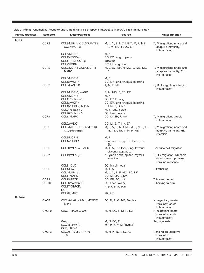
Table 7. Human Chemokine Receptor and Ligand Families of Special Interest to Allergy/Clinical Immunology
Family receptor Receptor Ligand/agonist Source Major function
I. CCCCR1 CCL3/MIP-1� CCL5/RANTES
CCL7/MCP-3M, L, N, E, MC, ME T, M, F, ME,
P, M, MC, F, EC, EPT, M migration; innate and
adaptive immunity,inflammation
CCL8/MCP-2 M, FCCL13/MCP-4, DC, EP, lung, thymusCCL14–16/HCC1–3 IntestineCCL23/MPIF DC, M, lung, liver
CCR2 CCL2/MCP-1 CCL7/MCP-3,MARC
M, L, EC, EP, N, MC, G, ME, DC,F
T, M migration; innate andadaptive immunity; TH1inflammation
CCL8/MCP-2 M, FCCL13/MCP-4 DC, EP, lung, thymus, intestine
CCR3 CCL5/RANTES T, M, F, ME E, B, T migration, allergicinflammation
CCL7/MCP-3, MARC P, M, MC, F, EC, EPCCL8/MCP-2 M, FCCL11/Eotaxin-1 EC, EP, E, lungCCL13/MCP-4 DC, EP, lung, thymus, intestineCCL15/HCC-2, MIP-5 DC, M, T, B, NKCCL24/Eotaxin 2 M, T, lung, spleenCCL26/Eotaxin 3 EC, heart, ovary
CCR4 CCL17/TARC DC, M, EP, F, SM T, M migration; allergicinflammation
CCL22/MDC DC, M, B, T, NK, EPCCR5 CCL3/MIP-1� CCL4/MIP-1�
CCL5/RANTESM, L, N, E, MC, ME M, L, N, E, F,
MC, BA, NK T, M, F, MET, M migration; innate and
adaptive immunity; HIVinfection
CCL8/MCP-2 M, FCCL14/HCC-1 Bone marrow, gut, spleen, liver,
SMCCR6 CCL20/MIP-3�, LARC M, T, N, EC, liver, lung, thymus,
placenta appendixDendritic cell migration
CCR7 CCL19/MIP-3� N, lymph node, spleen, thymus,intestine
T, DC migration; lymphoiddevelopment; primaryimmune response
CCL21/SLC EC, lymph nodeCCR8 CCL1/Gro� M, T, MC T trafficking
CCL4/MIP-1� M, L, N, E, F, MC, BA, NKCCL17/TARC DC, M, EP, F, SM
CCR9 CCL25/TECK DC, EP, EC, gut T homing to gutCCR10 CCL26/eotaxin-3 EC, heart, ovary T homing to skin
CCL27/CTACK, K, placenta, skinILCCCL28, MEC EP, EC
III. CXCCXCR CXCL8/IL-8, NAP-1, MDNCF,
MIP-2EC, N, P, G, ME, BA, NK N migration; innate
immunity; acuteinflammation
CXCR2 CXCL1–3/Gro�, Gro� M, N, EC, F, M, N, EC, F N migration; innateimmunity; acuteinflammation;
Gro� M, N, EC, F AngiogenesisCXCL5–8/ENA, EC, P, E, F, M (thymus)GCP, NAP-2
CXCR3 CXCL9–11/MIG, 1P-10, I-TAC
M, N, K, N, F, EC, G T migration; adaptiveimmunity; TH1inflammation
S58 ANNALS OF ALLERGY, ASTHMA & IMMUNOLOGY
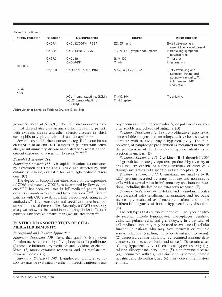
geometric mean of 6 �g/L). The ECP measurements havelimited clinical utility as an analyte for monitoring patientswith extrinsic asthma and other allergic diseases in whicheosinophils may play a role in tissue damage.666–672
Several eosinophil chemoattractants (eg, IL-5, eotaxin) areelevated in nasal and BAL samples in patients with activeallergic inflammatory disease associated with recent or con-current exposure to aerogenic allergens.303,304,672
Basophil Activation TestSummary Statement 138. A basophil activation test measuredby expression of CD63 and CD203c and detected by flowcytometry is being evaluated for many IgE-mediated disor-ders. (C)
The degree of basophil activation based on the expressionof CD63 and recently CD203c is determined by flow cytom-etry.672 It has been evaluated in IgE-mediated pollen, food,drug, Hymenoptera venom, and latex reactions.673–681 Sera ofpatients with CIU also demonstrate basophil-activating auto-antibodies.682 High sensitivity and specificity have been ob-served in most of these studies. Recently, a CD63 sensitivityassay was shown to be useful in monitoring clinical effects inpatients who receive omalizumab (Xolair) treatment.683
IN VITRO DIAGNOSTIC TESTS OF CELL-MEDIATED IMMUNITY
Background and Present ApplicationSummary Statement 139. Tests that quantify lymphocytefunction measure the ability of lymphocytes to (1) proliferate,(2) produce inflammatory mediators and cytokines or chemo-kines, (3) mount cytotoxic responses, and (4) regulate im-mune responses. (B)
Summary Statement 140. Lymphocyte proliferative re-sponses may be evaluated by either nonspecific mitogens (eg,
phytohemagglutinin, concanavalin A, or pokeweed) or spe-cific soluble and cell-bound antigens. (B)
Summary Statement 141. In vitro proliferative responses tosome soluble antigens, but not mitogens, have been shown tocorrelate with in vivo delayed hypersensitivity. The role,however, of lymphocyte proliferation as measured in vitro inthe pathogenesis of the delayed-type hypersensitivity tissuereaction is unclear. (B)
Summary Statement 142. Cytokines (IL-1 through IL-33)and growth factors are glycoproteins produced by a variety ofcells that are capable of altering activities of other cellsthrough interaction with specific surface receptors. (E)
Summary Statement 143. Chemokines are small (8 to 10kDa) proteins secreted by many immune and nonimmunecells with essential roles in inflammatory and immune reac-tions, including the late-phase cutaneous response. (E)
Summary Statement 144. Cytokine and chemokine profilesplay essential roles in allergic inflammation and are beingincreasingly evaluated as phenotypic markers and in thedifferential diagnosis of human hypersensitivity disorders.(B)
The cell types that contribute to the cellular hypersensitiv-ity reaction include lymphocytes, macrophages, dendriticcells, Langerhans cells, and granulocytes. In vitro tests ofcell-mediated immunity may be used to evaluate (1) cellularfunction in patients who may have recurrent or multipleserious infections (eg, fungal, mycobacterial and protozoan);(2) depressed cellular immunity (eg, acquired immune defi-ciency syndrome, sarcoidosis, and cancer); (3) certain casesof drug hypersensitivity; (4) chemical hypersensitivity (eg,toluene diisocyanate, beryllium); (5) autoimmune diseases(eg, rheumatoid arthritis, Guillain-Barre syndrome, chronichepatitis, and thyroiditis); and (6) many other inflammatoryentities.
Table 7. Continued
Family receptor Receptor Ligand/agonist Source Major function
CXCR4 CXCL12/SDF-1, PBSF EC, EP, lung B-cell development;myeloid cell development
CXCR5 CXCL13/BLC, BCA-1 EC, M, DC, lymph node, spleen B trafficking; lymphoiddevelopment
CXCR6 CXCL16 B, M, DC T migration? CXCL4/PF4 P, MK Inflammation
IIII. CX3CCX3CR1 CX3CL1/FRACTALKINE APC, DC, EC, T, SM T, NK trafficking and
adhesion; innate andadaptive immunity, TH1inflammation, MCchemotaxis
IV. XCXCRI
XCL1/ lymphotactin a, SCMl� T, MC, NK T traffickingXCL2/ Lymphotactin b,
SCMl�T, NK, spleen
Abbreviations: Same as Table 6; BA, pro-B cell line.
VOLUME 100, MARCH, 2008 S59
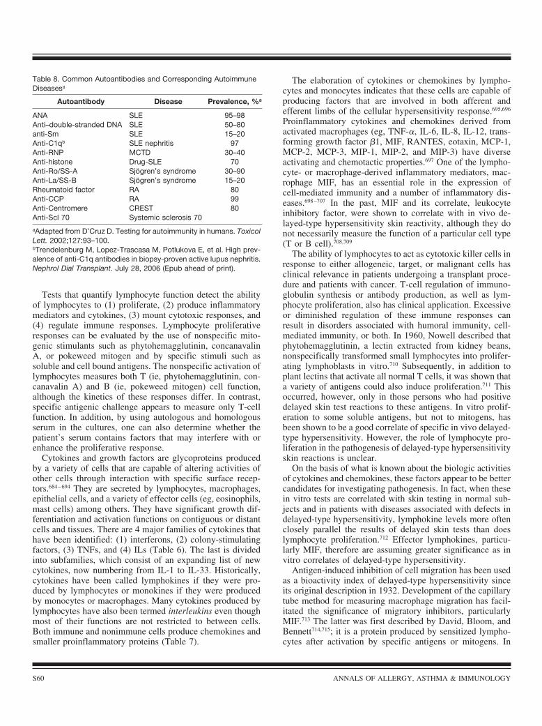
Tests that quantify lymphocyte function detect the abilityof lymphocytes to (1) proliferate, (2) produce inflammatorymediators and cytokines, (3) mount cytotoxic responses, and(4) regulate immune responses. Lymphocyte proliferativeresponses can be evaluated by the use of nonspecific mito-genic stimulants such as phytohemagglutinin, concanavalinA, or pokeweed mitogen and by specific stimuli such assoluble and cell bound antigens. The nonspecific activation oflymphocytes measures both T (ie, phytohemagglutinin, con-canavalin A) and B (ie, pokeweed mitogen) cell function,although the kinetics of these responses differ. In contrast,specific antigenic challenge appears to measure only T-cellfunction. In addition, by using autologous and homologousserum in the cultures, one can also determine whether thepatient’s serum contains factors that may interfere with orenhance the proliferative response.
Cytokines and growth factors are glycoproteins producedby a variety of cells that are capable of altering activities ofother cells through interaction with specific surface recep-tors.684–694 They are secreted by lymphocytes, macrophages,epithelial cells, and a variety of effector cells (eg, eosinophils,mast cells) among others. They have significant growth dif-ferentiation and activation functions on contiguous or distantcells and tissues. There are 4 major families of cytokines thathave been identified: (1) interferons, (2) colony-stimulatingfactors, (3) TNFs, and (4) ILs (Table 6). The last is dividedinto subfamilies, which consist of an expanding list of newcytokines, now numbering from IL-1 to IL-33. Historically,cytokines have been called lymphokines if they were pro-duced by lymphocytes or monokines if they were producedby monocytes or macrophages. Many cytokines produced bylymphocytes have also been termed interleukins even thoughmost of their functions are not restricted to between cells.Both immune and nonimmune cells produce chemokines andsmaller proinflammatory proteins (Table 7).
The elaboration of cytokines or chemokines by lympho-cytes and monocytes indicates that these cells are capable ofproducing factors that are involved in both afferent andefferent limbs of the cellular hypersensitivity response.695,696
Proinflammatory cytokines and chemokines derived fromactivated macrophages (eg, TNF-�, IL-6, IL-8, IL-12, trans-forming growth factor �1, MIF, RANTES, eotaxin, MCP-1,MCP-2, MCP-3, MIP-1, MIP-2, and MIP-3) have diverseactivating and chemotactic properties.697 One of the lympho-cyte- or macrophage-derived inflammatory mediators, mac-rophage MIF, has an essential role in the expression ofcell-mediated immunity and a number of inflammatory dis-eases.698–707 In the past, MIF and its correlate, leukocyteinhibitory factor, were shown to correlate with in vivo de-layed-type hypersensitivity skin reactivity, although they donot necessarily measure the function of a particular cell type(T or B cell).708,709
The ability of lymphocytes to act as cytotoxic killer cells inresponse to either allogeneic, target, or malignant cells hasclinical relevance in patients undergoing a transplant proce-dure and patients with cancer. T-cell regulation of immuno-globulin synthesis or antibody production, as well as lym-phocyte proliferation, also has clinical application. Excessiveor diminished regulation of these immune responses canresult in disorders associated with humoral immunity, cell-mediated immunity, or both. In 1960, Nowell described thatphytohemagglutinin, a lectin extracted from kidney beans,nonspecifically transformed small lymphocytes into prolifer-ating lymphoblasts in vitro.710 Subsequently, in addition toplant lectins that activate all normal T cells, it was shown thata variety of antigens could also induce proliferation.711 Thisoccurred, however, only in those persons who had positivedelayed skin test reactions to these antigens. In vitro prolif-eration to some soluble antigens, but not to mitogens, hasbeen shown to be a good correlate of specific in vivo delayed-type hypersensitivity. However, the role of lymphocyte pro-liferation in the pathogenesis of delayed-type hypersensitivityskin reactions is unclear.
On the basis of what is known about the biologic activitiesof cytokines and chemokines, these factors appear to be bettercandidates for investigating pathogenesis. In fact, when thesein vitro tests are correlated with skin testing in normal sub-jects and in patients with diseases associated with defects indelayed-type hypersensitivity, lymphokine levels more oftenclosely parallel the results of delayed skin tests than doeslymphocyte proliferation.712 Effector lymphokines, particu-larly MIF, therefore are assuming greater significance as invitro correlates of delayed-type hypersensitivity.
Antigen-induced inhibition of cell migration has been usedas a bioactivity index of delayed-type hypersensitivity sinceits original description in 1932. Development of the capillarytube method for measuring macrophage migration has facil-itated the significance of migratory inhibitors, particularlyMIF.713 The latter was first described by David, Bloom, andBennett714,715; it is a protein produced by sensitized lympho-cytes after activation by specific antigens or mitogens. In
Table 8. Common Autoantibodies and Corresponding AutoimmuneDiseasesa
Autoantibody Disease Prevalence, %a
ANA SLE 95–98Anti–double-stranded DNA SLE 50–80anti-Sm SLE 15–20Anti-C1qb SLE nephritis 97Anti-RNP MCTD 30–40Anti-histone Drug-SLE 70Anti-Ro/SS-A Sjögren’s syndrome 30–90Anti-La/SS-B Sjögren’s syndrome 15–20Rheumatoid factor RA 80Anti-CCP RA 99Anti-Centromere CREST 80Anti-Scl 70 Systemic sclerosis 70
aAdapted from D’Cruz D. Testing for autoimmunity in humans. ToxicolLett. 2002;127:93–100.bTrendelenburg M, Lopez-Trascasa M, Potlukova E, et al. High prev-alence of anti-C1q antibodies in biopsy-proven active lupus nephritis.Nephrol Dial Transplant. July 28, 2006 (Epub ahead of print).
S60 ANNALS OF ALLERGY, ASTHMA & IMMUNOLOGY
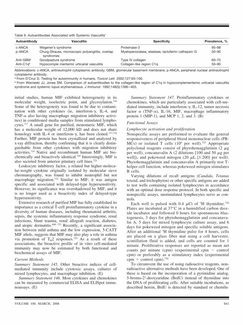
initial studies, human MIF exhibited heterogeneity in itsmolecular weight, isoelectric point, and glycosylation.716
Some of the heterogeneity was found to be due to contami-nation with other cytokines (ie, interferon-�, IL-4, andTNF-� also having macrophage migration inhibitory activi-ties) in conditioned media samples from stimulated lympho-cytes.717 A small gene for purified, monomeric MIF, whichhas a molecular weight of 12,000 kD and does not sharehomology with IL-4 or interferon-�, has been cloned.717,718
Further, MIF protein has been crystallized and analyzed byx-ray diffraction, thereby confirming that it is clearly distin-guishable from other cytokines with migration inhibitoryactivities.719 Native and recombinant human MIF are bio-chemically and bioactively identical.720 Interestingly, MIF isalso secreted from anterior pituitary cell lines.721
Leukocyte inhibitory factor, a related but higher-molecu-lar-weight cytokine originally isolated by molecular sievechromatography, was found to inhibit neutrophil but notmacrophage migration.722 Similar to MIF, it was antigenspecific and associated with delayed-type hypersensitivity.However, its significance was overshadowed by MIF, and itis no longer used as a bioactivity index of delayed-typehypersensitivity.
Extensive research of purified MIF has fully established itsimportance as a critical T-cell proinflammatory cytokine in adiversity of human diseases, including rheumatoid arthritis,sepsis, the systemic inflammatory response syndrome, renalinfections, blunt trauma, renal allograft reaction, diabetes,and atopic dermatitis.698–723 Recently, a significant associa-tion between mild asthma and the low expression, 5-CATTMIF allele, suggests that MIF may also play a role in asthmavia promotion of TH2 responses.724 As a result of theseassociations, the bioactive profile of in vitro cell-mediatedimmunity may now be estimated by both functional andbiochemical assays of MIF.
Current MethodsSummary Statement 145. Other bioactive indices of cell-mediated immunity include cytotoxic assays, cultures ofmixed lymphocytes, and macrophage inhibition. (E)
Summary Statement 146. Most cytokines and chemokinescan be measured by commercial ELISA and ELISpot immu-noassays. (E)
Summary Statement 147. Proinflammatory cytokines orchemokines, which are particularly associated with cell-me-diated immunity, include interferon-�, IL-12, tumor necrosisfactor � (TNF-�), IL-16, MIF, macrophage inflammatoryprotein 1 (MIP-1), and MCP 1, 2, and 3. (B)
Functional Assays
Lymphocyte activation and proliferationNonspecific assays are performed to evaluate the generalresponsiveness of peripheral blood mononuclear cells (PB-MCs) or isolated T cells (106 per well).725 Appropriatepolyclonal reagents consist of phytohemagglutinin (2 �gper well), concanavalin A (2 dilutions [100 and 50 �g perwell]), and pokeweed mitogen (20 �L [1:200] per well).Phytohemagglutinin and concanavalin A primarily test T-helper cell function, whereas pokeweed mitogen stimulatesB cells.
Varying dilutions of recall antigens (Candida, Tetanustoxoid, and trichophyton) or other specific antigens are addedto test wells containing isolated lymphocytes in accordancewith an optimal dose response protocol. In both specific andnonspecific assays, unstimulated lymphocytes serve as con-trols.
Each well is pulsed with 0.4 �Ci of 3H thymidine.726
Plates are incubated at 37°C in a humidified carbon diox-ide incubator and followed 0 hours for spontaneous blas-togenesis, 3 days for phytohemagglutinin and concanava-lin A, 5 days for mixed lymphocyte culture assay, and 6days for pokeweed mitogen and specific soluble antigens.After an additional 3H thymidine pulse for 4 hours, cellsare placed on a glass fiber mat using a cell harvester,scintillation fluid is added, and cells are counted for 1minute. Proliferative responses are reported as mean netcounts per minute (cpm) (experimental cpm � controlcpm) or preferably as a stimulatory index (experimentalcpm control cpm).726
To circumvent the use of using radioactive reagents, non-radioactive alternative methods have been developed. One ofthese is based on the incorporation of a pyrimidine analog,5-bromo-21-deoxyuridine (BrdU) instead of thymidine intothe DNA of proliferating cells. After suitable incubations, asdescribed herein, BrdU is detected by standard or chemilu-
Table 9. Autoantibodies Associated with Systemic Vasculitisa
Autoantibody Vasculitis Specificity Prevalence, %
c-ANCA Wegener’s syndrome Proteinase-3 95–98p-ANCA Churg-Strauss, microscopic polyangiiitis, overlap
syndromesMyeloperoxidase, elastase, lactoferrin cathepsin G 50–90
Anti-GBM Goodpasture syndrome Type IV collagen 60–75Anti-C1qb Hypocomple mentemic urticarial vasculitis Collagen-like region C1q 50–90
Abbreviations: c-ANCA, antineutrophil cytoplasmic antibody; GBM, glomerular basement membrane; p-ANCA, peripheral nuclear antineutrophilcytoplasmic antibody.a From D’Cruz D. Testing for autoimmunity in humans. Toxicol Lett. 2002;127:93–100.b From Wisnieski JJ, Jones SM. Comparison of autoantibodies to the collagen-like region of C1q in hypocomplementemic urticarial vasculitissyndrome and systemic lupus erythematosus. J Immunol. 1992;148(5):1396–403.
VOLUME 100, MARCH, 2008 S61
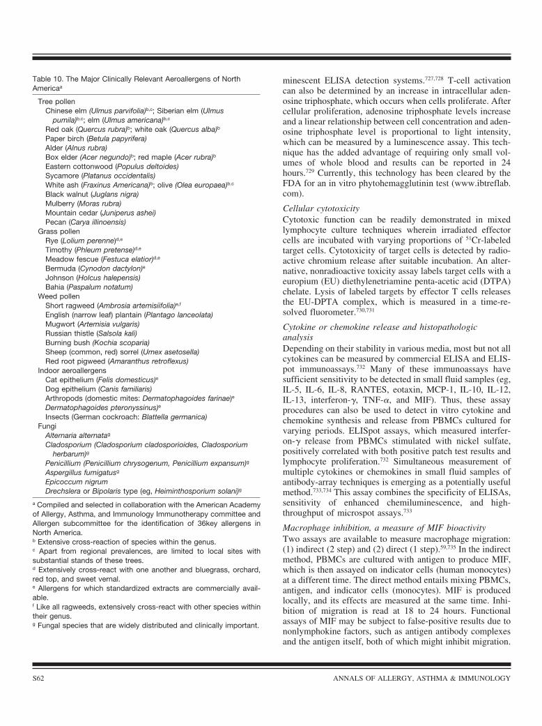
minescent ELISA detection systems.727,728 T-cell activationcan also be determined by an increase in intracellular aden-osine triphosphate, which occurs when cells proliferate. Aftercellular proliferation, adenosine triphosphate levels increaseand a linear relationship between cell concentration and aden-osine triphosphate level is proportional to light intensity,which can be measured by a luminescence assay. This tech-nique has the added advantage of requiring only small vol-umes of whole blood and results can be reported in 24hours.729 Currently, this technology has been cleared by theFDA for an in vitro phytohemagglutinin test (www.ibtreflab.com).
Cellular cytotoxicityCytotoxic function can be readily demonstrated in mixedlymphocyte culture techniques wherein irradiated effectorcells are incubated with varying proportions of 51Cr-labeledtarget cells. Cytotoxicity of target cells is detected by radio-active chromium release after suitable incubation. An alter-native, nonradioactive toxicity assay labels target cells with aeuropium (EU) diethylenetriamine penta-acetic acid (DTPA)chelate. Lysis of labeled targets by effector T cells releasesthe EU-DPTA complex, which is measured in a time-re-solved fluorometer.730,731
Cytokine or chemokine release and histopathologicanalysisDepending on their stability in various media, most but not allcytokines can be measured by commercial ELISA and ELIS-pot immunoassays.732 Many of these immunoassays havesufficient sensitivity to be detected in small fluid samples (eg,IL-5, IL-6, IL-8, RANTES, eotaxin, MCP-1, IL-10, IL-12,IL-13, interferon-�, TNF-�, and MIF). Thus, these assayprocedures can also be used to detect in vitro cytokine andchemokine synthesis and release from PBMCs cultured forvarying periods. ELISpot assays, which measured interfer-on-� release from PBMCs stimulated with nickel sulfate,positively correlated with both positive patch test results andlymphocyte proliferation.732 Simultaneous measurement ofmultiple cytokines or chemokines in small fluid samples ofantibody-array techniques is emerging as a potentially usefulmethod.733,734 This assay combines the specificity of ELISAs,sensitivity of enhanced chemiluminescence, and high-throughput of microspot assays.733
Macrophage inhibition, a measure of MIF bioactivityTwo assays are available to measure macrophage migration:(1) indirect (2 step) and (2) direct (1 step).59,735 In the indirectmethod, PBMCs are cultured with antigen to produce MIF,which is then assayed on indicator cells (human monocytes)at a different time. The direct method entails mixing PBMCs,antigen, and indicator cells (monocytes). MIF is producedlocally, and its effects are measured at the same time. Inhi-bition of migration is read at 18 to 24 hours. Functionalassays of MIF may be subject to false-positive results due tononlymphokine factors, such as antigen antibody complexesand the antigen itself, both of which might inhibit migration.
Table 10. The Major Clinically Relevant Aeroallergens of NorthAmericaa
Tree pollenChinese elm (Ulmus parvifolia)b,c; Siberian elm (Ulmus
pumila)b,c; elm (Ulmus americana)b,c
Red oak (Quercus rubra)b; white oak (Quercus alba)b
Paper birch (Betula papyrifera)Alder (Alnus rubra)Box elder (Acer negundo)b; red maple (Acer rubra)b
Eastern cottonwood (Populus deltoides)Sycamore (Platanus occidentalis)White ash (Fraxinus Americana)b; olive (Olea europaea)b,c
Black walnut (Juglans nigra)Mulberry (Moras rubra)Mountain cedar (Juniperus ashei)Pecan (Carya illinoensis)
Grass pollenRye (Lolium perenne)d,e
Timothy (Phleum pretense)d,e
Meadow fescue (Festuca elatior)d,e
Bermuda (Cynodon dactylon)e
Johnson (Holcus halepensis)Bahia (Paspalum notatum)
Weed pollenShort ragweed (Ambrosia artemisiifolia)e,f
English (narrow leaf) plantain (Plantago lanceolata)Mugwort (Artemisia vulgaris)Russian thistle (Salsola kali)Burning bush (Kochia scoparia)Sheep (common, red) sorrel (Umex asetosella)Red root pigweed (Amaranthus retroflexus)
Indoor aeroallergensCat epithelium (Felis domesticus)e
Dog epithelium (Canis familiaris)Arthropods (domestic mites: Dermatophagoides farinae)e
Dermatophagoides pteronyssinus)e
Insects (German cockroach: Blattella germanica)Fungi
Alternaria alternatag
Cladosporium (Cladosporium cladosporioides, Cladosporiumherbarum)g
Penicillium (Penicillium chrysogenum, Penicillium expansum)g
Aspergillus fumigatusg
Epicoccum nigrumDrechslera or Bipolaris type (eg, Heiminthosporium solani)g
a Compiled and selected in collaboration with the American Academyof Allergy, Asthma, and Immunology Immunotherapy committee andAllergen subcommittee for the identification of 36key allergens inNorth America.b Extensive cross-reaction of species within the genus.c Apart from regional prevalences, are limited to local sites withsubstantial stands of these trees.d Extensively cross-react with one another and bluegrass, orchard,red top, and sweet vernal.e Allergens for which standardized extracts are commercially avail-able.f Like all ragweeds, extensively cross-react with other species withintheir genus.g Fungal species that are widely distributed and clinically important.
S62 ANNALS OF ALLERGY, ASTHMA & IMMUNOLOGY
False-negative results may also occur because of individualvariation of indicator cells. Because of their complexity andtechnical skill requirements, functional MIF in vitro assaysare not commercially available. They are used chiefly forresearch of patient populations rather than individual patients.However, they may be of adjunctive clinical value for pur-poses of identifying certain pathogenetic factors, monitoringthe results of therapy and the clinical course of patients withdepressed cell-mediated immunity.
ImmunoassaysAs discussed herein, direct measurement of several cytokinesin blood, BAL, sputum, and other body fluids by commercialELISAs may act as biologic markers of acute or chronicinflammation and/or delayed-type hypersensitivity. Proin-flammatory cytokines or chemokines and their subfamiliesinclude interferon-�, TNF-�, IL-4, IL-5, IL-6, IL-8, RAN-TES, eotaxin, IL-12, IL-13, IL-15, IL-18, IL-19, IL-22, IL-23, IL-25, IL-31, IL-32, IL-33, and TNF-�. Proinflammatorycytokines or chemokines, which are particularly associatedwith cell-mediated immunity, include interferon-�, IL-12,TNF-�, IL-16, MIP-1, MCP-1, MCP-2, MCP-3, and MIF. Atest of interferon-� release by peripheral lymphocytes hasbeen recommended by the Centers for Disease Control andPrevention for diagnosis of latent tuberculosis.465 Increasedserum levels of MIF have been found in a wide range ofexogenous and autoinflammatory T-cell–mediated diseas-es.698–707 Normal serum concentrations of MIF range from 0.5to 8 �g/mL. In situ hybridization of MIF messenger RNA inmacrophages can also be documented in delayed-type hyper-sensitivity skin lesions.698
Nonspecific Screening Tests for Cellular ImmuneCompetencySummary Statement 148. Simple, cost-effective tests include(1) an absolute lymphocyte count, (2) the absolute number ofCD4� T cells, and (3) the CD4�/CD8� ratio. (B)
Several simple cost-effective screening tests are availablefor the evaluation of competency of cell-mediated immunity.These include (1) an absolute lymphocyte count (a count lessthan 1,200/mm3 suggests an abnormal immune response); (2)number of total T cells, as measured by anti-CD3 surfacemarkers; (3) estimation of CD4� helper cell and CD8� cyto-toxic populations by immunofluorescence staining with ap-propriate phenotypic cell markers; (4) measurement of T-celllymphocyte activation by IL-2 secretion or fluorescent anti-CD25 and/or anti–HLA-DR monoclonal antibodies; (5) de-termination of the relevant percentage of CD45 RO� CD29�
T-cell lymphocytes as an indication of memory cells; and (6)flow cytometric assay of the percentage of CD4�, CD25� FoxP3� immunoregulatory T cells. The latter cells may be in-creased after successful allergen immunotherapy.
Current Status of Cytokines and ChemokinesThese cellular products were first identified more than 25years ago and new ones are being discovered almost everyyear. As of 2007, there are 33 recognized IL cytokines and
many more that do not have the IL designation.696,741 Tables6 and 7 are summaries of selected cytokines and chemokines,respectively. They are derived from multiple cell sources andoften have redundant and overlapping biologic functions,which have been investigated extensively in knockout andtransfected animal models. Their chief utility in diagnosis ofhuman hypersensitivity is to determine the relative signifi-cance of cellular interactions involving dendritic cells, T-cellsubsets, NK-T cells, macrophages, NK cells, mast cells andepithelial cells in various immune inflammatory processes.For example, body tissue secretions that contain IL-4, IL-5,IL-10, and IL-13 are associated with IgE-mediated allergicinflammation, whereas the predominant cytokines found incell-mediated immunity inflammation are IL-12 and interfer-on-�. Likewise, in situ hybridization techniques enable iden-tification of specific cytokines in tissue biopsy samples. Cer-tain cytokines such as TNF-� and IL-5 are nonselective andare associated with various types of inflammation. Chemo-kines (eg, eotaxin, RANTES, MCP-1, MCP-2, MCP-3) canalso be identified by similar techniques.688,693
For the most part, cytokine and growth factor identificationand profiling in human disease are still research oriented.736–
741 However, a number of reports suggest that cytokine pro-files may be useful in differential diagnosis of a variety ofhuman diseases.742–749 For example, serologic profiles maydifferentiate infectious from noninfectious uveitis and may beof prognostic value in acute pancreatitis.742,743 Similar reportsconcerning the utility of cytokine profiling for the diagnosisand follow-up of allergic or allergy-related diseases have alsorecently appeared.734,750–760
Chemokines are small (8 to 10 kDa) proteins secreted bymany immune and nonimmune cells with essential roles ininflammatory and immune reactions, including the late-phasecutaneous response.697,761 Although principally chemoattrac-tants, they have diverse functions during an inflammatory orimmune response, which involve cellular recruitment, activa-tion, and differentiation.762 The processes of intravascularrolling, tethering, and diapedesis of inflammatory cells arecomplex and also involve integrins, adhesion molecules, andselectins.763–766 The approximately 50 human chemokine re-ceptors and their corresponding ligands are classified on thebasis of the number and structural characteristics of canonicalcysteine residues (Table 7).697,761,762 Understanding chemo-kine biology is at times confusing because a single chemo-kine can bind to multiple receptors and vice versa.
High serum levels of thymus and activation regulatorychemokine (TARC) (CCL17) and macrophage-derived che-mokine (MDC) (CCL22) preferentially attract TH2 subsets ofT-helper cells in patients with allergic disease.758 Once acti-vated and differentiated, TH2 cells express CCR3 and CCR4,receptors for eotaxin, RANTES, and TARC. Human CD25regulatory T cells express higher levels of CCR4, CCR5, andCCR8 compared with CD25 cells. Eotaxin (CCL11), its re-ceptor CCR3, and other ligands of CCR3 (CCL5, CCL7,CCL8, and CCL13) are coattractants for eosinophils in asth-ma.767 Lymphocytes of nonallergic asthma patients strongly
VOLUME 100, MARCH, 2008 S63
express CXCR4. Certain chemokines (CCL1, CCL2, CCL11,CCL17, CCL22) are preferentially produced in the presenceof TH2 cytokines. CCL2 may enhance IL-4 production inactivated T lymphocytes.
Specific chemokines have been reported to be associatedwith TH1 or TH2 cytokine production.768 Both CCR4 andCCR8 are associated with allergen-induced late asthmaticresponses.769 Both IL-8 and eotaxin levels were increased inpatients with severe asthma compared with patients with mildasthma.770,771 Fracktalkine (CX3CL1) contributes to mast cellrecruitment in asthma.772 Eotaxin levels are elevated in bothaspirin tolerant and intolerant patients.773
Although there is increased expression of RANTES,CCR3, and CCR5 in lesional skin, CC4R expression reflectsgreater severity in atopic dermatitis patients.773,774 Two che-mokine receptors (TARC, CCR3) appear to be targets fortreatment in atopic diseases.775,776
OTHER DIAGNOSTIC IMMUNOLOGIC TESTSSummary Statement 149. Investigation of non-IgE and non–cell-mediated clinical immunologic disorders may requiretests that indicate abnormal adaptive and innate immunereactions. (B)
Evaluation of non-IgE and non–cell-mediated clinical im-munologic diseases may include laboratory screening for (1)primary and acquired immunodeficiencies, (2) immune-me-diated gammopathies, (3) complement activation disorders,and (4) a diverse spectrum of autoimmune and vasculiticdisorders.
ImmunodeficiencyThe scope of diagnostic procedures for primary immunode-ficiency has been reviewed in the recently published PracticeParameter for Primary Immunodeficiencies. Current status ofimmune-based diagnostic and monitoring of HIV-acquiredimmunodeficiency and fully developed AIDS have been re-viewed elsewhere.777,778 It is generally agreed that HIV-1RNA levels and CD4 cell counts are important predictors ofsubsequent virologic and clinical outcomes.777
Immune-Mediated GammopathiesSummary Statement 150. Abnormal serum and urine proteins,including cryoglobulins, may be associated with several ab-normal immune syndromes. (B)
Drug-induced dysgammaglobulinemia, hypogammaglobu-linemia, and leukocytoclastic vasculitis may occasionally beconfused with premalignant or malignant gammopathy.779,780
Abnormal serum and urine protein levels are detected byelectrophoresis and immunofixation. Free light chains mayalso be demonstrated in serum.781 Cryoglobulins may alsodevelop, and these are usually classified as type I, type II, ortype III.782,783 Type I contains a single monoclonal IgG, typeII is a mixture of monoclonal IgG with polyclonal IgGs, andtype III is a mixture of polyclonal IgGs of different isotypes,most frequently IgG and IgM. Types II and III are also calledmixed cryoglobulins.784,785 Cryofibrinogenemia may also
have to be considered in the differential diagnosis of coldprecipitable proteins.786
Nonspecific Tests of Immunologic InflammationSummary Statement 151. The inflammatory consequencesinduced by immune functions may be detected by nonspecifictests, such as complete blood cell count with differential,sedimentation rate, C-reactive protein, and other acute-phasereactants. In some instances, functional assays of neutrophilsand macrophages may be necessary to pinpoint inflammatoryresponses. (B)
Routine laboratory tests, such as a complete blood cellcount with differential, sedimentation rate, C-reactive pro-tein, and other acute-phase reactants (eg, fibrinogen, ferritin),are useful in determining the inflammatory consequences ofinnate immunity.787–789 Functional assays of macrophages andneutrophils, the primary host defense cells, may indicateeither impaired defense (eg, decreased or absent chemotaxis,phagocytosis, bacterial killing, and cytokine or chemokinesynthesis) or uncontrolled inflammatory responses (ie, themacrophage activation syndrome).790–793 Neutrophil and mac-rophage function tests may only be available in specializedmedical centers. High ferritin levels associated with the mac-rophage activation syndrome can be used to monitor treat-ment of this disorder792
Complement ActivationSummary Statement 152. Evaluation of complement activa-tion with a decrease of C3 and C4 may indicate complementdeficiency, drug reactions, or the presence of immune com-plexes, which often are associated with increases in serumcryoglobulins and C1q binding. (B)
Although complement is a major component of innateimmunity to pathogenic microorganisms, it may also be ab-normally activated by adaptive immune pathways such asimmune complexes or cytotoxic antibodies. In most cases,these pathways of complement function can be estimated byimmune hemolysis (CH50 and AH50) and a functional ELISAof the mannan lectin binding pathway.794,795 Decrease of C4and C3 and increase in factor B are general screening tests forcomplement activation.796–798 Specialized laboratory centerscan also determine individual complement components. Eval-uation of both inherited and acquired forms of complementdeficiencies, including C1 esterase inhibitor, have been dis-cussed in Practice Parameters of Immunodeficiency. Immunecomplex activation of complement may be associated with anincrease in serum cryoglobulins and/or an increase of C1qbinding.784,785,799
AutoimmunitySummary Statement 153. Autoantibody profiles offer impor-tant diagnostic adjuncts in the diagnosis of collagen vasculardiseases, vasculitides, and cytotoxicity disorders. (B)
Many tissue antigens are capable of provoking autoim-mune responses when the milieu of genetic susceptibility (ie,major histocompatibility subtypes) and environmental inter-action is apropos. The discoveries of rheumatoid factor and
S64 ANNALS OF ALLERGY, ASTHMA & IMMUNOLOGY
antinuclear antibody in association with rheumatoid arthritisand systemic lupus erythematosus, respectively, providedimpetus for the clinical significance of autoimmunity.800 Ta-ble 8 is a partial list of common autoantibodies and theirprevalence in corresponding autoimmune diseases.800,801 Thy-roid autoantibodies may occur in up to 30% of patients withCIU.802,803 IgG autoantibodies to IgE or the � subunit ofFc�R1 may also be demonstrated in approximately 40% to50% of patients with CIU.804,805 The diagnostic or pathophys-iologic significance of these autoantibodies in CIU is as yetindeterminate.
Selection of any one or a combination of these tests shouldbe predicated on a reasonable clinical pretest probability.
VasculitidesSmall, medium, and large vessel vasculitides are most com-monly diagnosed by characteristic clinical features and bi-opsy with demonstration of appropriate immune complexeswithin vessel walls.800 Some cases of palpable purpura areassociated with type III cryoglobulins.780 A low C1q andanti-C1q antibodies806 may be associated with hypocomple-mentemic urticarial vasculitis. Several types of small andmedium vessel vasculitides are associated with antineutro-philic cytoplasmic and glomerular basement membrane anti-bodies (Table 9).
Human Cytotoxic AntibodiesAntibodies of this type may induce hemolytic anemia, neu-tropenia, or thrombocytopenia. These conditions may occurspontaneously or in association with drug therapy. Immune-mediated hemolytic anemia includes paroxysmal nocturnalhemoglobinuria, paroxysmal cold hemoglobinuria, and coldagglutinin disease.807–810 Direct and indirect Coombs tests areuseful screening tests for red cell autoantibodies. A gel mi-crocolumn assay is purported to increase sensitivity.811 Aquantitative antiglobulin consumption technique can detectIgG on granulocyte cell membranes, which occurs in theFelty syndrome.812 Because definitive tests are not generallyavailable in most clinical laboratories, immune-induced neu-tropenia and thrombocytopenia are less well studied.813–817
Drug-induced autoantibodies to red cells, neutrophils, andthrombocytes have been induced by a number of drugs butmost often they are associated with penicillin, propylthioura-cil, and quinine/quinidine, respectively.818–827
Analytic TechniquesThe clinician should be aware that there is considerableinterlaboratory variation of the methods described in thissection. Techniques that originally used agglutination, turbi-dimetry, nephelometry, double immunodiffusion, counterim-munoelectrophoresis, and indirect immunofluorescence haveevolved to ELISA, Western blotting, and in some situationsimmunoblot assays.828 However, ELISA antinuclear antibodyscreening assays may lack sensitivity for certain collagenvascular diseases and therefore require confirmatory indirectimmunofluorescence tests.829 In recognition of these possibleconfounders, future diagnostic accuracy of these tests will be
based on likelihood ratios.830 Because multiple autoantibodiestend to occur in autoimmune diseases by a process known asepitope spreading and specific autoantibody profiles mayhave greater diagnostic predictability or prognostication,multiplexed proteomic platforms are in current developmentfor SLE and rheumatoid arthritis.831,832 As yet, optimal con-ditions for autoantigen arrays have not been established sothese high throughput measures will require thorough valida-tion in the future.832
UNPROVEN TESTSSummary Statement 154. Procedures for which there is noevidence of diagnostic validity include cytotoxic tests, prov-ocation-neutralization, electrodermal testing, applied kinesi-ology, iridology, hair analysis, or food specific IgG, IgG4,and IgG/IgG4 antibody tests. (B)
Cytotoxic TestsThe cytotoxic test is performed by placing a drop of wholeblood or buffy coat as an unstained wet mount on a micro-scope slide precoated with a dried food extract. The techni-cian observes the unstained cells for changes in shape andappearance of the leukocytes. Swelling, vacuolation, crena-tion, or other cytotoxic changes in leukocyte morphology aretaken as evidence of allergy to the food.833,834 The test is timeconsuming and entirely subjective, and there are no standardsfor time of incubation, pH osmolarity, temperature, or otherconditions of the test.835 Controlled studies have shown thatresults are not reproducible and do not correlate with clinicalevidence of food allergy.836–840 It offers no reliable help inestablishing a diagnosis of food allergy.836,837
Provocation-NeutralizationThis procedure is purported to diagnose allergy to foods,chemicals, inhalant allergens, and endogenous hormones.Varying concentrations of test extracts of these substances aregiven to the patient by intracutaneous or subcutaneous injec-tion or sublingually. The patient records all subjective sen-sations for 10 minutes afterward, and any reported sensationis taken as a positive test result for allergy. In the event of apositive test result, other doses of the same substance aregiven until the sensation has disappeared, at which point theaction is said to be “neutralized.” Some proponents recom-mend measuring increase in the size of the injected wheal inthe intracutaneous provocation procedure, but the primaryindication of a positive result is the provocation and neutral-ization of symptoms. This procedure has been evaluated bydouble-blind, placebo-controlled trials, which showed thatresponses to test substances are no different from responses toplacebo.841 Furthermore, there is no rational immunologicexplanation for provocation and prompt neutralization ofsubjective symptoms under these conditions.841 Applicationof neutralizing injections of milk and wheat in a patient withunsuspected urticaria pigmentosa resulted in a potentiallylife-threatening reaction.842
VOLUME 100, MARCH, 2008 S65
Electrodermal DiagnosisThis procedure measures changes in skin resistance while thepatient is exposed to an allergen, either food or inhalant.Allergen exposure is done in various ways, the most commonof which is placing a sealed glass vial containing allergenextract onto an aluminum plate inserted in the electricalcircuit between the skin and the galvanometer. A drop in theelectrical resistance of the skin is said to indicate allergy.Although promoted by a single study, electrodermal testingor “Vega” cannot be recommended because its rationale isunsound and not evidence based.843–845 Two double-blinded,placebo-controlled, prospective studies revealed no signifi-cant differences between allergic patients and control, non-atopic volunteers.846,847
Applied KinesiologyThis technique is one in which change in muscle strength inextremities is measured before and after the patient is ex-posed to a test allergen. The usual exposure is performed byplacing a sealed glass vial of allergen extract on the patient’sskin. Measurement of muscle strength is measured in thecontralateral arm.848 Two controlled and blinded studies dem-onstrated that the technique was no more useful than place-bo.848,849
IridologyIridology attempts to relate the anatomical features in the iristo various systemic diseases.850 Several systematic reviewsconcluded that iridology as a diagnostic tool does not havescientific validity.851,852
Chemical Analysis of Body Fluids, Hair, or Other TissuesBased on unsupported theories that environmental chemicalsinduce allergies or a toxic effect on the immune system,certain practitioners have recommended measurement of var-ious exogenous environmental chemicals, particularly or-ganic solvents and pesticides in such endogenous substancesas amino acids, minerals, and various cytokines. These mea-surements have been made in samples of blood, urine, fat,and air. Exquisitely sensitive analytic chemistry techniquespermit detection of quantitation of almost any chemical atextremely low levels, but to date there has been no evidencethat allergic patients differ from nonallergic controls in theirbody burden of any of these compounds.
Hair analysis has important uses in screening for metalintoxication, but this does not necessarily carry over to itsutility for nutritional deficiencies or chronic diseases. In onestudy, duplicate hair samples of 2 healthy volunteers weresent to 13 different laboratories that performed multimineralhair analysis. Reported levels of most minerals varied con-siderably among identical samples. Six laboratories recom-mended food supplements, but the types and amounts variedwidely.853 In another study, hair analysis samples in patientsproven to be fish allergic by oral provocation were sent toseveral laboratories, which did not recognize fish-allergicpatients and, in fact, reported that other allergies were foundin these individuals.854
Specific IgG AntibodiesIgG antibodies to allergens such as foods can be detected andquantified by Unicap or ELISA techniques. The presence ofIgG antibodies, however, does not indicate allergy to theseenvironmental substances. Detection of IgG antibodies, IgGsubclasses, or IgG/IgG4 antibody ratios were discredited asreliable diagnostic tools.855,856 IgG antibodies to commonfoods can be detected in health and disease. This reflects thelikelihood that circulating immune complexes to foods occurin most normal individuals, particularly after a meal thatwould be considered a normal physiologic finding. It wastherefore concluded that food specific IgG or IgG subclassesshould not be used in the diagnostic evaluation of foodallergy.857,858
PART 2The purpose of this section is to provide evidence-basedguidance about the application of in vivo and in vitro diag-nostic tests to the evaluation of 5 common clinical entities ofunique interest to the allergist/immunologist. An essentialprerequisite to understanding the variables posed during in-dividual clinical assessments is the potency and availabilityof allergens that are used in both in vitro and in vivo testprocedures. Only a few of the protein allergen extracts havebeen standardized in biologic or mass units (ie, house dustmite, cat, ragweed/grass pollens, and insect venoms). Cus-tomized extraction of unusual pollens or testing with freshfoods is sometimes necessary. Patch tests are often appliedusing nonirritant concentrations of commercial products towhich a patient is exposed. If positive, special patch tests torelevant components of the product are subsequently tested.Although some laboratories can prepare a customized solid-phase immunosorbent for unusual allergens (eg, occupationalchemicals, latex, drugs), the validity of such tests is unknown.Since keeping every available extract or patch test reactant inthe office stock is impractical and testing every patient forevery known allergen is unnecessary, the practicing allergistmust choose from current catalogs of commercially availabletest reagents. This poses a considerable dilemma for evalua-tion of CD since the commercially available FDA-approvedpatch test reagents (T.R.U.E. TEST) may only account for25% to 30% of clinical contact sensitivity problems so furthertesting may be needed. Each of the foregoing sections willaddress specific issues that are germane to that particularclinical topic.
The situation with respect to diagnostic reagents requiredfor specific IgE testing is somewhat more complicated be-cause of the rapidly changing technology. For example, in thecase of new, multiplexed arrays for measurement of specificIgE and the ability to test for many allergenic determinantssimultaneously, the anticipated use of component-resolveddiagnosis may pave the way for using recombinant markersor partially purified allergens. As each of these diagnosticreactants is introduced into clinical practice, prospective cor-relative studies will be required to validate their respectiveclinical utility.
S66 ANNALS OF ALLERGY, ASTHMA & IMMUNOLOGY
ALLERGENS
Introduction and General ConsiderationsSummary Statement 155. Although North American inhalantallergens are botanically and ecologically diverse, severalexpert committees consisting of members with botanic andmycologic expertise have compiled and selected 36 key al-lergens in North America, based on Thommen’s postulates.(D)
Summary Statement 156. For individual patients, thechoice of test allergens is guided by the history and physicalexamination and the physician’s knowledge, training, andexperience. (B)
The catalogs of manufacturers that produce allergen ex-tracts or market in vitro tests list a wide range of pollens,molds, epidermals, insects, foods, and other substances avail-able for diagnostic allergy testing. Allergen extracts are com-mercially available for most recognized allergenic materials,although field collection and customized extraction of un-usual pollens or other substances or testing with fresh foodsis sometimes necessary. Allergen-bound solid matrix materi-als for in vitro immunoassay are available for approximatelythe same number of substances as skin test diagnostic aller-gen extracts, but some laboratories can prepare custom solid-phase immunosorbents for unusual allergens. Since keepingevery available extract in the office stock is impractical, andtesting every patient for every known allergen is unnecessary,the practicing allergist must choose from the variety of avail-able extracts.
Because North America is diverse botanically and demo-graphically, it is not possible to devise a universal list ofappropriate inhalant, food, and other allergens for testing inall patients with a given symptom complex. For pollens,regional lists are available but have generally been unsatis-factory because such lists are either too broad for a given areain the region or are incomplete and inaccurate. Thus, theselection of extracts for testing can be guided, but not di-rected, by the sciences of botany and mycology. As previ-ously discussed, the range of extracts stocked in the allergyoffice should reflect the following: (1) recent local aerobio-logic data obtained by a qualified counting station; (2) cor-relation between patients’ symptoms and aerobiologic data;(3) results of local and regional botanical and mold surveysconducted by a qualified botanist and mycologist, respec-tively; (4) knowledge of locally and regionally indigenousallergenic plants and other flora; (5) knowledge of foods inpatients’ diets; (6) knowledge of fungi prevalent in outdoorand indoor air; (7) knowledge about the clinical significance(sensitization) of allergens in the region; and (8) knowledgeof cross-reactivity patterns between allergens.
Unfortunately, this information is not available for allpotential allergens, particularly the fungi, in all areas. Oftenone must rely on the opinions and experience of local col-leagues in stocking the skin testing laboratory. In some areas,consultation with a local or regional allergy-immunologytraining program might prove useful. The Immunotherapy
Committee and the Allergen Subcommittee of the AAAAIcompiled and selected 36 key allergens in North America(Table 10).
For an individual patient, the choice of allergens for testingshould be guided primarily by the patient’s history and phys-ical examination and will reflect the physician’s knowledge,training, and experience. Indiscriminate testing is inconve-nient to patients and office staff and is an unwise use of healthcare resources. Parameters for appropriate numbers of skintests have been suggested by the Joint Task Force on PracticeParameters for Allergy and Immunology, distributed by theJoint Council of Allergy, Asthma and Immunology and re-considered in the previous section on “Numbers of SkinTests.”
The Skin Testing FormSummary Statement 157. A well-designed skin test or labo-ratory ordering form should provide useful information to theordering physician, his/her staff, health care providers, andother physicians who may be consulted in the future. (B)
A well-designed skin testing form or laboratory testorder form should reflect the physician’s knowledge oflocal aerobiology, foods, and possibly other substancesthat may have clinical relevance. Knowledge about cross-reactivity is also an essential prerequisite in the design ofthis form. As an important part of the medical record, theform should provide useful information to the orderingphysician, as well as to other physicians and health careproviders.
At a minimum, a skin/laboratory testing form should in-clude the following: (1) name, address, and telephone numberof the physician; (2) the patient’s name and the date oftesting; (3) name or initials of the person performing testing;(4) method(s) used for testing, (ie, prick, puncture, intracu-taneous; laboratory method); (5) measurement of reactionsizes of both wheal and erythema in mm should be recorded,and numerical grades (0-�4) are not recommended; (6) con-centration at which allergens are tested for both percutaneous(eg, 1:10 wt/vol or 100,000 AU/mL) and intracutaneous (eg,1:1,000 wt/vol or 1000 AU/mL) methods; (7) concentrationof histamine phosphate, histamine dihydrochloride, or othersubstances (ie, codeine phosphate) used for positive control(composition of material used for negative control); (8) re-sults of positive and negative control tests (in millimeters);(9) an unambiguous common name for all allergens tested;(10) when allergen mixes are used, a listing of the individualcomponents and precise ratio of the extract mix; (11) since itis not practical to record extract source, manufacturer’s lotnumber, and expiration date on the skin testing form for manyclinicians, these records may be kept separately; and (12) forspecific IgE testing, the quantitative result (in kIU/L) ispreferred to “class” results.
The following are optional: (1) abbreviated binomial Latinnomenclature (ie, Poa pratensis) for pollens and fungi inaddition to common names (Kentucky bluegrass) or genericdesignations (Baccharis spp). The manufacturers of allergen
VOLUME 100, MARCH, 2008 S67
extracts and in vitro materials are encouraged to provide thisinformation both in their catalogs and on product labels; (2)arrangement of test allergens by botanical classification; (3)especially in academic centers, it is useful to include timingof pollination periods. A typical example of a skin test formis included in the subsequent section on “Evaluation of In-halant Allergy.”
Specific Allergen Types
PollensSummary Statement 158. The best indicators in the selectionof appropriate pollens for clinical use are extensive preva-lence in the air and concurrent allergy symptoms duringannually recurrent seasons when such pollens are expected tobe present in the ambient air. (B)
Regional native plant geography is relatively well definedfor most areas, and lists of prevalent plants in various floristiczones are available from various sources.235,239,859,860 Suchlists, however, do not generally consider various plants suchas trees being introduced as ornamental species. Therefore,lists are only a starting point and should not be taken literallyor overinterpreted.
In selecting appropriate pollens for clinical use, the com-bination of extensive prevalence in the air and accompanyingallergic symptoms during annually recurring periods remainsthe best indicator of potential importance. Well-standardizedaeroallergen collection procedures can generate comparativedata from year to year and from collection station to collec-tion station in various localities and floristic regions. Thesedata can be used to establish the dates of onset and ending ofa flowering season, identify peak days of pollination, andquantitate the types and numbers of pollen and spores duringthe seasons of the year. Clinical application of these datashould consider the following.
First, collection sites relatively close to each other can haveboth quantitative and qualitative differences in da-ta.239,247,262,263,861 Second, single-site collection in a communitycan only roughly estimate an individual patient’s actual ex-posure. Use of a personal sampler worn throughout the day athome or at work may provide more meaningful data inselected cases. Spore and pollen counts may vary by as muchas 1,000 times at certain sites of activity.248,862 Third, in manyregions, several plant species concurrently shed windbornepollens that are both antigenically similar and morphologi-cally indistinguishable, making it impossible to discriminatemajor from minor source species even after extensive sourcesurveys. Botanical groups that especially reflect this diffi-culty in North America include the grasses, the chenopod-amaranth complex, and the ragweeds.863 Many families ofanemophilous plants, however, include 2 or more (and oftenmany more) species with pollens that are similar in form,allergenicity, and distribution.863 In some of these groups, 1 ormore shared allergens determine partial or complete cross-reactivity, but pollens of related species may contain distinc-tive or unique allergenic epitopes, as well as shared determi-nants.251,252–256,260,863 When available, cross-reactivity data can
simplify testing when sensitization has been demonstrated notto be species specific. These principles of cross-reactivity arebest illustrated among grass and tree pollens. Rye, timothy,blue, and orchard grasses share common allergens and arefrequently considered to be cross-reactive, although timothyalso has unique allergenic epitopes. Bermuda, Bahia, andJohnson grasses have separate and distinct allergenic proteinsand therefore should be tested separately when there is sig-nificant clinical exposure. Various trees also share majorallergens, including elder, birch, oak, hazel, and beech. Juni-per and cedar, trees of the cypress family, share commonallergens with the elder-birch family despite the fact that theyare separate and distinct species.
Unfortunately, characterization of cross-reactivity patternsbetween species or within families or other botanic groupshas proceeded slowly. Since clinically relevant allergenicdifference among related pollens occur commonly, valid sin-gle “representatives” of such groupings seldom can be iden-tified.256,260 When common airborne pollens cannot be judgedto be interchangeable in allergen content, testing and treat-ment with multiple locally prevalent pollens may be requiredto avoid clinically significant omissions.
Fourth, the dose response relationship in pollinosis orasthma is often difficult to define. The degree of clinicalsensitivity varies from the very sensitive patient with overtsymptoms at low pollen concentrations to those only devel-oping symptoms at high concentrations. Data on thresholdlevels are available only for grass and ragweed pollens andanimal epidermals.286,322,864–866 Although grass pollen concen-trations of 20 grains/m3 elicited rhinitis in some patients, alevel of 50 grains/ m3 was required to affect all sensitizedgrass patients.860 In some patients who are ragweed sensitiveand already “primed,” 7 to 15 pollen grains/m3 provokesymptoms.865,866
Fifth, pollens exposed to atmospheric pollutants may haveincreased allergenicity.867,868 The increased number of allergicindividuals in regions with high levels of air pollution may bea function of this factor rather than simply an alteration ofairway permeability to allergens.869
Sixth, diverse factors, including air temperature, relativehumidity, body position, ocular stimulation, respiratory in-fection, simultaneous exposure to airborne irritants, and otherpollens, may modify response to a specific pollen chal-lenge.235,243,262,264
Seventh, sensitization and allergy in children may dependon pollen load, with the influence of pollen load strongest onthe development of specific IgE and less on skin test reac-tivity and manifest allergies.870–872
Eighth, magnitude of pollination or spore production doesnot alone denote clinical relevance of airborne pollens. Pinepollens and Cladosporium spores appear in great numbers butseem less sensitizing than grass and tree (eg, oak and birch)pollens or Alternaria spores, which seem to be sensitizing atlow levels in ambient air.836 Pine pollen, in fact, is nonsen-sitizing.
S68 ANNALS OF ALLERGY, ASTHMA & IMMUNOLOGY
Ninth, skin reactivity or specific IgE tests may be shown topollens that occur at low levels or not at all in aerobiologicsurveys. In such cases, it is arguable that sensitization hasoccurred at levels not generally considered high enough toprovoke symptoms, that apparent sensitization reflects shar-ing of 1 or more allergenic epitopes with other importantpollen types (ie, cross-reactivity), that sensitization reflectsexposure to sufficiently high concentrations of a clinicallyrelevant anemiophilous pollen, that the results of the aeroal-lergen survey are not applicable to the patient’s actual envi-ronment, or that the demonstrated sensitization is not clini-cally relevant. In most of such situations, the truth remainsobscure and may vary from site to site. When a patient ishaving clear-cut seasonal symptoms unexplained by results ofroutine testing, the clinician might consider testing with moreesoteric allergens, perhaps based on a home visit.
FungiSummary Statement 159. The clinical significance of a singlefungus test reagent may be difficult to ascertain because ofimportant confounders, such as sampling method, cultureconditions, nonculturable species, allergenic differences be-tween spores and hyphae, and preferential ecologic niches.(A)
Summary Statement 160. For clinical purposes, molds areoften characterized as outdoors (Alternaria and Cladospo-rium species), indoors (Aspergillus and Penicillium species),or both (Alternaria, Aspergillus, and Penicillium species). (B)
Although there is diversity of opinion regarding the prev-alence and clinical importance of fungal allergy, there is astrong sense among clinicians that fungi contribute to symp-toms of respiratory allergy, with the dark-spored (dematia-ceous) asexual forms especially implicated. Apart from theassociation of spore blooms with thunderstorms, there is littleevidence, however, linking exposure to well-defined singleagents with isolated or recurring periods of morbidity.873,874
Establishing the practical importance of the various aller-genic fungi involves many of the same basic problems facedin pollen allergy.261,262,264,265,267–269,872 These include a largenumber of potentially sensitizing species, uncertain allergenicrelationships, and limited aerobiologic data. In addition, fungipresent distinctive concerns such as the limited value of fieldstudies when sources are microscopic, the apparent extremebiologic diversity of the organisms within the deuteromyceteform genera, the need to identify many fungus colonies inculture, the relative importance of apparently potent sensitiz-ers in both indoor and outdoor environments, and difficulty inidentifying many spores of the fleshy fungi.
For clinical purposes, molds are often characterized asoutdoor (Alternaria and Cladosporium species) and/or indoor(Aspergillus and Penicillium species). Traditional gravity orRotorod air sampling equipment does not collect fungalspores as efficiently as pollens, and many counting stationshave made no attempt at differentiating among the variousfungal forms.263,266
Several taxa of deuteromycetes (eg, major Cladosporiumspecies) occur universally in all but the coldest regions. Therelative prevalence of even the common types, however,remains to be defined by aeroallergen sampling methodshaving high sampling efficiency for small particles (ie, sporetraps). Since isolated spores within and among many groupsof fungi are similar, species determinations require carefulstudies of isolates in culture. Present efforts to achieve thesebasic and essential goals are still at an early stage of devel-opment for the deuteromycetes, although molecular charac-terization of airborne fungal spores appears to be a promisingadvance.269 The identification of the Ascomycetes and Basid-iomycetes is complicated by failure of many of these organ-isms to produce distinctive growth on laboratory media. Formost Basidiomycetes, this is a modest limitation since fieldcollections readily provide spores for extraction and insightinto relative species prevalence. Ascospores, however, typi-cally originate from minute fruiting bodies that are not easilyfound or identified. Even when laboratory propagation isfeasible, the spore harvest tends to be sparse. As a result, fewstudies of human reactivity to ascospores or basidiosporeshave been recorded, although IgE-mediated sensitization torepresentatives of both groups is demonstrable.875 These cur-rent investigations may lead to the inclusion of some of theseorganisms in future routine clinical testing.
When the fungal extracts are grown in the laboratory, thesource of specific organism for propagation and the culturemedia selected for use should be explicitly noted. Increasingevidence suggests that, at least among the Deuteromycetes,individual species should be assumed to differ allergenicallyuntil proven otherwise. Fungal extracts should derive fromauthenticated species and be labeled accordingly; extractsbearing only generic designations should be rejected. Labels,skin testing sheets, and specific IgE test reports should reflectcontemporary nomenclature, avoiding discarded designationseven if these are familiar. When recent taxonomic changethreatens confusion, both the new and old names should besupplied. At no time should extracts of a single organism besupplied under 2 or more deuteromycetal synonyms. Thehistoric tendency of taxonomists to give distinctive names todifferent life cycle stages of single fungus organisms is a factthat all must recognize. Although it is often proper to relateselected Deuteromycetes to their sexual stages, which aremore reliably classified, product labeling should show fromwhich growth form(s) the extract was derived, that is, thesexual or asexual stages.
At present, the optimal preparative approach for fungusextracts has not been defined, although a systematic evalua-tion of available options is clearly overdue. Such compari-sons must confront the tendency, especially among the Deu-teromycetes, to undergo somatic mutation and antigenic shiftsunder extended culture conditions. In addition, possible im-munospecific differences among strains of single taxa, onprimary isolation, are suggested by limited experience. Thesesources of diversity are of special concern because methodsfor assaying extracts for major fungal allergens are still
VOLUME 100, MARCH, 2008 S69
rudimentary. Similarly, the potential of fungus enzymes todegrade allergens of diverse origins (including other fungi)should be continually evaluated in practice. The apparentprimary importance of airborne spores as dispersive vectorsfor many common fungi is evident, but this does not precludea clinical role for hyphal and secreted products. Possibleallergenic difference among spores, hyphae, and metabolicproducts have been suggested without the emergence of de-finitive proof. Pending resolution of this issue and recogniz-ing that advantages of using separated spores may stillevolve, it seems appropriate, at least for Deuteromycetes, tobase fungal extracts on actively sporulating whole colonies.876
On the basis of this approach, the major allergen of Alterna-ria alternata (Alt a 29 subsequently Alt a 1) has been iden-tified.877
Insect and acarid allergensSummary Statement 161. Five Hymenoptera venom extractsare available for evaluation of anaphylactic reactions to hon-eybee, yellow jacket, yellow hornet, white faced hornet, andPolistes wasp. A whole-body extract is the only currentlyavailable diagnostic reagent for fire ant sting allergy. (A)
Summary Statement 162. Major inhalant acarid and insectallergens include several species of house dust mite andcockroach. (A)
Venom extracts are widely accepted as the standard re-agents for diagnostic testing and immunotherapy for Hyme-noptera anaphylactic allergy. The major allergenic venomproteins have been identified and most have enzymatic ac-tivities. Honeybee venom, the most thoroughly studied, con-tains the major allergen, Api m I, phospholipase A, hyaluron-idase, mellitin, apamin (an acid phosphatase), and severalhigher-molecular-weight molecules.158,878 Trace amounts ofkinins and histamine have been found in wasp venom.879,880
Five Hymenoptera venom extracts are available for clinicaluse: honeybee, yellow jacket, yellow hornet, white-facedhornet, and Polistes wasp. Honeybee venom is collected byinducing bees to sting against an electric grid. Commercialvespid venoms are primarily collected by microscopic dis-section of individual venom sacs. Venoms are supplied aslyophilized powder to be reconstituted with special diluentcontaining pasteurized HSA as a stabilizing agent.
Venom skin testing is the most useful and sensitive immu-nologic procedure for confirming immediate hypersensitivityto venoms.183,881 Circulating levels of venom specific IgE maybe measured in the laboratory.132 The original RAST methodis less sensitive than skin tests, but current methods (eg,ImmunoCap) demonstrate substantial improvement of sensi-tivity.882,883 The significance of skin or in vitro sensitization isunclear when the sting history is negative, but the conversesituation (negative test, results positive history) is also prob-lematic.131,159
The venoms used for treatment are the same as those usedfor skin testing. In cases of sensitivity to multiple venoms, amixture of yellow jacket, yellow hornet, and white-facedhornet venoms is available for treatment.158,884
Imported fire ant whole body extract is the only reagentpresently available for diagnostic testing and immunotherapyfor fire ant sting allergy.885 Although most imported fire antwhole body extracts appear to be useful for diagnosis andtreatment, some preparations contain variable concentrationsof relevant venom allergens. One possible explanation forvariability among imported fire ant whole body extract prep-arations may be seasonal variation in antigenic activity. Onestudy revealed a more than 100-fold difference in phospho-lipase activity in extracts prepared from ants collected inearly summer compared with winter.
Solenopsis invicta imported fire ant venom is currentlyavailable only for research purposes. Unique among Hyme-noptera venoms, fire ant venom contains 95% piperidinealkaloids.886 The small protein fraction contains phospho-lipase (0.1%) and hyaluronidase (0.1%). Sodium dodecylsulfate–polyacrylamide gel electrophoresis of the commercialimported fire ant venom product revealed bands identical tothose found in pure fire ant venom at approximately 15, 26,28, and 37 kD. These bands represent the molecular weightsof the 4 allergens identified in imported fire ant venom (Soli, I, II, III, and IV).887 Monoclonal antibody assays have beendeveloped for all of the antigens. Sol i 1, the venom phos-pholipase of imported fire ant venom, cross-reacts withvespid venom.888 All 4 Soli proteins are important allergens.Most patients react to all 4 allergens, whereas some reactalmost exclusively to a single allergen.
Mosquitoes (Diptera) and fleas (Siphonaptera) pierce theskin with their needle-like mouth parts and feed directly in acapillary or a pool of extravasated blood.889 Antigens respon-sible for mosquito bite reactivity include 4 different nondia-lyzable components extracted from the head-thorax portion ofthe mosquito and, more specifically, a high-molecular-weightprotein fraction isolated from the oral secretions. A dialyz-able low-molecular-weight material has been extracted fromthe oral secretion of the flea. Antigens involved in thesereactions have been obtained from extracts of whole body,body segments, and oral secretions of both mosquitoes andfleas. Studies of the efficacy of immunotherapy with whole-body extract of these insects have yielded variable results.The use of the low-molecular-weight hapten isolated fromflea saliva has been anecdotally effective.889
The most commonly reported and well-characterized ana-phylactic type of reaction to biting insects (Hemiptera) iscaused by the saliva of the genus Triatoma of the reduviidgroup.890 These are called kissing bugs, cone nose bugs, orassassin bugs and are commonly found in the southwesternUnited States from Texas to California. Rohr et al havereported the successful treatment of 5 patients with anaphy-lactic reactions to Triatoma, using immunotherapy with sal-ivary gland extract.891
Several other biting insect allergens have been identifiedand partially purified. Baur and coworkers have isolated,characterized, and analyzed the amino acid sequences of thespecific antigenic proteins from the midge, Chironomusthummi, already shown by other investigators to cause asth-
S70 ANNALS OF ALLERGY, ASTHMA & IMMUNOLOGY
ma.892 Baur et al demonstrated that chironomid hemoglobinswere the major inhalant allergens and that several differentspecies of midge hemoglobins were cross-reactive in RASTinhibition studies. Exposure to fish food containing midgelarvae is a possible inhalant source of sensitization.893
In the past decade, there has been an upsurge of homeinfestations by the multicolored Asian lady beetle, Harmoniaaxgridis. Hypersensitivity symptoms, including rhinitis,wheezing, and urticaria, have been reported.894
Inhalant insect allergens have long been recognized aspossible causes of clinical sensitivity. The earliest recognitionof this problem occurred in patient populations exposed toswarming mayflies or caddis flies at certain seasons of theyear.895 Localized mini-epidemics of asthma have also oc-curred after exposure to dust from insect larvae.896 Morerecently, the importance of cockroaches as an indoor allergenhas been emphasized.897 The major cockroach allergens (Blag I and Bla g II) have been isolated. Other inhalant allergensources have also been identified, although not all have beenpurified. The proteins share similar immunochemical proper-ties with other known allergens in that they are abundantlyavailable, retain their tertiary structure, and have the appro-priate size for airborne dispersal and inhalation.
House dust mites are a major source of the house dustallergen worldwide.898,899 House dust mites also are majorcauses of asthma.900 The pyroglyphid mites dominate andthese include Dermatophagoides pteronyssinus, Dermato-phagoides farinae, and Euroglyphus maynei.901 Blomia tropi-calis is endemic to Florida. The major allergens of D ptero-nyssinus (Der p I, Der p II) and D farinae (Der f I, Der f II)have been characterized. Tovey et al showed that dust mitefeces contain the clinically significant aeroallergen of housedust.902 Krills et al demonstrated many distinct D pteronys-sinus allergens.903
EpidermalsSummary Statement 163. Animal clinical sensitivity is mostoften associated with domestic pets (cats, dogs, birds) andlaboratory animals (rodents, rabbits). Specific testing isguided by history of appropriate animal exposure. (A)
Animal allergen sensitization may result in either trivial orsevere symptoms and may be both socially and occupation-ally important. Selection of animal allergens for testing isgenerally guided by history of animal exposure. Exhaustivetesting is rarely indicated. Cat allergen extracts have beenwell characterized (ie, Fel d l) and are derived from seba-ceous glandular protein in skin. To a lesser extent, allergenscan also be identified in cat saliva. Standardized cat extractsare commercially available. Similar antigen characterizationstudies are now available in several rodent laboratory ani-mals. Urinary allergens have also been demonstrated in thesespecies. Other mammalian extracts have been less carefullystudied, but clinically relevant sensitizations are easily de-monstrable to dog (the major allergen of which is Can f I),birds, and farm animals. On occasion, customized extract
preparation is indicated for evaluation of sensitization tomore exotic animals, including those found in zoos.
FoodsSummary Statement 164. Selection of food tests for IgE-mediated clinical sensitivity is usually tailored to the patient’stemporal history, which may be supplemented by a fooddiary. (A)
Immediate or IgE-mediated hypersensitivity to foods maybe particularly important in infants and young children. Manyof these clinical responses are severe and result in anaphy-laxis.613 In children younger than 3 years, the most importantfood allergens include milk, egg white, peanuts, soybean, andwheat. The prevalence of clinical sensitivity to food appearsto decline with age based on evidence that positive skinreactivity confirmed by double-blind oral food challenge isless than 2% of the general adult population. Despite theseobservations on relative prevalence, any individual at any agecan develop IgE-mediated sensitization to foods that cantrigger symptoms in gastrointestinal, skin, or respiratory or-gans. All clinicians caring for allergic patients must thereforebe aware of the indications for and limitations of food testing.Nearly any food can be allergenic, and cross-reactivity cannotgenerally be assumed. The absence of reactivity to one mem-ber of a group of botanically related foods cannot be taken asevidence for the lack of sensitivity to other foods in thegroup. Although many food allergens have been well char-acterized, standardized food extracts are not available.
Patient history is important in the selection of foods fortesting, and a patient’s spontaneous history may be supple-mented by a meticulous diary of foods eaten and symptomsobserved. This approach is indicated primarily in patientswith the potential for food-induced and food-dependent ex-ercise-induced anaphylaxis. Testing for the most commonallergenic foods usually detects most suspected food hyper-sensitivity, although more tests may be required dependingon the clinical situation. Many clinicians test with a limitedpanel of commonly allergenic foods when food allergy issuspected and there is no clear-cut history of symptomsoccurring after exposure to specific foods. Exhaustive testingto the 200 or so foods available for skin or specific IgE testingin patients presenting with anaphylactic reactions is rarelyindicated. The diagnostic yield and cost effectiveness of sucha strategy have been seriously questioned.271,272 The routineuse of food prick tests when the history is negative for foodallergy is not justified. Testing to a food product not com-mercially available may be indicated in certain clinical situ-ations. Furthermore, testing with commercially available ma-terials may produce false-negative results because ofalteration of relevant allergens by storage, cooking or thedigestive process. Additionally, the allergens of many plant-derived foods are labile, and testing with the fresh product orprick-prick testing may be necessary. This is especially ger-mane in the case of fruits and berries. Aerosolized foodproteins in certain occupational settings such as bakeries,
VOLUME 100, MARCH, 2008 S71
crab processing, or spice factories may induce severe formsof respiratory sensitization.
Antibiotics, other drugs, and chemicalsSummary Statement 165. Although commercial skin tests fordrugs, biologics, and chemicals are not available, specializedmedical centers prepare and use such tests under appropriateclinical situations. The validity of such tests is adjudged on acase by case basis. (C)
IgE-mediated mechanisms are implicated in adverse reac-tions to many antibiotics, pharmaceuticals, and biologic pro-teins such as insulin, protamine, heparin, streptokinase, andchymopapain. Penicilloyl polylysine, the skin test reagentthat tested for the major allergenic determinant in penicillin(ie, a penicilloyl major catabolic product) and detected 80%of penicillin-sensitive patients, is no longer commerciallyavailable. Many severe reactions, however, can only be con-firmed by minor penicillin metabolic determinants, whichoccur in lower (minor) concentrations.153,154 Routine clinicaltesting for standardized minor determinant mixtures is notfeasible because they are not commercially available. Somephysicians test with aged penicillin as a surrogate for minordeterminants in the hope that minor determinants will formon aging; however, studies of these aged test preparationshave not confirmed that this occurs. Some medical centersprepare major and minor determinants in their own laborato-ries. Cross-sensitivity to penicillin analogues, includingamoxicillin and imipenem, presumably occurs because ofreactive metabolites derived from the �-lactam ring.156 Cross-sensitivity to 1 of the monocyclic �-lactam antibiotics, az-treonam, has not been demonstrated thus far, and this drugcan be used with caution in patients with known prior peni-cillin sensitivity. It is also estimated that 6% to 15% ofpenicillin-sensitive patients will exhibit cross-sensitivity tothe first generation of the cephalosporin family of drugs, butthis may be as low as 1% to 2% in the case of second- andthird-generation analogs. Standardized skin test reagents forprediction of cephalosporin sensitivity are not available. Re-cently, it has also been demonstrated that a significant pro-portion of patients with clinical histories of �-lactam antibi-otic sensitivity and immediate skin test reactivity respondonly to side-chain specific determinants.904 The diagnosticvalidity of commercially available specific IgE tests for alldrugs, including penicillin, has not been confirmed. It isemphasized that negative specific IgE test results do not ruleout the possibility of penicillin allergy, and therefore suchtests should not be used to detect penicillin allergy.
In centers that have proper reagents, penicillin skin testsshould be used to evaluate the likelihood of an immediatehypersensitivity reaction in a patient with a history of apenicillin reaction when the clinical requirement for penicil-lin is strongly indicated and an effective alternate antibioticcannot be substituted.905 Under these conditions, 2 scenariosare possible. A skin test–negative patient may receive peni-cillin without anticipated problems. A skin test–positive pa-tient will require rapid oral or parenteral desensitization to
penicillin with close monitoring in the hospital. Skin testsmay also be used to determine whether IgE-medicated mech-anisms were involved in a reaction that occurred in the recentpast. It has been argued that penicillin skin testing could beperformed in individuals who have had a history of non–life-threatening reaction to penicillin to prevent antibiotic resis-tance (eg, vancomycin-resistant enterococcus) and allowmore efficacious and cost-effective selection of antibioticssince, in most instances, such patients have been found not tobe penicillin-sensitive when skin tested and subsequentlychallenged.905 This has been considered to be especially use-ful in the pediatric age group in which antibiotic use forpharyngitis, otitis media, and various other infections is fre-quent and recurrent. Routine penicillin testing before admin-istration of penicillin or related analogs in a history-negativepatient is not recommended.
Sulfonamide adverse reactions have increased significantlysince the advent of AIDS. Approximately 30% of thesereactions are attributed to IgE mechanisms, as detected bypositive skin test results with an immunogenic metabolite,sulfamethoxazoyl-poly-L-tyrosine.906 Most adverse reactionsare due to toxic hydroxylamine metabolites, which induce invitro cytotoxic reactions in peripheral blood monocytes ofpatients with AIDS.907–909 Clinical confirmation of these re-actions may be accomplished by a cautious graded oral chal-lenge protocol. If a positive response is obtained, an extendedoral desensitization or graded tolerance regimen is begun.910
Similar oral challenge testing and graded challenges havebeen accomplished with aspirin, isoniazid, rifampicin, sul-fasalazine, and allopurinol.911
Skin and in vitro tests may be used to detect sensitivity tovaccines that contain egg protein, other biologic large-mo-lecular-weight materials (enzymes, protamine, insulin, heter-ologous monoclonal antibodies, heparin, intravenous immu-noglobulin preparations, and other blood products), andcertain other drugs such as suxamethonium muscle relaxants.These materials are not available commercially. and testmaterials are usually prepared locally with fresh materials.Appropriate concentrations for testing with these materialshave not been well studied in large groups of patients andcontrols. The predictive value of negative skin or in vitro testresults to these substances is therefore not known, and testscan only be interpreted in the context of an individual pa-tient’s clinical situation. For example, in the case of reactionsto vaccines that contain traces of egg protein, other proteinsunrelated to egg may account for some reactions and thatmost egg-allergic children may tolerate such vaccines (ie,MMR vaccine).912
Large- and small-molecular-mass additives in foods anddrugsAlthough rarely considered, many chemicals (sulfonechlor-amide, azodyes, fragrances, parabens, vegetable gums) con-tained in food and drugs or used for processing biologicmaterials may induce classic IgE-mediated reactions. Large-molecular-weight substances may also induce similar reac-
S72 ANNALS OF ALLERGY, ASTHMA & IMMUNOLOGY
tions. Excipient chemicals may also induce contact urticaria(ie, butylated, hydroxyanisole). In addition to IgE-mediatedreactions, many excipients in food and drugs contain a sizablenumber of agents that may cause ACD. Allergenic food anddrug additives and excipients have been reviewed extensivelyelsewhere.913
Occupational AllergensSummary Statement 166. More than 300 low- and high-molecular-weight occupational allergens have been identi-fied. Test reagents for these agents are generally available inspecialized occupational allergy centers. (A)
More than 300 occupational allergens have been report-ed.266 Many of these are large molecular weight substancesthat only occur as significant inhalant agents in certain industriallocales. In addition, a large number of low-molecular-weightsubstances, including polyisocyanates, acid anhydrides, metallicsalts, aromatic amines, and azo dyes, have also been shown tocause allergic symptoms by classic immunologic mecha-nisms. A complete listing of these agents is available.276 Ifexposure to these compounds is by respiratory route, asthmaand/or hypersensitivity pneumonitis may ensue. The epider-mal route of exposure may induce ACD, the most commonimmunologic occupational disease. Less commonly, skin sen-sitization may also evoke clinical respiratory allergy.276
Miscellaneous Plant ProductsSummary Statement 167. A variety of plant or plant-derivedproteins or glycoproteins may be associated with systemicallergic symptoms. (A)
A variety of other plant products has been associated withallergic symptoms. These include kapok, papain, chymopa-pain, pyrethrum, cottonseed, flaxseed, condiments, psyllium,and bean products. Latex allergens contained in hospitalgloves, airborne sources, and medical appliances have in-creased in clinical importance since the introduction of uni-versal barrier precautions. Although NRL standardized skintest extracts are not commercially available, they exhibitsuperior sensitivity for detecting NRL clinical sensitivity incomparison to FDA-approved specific IgE assays.914–916 Test-ing with nonstandardized reagents from commercial sourcesor prepared locally is generally guided by clinical history ofsymptoms after exposure. Various vegetable gums are “hid-den” ingredients of commercial food and drug products, anda clear patient history of exposure is rare. In fact, they are soprevalent in the diet that exposure may be assumed. Testingwith vegetable gum extract may be indicated in selectedpatients with clear-cut symptoms not otherwise explained.
Contactant AllergensSummary Statement 168. Chemicals, plant resins, and lipidconstituents are the chief causes of ACD, which requirespatch testing for confirmation. (A)
Chemicals, plant resins and lipid materials are the chiefcauses of classic ACD. Complex topical medications maycontain potential antigens and additives. The major compo-
nent of a complex mixture may not be the sensitizer. The 23antigens in the FDA-certified T.R.U.E. TEST detect approx-imately 25% to 30% of all cases of ACD.469,504 For thisreason, an updated panel of 65 allergens has been designatedby the North American Contact Dermatitis Group. After thisscreening test panel, selected allergens based on the patient’shistory must then be used to supplement the screening panel(see section, “Evaluation of Contact Dermatitis”).
General Principles of Cross-reactivity of Plant-DerivedAllergensSummary Statement 169. As previously emphasized, knowl-edge of specific patterns of cross-reactivity among tree, grass,and weed pollens is essential in preparing an efficient panelof test reagents. (A)
Summary Statement 170. Although cross-reactivity amongrelated pollen families can usually be ascribed to specificepitopic determinants, more diffuse cross-reactivity due toplant profilins and cross-reactive carbohydrate determinantsmay also be present. (A)
Summary Statement 171. Cross-reactivity data on fungi areextremely sparse. (C)
Botanical taxonomy may be used to infer cross-reactivity,but this assumption depends on 2 premises. The first is thatthe more closely related plants are, the greater will be theirsimilarities and shared antigens. The second premise is thatthe present botanical classification truly reflects phylogeny.Two plants in the same genus might therefore be expected toshare at least some allergens, 2 in the same family shouldshare a lesser number. Distantly related plants would beexpected to show little if any cross-reactivity. However, evenin closely related species, unique allergenic epitopes mayexist and have clinical relevance.239
Cross-reactivity data on pollens are limited and extremelysparse on fungi. Pollen data suffer in some cases from beingderived from older techniques, being incomplete, or beingoccasionally contradictory. Several more recent studies in-vestigating characterized allergens have addressed cross-re-activity in a limited fashion. There have been few attempts tocollate this information859,917 (see Allergome.com). Data onconserved epitopes between genera and families have beendiscussed previously under “Number of Skin Tests.”
TreesAvailable information reveals marked diversity, with littlecross-reactivity except some notable exceptions.253,255–257 Co-nifers of the Cypress family (including cypresses, cedars, andjunipers) strongly cross-react. Thus, testing with a singlemember is probably adequate in most clinical situations. Theother conifers do not cross-react.917 Members of the 2 closelyrelated birch and beech families (beech, oak, birch, alder,hazel) cross-react with each other within the family but notcompletely across family boundaries. The birch family mem-bers appear to be the most closely related918,919; testing with 1or 2 should be adequate in most clinical situations. The olivefamily shows moderate cross-reactivity among olive, privet,
VOLUME 100, MARCH, 2008 S73
and ash.255,920 Information on other tree families is too frag-mentary to make useful recommendations. Indeed, a recentstudy failed to show correlation between regional pollencounts and percutaneous reactivity to tree pollens in patientswith seasonal allergic rhinitis.99
GrassesMost allergenically incriminated grasses belongs to the largeFescue subfamily or the northern pasture grasses. Extensiveresearch with the rye group antigens (eg, Lol p I) suggestsshared antigens and strong cross-reactivity across most of themembers of this subfamily that have been studied921–925; how-ever, complete cross-reactivity rarely occurs. Timothy andJohnson grasses may possess additional unique antigens.926 Inmost clinical circumstances it is reasonable to test with Tim-othy and/or Johnson grasses and 1 or 2 locally prevalentnorthern pasture grasses. Southern grasses, such as Bermudagrass, show greater diversity and should be tested separatelyin areas where these are common or when dealing with amobile population. Bermuda, although not sharing majorallergens with the northern pasture grasses, has been shownto cross-react with some western prairie grasses of minorsignificance.927,928 Bahia grass likewise does not appear tocross-react with northern grasses.929
WeedsThe composite family contains a number of potent sensitiz-ers, the most important of which are the ragweeds of thegenus Ambrosia. Short ragweed has been the most exten-sively studied, and several major and minor allergens aredescribed. Information on which ragweeds contain the majorallergens Amb a I (antigen E) is conflicting, but by RASTinhibition the 4 major species (short, giant, western, and falseragweed) all strongly cross-react.587 Other members of thesame tribe and other composites do not cross-react.930,931
Recent data on the sages and mugworts(genus Artemisia)suggest strong cross-reactivity.252 Thus, in many critical cir-cumstances it may be reasonable to test for 1 or 2 Ambrosiaspecies and a single Artemisia. Other compositae shouldbetested separately. The Chenopod and Amaranth familiesare closely linked and contain plants of major importance,especially in the western United States. Members show vary-ing degrees of cross-reactivity, even across family lines.859,932
The Atriplex weeds (salt bushes, wing scale, shad scale) arenearly identical antigenically, and testing for a single locallyprevalent species should be adequate in most cases. TheAmaranthus weeds (eg, redroot pigweed and Palmer’s ama-ranth) are likewise almost identical, whereas another memberof the family, western water hemp, shows some differences.The 2 major tumbleweeds, Russian thistle and burning bush,show only partial cross-reactivity. Lamb’s quarters shows thelargest degree of cross-reactivity with other family members.In endemic areas, testing for Russian thistle, burning bush, anAmaranthus, and an Atriplex should be sufficient in mostclinical situations.
Allergen CompendiumThe choice of extracts for testing and treatment should becontinuously refined in accord with scientific advances, bo-tanic and aerobiologic surveys, demographic trends, andavailability of relevant, defined reagents. Practice must bedirected by the best documented concepts of allergen preva-lence, geographic distribution, and immunochemical relation-ships.
From time to time patients may present with symptomscaused by previously unidentified substances that could bepotential new allergens. There is a role for testing thesepatients with properly prepared extracts of a new allergen.There is insufficient evidence, however, to justify tests forsuch agents as newsprint, sugar, cornstarch, tobacco smoke,orris root, cotton, smog, formaldehyde, and human dander.
An entirely satisfactory basis for establishing guidelinesfor choice of allergen extracts is not currently available. Abroad listing of allergens, based on botanic and aerobiologicsurveys of North America, the catalogs of various extract, andspecific IgE test manufacturers and miscellaneous othersources is presented in Table 11. For the pollens, fungi(currently alphabetical by genus), and foods, the list is orga-nized taxonomically. For other allergens, the list is alphabet-ical within categories. The most current Latin binomial no-menclature is used, and older names are listed in parentheses,for example, Aureobasidium (Pullularia). Likewise, the mostcommonly encountered vernacular names are listed and syn-onyms (some of which are more colloquial than factual) areprovided in brackets. The use of the common English namesfor definitive identification of regional plants is not advised.For instances, the tree commonly called Chinese elm (imply-ing Ulmus parvifolia) is properly termed Siberian elm (Ulmuspumillia). Similarly, the term cottonwood may apply to 5 ormore species in the genus Populus. Although reasonablycomprehensive, the list is not exhaustive. Pollens and otherallergens not in these lists were omitted because they werejudged to lack current evidence of allergenic impact. Numer-ous substances on the list are included even though theyprobably are of minor importance.
It is difficult to make clinically relevant recommendationfor testing with fungal extracts. In the listing of fungi, variousorganisms have been classified on the basis of what is cur-rently known about prevalence and clinical activity. Primarilybecause of problems of procurement and manufacture, how-ever, the capacity of many commonly prevalent spores toelicit IgE-mediated sensitization has not been evaluated. Italso seems likely that sampling in previously unstudied situ-ations will reveal new important forms; therefore, any listmust admit later additions and corrections, as analyses ofcollections become more factual.
ASSESSMENT OF INHALANT ALLERGYSummary Statement 172. The skin prick/puncture test is superior to intracutaneous testing for predicting nasal allergicsymptoms triggered by exposure to pollen. (B)
S74 ANNALS OF ALLERGY, ASTHMA & IMMUNOLOGY
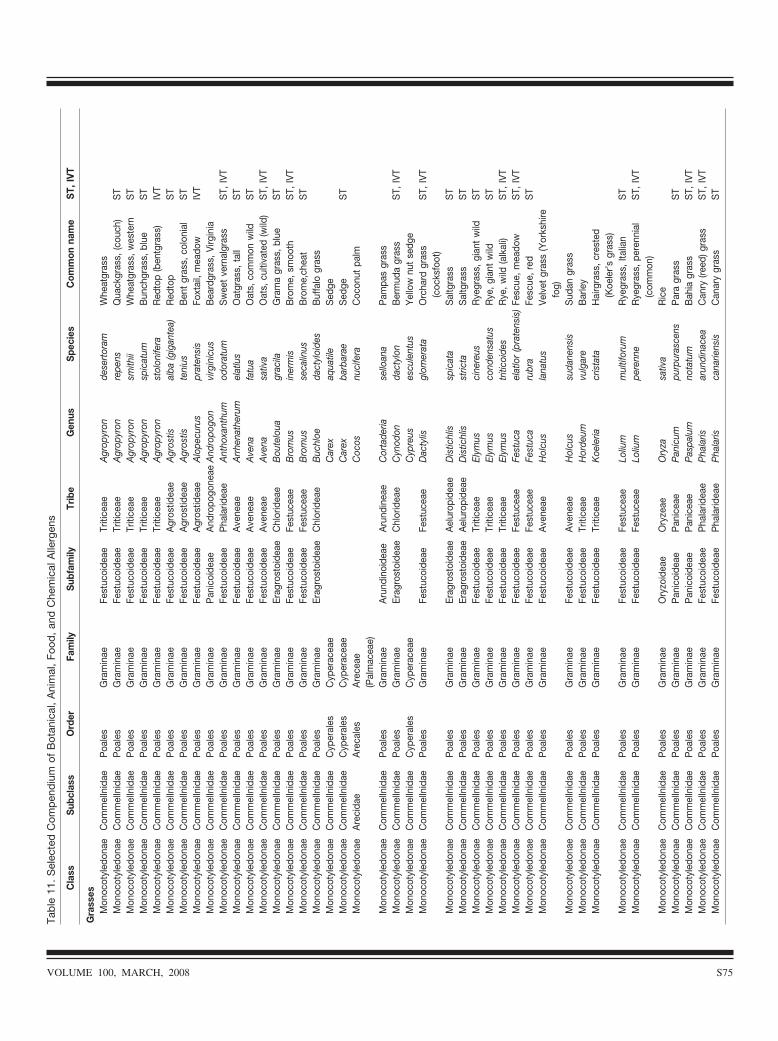
Tab
le11
.S
elec
ted
Com
pen
diu
mof
Bot
anic
al,
Ani
mal
,Fo
od,
and
Che
mic
alA
llerg
ens
Cla
ssS
ubcl
ass
Ord
erFa
mily
Sub
fam
ilyTr
ibe
Gen
usS
peci
esC
omm
onna
me
ST,
IVT
Gra
sses
Mon
ocot
yled
onae
Com
mel
lnid
aeP
oale
sG
ram
inae
Fest
ucoi
deae
Triti
ceae
Agr
opyr
onde
sert
oram
Whe
atgr
ass
Mon
ocot
yled
onae
Com
mel
lnid
aeP
oale
sG
ram
inae
Fest
ucoi
deae
Triti
ceae
Agr
opyr
onre
pens
Qua
ckgr
ass,
(cou
ch)
ST
Mon
ocot
yled
onae
Com
mel
lnid
aeP
oale
sG
ram
inae
Fest
ucoi
deae
Triti
ceae
Agr
opyr
onsm
ithii
Whe
atgr
ass,
wes
tern
ST
Mon
ocot
yled
onae
Com
mel
lnid
aeP
oale
sG
ram
inae
Fest
ucoi
deae
Triti
ceae
Agr
opyr
onsp
icat
umB
unch
gras
s,bl
ueS
TM
onoc
otyl
edon
aeC
omm
elln
idae
Poa
les
Gra
min
aeFe
stuc
oide
aeTr
itice
aeA
grop
yron
stol
onife
raR
edto
p(b
entg
rass
)IV
TM
onoc
otyl
edon
aeC
omm
elln
idae
Poa
les
Gra
min
aeFe
stuc
oide
aeA
gros
tidea
eA
gros
tisal
ba(g
igan
tea)
Red
top
ST
Mon
ocot
yled
onae
Com
mel
lnid
aeP
oale
sG
ram
inae
Fest
ucoi
deae
Agr
ostid
eae
Agr
ostis
teni
usB
ent
gras
s,co
loni
alS
TM
onoc
otyl
edon
aeC
omm
elln
idae
Poa
les
Gra
min
aeFe
stuc
oide
aeA
gros
tidea
eA
lope
curu
spr
aten
sis
Foxt
ail,
mea
dow
IVT
Mon
ocot
yled
onae
Com
mel
lnid
aeP
oale
sG
ram
inae
Pan
icoi
deae
And
ropo
gone
aeA
ndro
pogo
nvi
rgin
icus
Bea
rdgr
ass,
Virg
inia
Mon
ocot
yled
onae
Com
mel
lnid
aeP
oale
sG
ram
inae
Fest
ucoi
deae
Pha
larid
eae
Ant
hoxa
nthu
mod
orat
umS
wee
tve
rnal
gras
sS
T,IV
TM
onoc
otyl
edon
aeC
omm
elln
idae
Poa
les
Gra
min
aeFe
stuc
oide
aeA
vene
aeA
rrhe
nath
erum
elat
lus
Oat
gras
s,ta
llS
TM
onoc
otyl
edon
aeC
omm
elln
idae
Poa
les
Gra
min
aeFe
stuc
oide
aeA
vene
aeA
vena
fatu
aO
ats,
com
mon
wild
ST
Mon
ocot
yled
onae
Com
mel
lnid
aeP
oale
sG
ram
inae
Fest
ucoi
deae
Ave
neae
Ave
nasa
tiva
Oat
s,cu
ltiva
ted
(wild
)S
T,IV
TM
onoc
otyl
edon
aeC
omm
elln
idae
Poa
les
Gra
min
aeEr
agro
stoi
deae
Chl
orid
eae
Bou
telo
uagr
acila
Gra
ma
gras
s,bl
ueS
TM
onoc
otyl
edon
aeC
omm
elln
idae
Poa
les
Gra
min
aeFe
stuc
oide
aeFe
stuc
eae
Bro
mus
iner
mis
Bro
me,
smoo
thS
T,IV
TM
onoc
otyl
edon
aeC
omm
elln
idae
Poa
les
Gra
min
aeFe
stuc
oide
aeFe
stuc
eae
Bro
mus
seca
linus
Bro
me,
chea
tS
TM
onoc
otyl
edon
aeC
omm
elln
idae
Poa
les
Gra
min
aeEr
agro
stoi
deae
Chl
orid
eae
Buc
hloe
dact
yloi
des
Buf
falo
gras
sM
onoc
otyl
edon
aeC
omm
elln
idae
Cyp
eral
esC
yper
acea
eC
arex
aqua
tile
Sed
geM
onoc
otyl
edon
aeC
omm
elln
idae
Cyp
eral
esC
yper
acea
eC
arex
barb
arae
Sed
geS
TM
onoc
otyl
edon
aeA
reci
dae
Are
cale
sA
rece
ae(P
alm
acea
e)C
ocos
nuci
fera
Coc
onut
palm
Mon
ocot
yled
onae
Com
mel
lnid
aeP
oale
sG
ram
inae
Aru
ndin
oide
aeA
rund
inea
eC
orta
deria
sello
ana
Pam
pas
gras
sM
onoc
otyl
edon
aeC
omm
elln
idae
Poa
les
Gra
min
aeEr
agro
stoi
deae
Chl
orid
eae
Cyn
odon
dact
ylon
Ber
mud
agr
ass
ST,
IVT
Mon
ocot
yled
onae
Com
mel
lnid
aeC
yper
ales
Cyp
erac
eae
Cyp
reus
escu
lent
usY
ello
wnu
tse
dge
Mon
ocot
yled
onae
Com
mel
lnid
aeP
oale
sG
ram
inae
Fest
ucoi
deae
Fest
ucea
eD
acty
lisgl
omer
ata
Orc
hard
gras
s(c
ocks
foot
)S
T,IV
T
Mon
ocot
yled
onae
Com
mel
lnid
aeP
oale
sG
ram
inae
Erag
rost
oide
aeA
elur
opid
eae
Dis
tichl
issp
icat
aS
altg
rass
ST
Mon
ocot
yled
onae
Com
mel
lnid
aeP
oale
sG
ram
inae
Erag
rost
oide
aeA
elur
opid
eae
Dis
tichl
isst
ricta
Sal
tgra
ssS
TM
onoc
otyl
edon
aeC
omm
elln
idae
Poa
les
Gra
min
aeFe
stuc
oide
aeTr
itice
aeEl
ymus
cine
reus
Rye
gras
s,gi
ant
wild
ST
Mon
ocot
yled
onae
Com
mel
lnid
aeP
oale
sG
ram
inae
Fest
ucoi
deae
Triti
ceae
Elym
usco
nden
satu
sR
ye,g
iant
wild
ST
Mon
ocot
yled
onae
Com
mel
lnid
aeP
oale
sG
ram
inae
Fest
ucoi
deae
Triti
ceae
Elym
ustr
itico
ides
Rye
,wild
(alk
ali)
ST,
IVT
Mon
ocot
yled
onae
Com
mel
lnid
aeP
oale
sG
ram
inae
Fest
ucoi
deae
Fest
ucea
eFe
stuc
ael
atlo
r(p
rate
nsis
)Fe
scue
,mea
dow
ST,
IVT
Mon
ocot
yled
onae
Com
mel
lnid
aeP
oale
sG
ram
inae
Fest
ucoi
deae
Fest
ucea
eFe
stuc
aru
bra
Fesc
ue,r
edS
TM
onoc
otyl
edon
aeC
omm
elln
idae
Poa
les
Gra
min
aeFe
stuc
oide
aeA
vene
aeH
olcu
sla
natu
sVe
lvet
gras
s(Y
orks
hire
fog)
Mon
ocot
yled
onae
Com
mel
lnid
aeP
oale
sG
ram
inae
Fest
ucoi
deae
Ave
neae
Hol
cus
suda
nens
isS
udan
gras
sM
onoc
otyl
edon
aeC
omm
elln
idae
Poa
les
Gra
min
aeFe
stuc
oide
aeTr
itice
aeH
orde
umvu
lgar
eB
arle
yM
onoc
otyl
edon
aeC
omm
elln
idae
Poa
les
Gra
min
aeFe
stuc
oide
aeTr
itice
aeK
oele
riacr
ista
taH
airg
rass
,cre
sted
(Koe
ler’s
gras
s)M
onoc
otyl
edon
aeC
omm
elln
idae
Poa
les
Gra
min
aeFe
stuc
oide
aeFe
stuc
eae
Loliu
mm
ultif
orum
Rye
gras
s,Ita
lian
ST
Mon
ocot
yled
onae
Com
mel
lnid
aeP
oale
sG
ram
inae
Fest
ucoi
deae
Fest
ucea
eLo
lium
pere
nne
Rye
gras
s,pe
renn
ial
(com
mon
)S
T,IV
T
Mon
ocot
yled
onae
Com
mel
lnid
aeP
oale
sG
ram
inae
Ory
zoid
eae
Ory
zeae
Ory
zasa
tiva
Ric
eM
onoc
otyl
edon
aeC
omm
elln
idae
Poa
les
Gra
min
aeP
anic
oide
aeP
anic
eae
Pan
icum
purp
uras
cens
Par
agr
ass
ST
Mon
ocot
yled
onae
Com
mel
lnid
aeP
oale
sG
ram
inae
Pan
icoi
deae
Pan
icea
eP
aspa
lum
nota
tum
Bah
iagr
ass
ST,
IVT
Mon
ocot
yled
onae
Com
mel
lnid
aeP
oale
sG
ram
inae
Fest
ucoi
deae
Pha
larid
eae
Pha
laris
arun
dina
cea
Can
ry(re
ed)g
rass
ST,
IVT
Mon
ocot
yled
onae
Com
mel
lnid
aeP
oale
sG
ram
inae
Fest
ucoi
deae
Pha
larid
eae
Pha
laris
cana
riens
isC
anar
ygr
ass
ST
VOLUME 100, MARCH, 2008 S75
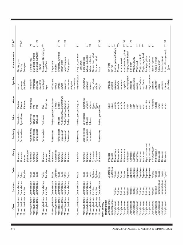
Cla
ssS
ubcl
ass
Ord
erFa
mily
Sub
fam
ilyTr
ibe
Gen
usS
peci
esC
omm
onna
me
ST,
IVT
Mon
ocot
yled
onae
Com
mel
lnid
aeP
oale
sG
ram
inae
Fest
ucoi
deae
Pha
larid
eae
Pha
laris
min
orC
anar
ygr
ass
Mon
ocot
yled
onae
Com
mel
lnid
aeP
olae
sG
ram
inae
Fest
ucoi
deae
Agr
ostid
eae
Phl
eum
prat
ense
Tim
othy
ST,
IVT
Mon
ocot
yled
onae
Are
cida
eA
reca
les
Are
cae
(Pal
mac
eae)
Pho
enix
dact
ylife
raD
ate
palm
Mon
ocot
yled
onae
Com
mel
lnid
aeP
oale
sG
ram
inae
Aru
ndin
oide
aeA
rund
inea
eP
hrag
mite
sco
mm
unis
Com
mon
reed
IVT
Mon
ocoy
tledo
nae
Com
mel
lnid
aeP
oale
sG
ram
inae
Fest
ucoi
deae
Fest
ucea
eP
oaco
mpr
essa
Blu
egra
ss,C
anad
aS
TM
onoc
oytle
dona
eC
omm
elln
idae
Poa
les
Gra
min
aeFe
stuc
oide
aeFe
stuc
eae
Poa
prat
ensi
s(p
alus
tris
)B
lueg
rass
,Ken
tuck
y(J
une)
ST,
IVT
Mon
ocoy
tledo
nae
Com
mel
lnid
aeP
oale
sG
ram
inae
Fest
ucoi
deae
Fest
ucea
eP
oasa
ndbe
rgii
Blu
egra
ass,
San
dber
g’s
ST
Mon
ocoy
tledo
nae
Are
cida
eA
reca
les
Are
ceae
(Pal
mac
eae)
Roy
ston
eare
gia
Roy
alP
alm
Mon
ocoy
tledo
nae
Com
mel
lnid
aeP
oale
sG
ram
inae
Pan
icoi
deae
And
ropo
gone
aeS
acch
arum
offic
inar
umS
ugar
cane
Mon
ocoy
tledo
nae
Com
mel
lnid
aeC
yper
ales
Cyp
erac
eae
Sci
rpus
valid
usB
ulru
shM
onoc
otyl
edon
aeC
omm
elln
idae
Poa
les
Gra
min
aeFe
stuc
iode
aeTr
itice
aeS
ecal
ece
real
eR
yegr
ass,
culti
vate
dS
T,IV
TM
onoc
otyl
edon
aeC
omm
elln
idae
Poa
les
Gra
min
aeP
anic
oide
aeA
ndro
pogo
noea
Sor
ghas
trum
nuta
nsIn
dian
gras
sM
onoc
otyl
edon
aeC
omm
elln
idae
Pol
aes
Gra
min
aeP
anic
oide
aeA
ndro
pogo
noea
Sor
ghum
hale
pens
eJo
hnso
ngr
ass
ST,
IVT
Mon
ocot
yled
onae
Com
mel
lnid
aeP
oale
sG
ram
inae
Pan
icoi
deae
And
ropo
gone
aeS
orgh
umsu
dane
nse
(vul
gare
-su
dane
nse)
Sud
angr
ass
Mon
ocot
yled
onae
Com
mel
lnid
aeP
oale
sG
ram
inae
Pan
icoi
deae
And
ropo
gone
aeS
orgh
umvu
igar
e(bi
colo
r)S
orgh
um,c
omm
oncu
ltiva
ted
ST
Mon
ocoy
tledo
nae
Com
mel
lnid
aeP
oale
sG
ram
inae
Erag
osto
idea
eS
poro
bole
aeS
poro
bolu
scr
ypta
ndru
sS
and
drop
seed
Mon
ocoy
tledo
nae
Com
mel
lnid
aeP
oale
sG
ram
inae
Fest
ucoi
deae
Triti
ceae
Triti
cum
aest
ivum
Whe
at,c
ultiv
ated
ST
Mon
ocoy
tledo
nae
Com
mel
lnid
aeP
oale
sG
ram
inae
Fest
ucoi
deae
Triti
ceae
Triti
cum
sativ
umW
heat
,cul
tivat
edIV
TM
onoc
oytle
dona
eA
reci
dae
Typh
ales
Typh
acea
eTy
pha
angu
stifo
llaN
arro
wLe
afca
ttai
lM
onoc
oytle
dona
eA
reci
dae
Typh
ales
Typh
acea
eTy
pha
latif
olla
Com
mon
catt
ail
Mon
ocoy
tledo
nae
Com
mel
lnid
aeP
oale
sG
ram
inae
Pan
icoi
deae
And
ropo
gone
aeZe
am
ays
Cor
nS
T,IV
TTr
ees,
shru
bs,
rela
ted
plan
tsG
ymno
sper
mae
Con
ifera
les
Pin
acea
eA
bies
conc
olor
Fir,
whi
teS
TG
ymno
sper
mae
Con
ifera
les
Pin
acea
eA
bies
proc
era
Fir,
red
ST
Dic
otyl
edon
aeR
osid
aeFa
bale
sM
imos
acea
eA
caci
aba
ileya
naA
caci
a,go
lden
(Bai
ley’
s)S
TD
icot
yled
onae
Ros
idae
Faba
les
Mim
osac
eae
Aca
cia
deal
bata
Mim
osa
STs
pD
icot
yled
onae
Ros
idae
Faba
les
Mim
osac
eae
Aca
cia
farn
esia
naA
caci
a,sw
eet
Dic
otyl
edon
aeR
osid
aeFa
bale
sM
imos
acea
eA
caci
alo
ngfo
liaA
caci
a(w
attle
),go
lden
ST,
IVT
Dic
otyl
edon
aeR
osid
aeS
apin
dale
sA
cera
ceae
Ace
rm
acro
phyl
lum
Map
le,c
oast
(big
leaf
)S
TD
icot
yled
onae
Ros
idae
Sap
inda
les
Ace
race
aeA
cer
negu
ndo
Box
elde
rS
T,IV
TD
icot
yled
onae
Ros
idae
Sap
inda
les
Ace
race
aeA
cer
plat
anoi
des
Map
le,N
orw
ayD
icot
yled
onae
Ros
idae
Sap
inda
les
Ace
race
aeA
cer
rubr
umM
aple
,red
ST
Dic
otyl
edon
aeR
osid
aeS
apin
dale
sA
cera
ceae
Ace
rsa
ccha
rinum
Map
le,s
ilver
(sof
t)S
TD
icot
yled
onae
Ros
idae
Sap
inda
les
Ace
race
aeA
cer
sacc
haru
mM
aple
,sug
ar(h
ard)
ST
Dic
otyl
edon
aeR
osid
aeS
apin
dale
sH
ippo
cast
anac
eae
Aes
culu
sfla
vaB
ucke
ye,y
ello
wD
icot
yled
onae
Ros
idae
Sap
inda
les
Hip
poca
stan
acea
eA
escu
lus
hipp
ocas
tanu
mC
hest
nut,
hors
eS
TD
icot
yled
onae
Ros
idae
Rut
ales
Sim
arou
bace
aeA
llant
him
sal
tissi
ma
Tree
ofhe
aven
ST
Dic
otyl
edon
aeR
oasi
dae
Faba
les
Mim
osac
eae
Alb
izzi
aju
libris
sin
Aca
cia,
Per
sian
Dic
otyl
edon
aeH
amam
elld
idae
Faga
les
Bet
ulac
eae
Aln
uscr
ispa
Ald
er,g
reen
Dic
otyl
edon
aeH
amam
elld
idae
Faga
les
Bet
ulac
eae
Aln
usgl
utin
osa
Ald
er,E
urop
ean
Dic
otyl
edon
aeH
amam
elld
idae
Faga
les
Bet
ulac
eae
Aln
usin
cana
(tenu
ifolia
)A
lder
,whi
te(s
peck
led)
(gre
y)S
T,IV
T
S76 ANNALS OF ALLERGY, ASTHMA & IMMUNOLOGY
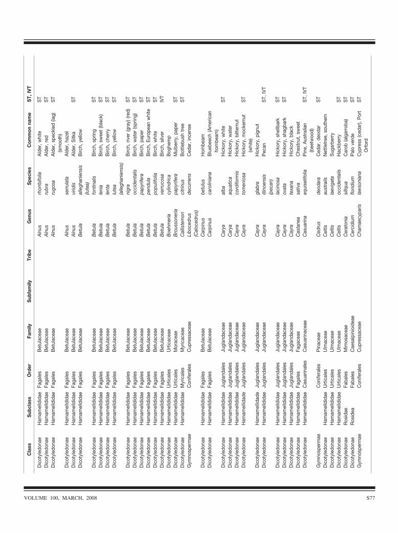
Cla
ssS
ubcl
ass
Ord
erFa
mily
Sub
fam
ilyTr
ibe
Gen
usS
peci
esC
omm
onna
me
ST,
IVT
Dic
otyl
edon
aeH
amam
elld
idae
Faga
les
Bet
ulac
eae
Aln
usrh
orrb
ifolia
Ald
er,w
hite
ST
Dic
otyl
edon
aeH
amam
elld
idae
Faga
les
Bet
ulac
eae
Aln
usru
bra
Ald
er,r
edS
TD
icot
yled
onae
Ham
amel
ldid
aeFa
gale
sB
etul
acea
eA
lnus
rugo
saA
lder
,spe
ckle
d(la
g)(s
moo
th)
ST
Dic
otyl
edon
aeH
amam
elld
idae
Faga
les
Bet
ulac
eae
Aln
usse
rrul
ata
Ald
er,h
azel
Dic
otyl
edon
aeH
amam
elld
idae
Faga
les
Bet
ulac
eae
Aln
usvi
ridis
Ald
er,S
itka
ST
Dic
otyl
edon
aeH
amam
elld
idae
Faga
les
Bet
ulac
eae
Bet
ula
alle
ghan
iens
is(lu
tea)
Birc
h,ye
llow
Dic
otyl
edon
aeH
amam
elld
idae
Faga
les
Bet
ulac
eae
Bet
ula
font
inal
isB
irch,
sprin
gS
TD
icot
yled
onae
Ham
amel
ldid
aeFa
gale
sB
etul
acea
eB
etul
ale
nia
Birc
h,sw
eet
(bla
ck)
ST
Dic
otyl
edon
aeH
amam
elld
idae
Faga
les
Bet
ulac
eae
Bet
ula
lent
aB
irch,
cher
ryS
TD
icot
yled
onae
Ham
amel
ldid
aeFa
gale
sB
etul
acea
eB
etul
alu
tea
(alle
ghan
iens
is)
Birc
h,ye
llow
ST
Dic
otyl
edon
aeH
amam
elld
idae
Faga
les
Bet
ulac
eae
Bet
ula
nigr
aB
irch,
river
(gra
y)(re
d)S
TD
icot
yled
onae
Ham
amel
ldid
aeFa
gale
sB
etul
acea
eB
etul
aoc
cide
ntal
isB
irch,
wat
er(s
prin
g)S
TD
icot
yled
onae
Ham
amel
ldid
aeFa
gale
sB
etul
acea
eB
etul
apa
pyrif
era
Birc
h,pa
per
ST
Dic
otyl
edon
aeH
amam
elld
idae
Faga
les
Bet
ulac
eae
Bet
ula
pend
ula
Birc
h,Eu
rope
anw
hite
ST
Dic
otyl
edon
aeH
amam
elld
idae
Faga
les
Bet
ulac
eae
Bet
ula
popu
lifol
iaB
irch,
whi
teS
TD
icot
yled
onae
Ham
amel
ldid
aeFa
gale
sB
etul
acea
eB
etul
ave
rruc
osa
Birc
h,si
lver
IVT
Dic
otyl
edon
aeH
amam
elld
idae
Urt
ical
esU
rtic
acea
eB
oehm
eria
cylin
dric
aB
oghe
mp
Dic
otyl
edon
aeH
amam
elld
idae
Urt
ical
esM
orac
eae
Bro
usso
neria
papy
rifer
aM
ulbe
rry,
pape
rS
TD
icot
yled
onae
Ham
amel
ldid
aeM
yric
ales
Myr
icac
eae
Cal
liste
mon
citr
inus
Bot
tlebu
shtr
eeS
TG
ymno
sper
mae
Con
ifera
les
Cup
ress
acea
eLi
boce
drus
(Cal
oced
rus)
decu
rren
sC
edar
,inc
ense
Dic
otyl
edon
aeH
amam
elld
idae
Faga
les
Bet
ulac
eae
Car
pinu
sbe
tulu
sH
ornb
eam
Dic
otyl
edon
aeH
amam
elld
idae
Faga
les
Bet
ulac
eae
Car
pinu
sca
rolin
iana
Blu
ebee
ch(A
mer
ican
horn
beam
)D
icot
yled
onae
Ham
amel
ldid
aeJu
glan
dale
sJu
glan
dace
aeC
arya
alba
Hic
kory
,whi
teS
TD
icot
yled
onae
Ham
amel
ldid
aeJu
glan
dale
sJu
glan
dace
aeC
arya
aqua
tica
Hic
kory
,wat
erD
icot
yled
onae
Ham
amel
ldid
aeJu
glan
dale
sJu
glan
dace
aeC
ayra
cord
iform
isH
icko
ry,b
itter
nut
Dic
otyl
edon
aeH
amam
ellid
ade
Jugl
anda
les
Jugl
anda
ceae
Cay
rato
men
iosa
Hic
kory
,moc
kern
ut(w
hite
)S
T
Dic
otyl
edon
aeH
amam
ellid
ade
Jugl
anda
les
Jugl
anda
ceae
Cay
ragl
abra
Hic
kory
,pig
nut
Dic
otyl
edon
aeH
amam
elld
idae
Jugl
anda
les
Jugl
anda
ceae
Cay
raill
inoe
nsis
(pec
an)
Pec
anS
T,IV
T
Dic
otyl
edon
aeH
amam
elld
idae
Jugl
anda
les
Jugl
anda
ceae
Cay
rala
cino
saH
icko
ry,s
hellb
ark
ST
Dic
otyl
edon
aeH
amam
elld
idae
Jugl
anda
les
Jugl
anda
ceae
Cay
raov
ata
Hic
kory
,sha
gbar
kS
TD
icot
yled
onae
Ham
amel
ldid
aeJu
glan
dale
sJu
glan
dace
aeC
ayra
texa
naH
icko
ry,b
lack
Dic
otyl
edon
aeH
amam
elld
idae
Faga
les
Faga
ceae
Cas
tane
asa
tiva
Che
stnu
t,sw
eet
Dic
otyl
edon
aeH
amam
elld
idae
Cas
uarin
ales
Cas
uarin
acea
eC
asua
rina
equi
setif
olia
Pin
e,A
ustr
alia
n(b
eetw
ood)
ST,
IVT
Gym
nosp
erm
aeC
onife
rale
sP
inac
eae
Ced
rus
deod
ara
Ced
ar,d
eoda
rS
TD
icot
yled
onae
Ham
amel
ldid
aeU
rtic
ales
Ulm
acea
eC
eltis
aust
rails
Net
tletr
ee,s
outh
ern
Dic
otyl
edon
aeH
amam
elld
idae
Urt
ical
esU
lmac
eae
Cel
tisla
evig
ata
Sug
arbe
rry
Dic
otyl
edon
aeH
amam
elld
idae
Urt
ical
esU
lmac
eae
Cel
tisoc
cide
ntai
lsH
ackb
erry
ST
Dic
otyl
edon
aeR
osid
aeFa
bale
sM
imos
acea
eC
erat
onia
sillq
uaC
arob
(alg
arro
ba)
ST
Dic
otyl
edon
aeR
osid
eaFa
bale
sC
aesa
lpin
ioid
eae
Cer
cidi
umflo
ridiu
mP
alo
verd
eS
TG
ymno
sper
mae
Con
ifera
les
Cup
ress
acea
eC
ham
aecy
paris
law
soni
ana
Cyp
ress
(ced
ar),
Por
tO
rfor
dS
T
VOLUME 100, MARCH, 2008 S77
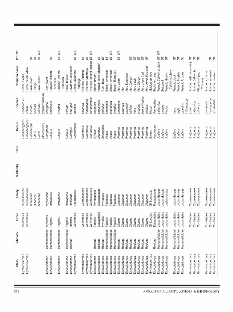
Cla
ssS
ubcl
ass
Ord
erFa
mily
Sub
fam
ilyTr
ibe
Gen
usS
peci
esC
omm
onna
me
ST,
IVT
Gym
nosp
erm
aeC
onife
rale
sC
upre
ssac
eae
Cha
mae
cypa
risno
otka
tens
isC
edar
,Ala
ska
Gym
nosp
erm
aeC
onife
rale
sC
upre
ssac
eae
Cha
mae
cypa
risob
tusa
Ced
ar,h
inok
lwhi
teA
reca
ceae
Cha
mae
rops
hum
ulis
Pal
m,d
war
fS
TR
utac
eae
Citr
ussi
nens
isO
rang
eS
TA
reca
ceae
Coc
os(A
reca
stru
m)
plum
osa
(rom
anzo
ffian
um)
Pal
m,q
ueen
ST,
IVT
Dic
otyl
edon
aeH
amam
elld
idae
Myr
ical
esM
yric
acea
eC
ompt
onia
pere
grin
aFe
rn,s
wee
tD
icot
yled
onae
Ham
amel
ldid
aeFa
gale
sB
etul
acea
eC
oryl
usam
eric
ana
Haz
elnu
t(fi
lber
t),A
mer
ican
ST
Dic
otyl
edon
aeH
amam
elld
idae
Faga
les
Bet
ulac
eae
Cor
ylus
avel
lana
Haz
elnu
t,(fi
lber
t)Eu
rope
anS
T,IV
T
Dic
otyl
edon
aeH
amam
elld
idae
Faga
les
Bet
ulac
eae
Cor
ylus
com
uta
Haz
el,b
eake
dD
icot
yled
onae
Ros
idae
Ros
ales
Ros
acea
eC
rate
gus
crus
-gal
liH
awth
orn,
cock
spur
Gym
nosp
erm
aeC
onife
rale
sTa
xodi
acea
eC
rypt
omer
iaja
poni
caC
edar
,Jap
anes
ere
d(su
gi)
ST,
IVT
Gym
nosp
erm
aeC
onife
rale
sC
upre
ssac
eae
Cup
ress
usar
izon
ica
Cyp
ress
,Ariz
ona
ST
Gym
nosp
erm
aeC
onife
rale
sC
upre
ssac
eae
Cup
ress
usm
acro
carp
aC
ypre
ss,M
onte
rey
ST
Gym
nosp
erm
aeC
onife
rale
sC
upre
ssac
eae
Cup
ress
usse
mpe
rvire
nsC
ypre
ss,f
uner
al(It
alia
n)S
T,IV
TD
icot
yled
onae
Ros
idae
Faba
les
Mim
osac
eae
Cyt
isus
scop
ariu
sS
cotc
hbr
oom
Dic
otyl
edon
aeR
osid
aeEl
aeag
naie
sEl
aeag
nace
aeEl
aegn
usan
gust
ifolia
Rus
sian
oliv
e(o
leas
ter)
ST
Dic
otyl
edon
aeR
osid
aeM
yrta
les
Myr
tace
aeEu
caly
ptus
glob
ulus
Gum
,blu
eS
TD
icot
yled
onae
Ham
amel
ldid
aeFa
gale
sFa
gace
aeFa
gus
amer
ican
aB
eech
,Am
eric
anS
TD
icot
yled
onae
Ham
amel
ldid
aeFa
gale
sFa
gace
aeFa
gus
gran
difo
liaB
eech
,Am
eric
anS
T,IV
TD
icot
yled
onae
Ham
amel
ldid
aeFa
gale
sFa
gace
aeFa
gus
sylv
atic
aB
eech
,Eur
opea
nD
icot
yled
onae
Ros
idae
Ole
ales
Ole
acea
eFr
anxi
nus
amer
ican
aA
sh,w
hite
ST,
IVT
Dic
otyl
edon
aeR
osid
aeO
leal
esO
leac
eae
Fran
xinu
sar
izon
ica
Ash
Dic
otyl
edon
aeR
osid
aeO
leal
esO
leac
eae
Fran
xinu
sex
cels
ior
Ash
,Eur
opea
nD
icot
yled
onae
Ros
idae
Ole
ales
Ole
acea
eFr
anxi
nus
latif
olia
Ash
,Ore
gon
ST
Dic
otyl
edon
aeR
osid
aeO
leal
esO
leac
eae
Fran
xinu
sni
gra
Ash
,bla
ckS
TD
icot
yled
onae
Ros
idae
Ole
ales
Ole
acea
eFr
anxi
nus
oreg
ona
Ash
,Ore
gon
ST
Dic
otyl
edon
aeR
osid
aeO
leal
esO
leac
eae
Fran
xinu
spe
nnsy
lvan
ica
Ash
,gre
en(re
d)S
TD
icot
yled
onae
Ros
idae
Ole
ales
Ole
acea
eFr
anxi
nus
velu
tina
Ash
,vel
vet
(Ariz
ona)
ST
Gym
nosp
erm
aeG
inkg
oale
sG
inkg
oace
aeG
inkg
obi
loba
Mai
denh
air
tree
Dic
otyl
edon
aeR
osid
aeEl
aeag
nale
sEl
aeag
nace
aeH
ippo
phae
rham
noid
esB
uckt
horn
,sea
Dic
otyl
edon
aeH
amam
elld
idae
Jugl
anda
les
Jugl
anda
ceae
Jugl
ans
calif
orni
caW
alnu
t,C
alifo
rnia
blac
kS
T,IV
TD
icot
yled
onae
Ham
amel
ldid
aeJu
glan
dale
sJu
glan
dace
aeJu
glan
sci
nere
aB
utte
rnut
ST
Dic
otyl
edon
aeH
amam
elld
idae
Jugl
anda
les
Jusl
anda
ceae
Jugl
ans
hind
sii
Wal
nut,
Hin
d’s
Cal
iforn
iabl
ack
ST
Dic
otyl
edon
aeH
amam
elld
idae
Jugl
anda
les
Jugl
anda
ceae
Jugl
ans
nigr
aW
alnu
t,bl
ack
ST
Dic
otyl
edon
aeH
amam
elld
idae
Jugl
anda
les
Jugl
anda
ceae
Jugl
ans
regi
aW
alnu
t,En
glis
hS
TD
icot
yled
onae
Ham
amel
ldid
aeJu
glan
dale
sJu
glan
dace
aeJu
glan
sru
pest
ris(m
icro
carp
a)W
alnu
t,A
rizon
aS
t
Gym
nosp
erm
aeC
onife
rale
sC
upre
ssac
eae
Juni
peru
sas
hel
Juni
per,
ash
(mou
ntai
n)S
TG
ymno
sper
mae
Con
ifera
les
Cup
ress
acea
eJu
nipe
rus
calif
orni
caJu
nipe
rC
alifo
rnia
ST
Gym
nosp
erm
aeC
onife
rale
sC
upre
ssac
eae
Juni
peru
sch
inen
sis
Pfit
zer
(Jun
iper
,C
hine
se)
ST
Gym
nosp
erm
aeC
onife
rale
sC
upre
ssac
eae
Juni
peru
sco
mm
unis
Juni
per,
com
mon
Gym
nosp
erm
aeC
onife
rale
sC
upre
ssac
eae
Juni
peru
sm
onos
perm
aJu
nipe
r,on
esee
dS
TG
ymno
sper
mae
Con
ifera
les
Cup
ress
acea
eJu
nipe
rus
occi
dent
ails
Juni
per,
wes
tern
ST
S78 ANNALS OF ALLERGY, ASTHMA & IMMUNOLOGY
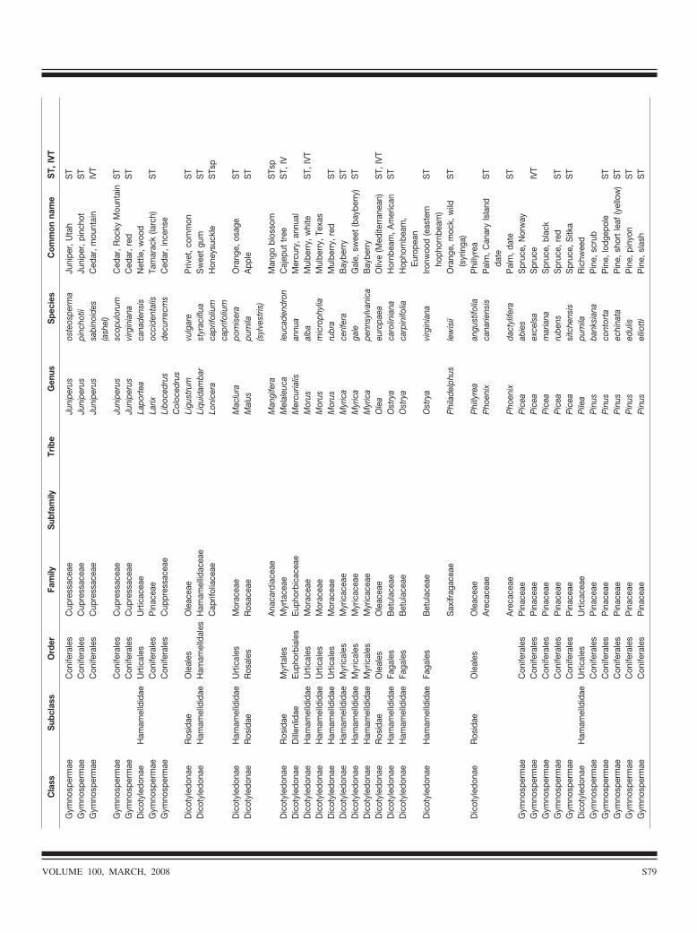
Cla
ssS
ubcl
ass
Ord
erFa
mily
Sub
fam
ilyTr
ibe
Gen
usS
peci
esC
omm
onna
me
ST,
IVT
Gym
nosp
erm
aeC
onife
rale
sC
upre
ssac
eae
Juni
peru
sos
teos
perm
aJu
nipe
r,U
tah
ST
Gym
nosp
erm
aeC
onife
rale
sC
upre
ssac
eae
Juni
peru
spi
ncho
tiiJu
nipe
r,pi
ncho
tS
TG
ymno
sper
mae
Con
ifera
les
Cup
ress
acea
eJu
nipe
rus
sabi
noid
es(a
shel
)C
edar
,mou
ntai
nIV
T
Gym
nosp
erm
aeC
onife
rale
sC
upre
ssac
eae
Juni
peru
ssc
opul
orum
Ced
ar,R
ocky
Mou
ntai
nS
TG
ymno
sper
mae
Con
ifera
les
Cup
ress
acea
eJu
nipe
rus
virg
inia
naC
edar
,red
ST
Dic
otyl
edon
aeH
amam
elld
idae
Urt
ical
esU
rtic
acea
eLa
port
eaca
nade
nsis
Net
tle,w
ood
Gym
nosp
erm
aeC
onife
rale
sP
inac
eae
Larix
occi
dent
alis
Tam
arac
k(la
rch)
ST
Gym
nosp
erm
aeC
onife
rale
sC
uppr
essa
ceae
Libo
cedr
usC
oloc
edru
sde
curr
ecm
sC
edar
,inc
ense
Dic
otyl
edon
aeR
osid
aeO
leal
esO
leac
eae
Ligu
stru
mvu
lgar
eP
rivet
,com
mon
ST
Dic
otyl
edon
aeH
amam
elld
idae
Ham
amel
ldal
esH
amam
ellid
acea
eLi
quid
amba
rst
yrac
iflua
Sw
eet
gum
ST
Cap
rifol
iace
aeLo
nice
raca
prifo
lium
capr
ifoliu
mH
oney
suck
leS
Tsp
Dic
otyl
edon
aeH
amam
elld
idae
Urt
ical
esM
orac
eae
Mac
lura
pom
iser
aO
rang
e,os
age
ST
Dic
otyl
edon
aeR
osid
aeR
osal
esR
osac
eae
Mal
uspu
mila
(syl
vest
ris)
App
leS
T
Ana
card
iace
aeM
angi
fera
Man
gobl
osso
mS
Tsp
Dic
otyl
edon
aeR
osid
aeM
yrta
les
Myr
tace
aeM
elal
euca
leuc
aden
dron
Caj
eput
tree
ST,
IVD
icot
yled
onae
Dill
enlid
aeEu
phor
bial
esEu
phor
bica
ceae
Mer
curia
lisan
nua
Mer
cury
,ann
ual
Dic
otyl
edon
aeH
amam
elld
idae
Urt
ical
esM
orac
eae
Mor
usal
baM
ulbe
rry,
whi
teS
T,IV
TD
icot
yled
onae
Ham
amel
ldid
aeU
rtic
ales
Mor
acea
eM
orus
mic
roph
ylia
Mul
berr
y,Te
xas
Dic
otyl
edon
aeH
amam
elld
idae
Urt
ical
esM
orac
eae
Mor
usru
bra
Mul
berr
y,re
dS
TD
icot
yled
onae
Ham
amel
ldid
aeM
yric
ales
Myr
icac
eae
Myr
ica
cerif
era
Bay
berr
yS
TD
icot
yled
onae
Ham
amel
ldid
aeM
yric
ales
Myr
icac
eae
Myr
ica
gale
Gal
e,sw
eet
(bay
berr
y)S
TD
icot
yled
onae
Ham
amel
ldid
aeM
yric
ales
Myr
icac
eae
Myr
ica
penn
sylv
anic
aB
aybe
rry
Dic
otyl
edon
aeR
osid
aeO
leal
esO
leac
eae
Ole
aeu
ropa
eaO
live
(Med
iterr
anea
n)S
T,IV
TD
icot
yled
onae
Ham
amel
ldid
aeFa
gale
sB
etul
acea
eO
stry
aca
rolin
iana
Hor
nbea
m,A
mer
ican
ST
Dic
otyl
edon
aeH
amam
elld
idae
Faga
les
Bet
ulac
eae
Ost
rya
carp
inifo
liaH
opho
rnbe
am,
Euro
pean
Dic
otyl
edon
aeH
amam
elld
idae
Faga
les
Bet
ulac
eae
Ost
rya
virg
inia
naIro
nwoo
d(e
aste
rnho
phor
nbea
m)
ST
Sax
ifrag
acea
eP
hila
delp
hus
lew
isii
Ora
nge,
moc
k,w
ild(s
yrin
ga)
ST
Dic
otyl
edon
aeR
osid
aeO
leal
esO
leac
eae
Phi
llyre
aan
gust
ifolia
Phi
llyre
aA
reca
ceae
Pho
enix
cana
riens
isP
alm
,Can
ary
Isla
ndda
teS
T
Are
cace
aeP
hoen
ixda
ctyl
ifera
Pal
m,d
ate
ST
Gym
nosp
erm
aeC
onife
rale
sP
inac
eae
Pic
eaab
ies
Spr
uce,
Nor
way
Gym
nosp
erm
aeC
onife
rale
sP
inac
eae
Pic
eaex
cels
aS
pruc
eIV
TG
ymno
sper
mae
Con
ifera
les
Pin
acea
eP
icea
mar
iana
Spr
uce,
blac
kG
ymno
sper
mae
Con
ifera
les
Pin
acea
eP
icea
rube
nsS
pruc
e,re
dS
TG
ymno
sper
mae
Con
ifera
les
Pin
acea
eP
icea
sitc
hens
isS
pruc
e,S
itka
ST
Dic
otyl
edon
aeH
amam
elld
idae
Urt
ical
esU
rtic
acea
eP
ilea
pum
ilaR
ichw
eed
Gym
nosp
erm
aeC
onife
rale
sP
inac
eae
Pin
usba
nksi
ana
Pin
e,sc
rub
Gym
nosp
erm
aeC
onife
rale
sP
inac
eae
Pin
usco
ntor
taP
ine,
lodg
epol
eS
TG
ymno
sper
mae
Con
ifera
les
Pin
acea
eP
inus
echi
nata
Pin
e,sh
ort
leaf
(yel
low
)S
TG
ymno
sper
mae
Con
ifera
les
Pin
acea
eP
inus
edul
isP
ine,
piny
onS
TG
ymno
sper
mae
Con
ifera
les
Pin
acea
eP
inus
ellio
tti
Pin
e,sl
ash
ST
VOLUME 100, MARCH, 2008 S79
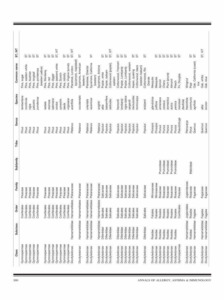
Cla
ssS
ubcl
ass
Ord
erFa
mily
Sub
fam
ilyTr
ibe
Gen
usS
peci
esC
omm
onna
me
ST,
IVT
Gym
nosp
erm
aeC
onife
rale
sP
inac
eae
Pin
usla
mbe
rtia
naP
ine,
suga
rG
ymno
sper
mae
Con
ifera
les
Pin
acea
eP
inus
mon
ticol
aP
ine,
wes
tern
whi
teS
TG
ymno
sper
mae
Con
ifera
les
Pin
acea
eP
inus
nigr
aP
ine,
Aus
tria
nS
TG
ymno
sper
mae
Con
ifera
les
Pin
acea
eP
inus
palu
stris
Pin
e,lo
ngle
afS
TG
ymno
sper
mae
Con
ifera
les
Pin
acea
eP
inus
pond
eros
aP
ine,
pond
eros
a(w
este
rnye
llow
)S
T
Gym
nosp
erm
aeC
onife
rale
sP
inac
eae
Pin
usra
diat
aP
ine,
Mon
tere
yS
TG
ymno
sper
mae
Con
ifera
les
Pin
acea
eP
inus
resi
nosa
Pin
e,re
dS
TG
ymno
sper
mae
Con
ifera
les
Pin
acea
eP
inus
sabi
nian
aP
ine,
digg
erS
TG
ymno
sper
mae
Con
ifera
les
Pin
acea
eP
inus
stro
bis
Pin
e,(e
aste
rn)w
hite
ST,
IVT
Gym
nosp
erm
aeC
onife
rale
sP
inac
eae
Pin
ussy
lves
tris
Pin
e,S
cotc
hG
ymno
sper
mae
Con
ifera
les
Pin
acea
eP
inus
taed
aP
ine,
lobl
olly
ST
Gym
nosp
erm
aeC
onife
rale
sP
inac
eae
Pin
usvi
rgin
iana
Pin
e,Vi
rgin
ia(s
crub
)S
TD
icot
yled
onae
Ham
amel
ldid
aeH
amam
elld
ales
Pla
tana
ceae
Pla
tanu
sac
erifo
liaP
lane
tree
,Lon
don
(syc
amor
e,m
aple
leaf
)S
T,IV
T
Dic
otyl
edon
aeH
amam
elld
idae
Ham
amel
ldal
esP
lata
nace
aeP
lata
nus
occi
dent
alis
Syc
amor
e,A
mer
ican
(eas
tern
)S
T
Dic
otyl
edon
aeH
amam
elld
idae
Ham
amel
ldal
esP
lata
nace
aeP
lata
nus
orie
ntal
isP
lane
tree
,Orie
ntal
Dic
otyl
edon
aeH
amam
elld
idae
Ham
amel
ldal
esP
lata
nace
aeP
lata
nus
race
mos
aS
ycam
ore,
Cal
iforn
ia(w
este
rn)
ST
Dic
otyl
edon
aeH
amam
elld
idae
Ham
amel
ldal
esP
lata
nace
aeP
lata
nus
wrig
htii
Syc
amor
e,A
rizon
aD
icot
yled
onae
Dill
enlid
aeS
alic
ales
Sal
icac
eae
Pop
ulus
alba
Pop
lar,
whi
teS
TD
icot
yled
onae
Dill
enlid
aeS
alic
ales
Sal
icac
eae
Pop
ulus
bals
amife
raP
opla
r,ba
lsam
ST
Dic
otyl
edon
aeD
illen
lidae
Sal
ical
esS
alic
acea
eP
opul
usde
ltoid
esC
otto
nwoo
d(p
opla
r),ea
ster
nS
T,IV
T
Dic
otyl
edon
aeD
illen
lidae
Sal
ical
esS
alic
acea
eP
opul
usfr
emon
tiiC
otto
nwoo
d,Fr
emon
tS
TD
icot
yled
onae
Dill
enlid
aeS
alic
ales
Sal
icac
eae
Pop
ulus
lom
bard
yP
opla
r,Lo
mba
rdy
ST
Dic
otyl
edon
aeD
illen
lidae
Sal
ical
esS
alic
acea
eP
opul
usni
gra-
italic
aP
opla
r,Lo
mba
rdy
ST
Dic
otyl
edon
aeD
illen
lidae
Sal
ical
esS
alic
acea
eP
opul
ussa
rgen
tiiC
otto
nwoo
d,w
este
rnD
icot
yled
onae
Dill
enlid
aeS
alic
ales
Sal
icac
eae
Pop
ulus
trem
uloi
des
Asp
en,q
uaki
ngS
TD
icot
yled
onae
Dill
enlid
aeS
alic
ales
Sal
icac
eae
Pop
ulus
tric
hoca
rpa
Cot
tonw
ood,
blac
k(w
este
rnba
lsam
)S
T
Dic
otyl
edon
aeD
illen
lidae
Sal
ical
esS
alic
acea
eP
opul
usw
isliz
enii
Cot
tonw
ood,
Rio
Gra
nde
ST
Dic
otyl
edon
aeR
osid
aeFa
bale
sM
imos
acea
eP
roso
pis
glan
dulo
saM
esqu
iteD
icot
yled
onae
Ros
idae
Faba
les
Mim
osac
eae
Pro
sopi
sju
liflo
raM
esqu
iteS
TD
icot
yled
onae
Ros
idea
Ros
ales
Ros
acea
eP
runo
idea
eP
runu
sar
men
iaca
Apr
icot
ST
Dic
otyl
edon
aeR
osid
eaR
osal
esR
osac
eae
Pru
noid
eae
Pru
nus
cera
sus
Che
rry
ST
Dic
otyl
edon
aeR
osid
eaR
osal
esR
osac
eae
Pru
noid
eae
Pru
nus
dom
estic
aP
lum
(pru
ne)
ST
Dic
otyl
edon
aeR
osid
eaR
osal
esR
osac
eae
Pru
noid
eae
Pru
nus
dulc
isA
lmon
dS
TD
icot
yled
onae
Ros
idea
Ros
ales
Ros
acea
eP
runo
idea
eP
runu
spe
rsic
aP
each
ST
Gym
nosp
erm
aeC
onife
rale
sP
inac
eae
Pse
udot
suga
men
zies
ii(ta
xifo
lia)
Fir,
Dou
glas
ST
Dic
otyl
edon
aeH
amam
elld
idae
Jugl
anda
les
Jugl
anda
ceae
Pte
roca
rya
frax
inifo
liaW
ingn
utD
icot
yled
onae
Ros
idea
Ros
ales
Ros
acea
eM
aloi
deae
Pyr
usco
mm
unis
Pea
rS
TD
icot
yled
onae
Ham
amel
ldid
aeFa
gale
sFa
gace
aeQ
uerc
usag
rifol
iaO
ak,C
alifo
rnia
(coa
st)
live
ST
Dic
otyl
edon
aeH
amam
elld
idae
Faga
les
Faga
ceae
Que
rcus
alba
Oak
,whi
teS
T,IV
TD
icot
yled
onae
Ham
amel
ldid
aeFa
gale
sFa
gace
aeQ
uerc
usbi
colo
rO
ak,b
lue
S80 ANNALS OF ALLERGY, ASTHMA & IMMUNOLOGY
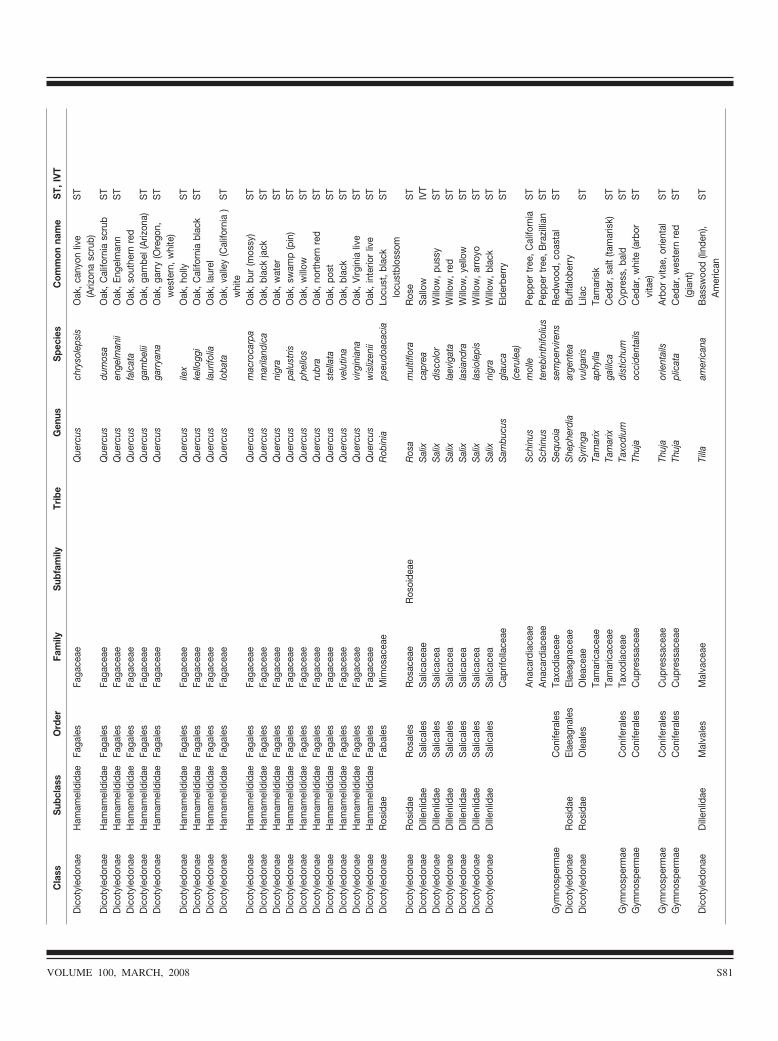
Cla
ssS
ubcl
ass
Ord
erFa
mily
Sub
fam
ilyTr
ibe
Gen
usS
peci
esC
omm
onna
me
ST,
IVT
Dic
otyl
edon
aeH
amam
elld
idae
Faga
les
Faga
ceae
Que
rcus
chry
sole
psis
Oak
,can
yon
live
(Ariz
ona
scru
b)S
T
Dic
otyl
edon
aeH
amam
elld
idae
Faga
les
Faga
ceae
Que
rcus
dum
osa
Oak
,Cal
iforn
iasc
rub
ST
Dic
otyl
edon
aeH
amam
elld
idae
Faga
les
Faga
ceae
Que
rcus
enge
lman
iiO
ak,E
ngel
man
nS
TD
icot
yled
onae
Ham
amel
ldid
aeFa
gale
sFa
gace
aeQ
uerc
usfa
lcat
aO
ak,s
outh
ern
red
Dic
otyl
edon
aeH
amam
elld
idae
Faga
les
Faga
ceae
Que
rcus
gam
belii
Oak
,gam
bel(
Ariz
ona)
ST
Dic
otyl
edon
aeH
amam
elld
idae
Faga
les
Faga
ceae
Que
rcus
garr
yana
Oak
,gar
ry(O
rego
n,w
este
rn,w
hite
)S
T
Dic
otyl
edon
aeH
amam
elld
idae
Faga
les
Faga
ceae
Que
rcus
ilex
Oak
,hol
lyS
TD
icot
yled
onae
Ham
amel
ldid
aeFa
gale
sFa
gace
aeQ
uerc
uske
llogg
iO
ak,C
alifo
rnia
blac
kS
TD
icot
yled
onae
Ham
amel
ldid
aeFa
gale
sFa
gace
aeQ
uerc
usla
urifo
liaO
ak,l
aure
lD
icot
yled
onae
Ham
amel
ldid
aeFa
gale
sFa
gace
aeQ
uerc
uslo
bata
Oak
,val
ley
(Cal
iforn
ia)
whi
teS
T
Dic
otyl
edon
aeH
amam
elld
idae
Faga
les
Faga
ceae
Que
rcus
mac
roca
rpa
Oak
,bur
(mos
sy)
ST
Dic
otyl
edon
aeH
amam
elld
idae
Faga
les
Faga
ceae
Que
rcus
mar
iland
ica
Oak
,bla
ckja
ckS
TD
icot
yled
onae
Ham
amel
ldid
aeFa
gale
sFa
gace
aeQ
uerc
usni
gra
Oak
,wat
erS
TD
icot
yled
onae
Ham
amel
ldid
aeFa
gale
sFa
gace
aeQ
uerc
uspa
lust
risO
ak,s
wam
p(p
in)
ST
Dic
otyl
edon
aeH
amam
elld
idae
Faga
les
Faga
ceae
Que
rcus
phel
los
Oak
,will
owS
TD
icot
yled
onae
Ham
amel
ldid
aeFa
gale
sFa
gace
aeQ
uerc
usru
bra
Oak
,nor
ther
nre
dS
TD
icot
yled
onae
Ham
amel
ldid
aeFa
gale
sFa
gace
aeQ
uerc
usst
ella
taO
ak,p
ost
ST
Dic
otyl
edon
aeH
amam
elld
idae
Faga
les
Faga
ceae
Que
rcus
velu
tina
Oak
,bla
ckS
TD
icot
yled
onae
Ham
amel
ldid
aeFa
gale
sFa
gace
aeQ
uerc
usvi
rgin
iana
Oak
,Virg
inia
live
ST
Dic
otyl
edon
aeH
amam
elld
idae
Faga
les
Faga
ceae
Que
rcus
wis
lizen
iiO
ak,i
nter
ior
live
ST
Dic
otyl
edon
aeR
osid
aeFa
bale
sM
imos
acea
eR
obin
iaps
eudo
acac
iaLo
cust
,bla
cklo
cust
blos
som
ST
Dic
otyl
edon
aeR
osid
aeR
osal
esR
osac
eae
Ros
oide
aeR
osa
mul
tiflo
raR
ose
ST
Dic
otyl
edon
aeD
illen
lidae
Sal
ical
esS
alic
acea
eS
alix
capr
eaS
allo
wIV
TD
icot
yled
onae
Dill
enlid
aeS
alic
ales
Sal
icac
eaS
alix
disc
olor
Will
ow,p
ussy
ST
Dic
otyl
edon
aeD
illen
lidae
Sal
ical
esS
alic
acea
Sal
ixla
evig
ata
Will
ow,r
edS
TD
icot
yled
onae
Dill
enlid
aeS
alic
ales
Sal
icac
eaS
alix
lasi
andr
aW
illow
,yel
low
ST
Dic
otyl
edon
aeD
illen
lidae
Sal
ical
esS
alic
acea
Sal
ixla
siol
epis
Will
ow,a
rroy
oS
TD
icot
yled
onae
Dill
enlid
aeS
alic
ales
Sal
icac
eaS
alix
nigr
aW
illow
,bla
ckS
TC
aprif
olla
ceae
Sam
bucu
sgl
auca
(cer
ulea
)El
derb
erry
ST
Ana
card
iace
aeS
chin
usm
olle
Pep
per
tree
,Cal
iforn
iaS
TA
naca
rdia
ceae
Sch
inus
tere
bint
hifo
lius
Pep
per
tree
,Bra
zilli
anS
TG
ymno
sper
mae
Con
ifera
les
Taxo
diac
eae
Seq
uoia
sem
perv
irens
Red
woo
d,co
asta
lS
TD
icot
yled
onae
Ros
idae
Elae
agna
les
Elae
agna
ceae
She
pher
dia
arge
ntea
Buf
falo
berr
yD
icot
yled
onae
Ros
idae
Ole
ales
Ole
acea
eS
yrin
gavu
lgar
isLi
lac
ST
Tam
aric
acea
eTa
mar
ixap
hylla
Tam
aris
kTa
mar
icac
eae
Tam
arix
galil
caC
edar
,sal
t(ta
mar
isk)
ST
Gym
nosp
erm
aeC
onife
rale
sTa
xodi
acea
eTa
xodi
umdi
stic
hum
Cyp
ress
,bal
dS
TG
ymno
sper
mae
Con
ifera
les
Cup
ress
acea
eTh
uja
occi
dent
ails
Ced
ar,w
hite
(arb
orvi
tae)
ST
Gym
nosp
erm
aeC
onife
rale
sC
upre
ssac
eae
Thuj
aor
ient
ails
Arb
orvi
tae,
orie
ntal
ST
Gym
nosp
erm
aeC
onife
rale
sC
upre
ssac
eae
Thuj
apl
icat
aC
edar
,wes
tern
red
(gia
nt)
ST
Dic
otyl
edon
aeD
illen
lidae
Mal
vale
sM
alva
ceae
Tilla
amer
ican
aB
assw
ood
(lind
en),
Am
eric
anS
T
VOLUME 100, MARCH, 2008 S81
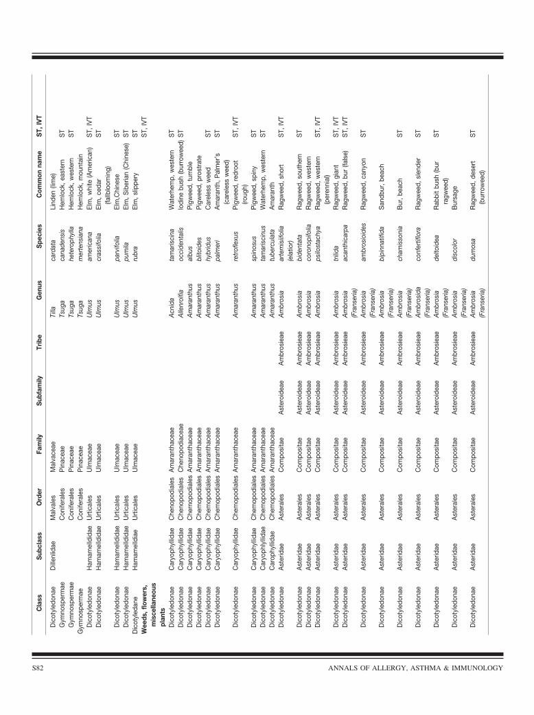
Cla
ssS
ubcl
ass
Ord
erFa
mily
Sub
fam
ilyTr
ibe
Gen
usS
peci
esC
omm
onna
me
ST,
IVT
Dic
otyl
edon
aeD
illen
lidae
Mal
vale
sM
alva
ceae
Tilla
card
ata
Lind
en(li
me)
Gym
nosp
erm
aeC
onife
rale
sP
inac
eae
Tsug
aca
nade
nsis
Hem
lock
,eas
tern
ST
Gym
nosp
erm
aeC
onife
rale
sP
inac
eae
Tsug
ahe
tero
phyl
laH
emlo
ck,w
este
rnS
TG
ymno
sper
mae
Con
ifera
les
Pin
acea
eTs
uga
mer
tens
iana
Hem
lock
,mou
ntai
nD
icot
yled
onae
Ham
amel
ldid
aeU
rtic
ales
Ulm
acea
eU
lmus
amer
ican
aEl
m,w
hite
(Am
eric
an)
ST,
IVT
Dic
otyl
edon
aeH
amam
elld
idae
Urt
ical
esU
lmac
eae
Ulm
uscr
assi
folia
Elm
,ced
ar(fa
llblo
omin
g)S
T
Dic
otyl
edon
aeH
amam
elld
idae
Urt
ical
esU
lmac
eae
Ulm
uspa
rvifo
liaEl
m,C
hine
seS
TD
icot
yled
onae
Ham
amel
ldid
aeU
rtic
ales
Ulm
acea
eU
lmus
pum
ilaEl
m,S
iber
ian
(Chi
nese
)S
TD
icot
yled
ane
Ham
amel
ldia
eU
rtic
ales
Ulm
acea
eU
lmus
rubr
aEl
m,s
lippe
ryS
TW
eeds
,flo
wer
s,m
isce
llane
ous
plan
ts
ST,
IVT
Dic
otyl
edon
aeC
aryo
phyl
lidae
Che
nopo
dial
esA
mar
anth
acea
eA
cnid
ata
mar
isci
naW
ater
hem
p,w
este
rnS
TD
icot
yled
onae
Car
yoph
yllid
aeC
heno
podi
ales
Che
nopo
diac
eae
Alle
nrol
fiaoc
cide
ntal
islo
dine
bush
(bur
row
eed)
ST
Dic
otyl
edon
aeC
aryo
phyl
lidae
Che
rnop
odia
les
Am
aran
thac
eae
Am
aran
thus
albu
sP
igw
eed,
tum
ble
Dic
otyl
edon
aeC
aryo
phyl
lidae
Che
mop
odia
les
Am
aran
thac
eae
Am
aran
thus
blito
ides
Pig
wee
d,pr
ostr
ate
Dic
otyl
edon
aeC
aryo
phyl
lidae
Che
mop
odia
les
Am
aran
thac
eae
Am
aran
thus
hybr
idus
Car
eles
sw
eed
ST
Dic
otyl
edon
aeC
aryo
phyl
lidae
Che
mop
odia
les
Am
aran
thac
eae
Am
aran
thus
palm
eri
Am
aran
th,P
alm
er’s
(car
eles
sw
eed)
ST
Dic
otyl
edon
aeC
aryo
phyl
lidae
Che
mop
odia
les
Am
aran
thac
eae
Am
aran
thus
retr
ofle
xus
Pig
wee
d,re
droo
t(ro
ugh)
ST,
IVT
Dic
otyl
edon
aeC
aryo
phyl
lidae
Che
mop
odia
les
Am
aran
thac
eae
Am
aran
thus
spin
osus
Pig
wee
d,sp
iny
ST
Dic
otyl
edon
aeC
aryo
phly
llida
eC
hem
opod
iale
sA
mar
anth
acea
eA
mar
anth
usta
mar
isci
nus
Wat
erhe
mp,
wes
tern
ST
Dic
otyl
edon
aeC
arop
hylli
dae
Che
mop
odia
les
Am
aran
thac
eae
Am
aran
thus
tube
rcul
ata
Am
aran
thD
icot
yled
onae
Ast
erid
aeA
ster
ales
Com
posi
tae
Ast
eroi
deae
Am
bros
ieae
Am
bros
iaar
tem
isiif
olia
(ela
tior)
Rag
wee
d,sh
ort
ST,
IVT
Dic
otyl
edon
aeA
ster
idae
Ast
eral
esC
ompo
sita
eA
ster
oide
aeA
mbr
osie
aeA
mbr
osia
bide
ntat
aR
agw
eed,
sout
hern
ST
Dic
otyl
edon
aeA
ster
idae
Ast
eral
esC
ompo
sita
eA
ster
oide
aeA
mbr
osie
aeA
mbr
osia
coro
nopi
folia
Rag
wee
d,w
este
rnD
icot
yled
onae
Ast
erid
aeA
ster
ales
Com
posi
tae
Ast
eroi
deae
Am
bros
ieae
Am
bros
iaps
ilost
achy
aR
agw
eed,
wes
tern
(per
enni
al)
ST,
IVT
Dic
otyl
edon
aeA
ster
idae
Ast
eral
esC
ompo
sita
eA
ster
oide
aeA
mbr
osie
aeA
mbr
osia
trili
daR
agw
eed,
gian
tS
T,IV
TD
icot
yled
onae
Ast
erid
aeA
ster
ales
Com
posi
tae
Ast
eroi
deae
Am
bros
ieae
Am
bros
ia(F
rans
eria
)ac
anth
icar
paR
agw
eed,
bur
(fals
e)S
T,IV
T
Dic
otyl
edon
aeA
ster
idae
Ast
eral
esC
ompo
sita
eA
ster
oide
aeA
mbr
osie
aeA
mbr
osia
(Fra
nser
ia)
ambr
osio
ides
Rag
wee
d,ca
nyon
ST
Dic
otyl
edon
aeA
ster
idae
Ast
eral
esC
ompo
sita
eA
ster
oide
aeA
mbr
osie
aeA
mbr
osia
(Fra
nser
ia)
bipi
nnat
ifida
San
dbur
,bea
ch
Dic
otyl
edon
aeA
ster
idae
Ast
eral
esC
ompo
sita
eA
ster
oide
aeA
mbr
osie
aeA
mbr
osia
(Fra
nser
ia)
cham
isso
nia
Bur
,bea
chS
T
Dic
otyl
edon
aeA
ster
idae
Ast
eral
esC
ompo
sita
eA
ster
oide
aeA
mbr
osie
aeA
mbr
osid
a(F
rans
eria
)co
nfer
tiflo
raR
agw
eed,
slen
der
ST
Dic
otyl
edon
aeA
ster
idae
Ast
eral
esC
ompo
sita
eA
ster
oide
aeA
mbr
osie
aeA
mbr
osia
(Fra
nser
ia)
delto
idea
Rab
bit
bush
(bur
ragw
eed)
ST
Dic
otyl
edon
aeA
ster
idae
Ast
eral
esC
ompo
sita
eA
ster
oide
aeA
mbr
osie
aeA
mbr
osia
(Fra
nser
ia)
disc
olor
Bur
sage
Dic
otyl
edon
aeA
ster
idae
Ast
eral
esC
ompo
sita
eA
ster
oide
aeA
mbr
osie
aeA
mbr
osia
(Fra
nser
ia)
dum
osa
Rag
wee
d,de
sert
(bur
row
eed)
ST
S82 ANNALS OF ALLERGY, ASTHMA & IMMUNOLOGY
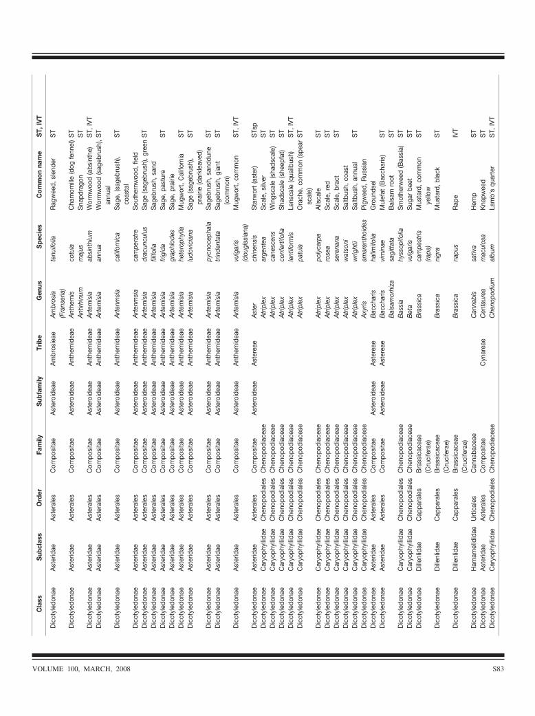
Cla
ssS
ubcl
ass
Ord
erFa
mily
Sub
fam
ilyTr
ibe
Gen
usS
peci
esC
omm
onna
me
ST,
IVT
Dic
otyl
edon
aeA
ster
idae
Ast
eral
esC
ompo
sita
eA
ster
oide
aeA
mbr
osie
aeA
mbr
osia
(Fra
nser
ia)
tenu
ifolia
Rag
wee
d,sl
ende
rS
T
Dic
otyl
edon
aeA
ster
idae
Ast
eral
esC
ompo
sita
eA
ster
oide
aeA
nthe
mid
eae
Ant
hem
isco
tula
Cha
mom
ille
(dog
fenn
el)
ST
Ant
irhin
umm
ajus
Sna
pdra
gon
ST
Dic
otyl
edon
aeA
ster
idae
Ast
eral
esC
ompo
sita
eA
ster
oide
aeA
nthe
mid
eae
Art
emis
iaab
sint
hium
Wor
mw
ood
(abs
inth
e)S
T,IV
TD
icot
yled
onae
Ast
erid
aeA
ster
ales
Com
posi
tae
Ast
eroi
deae
Ant
hem
idea
eA
rtem
isia
annu
aW
orm
woo
d(s
ageb
rush
),an
nual
ST
Dic
otyl
edon
aeA
ster
idae
Ast
eral
esC
ompo
sita
eA
ster
oide
aeA
nthe
mid
eae
Art
enm
sia
calif
orni
caS
age,
(sag
ebru
sh),
coas
tal
ST
Dic
otyl
edon
aeA
ster
idae
Ast
eral
esC
ompo
sita
eA
ster
oide
aeA
nthe
mid
eae
Art
enm
sia
cam
pers
tre
Sou
ther
nwoo
d,fie
ldD
icot
yled
onae
Ast
erid
aeA
ster
ales
Com
posi
tae
Ast
eroi
deae
Ant
hem
idea
eA
rtem
isia
drac
uncu
lus
Sag
e(s
ageb
rush
),gr
een
ST
Dic
otyl
edon
aeA
ster
idae
Ast
eral
esC
ompo
sita
eA
ster
oide
aeA
nthe
mid
eae
Art
emis
iafil
ifolia
Sag
ebru
sh,s
and
Dic
otyl
edon
aeA
ster
idae
Ast
eral
esC
ompo
sita
eA
ster
oide
aeA
nthe
mid
eae
Art
emis
iafr
igid
aS
age,
past
ure
ST
Dic
otyl
edon
aeA
ster
idae
Ast
eral
esC
ompo
sita
eA
ster
oide
aeA
nthe
mid
eae
Art
emis
iagr
aphl
odes
Sag
e,pr
airie
Dic
otyl
edon
aeA
ster
idae
Ast
eral
esC
ompo
sita
eA
ster
oide
aeA
nthe
mid
eae
Art
emis
iahe
tero
phyl
laM
ugw
ort,
Cal
iforn
iaS
TD
icot
yled
onae
Ast
erid
aeA
ster
ales
Com
posi
tae
Ast
eroi
deae
Ant
hem
idea
eA
rtem
isia
ludo
vici
ana
Sag
e(s
ageb
rush
),pr
airie
(dar
klea
ved)
ST
Dic
otyl
edon
aeA
ster
idae
Ast
eral
esC
ompo
sita
eA
ster
oide
aeA
nthe
mid
eae
Art
emis
iapy
cnoc
epha
laS
ageb
rush
,san
ddun
eS
TD
icot
yled
onae
Ast
erid
aeA
ster
ales
Com
posi
tae
Ast
eroi
deae
Ant
hem
idea
eA
rtem
isia
trin
dent
ata
Sag
ebru
sh,g
iant
(com
mon
)S
T
Dic
otyl
edon
aeA
ster
idae
Ast
eral
esC
ompo
sita
eA
ster
oide
aeA
nthe
mid
eae
Art
emis
iavu
lgar
is(d
ougl
asia
na)
Mug
wor
t,co
mm
onS
T,IV
T
Dic
otyl
edon
aeA
ster
idae
Ast
eral
esC
ompo
sita
eA
ster
oide
aeA
ster
eae
Ast
erch
inen
sis
Sta
rwor
t(a
ster
)S
Tsp
Dic
otyl
edon
aeC
aryo
phyl
lidae
Che
nopo
dial
esC
heno
podi
acea
eA
trip
lex
arge
ntea
Sca
le,s
ilver
ST
Dic
otyl
edon
aeC
aryo
phyl
lidae
Che
nopo
dial
esC
heno
podi
acea
eA
trip
lex
cane
scen
sW
ings
cale
(sha
dsca
le)
ST
Dic
otyl
edon
aeC
aryo
phyl
lidae
Che
nopo
dial
esC
heno
podi
acea
eA
trip
lex
conf
ertif
olia
Sha
dsca
le(s
heep
fat)
ST
Dic
otyl
edon
aeC
aryo
phyl
lidae
Che
nopo
dial
esC
heno
podi
acea
eA
trip
lex
lent
iform
isLe
nsca
le(q
uailb
ush)
ST,
IVT
Dic
otyl
edon
aeC
aryo
phyl
lidae
Che
nopo
dial
esC
heno
podi
acea
eA
trip
lex
patu
laO
rach
e,co
mm
on(s
pear
scal
e)S
T
Dic
otyl
edon
aeC
aryo
phyl
lidae
Che
nopo
dial
esC
heno
podi
acea
eA
trip
lex
poly
carp
aA
llsca
leS
TD
icot
yled
onae
Car
yoph
yllid
aeC
heno
podi
ales
Che
nopo
diac
eae
Atr
iple
xro
sea
Sca
le,r
edS
TD
icot
yled
onae
Car
yoph
yllid
aeC
heno
podi
ales
Che
nopo
diac
eae
Atr
iple
xse
rena
naS
cale
,bra
ctS
TD
icot
yled
onae
Car
yoph
yllid
aeC
heno
podi
ales
Che
nopo
diac
eae
Atr
iple
xw
atso
niS
altb
ush,
coas
tD
icot
yled
onae
Car
yoph
yllid
aeC
heno
podi
ales
Che
nopo
diac
eae
Atr
iple
xw
right
iiS
altb
ush,
annu
alS
TD
icot
yled
onae
Car
yoph
yllid
aeC
heno
podi
ales
Che
nopo
diac
eae
Axy
risam
aran
thoi
des
Pig
wee
d,R
ussi
anD
icot
yled
onae
Ast
erid
aeA
ster
ales
Com
posi
tae
Ast
eroi
deae
Ast
erea
eB
acch
aris
hallm
ifolia
Gro
unds
elD
icot
yled
onae
Ast
erid
aeA
ster
ales
Com
posi
tae
Ast
eroi
deae
Ast
erea
eB
acch
aris
vim
inae
Mul
efat
(Bac
char
is)
ST
Bal
sam
orhi
zasa
gitt
ata
Bal
sam
root
ST
Dic
otyl
edon
aeC
aryo
phyl
lidae
Che
nopo
dial
esC
heno
podi
acea
eB
assi
ahy
ssop
ifolia
Sm
othe
rwee
d(B
assi
a)S
TD
icot
yled
onae
Car
yoph
yllid
aeC
heno
podi
ales
Che
nopo
diac
eae
Bet
avu
lgar
isS
ugar
beet
ST
Dic
otyl
edon
aeD
illen
lidae
Cap
para
les
Bra
ssic
acea
e(C
ruci
fera
e)B
rass
ica
cam
pest
ris(ra
pa)
Mus
tard
,com
mon
yello
wS
T
Dic
otyl
edon
aeD
illen
lidae
Cap
para
les
Bra
ssic
acea
e(C
ruci
fera
e)B
rass
ica
nigr
aM
usta
rd,b
lack
ST
Dic
otyl
edon
aeD
illen
lidae
Cap
para
les
Bra
ssic
acea
e(C
ruci
fera
e)B
rass
ica
napu
sR
ape
IVT
Dic
otyl
edon
aeH
amam
elld
idae
Urt
ical
esC
anna
bace
aeC
anna
bis
sativ
aH
emp
ST
Dic
otyl
edon
aeA
ster
idae
Ast
eral
esC
ompo
sita
eC
ynar
eae
Cen
taur
eam
acul
osa
Kna
pwee
dS
TD
icot
yled
onae
Car
yoph
yllid
aeC
heno
podi
ales
Che
nopo
diac
eae
Che
nopo
dium
albu
mLa
mb’
squ
arte
rS
T,IV
T
VOLUME 100, MARCH, 2008 S83
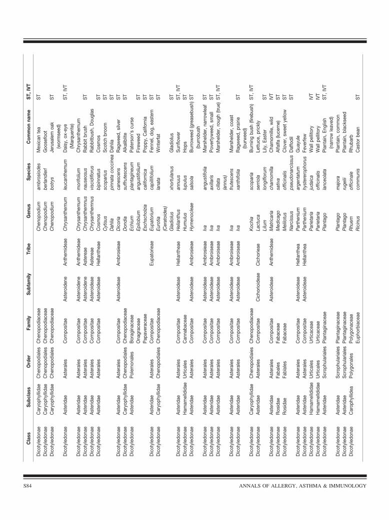
Cla
ssS
ubcl
ass
Ord
erFa
mily
Sub
fam
ilyTr
ibe
Gen
usS
peci
esC
omm
onna
me
ST,
IVT
Dic
otyl
edon
aeC
aryo
phyl
lidae
Che
nopo
dial
esC
heno
podi
acea
eC
heno
podi
umam
bros
iode
sM
exic
ante
aS
TD
icot
yled
onae
Car
yoph
yllid
aeC
heno
podi
ales
Che
nopo
diac
eae
Che
nopo
dium
berla
ndie
rlG
oose
foot
Dic
otyl
edon
aeC
aryo
phyl
lidae
Che
nopo
dial
esC
heno
podi
acea
eC
heno
podi
umbo
trys
Jeru
sale
moa
k(w
orm
seed
)S
T
Dic
otyl
edon
aeA
ster
idae
Ast
eral
esC
ompo
sita
eA
ster
oide
neA
nthe
mid
eae
Chr
ysan
them
umle
ucan
them
umD
aisy
,ox-
eye
(Mar
quer
ite)
ST,
IVT
Dic
otyl
edon
aeA
ster
idae
Ast
eral
esC
ompo
sita
eA
ster
oide
neA
nthe
mid
eae
Chr
ysan
them
umm
orifo
lium
Chr
ysan
them
umD
icot
yled
onae
Ast
erid
aeA
ster
ales
Com
posi
tae
Ast
eroi
dene
Ast
erea
eC
hrys
anth
emnu
sna
useo
sus
Rab
bit
brus
hS
TD
icot
yled
onae
Ast
erid
aeA
ster
ales
Com
posi
tae
Ast
eroi
deae
Ast
erea
eC
hrys
anth
emnu
svi
scid
iflor
usR
abbi
tbus
h,D
ougl
asD
icot
yled
onae
Ast
erid
aeA
ster
ales
Com
posi
tae
Ast
eroi
deae
Hel
lant
heae
Cos
mos
bipi
nnat
usC
osm
osS
TC
ytis
ussc
opar
ius
Sco
tch
broo
mS
TD
ahlia
pinn
ata
cocc
inea
Dah
liaS
TD
icot
yled
onae
Ast
erid
aeA
ster
ales
Com
posi
tae
Ast
eroi
deae
Am
bros
ieae
Dic
oria
cane
scen
sR
agw
eed,
silv
erS
TD
icot
yled
onae
Car
yoph
yllid
aeC
heno
podi
ales
Che
nopo
diac
eae
Don
dia
suffr
utes
cens
Alk
alib
lite
ST
Dic
otyl
edon
aeA
ster
idae
Pol
emon
ales
Bor
agin
acea
eEc
hium
plan
tagi
neum
Pat
erso
n’s
curs
eO
nagr
acea
eEp
ilobi
uman
gust
ifoliu
mFi
rew
eed
ST
Pap
aver
acea
eEs
chsc
holz
iaca
lifor
nica
Pop
py,C
alifo
rnia
ST
Dic
otyl
edon
aeA
ster
idae
Ast
eral
esC
ompo
sita
eEu
pato
rieae
Eupa
toriu
mca
pilli
foliu
mFe
nnel
,dog
,eas
tern
ST
Dic
otyl
edon
aeC
aryo
phyl
lidae
Che
nopo
dial
esC
heno
podi
acea
eEu
rotia
(Cer
atoi
des)
lana
taW
inte
rfat
ST
Gla
diol
usxh
ortu
lanu
sG
ladi
olus
ST
Dic
otyl
edon
aeA
ster
idae
Ast
eral
esC
ompo
sita
eA
ster
oide
aeH
elia
nthe
aeH
elia
nthu
san
nuus
Sun
flow
erS
T,IV
TD
icot
yled
onae
Ham
amel
ldid
aeU
rtic
ales
Can
naba
ceae
Hum
ulus
lupu
lus
Hop
sS
TD
icot
yled
onae
Ast
erid
aeA
ster
ales
Com
posi
tae
Ast
eroi
deae
Am
bros
ieae
Hym
enoc
leae
sals
ola
Bur
row
eed
(gre
aseb
ush)
(bur
robu
shS
T
Dic
otyl
edon
aeA
ster
idae
Ast
eral
esC
ompo
sita
eA
ster
oide
aeA
mbr
osie
aeIv
aan
gust
ifolia
Mar
shel
der,
narr
owle
afS
TD
icot
yled
onae
Ast
erid
aeA
ster
ales
Com
posi
tae
Ast
eroi
deae
Am
broi
seae
Iva
axill
aris
Pov
erty
wee
d,sm
all
ST
Dic
otyl
edon
aeA
ster
idae
Ast
eral
esC
ompo
sita
eA
ster
oide
aeA
mbr
oise
aeIv
aci
llata
(ann
us)
Mar
shel
der,
roug
h(tr
ue)
ST,
IVT
Dic
otyl
edon
aeA
ster
idae
Ast
eral
esC
ompo
sita
eA
ster
oide
aeA
mbr
oise
aeIv
afr
utes
cens
Mar
shel
der,
coas
tD
icot
yled
onae
Ast
erid
aeA
ster
ales
Com
posi
tae
Ast
eroi
deae
Am
broi
seae
Iva
xant
hifo
liaR
agw
eed,
prai
rie(b
urw
eed)
ST
Dic
otyl
edon
aeC
aryo
phyl
lidae
Che
nopo
diai
esC
heno
podi
acea
eK
ochi
asc
opar
iaB
urni
ngbu
sh(fi
rebu
sh)
ST,
IVT
Dic
otyl
edon
aeA
ster
idae
Ast
eral
esC
ompo
sita
eC
icho
rioid
eae
Cic
horie
aeLa
ctuc
ase
rrio
laLe
ttuc
e,pr
ickl
yLi
lum
long
iflor
umLi
ly,E
aste
rS
TD
icot
yled
onae
Ast
erid
aeA
ster
ales
Com
posi
tae
Ast
eroi
deae
Ant
hem
idea
eM
atric
aria
cham
omill
aC
ham
omill
e,w
ildIV
TD
icot
yled
onae
Ros
idae
Faba
les
Faba
ceae
Med
icag
osa
tiva
Alfa
lfa(lu
cern
e)S
TD
icot
yled
onae
Ros
idae
Fabl
ales
Faba
ceae
Mel
ilotu
sof
ficin
alis
Clo
ver,
swee
tye
llow
ST
Nar
ciss
usps
eudo
narc
issu
sD
affo
dil
ST
Dic
otyl
edon
aeA
ster
idae
Ast
eral
esC
ompo
sita
eA
ster
oide
aeH
ella
nthe
aP
arth
eniu
mar
gent
atum
Gua
yule
Dic
otyl
edon
aeA
ster
idae
Ast
eral
esC
ompo
sita
eA
ster
oide
aeH
ella
nthe
aP
arth
eniu
mhy
ster
erop
horu
sFe
verf
ewD
icot
yled
onae
Ham
amel
ldid
aeU
rtic
ales
Urt
icac
eae
Par
ieta
riaju
daic
aW
allp
ellit
ory
IVT
Dic
otyl
edon
aeH
amam
elld
idae
Urt
ical
esU
rtic
acea
eP
arie
taria
offic
inal
isW
allp
ellit
ory
IVT
Dic
otyl
edon
aeA
ster
idae
Scr
ophu
laria
les
Pla
ntag
inac
eae
Pla
ntag
ola
nceo
lata
Pla
ntai
n,En
glis
h(n
arro
wle
aved
)S
T,IV
T
Dic
otyl
edon
aeA
ster
idae
Scr
ophu
laria
les
Pla
ntag
inac
eae
Pla
ntag
om
ajor
aP
lant
ain,
com
mon
Dic
otyl
edon
aeA
ster
idae
Scr
ophu
laria
les
Pla
ntag
inac
eae
Pla
ntag
oru
gelii
Pla
ntai
n,bl
acks
eed
Dic
otyl
edon
aeC
arop
hylli
dea
Pol
ygon
ales
Pol
ygon
acea
eR
huem
offic
inal
eR
huba
rbEu
phor
biac
eae
Ric
inus
com
mun
isC
asto
rbe
anS
T
S84 ANNALS OF ALLERGY, ASTHMA & IMMUNOLOGY
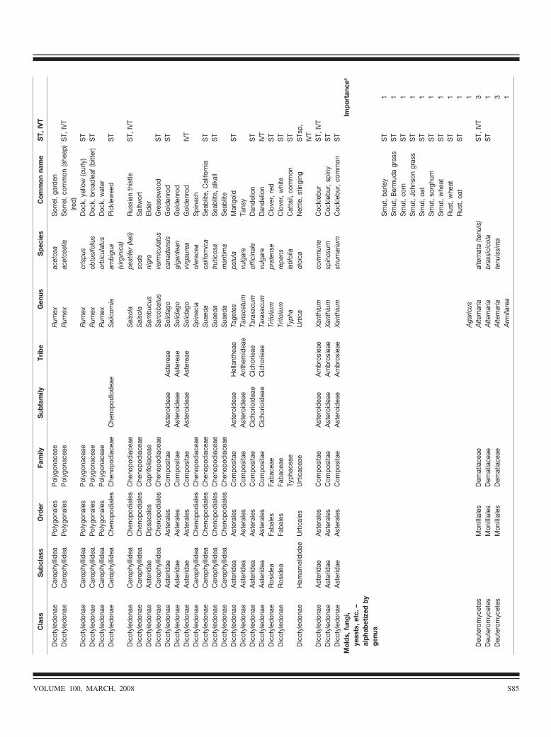
Cla
ssS
ubcl
ass
Ord
erFa
mily
Sub
fam
ilyTr
ibe
Gen
usS
peci
esC
omm
onna
me
ST,
IVT
Dic
otyl
edon
aeC
arop
hylli
dea
Pol
ygon
ales
Pol
ygon
acea
eR
umex
acet
osa
Sor
rel,
gard
enD
icot
yled
onae
Car
ophy
llide
aP
olyg
onal
esP
olyg
onac
eae
Rum
exac
etos
ella
Sor
rel,
com
mon
(she
ep)
(red)
ST,
IVT
Dic
otyl
edon
aeC
arop
hylli
dea
Pol
ygon
ales
Pol
ygon
acea
eR
umex
cris
pus
Doc
k,ye
llow
(cur
ly)
ST
Dic
otyl
edon
aeC
arop
hylli
dea
Pol
ygon
ales
Pol
ygon
acea
eR
umex
obtu
sifo
lius
Doc
k,br
oadl
eaf
(bitt
er)
ST
Dic
otyl
edon
aeC
arop
hylli
dea
Pol
ygon
ales
Pol
ygon
acea
eR
umex
orbi
cula
tus
Doc
k,w
ater
Dic
otyl
edon
aeC
arop
hylli
dea
Che
nopo
dial
esC
heno
podi
acea
eC
heno
podl
odea
eS
alic
orni
aam
bigu
a(v
irgin
ica)
Pic
klew
eed
ST
Dic
otyl
edon
aeC
arop
hylli
dea
Che
nopo
dial
esC
heno
podi
acea
eS
also
lape
stife
r(k
ali)
Rus
sian
this
tleS
T,IV
TD
icot
yled
onae
Car
ophy
llide
aC
heno
podi
ales
Che
nopo
diac
eae
Sal
sola
soda
Sal
twor
tD
icot
yled
onae
Ast
erid
aeD
ipsa
cale
sC
aprif
olia
ceae
Sam
bucu
sni
gra
Elde
rD
icot
yled
onae
Car
ophy
llide
aC
heno
podi
ales
Che
nopo
diac
eae
Sar
coba
tus
verm
icul
atus
Gre
asew
ood
ST
Dic
otyl
edon
aeA
ster
idae
Ast
eral
esC
ompo
sita
eA
ster
oide
aeA
ster
eae
Sol
idag
oca
nade
nsis
Gol
denr
odS
TD
icot
yled
onae
Ast
erid
aeA
ster
ales
Com
posi
tae
Ast
eroi
deae
Ast
erea
eS
olid
ago
giga
ntea
nG
olde
nrod
Dic
otyl
edon
aeA
ster
idae
Ast
eral
esC
ompo
sita
eA
ster
oide
aeA
ster
eae
Sol
idag
ovi
rgau
rea
Gol
denr
odIV
TD
icot
yled
onae
Car
ophy
llide
aC
heno
podi
ales
Che
nopo
diac
eae
Spi
naci
aol
erac
eaS
pina
chD
icot
yled
onae
Car
ophy
llide
aC
heno
podi
ales
Che
nopo
diac
eae
Sua
eda
calif
orni
caS
eabl
ite,C
alifo
rnia
ST
Dic
otyl
edon
aeC
arop
hylli
dea
Che
nopo
dial
esC
heno
podi
acea
eS
uaed
afr
utic
osa
Sea
blite
,alk
all
ST
Dic
otyl
edon
aeC
arop
hylli
dea
Che
nopo
dial
esC
heno
podi
acea
eS
uaed
am
ariti
ma
Sea
blite
Dic
otyl
edon
aeA
ster
idea
Ast
eral
esC
ompo
sita
eA
ster
oide
aeH
ella
nthe
aeTa
gete
spa
tula
Mar
igol
dS
TD
icot
yled
onae
Ast
erid
eaA
ster
ales
Com
posi
tae
Ast
eroi
deae
Ant
hem
idea
eTa
nace
tum
vulg
are
Tans
yD
icot
yled
onae
Ast
erid
eaA
ster
ales
Com
posi
tae
Cic
horio
idea
eC
icho
rieae
Tara
xacu
mof
ficin
ale
Dan
delio
nS
TD
icot
yled
onae
Ast
erid
eaA
ster
ales
Com
posi
tae
Cic
horio
idea
eC
icho
rieae
Tara
xacu
mvu
lgar
eD
ande
lion
IVT
Dic
otyl
edon
aeR
osid
eaFa
bale
sFa
bace
aeTr
ifoliu
mpr
aten
seC
love
r,re
dS
TD
icot
yled
onae
Ros
idea
Faba
les
Faba
ceae
Trifo
lium
repe
nsC
love
r,w
hite
ST
Typh
acea
eTy
pha
latif
olia
Cat
tail,
com
mon
ST
Dic
otyl
edon
aeH
amam
elld
idae
Urt
ical
esU
rtic
acea
eU
rtic
adi
oica
Net
tle,s
tingi
ngS
Tsp,
IVT
Dic
otyl
edon
aeA
ster
idae
Ast
eral
esC
ompo
sita
eA
ster
oide
aeA
mbr
osie
aeXa
nthi
umco
mm
une
Coc
kleb
urS
T,IV
TD
icot
yled
onae
Ast
erid
aeA
ster
ales
Com
posi
tae
Ast
eroi
deae
Am
bros
ieae
Xant
hium
spin
osum
Coc
kleb
ur,s
piny
ST
Dic
otyl
edon
aeA
ster
idae
Ast
eral
esC
ompo
sita
eA
ster
oide
aeA
mbr
osie
aeXa
nthi
umst
rum
ariu
mC
ockl
ebur
,com
mon
ST
Mol
ds,f
ungi
,ye
asts
,etc
.–al
phab
etiz
edby
genu
s
Impo
rtan
cea
Sm
ut,b
arle
yS
T1
Sm
ut,B
erm
uda
gras
sS
T1
Sm
ut,c
orn
ST
1S
mut
,Joh
nson
gras
sS
T1
Sm
ut,o
atS
T1
Sm
ut,s
orgh
umS
T1
Sm
ut,w
heat
ST
1R
ust,
whe
atS
T1
Rus
t,oa
tS
T1
Aga
ricus
1D
eute
rom
ycet
esM
onill
iale
sD
emat
iace
aeA
ltem
aria
alte
rnat
a(te
nuis
)S
T,IV
T3
Deu
tero
myc
etes
Mon
illia
les
Dem
atia
ceae
Alte
mar
iabr
assi
cico
laS
T1
Deu
tero
myc
etes
Mon
illia
les
Dem
atia
ceae
Alte
mar
iate
nuis
sim
a3
Arm
illar
ea1
VOLUME 100, MARCH, 2008 S85
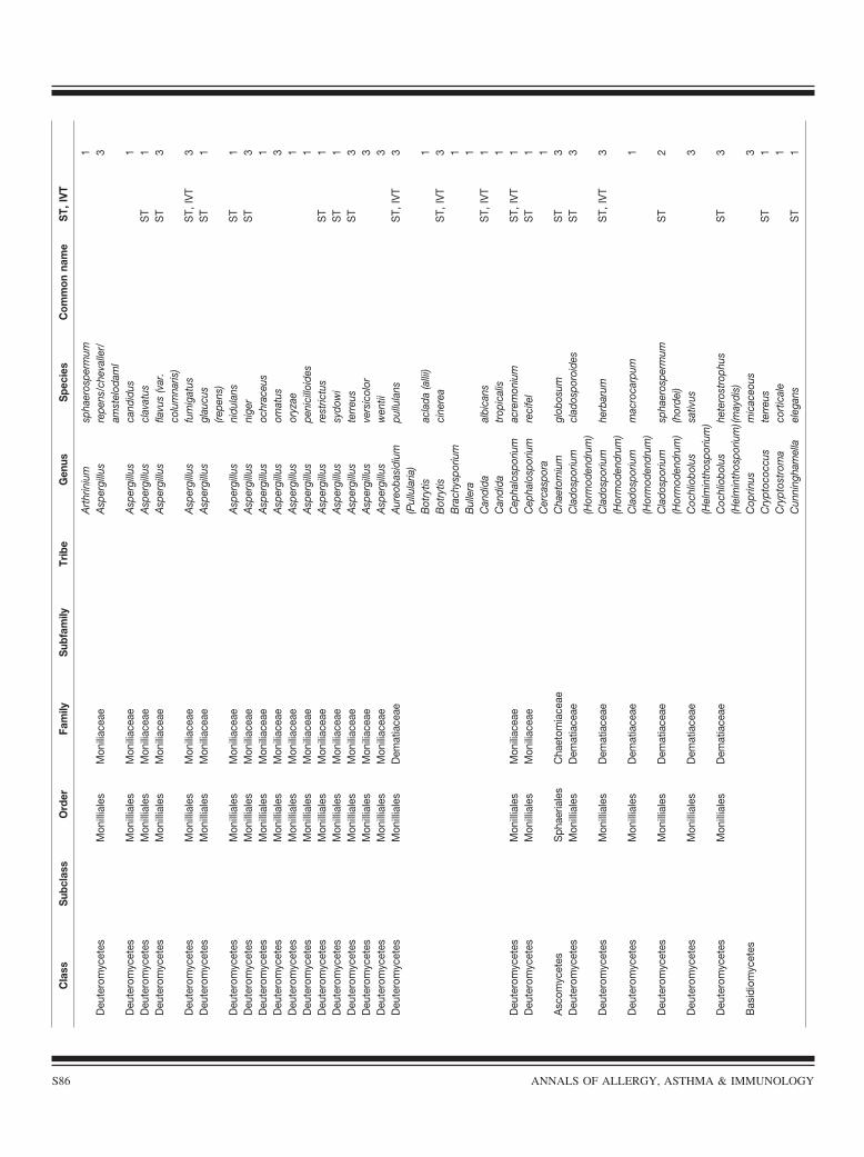
Cla
ssS
ubcl
ass
Ord
erFa
mily
Sub
fam
ilyTr
ibe
Gen
usS
peci
esC
omm
onna
me
ST,
IVT
Art
hrin
ium
spha
eros
perm
um1
Deu
tero
myc
etes
Mon
illia
les
Mon
iliac
eae
Asp
ergi
llus
repe
ns/c
heva
ller/
amst
elod
aml
3
Deu
tero
myc
etes
Mon
illia
les
Mon
iliac
eae
Asp
ergi
llus
cand
idus
1D
eute
rom
ycet
esM
onill
iale
sM
onili
acea
eA
sper
gillu
scl
avat
usS
T1
Deu
tero
myc
etes
Mon
illia
les
Mon
iliac
eae
Asp
ergi
llus
flavu
s(v
ar.
colu
mna
ris)
ST
3
Deu
tero
myc
etes
Mon
illia
les
Mon
iliac
eae
Asp
ergi
llus
fum
igat
usS
T,IV
T3
Deu
tero
myc
etes
Mon
illia
les
Mon
iliac
eae
Asp
ergi
llus
glau
cus
(repe
ns)
ST
1
Deu
tero
myc
etes
Mon
illia
les
Mon
iliac
eae
Asp
ergi
llus
nidu
lans
ST
1D
eute
rom
ycet
esM
onill
iale
sM
onili
acea
eA
sper
gillu
sni
ger
ST
3D
eute
rom
ycet
esM
onill
iale
sM
onili
acea
eA
sper
gillu
soc
hrac
eus
1D
eute
rom
ycet
esM
onill
iale
sM
onili
acea
eA
sper
gillu
som
atus
3D
eute
rom
ycet
esM
onill
iale
sM
onili
acea
eA
sper
gillu
sor
yzae
1D
eute
rom
ycet
esM
onill
iale
sM
onili
acea
eA
sper
gillu
spe
nici
lloid
es1
Deu
tero
myc
etes
Mon
illia
les
Mon
iliac
eae
Asp
ergi
llus
rest
rictu
sS
T1
Deu
tero
myc
etes
Mon
illia
les
Mon
iliac
eae
Asp
ergi
llus
sydo
wi
ST
1D
eute
rom
ycet
esM
onill
iale
sM
onili
acea
eA
sper
gillu
ste
rreu
sS
T3
Deu
tero
myc
etes
Mon
illia
les
Mon
iliac
eae
Asp
ergi
llus
vers
icol
or3
Deu
tero
myc
etes
Mon
illia
les
Mon
iliac
eae
Asp
ergi
llus
wen
tii3
Deu
tero
myc
etes
Mon
illia
les
Dem
atia
ceae
Aur
eoba
sidi
um(P
ullu
laria
)pu
llula
nsS
T,IV
T3
Bot
rytis
acla
da(a
llii)
1B
otry
tisci
nere
aS
T,IV
T3
Bra
chys
poriu
m1
Bul
lera
1C
andi
daal
bica
nsS
T,IV
T1
Can
dida
trop
ical
is1
Deu
tero
myc
etes
Mon
illia
les
Mon
iliac
eae
Cep
halo
spor
ium
acre
mon
ium
ST,
IVT
1D
eute
rom
ycet
esM
onill
iale
sM
onili
acea
eC
epha
losp
oriu
mre
cife
lS
T1
Cer
casp
ora
1A
scom
ycet
esS
phae
riale
sC
haet
omia
ceae
Cha
etom
ium
glob
osum
ST
3D
eute
rom
ycet
esM
onill
iale
sD
emat
iace
aeC
lado
spor
ium
(Hor
mod
endr
um)
clad
ospo
roid
esS
T3
Deu
tero
myc
etes
Mon
illia
les
Dem
atia
ceae
Cla
dosp
oriu
m(H
orm
oden
drum
)he
rbar
umS
T,IV
T3
Deu
tero
myc
etes
Mon
illia
les
Dem
atia
ceae
Cla
dosp
oriu
m(H
orm
oden
drum
)m
acro
carp
um1
Deu
tero
myc
etes
Mon
illia
les
Dem
atia
ceae
Cla
dosp
oriu
m(H
orm
oden
drum
)sp
haer
ospe
rmum
(hor
dei)
ST
2
Deu
tero
myc
etes
Mon
illia
les
Dem
atia
ceae
Coc
hlio
bolu
s(H
elm
inth
ospo
rium
)sativ
us3
Deu
tero
myc
etes
Mon
illia
les
Dem
atia
ceae
Coc
hlio
bolu
s(H
elm
inth
ospo
rium
)hete
rost
roph
us(m
aydi
s)S
T3
Bas
idio
myc
etes
Cop
rinus
mic
aceo
us3
Cry
ptoc
occu
ste
rreu
sS
T1
Cry
ptos
trom
aco
rtic
ale
1C
unni
ngha
mel
lael
egan
sS
T1
S86 ANNALS OF ALLERGY, ASTHMA & IMMUNOLOGY
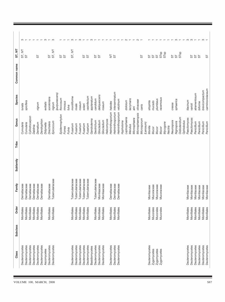
Cla
ssS
ubcl
ass
Ord
erFa
mily
Sub
fam
ilyTr
ibe
Gen
usS
peci
esC
omm
onna
me
ST,
IVT
Deu
tero
myc
etes
Mon
illia
les
Dem
atia
ceae
Cur
vula
rialu
nata
ST,
IVT
3D
eute
rom
ycet
esM
onill
iale
sD
emat
iace
aeC
urvu
laria
spic
ifera
ST
1D
eute
rom
ycet
esM
onill
iale
sD
emat
iace
aeC
ylin
droc
arpo
n1
Deu
tero
myc
etes
Mon
illia
les
Dem
atia
ceae
Dal
dini
a1
Deu
tero
myc
etes
Mon
illia
les
Dem
atia
ceae
Dem
atiu
mni
grum
ST
1D
eute
rom
ycet
esM
onill
iale
sD
emat
iace
aeD
endr
yphi
on1
Asc
omyc
etes
Did
ymel
laex
itial
is3
Deu
tero
myc
etes
Mon
illia
les
Dem
atia
ceae
Dre
chsl
era
soro
kini
ana
ST
1D
eute
rom
ycet
esM
onill
iale
sTu
berc
ular
iace
aeEp
icoc
cum
nigr
um(p
urpu
rasc
ens)
ST,
IVT
3
Epid
erm
ophy
ton
flocc
osum
ST
1Fo
mes
rimos
usS
T1
Fulv
iafu
lva
1D
eute
rom
ycet
esM
onill
iale
sTu
berc
ular
iace
aeFu
sariu
mm
onili
form
eS
T,IV
T3
Deu
tero
myc
etes
Mon
illia
les
Tube
rcul
aria
ceae
Fusa
rium
niva
le1
Deu
tero
myc
etes
Mon
illia
les
Tube
rcul
aria
ceae
Fusa
rium
rose
um3
Deu
tero
myc
etes
Mon
illia
les
Tube
rcul
aria
ceae
Fusa
rium
sola
niS
T1
Deu
tero
myc
etes
Mon
illia
les
Tube
rcul
aria
ceae
Fusa
rium
vasi
nfec
tum
ST
1B
asid
iom
ycet
esG
ando
derm
aap
plan
atum
3D
eute
rom
ycet
esM
onill
iale
sTu
berc
ular
iace
aeG
eotr
ichu
mca
ndid
umS
T1
Deu
tero
myc
etes
Mon
illia
les
Mon
iliaa
ceae
Glio
clad
ium
deliq
uesc
ens
ST
1D
eute
rom
ycet
esM
onill
iale
sM
onili
aace
aeG
liocl
adiu
mro
seum
1H
elic
omyc
es1
Deu
tero
myc
etes
Mon
illia
les
Dem
atia
ceae
Hel
min
thos
poriu
mha
lode
sIV
T1
Deu
tero
myc
etes
Mon
illia
les
Dem
atia
ceae
Hel
min
thos
poriu
min
ters
emin
atum
ST
1D
eute
rom
ycet
esM
onill
iale
sD
emat
iace
aeH
elm
inth
ospo
rium
velu
tinum
1H
ypho
lom
a1
Lept
osph
aeria
dolio
lum
2M
erul
ius
lacr
yman
s1
Mic
rosp
haer
aai
ni1
Mic
rosp
haer
opsi
sol
ivac
eae
2M
icro
spor
um(L
anos
um)
cani
sS
T1
Deu
tero
myc
etes
Mon
illia
les
Mon
iliac
eae
Mon
ilia
sito
phila
ST
1Zy
gom
ycet
esM
ucor
ales
Muc
orac
eae
Muc
orm
uced
oS
T1
Zygo
myc
etes
Muc
oral
esM
ucor
acea
eM
ucor
plum
beus
ST
1Zy
gom
ycet
esM
ucor
ales
Muc
orac
eae
Muc
orra
cem
osus
STs
p1
Myc
ogon
eS
Tsp
1N
ectr
ia1
Neu
rosp
ora
cras
saS
T1
Nig
rosp
ora
spha
eric
aS
T3
Oid
iode
ndru
mS
Tsp
1O
phio
bolu
s1
Deu
tero
myc
etes
Mon
illia
les
Mon
iliac
eae
Pae
cilo
myc
eslil
acin
um3
Deu
tero
myc
etes
Mon
illia
les
Mon
iliac
eae
Pae
cilo
myc
esva
rioti
ST
3D
eute
rom
ycet
esM
onill
iale
sM
onili
acea
eP
enic
illiu
mat
ram
ento
sum
ST
1D
eute
rom
ycet
esM
onill
iale
sM
onili
acea
eP
enic
illiu
mbi
form
eS
T1
Deu
tero
myc
etes
Mon
illia
les
Mon
iliac
eae
Pen
icill
ium
brev
icom
pact
um3
Deu
tero
myc
etes
Mon
illia
les
Mon
iliac
eae
Pen
icill
ium
carm
inov
iola
ceum
ST
1
VOLUME 100, MARCH, 2008 S87
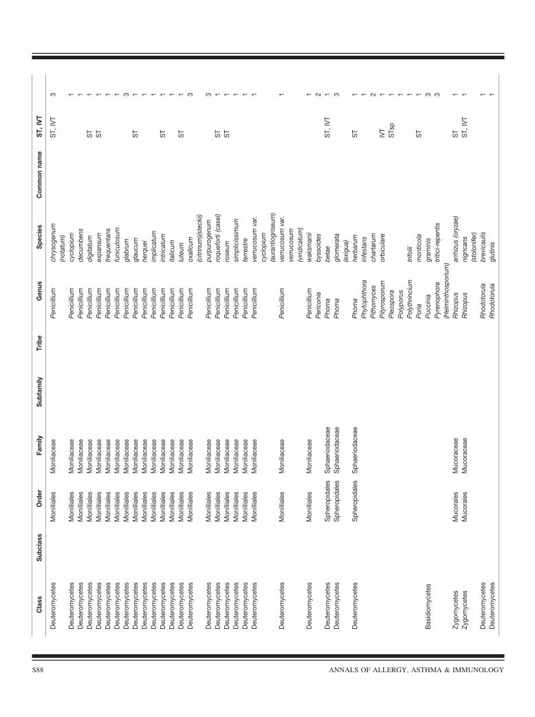
Cla
ssS
ubcl
ass
Ord
erFa
mily
Sub
fam
ilyTr
ibe
Gen
usS
peci
esC
omm
onna
me
ST,
IVT
Deu
tero
myc
etes
Mon
illia
les
Mon
iliac
eae
Pen
icill
ium
chry
soge
num
(not
atum
)S
T,IV
T3
Deu
tero
myc
etes
Mon
illia
les
Mon
iliac
eae
Pen
icill
ium
cycl
opiu
m1
Deu
tero
myc
etes
Mon
illia
les
Mon
iliac
eae
Pen
icill
ium
decu
mbe
ns1
Deu
tero
myc
etes
Mon
illia
les
Mon
iliac
eae
Pen
icill
ium
digi
tatu
mS
T1
Deu
tero
myc
etes
Mon
illia
les
Mon
iliac
eae
Pen
icill
ium
expa
nsum
ST
1D
eute
rom
ycet
esM
onill
iale
sM
onili
acea
eP
enic
illiu
mfr
eque
ntan
s1
Deu
tero
myc
etes
Mon
illia
les
Mon
iliac
eae
Pen
icill
ium
funi
culo
sum
1D
eute
rom
ycet
esM
onill
iale
sM
onili
acea
eP
enic
illiu
mgl
abru
m3
Deu
tero
myc
etes
Mon
illia
les
Mon
iliac
eae
Pen
icill
ium
glau
cum
ST
1D
eute
rom
ycet
esM
onill
iale
sM
onili
acea
eP
enic
illiu
mhe
rque
i1
Deu
tero
myc
etes
Mon
illia
les
Mon
iliac
eae
Pen
icill
ium
impl
icat
um1
Deu
tero
myc
etes
Mon
illia
les
Mon
iliac
eae
Pen
icill
ium
intr
icat
umS
T1
Deu
tero
myc
etes
Mon
illia
les
Mon
iliac
eae
Pen
icill
ium
italic
um1
Deu
tero
myc
etes
Mon
illia
les
Mon
iliac
eae
Pen
icill
ium
lute
umS
T1
Deu
tero
myc
etes
Mon
illia
les
Mon
iliac
eae
Pen
icill
ium
oxal
lcum
(citr
inum
)(ste
ckii)
3
Deu
tero
myc
etes
Mon
illia
les
Mon
iliac
eae
Pen
icill
ium
purp
urog
enum
3D
eute
rom
ycet
esM
onill
iale
sM
onili
acea
eP
enic
illiu
mro
quef
orti
(cas
el)
ST
1D
eute
rom
ycet
esM
onill
iale
sM
onili
acea
eP
enic
illiu
mro
seum
ST
1D
eute
rom
ycet
esM
onill
iale
sM
onili
acea
eP
enic
illiu
msi
mpl
icis
sim
um1
Deu
tero
myc
etes
Mon
illia
les
Mon
iliac
eae
Pen
icill
ium
terr
estr
e1
Deu
tero
myc
etes
Mon
illia
les
Mon
iliac
eae
Pen
icill
ium
verr
ucos
umva
r.cy
clop
ium
(aur
antio
gris
eum
)
1
Deu
tero
myc
etes
Mon
illia
les
Mon
iliac
eae
Pen
icill
ium
verr
ucos
umva
r.ve
rruc
osum
(viri
dica
tum
)
1
Deu
tero
myc
etes
Mon
illia
les
Mon
iliac
eae
Pen
icill
ium
wak
sman
ii1
Per
icon
iaby
ssoi
des
2D
eute
rom
ycet
esS
pher
opid
ales
Sph
aerio
dace
aeP
hom
abe
tae
ST,
IVT
1D
eute
rom
ycet
esS
pher
opid
ales
Sph
aerio
dace
aeP
hom
agl
omer
ata
(exi
gua)
3
Deu
tero
myc
etes
Sph
erop
idal
esS
phae
rioda
ceae
Pho
ma
herb
arum
ST
1P
hyto
phth
ora
infe
stan
s1
Pith
omyc
esch
arta
rum
2P
ityro
spor
umor
bicu
lare
IVT
1P
leos
pora
STs
p1
Pol
ypor
us1
Pol
ythr
inci
umtr
ifolii
1P
oria
mon
ticol
aS
T1
Bas
idio
myc
etes
Puc
cini
agr
amin
is3
Pyr
enop
hora
(Hel
min
thos
poriu
m)tr
itici
-rep
entis
3
Zygo
myc
etes
Muc
oral
esM
ucor
acea
eR
hizo
pus
arrh
izus
(ory
zae)
ST
1Zy
gom
ycet
esM
ucor
ales
Muc
orac
eae
Rhi
zopu
sni
gric
ans
(sto
loni
fer)
ST,
IVT
1
Deu
tero
myc
etes
Rho
doto
rula
brev
icau
lis1
Deu
tero
myc
etes
Rho
doto
rula
glut
inis
1
S88 ANNALS OF ALLERGY, ASTHMA & IMMUNOLOGY
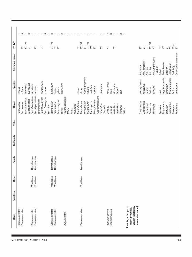
Cla
ssS
ubcl
ass
Ord
erFa
mily
Sub
fam
ilyTr
ibe
Gen
usS
peci
esC
omm
onna
me
ST,
IVT
Deu
tero
myc
etes
Rho
doto
rula
rose
us1
Deu
tero
myc
etes
Rho
doto
rula
rubr
umS
T3
Sac
char
omyc
esce
revi
slae
ST,
IVT
1S
copu
lario
psis
brev
icau
lisS
T3
Deu
tero
myc
etes
Mon
illia
les
Dem
atia
ceae
Spo
ndly
ocla
dium
atro
vire
ns1
Deu
tero
myc
etes
Mon
illia
les
Dem
atia
ceae
Spo
ndly
ocla
dium
aust
rale
ST
1S
porid
esm
ium
1S
poro
bolo
myc
essa
lmon
icol
orS
T1
Spo
robo
lom
yces
rose
us3
Spo
roth
rix1
Deu
tero
myc
etes
Mon
illia
les
Dem
atia
ceae
Ste
mph
yliu
mbo
tryo
sum
ST,
IVT
3D
eute
rom
ycet
esM
onill
iale
sD
emat
iace
aeS
tem
phyl
ium
sola
niS
T1
Str
epto
myc
esgr
iseu
sS
T1
Sui
llus
gran
ulat
us2
Zygo
myc
etes
Syn
ceph
alas
trum
1Ti
lletia
1To
rula
1To
rulo
psis
1Tr
icho
derm
are
esel
ST
1D
eute
rom
ycet
esM
onill
iale
sM
onili
acea
eTr
icho
derm
avi
ride
ST,
IVT
1Tr
icho
phyt
onm
enta
grop
hyte
sS
T1
Tric
hoph
yton
rubr
umS
T,IV
T1
Tric
hosp
oron
pullu
lans
IVT
1Tr
icho
thec
ium
(Cep
halo
thec
ium
)ro
seum
ST
1
Ulo
clad
ium
char
taru
mIV
T1
Uro
cyst
is1
Bas
idio
myc
etes
Ust
ilago
nuda
(triti
ci)
IVT
3B
asid
iom
ycet
esU
stila
goze
ae3
Vert
icill
ium
albo
-atr
umS
T1
Vert
icill
ium
leca
nii
1W
alle
mia
sebi
2Xy
laria
1In
sect
s,ar
thro
pods
,in
sect
prod
ucts
,ve
nom
(sor
ted
byve
rnac
ular
nam
e)C
ampo
notu
spe
nnsy
lvan
ica
Ant
,bla
ckS
TC
ampo
notu
sflo
ridan
usA
nt,c
arpe
nter
ST
Sol
enop
sis
richt
eri
Ant
,fire
ST
Sol
enop
sis
invi
cta
Ant
,fire
ST,
IVT
Myr
mec
iapi
losu
laA
nt,j
umpe
r(J
ack
juni
per)
IVT
Aph
idiu
ser
viA
phid
ST
Trog
oder
ma
angu
stum
solie
rB
erlin
beet
leIV
TS
imul
ium
ubiq
uitu
mB
lack
flyS
TC
hiro
nom
usth
umm
ithu
mm
iB
lood
wor
mIV
TP
silo
tret
ala
bida
Cad
disf
lyS
Tsp
Per
ipla
neta
amer
ican
aC
ockr
oach
,Am
eric
anS
T
VOLUME 100, MARCH, 2008 S89
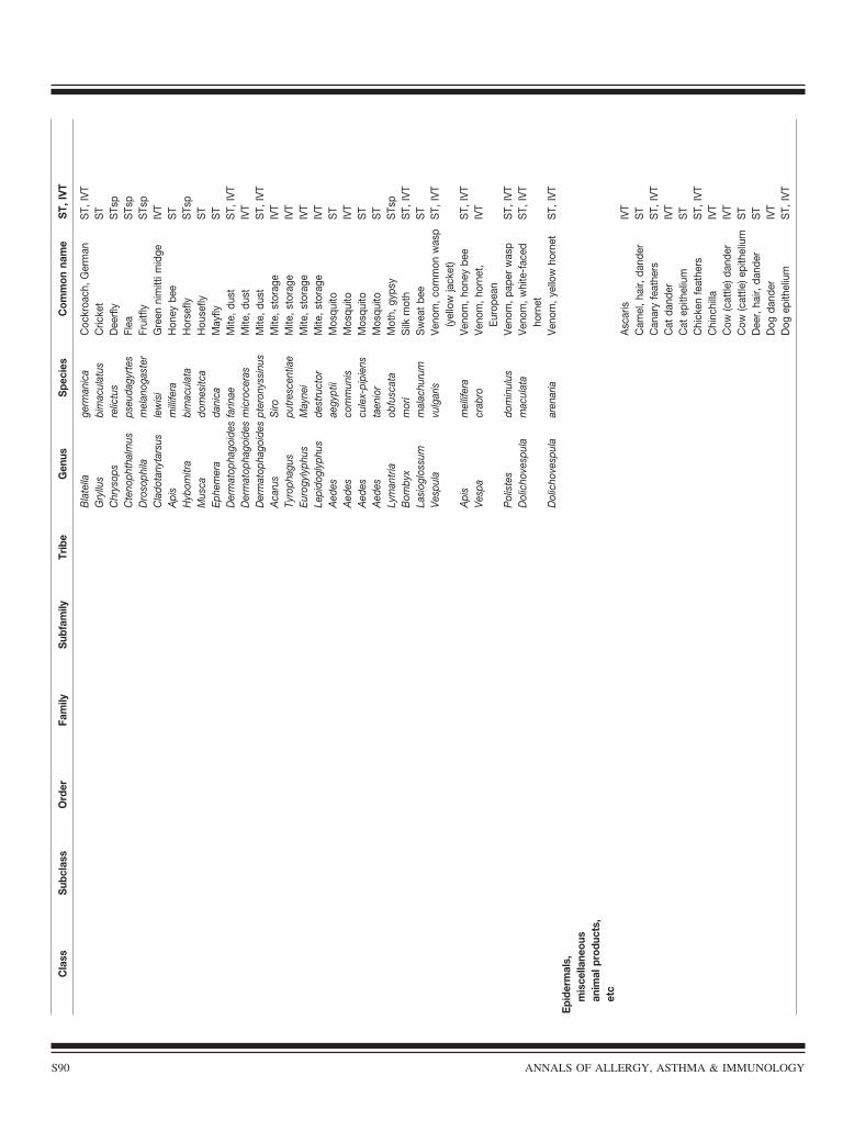
Cla
ssS
ubcl
ass
Ord
erFa
mily
Sub
fam
ilyTr
ibe
Gen
usS
peci
esC
omm
onna
me
ST,
IVT
Bla
tella
germ
anic
aC
ockr
oach
,Ger
man
ST,
IVT
Gry
llus
bim
acul
atus
Cric
ket
ST
Chr
ysop
sre
lictu
sD
eerf
lyS
Tsp
Cte
noph
thal
mus
pseu
dagy
rtes
Flea
STs
pD
roso
phila
mel
anog
aste
rFr
uitfl
yS
Tsp
Cla
dota
nyta
rsus
lew
isi
Gre
enni
mitt
imid
geIV
TA
pis
mill
ifera
Hon
eybe
eS
TH
ybom
itra
bim
acul
ata
Hor
sefly
STs
pM
usca
dom
esitc
aH
ouse
flyS
TEp
hem
era
dani
caM
ayfly
ST
Der
mat
opha
goid
esfa
rinae
Mite
,dus
tS
T,IV
TD
erm
atop
hago
ides
mic
roce
ras
Mite
,dus
tIV
TD
erm
atop
hago
ides
pter
onys
sinu
sM
ite,d
ust
ST,
IVT
Aca
rus
Siro
Mite
,sto
rage
IVT
Tyro
phag
uspu
tres
cent
iae
Mite
,sto
rage
IVT
Euro
gyly
phus
May
nei
Mite
,sto
rage
IVT
Lepi
dogl
yphu
sde
stru
ctor
Mite
,sto
rage
IVT
Aed
esae
gypt
iiM
osqu
itoS
TA
edes
com
mun
isM
osqu
itoIV
TA
edes
cule
x-pi
pien
sM
osqu
itoS
TA
edes
taen
ior
Mos
quito
ST
Lym
antr
iaob
fusc
ata
Mot
h,gy
psy
STs
pB
omby
xm
ori
Silk
mot
hS
T,IV
TLa
siog
loss
umm
alac
huru
mS
wea
tbe
eS
TVe
spul
avu
lgar
isVe
nom
,com
mon
was
p(y
ello
wja
cket
)S
T,IV
T
Api
sm
ellif
era
Veno
m,h
oney
bee
ST,
IVT
Vesp
acr
abro
Veno
m,h
orne
t,Eu
rope
anIV
T
Pol
iste
sdo
min
ulus
Veno
m,p
aper
was
pS
T,IV
TD
olic
hove
spul
am
acul
ata
Veno
m,w
hite
-fac
edho
rnet
ST,
IVT
Dol
icho
vesp
ula
aren
aria
Veno
m,y
ello
who
rnet
ST,
IVT
Epi
derm
als,
mis
cella
neou
san
imal
prod
ucts
,et
cA
scar
isIV
TC
amel
,hai
r,da
nder
ST
Can
ary
feat
hers
ST,
IVT
Cat
dand
erIV
TC
atep
ithel
ium
ST
Chi
cken
feat
hers
ST,
IVT
Chi
nchi
llaIV
TC
ow(c
attle
)dan
der
IVT
Cow
(cat
tle)e
pith
eliu
mS
TD
eer,
hair,
dand
erS
TD
ogda
nder
IVT
Dog
epith
eliu
mS
T,IV
T
S90 ANNALS OF ALLERGY, ASTHMA & IMMUNOLOGY
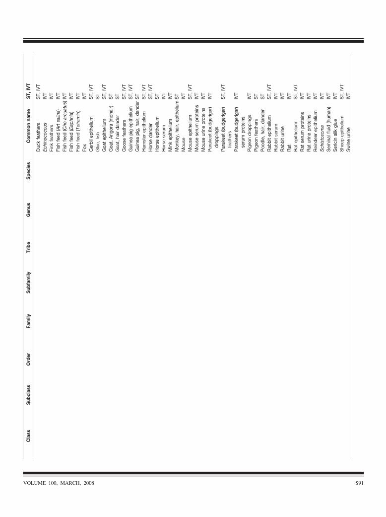
Cla
ssS
ubcl
ass
Ord
erFa
mily
Sub
fam
ilyTr
ibe
Gen
usS
peci
esC
omm
onna
me
ST,
IVT
Duc
kfe
athe
rsS
T,IV
TEc
hino
cocc
usIV
TFi
nkfe
athe
rsIV
TFi
shfe
ed(A
rtsa
lina)
IVT
Fish
feed
(Cho
arcu
atus
)IVT
Fish
feed
(Dap
hnia
)IV
TFi
shfe
ed(T
etra
min
)IV
TFo
xIV
TG
erbi
lepi
thel
ium
ST,
IVT
Glu
e,fis
hS
TG
oat
epith
eliu
mS
T,IV
TG
oat,
Ang
ora
(moh
air)
ST
Goa
t,ha
irda
nder
ST
Goo
sefe
athe
rsS
T,IV
TG
uine
api
gep
ithel
ium
ST,
IVT
Gui
nea
pig,
hair,
dand
erS
TH
amst
erep
ithel
ium
ST,
IVT
Hor
seda
nder
ST,
IVT
Hor
seep
ithel
ium
ST
Hor
sese
rum
IVT
Min
kep
ithel
ium
IVT
Mon
key,
hair,
epith
eliu
mS
TM
ouse
IVT
Mou
seep
ithel
ium
ST,
IVT
Mou
sese
rum
prot
eins
IVT
Mou
seur
ine
prot
eins
IVT
Par
akee
t(b
udge
rigar
)dr
oppi
ngs
IVT
Par
akee
t(b
udge
rigar
)fe
athe
rsS
T,IV
T
Par
akee
t(b
udge
rigar
)se
rum
prot
eins
IVT
Pig
eon
drop
ping
sIV
TP
igeo
nfe
athe
rsS
TP
oodl
e,ha
ir,da
nder
ST
Rab
bit
epith
eliu
mS
T,IV
TR
abbi
tse
rum
IVT
Rab
bit
urin
eIV
TR
atIV
TR
atep
ithel
ium
ST,
IVT
Rat
seru
mpr
otei
nsIV
TR
atur
ine
prot
eins
IVT
Rei
ndee
rep
ithel
ium
IVT
Sch
isto
som
aIV
TS
emin
alflu
id(h
uman
)IV
TS
eric
insi
lkgl
ueIV
TS
heep
epith
eliu
mS
T,IV
TS
win
eur
ine
IVT
VOLUME 100, MARCH, 2008 S91
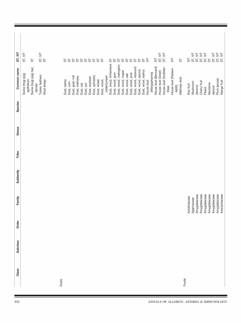
Cla
ssS
ubcl
ass
Ord
erFa
mily
Sub
fam
ilyTr
ibe
Gen
usS
peci
esC
omm
onna
me
ST,
IVT
Sw
ine
(hog
)(pi
g)ep
ithel
ium
ST,
IVT
Sw
ine
(hog
)(pi
g)ha
ir,da
nder
ST
Turk
eyfe
athe
rsS
T,IV
TW
ools
heep
ST
Dus
tsD
ust,
barle
yS
TD
ust,
corn
ST
Dus
t,gr
ain
mill
ST
Dus
t,m
attr
ess
ST
Dus
t,oa
tS
TD
ust,
rye
ST
Dus
t,so
ybea
nS
TD
ust,
upho
lstr
yS
TD
ust,
whe
atS
TD
ust,
woo
d,ce
dar/
juni
per
ST
Dus
t,w
ood,
fir/h
emlo
ckS
TD
ust,
woo
d,gu
mS
TD
ust,
woo
d,m
ahog
any
ST
Dus
t,w
ood,
map
leS
TD
ust,
woo
d,oa
kS
TD
ust,
woo
d,pi
neS
TD
ust,
woo
d,re
dwoo
dS
TD
ust,
woo
d,sp
ruce
ST
Dus
t,w
ood,
wal
nut
ST
Hou
sedu
st(A
llerg
opha
rma)
IVT
Hou
sedu
st(B
enca
rd)
IVT
Hou
sedu
st(G
reer
)S
T,IV
TH
ouse
dust
(Hol
liste
r-S
tier)
ST,
IVT
Hou
sedu
st(P
aste
urA
600)
IVT
Mat
tres
sdu
stS
TFo
ods
Act
idin
iace
aeK
iwif
ruit
IVT
Aga
ricac
eae
Mus
hroo
mS
T,IV
TA
myg
dala
ceae
Alm
ond
ST,
IVT
Am
ygda
lace
aeC
herr
yfr
uit
ST,
IVT
Am
ygda
lace
aeP
each
ST,
IVT
Am
ygda
lace
aeN
ecta
rine
ST
Am
ygda
lace
aeA
pric
otS
T,IV
TA
myg
dala
ceae
Plu
m(p
rune
)S
T,IV
TA
naca
rdia
ceae
Man
gofr
uit
ST,
IVT
S92 ANNALS OF ALLERGY, ASTHMA & IMMUNOLOGY
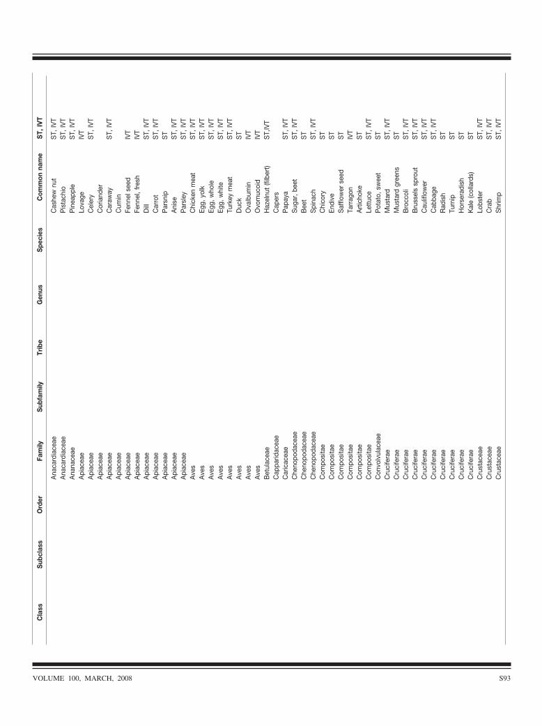
Cla
ssS
ubcl
ass
Ord
erFa
mily
Sub
fam
ilyTr
ibe
Gen
usS
peci
esC
omm
onna
me
ST,
IVT
Ana
card
iace
aeC
ashe
wnu
tS
T,IV
TA
naca
rdia
ceae
Pis
tach
ioS
T,IV
TA
nana
ceae
Pin
eapp
leS
T,IV
TA
piac
eae
Lova
geIV
TA
piac
eae
Cel
ery
ST,
IVT
Api
acea
eC
oria
nder
Api
acea
eC
araw
ayS
T,IV
TA
piac
eae
Cum
inA
piac
eae
Fenn
else
edIV
TA
piac
eae
Fenn
el,f
resh
IVT
Api
acea
eD
illS
T,IV
TA
piac
eae
Car
rot
ST,
IVT
Api
acea
eP
arsn
ipS
TA
piac
eae
Ani
seS
T,IV
TA
piac
eae
Par
sley
ST,
IVT
Ave
sC
hick
enm
eat
ST,
IVT
Ave
sEg
g,yo
lkS
T,IV
TA
ves
Egg,
who
leS
T,IV
TA
ves
Egg,
whi
teS
T,IV
TA
ves
Turk
eym
eat
ST,
IVT
Ave
sD
uck
ST
Ave
sO
valb
umin
IVT
Ave
sO
vom
ucoi
dIV
TB
etul
acea
eH
azel
nut
(filb
ert)
ST,
IVT
Cap
parid
acea
eC
aper
sC
aric
acea
eP
apay
aS
T,IV
TC
heno
poda
ceae
Sug
ar,b
eet
ST,
IVT
Che
nopo
dace
aeB
eet
ST
Che
nopo
dace
aeS
pina
chS
T,IV
TC
ompo
sita
eC
hico
ryS
TC
ompo
sita
eEn
dive
ST
Com
posi
tae
Saf
flow
erse
edS
TC
ompo
sita
eTa
rrag
onIV
TC
ompo
sita
eA
rtic
hoke
ST
Com
posi
tae
Lett
uce
ST,
IVT
Con
volv
ulac
eae
Pot
ato,
swee
tS
TC
ruci
fera
eM
usta
rdS
T,IV
TC
ruci
fera
eM
usta
rdgr
eens
ST
Cru
cife
rae
Bro
ccol
iS
T,IV
TC
ruci
fera
eB
russ
els
spro
utS
T,IV
TC
ruci
fera
eC
aulif
low
erS
T,IV
TC
ruci
fera
eC
abba
geS
T,IV
TC
ruci
fera
eR
adis
hS
TC
ruci
fera
eTu
rnip
ST
Cru
cife
rae
Hor
sera
dish
ST
Cru
cife
rae
Kal
e(c
olla
rds)
ST
Cru
stac
eae
Lobs
ter
ST,
IVT
Cru
stac
eae
Cra
bS
T,IV
TC
rust
acea
eS
hrim
pS
T,IV
T
VOLUME 100, MARCH, 2008 S93
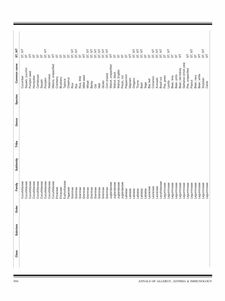
Cla
ssS
ubcl
ass
Ord
erFa
mily
Sub
fam
ilyTr
ibe
Gen
usS
peci
esC
omm
onna
me
ST,
IVT
Cuc
urbi
tace
aeC
ucum
ber
ST,
IVT
Cuc
urbi
tace
aeS
quas
h,zu
cchi
niS
TC
ucur
bita
ceae
Pum
pkin
seed
IVT
Cuc
urbi
tace
aeH
oney
dew
ST
Cuc
urbi
tace
aeC
anta
loup
eS
TC
ucur
bita
ceae
Squ
ash
ST
Cuc
urbi
tace
aeP
umpk
inS
T,IV
TC
ucur
bita
ceae
Wat
erm
elon
ST
Cuc
urbi
tace
aeM
elon
s,un
spec
ified
IVT
Eric
acea
eC
ranb
erry
ST
Eric
acea
eB
lueb
erry
ST
Euph
orbi
acea
eTa
pioc
aS
TFa
gace
aeC
hest
nut
ST
Gra
min
aeR
ice
ST,
IVT
Gra
min
aeR
yeS
T,IV
TG
ram
inae
Ric
e,W
ildS
TG
ram
inae
Mill
etse
edIV
TG
ram
inae
Whe
atS
T,IV
TG
ram
inae
Bar
ley
ST,
IVT
Gra
min
aeO
atS
T,IV
TG
ram
inae
Mal
tS
T,IV
TG
ram
inae
Glu
ten
ST,
IVT
Gra
min
aeC
orn
(mai
ze)
ST,
IVT
Jugl
anda
ceae
Wal
nut,
unsp
ecifi
edIV
TJu
glan
dace
aeW
alnu
t,bl
ack
ST
Jugl
anda
ceae
Wal
nut,
Engl
ish
ST
Jugl
anda
ceae
Pec
an,n
utS
TLa
biat
aeP
eppe
rmin
tS
TLa
biat
aeM
ajor
amIV
TLa
biat
aeO
rega
noS
T,IV
TLa
biat
aeTh
yme
ST,
IVT
Labi
atae
Bas
ilS
T,IV
TLa
biat
aeS
age
ST
Laur
acea
eB
ayle
afS
TLa
urac
eae
Cin
nam
onS
T,IV
TLa
urac
eae
Avo
cado
ST,
IVT
Lecy
thid
acea
eB
razi
lnut
ST,
IVT
Legu
min
osae
Pea
,gre
enS
TLe
gum
inos
aeLe
ntils
ST,
IVT
Legu
min
osae
Bea
n,lim
aS
TLe
gum
inos
aeB
ean,
pint
oS
TLe
gum
inos
aeB
ean,
red
kidn
eyIV
TLe
gum
inos
aeG
arba
nzo
(chi
ckpe
a)Le
gum
inos
aeP
ea,u
nspe
cifie
dIV
TLe
gum
inos
aeP
eanu
tS
T,IV
TLe
gum
inos
aeB
ean,
navy
ST
Legu
min
osae
Bea
n,w
hite
IVT
Legu
min
osae
Soy
bean
ST,
IVT
Legu
min
osae
Car
obIV
T
S94 ANNALS OF ALLERGY, ASTHMA & IMMUNOLOGY
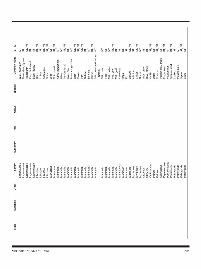
Cla
ssS
ubcl
ass
Ord
erFa
mily
Sub
fam
ilyTr
ibe
Gen
usS
peci
esC
omm
onna
me
ST,
IVT
Legu
min
osae
Gua
r,gu
argu
mIV
TLe
gum
inos
aeB
ean,
strin
g(g
reen
)S
TLe
gum
inos
aeTr
agac
anth
ST,
IVT
Legu
min
osae
Pea
,bla
ck-e
yed
ST
Legu
min
osae
Bea
n,ki
dney
ST
Lilia
ceae
Gar
licS
T,IV
TLi
liace
aeC
hive
sS
TLi
liace
aeA
spar
agus
ST,
IVT
Lilia
ceae
Oni
onS
T,IV
TM
alva
ceae
Okr
aS
TM
alva
ceae
Cot
tons
eed
ST,
IVT
Mam
mal
iaA
lpha
-lact
albu
min
IVT
Mam
mal
iaW
hey
IVT
Mam
mal
iaM
utto
n(la
mb)
ST,
IVT
Mam
mal
iaLi
ver,
beef
ST
Mam
mal
iaB
eta-
lact
oglo
bulin
IVT
Mam
mal
iaB
eef
ST,
IVT
Mam
mal
iaC
asei
nS
T,IV
TM
amm
alia
Por
kS
T,IV
TM
amm
alia
Gel
atin
ST
Mam
mal
iaEl
km
eat
IVT
Mam
mal
iaR
abbi
tS
T,IV
TM
amm
alia
Milk
,pow
dere
d(A
lfare
,N
estle
)IV
T
Mam
mal
iaM
ilk,m
are
IVT
Mam
mal
iaVe
alS
TM
amm
alia
Milk
,boi
led
IVT
Mam
mal
iaM
ilk,c
owS
T,IV
TM
amm
alia
Milk
,goa
tS
TM
aran
tace
aeA
rrow
root
ST
Mor
acea
eH
ops
ST
Mor
acea
eFi
gS
TM
usac
eae
Ban
ana
ST,
IVT
Myr
tace
aeA
llspi
ceS
TM
yrta
ceae
Clo
ves
ST,
IVT
Myr
tace
aeG
uava
IVT
Ole
acea
eO
live,
gree
nS
TO
leac
eae
Oliv
e,bl
ack
ST
Orc
hida
ceae
Vani
llaS
T,IV
TP
alm
aeD
ates
ST
Pal
mae
Coc
onut
ST,
IVT
Sol
anac
eae
Pep
per,
bell,
gree
nS
TP
apav
erac
eae
Pop
pyse
edS
T,IV
TP
assi
flora
ceae
Pas
sion
frui
tIV
TP
edal
iace
aeS
esam
ese
edS
T,IV
TP
elec
ypod
aS
callo
pS
TP
elec
ypod
aM
usse
l,bl
ueIV
TP
elec
ypod
aO
yste
rS
T,IV
TP
elec
ypod
aC
lam
ST
VOLUME 100, MARCH, 2008 S95
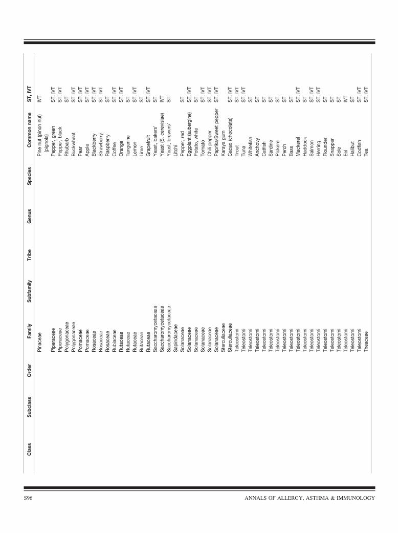
Cla
ssS
ubcl
ass
Ord
erFa
mily
Sub
fam
ilyTr
ibe
Gen
usS
peci
esC
omm
onna
me
ST,
IVT
Pin
acea
eP
ine
nut
(pin
onnu
t)(p
igno
la)
IVT
Pip
erac
eae
Pep
per,
gree
nS
T,IV
TP
iper
acea
eP
eppe
r,bl
ack
ST,
IVT
Pol
ygon
acea
eR
huba
rbS
TP
olyg
onac
eae
Buc
kwhe
atS
T,IV
TP
omac
eae
Pea
rS
T,IV
TP
omac
eae
App
leS
T,IV
TR
osac
eae
Bla
ckbe
rry
ST,
IVT
Ros
acea
eS
traw
berr
yS
T,IV
TR
osac
eae
Ras
pber
ryS
TR
ubia
ceae
Cof
fee
ST,
IVT
Rut
acea
eO
rang
eS
T,IV
TR
utac
eae
Tang
erin
eS
TR
utac
eae
Lem
onS
T,IV
TR
utac
eae
Lim
eS
TR
utac
eae
Gra
pefr
uit
ST,
IVT
Sac
char
omyc
etac
eae
Yea
st,b
aker
s’S
TS
acch
arom
ycet
acea
eY
east
(S.c
erev
isia
e)IV
TS
acch
arom
ycet
acea
eY
east
,bre
wer
s’S
TS
apin
dace
aeLi
tchi
Sol
anac
eae
Pep
per,
red
ST
Sol
anac
eae
Eggp
lant
(aub
ergi
ne)
ST,
IVT
Sol
anac
eae
Pot
ato,
whi
teS
TS
olan
acea
eTo
mat
oS
T,IV
TS
olan
acea
eC
hili
pepp
erS
T,IV
TS
olan
acea
eP
aprik
a/S
wee
tpe
pper
ST,
IVT
Ste
rcul
iace
aeK
aray
agu
mS
terc
ulia
ceae
Cac
ao(c
hoco
late
)S
T,IV
TTe
leos
tom
iTr
out
ST,
IVT
Tele
osto
mi
Tuna
ST,
IVT
Tele
osto
mi
Whi
tefis
hS
TTe
leos
tom
iA
ncho
vyS
TTe
leos
tom
iC
atfis
hS
TTe
leos
tom
iS
ardi
neS
TTe
leos
tom
iP
icke
rel
ST
Tele
osto
mi
Per
chS
TTe
leos
tom
iB
ass
ST
Tele
osto
mi
Mac
kere
lS
T,IV
TTe
leos
tom
iH
addo
ckS
TTe
leos
tom
iS
alm
onS
T,IV
TTe
leos
tom
iH
errin
gS
T,IV
TTe
leos
tom
iFl
ound
erS
TTe
leos
tom
iS
napp
erS
TTe
leos
tom
iS
ole
ST
Tele
osto
mi
Eel
IVT
Tele
osto
mi
Hal
ibut
ST
Tele
osto
mi
Cod
fish
ST,
IVT
Thea
ceae
Tea
ST,
IVT
S96 ANNALS OF ALLERGY, ASTHMA & IMMUNOLOGY
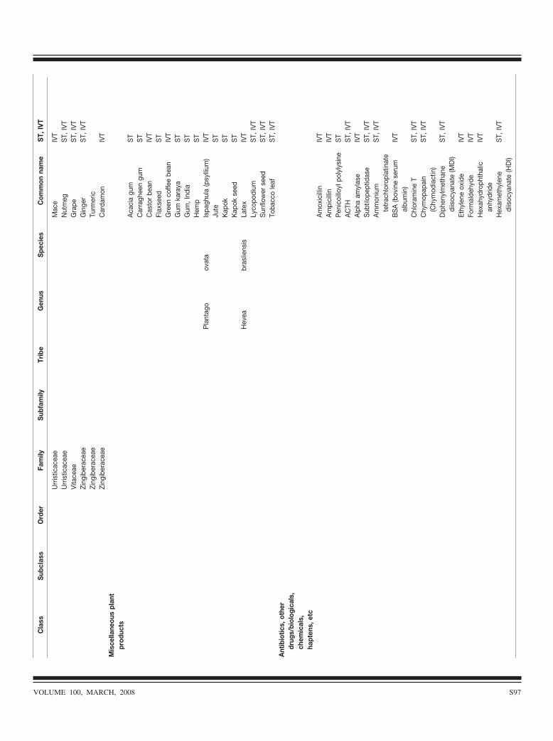
Cla
ssS
ubcl
ass
Ord
erFa
mily
Sub
fam
ilyTr
ibe
Gen
usS
peci
esC
omm
onna
me
ST,
IVT
Urr
istic
acea
eM
ace
IVT
Urr
istic
acea
eN
utm
egS
T,IV
TVi
tace
aeG
rape
ST,
IVT
Zing
iber
acea
eG
inge
rS
T,IV
TZi
ngib
erac
eae
Turm
eric
Zing
iber
acea
eC
arda
mon
IVT
Mis
cella
neou
spl
ant
prod
ucts
Aca
cia
gum
ST
Car
ragh
een
gum
ST
Cas
tor
bean
IVT
Flax
seed
ST
Gre
enco
ffee
bean
IVT
Gum
kara
yaS
TG
um,I
ndia
ST
Hem
pS
TP
lant
ago
ovat
aIs
pagh
ula
(psy
llium
)IV
TJu
teS
TK
apok
ST
Kap
okse
edS
TH
evea
bras
liens
isLa
tex
IVT
Lyco
podi
umS
T,IV
TS
unflo
wer
seed
ST,
IVT
Toba
cco
leaf
ST,
IVT
Ant
ibio
tics,
othe
rdr
ugs/
biol
ogic
als,
chem
ical
s,ha
pten
s,et
cA
mox
icill
inIV
TA
mpi
cilli
nIV
TP
enic
oillo
ylpo
lyly
sine
ST
AC
THS
T,IV
TA
lpha
amyl
ase
IVT
Sub
tilop
eptid
ase
ST,
IVT
Am
mon
ium
tetr
achl
orop
latin
ate
ST,
IVT
BS
A(b
ovin
ese
rum
albu
min
)IV
T
Chl
oram
ine
TS
T,IV
TC
hym
opap
ain
(Chy
mod
iact
in)
ST,
IVT
Dip
heny
lmet
hane
diis
ocya
nate
(MD
I)S
T,IV
T
Ethy
lene
oxid
eIV
TFo
rmal
dehy
deIV
TH
exah
ydro
phth
alic
anhy
drid
eIV
T
Hex
amet
hyle
nedi
isoc
yana
te(H
DI)
ST,
IVT
VOLUME 100, MARCH, 2008 S97
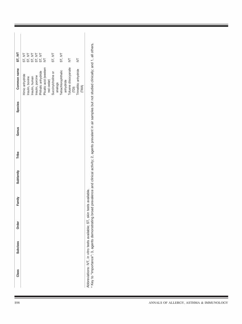
Cla
ssS
ubcl
ass
Ord
erFa
mily
Sub
fam
ilyTr
ibe
Gen
usS
peci
esC
omm
onna
me
ST,
IVT
Him
ican
hydr
ide
ST,
IVT
Insu
lin,b
ovin
eS
T,IV
TIn
sulin
,hum
anS
T,IV
TIn
sulin
,por
cine
ST,
IVT
Pht
halic
anhy
drid
eS
T,IV
TP
licat
icac
id(w
este
rnre
dce
dar)
IVT
Suc
ciny
lcho
line
oran
alog
sS
T,IV
T
Tetr
achl
orop
htha
lican
hydr
ide
ST,
IVT
Tolu
ene
diis
ocya
nate
(TD
I)IV
T
Trim
ellit
ican
hydr
ide
(TM
A)
IVT
Ab
bre
viat
ions
:IV
T,in
vitr
ote
sts
avai
lab
le;
ST,
skin
test
sav
aila
ble
.a
Key
to“i
mp
orta
nce”
:3,
agen
tsd
emon
stra
ting
bro
adp
reva
lenc
ean
dcl
inic
alac
tivity
;2,
agen
tsp
reva
lent
inai
rsa
mp
les
but
not
stud
ied
clin
ical
ly;
and
1,al
loth
ers.
S98 ANNALS OF ALLERGY, ASTHMA & IMMUNOLOGY
Summary Statement 173. A skin prick/puncture test issuperior to intracutaneous testing for predicting allergic rhi-nitis and allergic asthma triggered by cat allergen exposure.(B)
Summary Statement 174. The skin prick/puncture test canbe used to rule out allergic rhinitis and allergic asthma trig-gered by cat allergen exposure. (B)
Summary Statement 175. Knowledge of allergen cross-reactivity and local aerobiology is important in selectingappropriate allergens and in minimizing the number of aller-gens required for skin and specific IgE tests. (D)
Summary Statement 176. In general, skin prick/puncturetesting is more sensitive for identifying sensitization to in-halant allergens and confirming clinical allergy. However,dated specific IgE assays with defined quantifiable thresholdlevels can also predict positive respiratory responses afterallergen exposure. (B)
Summary Statement 177. Demonstration of sensitization toan occupational agent by specific IgE and/or skin testingalone is insufficient to establish a diagnosis of OA. (B)
Summary Statement 178. Skin prick testing with certainwell-characterized occupational protein allergens possessesadequate sensitivity such that a negative skin test result(�3-mm-wheal diameter) can be used to rule out clinicalallergy. (B)
Summary Statement 179. Test performance characteristicsof specific IgE assays and skin testing for detection of chem-ical IgE-mediated sensitization must undergo validation andreproducibility in controlled studies using standardized anti-gens and assay protocols before these can be consideredreliable for routine evaluation of workers suspected of OA.(B)
Summary Statement 180. In patients undergoing evaluationfor suspected work-related natural rubber latex (NRL) al-lergy, a positive skin prick test result with a NRL extract (ifavailable) is preferred to demonstration of elevated specificIgE with an FDA-cleared assay due to higher sensitivity ofthe former. Current IgE-mediated allergy and asthma causedby NRL allergens is highly unlikely in the presence of anegative skin prick test result with a reliable crude NRLallergen extract. Elevated in vitro specific IgE levels can beused to confirm NRL allergy, but a negative result does notexclude NRL allergen sensitization. (B)
Clinical Indications and UtilitySkin testing and specific IgE evaluation of specific IgE aremethods used to demonstrate IgE-mediated sensitization toinhalant allergens. In clinical practice, skin and/or specificIgE testing that demonstrate specific IgE for inhalant aeroal-lergens are utilized to (1) confirm or exclude a suspecteddiagnosis of allergic rhinitis, allergic conjunctivitis, or asthmatriggered by aeroallergens; (2) determine the need for envi-ronmental control recommendations to reduce exposure tooutdoor or indoor aeroallergens; (3) demonstrate sensitizationto inhalant occupational allergens, which cause OA or rhini-
tis; and (4) guide selection of inhalant allergens for inclusionin allergen immunotherapy extracts.
A clinician must be familiar with performance charac-teristics of skin testing and specific IgE measurement sothat test results are applied accurately to diagnose and treatallergic respiratory disorders. To optimally define testperformance, a method should be reproducible and vali-dated against a diagnostic benchmark or gold standard. Amedical history is subjective and not adequate alone fordefining clinical sensitivity or specificity of in vivo orspecific IgE tests.101,120 –123,933 Evaluation of symptoms orphysiologic responses during direct allergen challengetests under supervision of a physician or in associationwith natural exposure to inhalant allergens (ie, pollen, cat,house dust mite) are appropriate ways to validate skinprick/puncture tests, intracutaneous tests, and specific IgEassays.111,167,934 Although such validation studies are lim-ited both in number and scope of allergens evaluated, theyprovide objective evidence for defining test characteristics,including sensitivity, specificity, predictive values, andlikelihood ratios.
Performance of Skin Tests in Evaluation of InhalantAllergyIn clinical practice, skin prick/puncture testing is used as aninitial screening test and is often followed by intracutaneoustesting for inhalant allergens eliciting negative prick testresults. This long-standing practice is based on the assump-tion that intracutaneous testing has greater sensitivity thanskin prick/puncture testing. However, there is evidence that apositive intracutaneous test result at a fixed dose of 1:500 or1:1,000 (wt/vol) in the absence of skin prick reactivity cor-relates poorly with clinical sensitivity.167,171 When seasonalallergic rhinitis was confirmed by nasal challenge with grasspollen allergen, only 11% of patients with positive intracu-taneous test results (and negative prick/puncture test results)exhibited a positive challenge test result, and this outcomewas identical to symptomatic patients with negative intracu-taneous test results. In contrast, 68% of symptomatic patientswith skin prick/puncture test reactivity to grass pollen exhib-ited positive nasal challenge test results.167 Based on historyalone, a positive skin prick/puncture test result to cat definedby a wheal 3 mm or greater than the negative control pos-sessed 90% sensitivity and 90% specificity.97 Wood et alreported that skin prick/puncture testing using a 27-gaugehypodermic needle exhibited 94% sensitivity, 80% specific-ity, 90% positive predictive value, and 87% negative predic-tive value for identifying subjects with increased upper re-spiratory tract symptoms elicited by live cat exposure. Foridentifying subjects with lower respiratory tract symptomsafter cat exposure, skin prick testing had 84% sensitivity (vs35% for intracutaneous tests), 87% specificity, 88% positivepredictive value, 82% negative predictive value, and 97%sensitivity for predicting a reduction in FEV1 during live catchallenge.111
VOLUME 100, MARCH, 2008 S99
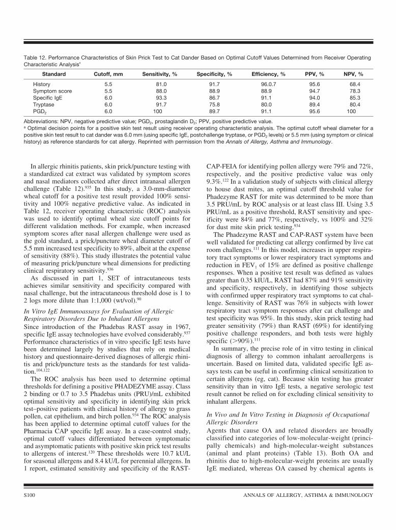
In allergic rhinitis patients, skin prick/puncture testing witha standardized cat extract was validated by symptom scoresand nasal mediators collected after direct intranasal allergenchallenge (Table 12).935 In this study, a 3.0-mm-diameterwheal cutoff for a positive test result provided 100% sensi-tivity and 100% negative predictive value. As indicated inTable 12, receiver operating characteristic (ROC) analysiswas used to identify optimal wheal size cutoff points fordifferent validation methods. For example, when increasedsymptom scores after nasal allergen challenge were used asthe gold standard, a prick/puncture wheal diameter cutoff of5.5 mm increased test specificity to 89%, albeit at the expenseof sensitivity (88%). This study illustrates the potential valueof measuring prick/puncture wheal dimensions for predictingclinical respiratory sensitivity.936
As discussed in part 1, SET of intracutaneous testsachieves similar sensitivity and specificity compared withnasal challenge, but the intracutaneous threshold dose is 1 to2 logs more dilute than 1:1,000 (wt/vol).90
In Vitro IgE Immunoassays for Evaluation of AllergicRespiratory Disorders Due to Inhalant AllergensSince introduction of the Phadebas RAST assay in 1967,specific IgE assay technologies have evolved considerably.937
Performance characteristics of in vitro specific IgE tests havebeen determined largely by studies that rely on medicalhistory and questionnaire-derived diagnoses of allergic rhini-tis and prick/puncture tests as the standards for test valida-tion.104,122
The ROC analysis has been used to determine optimalthresholds for defining a positive PHADEZYME assay. Class2 binding or 0.7 to 3.5 Phadebas units (PRU)/mL exhibitedoptimal sensitivity and specificity in identifying skin pricktest–positive patients with clinical history of allergy to grasspollen, cat epithelium, and birch pollen.934 The ROC analysishas been applied to determine optimal cutoff values for thePharmacia CAP specific IgE assay. In a case-control study,optimal cutoff values differentiated between symptomaticand asymptomatic patients with positive skin prick test resultsto allergens of interest.120 These thresholds were 10.7 kU/Lfor seasonal allergens and 8.4 kU/L for perennial allergens. In1 report, estimated sensitivity and specificity of the RAST-
CAP-FEIA for identifying pollen allergy were 79% and 72%,respectively, and the positive predictive value was only9.3%.122 In a validation study of subjects with clinical allergyto house dust mites, an optimal cutoff threshold value forPhadezyme RAST for mite was determined to be more than3.5 PRU/mL by ROC analysis or at least class III. Using 3.5PRU/mL as a positive threshold, RAST sensitivity and spec-ificity were 84% and 77%, respectively, vs 100% and 32%for dust mite skin prick testing.934
The Phadezyme RAST and CAP-RAST system have beenwell validated for predicting cat allergy confirmed by live catroom challenges.111 In this model, increases in upper respira-tory tract symptoms or lower respiratory tract symptoms andreduction in FEV1 of 15% are defined as positive challengeresponses. When a positive test result was defined as valuesgreater than 0.35 kIU/L, RAST had 87% and 91% sensitivityand specificity, respectively, in identifying those subjectswith confirmed upper respiratory tract symptoms to cat chal-lenge. Sensitivity of RAST was 76% in subjects with lowerrespiratory tract symptom responses after cat challenge andtest specificity was 95%. In this study, skin prick testing hadgreater sensitivity (79%) than RAST (69%) for identifyingpositive challenge responders, and both tests were highlyspecific (�90%).111
In summary, the precise role of in vitro testing in clinicaldiagnosis of allergy to common inhalant aeroallergens isuncertain. Based on limited data, validated specific IgE as-says tests can be useful in confirming clinical sensitization tocertain allergens (eg, cat). Because skin testing has greatersensitivity than in vitro IgE tests, a negative serologic testresult cannot be relied on for excluding clinical sensitivity toinhalant allergens.
In Vivo and In Vitro Testing in Diagnosis of OccupationalAllergic DisordersAgents that cause OA and related disorders are broadlyclassified into categories of low-molecular-weight (princi-pally chemicals) and high-molecular-weight substances(animal and plant proteins) (Table 13). Both OA andrhinitis due to high-molecular-weight proteins are usuallyIgE mediated, whereas OA caused by chemical agents is
Table 12. Performance Characteristics of Skin Prick Test to Cat Dander Based on Optimal Cutoff Values Determined from Receiver OperatingCharacteristic Analysisa
Standard Cutoff, mm Sensitivity, % Specificity, % Efficiency, % PPV, % NPV, %
History 5.5 81.0 91.7 96.0.7 95.6 68.4Symptom score 5.5 88.0 88.9 88.9 94.7 78.3Specific IgE 6.0 93.3 86.7 91.1 94.0 85.3Tryptase 6.0 91.7 75.8 80.0 89.4 80.4PGD2 6.0 100 89.7 91.1 95.6 100
Abbreviations: NPV, negative predictive value; PGD2, prostaglandin D2; PPV, positive predictive value.a Optimal decision points for a positive skin test result using receiver operating characteristic analysis. The optimal cutoff wheal diameter for apositive skin test result to cat dander was 6.0 mm (using specific IgE, postchallenge tryptase, or PGD2 levels) or 5.5 mm (using symptom or clinicalhistory) as reference standards for cat allergy. Reprinted with permission from the Annals of Allergy, Asthma and Immunology.
S100 ANNALS OF ALLERGY, ASTHMA & IMMUNOLOGY
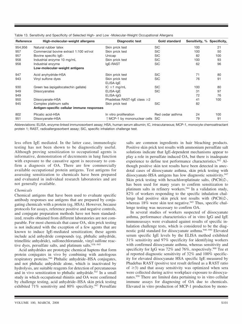
less often IgE mediated. In the latter case, immunologictesting has not been shown to be diagnostically useful.Although proving sensitization to occupational agents isinformative, demonstration of decrements in lung functionwith exposure to the causative agent is necessary to con-firm a diagnosis of OA. There are few commerciallyavailable occupational protein antigens. Test antigens forassessing sensitization to chemicals have been preparedand evaluated in individual research laboratories and arenot generally available.
ChemicalsChemical antigens that have been used to evaluate specificantibody responses use antigens that are prepared by conju-gating chemicals with a protein (eg, HSA). However, becauseprotocols for assays, reference positive and negative controls,and conjugate preparation methods have not been standard-ized, results obtained from different laboratories are not com-parable. For most chemicals that cause OA, skin prick testingis not indicated with the exception of a few agents that areknown to induce IgE-mediated sensitization; these agentsinclude acid anhydride compounds (eg, phthalic anhydride,trimellitic anhydride), sulfonechloramide, vinyl sulfone reac-tive dyes, persulfate salts, and platinum salts.938–945
Acid anhydrides are prototypic chemical haptens that formprotein conjugates in vivo by combining with autologousrespiratory proteins.946 Phthalic anhydride–HSA conjugates,and not phthalic anhydride alone, which is inactivated byhydrolysis, are suitable reagents for detection of percutaneousand in vivo sensitization to phthalic anhydride.947 In a smallstudy in which occupational rhinitis and OA were confirmedby challenge testing, acid anhydride–HSA skin prick testingexhibited 71% sensitivity and 80% specificity.947 Persulfate
salts are common ingredients in hair bleaching products.Positive skin prick test results with ammonium persulfate saltsolutions indicate that IgE-dependent mechanisms appear toplay a role in persulfate induced OA, but there is inadequateexperience to define test performance characteristics.942 Al-though positive skin test results have been detected in anec-dotal cases of diisocyanate asthma, skin prick testing withdiisocyanate-HSA antigens has low diagnostic sensitivity.947
Skin prick testing with hexachloroplatinate salts, (PtCl6)2-,has been used for many years to confirm sensitization toplatinum salts in refinery workers.944 In a validation study,82% of workers responding to the specific inhalation chal-lenge had positive skin prick test results with (PtCl6)2-,whereas 18% were skin test negative.945 Thus, specific chal-lenge testing was necessary to confirm OA.
In several studies of workers suspected of diisocyanateasthma, performance characteristics of in vitro IgG and IgEimmunoassays were evaluated against results of specific in-halation challenge tests, which is considered to be the diag-nostic gold standard for diisocyanate asthma.948–950 Elevatedserum specific IgE levels by the ELISA method exhibited31% sensitivity and 97% specificity for identifying workerswith confirmed diisocyanate asthma, whereas sensitivity andspecificity for IgG was 72% and 76%, respectively.900 Tee etal reported diagnostic sensitivity of 32% and 100% specific-ity for elevated diisocyanate HSA specific IgE measured byPhadebas RAST (positive test result defined as a RAST ratioof �3) and that assay sensitivity was optimized when serawere collected during active workplace exposure to diisocya-nates.950 There are limited data pertaining to in vitro cellularimmune assays for diagnosing of OA due to chemicals.Elevated in vitro production of MCP-1 production by mono-
Table 13. Sensitivity and Specificity of Selected High- and Low -Molecular-Weight Occupational Allergens
Reference High-molecular-weight allergens Diagnostic test Gold standard Sensitivity, % Specificity,
954,956 Natural rubber latex Skin prick test SIC 100 21957 Commercial bovine extract 1:100 wt/vol Skin prick test SIC 100 50957 Bovine specific IgE- Unicap SIC 82 100958 Industrial enzyme 10 mg/mL Skin prick test SIC 100 93958 Industrial enzyme IgE-RAST SIC 62 96
Low-molecular-weight antigens
947 Acid anyhydride-HSA Skin prick test SIC 71 80943 Vinyl sulfone dyes Skin prick test SIC 76 91
ELISA-IgE930 Green tea (epigallocatechin gallate) IC �1 mg/mL SIC 100 80949 Diisocyanates ELISA-IgE SIC 31 97949 ELISA-IgG 72 76950 Diisocyanate-HSA Phadebas RAST-IgE class �2 41 100945 Complex platinum salts Skin prick test SIC 82
Antigen-specific cellular immune responses
802 Plicatic acid-HSA In vitro proliferation Red cedar asthma 24 100951 Diisocyanate-HSA 1MCP-1 by mononuclear cells SIC 79 91
Abbreviations: ELISA, enzyme-linked immunosorbent assay; HSA, human serum albumin; IC, intracutaneous; MCP-1, monocyte chemoattractantprotein 1; RAST, radioallergosorbent assay; SIC, specific inhalation challenge test.
VOLUME 100, MARCH, 2008 S101
nuclear cells after stimulation with diisocyanate-HSA anti-gens exhibited 79% sensitivity and 91% specificity in iden-tification of exposed workers with confirmed OA.Replication of these results, however, in larger diisocyanate-exposed populations is needed before this assay is adopted forclinical use.951
Protein Allergens in the WorkplaceInhalant proteins encountered at work readily induce sensiti-zation and elicit allergic contact urticaria, rhinoconjunctivitis,and/or asthma at work. In general, skin prick testing withprotein allergens (or high-molecular-weight sensitizers) arehighly sensitive tools for evaluating workers with suspectedOA. In the case of NRL, skin prick testing has greatersensitivity compared with FDA-approved in vitro specificIgE assays.916
A nonammoniated latex extract was reported to possessboth high sensitivity (99%) and specificity (100%) at a testconcentration of 100 �g/mL in identifying health care work-ers with latex allergy based on medical history.952,953 In astudy that confirmed NRL induced OA based on responses tocontrolled challenge with NRL powdered gloves, skin pricktesting with a well-characterized nonammoniated NRL ex-tract exhibited 100% sensitivity, 100% negative predictivevalue, and 21% specificity. Specific IgE reactive with NRLwas not evaluated in this study.954 However, a negative skintest result alone virtually excluded NRL-induced OA definedby a positive specific inhalation challenge.954 Among healthcare workers reporting a history of NRL allergy, skin pricktesting with standardized commercial NRL extracts has su-perior sensitivity for detecting NRL sensitization comparedwith FDA-approved in vitro specific IgE assays.914–916 Animportant practical limitation in the United States is the lackof a standardized commercial NRL skin test reagent. Becauseuncharacterized powdered glove extracts prepared by physi-cians have variable diagnostic sensitivity (64% to 96%), anegative test result must be interpreted cautiously.955 In arecent study, CAP and UniCAP immunoassays identified53% to 77% of NRL skin test prick–positive health careworkers with latex allergy.956
A similar study design was used to investigate test char-acteristics of skin prick testing with a commercial bovineallergen extract (1:100 wt/vol), which provided 100% sensi-tivity, 50% specificity, and 100% negative predictive value inidentifying OA based on specific challenge testing with bo-vine dander extract. On the other hand, bovine dander spe-cific IgE measured by quantitative fluoroenzymatic immuno-assay (UniCAP) exhibited 82% sensitivity and 100%specificity.957
Finally, in a study of workers exposed to industrial en-zymes that used specific inhalation testing as the diagnosticgold standard, investigators reported 100% sensitivity and93% specificity for skin prick testing with enzyme solutions(10 mg/mL) compared with 62% sensitivity and 96% speci-ficity for an enzyme allergosorbent IgE assay.958
ASSESSMENT OF FOOD ALLERGYAn adverse reaction to food can result from nonimmune (eg,intolerance, pharmacologic effects) or immune (allergy) ori-gins. Immune-mediated adverse reactions to foods (food al-lergy) may be attributable to IgE antibody–mediated mecha-nisms (eg, food-induced anaphylaxis), cellular mechanismswith no detectable food specific IgE antibodies (primarilygastrointestinal disorders), and disorders in which both IgEantibody–mediated and cellular mechanisms have been iden-tified (eosinophilic gastrointestinal disorders, atopic dermati-tis).959
Summary Statement 181. The primary tools available toevaluate patients’ adverse reactions to foods include history(including diet records), physical examination, prick/punctureskin tests, serum tests for food specific IgE antibodies, trialelimination diets, and oral food challenges. (B)
The general aims of diagnosis are to determine if food iscausing the disorder under evaluation and, if so, to identifyspecific causal food(s). A proper diagnosis will allow thepatient to receive instructions regarding avoidance of prob-lematic foods. Just as important, specific diagnosis will pre-vent unnecessary and potentially deleterious dietary restric-tions when a suspected food allergy is not present. Thediagnostic tools available to the clinician include simple andrelatively inexpensive tests, such as the clinical history, phys-ical examination (that may reveal associated atopic disordersand raise the likelihood of a food allergy960), prick/punctureskin tests, and serum tests for food specific IgE. Additionaltests (oral food challenges) are more involved timewise, maybe more expensive, and may carry additional risks. Therational selection and interpretation of diagnostic tests requirean appreciation for the utility of the tests themselves and anevaluation of the level of certainty required for the diagno-sis.959
Summary Statement 182. A detailed dietary history, attimes augmented with written diet records, is necessary todetermine the likelihood that food is causing the disorder,identify the specific food, and determine the potential immu-nopathophysiology. (D)
The history is the starting point at which the clinician mustdecide on the possibility that food is a potential cause of adisorder or reaction. The features of the reaction may alsoindicate whether the pathophysiology of the disorder may benon–immune-mediated (intolerance, pharmacologic reaction)or allergic, and if the latter, whether it is IgE mediated orassociated or not (thereby guiding further diagnostic evalua-tion). Historical points of interest include age of the patient;a list of suspect foods, ingredients, or labels for manufacturedproducts; the amount of food necessary to elicit a reaction;the route of exposure eliciting a reaction; the typical timeinterval between exposure and onset of symptoms; clinicalmanifestations of reaction(s) after exposure to each food;duration of symptoms; ancillary events (exercise, use ofNSAIDS, alcohol); treatment of reactions and patient re-
S102 ANNALS OF ALLERGY, ASTHMA & IMMUNOLOGY
sponse; and the consistency with which a reaction occurs onexposure.
Key points in the history, such as symptoms and timing ofonset after ingestion or chronicity, may identify reactionslikely to be dependent on IgE antibody (eg, sudden reactionssuch as anaphylaxis), those that are associated with IgE toparticular foods in many but not all cases (eg, chronic disor-ders such as atopic dermatitis), or disorders not associatedwith IgE antibodies or ones unlikely to be allergic in nature(eg, lactose intolerance, isolated gastrointestinal disorders ofinfancy).959
In addition to identifying a pathophysiologic basis, thehistory may indicate specific food triggers and a starting pointto estimate the probability that a particular food is causal (eg,prior probability of an association). Diet records, includingreview of labels from packaged foods, may facilitate identi-fication of specific triggers.961,962 Common reasoning wouldindicate that a food previously tolerated on a routine basis isless likely to be a trigger than one eaten rarely. Similarly, fora person with a previously confirmed food allergy to a ubiq-uitous food (eg, milk, peanut) who reacts to a specific meal,consideration that the previously identified allergen may bepresent as a hidden ingredient or contaminant should beentertained. Age is important since the epidemiology of foodallergy indicates a higher probability of reactions to cow’smilk, egg, wheat, and soy in infants; peanuts, tree nuts,seafood, and raw fruits in older children and adults; and apredilection of certain food-related disorders in infants andchildren (atopic dermatitis, enterocolitis).959 Consistent reac-tions, particularly acute ones, to a specific food raise theprobability that the food is causal; in 1 study of infants,specificity of urticaria after ingestion of cow’s milk was 0.77,but specificity of atopic dermatitis was only 0.22.95 Indeed,the history is notoriously poor in identifying causal foods forchronic disorders such as atopic dermatitis when comparedwith outcomes of definitive oral food challenges.590,960,963
Food allergy is commonly suspected but rarely incriminatedin chronic urticaria and/or angioedema.
Summary Statement 183. With regard to evaluations forIgE antibody–associated food allergies, tests for food specificIgE antibody include percutaneous skin tests (prick/puncturetests) and serum assays. In general, these tests are highlysensitive (generally �85%) but only modestly specific (ap-proximately 40% to 80%) and therefore are well suited foruse when suspicion of a particular food or foods is high. Theyare not effective for indiscriminate screening (eg, using pan-els of tests without consideration of likely causes) and there-fore generally should not be used for that purpose. (B)
Modalities to determine the presence of IgE antibody tospecific foods include prick/puncture tests and serum assays.Both techniques merely detect the presence of antibody (sen-sitization) and do not necessarily indicate, by themselves, thatingestion would result in clinical reactions. Even infants canbe tested.95,964 Commercial reagents for food allergy skintesting have not yet been standardized and may have varyingconcentrations of relevant proteins.965,966 Clinically important
but labile proteins, particularly ones in fruits and vegetables,may degrade, making extracts inadequate for an evaluation;fresh extracts may therefore be needed to evaluate sensitiza-tion to these allergens.965 A retrospective review of medicalrecords (1,152 children) concerning prick/puncture tests withfoods indicate a very low rate of generalized reactions (521per 100,000 tested; 95% confidence interval, 105–937), butall reactions in infants (n � 6) occurred in infants youngerthan 6 months of age tested with fresh food specimens.967
Another means to detect food specific IgE is serologic todetermine the presence of food specific IgE antibodies in theserum. There are a variety of manufacturers, substrates, andmanners of reporting results as discussed in part 1.
The clinical utility of prick/punctures testing and serumfood specific IgE has been evaluated in various referralpopulations of infants and children evaluated by oral foodchallenges for suspected food allergy.140,589–591,960,963,968–970
Sensitivity of a positive test result is generally more than 0.7and in most studies exceeds 0.85; specificity is lower, gen-erally in the range of 0.4 to 0.8. Test utility varies by intrinsicfeatures of the test (technique, definition of positive, type offood) and features of the population tested (age, disease).These test characteristics generally indicate that a negativetest result has a high utility to rule out IgE-mediated reactionsto the food tested but that a positive test result may not beassociated with true clinical reactions. Consequently, panelsof food allergy tests should not be performed without con-sideration of the history because one may be faced withnumerous irrelevant positive results (particularly in disorderswith high total IgE antibody).960 The serum assay for specificIgE may be less sensitive than skin prick tests,970,971 so if thereis a suspicion of a reaction when in vitro test results arenegative, a skin test may detect sensitization.589 However, inmany cases the sensitivity is similar.589,590,960
Summary Statement 184. Intracutaneous skin tests forfoods are potentially dangerous, are overly sensitive, increasethe chance of a false-positive test result, and are not recom-mended. (D)
Intracutaneous allergy skin tests with food extracts give anunacceptably high false-positive rate, can elicit systemic re-actions (rarely an issue for prick tests), and should generallynot be used.972
Summary Statement 185. Based on studies in infants andchildren, increasingly higher concentrations of food specificIgE antibodies (reflected by increasingly larger percutaneousskin test size and/or higher concentrations of food-specificserum IgE antibody) correlate with an increasing risk for aclinical reaction. (B)
Studies in children support the notion that increasinglyhigher concentrations of food specific IgE antibody, reflectedby increasingly larger prick/puncture test results or highserum IgE antibody concentrations, are correlated with in-creased risks for clinical reactions.85,589,590,968–973 Thus, insteadof considering a test result for IgE as positive or negative withone decision point (positive-negative at “detectable” serumfood specific IgE or a particular skin test size such as 3 mm),
VOLUME 100, MARCH, 2008 S103
additional clinical utility may be achieved through consider-ation of prick/puncture test result size and serum food-spe-cific antibody concentration. Various studies have correlatedreaction likelihood with test results in this re-gard,85,140,589,590,964,968–973 but it is clear that results may vary bytechnique, food involved, age group studied, the specificpatient history, and the disorder under consideration. Al-though the size of the prick/puncture skin test result orconcentration of food-specific IgE antibody by in vitro assaymay be positively correlated with an increasing likelihood ofa clinical reaction, the level of IgE is poorly correlated withclinical manifestations of the allergy (eg, severity or dosecausing a reaction).974–976
Summary Statement 186. A trial elimination diet may behelpful to determine if a disorder with frequent or chronicsymptoms is responsive to dietary manipulation. (D)
In the evaluation of disorders with chronic symptoms forwhich foods may be causal (eg, atopic dermatitis, gastroin-testinal symptoms), elimination of suspected causal foodsmay be undertaken to determine whether symptoms are dietresponsive. There are no studies to define the utility of thisapproach. Factors that may complicate interpretation of sucha trial (eg, a trial failure when the disorder is truly foodresponsive) include incomplete removal of causal foods, se-lection of the wrong foods to eliminate, inadequate timeallowed for resolution of chronic inflammation (eg, atopicdermatitis), and additional triggers may be causing symptoms(eg, skin infection in atopic dermatitis). The underlyingpathophysiology is not a significant consideration in usingelimination trials. Selection of foods to eliminate may bebased on a variety of factors, including historical features,results of tests, and epidemiologic considerations. Informa-tion concerning strict adherence to the diet must be carefullyreviewed, similar to what is needed for treatment of foodallergy after a definitive diagnosis. Diets may vary fromdirected ones (removal of one or a few targeted foods), evenmore restricted ones with elimination of most allergenicfoods (eg, a prescribed diet without major allergens andlimited numbers of allowed foods), or even to extreme oneswith essentially no source of potential allergen (eg, use ofamino acid–based formula alone or with a few other provensafe foods). A positive response to an elimination diet shouldnot be construed as a definitive diagnosis unless there iscompelling supportive evidence regarding specific foods. An-other use for an elimination diet is to establish baseline statusbefore undertaking oral food challenges; the response to oralfood challenge is potentially definitive but must be performedfor each food under consideration. Severe reactions haveoccurred when previously ingested, IgE antibody–positivefoods were added back to the diet after they had been re-moved from the diet for a period.977
Summary Statement 187. Graded oral food challenge is auseful means to diagnose an adverse reaction to food. (B)
The oral food challenge is performed by having the patientingest increasing amounts of the suspected food under phy-sician observation over hours or days.961,962 This represents a
definitive test for tolerance since ingestion of a relevantamount of the food with no reaction excludes the diagnosis ofan adverse reaction to the tested food. The test result is opento misinterpretation when not done in a masked manner.Therefore, procedures to reduce this possibility need to beimplemented, such as masking the challenge substance(blinding) and using placebos. The format of a food challengecan be applied to evaluate any type of adverse event attrib-uted to foods due to both allergic and nonallergic hypersen-sitivity mechanisms.
The challenge procedure, its risks, and its benefits must bediscussed with the patient and/or the caregiver. Several fac-tors are considered, including the evaluation of the likelihoodthat the food will be tolerated, the nutritional and social needfor the food, and ability of the patient to cooperate with thechallenge. In limited circumstances, the food could be ad-ministered with potential adverse reactions monitored athome by the patient and parents. This may be considered ifthe expected adverse reactions are delayed in onset, non–IgEmediated, atypical (eg, headache, behavioral issues), mildgastrointestinal, and not potentially anaphylactic. On theother hand, if there is a reasonable potential for an acuteand/or severe reaction, or if there is strong patient anxiety,physician supervision is recommended.
Except in the uncommon circumstances described previ-ously, oral food challenges are undertaken under direct med-ical supervision. A risk evaluation must be made regardinglocation of challenge (office, hospital, intensive care unit) andpreparation (eg, with or without an intravenous line in place).These decisions are based on the same types of data evaluatedfor the consideration of food allergy in the early diagnosticprocess: the history of the possible food allergic reaction,patient’s medical history, and prick/puncture skin test results.Although generally considered a safe procedure when under-taken by qualified personnel, it must be appreciated that oralchallenges can elicit severe, anaphylactic reactions, so thephysician must be immediately available and comfortablewith this potential and be prepared with emergency medica-tions and equipment to promptly treat such a potentiallylife-threatening reaction.978 In high-risk challenges, it mayalso be prudent to have intravenous access before commenc-ing challenges.962,979 Even non–IgE antibody–mediated foodallergic reactions can be severe, such as food protein–inducedenterocolitis syndrome that may include lethargy, dehydra-tion, and hypotension, and may be complicated by acidosisand methemoglobinemia.980 The details regarding undertak-ing an oral food challenge are described in the publishedFood Allergy Practice Parameter and other resources.961,962,981
Challenges can be performed openly, with the patient ingest-ing the food in its natural form; single-blind, with the foodmasked and the patient unaware if the test substance containsthe target food; or double-blind and placebo-controlled, withneither patient nor physician or medical professional knowingwhich challenges contain the food being tested. Although theopen challenge is most prone to bias, it is easy to performsince no special preparation is needed to mask the food.
S104 ANNALS OF ALLERGY, ASTHMA & IMMUNOLOGY
Indeed, if the patient tolerates the ingestion of the food, thereis little concern about bias. Bias becomes an issue when thechallenge food causes symptoms, particularly subjectiveones. Therefore, open challenges are a good option forscreening when several foods are under consideration, and ifa food is tolerated, nothing further is needed. If there is areaction to an open challenge used in the clinical setting, andthere is concern that the reaction may not have been physi-ologic, the format could be altered to include blinding andcontrols. False-negative rates for double-blind, placebo-con-trolled food changes are low (usually �3%).982 After a neg-ative challenge, consideration should be given to having thepatient eat the food prepared in the same manner and amountthat caused the original reaction.
Summary Statement 188. A number of additional diagnos-tic tests are under investigation, including APTs (APTs) andtests for IgE binding to specific epitopes. (B)
Various additional diagnostic tests, particularly APTs, areunder evaluation and are at various stages of acceptance orstill under research scrutiny investigation of food aller-gy.95,523–530,983–985 The test response is noted in the days afterapplication and may potentially identify food triggers that arenot associated with IgE antibodies, which is a particular issuefor gastrointestinal food allergies.945,947 Although increasingstudies, primarily from Europe, are assessing the utility ofthese tests, more work is needed on standardization andclinical correlation before widespread routine clinical use canbe advocated.946 Several recent European experiences suggestthat the ultimate utility of APTs in reducing the need for oralfood challenges may be limited.526,530 Additional diagnostictests under development may use proteins of particular rele-vance or map informative protein epitopes for improveddiagnosis of IgE antibody–mediated reactions.
Summary Statement 189. The rational selection, applica-tion, and interpretation of tests for food specific IgE antibod-ies require consideration of the epidemiology and underlyingimmunopathophysiology of the disorder under investigation,estimation of prior probability that a disorder or reaction isattributable to particular foods, and an understanding of thetest utility and limitations. (D)
Tests for food allergy, like other medical tests, are neither100% sensitive nor 100% specific. The diagnostic utility of atest in regard to making a diagnosis in an individual patient isinfluenced by (1) the possibility of the disease existing in theindividual being tested (prior probability) and (2) the char-acteristics of the test itself (sensitivity, specificity). To deter-mine prior probability, it is necessary to undertake a carefulhistory and to understand the epidemiologic features of foodallergic disorders. Description of the latter is beyond thescope of this Practice Parameter but may be found in re-views959,986,987 and the Food Allergy Practice Parameter.981
To evaluate the clinical utility of a test, studies are per-formed comparing outcomes of oral food challenges (prefer-ably double-blind, placebo-controlled food challenges).Many studies use open food challenges with objective symp-
toms as an end point, or sometimes they rely on convincingclinical histories.
Such studies have reported skin test sizes above whichclinical reactions are virtually 100% likely (eg, wheal diam-eter �8 mm for cow’s milk or peanut and �7 mm for egg ininfants) or levels of IgE antibody at or above which reactionsare more than 95% likely (eg, 7 kIU/L for egg, 15 kIU/L formilk, 14 kIU/L for peanut and 20 kIU/L for cod fish, mea-sured by Pharmacia CAP-System FEIA) in atopic children ata median age of 3.8 years.85,589 This approach is helpful inidentifying persons who are likely to be clinically allergic, forwhom an oral food challenge is not indicated. Conversely,progressively lower levels of food specific IgE (reflected bysmaller skin test results or lower serum test results) areassociated with a better chance to tolerate the food.589,974,988
As described herein, the clinician must appreciate that thepredictive data are reflective of the characteristics of thetested population. For example, studies of young children(younger than 2 years) show that 95% reacted to egg if theirIgE level was more than 2 kIU/L or milk if their IgE level wasmore than 5 kIU/L, values lower than those calculated forolder children.969,970 Additional studies have confirmed age-related differences among children in regard to the foodspecific IgE concentrations indicative of a high risk of reac-tion.591,989 Another study also confirmed the utility of thresh-old values,589,990 although there are some discrepancies in theactual values associated with specific outcomes (eg, predic-tive values were higher in a German study of children).591 Asa note of caution, reactions may occur when at risk childrenhave undetectable food specific IgE (eg, approximately 20%with egg or peanut specific IgE �0.35 kIU/L have clinicalreactions to these foods); most970,971 but not all971,976 suchpatients have a positive prick/puncture test results, indicatinghigher sensitivity of the test. As more studies emerge com-paring serum and prick/puncture skin test results with clinicaloutcomes in wider age groups and populations with variousdisorders, further conclusions of test utility will be possible.In regard to skin prick testing, various reagents (fresh food,commercial extracts) and techniques of testing (probe type,location on the body, method of measurement, timing ofmeasurement) are variables that affect final results and areadditional obstacles in regard to applying study results to aparticular patient.141
An additional and complementary way to interpret teststakes into consideration the individual’s history to establish aprior probability on which to interpret a test result. Forexample, at a serum concentration of peanut specific IgE of 2kIU/L, children with a history of a peanut allergic reactionhad approximately a 50% chance of tolerating peanut.974
However, in a group of children with a positive test result butno history of a reaction, 50% tolerated peanut at a peanutspecific IgE concentration of 5 kIU/L. Similarly, a wheal sizeof 3 mm to peanut in children with atopic dermatitis wasassociated with a positive predictive value of 61%, whereasthe same wheal size had a positive predictive value of 28% inchildren at low risk according to their clinical histories.93,973
VOLUME 100, MARCH, 2008 S105
These examples illustrate the importance of clinical history(including age and frequency of reactions) and calculation ofa prior probability for allergy in regard to test interpretation.
A means to apply prior probability and test results in aparticular patient to improve diagnostic accuracy is throughthe use of a calculated likelihood ratio. The likelihood ratio issimply the ratio of the odds that the patient whose test resultsfall within a particular range has the disease divided by theodds that they do not. The formula can most conveniently beexpressed as: (likelihood ratio � sensitivity)/(1 � specificity)as applies to a positive test result. To be useful, a likelihoodratio needs to be determined for each diagnostic test used inevaluating the probability of food allergy. Unfortunately, thisis not available for most food allergy tests. When the likeli-hood ratio is known, a pretest probability (based for exampleon the medical history) is estimated and a nomogram can beused to determine the posttest probability that a person hasthe disorder.
Although likelihood ratios are not calculated for most testsof food allergy, the concept of likelihood ratio and pretestprobability has practical implications for routine practice.Consider, for example, 3 individuals: (1) a child with 3 severeallergic reactions to peanut requiring epinephrine, (2) a childwith chronic atopic dermatitis who eats peanuts but has nohistory of a reaction to peanut, and (3) a nonatopic child whosometimes has headaches on days he eats peanut. Each pa-tient is tested by prick/puncture testing to peanut and has a4-mm wheal, a positive test result with modest sensitivity(approximately 50%), and good specificity (approximately90%). The meaning of a 4-mm wheal to peanut when therehas been recurrent anaphylaxis in patient 1 (high prior prob-ability of peanut allergy, virtually 100%) is that it confirmsreactivity and no food challenge should be undertaken. In achronic condition like atopic dermatitis in patient 2, a modestsize skin test may reflect clinical reactivity in only approxi-mately half of patients (depending also on age) and may be arelevant positive in this scenario, needing confirmation byother means (oral food challenge) or additional testing toimprove diagnostic accuracy (serum test). The test result inpatient 3 with headaches is most likely of no clinical concernbecause the pretest probability is essentially zero. Consider-ing again the patient with multiple episodes of peanut-relatedanaphylaxis, if there were no wheal to peanut, the clinicianwould not be likely to trust the result because the pretestprobability is so high that the correct course of action wouldbe to repeat the skin test or perform an in vitro test andconsider a supervised oral food challenge if the test resultwere negative. Similarly, one could argue that a test forpeanut causing migraines is not necessary since the priorprobability is so low. Thus, 1 test (eg, prick/puncture) canprovide pretest probability for another test (eg, oral foodchallenge).
It therefore is important to remember that every patientmust be evaluated individually and the history taken as care-fully as possible. Otherwise one risks obtaining a falselypositive or negative history that could skew interpretation of
subsequent tests since they depend on the pretest probabilitygenerated by the history. This emphasizes the fact that face-to-face evaluation of the patient is essential and that remotepractice of allergy is not valid.
At this time, there are a number of publications about thediagnostic utility of IgE antibody tests for egg, milk, andpeanut for children at a range of ages and clinical circum-stances that show excellent predictive ability. The studies onthese same age groups have not determined strong diagnosticutility for soy or wheat,589,591,974–990 and more studies areneeded for determination of results for additional foods, clin-ical problems and ages, and the specific impact of cross-reactive homologous proteins in reagents currently used fortesting.
ASSESSMENT OF STINGING INSECT ALLERGY
Clinical Indications and UtilitySummary Statement 190. Diagnostic skin and/or specific IgEtests are used to confirm clinical sensitivity to venoms in apatient with a history of a prior systemic reaction. (B)
Summary Statement 191. Although diagnostic tests identifyspecies specificity of venom sensitization, they do not reli-ably predict severity of the sting reaction. (B)
The diagnosis of insect sting allergy requires confirmationof the clinical history with an accurate diagnostic test. This ismost important in patients who require venom immunother-apy such as those with a history of systemic reactions tostings. Testing is not usually performed in those who havehad only large local reactions to stings because they haveonly a 5% risk of systemic reaction to subsequent stings.991–993
Testing is also not recommended to individuals without ahistory of a systemic reaction to stings because 25% of suchpersons will have positive diagnostic venom test results.994
The overall clinical significance of this finding (asymptom-atic skin sensitization) is uncertain, but there is an estimated15% chance of systemic reaction in such individuals.995 Di-agnostic tests are also used during venom immunotherapy todetermine whether the sensitivity has diminished or disap-peared.996
The utility of diagnostic tests for insect allergy is limited toidentifying the presence and species specificity of venomsensitization. Although there is a statistical correlation, thestrength of the venom sensitivity shown by either skin orspecific IgE diagnostic tests does not reliably predict theclinical severity of the sting reaction. Some patients havevery strong test results but only local swelling reaction to asting, whereas others have barely detectable sensitivity andyet have life-threatening anaphylaxis when stung.
Diagnostic Reagents for Hymenoptera and Fire AntsSummary Statement 192. Standardized honeybee, Polistes,and Vespula antigens are commercially available as skin testreagents. (A)
Summary Statement 193. The skin test reagent available forevaluation of imported fire sting allergy is a nonstandardizedwhole-body extract. (C)
S106 ANNALS OF ALLERGY, ASTHMA & IMMUNOLOGY
Summary Statement 194. In the case of a history of ana-phylaxis to Hymenoptera venoms, intracutaneous skin testsare generally performed to 5 of the available venoms in adose response protocol (up to 1 �g/mL [wt/vol]) when pre-liminary prick/puncture test results are negative. (B)
Summary Statement 195. The FDA-cleared specific IgEassays have comparable specificity but decreased sensitivitycompared with venom skin tests. (B)
Hymenoptera venom extracts are widely accepted as thestandard reagents for diagnostic testing and immunotherapyfor insect sting allergy. The commercially available productsare lyophilized protein extracts for honeybee, Vespula (yel-low jacket), and Polistes wasp venoms. The last 2 are mix-tures of clinically relevant species. Also available are 2Dolichovespula venoms (yellow hornet and white-faced hor-net). A mixed vespid venom product that contains equal partsof the 3 Vespula venoms (yellow jacket, yellow hornet, andwhite-faced hornet) is available for treatment but is not rec-ommended for diagnostic use. Honeybee venom is standard-ized for the content of phospholipase A (Api m 1), the majorallergen in honeybee venom. Vespula venoms are standard-ized for their content of hyaluronidase. However, the primaryvespid venom allergen is a nonenzymatic protein designatedas antigen 5 (eg, Ves v 5). This could be a basis for a possiblediscrepancy between skin test results and sting response.Lyophilized venom products are reconstituted and dilutedwith buffered saline diluent that contains 0.03% HSA, whichfunctions to stabilize the small amounts of protein allergensin the solutions and prevent adsorption to the walls of thevials. Although the activity of the allergens is stable for 12months at the full concentration (100 �g/mL [wt/vol]), it maydecay more rapidly at lower concentrations used for skintesting or early immunotherapy. Venoms for laboratory useare typically dialyzed to remove small-molecular-weightcomponents, which can interfere in some assays. Dialyzedvenoms may be more accurate for skin testing and are avail-able in Europe but not in the United States.
Imported fire ant whole-body extract is the only reagentpresently available for diagnostic skin testing and immuno-therapy for fire ant sting allergy. Most imported fire antwhole-body extracts have been shown to contain sufficientvenom allergens to be useful for diagnosis and treatment, butsome preparations contain variable quantities of the relevantallergens.997–999 Fire ant prick/puncture tests are performedfirst at the dose recommended by the manufacturer. If theresults are negative, intracutaneous skin tests may be startedwith concentrations as low as 1:1,000,000 (wt/vol) in highlysensitive patients but are considered to be indicative of thepresence of specific IgE antibodies if a positive responseoccurs at a concentration of 1:500 (wt/vol) or less1000
Venom skin tests are generally performed using the intra-cutaneous technique of injecting a small volume (0.02 to 0.03mL) superficially in the skin to raise a bleb of 3 to 4 mm. Theprick/puncture method (with a venom concentration of 1.0�g/mL [wt/vol]) is used for preliminary skin testing, espe-cially in patients with a history of very severe anaphylaxis,
but when these results are negative, intracutaneous tests arerequired for diagnosis. Venom skin tests are generally per-formed with all 5 of the available venoms (and/or fire antwhole-body extract when indicated). The prick/puncturemethod is unable to detect the allergy in most patients (usingconcentrations �1.0 �g/mL [wt/vol]), but like all venom skintests, can cause false-positive irritative reactions when veryhigh concentrations are used (�1 �g/mL [wt/vol]). Intracu-taneous skin tests are generally performed beginning withconcentrations of 0.01 �g/mL (wt/vol) or less, but when theygive negative results at the lowest concentration, the skintests are repeated serially at 10-fold higher concentrationsuntil a positive response occurs or until the result is negativeat a concentration of 1.0 �g/mL. Higher concentrations cancause false-positive reactions in some cases.
Serologic diagnostic tests provide measurements of ven-om-specific IgE antibodies in serum using the FDA-clearedassays, including RASTs and RAST modifications. Modifiedassays (see part 1) have shown improved accuracy but arestill subject to the validity and reproducibility of the clinicallaboratory performing the assays.132 This means that homol-ogous internal controls (eg, specific venoms) are essential forin vitro venom tests.
Performance Characteristics of Insect Venom Tests (Prick,Intracutaneous, Specific IgE)Summary Statement 196. Paradoxically, as many as 16% ofinsect allergic patients with negative venom skin test resultshave positive results in currently available specific IgE invitro tests. (B)
Summary Statement 197. A small percentage of patients(1%) with negative results to both skin and in vitro tests mayexperience anaphylaxis after a field sting. (B)
Summary Statement 198. A skin test refractory periodlasting up to 6 weeks after a venom sting has been demon-strated by recent data. (B)
Although venom skin tests have been said to be highlyaccurate, recent studies have focused on deficiencies in bothsensitivity and specificity, which are related to the testingreagents and to the natural history of this condition. Venomskin tests are more sensitive than specific IgE tests, sinceinsect allergic patients with positive skin test results havenegative specific IgE results in 15% to 20% of cases. How-ever, it has been reported that skin test results are negative in10% to 30% of patients with a convincing history of systemicreaction to a sting.188,1000,1001 This may occur in patients withnear life-threatening reactions to a specific venom, as previ-ously noted under Summary Statement 32.188 Some of thesepatients will have a positive specific IgE test result so thatcomprehensive testing (both skin and specific IgE tests)rarely misses the diagnosis. Some patients with negative skinand specific IgE test results but a positive history may notexperience a reaction to a field sting. This suggests that eitherthe history itself is a poor predictor or the specific IgE thatwas initially present has disappeared over time. Conversely,1% of patients with a positive history of an allergic sting
VOLUME 100, MARCH, 2008 S107
reaction have negative results to both skin and specific IgEdiagnostic tests and yet may have a systemic reaction to asubsequent sting.1001 Absolute reconciliation of these resultsis difficult. Stated otherwise, although available tests arealmost always sufficient for diagnosis, they are not 100%foolproof.
One possible reason for negative venom skin test results ininsect allergic patients is the refractory period that occursafter an allergic reaction to a sting. One published reportdescribes this phenomenon in 50% of patients tested within 1week after the sting reaction, but more than half of the skintest–negative patients had positive specific IgE results at thesame time, such that the diagnosis was made in 79% usingboth test methods.1002 In the other 21% of patients the testresults were positive only 6 weeks after the sting reaction.When venom skin test and specific IgE test results are neg-ative more than 6 weeks after the sting reaction, it has beenrecommended that the tests should be repeated at a later date.This is based on the observation that venom skin test re-sponses may vary over time such that relatively mild sensi-tivity may fluctuate around the lower level of detection andgive negative results on one occasion and positive on another.This observation has been reported in one clinical investiga-tion.616
The immunologic specificity of venom skin tests is excel-lent but clinically limited. Positive skin test results invariablydemonstrate the presence of venom specific IgE antibodiesbut are not absolute indicators of clinical allergic reactions tostings. Studies of the natural history of insect allergy haverevealed that clinical reactivity is variable and can disappeardespite the persistence of sensitization demonstrated withdiagnostic tests.995,1003–1006 The risk of systemic reaction to asting in patients with positive venom skin test results and ahistory of previous systemic reactions has been reported to beas high as 61% and as low as 30%.1005–1007 This broad rangehas been explained by multiple factors, including the age ofthe patient, the severity of prior sting reactions, the degree ofskin test (or specific IgE) sensitivity to venoms, and variablesrelating to the insects themselves.
Specific IgE in vitro tests have shown improved sensitivityusing modified assay methods.134 The sensitivity of these testsis still not as good as skin tests, but the specificity is com-parable. Many such assays show reduced accuracy when thelevel of venom IgE is in the low range. Compared withvenom skin tests, current serologic tests still give false-negative results in some cases, but the converse has becomeequally important. As many as 10% of insect allergic patientswith negative venom skin test results have positive results inthe most highly sensitive specific IgE in vitro assays.1001 Asingle report of a Western blot technique claims equivalentsensitivity and specificity to skin tests, provided that specificbands for antigen 5 or hyaluronidase are measured.1008 Theclinical significance of positive specific IgE tests in a patientwith negative skin test results is uncertain, but this situationclearly indicates the potential for insect sting reactions as
shown in case reports of such patients who had systemicreactions to challenge stings.188
Complementary Skin and Specific IgE TestingSummary Statement 199. Because of predictive inconsisten-cies of both skin and serum specific IgE tests, patients with aconvincing history of venom-induced systemic reactionsshould be evaluated by both methods. (D)
The performance characteristics of the diagnostic testsdescribed herein provide a clear rationale for the combineduse of the skin tests and serologic tests. Neither test alone isfully accurate, and some insect allergic patients (by history)show positive results to only one but not the other test. It istherefore important to perform the other test when 1 testresult is negative in a patient with a clear history of severereaction to stings. This is also true for individual venoms. Theneed to perform specific IgE tests or repeat skin testing wheninitial skin test results are negative is most clear in patientswho have had severe anaphylactic reactions, but there is noconsensus about whether this should be done in all patientswith negative skin test results who are candidates for venomimmunotherapy based on their history of systemic allergicreactions to stings.132,1019
Cross-allergenicitySummary Statement 200. Cross-allergenicity among insectvenoms is (1) extensive among vespid venoms, (2) consider-able between vespids and Polistes, (3) infrequent betweenbees and vespids, and (4) very limited between yellow jacketsand imported fire ants. (B)
Venoms of different species and genera may demonstratecross-allergenicity consistent with phylogeny or across phy-logenetic lines. There is infrequent specific IgE cross-aller-genicity between the venoms of honeybees and vespids.1010
Both hyaluronidase and cross-reacting carbohydrate determi-nants have been attributed as the basis for this cross-allerge-nicity.1010,1011 The clinical significance of IgE antibodies withsuch cross-allergenicity is unclear but is thought to be mini-mal. There is also very limited cross-allergenicity betweenyellow jacket and fire ant venoms, which is of unknownclinical significance.1010 Bumblebee venom allergy can bediagnosed by specific IgE testing, but there is no approveddiagnostic material for skin testing in the United States.Although some cross-allergenicity exists with honeybeevenom, most bumblebee allergic patients have negative testresults for honeybee venom skin test reagents.1012,1013
There is more extensive cross-allergenicity among thevespid venoms. Vespula venoms (yellow jacket species, hor-nets) show almost complete cross-allergenicity, which ismanifested by positive diagnostic test results to all 3 vespidvenom reagents in most yellow jacket allergic patients. Thereare some individuals who show positive test results to only 1of these venoms, and it is clear that some species have uniqueallergenic determinants.1014 Polistes wasp venom is not asclosely related to the other vespids. More than half of yellowjacket allergic patients have positive venom test results to
S108 ANNALS OF ALLERGY, ASTHMA & IMMUNOLOGY
Polistes venom as well. In almost half of these cases, the IgEantibodies can be shown to be fully cross-allergenic by dem-onstrating complete inhibition of the Polistes specific IgE testby the addition of yellow jacket venom in the assay.1015 Theother half of the patients have separate and distinct sensitiv-ities to yellow jacket and Polistes venom allergens. A specificIgE inhibition test can be used to exclude the need for waspvenom immunotherapy in many patients whose tests showmultiple vespid venom sensitivities. Unfortunately, this test isnot commercially available.
Number and Frequency of TestsSummary Statement 201. If Hymenoptera venom sensitivityis suspected, initial prick/puncture tests followed by serialend point titration with intracutaneous tests may be required.(B)
Summary Statement 202. Venom skin test may be repeatedonce or twice at 3- to 6-month intervals to confirm thediagnosis in a patient who initially had negative test results.(D)
When testing is started with prick/puncture tests, the com-plete set of 5 Hymenoptera venoms should be used, as well aspositive and negative controls. If fire ant sting has beenconfirmed, prick/puncture and intracutaneous testing is lim-ited to this single insect. When prick/puncture test results forHymenoptera venoms are negative, as they are in most cases,serial intracutaneous tests that use the same materials (includ-ing intracutaneous controls) may begin at concentrations of0.001 and end at 1.0 �g/mL.1016
Venom skin tests may be repeated once or twice at 3- to6-month intervals when necessary to make the diagnosis in apatient who has negative initial diagnostic test results. Thismay also be useful when initial skin tests show inconsistentresults for the vespid venoms, so as to clarify whether addi-tional venoms should be included in immunotherapy. In pa-tients who are treated with venom immunotherapy, someclinicians may repeat skin tests every 2 to 5 years to deter-mine whether the patient has lost sensitivity. In patients whoare not treated, there is generally no need to repeat skin tests,but examination for loss of sensitivity may be of interest after2 to 5 years.
Challenge TestingSummary Statement 203. When the diagnosis is highly sus-pected but not proved by skin and specific IgE tests, super-vised live insect challenge sting may confirm clinical sensi-tivity. Nevertheless, most of patients with suspected venomallergy do not require live stings. (D)
The importance of supervised challenge testing in clinicalpractice has been established for food and drug allergy, and asimilar rationale may be used in some cases of insect stingallergy. In research studies for the efficacy of venom immu-notherapy and to determine the relapse rate after discontinu-ing venom immunotherapy, live sting challenge has beenused as the gold standard. However, even among untreatedpatients with a compelling history of allergic sting reactions
and positive venom skin test results, approximately half willnot react to a challenge sting.1003,1005,1006 Some investigatorshave therefore suggested that many patients do not needvenom immunotherapy and that these individuals should beidentified by deliberate sting challenge. Although it is truethat no available test can reliably distinguish those who willreact to a sting from those who will not in every case, theoutcome of sting challenge is also not fully reproducible. Upto 20% will react to a subsequent sting after experiencing aninitial negative challenge sting.1003 Therefore, sting challengeis less sensitive than venom skin tests and only somewhatmore specific. Although deliberate sting challenge in theUnited States is limited because of both practical and ethicalconcerns, it is clear that specific patients would benefit fromsupervised challenge with live insects if this procedure wasmore widely available in regional allergy and anaphylaxiscenters. Nevertheless, most patients with suspected venomallergy do not require live sting challenges.
ASSESSMENT OF DRUG ALLERGYSummary Statement 204. Evaluation of drug specific IgEantibodies induced by many high-molecular-weight and sev-eral low-molecular-weight agents is often highly useful forconfirming the diagnosis and prediction of future IgE-medi-ated reactions, such as anaphylaxis and urticaria. (B)
Summary Statement 205. Neither immediate skin nor testsfor specific IgE antibodies are diagnostic of cytotoxic, im-mune complex, or cell-mediated drug-induced allergic reac-tions. (B)
IgE-mediated mechanisms are important in adverse reac-tions to many antibiotics, pharmaceuticals, and biologic pro-teins, such as insulin, protamine, and heparin. In the case ofimmediate hypersensitivity reactions mediated by IgE anti-bodies, demonstration of the presence of drug specific IgE isusually taken as sufficient evidence that the individual is atsignificant risk of having an anaphylactic reaction if the drugis administered. This is helpful in the case of high-molecular-weight agents. However, insufficient knowledge about drugdegradation products and/or metabolites and how they areconjugated with body proteins has been an impediment todeveloping either skin or specific IgE assays for most small-molecular-weight drug chemicals.
Evaluation of drug specific IgE antibodies induced bymany high-molecular-weight and several low-molecular-weight agents is often highly useful for confirming the diag-nosis and prediction of future IgE-mediated reactions, such asanaphylaxis and urticaria.1017–1019 Immediate-type skin testsare usually the most sensitive diagnostic tests but in certaincases where skin testing is not possible (ie, a negative hista-mine control test result, dermatographism, or generalizedeczema), specific IgE assays (eg, Immulite, ImmunoCap) areavailable though not adequately standardized for either neg-ative or positive predictability. In the case of small-molecu-lar-weight drugs, validated and reliable skin test reagents areonly available for penicillin.1020 They have excellent negativepredictive values in predicting that severe reactions to peni-
VOLUME 100, MARCH, 2008 S109
cillin will not occur. Immunoassays for penicillin specific IgEantibodies are less sensitive than skin tests, and therefore skintesting is preferred. Neither immediate skin nor specific IgEtests for IgE antibodies are diagnostic of cytotoxic, immunecomplex, or cell-mediated drug-induced allergic reactions.
Summary Statement 206. The availability of specific lab-oratory tests for non–IgE-mediated drug allergies is limited.(C)
Summary Statement 207. Atopy patch tests, lymphocyteproliferation tests, and basophil activation tests are additionaldiagnostic tests for drug allergy. Further studies are requiredto confirm their clinical utility in the evaluation of drugallergic patients. (B)
Both direct and indirect Coombs test results are oftenpositive in drug-induced hemolytic anemia. This may reflectthe presence of complement and/or drug on the red cellmembrane or an Rh determinant autoantibody (eg, as occurswith �-methyldopa). Although these may be useful as diag-nostic adjuncts, elevated levels can occur in individuals whoreceive the drug and do not experience a clinical reaction.1020
Specific antibody tests for drug-induced neutropenia orthrombocytopenia have been reported from specific researchlaboratories but are not clinically available. Furthermore,using small-molecular-weight native drugs for these in vitrocytotoxicity tests may be insufficient because they may not beimmunogenoric unless coupled to protein or patients mayonly react to specific drug metabolites.1021 Furthermore, test-ing with the native drug may be insufficient or patients mayreact to a variety of drug metabolites.981 Drug-specific testsare generally available in specific research laboratories andtherefore are not clinically applicable for most drugs.
Summary Statement 208. A graded challenge (test dose) isa procedure to determine if a drug is safe to administer and isintended for patients who are unlikely to be allergic to thegiven drug. In contrast to desensitization, a graded challengedoes not modify the immune response to a drug. (B)
Graded challenge (ie, test dosing), is intended for patientsunlikely to have an IgE-mediated reaction to a drug and doesnot modify an individual’s immune response to a givendrug.1022–1025 The objective of graded challenge is to introducea medication cautiously so as not to induce a severe reaction.Although it is not possible to be absolutely certain that apatient is not allergic to a drug because valid diagnostic testsare not available for most drugs, the procedure is intended forpatients who, after a full evaluation, have low pretest prob-ability of being allergic to the given drug. The starting dosefor graded challenge is generally higher than for drug desen-sitization, and the number of steps in the procedure may be 2or several. It is postulated that a graded challenge consistingof more than 4 or 5 steps may induce modifications ofimmune effector cells and therefore induce tolerance in thepatient. Since tolerance status is impossible to predict, futureadministrations of the drug should be given cautiously. Thetime intervals between doses are dependent on the type ofprevious reaction, and the entire procedure may take hours ordays to complete. Readministration of a drug via graded
challenge is absolutely contraindicated if it caused a severenon–IgE-mediated reaction such as Stevens-Johnson syn-drome, toxic epidermal necrolysis, or exfoliative dermatitis.
Summary Statement 209. Atopy patch tests, lymphocyteproliferation tests, and basophil activation tests are additionaldiagnostic tests for drug allergy. Further studies are requiredto confirm their clinical utility in the evaluation of drugallergic patients. (B)
In recent years there have been reports concerning thediagnostic utility of APTs with drugs in non–IgE-mediatedcutaneous drug reactions.1026,1027 A positive reaction may beuseful by identifying a specific drug in a patient receivingmultiple drugs, provided that it is properly compared with agroup of negative controls. The lack of standardization ofreagent concentrations may limit the clinical usefulness ofthis procedure. The lymphocyte proliferation test has beenstudied as an in vitro correlate of drug-induced cellular reac-tions.1028 This is used primarily as a retrospective test and isnot clinically available in most medical centers. There isconsiderable disagreement among investigators about thevalue of this assay in evaluating drug allergies because nei-ther its positive nor negative predictive values have beensystematically investigated. One potential advantage of thetest for some patients is that it is possible to obtain in vitroevidence of lymphocyte transformation by the parent drugitself and liver microsomal products of the drug, therebybypassing the need for precise knowledge of metabolic de-terminants.1028,1029 Although the general clinical applicabilityof these tests has not been validated in any large-scale study,a number of investigators have shown that drugs may induceboth CD4� and CD8� T-cell responses and drug-specific TH1and/or TH2 responses.1030–1034
Basophil activation tests have recently been used in thediagnosis of drug allergy. Basophil activation tests are invitro tests that measure expression of activation markers,principally CD63 203C on the surface of basophils. Thesetests are typically performed by incubating peripheral bloodsamples with allergen and IL-3 to enhance the expression ofCD63, which is detected by flow cytometry. This method hasbeen reported by European investigators in a variety of casesof drug hypersensitivity reactions with drugs such as neuro-muscular blocking agents, �-lactams, and NSAIDs and byAustralian investigators for succinylated gelatin.677,1035–1037 Ina study of patients with perioperative allergic reactions toneuromuscular blocking agents, intracutaneous skin testingwith a 1:100 dilution was more sensitive (100%) comparedwith basophil activation tests using CD63 (64%).1040 Thespecificity of the basophil activation test was relatively high(93%), whereas the specificity of intracutaneous tests variedbetween 63% (1:100 dilution) and 100% (1:1,000 dilution)for muscle relaxant allergy. Another study evaluated basophilactivation tests in 60 subjects with a history of aspirin orNSAID-induced respiratory and/or cutaneous reactions.1037 Inthis study, the basophil activation test with aspirin had aspecificity of 100% and a sensitivity of 43%. Further confir-
S110 ANNALS OF ALLERGY, ASTHMA & IMMUNOLOGY
matory studies, especially with commercially available tests,are needed before its general acceptance as a diagnostic tool.
PenicillinSummary Statement 210. Penicillin skin testing is the mostreliable method for evaluating IgE-mediated penicillin al-lergy provided that the necessary reagents are available.When performed with both major and minor determinants,the negative predictive value of penicillin skin testing forimmediate reactions approaches 100%, whereas the positivepredictive value is between 40% and 100%. (B)
Penicillin is the only low-molecular-weight agent forwhich validated testing has been documented.1019,1020 Themajor determinant of penicillin, penicilloyl polylysine, is theonly skin test reagent licensed for antibiotic skin testing.Currently, neither minor nor major determinant reagents areavailable commercially in the United States. Some medicalcenters prepare these reagents for their own local institutionaluse.
Immediate-type penicillin allergy cannot be accurately di-agnosed by history alone. This observation is partially ex-plained by the fact that patients with documented penicillinspecific IgE may lose their sensitivity over time.1038 Addi-tionally, patients with vague reaction histories may be aller-gic and demonstrate positive skin test results. Overall, ap-proximately one third of patients with positive penicillin skintest results report vague reaction histories.1039 Penicillin skintesting is the most reliable method for evaluating IgE-medi-ated penicillin allergy. Specific IgE tests (RAST, Immuno-CAP, or ELISA) are less sensitive and specific comparedwith skin testing.1040–1042 Penicillin skin testing detects thepresence or absence of penicillin specific IgE antibodies, andit is neither useful nor indicated for clearly non–IgE-mediatedreactions (ie, penicillin-induced hemolytic anemia, serumsickness-like reaction, or ACD).
Ideally, both major and minor determinant reagents areused for skin testing. Currently, the major determinant is notcommercially available as penicilloyl-polylysine (PrePen) ina premixed 6 � 10�5M solution but, as cited herein, it hasbeen prepared for local use in various medical centers. Al-though not actually a minor determinant, penicillin G iscommercially available and traditionally has been used forskin testing at a concentration of 10,000 U/mL. The otherminor determinants (penicilloate and penilloate) are used forskin testing at 0.01M but have never been commerciallyavailable in the United States. Penicillin G left in solution(“aged” penicillin) does not spontaneously degrade to formseparable minor determinants and therefore cannot be used asa substitute for the other minor determinants.1043 The negativepredictive value of penicillin skin testing (using penicilloyl-polylysine, penicillin G, and penicilloate and/or penilloate)for serious immediate-type reactions approaches100%,1019,1044,1045 and the positive predictive value (based onlimited challenges of skin test–positive patients) is between40% and 100%.1019,1045,1046
Likelihood ratios for positive skin test results based on ahistory of penicillin allergy have been calculated based on theresults of 4 studies involving a total of 9,526 patients who hadpenicillin skin testing performed.1047 The overall likelihoodratio for a patient with a history of penicillin allergy to havea positive penicillin skin test result was 1.9 (95% confidenceinterval, 1.5–2.5). Conversely, the negative likelihood ratiowas 0.5 (95% confidence interval, 0.4–0.6), indicating thelikelihood that a patient without a history of penicillin allergywould have a positive penicillin skin test result.
Summary Statement 211. Skin testing with penicilloyl-polylysine and penicillin G appears to have adequate negativepredictive value in the evaluation of penicillin allergy. (C)
When penicilloyl-polylysine was available, most allergistsperformed penicillin skin tests with only penicilloyl-polyly-sine and penicillin G. However, some studies report thatapproximately 10% to 20% of penicillin allergic patientsshow skin test reactivity only to penicilloate or pe-nilloate.1019,1044,1048–1053 The clinical significance of these find-ings is uncertain. Penicillin challenges of individuals skin testnegative to penicilloyl-polylysine and penicillin G1046,1049
have similar reaction rates compared with individuals skintest negative to the full set of major and minor penicillindeterminants.1019,1044,1045 Therefore, based on the available lit-erature, skin testing with penicilloyl-polylysine and penicillinG appears to have adequate negative predictive value in theevaluation of penicillin allergy. To date, the positive predic-tive value of penicillin skin tests has not been carefullystudied.
Penicillin skin testing should only be performed by per-sonnel skilled in the application and interpretation of this typeof skin testing, with preparedness to treat potential anaphy-laxis. Appropriate positive (histamine) and negative (saline)controls should be placed. First, full-strength reagents areapplied by the prick/puncture technique, and if the results arenegative, intracutaneous testing should be performed. Thereis no uniform agreement on what constitutes a positive skintest response, but most experts agree that it is defined by thesize of the wheal that should be 3 mm or greater than that ofthe negative control for either prick/puncture or intracutane-ous tests. Penicillin skin testing, using the reagents describedherein and proper technique, are safe, with only a rare risk(0.1%–2%) of a systemic reaction occurring.1044,1050 Patientswith a history of anaphylaxis to �-lactams and a history ofdrug reactions occurring within an hour may be at greater riskfor systemic reactions to skin testing with �-lactams.1051
Summary Statement 212. Penicillin skin test–negative pa-tients (as determined by testing with major and minor deter-minants) may receive penicillin, and depending on whichskin test reagents are used and the reaction history, the firstdose may need to be given via a test challenge with a lowerdose under observation. (D)
Penicillin skin testing is indicated in patients who have areaction history consistent with a possible IgE-mediatedmechanism. Penicillin skin testing may be performed elec-tively (when patients are well and not in immediate need of
VOLUME 100, MARCH, 2008 S111
antibiotic therapy) or only when treatment with a penicillincompound is contemplated.
There is lack of agreement regarding the need to performan elective challenge with penicillin immediately after anegative penicillin skin test result. Surveys of patient whoexhibited negative penicillin skin test results (without subse-quently being challenged with penicillin) found that a largeproportion were not given �-lactam antibiotics because offear expressed by either the patient or the treating physi-cian.1052 In an enclosed health maintenance organization set-ting, review of medical records found that subsequent pre-scriptions for penicillins in penicillin skin test–negativepatients were comparable in those individuals who were orwere not challenged with oral penicillin after their skin test(47% vs 48% during the year after the skin test).1053 Ifpenicillin skin testing is performed with only penicilloyl-polylysine and penicillin G, initial administration of penicil-lin, depending on the pretest probability of the patient beingallergic, may need to be done via graded challenge (ie, 1/100of the dose, followed by the full dose, assuming no reactionoccurs during a brief observation period).
Summary Statement 213. In the absence of validated skintest reagents, the approach to patients with a history ofpenicillin allergy is similar to that of other antibiotics forwhich no validated in vivo or in vitro diagnostic tests areavailable. Therapeutic options include (1) prescribing an al-ternative antibiotic, (2) performing a graded challenge, and(3) performing penicillin desensitization. (D)
Currently, penicilloyl-polylysine, the major determinant ofpenicillin, is not commercially available. Penicillin testingwithout the major determinant fails to identify most penicillinallergic patients. Therefore, some medical centers preparethese reagents for local, institutional use only. In the absenceof validated commercial or locally prepared skin test re-agents, therapeutic options include (1) prescribing an alter-native antibiotic, (2) performing a graded challenge, and (3)performing penicillin desensitization. If a therapeuticallyequivalent antibiotic is available, this would typically be thesafest choice. However, in some cases penicillin would be thedrug of choice. In this scenario, the decision of performing agraded challenge or desensitization would be based on factorssuch as the documentation and description of the reaction topenicillin, the time elapsed since the allergic reaction, andpresence of comorbid conditions (eg, coronary artery dis-ease). For example, in a healthy patient with a childhoodhistory of a morbilliform eruption to penicillin 30 years prior,a graded challenge could be considered. In contrast, a patientwith congestive heart failure and a history of anaphylaxis topenicillin 2 years ago should likely undergo an empiric pen-icillin desensitization.
Summary Statement 214. In patients who have reacted tosemisynthetic penicillins, consideration should be given toskin test the implicated antibiotic and penicillin determinants.(B)
Some patients with immediate-type reactions to amoxicil-lin or ampicillin are able to tolerate other penicillin-class
compounds.1054,1055 These individuals appear to have reactionsdirected at the R-group side chain, which distinguishes thechemical structure of different penicillin-class compounds.These patients may have skin test results that are positive toa nonirritating concentration of either amoxicillin or ampicil-lin but test negative to penicillin major and minor determi-nants. Therefore, when skin testing patients who have reactedto semisynthetic penicillins, consideration should be given toinclude the implicated antibiotic and penicillin determinants.The negative predictive value of skin testing with nativesemisynthetic penicillins is unknown, and there is no consen-sus regarding the appropriate concentration that should beused.
Other AntibioticsSummary Statement 215. There are no validated diagnostictests of sufficient sensitivity for evaluation of IgE-mediatedallergy to antibiotics other than penicillin. (C)
Summary Statement 216. Skin testing with nonirritatingconcentrations of other antibiotics is not standardized. Anegative skin test result does not rule out the possibility of animmediate-type allergy. A positive skin test result suggeststhe presence of drug specific IgE antibodies, but the predic-tive value is unknown. (C)
Most patients with immediate allergic reactions to cepha-losporins react to the R1 side chain rather than the �-lactamring, and skin test results are often positive in such pa-tients.1056 Specific IgE test results have been positive in somepatients with histories of cephalosporin allergy, some ofwhom had negative skin test results.1057 A positive cephalo-sporin skin test result (using a nonirritating concentration)implies the presence of drug specific IgE antibodies, and thepatient should receive an alternate drug or undergo desensi-tization. A negative cephalosporin skin test result (using anonirritating concentration) does not rule out the presence ofdrug specific IgE antibodies. IgE antibodies to cephalosporinmetabolic products not used in the testing may be present butnot detectable. Therefore, since the negative predictive valueof cephalosporin skin testing is unknown, a cautious gradedchallenge should be performed (eg, 1/100 of the therapeuticdose, increasing tenfold every 30 to 60 minutes up to the fulltherapeutic dose) in cases of negative skin test results. Thenumber of steps in the graded challenge and the pace of thechallenge are determined by the reaction history. If the pre-vious history is consistent with a severe IgE-mediated reac-tion, rapid desensitization may be undertaken instead. Eval-uation of IgE-mediated allergy to other �-lactams (eg,aztreonam, carbapenems) is analogous to cephalosporins inthat relevant degradation products are unknown, and thusthere are no standardized skin test reagents available. Skintesting with a nonirritating concentration of non-�-lactamshas the same limitation and questionable predictive value aswith cephalosporins.
For most non–�-lactam antibiotics, there are case reportsof positive skin test results with the native drug; however,large-scale validation of such skin testing has not been ac-
S112 ANNALS OF ALLERGY, ASTHMA & IMMUNOLOGY
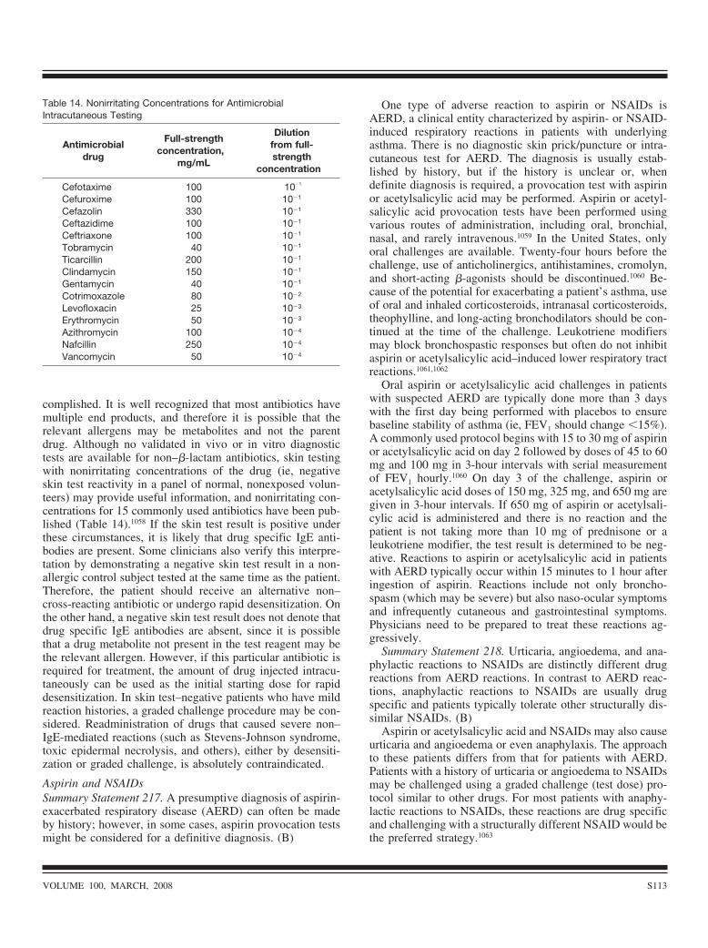
complished. It is well recognized that most antibiotics havemultiple end products, and therefore it is possible that therelevant allergens may be metabolites and not the parentdrug. Although no validated in vivo or in vitro diagnostictests are available for non–�-lactam antibiotics, skin testingwith nonirritating concentrations of the drug (ie, negativeskin test reactivity in a panel of normal, nonexposed volun-teers) may provide useful information, and nonirritating con-centrations for 15 commonly used antibiotics have been pub-lished (Table 14).1058 If the skin test result is positive underthese circumstances, it is likely that drug specific IgE anti-bodies are present. Some clinicians also verify this interpre-tation by demonstrating a negative skin test result in a non-allergic control subject tested at the same time as the patient.Therefore, the patient should receive an alternative non–cross-reacting antibiotic or undergo rapid desensitization. Onthe other hand, a negative skin test result does not denote thatdrug specific IgE antibodies are absent, since it is possiblethat a drug metabolite not present in the test reagent may bethe relevant allergen. However, if this particular antibiotic isrequired for treatment, the amount of drug injected intracu-taneously can be used as the initial starting dose for rapiddesensitization. In skin test–negative patients who have mildreaction histories, a graded challenge procedure may be con-sidered. Readministration of drugs that caused severe non–IgE-mediated reactions (such as Stevens-Johnson syndrome,toxic epidermal necrolysis, and others), either by desensiti-zation or graded challenge, is absolutely contraindicated.
Aspirin and NSAIDsSummary Statement 217. A presumptive diagnosis of aspirin-exacerbated respiratory disease (AERD) can often be madeby history; however, in some cases, aspirin provocation testsmight be considered for a definitive diagnosis. (B)
One type of adverse reaction to aspirin or NSAIDs isAERD, a clinical entity characterized by aspirin- or NSAID-induced respiratory reactions in patients with underlyingasthma. There is no diagnostic skin prick/puncture or intra-cutaneous test for AERD. The diagnosis is usually estab-lished by history, but if the history is unclear or, whendefinite diagnosis is required, a provocation test with aspirinor acetylsalicylic acid may be performed. Aspirin or acetyl-salicylic acid provocation tests have been performed usingvarious routes of administration, including oral, bronchial,nasal, and rarely intravenous.1059 In the United States, onlyoral challenges are available. Twenty-four hours before thechallenge, use of anticholinergics, antihistamines, cromolyn,and short-acting �-agonists should be discontinued.1060 Be-cause of the potential for exacerbating a patient’s asthma, useof oral and inhaled corticosteroids, intranasal corticosteroids,theophylline, and long-acting bronchodilators should be con-tinued at the time of the challenge. Leukotriene modifiersmay block bronchospastic responses but often do not inhibitaspirin or acetylsalicylic acid–induced lower respiratory tractreactions.1061,1062
Oral aspirin or acetylsalicylic acid challenges in patientswith suspected AERD are typically done more than 3 dayswith the first day being performed with placebos to ensurebaseline stability of asthma (ie, FEV1 should change �15%).A commonly used protocol begins with 15 to 30 mg of aspirinor acetylsalicylic acid on day 2 followed by doses of 45 to 60mg and 100 mg in 3-hour intervals with serial measurementof FEV1 hourly.1060 On day 3 of the challenge, aspirin oracetylsalicylic acid doses of 150 mg, 325 mg, and 650 mg aregiven in 3-hour intervals. If 650 mg of aspirin or acetylsali-cylic acid is administered and there is no reaction and thepatient is not taking more than 10 mg of prednisone or aleukotriene modifier, the test result is determined to be neg-ative. Reactions to aspirin or acetylsalicylic acid in patientswith AERD typically occur within 15 minutes to 1 hour afteringestion of aspirin. Reactions include not only broncho-spasm (which may be severe) but also naso-ocular symptomsand infrequently cutaneous and gastrointestinal symptoms.Physicians need to be prepared to treat these reactions ag-gressively.
Summary Statement 218. Urticaria, angioedema, and ana-phylactic reactions to NSAIDs are distinctly different drugreactions from AERD reactions. In contrast to AERD reac-tions, anaphylactic reactions to NSAIDs are usually drugspecific and patients typically tolerate other structurally dis-similar NSAIDs. (B)
Aspirin or acetylsalicylic acid and NSAIDs may also causeurticaria and angioedema or even anaphylaxis. The approachto these patients differs from that for patients with AERD.Patients with a history of urticaria or angioedema to NSAIDsmay be challenged using a graded challenge (test dose) pro-tocol similar to other drugs. For most patients with anaphy-lactic reactions to NSAIDs, these reactions are drug specificand challenging with a structurally different NSAID would bethe preferred strategy.1063
Table 14. Nonirritating Concentrations for AntimicrobialIntracutaneous Testing
Antimicrobialdrug
Full-strengthconcentration,
mg/mL
Dilutionfrom full-strength
concentration
Cefotaxime 100 10�1
Cefuroxime 100 10�1
Cefazolin 330 10�1
Ceftazidime 100 10�1
Ceftriaxone 100 10�1
Tobramycin 40 10�1
Ticarcillin 200 10�1
Clindamycin 150 10�1
Gentamycin 40 10�1
Cotrimoxazole 80 10�2
Levofloxacin 25 10�3
Erythromycin 50 10�3
Azithromycin 100 10�4
Nafcillin 250 10�4
Vancomycin 50 10�4
VOLUME 100, MARCH, 2008 S113
Perioperative AnaphylaxisSummary Statement 219. Skin testing is a useful diagnostictool in cases of perioperative anaphylaxis, and when skintesting is used to guide subsequent anesthetic agents, the riskof recurrent anaphylaxis to anesthesia is low. (C)
Skin testing has been reported to be of diagnostic utility inidentifying the causative agent in cases of anaphylaxis duringgeneral anesthesia in both retrospective and prospective stud-ies.1064,1065 Intracutaneous testing is the most widely usedmethod and has been determined to be a valid and reproduc-ible method in several studies.1064,1065 Prick testing has alsobeen evaluated, and 2 prospective studies confirmed thatprick testing was a useful diagnostic tool and highly corre-lated with intracutaneous skin testing.1036,1065 For propofolreactions, intracutaneous testing is more reliable.1066,1067 Skintesting has not been applied to any gold standard for anes-thetic allergy due to the inherent dangers with challenging apatient with a history of anaphylaxis and the inherent phar-macologic effects of the anesthetic. When skin testing is usedto guide subsequent anesthetic agents, the risk of recurrentanaphylaxis to anesthesia is low.1066,1068 Nevertheless, false-negative skin test results have been reported and the true-negative predictive value remains unknown.1066
The concentrations and dilutions for skin testing used indifferent studies is varied.1069 One approach recommended bythe French Society of Anesthesiology and used in a study of789 patients being evaluated for allergic reactions to anes-thetics uses a combination of prick and intracutaneoustests.1070 The drugs tested in this study included neuromus-cular blocking agents, antibiotics, hypnotics, opioids, andothers. Prick tests are performed with undiluted drug, with theexception of atracurium, mivacurium, and morphine, whichare tested using a 1:10 (wt/vol) dilution. Intracutaneous testsare performed with 0.02 to 0.05 mL of serial dilutions of thedrug every 15 minutes. The initial dilution is 10�4 (wt/vol) ifthe prick test result is positive and 10�3 (wt/vol) when theprick test result is negative and subsequent intracutaneoustests are performed at 10-fold higher concentrations up to10�1 (wt/vol) for most drugs. The final testing dilution formorphine, rocuronium, and cisatracurium is 10�2 (wt/vol),and for atracurium, and mivacurium a maximal dilution of10�3 (wt/vol) is recommended.
Specific IgE tests for detecting sensitization to neuromus-cular blocking agents and latex have been used before generalanesthesia to prevent anaphylaxis during surgery.1071 In thisFrench study, specific IgE was positive in 79% of cases ofanaphylaxis attributed to neuromuscular blocking agents and88% of cases attributed to latex.1071 For neuromuscular block-ing agents, skin testing appears to have greater sensitivity;however, a few patients may have positive specific IgE testresults with negative skin test results.
Tryptase has been evaluated for its diagnostic value inperioperative anaphylaxis. In the previously cited Frenchstudy, 112 of 175 patients (64%) with anaphylaxis had atryptase level of more than 25 �g/L, whereas only 9 of 84
anaphylactoid reactions (11%) had an elevated tryptaselevel.1071 For diagnosis of perioperative anaphylaxis,tryptase measurements had a positive predictive value of92.6% but a negative predictive value of 54%. The positivelikelihood ratio for an anaphylactic event based on atryptase level was 6.0 and the negative likelihood ratio was0.4.
ChemotherapeuticsSummary Statement 219. Skin testing is not helpful in casesof taxane-induced anaphylactoid reactions. (C)
Summary Statement 220. Skin testing to carboplatin yieldsfavorable predictive values. (C)
Summary Statement 221. Skin testing with asparaginasebefore treatment is recommended but does not identify allpatients at risk of reactions. (C)
Hypersensitivity reactions have been reported for virtuallyall commonly used chemotherapeutic agents. Reactions rangefrom mild cutaneous eruptions to fatal anaphylaxis. In somecases, it is difficult to determine whether a reaction is ana-phylactic (ie, mediated by drug specific IgE antibodies) oranaphylactoid (due to nonimmune degranulation of mast cellsand basophils as occurs with Cremophor-EL, a solvent usedfor many cancer chemotherapy drugs). For some chemother-apeutics, skin testing may help identifying patients at highrisk for allergic reactions to chemotherapy.
In the taxane family, paclitaxel and docetaxel produceanaphylactoid reactions in as many as 42% of patients on firstadministration and rarely (3%) with subsequent cycles.1072
The pathophysiology of these reactions is unknown but un-likely to be IgE mediated because skin test results to taxanesare negative in patients with these anaphylactoid reactions1062
and prophylactic therapy with antihistamines and corticoste-roids reduces hypersensitivity reactions to approximately4%.1073 Test dose protocols have been used to reduce theincidence of reactions and cost of drug wastage. However, thelargest study to date using test dosing of paclitaxel in 130patients revealed no significant difference in hypersensitivityreactions compared with patients treated without using thetest dosing protocol (2.3% in test dose group vs 6.2% incontrol group, P � .20).1074 Of note, 1 severe reaction oc-curred in the non–test dose group, but none were observed inthe test dose group. Finally, the test dose strategy was actu-ally more expensive (increased cost of $6,100 for 130 pa-tients).
Platinum compounds (cisplatin, carboplatin, and oxiplatin)typically cause hypersensitivity reactions after completion ofseveral treatment courses, suggesting an immunologic mech-anism.1075,1076 Skin testing with carboplatin has been shown tohelp predict patients who will have allergic reactions tocarboplatin. A study of 47 patients receiving carboplatin forgynecologic malignancies had intracutaneous tests with 0.02mL of undiluted carboplatin and found that a negative skintest result accurately predicted the absence of an allergicreaction in 166 of 168 courses of therapy.1077 This rate ofreactivity was lower than historical controls who had a 27%
S114 ANNALS OF ALLERGY, ASTHMA & IMMUNOLOGY
incidence of allergic reactions. A larger study of 126 womenwith gynecologic cancers performed intracutaneous skin testswith 0.02 mL of undiluted carboplatin in women who hadreceived more than 6 courses of platinum-based chemother-apy.1078 Of 668 negative skin test results, 10 were associatedwith hypersensitivity reactions (1.5% false negatives butnone were severe. Although most patients with positive testresults did not receive further carboplatin, 7 patients who hadpositive skin test results received carboplatin and 6 of 7 hadallergic reactions. On the basis of this information, it has beenrecommended that skin testing to carboplatin be performedbefore the eighth cycle of chemotherapy.1079
Immediate-type reactions to asparaginase occur in as manyas 43% of patients, and the reaction rate increases after thefourth weekly dose.1080 It is unknown whether the mechanismis anaphylactic or anaphylactoid, and it may be different indifferent patients. Use of skin testing with asparaginase be-fore treatment is recommended but does not identify allpatients at risk of reactions.1080 In addition to false-negativeskin test results, false-positive skin test results may alsooccur.1080 Polyethylene glycolated–asparaginase has alsobeen reported to cause anaphylaxis, and skin test results toEscherichia coli derived granulocyte colony-stimulating fac-tor was positive, suggesting an IgE-mediated mechanisminvolving bacteria-specific antigens.1081 This report suggeststhat patients with allergic reactions to E coli–derived aspar-aginase should avoid other products synthesized with recom-binant E coli systems and that skin testing may be helpful inconfirming such cross-reactivity, although the predictivevalue of this testing is unknown.
Local AnestheticsSummary Statement 223. Skin testing for diagnosis of localanesthetic allergy is limited by false-positive reactions. Thegold standard for establishing a diagnosis of local anestheticallergy is the provocative challenge. (C)
Local anesthetics are commonly used and adverse reac-tions to injections of local anesthetics may occur. Nearlyall of these reactions are due to vasovagal reactions, anx-iety, psychosomatic, or toxic effects. IgE-mediated or ana-phylactoid reactions to local anesthetics are extremely rareand have been documented in only a few case re-ports.1082,1083 The gold standard for establishing a diagnosisof local anesthetic allergy is the provocative challenge.Skin testing has also been used as a diagnostic tool; however,several studies have indicated false-positive intracutaneousskin test results.1084–1086 In patients who subsequently toleratea provocative challenge without an adverse reaction, false-positive intracutaneous test results occur in approximately19% and 9% of history-negative and history-positive patients,respectively.1087 On the basis of the low pretest probability ofIgE-mediated local anesthetic allergy and the occurrence offalse-positive results, it is unclear whether intracutaneoustests have any benefit in the diagnostic approach to localanesthetic allergy.1088,1089 Rare patients may also have positiveskin test results to methylparaben additives in the local an-
esthetics and some of these may be false-positive skin testresults because subsequent subcutaneous challenge to localanesthetic with methylparabens are often negative.1090 Sub-cutaneous local anesthetic challenges using a graded incre-mental approach after skin tests have been reported as a safemethod in a study of 236 patients with histories of adversereactions to local anesthetics.1088 Rechallenge without priorskin tests was reported to be an easy and cheap alternative toskin testing.1091 However, the possibility of a rare, systemicreaction must still be kept in mind.
CorticosteroidsSummary Statement 224. The specificity and sensitivity ofskin tests for systemic corticosteroid allergy are unknown,and cases of corticosteroid allergy with negative skin testresults to the implicated corticosteroid have been reported.(D)
Immediate-type allergic reactions to corticosteroids arerare. The mechanisms of these reactions remain unclear, andboth IgE- and non–IgE-mediated reactions have been pro-posed.1092 Skin testing has been used in the diagnosis ofcorticosteroid hypersensitive reactions with variable results.Prick and intracutaneous tests have been used with a varietyof concentrations1093–1095 and have been found to be nonirri-tating in normal controls even up to undiluted concentra-tions.1096 The specificity and sensitivity of skin tests forcorticosteroid allergy are unknown, and cases of corticoste-roid allergy with negative skin test results to the implicatedcorticosteroid have been reported, including a case with apositive provocative challenge.1095 Finally, other componentsadded to corticosteroid preparations (eg, carboxymethylcel-lulose) have been reported to be responsible for anaphylaxisafter injection of parenteral corticosteroids.1097
Additives and PreservativesSummary Statement 225. For most allergic reactions to addi-tives, skin tests are of no diagnostic value, and placebo-controlled oral challenges are required. (C)
The number of additives used by the food and drug indus-tries is extensive. Only a small number of additives have beenimplicated in IgE-mediated or other adverse reactions. Formany additives, including tartrazine, aspartame, sodium ben-zoate, butylated hydroxyanisole, butylated hydroxytoluene,and FD&C dyes, skin tests are of no diagnostic value, andplacebo-controlled oral challenges are required.981 In rarecases of sulfite sensitivity, positive skin test results to sulfiteshave been described.1098,1099 Natural food additives, such asannatto, saffron, carmine, and erythritol, have been describedto rarely cause anaphylaxis and positive skin test results havebeen demonstrated.1100–1103 Antibacterial additives such asparabens and benzylkonium chloride may also induce IgE-mediated symptoms.909
ASSESSMENT OF ALLERGIC CONTACTDERMATITISSummary Statement 226. Contact dermatitis is a commonskin disorder seen by allergists and dermatologists and can
VOLUME 100, MARCH, 2008 S115
present with a spectrum of morphologic cutaneous reactions.(C)
Contact dermatitis is a common skin problem for which 5.7million physician visits per year are made.468,1104 All agegroups are affected, with a slight female preponderance basedon a large population-based survey of public health issues.1105
The acute clinical expression of CD is characterized byredness, edema, papules, vesiculation, weeping, crusting, andpruritus most commonly recognized as eczema, a nonspecificterm applied to a number of dermatitides, including atopicdermatitis. Prolonged persistence of this dermatitis may beassociated with acneiform eruptions secondary to irritation offollicular function, hypopigmentation or hyperpigmentationdue to alterations in melanocytic biology, skin thickening,lichenification, and fissuring. Exposure to UV light mostcommonly causes a phototoxic or sunburn type of reactionand less commonly a photoallergic reaction when the UVlight interacts with chemical agents (ie, fragrances, PABA,plants, parsnips, figs, or several ingested drugs) inducingphotosensitization of various forms.
Summary Statement 227. The initial approach to clinicaldiagnosis of CD is to distinguish between ACD and ICD. (C)
Contact dermatitis encompasses all adverse cutaneous re-actions that result from the direct contact of an exogenousagent (a foreign molecule, UV light, or temperature) to thesurface of the skin or mucous membranes. The skin can reactimmunologically and/or nonimmunologically to such exoge-nous agents. The inflammatory process resulting from anallergic substance is mediated through immunologic mecha-nisms, whereas irritant reactions result from direct tissuedamage, which initiate alternative inflammatory reactions.However, the distinction between ACD and ICD has becomeincreasingly blurred. Often these exogenous forms of derma-titis must be distinguished from endogenous dermatitis (ie,atopic dermatitis, nummular eczema, dyshidrosis).1105 It is notunusual for an exogenous dermatitis to be superimposed onan endogenous eruption, most commonly encountered whencompresses or topical antibiotics are used too long on barrierimpaired skin.
Based on several studies, the bulk of exogenous cases arediagnosed as ICD. The appropriate diagnosis is made byevaluating the location and evolution of the inflammation,together with morphologic nuances, to arrive at a probablediagnosis. Patch testing remains the most useful method forconfirming ACD. Irritant contact dermatitis is a diagnosis ofexclusion without firm criteria or when patch test results forACD are negative. However, if patch tests fail to test for theappropriate substance, an ICD diagnosis could be incorrect.
Summary Statement 228. The inflammatory lesions of CDmay result from either ACD or ICD mechanisms. Factors thataffect response to the contact agent include the agent itself,the patient, the type and degree of exposure, and the envi-ronment. (A)
The potential for substances that could cause either ICD orACD is variable. Thus, detergents have a higher irritancyindex, whereas nickel is a major allergenic contactant chem-
ical. The severity of the CD ranges from a mild, short-livedcondition to a severe, persistent, but rarely life-threatening,disease. The thickness and integrity of the skin influence thepotential for developing ICD or ACD. Thinner skin sites,such as the eyelids, ear lobes, and genital areas, are mostvulnerable, whereas the thicker palms and soles are moreresistant to injury induced by irritation or sensitization. Ex-posure time to allergenic contactants, which usually definesboth induction and elicitation phases of ACD, varies frombeing brief (eg, poison ivy) to protracted (eg, nickel in jew-elry or other chemicals in the work environment). Similarly,irritant substances may damage the skin in either the short orlong term.
Summary Statement 229. Tissue reactions to contactantsare attributable primarily to cellular immune mechanismsexcept for contact urticaria. (A)
Contact dermatitis reactions are noted almost exclusivelyat the site of exposure with the putative antigen. Most ACDantigens are small-molecular-weight molecules or haptensthat become immunogenic after conjugation with proteins inthe skin.1106 Less commonly, large-molecular-weight peptidesor proteins (eg, latex, cashew nuts) may both induce and elicitthe classic inflammatory lesions of ACD.
Summary Statement 230. Irritant contact dermatitis is usu-ally the result of nonimmunologic, direct tissue reaction andmust be clearly differentiated from ACD. (A)
Irritant contact dermatitis is generally a multifactorial re-sponse that involves contact with a substance that chemicallyabrades, physically irritates, or damages the skin.1107 Irritationis usually a direct cytotoxic reaction produced by a widevariety of agents (eg, chemicals, detergents, solvents, alcohol,creams, lotions, ointments, and powders) and by contributoryphysical factors that include excessive scrubbing, washing,overhydration, improper drying, perspiration, and tempera-ture extremes. Any impairment to the epidermal barrier layer(eg, fissuring, superhydration) increases skin susceptibility toan irritant defect. The evolution and resolution of ICD areless predictable than those of ACD. The clinical presentationof ICD is more limited to the skin site directly in contact withthe offending agent(s) with little or no extension beyond thesite of contact.
Summary Statement 231. The diagnosis of ACD is sus-pected from the clinical presentation of the rash, which thenmust be supported by a history of exposure to a putative agentand subsequently confirmed by patch testing whenever this ispossible. (C)
The suspicion of ACD is the first step in making thediagnosis. Thus, the history remains an essential part of thediagnosis and subsequent management of this disease. Al-though history can strongly suggest the cause of ACD, it hasbeen reported that experienced physicians accurately predictthe sensitizer in only 10% to 20% of patients with ACD whenrelying solely on the history and physical examination.1108
For ACD to occur, the site of inflammation must havecome in direct contact with the offending agent. Initially, thearea may itch, burn, or sting. The evolution of the lesion
S116 ANNALS OF ALLERGY, ASTHMA & IMMUNOLOGY
depends on multiple factors, including the innate allergenicityor irritancy of the agent, the integrity of the involved skin,environmental conditions, a history of prior reactions, andimmunocompetency status. Activities that involve exposureto sun, water, or airborne allergens may affect the skindistribution. Remissions and exacerbations may be related toweekends, vacations, and work schedule.
Work history must be carefully reviewed. The exact natureof the work duration of each activity and similar skin effectsin coworkers may be relevant. Recent changes in procedureor chemical exposures, including vapors and fumes, must beprobed. Protective wear and compliance with its use may givea clue as to the nature of the suspected allergen. Certain jobsrequire frequent hand washing and the use of special cleans-ing agents that not only may impair skin barrier but also maycause irritant hand dermatitis. Although moisturizers afterhand washing may prevent dehydration, they may expose thepatient to unsuspected allergens in the moisturizer prepara-tion. Since the worker may be unaware of specific chemicalsto which he/she is exposed, material safety data sheets mayhave to be obtained from the manufacturer.1109 Chronologicexposure histories and other activities must be obtained.
Hobbies and nonwork activity, such as gardening, mac-rame, painting, ceramic work, carpentry, and photography,may be sources of exposure to other contactants. Obtaining adetailed history of animal exposure is essential. Pet dander,products used on pets, and traces of outdoor allergens all cancause ACD. The history should also include response toprevious treatment. Many patients will have tried to eliminatemultiple agents or have used various remedies before seeinga physician.
Summary Statement 232. The skin site of the dermatitis isimportant in the diagnosis of ACD because the area of pre-dominant involvement and the regional distribution of thelesions often reflect the area of contact with the allergen. (A)
All inflammatory and spongiotic clinical reactions shouldinclude ACD as a possibility. Each lesional site usuallycorresponds to the site of contact with the putative allergen,and the physical appearance of the lesion may also suggestthe potential for ACD. Particular attention should be given tocertain anatomical sites, which include eyelids, face, neck,scalp, hands, axillae, lower extremities, and the anogenitalarea.1110–1116
Summary Statement 233. Epicutaneously applied patchtests are the standardized diagnostic procedures to confirmACD. (A)
Patch testing is the gold standard for identification of acontact allergen.1107 Although occlusive patch testing is themost common technique, open, prophetic (provocative), re-peated insult, photopatch, and atopy patch tests are alsoavailable if special situations indicate their use. For example,open patch tests are preferred for potential photosensitizers,volatile substances, mascara, antiperspirants, shaving creams,dentifrices, and strong topical medicaments that could act asrelative primary irritants. If photosensitization is suspected,photopatch tests should be performed by a physician with
expertise in UV radiation. Duplicate applications of the sus-pected photocontactant(s) are placed on each side of theupper back. One side is irradiated with 5 J cm�2 of UV-A 24to 48 hours later, and both radiated and unradiated sides areread 48 hours later.
The number of appropriate patch tests required to diagnoseACD may vary, depending on the nature of the clinicalproblem and the potential for significant allergen exposure.The value of a test depends on whether the clinical presen-tation warrants its use, the quality of reagents used, the timingof the application, an appropriate interpretation of the reac-tion, and establishing relevance for the benefit of the patient.Although the application of allergen patch testing is rathersimple, allergen selection, the proper test concentration, andinterpretation of the test require expertise. Clinical researchdefining the validity of each of these components has beenextensive. Such data are well described in textbooks andprevious practice parameters (Practice Parameter for AllergyDiagnostic Testing and Contact Dermatitis: A Practice Pa-rameter).235,1108–1120 These sources provide details for the pur-chase and/or preparation of allergens and materials for appli-cation, forms for record keeping, preparation of patch testsites, application of the allergens, times of reading, andinterpretation according to internationally approved guide-lines.1117,1120 Because it is impractical to test an unlimitednumber of contactants, standardized sets have been designedand validated by collaborative dermatologic research societ-ies.469,481,482,1121,1122 These vary somewhat to reflect differencesin exposure patterns in different parts of the world. Newallergens are added from time to time, depending on changesof product utilization and exposure patterns. Since 2001, theNorth American Contact Dermatitis Group has enlarged itsstandard panel to 65 allergens and/or allergen mixes. How-ever, use of the FDA-certified antigen panel available in theUnited States can fully evaluate approximately 25% to 30%of patients with ACD, especially those patients who areallergic to rubber, metals, fragrances, cosmetics, and medi-caments.469
Summary Statement 234. Patch tests are indicated in anypatient with a chronic, pruritic, eczematous, or lichenifieddermatitis if underlying or secondary ACD is suspected. (C)
Virtually any eczematous lesion could be caused or aggra-vated by a contactant.481,482,1107,1121–1124 The decision to patchtest under these circumstances is often independent of thehistory because the patient may be unaware of any relevantexposure. Based on repetitive tests in patients with the angryback syndrome, it is recommended that patch testing shouldbe deferred until the underlying dermatitis is no longer acuteor severe.507 Under such circumstances, the entire skin maybe irritable and false-positive reactions may occur. There isalways the possibility that a positive patch test result maytrigger an exacerbation of the original dermatitis. In thissituation, however, negative patch test results to a standardbattery of allergens can be valuable in excluding a suspectedagent.
VOLUME 100, MARCH, 2008 S117
Summary Statement 235. Patch test results are affected byoral corticosteroids but not by antihistamines. (A)
Immunocompromised adult patients, including those tak-ing oral corticosteroids (�20 mg of prednisone per day or itsequivalent) or those undergoing cancer chemotherapy, mayshow diminished or absent reactivity to the patch tests.1125,1126
A multicenter, randomized, double-blind study revealed thatsystemic steroids in doses of less than 20 mg/d were notlikely to suppress strongly positive patch test results, but theycould suppress milder responses.1126 The same study con-cluded that equivalent doses of prednisone did not affectirritant responses1126 The effect of systemic corticosteroids onthe results of patch testing is less understood for children.
The skin site where the patch tests are to be applied shouldhave had no topical potent corticosteroid or calcineurin in-hibitor applied for 5 to 7 days before testing since localanti-inflammatory effects of these agents can diminish orobliterate a possible positive test result. Systemic antihista-mines do not affect the interpretation of patch tests. Surpris-ingly, patients who have late HIV disease are still reactive tocontact allergens.1127
Summary Statement 236. Reading and interpretation ofpatch tests should conform to principles developed by theInternational Contact Dermatitis Research Group and theNorth American Contact Dermatitis Research Group. (A)
Summary Statement 237. A 96-hour reading may be nec-essary because 30% of relevant allergens that are negative atthe 48-hour reading become positive in 96 hours. (A)
The initial reading of patch test results should be per-formed 48 hours after their application. Tests may need to beread 30 minutes after removal of the patch to allow resolutionof erythema due to occluding pressure or the tape and/orchamber if present. Ideally, there should be an additionalreading 3 to 4 days after the initial application and occasion-ally after 7 days for certain contactants.483,1128,1129 A collabo-rative study documented that approximately 30% of relevantallergens that were negative at the 48-hour reading becomepositive at a 96-hour reading.484 Conversely, some irritantreactions at 48 hours tended to disappear by 96 hours. Thereading itself is based on a nonlinear, descriptive scale thatwas developed and validated by the International ContactDermatitis Research Group.1130 The details of this ratingsystem and corresponding clinical interpretation with a visualkey may be found in the parameter, Contact Dermatitis: APractice Parameter. In general, there is good concordance ofpositive patch test results between individual Finn Chambertests and the T.R.U.E. TEST technology and between differ-ent commercial manufacturers.503–505 The relevance of posi-tive reactions to clinical ACD can only be established bycarefully correlating the history, including exposure to theallergen with the test results.504,1135Laser Doppler perfusionimaging of cutaneous blood flow has been proposed as analternative to visual reading.1131 This technique correlateswith visual scoring but is not useful in distinguishing betweenallergic and irritant reactions.1132,1133
Summary Statement 238. Nonstandardized and customizedpatch testing is often required, depending on the patient’sexposure history. (C)
When an agent not included in the standard set is sus-pected, kits for specific occupations (eg, beauty operators,machinists) and exposures (eg, shoes, plants, photoallergens)permit identification of many other significant allergens. Notinfrequently, it may be necessary to customize patch tests inaccordance with a patient’s specific exposure history.“Leave-on” cosmetics (eg, nail polish, lipstick, rouge, foun-dation), clothing, gloves, and foods may be applied “as is.”“Wash-off” cosmetics (eg, shampoos, conditioners, cleans-ers) should be diluted (10�2 or 10�3) before applica-tion.1118,1131 Other household and industrial products shouldonly be tested after ascertaining their safety in material safetydata sheet background information and in accord with anauthoritative text on patch test concentrations.1106 Even afterthis research, nonirritant concentrations may need to be per-formed in nonexposed controls if more precise toxic infor-mation cannot be obtained.
If photosensitization is suspected, photo patch tests shouldbe performed by a physician with expertise in UV radiation.Duplicate applications of the suspected photo contactant(s)are placed on each side of the upper back. One side isirradiated with 5 J cm�2 of UV-A 24 to 48 hours later, andboth irradiated and unradiated sides are measured 48 hoursafter irradiation.1134
Summary Statement 239. A problem-oriented approach todiagnostic patch testing using evidence-based principles oflikelihood ratios and posttest probability is more likely toconfirm clinical ACD than a randomly selected patch testapproach. (B)
Recently, the question of proper pretesting probabilitymeasurement has been raised with the purpose of discourag-ing random patch testing, which has a low pretest predictiveprobability.499 It is postulated that pretest probabilities can beestimated by the data of large-scale prevalence studies ofcontact allergy in the general population. Using these data,likelihood ratios and postpatch test probability of contactallergy can be ascertained.499
Summary Statement 240. Several in vitro procedures arebeing investigated for the diagnosis of ACD. (A)
The potential for induction and elicitation of sensitizationis augmented if the allergen also has the ability to induceirritant signals, presumably through the innate immune sys-tem.1135 Irritant signals may induce the synthesis and releaseof proinflammatory cytokines, such as TNF-�, IL-1, IL-8,and granulocyte-macrophage colony-stimulating factor.1135
Thus, there is a rationale for developing alternative in vitrodiagnostic tests. The lymphocyte transformation test ismostly used for research purposes.235 Recently, an enzyme-linked immunospot (ELISPOT) assay, specifically designedto detect contactant-induced cellular release of cytokines(interferon-�, IL-2, IL-4) by the patient’s peripheral mono-nuclear cells, was compared with patch tests and lymphocytetransformation test. Overall, there was a statistically signifi-
S118 ANNALS OF ALLERGY, ASTHMA & IMMUNOLOGY
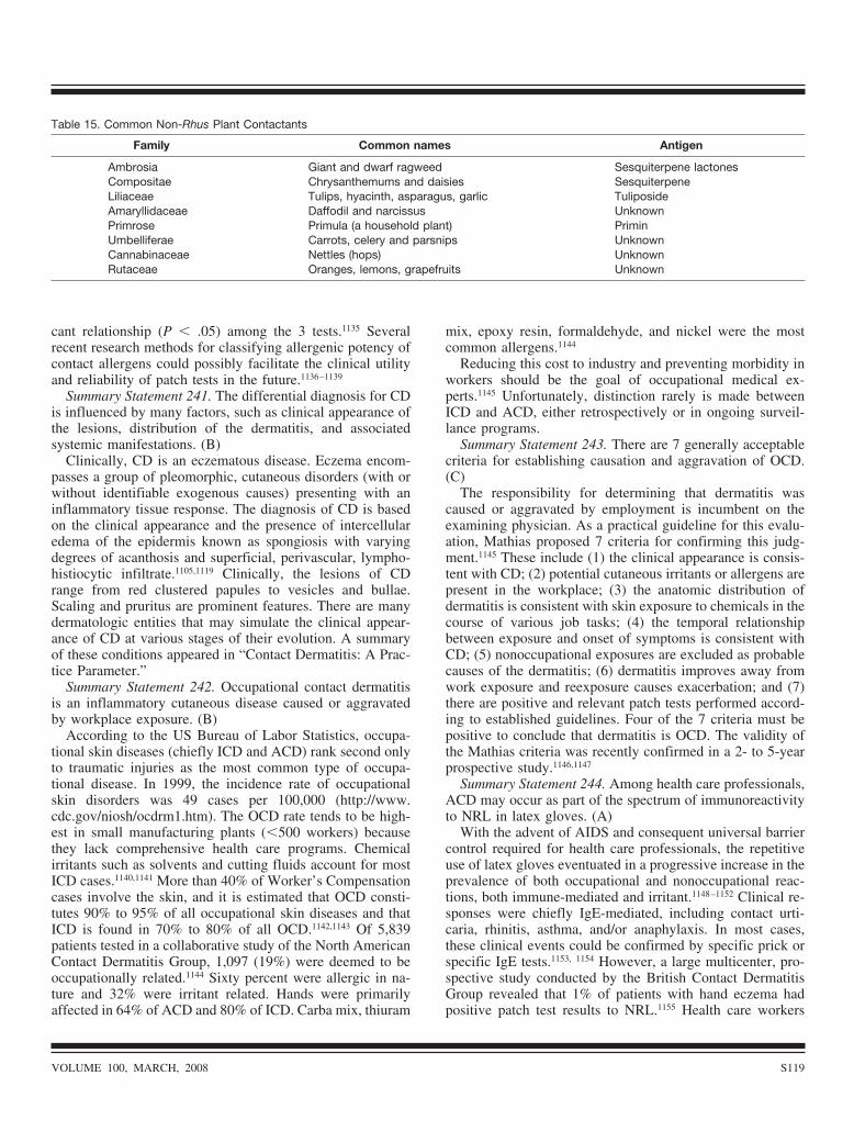
cant relationship (P � .05) among the 3 tests.1135 Severalrecent research methods for classifying allergenic potency ofcontact allergens could possibly facilitate the clinical utilityand reliability of patch tests in the future.1136–1139
Summary Statement 241. The differential diagnosis for CDis influenced by many factors, such as clinical appearance ofthe lesions, distribution of the dermatitis, and associatedsystemic manifestations. (B)
Clinically, CD is an eczematous disease. Eczema encom-passes a group of pleomorphic, cutaneous disorders (with orwithout identifiable exogenous causes) presenting with aninflammatory tissue response. The diagnosis of CD is basedon the clinical appearance and the presence of intercellularedema of the epidermis known as spongiosis with varyingdegrees of acanthosis and superficial, perivascular, lympho-histiocytic infiltrate.1105,1119 Clinically, the lesions of CDrange from red clustered papules to vesicles and bullae.Scaling and pruritus are prominent features. There are manydermatologic entities that may simulate the clinical appear-ance of CD at various stages of their evolution. A summaryof these conditions appeared in “Contact Dermatitis: A Prac-tice Parameter.”
Summary Statement 242. Occupational contact dermatitisis an inflammatory cutaneous disease caused or aggravatedby workplace exposure. (B)
According to the US Bureau of Labor Statistics, occupa-tional skin diseases (chiefly ICD and ACD) rank second onlyto traumatic injuries as the most common type of occupa-tional disease. In 1999, the incidence rate of occupationalskin disorders was 49 cases per 100,000 (http://www.cdc.gov/niosh/ocdrm1.htm). The OCD rate tends to be high-est in small manufacturing plants (�500 workers) becausethey lack comprehensive health care programs. Chemicalirritants such as solvents and cutting fluids account for mostICD cases.1140,1141 More than 40% of Worker’s Compensationcases involve the skin, and it is estimated that OCD consti-tutes 90% to 95% of all occupational skin diseases and thatICD is found in 70% to 80% of all OCD.1142,1143 Of 5,839patients tested in a collaborative study of the North AmericanContact Dermatitis Group, 1,097 (19%) were deemed to beoccupationally related.1144 Sixty percent were allergic in na-ture and 32% were irritant related. Hands were primarilyaffected in 64% of ACD and 80% of ICD. Carba mix, thiuram
mix, epoxy resin, formaldehyde, and nickel were the mostcommon allergens.1144
Reducing this cost to industry and preventing morbidity inworkers should be the goal of occupational medical ex-perts.1145 Unfortunately, distinction rarely is made betweenICD and ACD, either retrospectively or in ongoing surveil-lance programs.
Summary Statement 243. There are 7 generally acceptablecriteria for establishing causation and aggravation of OCD.(C)
The responsibility for determining that dermatitis wascaused or aggravated by employment is incumbent on theexamining physician. As a practical guideline for this evalu-ation, Mathias proposed 7 criteria for confirming this judg-ment.1145 These include (1) the clinical appearance is consis-tent with CD; (2) potential cutaneous irritants or allergens arepresent in the workplace; (3) the anatomic distribution ofdermatitis is consistent with skin exposure to chemicals in thecourse of various job tasks; (4) the temporal relationshipbetween exposure and onset of symptoms is consistent withCD; (5) nonoccupational exposures are excluded as probablecauses of the dermatitis; (6) dermatitis improves away fromwork exposure and reexposure causes exacerbation; and (7)there are positive and relevant patch tests performed accord-ing to established guidelines. Four of the 7 criteria must bepositive to conclude that dermatitis is OCD. The validity ofthe Mathias criteria was recently confirmed in a 2- to 5-yearprospective study.1146,1147
Summary Statement 244. Among health care professionals,ACD may occur as part of the spectrum of immunoreactivityto NRL in latex gloves. (A)
With the advent of AIDS and consequent universal barriercontrol required for health care professionals, the repetitiveuse of latex gloves eventuated in a progressive increase in theprevalence of both occupational and nonoccupational reac-tions, both immune-mediated and irritant.1148–1152 Clinical re-sponses were chiefly IgE-mediated, including contact urti-caria, rhinitis, asthma, and/or anaphylaxis. In most cases,these clinical events could be confirmed by specific prick orspecific IgE tests.1153, 1154 However, a large multicenter, pro-spective study conducted by the British Contact DermatitisGroup revealed that 1% of patients with hand eczema hadpositive patch test results to NRL.1155 Health care workers
Table 15. Common Non-Rhus Plant Contactants
Family Common names Antigen
Ambrosia Giant and dwarf ragweed Sesquiterpene lactonesCompositae Chrysanthemums and daisies SesquiterpeneLiliaceae Tulips, hyacinth, asparagus, garlic TuliposideAmaryllidaceae Daffodil and narcissus UnknownPrimrose Primula (a household plant) PriminUmbelliferae Carrots, celery and parsnips UnknownCannabinaceae Nettles (hops) UnknownRutaceae Oranges, lemons, grapefruits Unknown
VOLUME 100, MARCH, 2008 S119
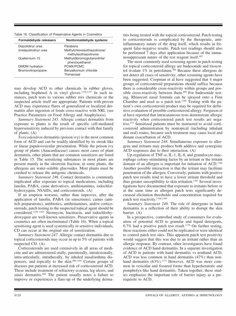
may develop ACD to other chemicals in rubber gloves,including bisphenol A in vinyl gloves.1156,1157 In such in-stances, patch tests to various rubber mix chemicals or thesuspected article itself are appropriate. Patients with provenACD may experience flares of generalized or localized der-matitis after ingestion of foods cross-reactive with NRL (seePractice Parameters on Food Allergy and Anaphylaxis).
Summary Statement 245. Allergic contact dermatitis fromexposure to plants is the result of specific cell-mediatedhypersensitivity induced by previous contact with that familyof plants. (A)
Toxicodendron dermatitis (poison ivy) is the most commonform of ACD and can be readily identified by its streak-likeor linear papulovesicular presentation. While the poison ivygroup of plants (Anacardiaceae) causes most cases of plantdermatitis, other plants that are common sensitizers are listedin Table 15. The sensitizing substances in most plants arepresent mainly in the oleoresin fraction; in some plants, theallergens are water-soluble glucosides. Most plants must becrushed to release the antigenic chemicals.
Summary Statement 246. Contact dermatitis is commonlyimplicated after exposure to topical medications, includinglanolin, PABA, caine derivatives, antihistamines, iodochlor-hydroxyquin, NSAIDs, and corticosteroids. (A)
If an eruption worsens, rather than improves, after theapplication of lanolin, PABA (in sunscreens), caines (anti-itch preparations), antibiotics, antihistamines, and/or cortico-steroids, patch testing to the suspected topical agent should beconsidered.1158–1163 Neomycin, bacitracin, and iodochlorhy-droxyquin are well-known sensitizers. Preservative agents incosmetics are often incriminated (Table 16). When a topicalsensitizing agent is used systemically in sensitive individuals,CD can occur at the original site of sensitization.
Summary Statement 247. Allergic contact dermatitis due totopical corticosteroids may occur in up to 5% of patients withsuspected CD. (A)
Corticosteroids are used extensively in all areas of medi-cine and are administered orally, parenterally, intralesionally,intra-articularly, intrathecally, by inhaled nasal/asthma dis-pensers, and topically to the skin.486,1164 Certain groups ofdiseases put patients at increased risk of corticosteroid ACD.These include treatment of refractory eczema, leg ulcers, andstasis dermatitis.486 The patient usually notes a failure toimprove or experiences a flare-up of the underlying derma-
titis being treated with the topical corticosteroid. Patch testingto corticosteroids is complicated by the therapeutic, anti-inflammatory nature of the drug itself, which results in fre-quent false-negative results. Patch test readings should alsobe performed 7 days after application because of the immu-nosuppressant nature of the test reagent itself.487
The most commonly used screening agents in patch testingfor topical corticosteroid allergy are budesonide and tixocor-tol tivalate 1% in petrolatum.488 Because these allergens donot detect all cases of sensitivity, other screening agents havebeen suggested. Coopman et al have suggested that 4 majorgroups of corticosteroid preparations should suffice becausethere is considerable cross-reactivity within groups and pos-sible cross-reactivity between them.489 For budesonide test-ing, Rhinocort nasal formula can be sprayed onto a FinnChamber and used as a patch test.1165 Testing with the pa-tient’s own corticosteroid product may be required for defin-itive evaluation of possible corticosteroid allergy. Ferguson etal have reported that intracutaneous tests demonstrate allergicreactivity when corticosteroid patch test results are nega-tive.477 Sensitized patients must be instructed to avoid corti-costeroid administration by nontopical (including inhalantand oral) routes, because such treatment may cause local anddistant exacerbation of ACD.
Summary Statement 248. Simultaneous exposure to aller-gens and irritants may produce both additive and synergisticACD responses due to their interaction. (A)
Up-regulation of TNF-�, IL-1, IL-8, and granulocyte-mac-rophage colony-stimulating factor by an irritant or the irritantdomain of an allergen is important for initiation of ACD.1166
Another possible interaction is that the irritant may facilitatepenetration of the allergen. Conversely, patients with positivepatch test results tend to have a lower irritant threshold andthus greater susceptibility to skin irritation.1167 Several inves-tigations have documented that exposure to irritants before orat the same time as allergen patch tests significantly de-creased elicitation thresholds and concentration required forpatch test reactivity.1168,1169
Summary Statement 249. The role of detergents in handdermatitis is a reflection of their ability to disrupt the skinbarrier. (A)
In a prospective, controlled study of consumers for evalu-ation of potential ACD to granular and liquid detergents,0.7% had a positive patch test result.1170 On further testing,these reactions either could not be replicated or were identicalto control patch test sites. This apparent patch test positivitywould suggest that this was due to an irritant rather than anallergic response. By contrast, other investigators have foundevidence of ACD hand dermatitis. In a separate investigationof ACD in patients with hand dermatitis vs nonhand ACD,ACD was less common in hand dermatitis (47%) than non-hand dermatitis (63%).1171 However, ACD was more com-mon in vesicular and fissured forms than hyperkeratotic andpompholyx-like hand dermatitis. Taken together, these stud-ies emphasize the important role of barrier injury as a pre-requisite to ACD.
Table 16. Classification of Preservative Agents in Cosmetics
Formaldehyde releasers Nonformaldehyde systems
Diazolidinyl urea ParabensImidazolindinyl urea Methylchloroisothiazolinone/
methylisothiazolinoneQuaternium-15 Methyldibromoglutaronitrile/
phenoxyethanolDMDM hydration PCMX/PCMCBromonitropropane Benzalkonium chloride
Thimerosal
S120 ANNALS OF ALLERGY, ASTHMA & IMMUNOLOGY
Summary Statement 250. Allergic contact dermatitis is asignificant clinical problem in children. (A)
Although less frequent in the first years of life (ie, beforethe age of 10 years), the rate of occurrence beginning at thisage and through teen years attains and even exceeds thatobserved in adults.1172,1173 The order and prevalence of ACDto individual allergens are generally comparable to a generaladult population with nickel, fragrances, and rubber chemi-cals being similar in occurrence in the 2 groups of patients.1174
The influence of fashion trends, hobbies, and lifestyle activ-ity, such as body piercing, decorative skin paintings (eg,black henna tattoo), natural remedies, and cosmetics (eg, teatree oil)m or the use of products with fragrances and herbalingredients are important determinants for ACD in this agegroup.1174–1176
ACKNOWLEDGMENTSPublished Practice Parameters of the Joint Task Force onPractice Parameters for Allergy & Immunology include:
1. Practice parameters for the diagnosis and treatment ofasthma. J Allergy Clin Immunol. 1995;96:S707–870.
2. Practice parameters for allergy diagnostic testing. AnnAllergy. 1995; 75:543–625.
3. Practice parameters for the diagnosis and managementof immunodeficiency. Ann Allergy. 1996;76:282–294.
4. Practice parameters for allergen immunotherapy. J Al-lergy Clin Immunol. 1996;98:1001–1011.
5. Disease management of atopic dermatitis: a practiceparameter. Ann Allergy. 1997;79:197–211.
6. The diagnosis and management of anaphylaxis. J Al-lergy Clin Immunol. 1998;101:S465–528.
7. Algorithm for the diagnosis and management of asthma:a practice parameter update. Ann Allergy. 1998;81:415–420.
8. Diagnosis and management of rhinitis: parameter doc-uments of the Joint Task Force on Practice parameters inAllergy, Asthma and Immunology. Ann Allergy. 1998;81:S463–518.
9. Parameters for the diagnosis and management of sinus-itis. J Allergy Clin Immunol. 1998;102:S107-S144.
10. Stinging insect hypersensitivity: a practice parameter. JAllergy Clin Immunol. 1999;103:963–980.
11. Disease management of drug hypersensitivity: a prac-tice parameter. Ann Allergy. 1999; 83:S665 – S700.
12. Diagnosis and management of urticaria: a practiceparameter. Ann Allergy. 2000;85:S521-S544.
13. Allergen immunotherapy: a practice parameter. AnnAllergy. 2003;90:SI-S540.
14. Symptom severity assessment of allergic rhinitis: part I.Ann Allergy. 2003;91:105–114.
15. Disease management of atopic dermatitis: an updatedpractice parameter. Ann Allergy. 2004;93:S1-S21.
16. Stinging insect hypersensitivity: a practice parameterupdate. J Allergy Clin Immunol. 2004;114(4):869–886.
17. The diagnosis and management of anaphylaxis: anupdated practice parameter. J Allergy Clin Immunol. 2005;115(3):S483-S523.
18. Practice parameter for the diagnosis and managementof primary immunodeficiency. Ann Allergy. 2005;94:S1-S63.
19. Attaining optimal asthma control: a practice parameter.J Allergy Clin Immunol. 2005;116:S3-S11.
20. Slavin RG, Spector SL, Bernstein IL, et al. The diag-nosis and management of sinusitis: a practice parameter up-date. J Allergy Clin Immunol. 2006;116:S13-S47.
21. Chapman J, Bernstein IL, Lee RE, et al. Food allergy:a practice parameter. Ann Allergy. 2006;96:S1–68.
22. Beltrani VS, Bernstein IL, Cohen DE, Fonacier L, et al.Contact dermatitis: a practice parameter. Ann Allergy. 2006;97:S1-S37.
23. Joint Task Force on Practice Parameters; AmericanAcademy of Allergy, Asthma and Immunology; AmericanCollege of Allergy, Asthma and Immunology; Joint Councilof Allergy, Asthma and Immunology Allergen immunother-apy: a practice parameter second update. J Allergy ClinImmunol. 2007;120(3 suppl):S25-S85.
These parameters are also available on the internet at:http://www.jcaai.org.
The Joint Task Force has made a concerted effort toacknowledge all contributors to this parameter. If any con-tributors have been excluded inadvertently, the Task Forcewill ensure that appropriate recognition of such contributionsis made subsequently.
These parameters were developed by the Joint Task Forceon Practice Parameters, representing the American Academyof Allergy, Asthma and Immunology, the American Collegeof Allergy, Asthma and Immunology, and the Joint Councilof Allergy, Asthma and Immunology. Chief Editor, I. Leo-nard Bernstein, MD, Clinical Professor of Medicine andEnvironmental Health, University of Cincinnati College ofMedicine, Cincinnati, Ohio; Parameter Workgroup, James T.Li, MD, PhD – Co-Chairman, Division of Allergic Diseasesand Internal Medicine, Mayo Clinic, Rochester, Minnesota; I.Leonard Bernstein, MD – Co-Chairman, Clinical Professor ofMedicine and Environmental Health, University of Cincin-nati College of Medicine, Cincinnati, Ohio; David I. Bern-stein, MD, Clinical Professor of Medicine and EnvironmentalHealth, University of Cincinnati College of Medicine, Cin-cinnati, Ohio; Robert Hamilton, PhD, D. ABMLI, Professorof Medicine and Pathology, Johns Hopkins University Schoolof Medicine, Baltimore, Maryland; Sheldon L. Spector, MD,Clinical Professor of Medicine, UCLA School of Medicine,Los Angeles, California; Ricardo Tan, MD, California Al-lergy and Asthma Medical Group, Los Angeles, California;Scott Sicherer, MD, Associate Professor of Pediatrics, JaffeFood Allergy Institute, Mount Sinai School of Medicine,New York, New York; David B.K. Golden, MD, AssociateProfessor of Medicine, Johns Hopkins University, Baltimore,MD; David A. Khan, MD, Associate Professor of InternalMedicine, University of Texas Southwestern Medical Center,Dallas, Texas; Task Force Reviewers, Richard A. Nicklas,MD, Clinical Professor of Medicine, George WashingtonMedical Center, Washington, DC; Jay M. Portnoy, MD,Chief, Section of Allergy, Asthma & Immunology, The Chil-
VOLUME 100, MARCH, 2008 S121
dren’s Mercy Hospital, Professor of Pediatrics, University ofMissouri-Kansas City School of Medicine, Kansas City, Mis-souri; Joann Blessing-Moore, MD, Clinical Associate Profes-sor of Medicine and Pediatrics, Stanford University MedicalCenter, Department of Immunology, Palo Alto, California;Linda Cox, MD, Assistant Clinical Professor of Medicine,Nova Southeastern University College of Osteopathic Medi-cine, Davie, Florida, David M. Lang, MD, Head, Allergy/Immunology Section, Division of Medicine, Director, Al-lergy and Immunology Fellowship Training Program,Cleveland Clinic Foundation, Cleveland, Ohio; John Oppen-heimer, MD, Department of Internal Medicine, New JerseyMedical School, Pulmonary and Allergy Associates, Morris-town, New Jersey; Christopher C. Randolph, MD, ClinicalProfessor of Pediatrics, Allergy/Immunology Section, YaleUniversity; Diane E. Schuller, MD, Professor of Pediatrics,Pennsylvania State University Milton S. Hershey MedicalCollege, Hershey, Pennsylvania; Stephen A. Tilles, MD,Clinical Assistant Professor of Medicine, University ofWashington School of Medicine, Redmond, Washington;Dana V. Wallace, MD, Assistant Clinical Professor, NovaSoutheastern University, Davie, Florida; Consultants, EstelleLevetin, PhD, Professor of Biology and Chair, Faculty ofBiological Science, University of Tulsa, Tulsa, Oklahoma;Richard Weber, MD, Professor of Medicine, National JewishMedical & Research Center, Denver, CO, Professor of Med-icine, University of Colorado Health Sciences Center, Au-rora, Colorado.
REFERENCES1. Taylor JG, Walker J. Charles Harrison Blackley (1820–1990). Clin
Allergy. 1973;3:103–108. (IIb)2. Feinberg SM. Allergy in Practice. 2nd ed. Chicago, IL: Year Book
Medical Publishers; 1946. (IIb)3. Indrajana T, Spieksma STH, Voorhorst R. Comparative study of the
intracutaneous, scratch and prick test in allergy. Ann Allergy. 1971;12:639–650. (III)
4. Lewis T. Vascular reactions of skin to injury; reaction to stroking:urticaria and factitia. Heart. 1924;11:119–139. (IIa)
5. Squire JR. The relationship between horse dandruff and horse serumantigens in asthma. Clin Sci. 1950;9:127–150. (IIa)
6. Antico A, Di Berardino L. Prilotest, an innovative disposable skinpuncture test: qualitative aspects. Allerg Immunol (Paris). 1994;26(8):297–301. (IIa)
7. Roovers MH, Gerth van Wijk R, Dieges PH, et al. Phazet skin pricktests versus conventional prick tests with allergens and histamine inchildren. Ann Allergy. 1990;64(2):166–169. (III)
8. Nelson HS, Kolehmainen C, Lahr J, et al. A comparison of multi-headed devices for allergy skin testing [Letter to the Editor]. J AllergyClin Immunol. 2004;113:1218–1219. (III)
9. Occupational Safety and Health Administration, Hazard InformationBulletin, Sept. 21, 1995. (IV)
10. Huber B, Berdel D. A comparison of the multi-test with the prick testand multi-test 2. Clin Allergy. 1983;13:467–472. (III)
11. Adinoff AD, Rosloniec DM, McCall LL, et al. A comparison of sixepicutaneous devices in the performance of immediate hypersensitiv-ity skin testing. J Allergy Clin Immunol. 1989;84:168–174. (III)
12. Garibaldi E, Slavin R. Positive multi-test reactions do not cause falsepositive reactions at adjacent sites. Ann Allergy. 1990;65:481–484.(III)
13. Basomba A, Sastre A, Pelaez A, et al. Standardization of the prick
test. Allergy. 1985;40:395–399. (III)14. Menardo JL, Bousquet J, Michel F. Comparison of three prick test
methods with the intradermal test and with the RAST in the diagnosisof mite allergy. Ann Allergy. 1982;48:235–239. (III)
15. Nierop G, Voorhorst R, Temmerman-Van de Vijver RL. Atopic skintest reevaluated. X. Comparison of a perfected technique of intracu-taneous skin testing and the prick test performed with a modifiedMorrow Brown needle. Ann Allergy. 1981;46:105–109. (III)
16. Holgersson M, Stalenhein G, Dreborg S. The precision of skin pricktest with Phazet, the Osterballe needle and the bifurcated needle.Allergy. 1985;40(Suppl):64–65. (III)
17. Berkowitz RB, Tinkelman DG, Lutz C, et al. Evaluation of theMulti-Test device for immediate hypersensitivity skin testing. J Al-lergy Clin Immunol. 1992;90:979–985. (III)
18. Murphree JT, Kniker WT. Correlation of immediate skin test re-sponses to antigens introduced by Multi-Test and intracutaneousroutes. Ann Allergy. 1979;43:279–285. (III)
19. Aas K. Some variables in skin prick testing. Standardization ofClinical (Biological) Methods Workshop No. 4. Allergy. 1980;36:250–252. (III)
20. Mahan C, Spector S, Siegel S, et al. Validity and reproducibility ofMulti-Test skin test device. Ann Allergy. 1993;71:25–28. (III)
21. Terho BO, Husman K, Kivekas J, et al. Histamine control affects thewheal produced by the adjacent diluent control in skin prick tests.Allergy. 1989;44:30–32. (III)
22. Demoly P, Bousquet J, Manderscheid JC, et al. Precision of skin prickand puncture tests with nine methods. J Allergy Clin Immunol. 1991;88:758–762. (III)
23. Bousquet J, Michel FB. Precision of prick and puncture tests. JAllergy Clin Immunol. 1992;90:870–872. (III)
24. Koller DY, Pirker C, Jarisch R, et al. Influence of the histaminecontrol on skin reactivity in skin testing. Allergy. 1992;47:58–59. (III)
25. Nelson HS, Rosloniec DM, McCall LI, et al. Comparative perfor-mance of five commercial prick skin test devices. J Allergy ClinImmunol. 1993;92:750–756. (IIb)
26. Corder WT, Hogan MB, Wilson NW. Comparison of two disposableplastic skin test devices with the bifurcated needle for epicutaneousallergy testing. Ann Allergy Asthma Immunol. 1996;77:222–226. (III)
27. Nelson HS, Lahr J, Buchmeier A, McCormick D. Evaluation ofdevices for skin prick testing. J Allergy Clin Immunol. 1998;101:153–156. (III)
28. Rhodius R, Wickens K, Cheng S, et al. A comparison of two skin testmethodologies and allergens from two different manufacturers. AnnAllergy Asthma Immunol. 2002;88(4):374–379. (III)
29. Sangsupawanich P, Chamnanphol S, Konrungsrisomboon D. Evalu-ation of three methods for using the Duotip-Test device for skintesting. Asian Pac J Allergy Immunol. 2000;18(3):153–156. (III)
30. Oppenheimer J, Nelson HS. Skin testing. Ann Allergy. Asthma Im-munol 2006;96(SI):56–512. (III)
31. Antico A, Lima G, Arisi M, Ostan A, Morrica B. Assay of prick testinoculum volume. I. Use and reliability of a gamma camera-basedmethod. Ann Allergy. Asthma Immunol 2000;85(2))140–144. (IIb)
32. Kaleyias J, Papaioannou D, Manoussakis M, et al. Skin-prick testfindings in atopic asthmatic children: a follow-up study from child-hood to puberty. Pediatr Allergy Immunol. 2002;13(5):368–374. (IIb)
33. Cantani A, Micera M. Epidemiology of atopy in 220 children. Diag-nostic reliability of skin prick tests and total and specific IgE levels.Minerva Pediatr 2003;55(2):129–137, 138–142. (III)
34. Kuehr J, Karmaus W, Frischer T, et al. Longitudinal variability of skinprick test results. Clin Exp Allergy. 1992;22(9):839–844. (IIb)
35. Illi S, Garcia-Marcus L, Hernando V, Guillen JJ, et al. Reproducibilityof skin prick test results in epidemiologic studies: a comparison of twodevices. Allergy. 1998;53(4):353–358. (III)
36. Johnston SL, Clough JB, Pattemore PK, et al. Longitudinal changes inskin-prick test reactivity over 2 years in a population of schoolchil-dren with respiratory symptoms. Clin Exp Allergy. 1992;22(10):948–957. (III)
37. Bodtger U, Jacobsen CR, Poulsen LK, et al. Long-term repeatability
S122 ANNALS OF ALLERGY, ASTHMA & IMMUNOLOGY
of the skin prick test is high when supported by history or allergen-sensitivity tests: a prospective clinical study. Allergy. 2003;58(11):1180–1186. (IIb)
38. Cook TJ, MacQueen DM, Wittig JH, et al. Degree and duration ofskin test suppression and side effects with antihistamines. J AllergyClin Immunol. 1973;51:71–77. (III)
39. Almind M, Dirksen A, Nielsen NH, et al. Durtion of the inhibitoryactivity on histamine-induced skin wheals of sedative and non-sedative antihistamines. Allergy. 1988;43(8):593–6. (III)
40. Simons FE, Simons KJ. Clinical pharmacology of new histamine H1receptor antagonists. Clin Pharmakinet. 1999;36(5):329–52. (III)
41. Rao KS, Menon PK, Hillman BC, et al. Duration of the suppressiveeffect of tricyclic antidepressants on histamine-induced wheal-and-flare reactions in human skin. J Allergy Clin Immunol. 1988;82:752–757. (III)
42. Miller J, Nelson HS. Suppression of immediate skin tests by raniti-dine. J Allergy Clin Immunol. 1989;84:895–899. (III)
43. Harvey RP, Schocket AL. The effect of H1 and H2 blockade oncutaneous histamine response in man. J Allergy Clin Immunol. 1980;65(2):136–9. (III)
44. Saarinen JV, Harvima RJ, Horsmanheimo M, et al. Modulation of theimmediate allergic wheal reaction in the skin by drugs inhibiting theeffects of leukotriene C4 and prostaglandin D2. Eur J Clin Pharma-col. 2001;57(1):1–4. (IIb)
45. Hill SL III, Krouse JH. The effects of montelukast on intradermalwheal and flare. Otolaryngol Head Neck Surg. 2003;129(3):199–203.(III)
46. Slott RI, Zweiman B. A controlled study of the effect of corticoste-roids on immediate skin test reactivity. J Allergy Clin Immunol.1974;54:229–235. (IIb)
47. Des Roches A, Paradis L, Bougeard YH, et al. Long-term oralcorticosteroid therapy does not alter the results of immediate-typeallergy skin prick tests. J Allergy Clin Immunol. 1996;98(3):522–7.(IIb)
48. Olson R, Karpink MH, Shelanski S, et al. Skin reactivity to codeineand histamine during prolonged corticosteroid therapy. J Allergy ClinImmunol. 1990;86:153–159. (IIb)
49. Pipkorn U, Hammarlund A, Enerbach L. Prolonged treatment withtopical glucocorticoids results in an inhibition of the allergen-inducedweal-and-flare response and a reduction in skin mast cell numbers andhistamine content. Clin Exp Allergy. 1989;19:19–25. (III)
50. Cole ZA, Clough GF, Church MK. Inhibition by glucocorticoids ofthe mast cell-dependent weal and flare response in human skin in vivo.Br J Pharmacol. 2001;132:286–292. (III)
51. Narasimha SK, Srinivas CR, Mathew AC. Effect of topical cortico-steroid application frequency on histamine-induced wheals. Int JDermatol. 2005;44(5):425–427. (III)
52. Herrscher RF, Ksper C, Sullivan TJ. Endogenous cortisol regulatesimmunoglobulin E-dependent late phase reactions. J Clin Invest.1992;90(2):596–603. (III)
53. Peebles RS Jr., Togias A, Bickel CA, et al. Endogenous glucocorti-coids and antigen-induced acute and late phase pulmonary responses.Clin Exp Allergy. 2000;30(9):1257–65. (IIb)
54. Kirmaz C, Yuksel H, Mete N, et al. Is the menstrual cycle affectingthe skin prick test reactivity? Asian Pac J Allergy Immunol. 2004;22(4):197–203. (III)
55. Van Nickerk CH, Prinsloo AE. Effect of skin pigmentation on theresponse to intradermal histamine. Int Arch Allergy Appl Immunol.1985;76(1):73–5. (III)
56. Ronchetti R, Villa MP, Rennerova Z, Haluszka J, et al. Allergen skinweal/radioallergosorbent test relationship in childhood populationsthat differ in histamine skin reactivity: a multi-national survey. ClinExp Allergy. 2005;35(1):70–74. (III)
57. Vocks E, Stander K, Rakoski J, et al. Suppression of immediate-typehypersensitivity elicitation in the skin prick test by ultraviolet Birradiation. Photodermatol Photoimmunol Photomed. 1999;15(6):236–40. (III)
58. Nelson HS. Diagnostic procedures in allergy, I: allergy skin testing.
Ann Allergy. 1983;51:411–417. (III)59. Bernstein IL. Proceedings of the task force on guidelines for stan-
dardizing old and new technologies used for the diagnosis and treat-ment of allergic diseases. J Allergy Clin Immunol. 1988;82:488–499.(IV)
60. Norman PS. Allergy. In: Middleton E, Reed CE, Ellis EF, eds.Principles and Practice in Allergy: In Vivo Methods of Study ofAllergy. Skin and Mucosal Tests, Techniques and Interpretation. 2nded. St Louis, MO: Mosby; 1983:295–302. (IV)
61. Manardo JL, Bousquet J, Rodiere M, et al. Skin test reactivity ininfancy. J Allergy Clin Immunol. 1985;75:646–651. (III)
62. Campo P, Kaira HK, Levin L, et al. Influence of dog ownership andhigh endotoxin on wheezing and atopy during infancy. J Allergy ClinImmunol. 2006 Dec; 118(6):1271–8. (IIb)
63. Barbee RA, Brown WGH, Kaltenborn W, et al. Allergen skin testreactivity in a community population sample: correlation with age,histamine skin reactions and total serum immunoglobulin E. J AllergyClin Immunol. 1981;68:15–19. (III)
64. Joseph CLM, Ownby DR, Peterson EL, et al. Racial differences inphysiologic parameters related to asthma among middle-class chil-dren. Chest 2000;117:1336–1344. (III)
65. Celedon JC, Sredl D, Weiss ST, et al. Ethnicity and skin test reactivityto aeroallergens among asthmatic children in Connecticut. Chest2004;125:85–92. (III)
66. Gleich GJ, Larson JB, Jones RT, et al. Measurement of the potency ofallergy extracts by their inhibitory capacities in the radioallergosor-bent test. J Allergy Clin Immunol. 1974;53:158–169. (LB)
67. Turkeltaub PC, Rastogi SC, Baer H, et al. A standardized quantitativeskin-test assay of allergen potency and stability: studies on the aller-gen dose-response curve and effect of wheal, erythema, and thepatient selection on assay results. J Allergy Clin Immunol. 1982;70:343–352. (IIb)
68. Baer H. In: Shaeffer M, Sisk C, Brede HD, eds. Potency units forallergenic extracts in the USA: regulatory control and standardizationof allergenic extracts. In: Fourth International Paul-Ehrlich-Seminar.October 16–17, 1985. Stuttgart, NY: Gustav Fisher Verlag, Stuttgart;1986;167–168. (IV)
69. Aas K. Some variables in skin prick testing. Allergy. 1980;35:250–252. (III)
70. Dreborg S. Skin tests used in type 1 allergy testing. Position paperprepared by the subcommittee on skin tests of the European Academyof Allergology and Clinical Immunology. Allergy. 1989;44(Suppl):22–59. (IV)
71. Sampson HA. Comparative study of commercial food antigen extractsfor the diagnosis of food hypersensitivity. J Allergy Clin Immunol.1988;82(5):718–726. (IIb)
72. Skamstrup Hansen K, Bindslev-Jensen C, Skov PS, et al. Standard-ization of food allergen extracts for skin prick test. J ChromatogrBiomed Sci Appl. 2001;756(1–2):57–69. (IIb)
73. Nelson HS. Effect of preservatives and conditions of storage on thepotency of allergy extracts. J Allergy Clin Immunol. 1981;67:64–67.(III)
74. Niemeijer NR, Kauffman HF, van Hove W, et al. Effect of dilution,temperature, and preservatives on the long-term stability of standard-ized inhalant allergen extracts. Ann Allergy. Asthma Immunol 1996;76(6):535–540. (III)
75. Schmid-Grendelmeier P, Crameri R. Recombinant allergens for skintesting. Int Arch Allergy Immunol. 2001;125(2):96–111. (IV)
76. van Hage-Hamsten M, Pauli. Provocation testing with recombinantallergens. Methods 2004;32(3):281–291. (IV)
77. Ownby DR, Andreson JA. An improved prick skin-test procedure foryoung children. J Allergy Clin Immunol. 1982;69:533–535. (III)
78. Bjorksten F, Haahtela T, Backman A, et al. Assay of the biologicactivity of allergen skin test preparations. J Allergy Clin Immunol.1984;73:324–331. (IIb)
79. McCann WA, Ownby DR. The reproducibility of the allergy skin testscoring and interpretation by board-certified/board-eligible allergists.Ann Allergy. Asthma Immunol 2002;89(4):368–371. (III)
VOLUME 100, MARCH, 2008 S123
80. Poulsen LK, Liisberg C, Bindslev-Jensen C, et al. Precise area deter-mination of skin-prick tests: validation of a scanning device andsoftware for a personal computer. Clin Exp Allergy. 1993;23(1):61–68. (IIb)
81. Poulsen LK, Bindslev-Jensen C, Rihoux JP. Quantitative determina-tion of skin reactivity by two semiautomatic devices for skin prick testarea measurements. Agents Action. 1994;June 41 Spec No.:C134–5.(III)
82. Bordignon V, Parmiani S. Differential diagnosis by the endpointmethod in patients skin-reactive to more than one inhalant allergen.J Investig Allergol Clin Immunol. 2002;12(4):272–278. (III)
83. Antico A. Morphometry in skin-test methodological studies – valida-tion of the point-counting technique for precise area determination.Allerg Immunol (Paris). 2004;36(6):219–224. (IIb)
84. Wohrl S, Vigl K, Binder M, et al. Automated measurement of skinprick tests: an advance towards exact calculation of wheal size. ExperDermatol 2006;15:119–124. (III)
85. Sporik R, Hill DJ, Hosking CS. Specificity of allergen skin testing inpredicting positive open food challenges to milk, egg and peanut inchildren. Clin Exp Allergy. 1999;30:1540–1546. (IIb)
86. Roberts G, Lack G. Food allergy – getting more out of your skin pricktests. Clin Exp Allergy. 2000;30:1495–1498. (IV)
87. Day JH, Briscoe MP. Environmental exposure unit: a system to testanti-allergic treatment. Ann Allergy Asthma Immunol. 1999;83(2):83–89. (IIa)
88. Pastorello EA, Codecasa LR, Pravettoni V, et al. Clinical reliability ofdiagnostic tests in allergic rhinoconjunctivitis. Boll 1st Sieroter Milan.1988;67(5–6):377–385. (IIb)
89. Barreto BA, Daher S, Naspitz CK, et al. Specific and non-specificnasal provocation tests in children with perennial allergic rhinitis.Allergol Immunopathol (Madr).2001: 29(6):255–263. (IIb)
90. Gungor A, Houser SM, Aquino BF, et al. A comparison of skinendpoint titration and skin-prick testing in the diagnosis of allergicrhinitis. Ear Nose Throat J. 2004;83(1):54–60. (IIb)
91. Krouse JH, Sadrazodi K, Kerswill K. Sensitivity and specificity ofprick and intradermal testing in predicting response to nasal provo-cation with timothy grass antigen. Otolaryngol Head Neck Surg.2004;131(3):215–219. (IIb)
92. Krouse JH, Shah AG, Kerswill K. Skin testing in predicting responseto nasal provocation with Alternaria. Laryngoscope. 2004;114:1389–1393. (III)
93. Eigenmann PA, Sampson HA. Interpreting skin prick tests in theevaluation of food allergy in children. Pediatr Allergy Immunol.1998;9(4):186–191. (IIb)
94. Sharnan J, Kumar L, Singh S. Comparison of results of skin pricktests, enzyme-linked immunosorbent assays and food challenges inchildren with respiratory allergy. J Trop Pediatr. 2001;47(6):367–368. (III)
95. Saarinen KM, Suomalainen H, Savilahti E. Diagnostic value of skin-prick and patch tests and serum eosinophil cationic protein and cow’smilk-specific IgE in infants with cow’s milk allergy. Clin Exp Allergy.2001;31(3):423–429. (IIb)
96. Kagan R, Hayami D, Joseph L, et al. The predictive value of a positiveprick skin test to peanut in atopic, peanut-naıve children. Ann AllergyAsthma Immunol. 2003;90(6):640–645. (IIb)
97. Adinoff AD, Rosloniec DM, McCall LL, et al. Immediate skin testreactivity to Food and Drug Administration-approved standardizedextracts. J Allergy Clin Immunol. 1990;86(5):766–774. (IIb)
98. Williams PB, Ahlstedt S, Barnes JH, et al. Are our impressions ofallergy test performances correct? Ann Allergy Asthma Immunol.2003;91(1):26–33. (IIb)
99. White JF, Levin L, Villareal M, et al. Lack of correlation betweenregional pollen counts and percutaneous reactivity to tree pollenextracts in patients with seasonal allergic rhinitis. Ann Allergy AsthmaImmunol. 2005;94(2):240–246. (IIb)
100. Bernstein DI, Bernstein IL, Gaines WG, et al. Characterization of skinprick testing responses for detecting sensitization to detergent en-zymes at extreme dilutions: Inability of the RAST to detect lightly
sensitized individuals. J Allergy Clin Immunol. 1994;498–507. (IIa)101. Tschopp JM, SAistek D, Schindler C, et al. Current allergic asthma
and rhinitis: diagnostic efficiency of three commonly used atopicmarkers (IgE, skin prick tests, and Phadiatop). Results from 8329randomized adults from the SAPALDIA Study. Swiss Study on AirPollution and Lung Diseases in Adults. Allergy. 1998;53(6):608–613.(IIb)
102. Liccardi G, Dente B, Triggiani M, et al. A multicenter evaluation ofthe CARLA system for the measurement of specific IgE antibodies vs.other different methods and skin prick tests. J Investig Allergol ClinImmunol. 2002;12(4):235–241. (LB)
103. Montojo J, Rubio I. Comparison of Phadiatop and skin tests in 130patients with suspected allergic rhinitis. Acta Otorhinolaryngol Esp.2003;54(8):540–546. (III)
104. Ricci G, Capelli M, Miniero R, et al. A comparison of differentallergometric tests, skin prick test, Pharmacia UniCAP and ADVIACentaur, for diagnosis of allergic diseases in children. Allergy. 2003;58(1):38–45. (LB)
105. Hansen TK, Host A, Bindslev-Jensen C. An evaluation of the diag-nostic value of different skin tests with egg in clinically egg-allergicchildren having atopic dermatitis. Pediatr Allergy Immunol. 2004;15(5):428–434. (III)
106. Simons JP, Rubinstein EN, Kogut VJ, et al. Comparison of Multi-TestII skin prick testing to intradermal dilutional testing. OtolaryngolHead Neck Surg 2004;130(5):536–544. (III)
107. Chinoy B, Yee E, Bahna SL. Skin testing versus radioallergosorbenttesting for indoor allergens. Clin Mol Allergy 2005;3(1):4. (III)
108. Bodtger U, Poulsen LK, Malling HJ. Asymptomatic skin sensitizationto birch predicts later development of birch pollen allergy in adults: a3-year follow-up study. J Allergy Clin Immunol. 2003;111(1):149–154. (III)
109. Graif Y, Yigla M, Tov N, et al. Value of a negative aeroallergenskin-prick test result in the diagnosis of asthma in young adults. Chest2002;122:821–825. (IIa)
110. Gendo K, Larson EB. Evidence-based diagnostic strategies for eval-uating suspected allergic rhinitis. Ann Intern Med. 2004;140:278–289. (IV)
111. Wood RA, Phipatanakul W, Hamilton RG, et al. A comparison of skinprick tests, intradermal skin tests, and RASTs in the diagnosis of catallergy. J Allergy Clin Immunol. 1999;03:773–779. (III)
112. Petersson G, Dreborg S, Ingestad R. Clinical history, skin prick testand RAST in the diagnosis of birch and timothy pollinosis. Allergy.1986;41:398–407. (III)
113. Clarke PS. The diagnosis of perennial rhinitis due to house dust mite(Dermatophagoides pteronyssinus) demonstrated by nasal provoca-tion tests. Ann Allergy. 1987;59:25–28. (III)
114. Escudero AI, Sanchez-Guerrero IM, Mora AM, Soriano V, Lopez JD,Garcia FJ, et al. Cost-effectiveness of various methods of diagnosinghypersensitivity to Alternaria. Allergol Immunopathol (Madr). 1993;21:153–157 (III)
115. Hill DJ, Heine RG, Hosking CS. The diagnostic value of skin pricktesting in children with food allergy. Pediatr Allergy Immunol. 2004;15(5):435–441. (III)
116. Knight AK, Shreffler WG, Sampson HA, et al. Skin prick test to eggwhite provides additional diagnostic utility to serum egg white-specific IgE antibody concentration in children. J Allergy Clin Immu-nol. 1006; 117(4):842–7. (IIb)
117. Verstege A, Mehl A, Rolinck-Werninghaus C, et al. The predictivevalue of the skin prick test wheal size for the outcome of oral foodchallenges. Clin Exp Allergy. 2005;25(9):1220–6. (IIb)
118. Ho MH, Heine RG, Wong W, et al. Diagnostic accuracy of skin pricktesting in children with tree nut allergy. J Allergy Clin Immunol.2006;117(6):1506–8. (IIb)
119. Niemeijer NR, Fluks AF, de Monchy JG. Optimization of skin testing.II. Evaluation of concentration and cutoff values, as compared withRAST and clinical history, in a multicenter study. Allergy. 1993;48(7):498–503. (III)
120. Pastorello EA, Incorvaia C, Ortolani C, et al. Studies on the relation-
S124 ANNALS OF ALLERGY, ASTHMA & IMMUNOLOGY
ship between the level of specific IgE antibodies and the clinicalexpression of allergy: I. Definition of levels distinguishing patientswith symptomatic from patients with asymptomatic allergy to com-mon aeroallergens. J Allergy Clin Immunol. 1995;96(5):580–587.(IIb)
121. Weintraub JM, Sparrow D, Weiss ST. Receiver operating character-istics curve analysis of cutaneous skin test reactions to predict hayfever and asthma symptoms in the Normative Aging Study. Allergy.2001;56(3):243–246. (III)
122. Schafer T, Hoelscher B, Adam H, et al. Hay fever and predictive valueof prick test and specific IgE antibodies: a prospective study inchildren. Pediatr Allergy Immunol. 2003;14(2):120–129. (III)
123. Li JT, Andrist D, Bamlet WR, et al. Accuracy of patient prediction ofallergy skin test results. Ann Allergy Asthma Immunol. 2000;85(5):382–384. (III)
124. Methods of the Laboratory of Allergenic Products, Laboratory ofAllergic Products. Bethesda, MD: OBRR. (IV)
125. Malling HJ. Proposed guidelines for quantitative skin prick test pro-cedure to determine the biological activity of allergenic extracts usingparallel line assay. Allergy. 1987;42(5):391–394. (IV)
126. Son DY, Scheurer S, Hoffmann A, et al. Pollen-related food allergy:cloning and immunological analysis of isoforms and mutants of Mald 1, the major apple allergen, and Bet v 1, the major birch pollenallergen. Eur J Nutr. 1999;38(4):201–15. (LB)
127. Bolhaar ST, van de Weg WE, van Ree R, et al. In vivo assessmentwith prick-to-prick testing and double-blind, placebo-controlled foodchallenge of allergenicity of apple cultivars. J Allergy Clin Immunol.2005;116(5):1080–6. (IIb)
128. Lin RY, Erlich ER, Don PC. Skin prick test responses to codeine,histamine, and ragweed utilizing the Multitest device. Ann Allergy.1990;65(3):222–226. III)
129. Vohlonen I, Terho EO, Koivikko A, et al. Reproducibility of the skinprick test. Allergy. 1989;44(8):525–31. (III)
130. Kochuyt AM, Van Hoeyveld EM, Stevens EA. Prevalence and clin-ical relevance of specific immunoglobulin E to pollen caused bysting-induced specific immunoglobulin E to cross-reacting carbohy-drate determinants in Hymenoptera venoms. Clin Exp Allergy. 2005;35(4):441–447. (III)
131. Golden DB, Tracy JM, Freeman TM, et al. Insect Committee of theAmerican Academy of Allergy, Asthma and Immunology. Negativevenom skin test results in patients with histories of systemic reactionto a sting. J Allergy Clin Immunol. 2003;112(3):495–498. (IV)
132. Hamilton RG. Diagnostic methods for insect sting allergy. Curr OpinAllergy Clin Immunol. 2004;4(4):297–306. (IV)
133. Miadonna A, Leggieri E, Tedeschi A, et al. Clinical significance ofspecific IgE determination on nasal secretion. Clin Allergy. 1983;13(2):155–164. (III)
134. Leonardi A, Battista MC, Gismondi M, et al. Antigen sensitivityevaluated by tear-specific and serum-specific IgE, skin tests, andconjunctival and nasal provocation tests in patients with ocular aller-gic disease. Eye. 1993;7:461–464. (IIb)
135. Weschta M, Rimek D, Formanek M, et al. Local production ofAspergillus fumigatus specific immunoglobulin E in nasal polyps.Laryngoscope. 2003;113(10):1798–1802. (III)
136. Small P, Barrett D, Frenkiel S, et al. Local specific IgE production innasal polyps associated with negative skin tests and serum RAST. AnnAllergy. 1985;55(5):736–9. (III)
137. Gurgendze GV, Baraban EI, Gamkrelidze AG. Local humoral immu-nity in patients with pollen allergy. Allergol Immunopathol (Madr).1990;18(6):315–9. (III)
138. Tamura G, Satoh K, Chao CL, et al. Do diagnostic procedures otherthan inhalation challenge predict immediate bronchial responses toinhaled allergen? Clin Exp Allergy. 1991;21(4):497–502. (III)
139. Marcucci F, Passalacqua G, Canonica GW, et al. Measurement ofnasal IgE in an epidemiological study: assessment of its diagnosticvalue in respiratory allergy. Allerg Immunol (Paris). 2004;36(6):225–31. (III)
140. Roberts G, Lack G. Diagnosing peanut allergy with skin prick and
specific IgE testing. J Allergy Clin Immunol. 2005;115(6):1291–6.(IIb)
141. Hauswirth DW, Burks AW. Banana anaphylaxis with a negativecommercial skin test. J Allergy Clin Immunol. 2005;115(3):632–3.(III)
142. Lim DL, Neo KH, Goh DL, et al. Missing parvalbumin: implicationsin diagnostic testing for tuna allergy. J Allergy Clin Immunol. 2005;115(4):874–5. (LB)
143. Leduc V, Moneret-Vautrin DA, Tzen JT, et al. Identification ofoleosins as major allergens in sesame seed allergic patients. Allergy.2006;61(3):349–56. (LB)
144. Munitz A, Piliponsky AM, Levi-Schaffer F. IgE-independent activa-tion of human mast cells indicates their role in the late phase reactionof allergic inflammation. Cell Tissue Bank. 2003;4(1):25–8. (LB)
145. Gould HJ, Takhar P, Harries HE, et al. Germinal centre reactions inallergic inflammation. Trends Immunol. 2006;27(10):446–452. (IIb)
146. Devenney I, Falth-Magnusson K. Skin prick tests may give general-ized allergic reactions in infants. Ann Allergy Asthma Immunol. 2000;85(6):457–460. (III)
147. Bernstein DI, Wanner M, Borish L, et al; Immunotherapy Committee,American Academy of Allergy, Asthma and Immunology. Twelve-year survey of fatal reactions to allergen injections and skin testing:1990–2001. J Allergy Clin Immunol. 2004;113(6):1129–1136. (III)
148. Lieberman P, Kemp SF, Oppenheimer J, et al. The diagnosis andmanagement of anaphylaxis. J Allergy Clin Immunol. 2005;115:S483–523. (IV)
149. Ober AI, MacLean JA, Hannaway PJ. Letter. Life-threatening ana-phylaxis to venom immunotherapy in a patient taking an angiotensin-convertin enzyme inhibitor. J Allergy Clin Immunol. 2003;112:1008–1009. (IV)
150. Lieberman P, Kemp SF, Oppenheimer J, et al. Reply to Letter. Thediagnosis and management of anaphylaxis. J Allergy Clin Immunol.2005;116(4):933–935. (IV)
151. Stupf JL, Shehab N, Patel AC. Safety of angiotensin-convertingenzyme inhibitors in patients with insect venom allergies. Ann Phar-macother. 2006;40(4):699–703. (IV)
152. Lockey RF, Buckantz SC. In: Weiss EB, Siegel MS, eds. BronchialAsthma, Mechanisms and Therapeutics: Diagnostic Tests and Hypo-sensitization Therapy in Asthma. Boston, MA: Little Brown & Co;1976:613–617. (IV)
153. Sogn DD, Evans R, Shepherd GM, et al. Results of the NationalInstitute of Allergy and Infectious Diseases Collaborative ClinicalTrial to test the predictive value of skin testing with major and minorpenicillin derivatives in hospitalized adults. Arch Intern Med. 1992;152:1025–1032. (III)
154. Saxon A, Beall GN, Rohr AS, et al. Immediate hypersensitivityreactions to beta-lactam antibiotics. Ann Intern Med. 1987;107:204–215. (III)
155. Anderson JA. Allergic reactions to drugs and biologic agents. JAMA.1992;268:2845–2857. (IV)
156. Saxon A, Adelman DC, Patel A, et al. Imipenem cross-reactivity withpenicillin in humans. J Allergy Clin Immunol. 1988;82:231–218. (IIb)
157. Langley JM, Halperin SA, Bortolussi R. History of penicillin allergyand referral for skin testing: evaluation of a pediatric penicillin allergytesting program. Clin Invest Med. 2002;25(5):181–184. (III)
158. Valentine MD, Lichtenstein LM. Anaphylaxis and stinging insecthypersensitivity. JAMA. 1987;258:2881–2885. (IIa)
159. Golden DBK. Diagnosis and prevalence of stinging insect allergy.Clin Rev Allergy. 1987;5:119–136. (IV)
160. Stafford CT. Hymenoptera venom immunotherapy: current guide-lines. Immunol Allergy Prac. 1985;7:322–330. (IV)
161. Brown SG, Haas MA, Black JA, et al. In vitro testing to diagnosevenom allergy and monitor immunotherapy: a placebo-controlled,crossover trial. Clin Exp Allergy. 2004;34(5):792–800. (IIa)
162. Markman M, Zanotti K, Peterson G, et al. Expanded experience withan intradermal skin test to predict for the presence or absence ofcarboplatin hypersensitivity. J Clin Oncol. 2003;21(24):4611–4614.(III)
VOLUME 100, MARCH, 2008 S125
163. Mertes PM, Laxenaire MC. Adverse reactions to neuromuscularblocking agents. Curr Allergy Asthma Rep. 2004;4(1):7–16. (IV)
164. Soetens FM, Smolders FJ, Meeuwis HC, et al. Intradermal skin testingin the investigation of suspected anaphylactic reactions during anes-thesia – a retrospective survey. Acta Anaesthesiol Belg. 2003;54(1):59–63. (IV)
165. Lee AY, Chey WY, Choi J, et al. Insulin-induced drug eruptions andreliability of skin tests. Acta Derm Venereol. 2002;82(2):114–117.(III)
166. Berkun Y, Haviv YS, Schwartz LB, et al. Heparin-induced recurrentanaphylaxis. Clin Exp Allergy. 2004;34(12):1916–1918. (IV)
167. Nelson HS, Oppenheimer JJ, Buchmeier A, et al. An assessment ofthe role of intradermal skin testing in the diagnosis of clinicallyrelevant allergy to timothy grass. J Allergy Clin Immunol. 1996;97:1193–1201. (III)
168. Scherr M, Grater WC, Baer H, et al. Report of the Committee onStandardization I. A method of evaluating skin response. Ann Allergy.1971;29:30–34. (IV)
169. Dreborg S, Nilsson G, Zetterstrom O. The precision of intracutaneousskin test (ICT) with timothy pollen allergen preparation using twodifferent techniques. Ann Allergy. 1987;58:33–35. (III)
170. Niemeijer NR, Goedewaagen B, Kauffman HF, et al. Optimization ofskin testing. I. Choosing allergen concentrations and cutoff values byfactorial design. Allergy. 1993;48(7):491–497. (III)
171. Schwindt CD, Hutcheson PS, Leu S-Y, et al. Role of intradermal skintests in the evaluation of clinically relevant respiratory allergy as-sessed using patient history and nasal challenges. Ann Allergy AsthmaImmunol. 2005;94:627–633. (IIb)
172. Gottlieb PM, Stupniker S, Askovitz SI. The reproducibility of intra-dermal skin tests: a controlled study. Ann Allergy. 1960;18:949–960.(IIb)
173. Brown WG, Halonen MJ, Kaltenborn WT, et al. The relationship ofrespiratory allergy, skin test reactivity and serum IgE in a communitypopulation sample. J Allergy Clin Immunol. 1979;63:328–335. (III)
174. Oppenheimer J, Nelson HS. Skin testing: a survey of allergists. AnnAllergy Asthma Immunol. 2006;96(1):19–23. (IV)
175. Torres MJ, Sanchez-Sabate E, Alvarez J, et al. Skin test evaluation innonimmediate allergic reactions to penicillins. Allergy. 2004;59(2):219–224. (III)
176. Scherer K, Bircher AJ. Hypersensitivity reactions to fluoroquinolones.Curr Allergy Asthma Rep. 2005;5(1):15–21. (LB)
177. Moneret-Vautrin DA, Kanny G. Anaphylaxis to muscle relaxants:rational for skin tests. Allerg Immunol (Paris). 2002;34(7):233–240.(III)
178. Roelofse JA, van der Bijl P. An anaphylactic reaction to protaminesulfate. Anesth Prog. 1991;38(3):99–100. (IV)
179. Yokoyama H, Fukumoto S, Koyaa H, et al. Insulin allergy; desensi-tization with crystalline zinc-insulin and steroid tapering. DiabetesRes Clin Pract. 2003;61(3):161–166. (IV)
180. Adachi A, Fukunaga A, Horikawa T. A case of human insulin allergyinduced by short-acting and intermediate-acting insulin but not bylong-acting insulin. Int J Dermatol. 2004;43(8):597–599. (IV)
181. Golden DB. Insect sting allergy and venom immunotherapy: a modeland a mystery. J Allergy Clin Immunol. 2005;115(3):439–447. (IV)
182. Nikolaizik WH, Weichel M, Blaser K, et al. Intracutaneous tests withrecombinant allergens in cystic fibrosis patients with allergic bron-chopulmonary aspergillosis and Aspergillus allergy. Am J Respir CritCare Med. 2002;165:916–921.(IIb)
183. Jeep S, Reiprich G, Kunkel G. Yellow jacket allergy. Comparison ofskin prick tests and intradermal tests with three different yellow jacketvenom extracts. Allergy. 1992;47(1):35–40. (III)
184. Niemeijer NR, Fluks AF, de Monchy JG. Optimization of skin testing.II. Evaluation of concentration and cutoff values, as compared withRAST and clinical history, in a multicenter study. Allergy. 1993;48(7):473–475. (III)
185. Perera MG, Bernstein IL, Michael JG, et al. Predictability of theradioallergosorbent test (RAST) in ragweed pollenosis. Am Rev Re-spir Dis. 1975;111:605–610. (IIb)
186. Purohit A, Laffer S, Metz-Favre C, et al. Poor association betweenallergen-specific serum immunoglobulin E levels, skin sensitivity andbasophil degranulation: a study with recombinant birch pollen aller-gen Bet v 1 and an immunoglobulin E detection system measuringimmunoglobulin E capable of binding to Fc�R1. Clin Exp Allergy.2005;35:186–192. (III)
187. Bobbitt RC Jr., Crandall MS, Venkataraman A, et al. Characterizationof a population presenting with suspected mold-related health effects.Ann Allergy Asthma Immunol. 2005;94:39–44. (III)
188. Kontou-Fili K. Patients with negative skin tests. Curr Opin AllergyClin Immunol. 2002;2(4):353–357. (IV)
189. Fricker M, Helbling A, Schwartz L, et al. Hymenoptera sting andurticaria pigmentosa: clinical findings and results of immunotherapyin 10 patients. J Allergy Clin Immunol. 1997;100:11–15. (III)
190. Ludolph-Hauser D, Rueff F, Fries C, et al. Constitutively raised serumconcentrations of mast-cell tryptase and severe anaphylactic reactionsto Hymenoptera stings. Lancet. 2001;357:361–362. (III)
191. Nugent JS, Quinn JM, McGrath CM, et al. Determination of theincidence of sensitization after penicillin skin testing. Ann AllergyAsthma Immunol. 2003;90(4):398–403. (III)
192. Kubota T, Mukai K, Minegishi Y, et al. Different stabilities of thestructurally related receptors for IgE and IgG on the cell surface aredetermined by length of the stalk region in their alpha-chains. J Im-munol. 2006;176(11):7008–14. (LB)
193. Lockey RF, Turkeltaub PC, Olive CA, et al. The Hymenoptera venomstudy. II. Skin test results and safety of venom skin testing. J AllergyClin Immunol. 1989;84:967–974. (III)
194. Turkeltaub PC, Gergen PJ. The risk of adverse reactions from percu-taneous prick-puncture allergen skin testing, venipuncture and bodymeasurements: data from the second National Health and NutritionExamination Survey 1976–80 (NHES II). J Allergy Clin Immunol.1989;84:886–890. (IV)
195. Lockey RF, Benedict LM, Turkeltaub PC, et al. Fatalities fromimmunotherapy (IT) and skin testing (ST). J Allergy Clin Immunol.1987;79:660–677. (IV)
196. Dolovich J, Hargreave FE, Chalmers R, et al. Late cutaneous allergicresponses in isolated IgE-dependent reactions. J Allergy Clin Immu-nol. 1973;52:38–46. (IIb)
197. Solley GO, Gleich GJ, Jordan RE, et al. The late phase of theimmediate wheal and flare skin reaction. Its dependence upon IgEantibodies. J Clin Invest. 1976;58:408–420. (IIb)
198. Atkins PC, Martin GL, Yost R, et al. Late onset reactions in humans:correlation between skin and bronchial reactivity. Ann Allergy. 1988;60:27–30. (IIb)
199. Gronneberg R. Inhibition of the late phase response to anti-IgE byprevious mast cell activation with compound 48/80. Allergy. 1984;39:119–123. (III)
200. Gronneberg R. Inhibition of the late phase response to anti-human IgEin man by oral tranexamic acid. Allergy. 1984;39:115–118. (III)
201. Gronneberg R, Standberg K. Effect of betamethasone on the dualreaction to anti-human IgE in man: influence of time interval betweenadministration of drug and anti-IgE in man. Allergy. 1985;40:223–228. (III)
202. Haselden BM, Kay AB, Larche M. Immunoglobulin E-independentmajor histocompatibility complex-restricted T cell peptide epitope-induced late asthmatic reactions. J Exp Med. 1999;189(12):1885–1894. (IIb, LB)
203. Lin RY. Delayed hypersensitivity to pollen skin prick tests andseasonal rhinitis. J Allergy Clin Immunol. 1995;95(4):1–3. (III)
204. Lierl MB. Isolated late-cutaneous reactions to allergen skin testing inchildren. Ann Allergy. Asthma Immunol 2000;84:294–298. (III)
205. deShazo RD, Boehm TM, Kumar D, et al. Dermal hypersensitivityreactions to insulin: correlations of three patterns to their histopathol-ogy. J Allergy Clin Immunol. 1982;69:729–737. (III)
206. Lemanske RF, Kaliner MA. Late phase allergic reactions. Int J Der-matol. 1983;22:401–409. (IV)
207. Dolovich J, Denberg J, Kwee YN, et al. Does non-immunologic mast
S126 ANNALS OF ALLERGY, ASTHMA & IMMUNOLOGY
cell mediator release/activation elicit a late cutaneous response? AnnAllergy. 1983;50:241–244. (IV)
208. Dor PJ, Vervolet D, Sapene M, et al. Induction of late cutaneousreaction by kallikrein injection: comparison with allergic-like lateresponse to compound 48/80. J Allergy Clin Immunol. 1983;71:363–370. (IIb)
209. Tsicopoulos A, Fahy O, Tonnel AB. Delayed-type hypersensitivityreactions to nominal protein antigens and to environmental allergens:similarities and differences. Eur J Dermatol. 1999;9(4):261–268. (III)
210. Ying S, Barata LT, Meng Q, et al. High-affinity immunoglobulin Ereceptor (Fc epsilon RI)-bearing eosinophils, mast cells, macrophagesand Langerhans’ cells in allergen-induced late-phase cutaneous reac-tions in atopic subjects. Immunology. 1998;93(2):281–288. (IIb, LB)
211. Irani AM, Huang C, Xia HZ, et al. Immunohistochemical detection ofhuman basophils in late-phase skin reactions. J Allergy Clin Immunol.1998;101(3):354–362. (LB)
212. Oldfield WLG, Larche M, Kay AB. Effect of T-cell peptides derivedfrom Fel d 1 on allergic reactions and cytokine production in patientssensitive to cats: a randomized controlled trial. Lancet. 2002;360:47–53. (IIa)
213. Barata LT, Ying S, Meng Q, et al. IL-4- and IL-5-positive T lympho-cytes, eosinophils, and mast cells in allergen-induced late-phase cu-taneous reactions in atopic subjects. J Allergy Clin Immunol. 1998;101(2):222–230. (IIb, LB)
214. Okada M, Terui T, Honda M, et al. Cutaneous late phase reaction inadult atopic dermatitis patients with high serum IgE antibody toDermatophagoides farinae: correlation with IL-5 production by aller-gen-stimulated peripheral blood mononuclear cells. J Dermatol Sci.2002;29(2):73–84. (IIb, LB)
215. Nuri-Aria KT, Wilson D, Francis JN, et al. CCR4 in human allergen-induced late responses in the skin and lung. Eur J Immunol. 2002;32(7):1933–8. (IIb, LB)
216. Ying S, Robinson DS, Meng Q, et al. C-C chemokines in allergen-induced late-phase cutaneous responses in atopic subjects: associationof eotaxin with early 6-hour eosinophils, and of eotaxin-2 and mono-cyte chemoattractant protein-4 with the later 24-hour tissue eosino-philia, and relationship to basophils and other C-C chemokines(monocyte chemoattractant protein-3 and RANTES). J Immunol.1999;163(7)3976–84. (IIb, LB)
217. Zweiman B, Von Allmen C. Temporal patterns of mediator releaseduring developing cutaneous late-phase reactions. Clin Exp Allergy.2000;30(6):856–862. (IIb, LB)
218. Ying S, Meng Q, Barata LT, et al. Macrophage inflammatory pro-tein-1 alpha and C-C chemokine receptor-1 in allergen-induced skinlate-phase reactions: relationship to macrophages, neutrophils, ba-sophils, eosinophils and T lymphocytes. Clin Exp Allergy. 2001;31(11):1724–1731. (IIb, LB)
219. Menzies-Gow A, Ying S, Sabroe I, et al. Eotaxin (CCL11) andeotaxin-2 (CCL24) induce recruitment of eosinophils, basophils, neu-trophils, and macrophages as well as features of early- and late-phaseallergic reactions following cutaneous injection in human atopic andnonatopic volunteers. J Immunol. 2002;169(5):2712–2718. (IIb, LB)
220. Durham SR, Walker SM, Varga E-M, et al. Long-term clinical effi-cacy of grass-pollen immunotherapy. N Engl J Med. 1999;341(7):468–475. (IIa)
221. Ferrer M, Burches F, Pelaez A, et al. Double-blind, placebo-controlledstudy of immunotherapy with Parietaria judaca: clinical efficacy andtolerance. J Investig Allergol Clin Immunol. 2005;15(4):283–92. IIa)
222. Parker WA Jr, Whisman BA, Apaliski SJ, et al. The relationshipsbetween late cutaneous responses and specific antibody responseswith outcome of immunotherapy for seasonal allergic rhinitis. J Al-lergy Clin Immunol. 1989;84(5):667–77. (IIb)
223. Alexander C, Ying S, Kay AB, et al. Fel d 1-derived T cell peptidetherapy induces recruitment of CD4�CD25 �; CD4� interferon-�� Thelper type 1 cells to sites of allergen-induced late-phase skin reac-tions in cat-allergic subjects. Clin Exp Allergy. 2005;38:52–58. (IIb,LB)
224. Katoh N, Hirano S, Suehiro M, et al. The characteristics of patients
with atopic dermatitis demonstrating a positive reaction in a scratchtest after 48 hours against house dust mite antigen. J Dermatol.2004;31(9):720–726. (III)
225. Reelers R, Busche M, Wittmann M, et al. Birch pollen-related foodstrigger atopic dermatitis in patients with specific cutaneous T-cellresponses to birch pollen antigens. J Allergy Clin Immunol. 1999;104(2):466–72. (IIb)
226. Romano A, Viola M, Mondino C, et al. Diagnosing nonimmediatereactions to penicillins by in vivo tests. Int Arch Allergy Immunol.2002;129(2):169–174. (III)
227. Barbaud A, Goncalo M, Bruynzeel D, et al. European Society ofContact Dermatitis. Guidelines for performing skin tests with drugs inthe investigation of cutaneous adverse drug reactions. Contact Der-matitis. 2001;45(6):321–328. (IV)
228. Lammintausta K, Kortekangas-Savolainen O. The usefulness of skintests to prove drug hypersensitivity. Br J Dermatol. 2005;152(5):968–974. (III)
229. Hausermann P, Bircher AJ. Immediate and delayed hypersensitivity toceftriaxone, and anaphylaxis is due to intradermal testing with otherbeta-lactam antibiotics, in a previously amoxicillin-sensitized patient.Contact Dermatitis. 2002;47(5):311–312. (IV)
230. Koch P, Munssinger T, Rupp-John C, et al. Delayed-type hypersen-sitivity skin reactions caused by subcutaneous unfractionated andlow-molecular-weight heparins: tolerance of a new recombinant hiru-din. J Am Acad Dermatol. 2000;42(4):612–619. (III)
231. Geba GP, Ptak W, Askenase PW. Topical tacrolimus and cyclosporinA differentially inhibit early and late effector phases of cutaneousdelayed-type and immunoglobulin E hypersensitivity. Immunology.2001;104(2):235–42. (IIb)
232. Alam R, DeJarnatt A, Stafford S, et al. Misoprostol inhibits thecutaneous late-phase allergic responses to antigens: results of a dou-ble-blind placebo-controlled randomized study and an investigationinto the mechanism of action. Am J Ther. 1995;2(10):749–54. (IIa)
233. Taborda-Barata L, Jacobson M, Walker S, et al. Effect of cetirizineand prednisolone on cellular infiltration and cytokine mRNA expres-sion during allergen-induced late cutaneous responses. Clin Exp Al-lergy. 1996;26(1):68–78. (IIb)
234. De Weck AL, Derer T, Bahre M. Investigation of the anti-allergicactivity of azelastine on the immediate and late-phase reactions toallergens and histamine using telethermography. Clin Exp Allergy.2000;30(2):283–7. (III)
235. Bernstein IL, Storms WW. Practice parameters for allergy diagnostictesting. Co-sponsored by the Joint Task Force on Practice Parameters,The American Academy of Allergy, Asthma and Immunology and theAmerican College of Allergy, Asthma and Immunology. Ann AllergyAsthma Immunol. 1995;75:543–625. (IV)
236. Heinzerling L, Frew AJ, Bindslev-Jensen C, et al. Standard skin pricktesting and sensitization to inhalant allergens across Europe – a surveyfrom the GA2LEN network. Allergy. 2005;60:1287–1300. (III)
237. Arbes SJ Jr, Gergen PJ, Elliott L, et al. Prevalences of positive skintest responses to 10 common allergens in the US population: resultsfrom the third National Health and Nutrition Examination Survey. JAllergy Clin Immunol. 2005;116(2):377–83. (III)
238. Holmquist L, Vesterberg O. Quantification of birch and grass pollenallergens in indoor air. Indoor Air. 1999;9(2):85–91. (III)
239. Thommen AA. Which plants cause hay fever? In: Eds: Coca AF,Walzer M, Thommen AA, eds. Asthma and Hay Fever in Theory andPractice. Springfield, IL: Charles C. Thomas; 1931:546–552. (IV)
240. Jacinto CM, Nelson RP, Bucholtz GA, et al. Nasal and bronchialprovocation challenges with bayberry (Myrica cerifera) pollen extract.J Allergy Clin Immunol. 1992;90:312–318. (IIb)
241. Day JH, Briscoe MP, Rafeiro E, et al. Randomized double-blind com-parison of cetirizine and fexofenadine after pollen challenge in the En-vironmental Exposure Unit: duration of effect in subjects with seasonalallergic rhinitis. Allergy Asthma Proc. 2004;25(1):59–68. (IIa)
242. Ranta H, Oksanen A, Hokkanen T, et al. Masting by Betula-species;applying the resource budget model to north European data sets. IntJ Biometeorol. 2005;49(3):146–51. (III)
VOLUME 100, MARCH, 2008 S127
243. Ahlholm JU, Helander ML, Savolainen J. Genetic and environmentalfactors affecting the allergenicity of birch [Betula pubescens ssp.Ezerepanovii (Orl.) Hamet-ahti] pollen. Clin Exp Allergy. 1998;28(11):1384–8. (LB)
244. Taylor PE, Flagan RC, Valenta R, et al. Release of allergens asrespirable aerosols: a link between grass pollen and asthma. J AllergyClin Immunol. 2002;109(1):51–6. (LB)
245. Taylor PE, Flagan RC, Miguel AG, et al. Birch pollen rupture and therelease of aerosols of respirable allergens. Clin Exp Allergy. 2004;34(10):1591–6. (LB)
246. Yli-Panula E, Takahashi Y, Rantiio-Lehtimaki A. Comparison ofdirect immunostaining and electroimmunoassay for analysis of air-borne grass-pollen antigens. Allergy. 1997;52(5):541–6. (LB)
247. Silvers WS, Ledoux RA, Dolen WK, et al. Aerobiology of theColorado Rockies: pollen count comparisons between Vail and Den-ver, Colorado. Ann Allergy. 1992;69(5):421–6. (III)
248. Fiorina A, Legnani D, Fasano V, et al. Pollen, mite and mouldsamplings by a personal collector at high altitude in Nepal. J InvestigAllergol Clin Immunol. 1998;8(2):85–8. (III)
249. Radauer C, Breiteneder H. Pollen allergens are restricted to fewprotein families and show distinct patterns of species distribution. JAllergy Clin Immunol. 2006;117(1):141–7. (LB)
250. Asturias JA, Ibarrola I, Eseverri JL, et al. PCR-based cloning andimmunological characterization of Parietaria judaica pollen profilin.J Investig Allergol Clin Immunol. 2004;14(1):43–8. (LB)
251. Gadermaier G, Dedic A, Obermeyer G, et al. Biology of weed pollenallergens. Curr Allergy Asthma Rep. 2004;4(5):391–400. (IV)
252. Wopfner N, Gadermaier G, Egger M, et al. The spectrum of allergensin ragweed and mugwort pollen. Int Arch Allergy Immunol. 2005;138(4):337–46. (LB)
253. Asturias JA, Ibarrola I, Fernandez J, et al. Pho d 2, a major allergenfrom date palm pollen, is a profilin: cloning, sequencing, and immu-noglobulin E cross-reactivity with other profilins. Clin Exp Allergy.2005;35(3):374–81. (LB)
254. Barderas R, Villalba M, Rodriguez R. Recombinant expression, pu-rification and cross-reactivity of chenopod profilin: rChe a 2 as a goodmarker for profilin sensitization. Biol Chem. 2004;385(8):731–7. (LB)
255. Niederberger V, Purohit A, Oster JP, et al. The allergen profile of ash(Fraxinus excelsior) pollen: cross-reactivity with allergens from var-ious plant species. Clin Exp Allergy. 2002;32(6):933–41. (III)
256. Mari A, Wallner M, Ferreira F. Fagales pollen sensitization in abirch-free area: a respiratory cohort survey using Fagales pollenextracts and birch recombinant allergens (rBet v 1, rBet v 2, rBet v 4).Clin Exp Allergy. 2003;33(10):1419–28. (III)
257. Compes E, Hernandez E, Quirce S, et al. Hypersensitivity to blacklocust (Robinia pseudoacacia) pollen: “allergy mirages.” Ann AllergyAsthma Immunol. 2006;96(4):586–92. (III)
258. Burastero SE, Paolucci C, Breda D, et al. T-cell receptor-mediatedcross-allergenicity. Int Arch Allergy Immunol. 2004;135(4):296–305.(III)
259. Fumanaviciene R, Sutton BJ, Glaser F, et al. An attempt to defineallergen-specific molecular surface features: a bioinformatics ap-proach. Bioinformatics. 2005;21(23):4201–4204. (III)
260. Ebo DG, Hagendorens MM, Bridts CH, et al. Sensitization to cross-reactive carbohydrate determinants and the ubiquitous proteinprofilin: mimickers of allergy. Clin Exp Allergy. 2004;34(1):137–44.(III)
261. Rantio-Lehtimaki A. Mould spores and yeasts in outdoor air. Allergy.1985;40 (suppl):3:17–20. (III)
262. Li DW, Kendrick B. A year-round study on functional relationships ofairborne fungi with meteorological factors. Int J Biometeorol. 1995;39(2):74–80. (III)
263. Cage BR, Schreiber K, Barnes C, et al. Evaluation of four bioaerosolsamplers in the outdoor environment. Ann Allergy Asthma Immunol.1996;77(5):401–6. (III)
264. Sabariego S, Diaz de la Guardia C, Alba F. The effect of meteoro-logical factors on the daily variation of airborne fungal spores inGranada (southern Spain). Int J Biometeorol. 2000;44(1):1–5. (III)
265. Khan NN, Wilson BL. An environmental assessment of mold con-centrations and potential mycotoxin exposures in the greater South-east Texas area. J Environ Sci Health A Tox Hazard Subst EnvironEng. 2003;38(12):2759–72. (III)
266. Lee KS, Teschke K, Brauer M, et al. A field comparison of fourfungal aerosol sampling instruments: inter-sampler calibrations andcaveats. Indoor Air. 2004;14(5):367–72. (III)
267. Ren P, Jankun TM, Leaderer BP. Comparisons of seasonal fungalprevalence in indoor and outdoor air and in house dusts of dwellingsin one Northeast American county. J Exp Anal Environ Epidemiol.1999;9(6):560–8. (III)
268. Spicer, Gangloff H. Establishing site specific reference levels forfungi in outdoor air for building evaluation. J Occup Environ Hyg.2005;2(5):257–266. (III)
269. Kauserud H, Lie M, Stensrud O, et al. Molecular characterization ofairborne fungal spores in boreal forests of contrasting human distur-bance. Mycologia. 2005;97(6):1215–24. (LB)
270. Egger M, Mutschlechner S, Wopfner N, et al. Pollen-food syndromesassociated with weed pollinosis: an update from the molecular pointof view. Allergy. 2006;6(4):461–476. (LB)
271. Stricker WE, Anorve-Lopez E, Reed CE. Food skin testing in patientswith idiopathic anaphylaxis. J Allergy Clin Immunol. 1986;77:516–519. (III)
272. Yunginger JW. Anaphylaxis: an overview from one referral clinic(editorial). Ann Allergy Asthma Immunol. 2006;97:3. (IV)
273. Spector S, Gutman A, Bernstein IL. Practice Parameters for theDiagnosis and Treatment of Asthma. J Allergy Clin Immunol. 1995;96:707–870. (IV)
274. Abelson M, Chambers WA, Smith M. Conjunctival allergenchallenge: a clinical approach to studying allergic conjunctivitis. ArchOphthalmol. 1990;108:84–88. (III)
275. Akerlund A, Andersson M, Leflein J, et al. Clinical trial design, nasalallergen challenge models and consideration of relevance to pediat-rics, nasal polyposis and different classes of medication. J AllergyClin Immunol. 2005;115:S460–S482. (IV)
276. Bernstein IL, Yeung MC, Malo JL, Bernstein DI, eds. Asthma in theWorkplace. 3rd ed. New York, NY: Taylor and Francis Group; 2006.(IV)
277. Tuft L. The value of eye tests with inhalant allergens: a clinical study.Ann Allergy. 1967;25:183–191. (III)
278. Abelson M, Loeffler O. Conjunctival allergen challenge: models inthe investigation of ocular allergy. Curr Allergy Asthma Rep. 2003;3:363–368. (IV)
279. Moller C, Bjorkstein B, Nilsson G, et al. The precision of the con-junctival provocation test. Allergy. 1984;39:37–41. (III)
280. Raizman MB. Conjunctival challenge. In: Spector SL, ed. ProvocativeTesting in Clinical Practice. New York, NY: Marcel Dekker; 1995:693–671. (IV)
281. Friedlander MH. Objective measurement of allergic reactions in theeye. Curr Opin Allergy Clin Immunol. 2004;4:447–453. (IV)
282. Leonardi A, Borghesan F, Faggian D, et al. Tear and serum solubleleukocyte activation markers in conjunctival allergic diseases. Am JOphthalmol. 2000;129(2):151–8. (III)
283. Monteseirin J, Fernandez-Pineda I, Chacon P, et al. Myeloperoxidaserelease after allergen-specific conjunctival challenge. J Asthma. 2004;41(6):639–43. (III)
284. Barreto BA, Daher S, Naspitz CK, et al. Specific and non-specificnasal provocation tests in children with perennial allergic rhinitis.Allergol Immunopathol (Madr). 2001:29(6):255–63. (III)
285. Rajakulasingam K. Nasal provocation testing. In: Adkinson NF,Yunginger JW, Busse WW, et al, eds. Middleton’s Allergy: Principlesand Practice. 6th ed. Philadelphia, PA: Mosby; 2003:644–655. (IV)
286. Cornell JT. Quantitative intranasal pollen challenge, I: apparatusdesign and technique. J Allergy 1967;39:358–367. (III)
287. Lityakova LI, Baraniuk JN. Nasal provocation testing: a review. AnnAllergy Asthma Immunol. 2001;86:355–364. (IV)
288. Demoly P, Campbell A, Lebel B, et al. Experimental models inrhinitis. Clin Exp Allergy. 1999;29(3):72–76. (IV)
S128 ANNALS OF ALLERGY, ASTHMA & IMMUNOLOGY
289. Persi L, Demoly P, Harris AG, et al. Comparison between nasalprovocation tests and skin tests in patients treated with loratadine andcetirizine. J Allergy Clin Immunol. 1999;103:591–594. (IIb)
290. Togias AA, Naclerio RM, Proud D, et al. Mediator release duringnasal provocation: a model to investigate the pathophysiology ofrhinitis. Am J Med. 1985;79:26–33. (III)
291. Ronborg SM, Mosbech H, Poulsen LK. Exposure chamber for aller-gen challenge. A placebo-controlled, double-blind trial in house-dustmite asthma. Allergy. 1997;52(8):821–8. (IIb)
292. Spector SL, English G and Jones J. Clinical and nasal biopsy responseto treatment of perennial rhinitis. J Allergy Clin Immunol. 1980;66:129–137. (IIb)
293. Wilson AM, Sims EJ, Robb F, et al. Peak inspiratory flow rate is moresensitive than acoustic rhinometry or rhinomanometry in detectingcorticosteroid response with nasal histamine challenge. Rhinology.2003;41:16–20. (IIb)
294. Druce HM. Nasal provocation challenge: strategies for experimentaldesign. Ann Allergy. 1988;60:191–195. (IV)
295. Schumacher MJ, Pain MCF. Nasal challenge testing in grass pollenhay fever. J Allergy Clin Immunol. 1979;64:202–208. (III)
296. Solomon WR. Nasal provocative testing. In: Spector SL, ed. Provoc-ative Testing in Clinical Practice. New York, NY: Marcel Dekker;1995:647–692. (IV)
297. Mamikoglu B, Houser SM, Corey JP. An interpretation method forobjective assessment of nasal congestion with acoustic rhinometry.Laryngoscope. 2002;112:926–929. (III)
298. Andersson M, Greiff L, Svensson C, et al. Various methods for testingnasal responses in vivo: a critical review. Acta Otolaryngol. 1995;115:705–713. (IV)
299. Malm L, Gerth van Wijk R, Bachert C. Guidelines for nasal provo-cation with aspects on nasal patency, airflow, and airflow resistance.International Committee on Objective Assessment of the Nasal Air-ways, International Rhinologic Society. Rhinology. 2000;38:1–6. (IV)
300. Nathan RA, Eccles R, Howarth PH, et al. Objective monitoring ofnasal patency and nasal physiology in rhinitis. J Allergy Clin Immu-nol. 2005;115:S442–S459. (IV)
301. Bellussi L, De Lauretis A, D’Onza M, et al. Specific nasal provocativetest in allergic rhinitis diagnosis: reliability and standardization. ActaOtorhinolaryngol Ital. 2002;22(4):208–14. (III)
302. Ciprandi G, Vizzaccaro A, Cirillo I, et al. Nasal eosinophils displaythe best correlation with symptoms, pulmonary function and inflam-mation in allergic rhinitis. Int Arch Allergy Immunol. 2005;136:266–272. (III)
303. Salib RJ, Lau LC, Howarth PH. Nasal lavage fluid concentrations ofeotaxin-1 (CCL11) in naturally occurring allergic rhinitis: relationshipto disease activity, nasal luminal eosinophil influx, and plasma proteinexudation. Clin Exp Allergy. 2005;35(8):995–1002. (III)
304. Noah TL, Tudor GE, Ivins SS, et al. Repeated measurement of nasallavage fluid chemokines in school-age children with asthma. AnnAllergy Asthma Immunol. 2006;96(2):304–10. (III)
305. Spector SL. Bronchial inhalation challenge with antigens. J AllergyClin Immunol. 1979;64:580. (IV)
306. AARC Clinical Practice Guideline. Respir Care. 2001;46(5):523–30.(IV)
307. Spector S, Farr R. Allergen inhalation challenges.In: Spector SL, ed.Provocation Testing in Clinical Practice. New York, NY: MarcelDekker; 1995:325–368. (IV)
308. Chai H, Farr RS, Froehlich LA, et al. Standardization of bronchialchallenge procedures. J Allergy Clin Immunol. 1975;56:323. (III)
309. Cockcroft DW, Murdock KY, Kirby J, Hargreave F. Prediction ofairway responsiveness to allergen from skin sensitivity to allergen andairway responsiveness to histamine. Am Rev Respir Dis. 1987;135:264–267. (IIb)
310. Ryan G, Dolovich MB, Obminski G, et al. Standardization of inha-lation provocation tests: influence of nebulizer output, particle sizeand method of inhalation. J Allergy Clin Immunol. 1981;67:156. (III)
311. Fish JE, Peters SP. Bronchial challenge testing. In: Adkinson NF,Yunginger JW, Busse WW, et al, eds. Middleton’s Allergy: Prin-
ciples and Practice. 6th ed. Philadelphia, PA: Mosby; 2003:657– 670. (IV)
312. Pepys J, Hutchcroft JJ. Bronchial provocation tests in etiologic diag-nosis and analysis of asthma. Am Rev Respir Dis. 1975;112:829. (III)
313. Horak K, Jager S. The Vienna challenge chamber – a new method forallergen exposition tests. Wien Klin Wochenschr. 1987;99:509–510.(III)
314. American Conference for Government Industrial Hygienists. Docu-mentation of the Threshold Limit Values and Biological ExposureIndices. 2007; 7th ed. Cincinnati, OH: ACGIH Signature Publications.(IV)
315. Vandenplas O, Cartier A, Malo J-L. Occupational challenge tests. In:Bernstein IL, Chan-Yeung M, Malo J-L, Bernstein DI, eds. Asthma inthe Workplace. 3rd ed. New York, NY: Taylor and Francis Group;2006:227–252. (III)
316. Malo J-L, Chan-Yeung M. Occupational asthma. J Allergy Clin Im-munol. 2001;108:317–328. (IV)
317. Tarlo SM, Boulet LP, Cartier A, et al. Canadian Thoracic Societyguidelines for occupational asthma. Can Respir J. 1998;5:289–300.(IV)
318. Cartier A, Bernstein IL. Guidelines for bronchoprovocation in theinvestigation of occupational asthma: report of the Subcommittee onBronchoprovocation for Occupational Asthma. J Allergy Clin Immu-nol. 1989;84:823–829. (IV)
319. Lesage J, Perrault G. Environmental monitoring of chemical agents.In: Bernstein IL, Chan-Yeung M, Malo JL, Bernstein DI, eds. Asthmain the Workplace. 3rd ed. New York, NY: Taylor and Frances Group;2006:297–318. (IV)
320. Gannon DFG, Newton DT, Belcher J, Paritin CI, et al. Developmentof OASYS-2: a system for the analysis of serial measurement of peakexpiratory flow in workers with suspected occupational asthma. Tho-rax. 1996;51:484–489. (III)
321. Corren J, Spector S, Fuller L, et al. Effects of zafirlukast upon clinical,physiologic and inflammatory responses to natural cat allergen expo-sure. Ann Allergy Asthma Immunol. 2001;87:211–217. (IIb)
322. Wood RA, Eggleston PA. Environmental challenges to animal aller-gens. In: Spector SL, ed. Provocation Testing in Clinical Practice.New York, NY: Marcel Dekker; 1995:369–381. (IV)
323. Gordon S, Bush RK, Newman Taylor AJ. Laboratory animal, insect,fish and shellfish allergy. In: Bernstein IL, Chan-Yeung M, Malo J-L,Bernstein DI, eds. Asthma in the Workplace. 3rd ed. New York, NY:Taylor and Francis Group; 2006:415–435. (IV)
324. Bush RK, Wood RA, Eggleston PA. Laboratory animal allergy. JAllergy Clin Immunol. 1998;102:99–112. (IV)
325. Brightling CE. Clinical applications of induced sputum. Chest. 2006;129(5):1344–8. (IV)
326. Hamzaoui A, Chaouch N, Grairi H, et al. Inflammatory process ofCD8(�) CD28(-) T cells in induced sputum from asthmatic patients.Mediators Inflamm. 2005;3:160–166. (III)
327. Ying S, O’Connor BJ, Meng Q, et al. Expression of prostaglandinE(2) receptor subtypes on cells in sputum from patients with asthmaand controls: effect of allergen inhalational challenge. J Allergy ClinImmunol. 2004;114(6):1309–16. (III)
328. Kanazawa H, Tochino Y, Ichimaru Y, et al. Role of vascular endo-thelial growth factor in pulmonary endothelial cell injury by exercisechallenge in asthmatic patients. J Asthma. 2006;43(4):267–71. (III)
329. Cho SH, Stanciu LA, Holgate ST, et al. Increased interleukin-4,interleukin-5, and interferon-gamma in airway CD4� and CD8� Tcells in atopic asthma. Am J Respir Crit Care Med. 2005;171(3):224–30. (III)
330. Zeibecoglou K, Ying S, Meng Q, et al. Macrophage subpopulationsand macrophage-derived cytokines in sputum of atopic and nonatopicasthmatic subjects and atopic and normal control subjects. J AllergyClin Immunol. 2000;106(4):697–704. (III)
331. Pizzichini E, Pizzichini MM, Efthimiadis A, et al. Measuring airwayinflammation in asthma: eosinophils and eosinophilic cationic proteinin induced sputum compared with peripheral blood. J Allergy ClinImmunol. 1997;99(4):539–44. (IIb)
VOLUME 100, MARCH, 2008 S129
332. Tsoumakidou M, Papadopouli E, Tzanakis N, et al. Airway inflam-mation and cellular stress in noneosinophilic atopic asthma. Chest.2006;129(5):1194–202. (III)
333. Janson C, Bjornsson E, Enander I, et al. Seasonal variation in serumeosinophilic cationic protein (S-ECP) in a general population sample.Respir Med. 1997;91(6):347–9. (III)
334. O’Sullivan S, Roquet A, Dahlen B, et al. Urinary excretion of inflam-matory mediators during allergen-induced early and late phase asth-matic reactions. Clin Exp Allergy. 1998;28(11):1309–12. (III)
335. Lommatzsch M, Julius P, Kuepper M, et al. The course of allergen-induced leukocyte infiltration in human and experimental asthma. JAllergy Clin Immunol. 2006;118(1):91–7. (III)
336. Kim CK, Kim SW, Kim YK, et al. Bronchoalveolar lavage eosinophilcationic protein and interleukin-8 levels in acute asthma and acutebronchiolitis. Clin Exp Allergy. 2005;35(5):591–7. (III)
337. Tang LF, Du LZ, Chen ZM, et al. Levels of matrix metalloprotein-ase-9 and its inhibitor in bronchoalveolar lavage cells of asthmaticchildren. Fetal Pediatr Pathol. 2006;25(1):1–7. (III)
338. Morgan AJ, Guillen C, Symon FA, et al. Expression of CXCR6 andits ligand CXCL16 in the lung in health and disease. Clin Exp Allergy.2005;35(12):1572–80. (III)
339. Morgan AJ, Symon FA, Berry MA, et al. IL-4-expressing bronchoal-veolar T cells from asthmatic and healthy subjects preferentiallyexpress CCR 3 and CCR 4. J Allergy Clin Immunol. 2005;116(3):590–600. (III)
340. Wu J, Kobayashi M, Sousa EA, et al. Differential proteomic analysisof bronchoalveolar lavage fluid in asthmatics following segmentalantigen challenge. Mol Cell Proteomics. 2005;4(9):1251–64. (III)
341. Candiano G, Bruschi M, Pedemonte N, et al. Gelsolin secretion ininterleukin-4 treated bronchial epithelia and in asthmatic airways.Am J Respir Crit Care Med. 2006;173(6):685. (III, LB)
342. Ghosh S, Janocha AJ, Aronica MA, et al. Nitrotyrosine proteomesurvey in asthma identifies oxidative mechanism of catalases inacti-vation. J Immunol. 2006;176(9):5587–97. (III, LB)
343. de Torre C, Ying SX, Munson PJ, et al. Proteomic analysis ofinflammatory biomarkers in bronchoalveolar lavage. Proteomics.2006;6(13):3949–3957. (III).
344. Langley SJ, Goldthorpe S, Custovic A, et al. Relationship amongpulmonary function, bronchial reactivity, and exhaled nitric oxide ina large group of asthmatic patients. Ann Allergy Asthma Immunol.2003;91(4):398–404. (III)
345. Steerenberg PA, Janssen NA, de Meer G, et al. Relationship betweenexhaled NO, respiratory symptoms, lung function, bronchial hyper-responsiveness and blood eosinophilia in school children. Thorax.2003;58(3):242–5. (III)
346. Olin AC, Alving K, Toren K. Exhaled nitric oxide: relation to sensi-tization and respiratory symptoms. Clin Exp Allergy. 2004;34(2):221–6. (III)
347. Berkman N, Avital A, Breuer R, et al. Exhaled nitric oxide in thediagnosis of asthma: comparison with bronchial provocation tests.Thorax. 2005;60:383–388. (III)
348. Smith AD, Cowan JO, Brassett KP, et al. Use of exhaled nitric oxidemeasurements to guide treatment in chronic asthma. N Engl J Med.2005;352(21):2163–73. (IIb)
349. Eihalawani SM, Ly NT, Mahon RT, et al. Exhaled nitric oxide as apredictor of exercise-induced bronchoconstriction. Chest. 2003;123(2):639–43. (III)
350. Bonetto G, Corradi M, Carraro S, et al. Longitudinal monitoring oflung injury in children after acute chlorine exposure in a swimmingpool. Am J Respir Crit Care Med. 2006;174:545–549. (III)
351. Hunt J. Exhaled breath condensate: an evolving tool for noninvasiveevaluation of lung disease. J Allergy Clin Immunol. 2002;110(1):28–34. (IV)
352. Hanazawa T, Kharitonov A, Barnes PJ. Increased nitrotyrosine inexhaled breath condensate of patients with asthma. Am J Respir CritCare Med. 2000;162:1273–1276. (III)
353. Corradi M, Folesani G, Andreoli R, et al. Aldehydes and glutathione
in exhaled breath condensate of children with asthma exacerbation.Am J Respir Crit Care Med. 2003;167:395–399. (III)
354. Ko FWS, Lau CYK, Leung TF, et al. Exhaled breath condensatelevels of eotaxin and macrophage-derived chemokine in stable adultasthma patients. Clin Exp Allergy. 2006;36:44–51. (III)
355. Simpson JL, Wood LG, Gibson PG. Inflammatory mediators in ex-haled breath, induced sputum and saliva. Clin Exp Allergy. 2005;35180–85. (III)
356. Joos L, Patuto N, Chhajed PN, et al. Diagnostic yield of flexiblebronchoscopy in current clinical practice. Swiss Med Wkly. 2006;136(9–10):155–9. (III)
357. Kennedy TC, Franklin WA, Prindiville SA, et al. High prevalence ofoccult endobronchial malignancy in high risk patients with moderatesputum atypia. Lung Cancer. 2005;49(2):187–91. (III)
358. Karnak D, Avery RK, Gildea TR, et al. Endobronchial fungal disease:an under-recognized entity. Respiration. July 20, 2006; (Epub aheadof print). (IV)
359. Dincer I, Demir A, Akin H, et al. A giant endobronchial inflammatorypolyp. Ann Thorac Surg. 2005;80(6):2353–6. (IV)
360. Boogaard R, Huijsmans SH, Pijnenburg MW, et al. Tracheomalaciaand bronchomalacia in children: incidence and patient characteristics.Chest. 2005;128(5):3391–7. (III)
361. Maclennan C, Hutchinson P, Holdsworth S, et al. Airway inflamma-tion in asymptomatic children with episodic wheeze. Pediatr Pulmo-nol. 2006;41(6):577–83. (III)
362. Saito J, Harris WT, Gelfond J, et al. Physiologic, bronchoscopic, andbronchoalveolar lavage fluid findings in young children with recurrentwheeze and cough. Pediatr Pulmonol. Jun3 15, 2006 (Epub ahead ofprint). (III)
363. Priftis KN, Anthracopoulos MB, Mermiri D, et al. Bronchial hyper-responsiveness, atopy, and bronchoalveolar lavage eosinophils in per-sistent middle lobe syndrome. Pediatr Pulmonol. July 14, 2006 (Epubahead of print). (III)
364. Fauroux B, Aynie V, Larroquet M, et al. Carcinoid and mucoepider-moid bronchial tumours in children. Eur J Pediatr. 2005;164(12):748–52. (IV)
365. Out TA, van de Graaf EA, van den Berg NJ, et al. IgG subclasses inbronchoalveolar lavage fluid from patients with asthma. Scand J Im-munol. 1991;33(6):719–27. (III)
366. de Nadai P, Charbonnier A-S, Chenivesse C, et al. Involvement ofCCL18 in allergic asthma. J Immunol. 2006;176:6286–6293. (III)
367. Montes-Vizuet R, Vega-Miranda A, Valencia-Maqueda E, et al. CCchemokine ligand 1 is released into the airways of atopic asthmatics.Eur Respir J. 2006;28(1):59–67. (III)
368. Navarro C, Mejia M, Gaxiola M, et al. Hypersensitivity pneumonitis:a broader perspective. Treat Respir Med. 2006;5(3):167–79. (IV)
369. Lacasse Y, Cormier Y. Hypersensitivity pneumonitis. Orphanet JRare Dis. 2006;1(1):25 (Epub ahead of print). (IV)
370. Veillette M, Cormier Y, Israel-Asayaq E, et al. Hypersensitivitypneumonitis in a hardwood processing plant related to heavy moldexposure. J Occup Environ Hyg. 2006;3(6):301–7. (III)
371. Uphoff TS, Highsmith WE Jr. Introduction to molecular cystic fibro-sis testing. Clin Lab Sci. 2006;19(1):24–31. (LB)
372. Dahl M, Tybjaerg-Hansen A, Lange P, et al. Asthma and COPD incystic fibrosis intron-8 5T carriers: a population-based study. RespirRes. 2005;6:113. (III)
373. McGarvey LP, Dunbar K, Martin SL, et al. Cytokine concentrationsand neutrophil elastase activity in bronchoalveolar lavage and inducedsputum from patients with cystic fibrosis, mild asthma and healthyvolunteers. J Cyst Fibros. 2002;1(4):269–75. (III)
374. Eden E, Hammel J, Rouhani FN, et al. Asthma features in severealpha1-antitrypsin deficiency: experience of the National Heart, Lungand Blood Institute Registry. Chest. 2003;123(3):765–71. (III)
375. Miravitlles M, Vila S, Torrella M, et al. Influence of deficient al-pha10-anti-trypsin phenotypes on clinical characteristics and severityof asthma in adults. Respir Med. 2002;96)3):186–92. (III)
376. Piitulainen E, Sveger T. Respiratory symptoms and lung function in
S130 ANNALS OF ALLERGY, ASTHMA & IMMUNOLOGY
young adults with severe alpha(1)-antitrypsin deficiency PPiZZ). Tho-rax. 2002;57(8):705–8. (III)
377. Eden E, Strange C, Holladay B, et al. Asthma and allergy in alpha-1antitrypsin deficiency. Respir Med. 2006;100(8):1384–91. (III)
378. van Veen IH, Ten Brinke A, van der Linden AC, et al. Deficientalpha-1-antitrypsin phenotypes and persistent airflow limitation insevere asthma. Respir Med. 2006;100(9):1534–9. (III)
379. von Ehrenstein OS, Maier EM, Weiland SK, et al. Alpha1 antitrypsinand the prevalence and severity of asthma. Arch Dis Child. 2004;89(3):230–1. (III)
380. Mahadeva R, Gaillard M, Pillay V, et al. Characterization of a newvariant of alpha(1)-antitrypsin E (Johannesburg) (H15N) in associa-tion with asthma. Hum Mutat. 2001;17)2):156. (IV)
381. Pillay V, Halsall DJ, Gaillard C, et al. A novel polymorphism(471C–�T) in alpha-1-antitrypsin in a patient with asthma. HumMutat. 2001;17(2):155–6. (IV)
382. Sigsgaard T, Brandslund I, Omland O, et al. S and Z alpha1-antitrypsin alleles are risk factors for bronchial hyperresponsivenessin young farmers: an example of gene/environment interaction. EurRespir J. 2000;16(1):50–5. (III)
383. Dusmet M, McKneally MF. Bronchial and thymic carcinoid tumors:a review. Digestion. 1994;55 (suppl)3:70–76. (IV)
384. Chemli J, Krid S, Tfefha A, et al. Systemic infantile mastocytosis:about a case with respiratory and digestive involvement. Arch Pediatr.2003;10(10)898–902. (IV)
385. Bayle JY, Arnouk H, de Perthuis D, et al. Indolent mastocytosis andbronchial hyperreactivity: a case report. Rev Mal Respir. 1994;11(5):503–6. (IV)
386. Boyden SV, Sorkin E. Antigens of mycobacterium tuberculosis. AdvTuberc Res. 1956;7:17–51. (IIb, LB)
387. Kimura M, Comstock GS, Mori T. Comparison of erythema andinduration as results of tuberculin tests. Int J Tuberc Lung Dis.2005;9(8):853–7. (III)
388. Hansen KN, Heltberg I, Hjelt K. Sensitivity to tuberculin and sensitinsfrom atypical mycobacteria (M. chelonae subsp. Abscessus, M. avium,M. intracellulare, M. scrofulaceum) in 100 Danish school children.Dan Med Bull. 1989;36(4):399–401. (III)
389. Snider DE. The tuberculin skin test. Am Rev Respir Dis. 1982;112:108–118. (III)
390. Marsh BJ, San Vicente J, von Reyn CF. Utility of dual skin tests toevaluate tuberculin skin test reactions of 10 to 14 mm in healthcareworkers. Infect Control Hosp Epidemiol. 2003;24(11):821–824. (III)
391. Berkel GM, Cobelens FG, de Vries G, et al. Tuberculin skin test:estimation of positive and negative predictive values from routinedata. Int J Tuberc Lung Dis. 2005;9(3):310–316. (III)
392. Bass JB Farer LS, Hopewell PC, et al. Diagnostic standards andclassification of tuberculosis: Position Statement of the ATS andCDC. Am Rev Respir Dis. 1990;142:725–735. (IV)
393. Seibert AF, Bass JB. Tuberculin skin testing: guidelines for the 1990s.J Respir Dis. 1990;11:225–234. (IV)
394. Bierrenbach AL, Cunha SS, Barreto ML, et al. Tuberculin reactivityin a population of schoolchildren with high BCG vaccination cover-age. Rev Pana Salud Publica. 2003;13(5):285–293. (III)
395. Leigh JE, Barousse M, Swoboda RK, et al. Candida-specific systemiccell-mediated immune reactivities in human immunodeficiency virus-positive persons with mucosal candidiasis. J Infect Dis. 2001;183(2):277–285. (III)
396. Klein RS, Flanigan T, Schuman P, et al. Criteria for assessing cuta-neous anergy in women with or at risk for HIV infection. HIVEpidemiologic Research Study Group. J Allergy Clin Immunol. 1999;103(1):93–98. (III)
397. Maher J, Kelly P, Hughes P, et al. Skin anergy and tuberculosis.Respir Med. 1992;86(6):481–484. (III)
398. Mahadevan B, Mahadevan S, Serane VT, et al. Tuberculin reactivityin tuberculous meningitis. Indian J Pediatr. 2005;72(3):213–215. (III)
399. Kilian HD, Nielsen G. Cell-mediated and humoral immune responsesto BCG and rubella vaccinations and to recall antigens in onchocer-ciasis patients. Trop Med Parasitol. 1989;40(4):445–453. (III)
400. Froebel KS, Bollert FG, Jellema J, et al. Immunodeficiency in non-tuberculous mycobacterial disease. Respir Med. 1997;91(2):95–101.(III)
401. Kondo Y, Uchino J, Sawaguchi Y, et al. Evaluation of multi skin testin colorectal cancer patients and effects of serum immunosuppressivefactor and cytokine production of peripheral mononuclear cells. Am JClin Oncol. 1996;19(2):159–163. (III)
402. Liu AY, Wagner WO, Piedmonte MR, et al. Anergic response todelayed hypersensitivity skin testing: a predictor of early mortality inheart transplant recipients. Chest. 1993;104(6):1668–1672. (III)
403. Poduval RD, Hammes MD. Tuberculosis screening in dialysis pa-tients – is the tuberculin test effective? Clin Nephrol. 2003;59(6):436–440. (III)
404. Mutchnick MG, Cohnen IA, Elta GH. Persistent immune deficiency inpatients with alcoholic hepatitis. Am J Gastroenterol. 1990;85(4):428–434. (III)
405. Smith AJ, Vollmer-Conna U, Bennett B, et al. Influences of distressand alcohol consumption on the development of a delayed-type hy-persensitivity skin test response. Psychosom Med. 2004;66(4):614–619. (III)
406. French AL, McCullough ME, Rice KT, et al. The use of tetanustoxoid to elucidate the delayed-type hypersensitivity response in anolder, immunized population. Gerontology. 1998;44(1):56–60. (III)
407. Pumhirun P, Wasuwat P. Anergy testing in patients with head andneck cancer. Asian Pac J Allergy Immunol. 2003;21(3):189–92. (III)
408. Hershman MJ, Cheadle WG, Appel SH, et al. Comparison of antibodyresponse with delayed hypersensitivity in severely injured patients.Arch Surg. 1989;124:339–341. (III)
409. Bystryn JC, Oratz R, Roses D, et al. Relationship between immuneresponse to melanoma vaccine immunization and clinical outcome instage II malignant melanoma. Cancer. 1992;69:1157–1164. (III)
410. Coaccioli S, Di Cato L, Marioli D, et al. Impaired cutaneous cell-mediated immunity in newly diagnosed rheumatoid arthritis. Panmin-erva Med. 2000;42(4):263–166. (III)
411. Wiebke EA, Rosenberg SA, Lotze MT. Acute immunologic effects ofinterleukin-2 therapy in cancer patients: decreased delayed type hy-persensitivity response and decreased proliferative response to solubleantigens. J Clin Oncol. 1988;6(9):1440–1449. (III)
412. Morell F, Levy G, Orriols R, et al. Delayed cutaneous hypersensitivitytests and lymphopenia as activity markers in sarcoidosis. Chest.2002;121(4):1239–1244. (III)
413. Elliott ST, Hanifin JM. Delayed cutaneous hypersensitivity and lym-phocyte transformation: dissociation in atopic dermatitis. Arch Der-matol. 1979;115(1):36–39. (III)
414. Pauly CR, Artis WM, Jones HE. Atopic dermatitis, impaired cellularimmunity, and molluscum contagiosum. Arch Dermatol. 1978;2224(3):391–393. (IV)
415. Bates SE, Suen JY, Tranum BL. Immunologic skin testing andinterpretation: a plea for uniformity. Cancer. 1979;43:2306–2314.(IV)
416. Tryphonas H. Approaches to detecting immunotoxic effects of envi-ronmental contaminants in humans. Environ Health Perspect. 2001;109(suppl 6):877–884. (IV)
417. Rudd RM, Gellert AR, Venning M. Comparison of Mantoux, tine, and“Imotest” tuberculin tests. Lancet. 1982;2(8297):515–518. (III)
418. Anand JK, Roberts JT. Disposable tuberculin tests: available andneeded: a review. Public Health. 1991;105(3):257–259. (IV)
419. Sokal JE. Editorial: measurement of delayed skin test responses.N Engl J Med. 1975;293:501–502. (IV)
420. Toivgoogiin A, Toyota M, Yasuda N, et al. Validity of using tuber-culin skin test erythema measurement for contact investigation duringa tuberculosis outbreak in schoolchildren previously vaccinated withBCG. J Epidemiol. 2005;15(2):56–64. (III)
421. Froeschle JE, Ruben FL, Bloh AM. Immediate hypersensitivity reac-tions after use of tuberculin skin testing. Clin Infect Dis. 2002;34:e12–e13. (IV)
422. Youssef E, Wooltorton E. Serious allergic reactions following tuber-culin skin tests. CMAJ. 2005;5:173. (IV)
VOLUME 100, MARCH, 2008 S131
423. Buckley CE. Delayed hypersensitivity skin testing. In: Rose NR,Friedman H, Fahey JL, eds. Manual of Clinical Immunology. 3rd ed.Washington, DC: American Society for Microbiology; 1986:260–273. (IV)
424. Shastri AR, Serane VT, Mahadevan S, et al. Qualitative tuberculinresponse in the diagnosis of tuberculosis in apparently healthy school-children. Int J Tuberc Lung Dis. 2003;7(11):1092–1096. (III)
425. Tat D, Polenakovik H, Herchline T. Comparing interferon-gammarelease assay with tuberculin skin test readings at 48–72 hours and144–168 hours with use of 2 commercial reagents. Clin Infect Dis.2005;40(2):246–250. (III)
426. Esch RE, Buckley CE III. A novel Candida albicans skin test antigen:efficacy and safety in man. J Biol Stand. 1988;16:33–43. (III)
427. Steele RW, Suttle BE, LeMaster PC, et al. Screening for cell-mediatedimmunity in children. Am J Dis Child. 1976;130:1218–1221. (III)
428. Villarino ME, Geiter LJ, Schulte JM, et al. Purified protein derivativetuberculin and delayed-type hypersensitivity skin testing in migrantfarm workers at risk for tuberculosis and HIV coinfection. AIDS.1994;8(4):477–481. (III)
429. Morrow R, Fanta J, Kerlen S. Tuberculosis screening and anergy in ahomeless population. J Am Board Fam Pract. 1997;10(1):1–5. (III)
430. Hussain R, Toossi Z, Hasan R, et al. Immune response profile inpatients with active tuberculosis in a BCG vaccinated area. SoutheastAsian J Trop Med Public Health. 1997;28(4):764–773. (III)
431. Ashford DA, Hajjeh RA, Kelley MF, et al. Outbreak of histoplasmosisamong cavers attending the National Speleological Society AnnualConvention, Texas, 1994. Am J Trop Med Hyg. 1999;60(6):899–903.(III)
432. Zhao B, Xia X, Yin J, et al. Epidemiological investigation of His-toplasma capsulatum infection in China. Chin Med J (Engl). 2001;114(7):743–6. (III)
433. Vail GM, Mocherla S, Wheat LJ, et al. Cellular immune response inHIV-infected patients with histoplasmosis. J Acquir Immune DeficSyndr. 2002;29(1):49–53. (III)
434. Klein BS, Bradsher RW, Vergeront JM, et al. Development of long-term specific cellular immunity after acute Blastomyces dermatitidisinfection: assessments following a large point-source outbreak inWisconsin. J Infect Dis. 1990;161(1):97–101. (III)
435. Intradermal BCG: partial protection against tuberculosis in children:unproven efficacy of multipuncture administration. Prescrire Int.2003;12(68):226–229. (III)
436. Tissot F, Zanetti G, Francioli P, et al. The influence of bacilleCalmette-Guerin vaccination on size of tuberculin skin test reaction:to what size? Clin Infect Dis. 2005;40(2):211–217. (III)
437. Montecalvo MA, Wormser GP. Selective tuberculin anergy: casereport and review. Mt Sinai J Med. 1994;61(4):363–365. (IV)
438. Markowitz N, Hansen NI, Wilcosky TC, et al. Tuberculin and anergytesting in HIV-seropositive and HIV-seronegative persons. Pulmo-nary Complications of HIV Infection Study Group. Ann Intern Med.1993;119(3):241–243. (III)
439. Chin DP, Osmond D, Page-Shafer K, et al. Reliability of anergy skintesting in persons with HIV infection. The Pulmonary Complicationsof HIV Infection Study Group. Am J Respir Crit Care Med. 1996;153(6):1982–1984. (III)
440. Anergy skin testing and tuberculosis (corrected) preventive therapyfor HIV-infected persons: revised recommendations. Centers for Dis-ease Control and Prevention. MMWR Recomm Rep. 1997;46:1–10.(IV)
441. Johnson JL, Nyole S, Okwera A, et al. Instability of tuberculin andCandida skin test reactivity in HIV-infected Ugandians. Am J RespirCrit Care Med. 1 998;158:1790–1796. (III)
442. Caiaffa WT, Graham NM, Galai N, et al. Instability of delayed-typehypersensitivity skin test anergy in human immunodeficiency virusinfection. Arch Intern Med 1995;155(19):2111–2117. (III)
443. Blatt SP, Hendrix CW, Butzin CA, et al. Delayed-type hypersensitiv-ity skin testing predicts progression to AIDS in HIV-infected patients.Ann Intern Med. 1993;119(3):177–184. (III)
444. Raszka WV, Moriarty RA, Ottolini MG, et al. Delayed-type hyper-
sensitivity skin testing in human immunodeficiency virus-infectedpediatric patients. J Pediatr. 1996;129(2):245–250. (IIb)
445. Smith Rogers A, Ellenberg JH, Douglas SD, et al. The prevalence ofanergy in human immunodeficiency virus-infected adolescents andthe association of delayed-type hypersensitivity with subject charac-teristics. J Adolesc Health. 2000;27(6):384–390. (III)
446. Brown AE, Markowitz L, Nitayaphan S, et al. DTH responsiveness ofHIV-infected Thai adults. J Med Assoc Thai. 2000;83(6):633–639.(III)
447. Mackley CL, Marks JG Jr, Anderson BE. Delayed-type hypersensi-tivity to lidocaine. Arch Dermatol. 2003;139(3):343–346. (IV)
448. Garcia Robaina JC, Sanchez Machin I, Fernandez-Caldas E, et al.Delayed systemic reactions with flare-ups of previously negativeintradermal skin tests to heparin. Allergy. 2003;58(7):685–686. (IV)
449. Jappe U, Juschka U, Kuner N, et al. Fondaparinux, a suitable alter-native in cases of delayed-type allergy to heparins and semisyntheticheparinoids? a study of 7 cases. Contact Dermatitis. 2004;51(2):67–72. (III)
450. Kanny G, Ichler W, Morissest M, et al. T cell-mediated reactions toiodinated contrast media: evaluation by skin and lymphocyte activa-tion tests. J Allergy Clin Immunol. 2005;115(1):179–185. (III)
451. Kraut A, Coodin M, Plessis R, et al. Predictors of positive tuberculinskin test (TST) results after 2-step TST among health care workers inManitoba, Canada. Clin Infect Dis. 2004;39(11):e113–118. (III)
452. Gaga M, Frew AJ, Varney VA, et al. Eosinophil activation and Tlymphocyte infiltration in allergen-induced late phase skin reactionsand classical delayed-type hypersensitivity reactions. J Immunol.1991;147:816–822. (IIb, LB)
453. Kuramoto Y, Tagami H. Histopathologic pattern analysis of humanintracutaneous tuberculin reaction. Am J Dermatopathol. 1989;11(4):329–337. (III, LB)
454. Eberlein-Konig B, Jung C, Rakoski J, et al. Immunohistochemicalinvestigation of the cellular infiltrates at the sites of allergoid-inducedlate-phase cutaneous reactions associated with pollen allergen-specific immunotherapy. Clin Exp Allergy. 1999;29(12):1641–1647.(III, LB)
455. Macfarlane AJ, Kon OM, Smith SJ, et al. Basophils, eosinophils, andmast cells in atopic and nonatopic asthma and in late-phase allergicreactions in the lung and skin. J Allergy Clin Immunol. 2000;105(1):99–107. (LB)
456. Schatz M, Patterson R, Kloner R, et al. The prevalence of tuberculosisskin tests in a steroid-treated asthmatic population. Ann Intern Med.1976;84:261–265. (III)
457. England RW, Nugent JS, Gratwohl KW, et al. High-dose inhaledfluticasone and delayed hypersensitivity skin testing. Chest. 2003;123:1014–1017. (IIa)
458. Slois BS, Plitman JD, Haas DW. The case against anergy testing as aroutine adjunct to tuberculin skin testing. JAMA. 2000;283(15):2003–2007. (IV)
459. Brock I, Weldingh K, Lillebaek T, et al. Comparison of tuberculinskin test and new specific blood test in tuberculosis contacts. Am JRespir Crit Care Med. 2004;170(1)65–69. (III)
460. Pai M, Riley LW, Colford JM Jr. Interferon-gamma assays in theimmunodiagnosis of tuberculosis: a systemic review. Lancet InfectDis. 2004;4(12):761–776. (IIb)
461. Dheda K, Udwadia ZF, Huggett JF, et al. Utility of the antigen-specific interferon-gamma assay for the management of tuberculosis.Curr Opin Pulm Med. 2005;11(3):195–202. (IV)
462. Brodie D, Schluger NW. The diagnosis of tuberculosis. Clin ChestMed. 2005;26(2):247–271. (IV)
463. Ferrara G, Losi M, Meacci M, et al. Routine hospital use of acommercial whole blood interferon- {gamma} assay for tuberculosisinfection. Am J Respir Crit Care Med. 2005 172(2):631–5. (LB)
464. Kang YA, Lee HW, Yoon HI, et al. Discrepancy between the tuber-culin skin test and the whole-blood interferon gamma assay for thediagnosis of latent tuberculosis infection in an intermediate tubercu-losis-burden country. JAMA. 2005;293(22):2756–2761. (LB)
465. Menzies D, Pai M, Comstock G. Meta-analysis: New Tests for the
S132 ANNALS OF ALLERGY, ASTHMA & IMMUNOLOGY
Diagnosis of Latent Tuberculosis Infection: Areas of Uncertainty andRecommendations for Research. Ann Intern Med. 2007;146:340–354.(Ia)
466. Jing W, Patel M, Nathanson M, et al. Acute transverse myelitisassociated with tuberculin skin test (PPD). Neurology. 1998;50(6):1921–1922. (IV)
467. Jadassohn J. Excerpts from Classics in Allergy. Columbus, OH: RossLaboratories; 1969:26–27 (IV)
468. Beltrani VS, Beltrani VP. Contact dermatitis. Ann Allergy AsthmaImmunol. 1997;78(2):160–173. (IV)
469. Belsito DV. Patch testing with a standard allergen (“screening”) tray:rewards and risks. Dermatol Ther. 2004;17:231–239. (IV)
470. Cronin E. Comparison of A1-test and Finn chamber. Contact Derma-titis. 1978;4:301–302. (III)
471. Fisher AA. Contact Dermatitis. 3rd ed. Philadelphia, PA: Lea &Febiger; 1986. (IV)
472. Jordan WP Jr. The American Academy of Dermatology patch testtray. Arch Dermatol. 1986;122:1127–1128. (IV)
473. Chen H, Chang X, Du D, et al. Microemulsion-based hydrogel for-mulation of ibuprofen for topical delivery. Int J Pharm. 2006;315(1–2):52–8. (III)
474. Rietschel RL. The patch test as an exercise in cutaneouspharmacokinetics: does compound exist? Arch Dermatol. 1992;128:678–679. (IV)
475. Deleo VA. Photocontact dermatitis. Dermatol Ther. 2004;17:279–288. (IV)
476. Brancaccio RR, Alvarez MS. Contact allergy to food. Dermatol Ther.2004;17:302–313. (IV)
477. Alvarez MS, Jacobs S, Jiang B, et al. Photocontact allergy to diallyldisulfide. Am J Contact Dermat. 2003;14:161–165. (III)
478. Bowers AG. Phytophotodermatitis. Am J Contact Dermat. 1999;10:89–93. (IV)
479. Skog E, Forsbeck M. Comparison between 24- and 48-hour exposuretime in patch testing. Contact Dermatitis. 1978;4:362–364. (III)
480. Rudzki E, Zakrzewski Z, Prokopczyk G, et al. Patch tests withpotassium dichromate removed after 24 and 48 hours. Contact Der-matitis. 1976;2:309–310. (III)
481. Pratt MD, Belsito DV, DeLeo VA, et al. North American ContactDermatitis Group patch-test results, 2001–2002 study period. Derma-titis. 2004;15(4):176–183. (III)
482. Britton JER, Wilkinson SM, English JSC, et al. The British standardseries of contact dermatitis allergens: validation in clinical practice andvalue for clinical governance. Br J Derm. 2003;148:259–264. (III)
483. Mathias CGT, Maibach HI. When to read the patch test? Int JDermatol. 1979;18:127–128. (IV)
484. Fregert S, Hjorth N, Magnusson B, et al. Epidemiology of contactdermatitis. Trans St John’s Hosp Dermatol Soc. 1984;55:17–35. (III)
485. Staberg B, Klemp P, Serup J. Patch test responses evaluated bycutaneous blood flow measurements. Arch Dermatol. 1984;120:741–746. (III)
486. Bennett ML, Fountain JM, McCarty MA, et al. Contact allergy tocorticosteroids in patients using inhaled or intranasal corticosteroidsfor allergic rhinitis or asthma. Am J Contact Dermat. 2001;12(4):193–196. (IV)
487. Isaksson M. Corticosteroids. Dermatol Ther. 2004;17:314–320. (IV)488. Ferguson AD, Emerson RM, Englis JS. Cross-reactivity patterns to
budesonide. Contact Dermatitis. 2002;47(6):337–340. (III)489. Coopman S, Degreef H, Dooms-Groossens A. Identification of cross
reaction patterns in allergic contact dermatitis from topical cortico-steroids. Br J Dermatol. 1989;121:27–34. (III)
490. Warshaw EM, Zug KA. Sesquiterpene lactone allergy. Am J ContactDermat. 1996;7:1–23. (IV)
491. Smith HR, Armstrong DKB, Holloway D, et al. Skin irritation thresh-olds in hairdressers: implications for the development of hand der-matitis. Br J Dermatol. 2002;146:849–852. (III)
492. Suneja T, Belsito DV. Thimerosal in the detection of clinically rele-vant allergic contact reactions. J Am Acad Dermatol. 2001;45(1):23–7. (III)
493. Freiman A, Al-Layali A, Sasseville D, et al. Patch testing withthimerosal in a Canadian center: an 11-year experience. Am J ContactDermatitis. 2003;14(3):138–43. (III)
494. Villarama CD, Maibach HI. Correlation of patch test reactivity andthe repeated open application test (ROAT)/provocation use test(PUT). Food Chemical Toxicol. 2004;42:1719–1725. (IV)
495. Dickel H, Taylor JS, Bickers DR, et al. Multiple patch-test reactions:a pilot evaluation of a combination approach to visualize patterns ofmultiple sensitivity in patch-test databases and a proposal for a mul-tiple sensitivity index. Am J Contact Dermat. 2003;14(4):224. (III)
496. Bauer A, Geier J, Elsner P. Type IV allergy in the food processingindustry: sensitization profiles in bakers, cooks and butchers. ContactDermatitis. 2002;46(4):228–235. (III)
497. Farm G. Contact allergy to colophony. Clinical and experimentalstudies with emphasis on clinical relevance. Acta Derm VenereolSuppl (Stockh). 1998;201:1–42. (III)
498. Jensen CD, J Ohansen JD, Menne T, et al. Methyldibromoglutaroni-trile contact allergy: effect of single versus repeatedly daily exposure.Contact Dermatitis. 2005;52(2):88–92. (III)
499. van der Valak PG, Devos SA, Coenraads PJ. Evidence-based diag-nosis in patch testing. Contact Dermatitis. 2003;48(3):121–125. (IV)
500. Bruynzeel DP, Maibach HI. Excited skin syndrome (angry back. ArchDermatol. 1986;122:323–328. (III)
501. Fischer T, Maibach HI. Patch testing in allergic contact dermatitis inexogenous dermatoses. In: Menne T, Maibach HI, eds. EnvironmentalDermatitis. Boca Raton, FL: CRC Press; 1991:94–95. (IV)
502. Menne T, Brandup F, Thestrup-Pedersen K, et al. Patch test reactivityto nickel alloys. Contact Dermatitis. 1987;26:255–259. (IV)
503. Schiessl C, Wolber C, Strohal R. Reproducibility of patch tests:comparison of identical test allergens from different commercialsources. Contact Dermatitis. 2004;59(1):27–30. (III)
504. Fischer T, Kreilgard B, Maiabach HI. The true value of the T.R.U.E.Test for allergic contact dermatitis. Curr Allerg Asthma Rep. 2001;1(4):316–322. (III)
505. Wilkinson JD, Bruynzeel DP, Ducombs G, et al. European multi-center study of T.R.U.E. Test™, Panel 2. Contact Dermatitis. 1990;22:218–225. (III)
506. Goh CL. Comparative study of T.R.U.E. Test® and Finn Chamberpatch test techniques in Singapore. Contact Dermatitis. 1992;27:84–89. (III)
507. Duarte I, Lazzarini R, Bedrikow R. Excited skin syndrome: study of39 patients. Am J Contact Dermat. 2002;13(2):59–65. 9 (III)
508. Durate I, Lazzarini, Buense R. Interference of the position of sub-stances in an epicutaneous patch test battery with the occurrence offalse-positive results. Am J Contact Dermat. 2002;13(3):125–132.(III)
509. Masiah J, Brenner S. A systemic reaction to patch testing for theevaluation of acute generalized exanthematous pustulosis. Arch Der-matol. 2003;139:1181–1183. (IV)
510. Nishimura T, Yoshioka K, Katoh J, et al. Pustular drug eruptioninduced by diltiazem HCL. Skin Res. 1991;33(suppl 10):251–254.(IV)
511. Vincente-Callleja JM, Aguirre A, Landa N, et al. Acute generalizedexanthematous pustulosis due to diltiazem: confirmation by patchtesting. Br J Dermatol. 1997;137:837–839. (IV)
512. Niinimaki A. Double-blind placebo-controlled peroral challenges inpatients with delayed-type allergy to balsam of Peru. Contact Der-matitis. 1995;33(2):78–83. (IIb)
513. Watanabe C, Kawada A. Exfoliative dermatitis from Chinese herbs(decoction) – an example of purely epidermal contact-type hypersen-sitivity to ingested medicines. J Dermatol. 1977;4(3):109–114. (IV)
514. Corazza M, Mantovani L, Montanari A, et al. Allergic contact der-matitis from transdermal estradiol and systemic contact dermatitisfrom oral estradiol: a case report. J Reprod Med. 2002;47(6):507–509.(IV)
515. Ravenscroft J, Goulden V, Wilkinson M. Systemic allergic contactdermatitis to 8-methoxypsoralen (8-MOP). J Am Acad Dermatol.2001;45(6 suppl):S218–219. (IV)
VOLUME 100, MARCH, 2008 S133
516. Moller H, Ohlsson K, Linder C, et al. The flare-up reactions aftersystemic provocation in contact allergy to nickel and gold. ContactDermatitis. 1999;40(4):200–204. (IIb)
517. Isaksson M, Bruze M. Allergic contact dermatitis in response tobudesonide reactivated by inhalation of the allergen. J Am AcadDermatol. 2002;46(6):880–885. (IIb)
518. Bygum A, Mortz CG, Andersen KE. Atopy patch tests in young adultpatients with atopic dermatitis and controls: dose-response relation-ship, objective reading, reproducibility and clinical interpretation.Acta Derm Venereol. 2003;83(1):18–23. (IIb)
519. Ingordo V, Dalle Nogare R, Colecchia B, et al. Is the atopy patch testwith house dust mites specific for atopic dermatitis? Dermatology.2004;209(4):276–283. (IIb)
520. Taskapan O, Harmanyeri Y. Atopy patch test reactions to house dustmites in patients with scabies. Acta Derm Venereol. 2005;85(2):123–125. (IIb)
521. Seidenari S, Giusti F, Bertoni L, et al. Combined skin prick and patchtesting enhances identification of peanut-allergic patients with atopicdermatitis. Allergy. 2003;58(6):495–499. (III)
522. Turjanmaa K. “Atopy patch tests” in the diagnosis of delayed foodhypersensitivity. Allerg Immunol (Paris). 2002;34(3):95–97. (IV)
523. Stromberg L. Diagnostic accuracy of the atopy patch test and theskin-prick test for the diagnosis of food allergy in young children withatopic eczema/dermatitis syndrome. Acta Paediatr. 2002;91(10):11044–1049. (III)
524. Rokaite R, Labanauskas L, Vaideliene L. Role of the skin patch testin diagnosing food allergy in children with atopic dermatitis. Medi-cina (Kaunas). 2004;40(11):1081–1087. (III)
525. Roehr CC, Reibel S, Ziegert M, et al. Atopy patch tests, together withdetermination of specific IgE levels, reduce the need for oral foodchallenges in children with atopic dermatitis. J Allergy Clin Immunol.2001;107(3):548–553. (IIb)
526. Mehl A, Rolinck-Werninghaus C, Staden U, et al. The atopy patch testin the diagnostic workup of suspected food-related symptoms inchildren. J Allergy Clin Immunol. 2006;118:923–929. (III)
527. Niggemann B, Reibel S, Roehr CC, et al. Predictors of positive foodchallenge outcome in non-IgE-mediated reactions to food in childrenwith atopic dermatitis. J Allergy Clin Immunol. 2001;108(6):1053–1058. (IIb)
528. Spergel JM, Beausoleil JL, Mascarenhas M, et al. The use of skinprick tests and patch tests to identify causative foods in eosinophilicesophagitis. J Allergy Clin Immunol. 2002;109(2):363–368. (III)
529. Spergel JM, Andrews T, Brown-Whitehorn TF, et al. Treatment ofeosinophilic esophagitis with specific food elimination diet directedby a combination of skin prick and patch tests. Ann Allergy AsthmaImmunol. 2005;95(4):336–43. (III)
530. Darsow U, Laifaoui J, Kerschenlohr K, et al. The prevalence ofpositive reactions in the atopy patch test with aeroallergens and foodallergens in subjects with atopic eczema: a European multicenterstudy. Allergy. 2004;59(12):1318–1325. (III)
531. Barbaud A. Drug patch testing in systemic cutaneous drug allergy.Toxicology. 2005;209:209–216. (IV)
532. Niggemann B, Ziegert M, Reibel S. Importance of chamber size forthe outcome of atopy patch testing in children with atopic dermatitisand food allergy. J Allergy Clin Immunol. 2002;110(3):515–516. (III)
533. Kalach N, Soulaines P, de Boissieu D, et al. A pilot study of theusefulness and safety of a ready-to-use atopy patch test (Diallertest)versus a comparator (Finn Chamber) during cow’s milk allergy inchildren. J Allergy Clin Immunol. 2005;116(6):1321–6. (III)
534. Ozkaya-Bayazit E. Topical provocation in fixed drug eruption due tometamizol and naproxen. Clin Exp Dermatol. 2004;29(4):419–422. (III)
535. Nicholson M, Willis CM. The influence of patch test size and designon the distribution of erythema induced by sodium lauryl sulfate.Contact Dermatitis. 1999;41(5):264–267. (III)
536. Brasch J, Szliska C, Grabbe J. More positive patch test reactions withlarger test chambers? Results from a study group of the GermanContact Dermatitis Research Group (DKG). Contact Dermatitis.1997;37(3):181–120. (III)
537. Loffler H, Freyschmidt-Paul P, Effendy I, et al. Pitfalls of irritantpatch testing using different test chamber sizes. Am J Contact Dermat.2001;12(1):28–32. (III)
538. Dickel H, Bruckner TM, Erdmann SM, et al. The “strip” patch test:results of a multicentre study towards a standardization. Arch Der-matol. Res 2004;296(5):212–219. (III)
539. Shupack JL, Andersen SR, Romano SJ. Human skin reaction toethylene oxide. J Lab Clin Med. 1981;98(5):723–729. (III)
540. Farage M, Maibach HI. The vulvar epithelium differs from the skin:implications for cutaneous testing to address topical vulvar exposures.Contact Dermatitis. 2004;51:201–209. (III)
541. Schnuch A, Kelterer D, Bauer A, et al. Quantitative patch and re-peated open application testing in methyldibromoglutaronitrile-sensitive patients. Contact Dermatitis. 2005;52(4):197–206. (III)
542. Darsow U, Vieluf D, Ring J. Atopy patch test with different vehiclesand allergen concentrations: an approach to standardization. J AllergyClin Immunol. 1995;95(3):677–684. (III)
543. Heinemann C, Schliemann-Willers S, Kelterer D, et al. The atopypatch test – reproducibility and comparison of different evaluationmethods. Allergy. 2002;57(7):641–645. (III)
544. Langeveld-Wildschut EG, Bruijnzeel PL, Mudde GC, et al. Clinicaland immunologic variables in skin of patients with atopic eczema andeither positive or negative atopy patch test reactions. J Allergy ClinImmunol. 2000;105(5):1008–1016. (IIb, LB)
545. Ishizaka K, Ishizaka T, Hornbrook M. Physico-chemical properties ofhuman reaginic antibody. IV. Presence of a unique immunoglobulinas a carrier of reaginic activity. J Immunol. 1966;97:75–85. (LB)
546. Ishizaka K, Ishizaka T, Hornbrook M. Physico-chemical properties ofreaginic antibody, V: correlation of reaginic activity with �E-globulinantibody. J Immunol. 1966;97:849–852. (LB)
547. Ishizaka K, Ishizaka T. Identification of �E antibodies as a carrier ofreaginic activity. J Immunol. 1967;99:1187–1198. (LB)
548. Ishizaka K, Ishizaka T. Physiochemical properties of reaginic anti-body. I. Association of reaginic activity with an immunoglobulinother than �A- or �G-globulin. J Allergy. 1966;37:169–185. (LB)
549. Wide L, Bennich H, Johansson SG. Diagnosis of allergy by an in vitrotest for allergen antibodies. Lancet. 1967;2(7526):1105–7. (III)
550. Bousquet J, Michel F-B. In vitro methods for study of allergy. Skintests, techniques and interpretation. In: Middleton E Jr, Reed CE,Elliis EF, eds. Principles and Practice in Allergy: In vivo Methods ofStudy of Allergy. Skin and Mucosal Tests, Techniques and Interpre-tation. 4th ed. St Louis, MO: Mosby Year Book; 1993:573. (IV)
551. Scibilia J, Pastorello EA, Zisa G, et al. Wheat allergy: a double-blind,placebo-controlled study in adults. J Allergy Clin Immunol. 2006;117:433–439. (III)
552. Stenius B, Wide L, Seymour WM, et al. Clinical significance ofspecific IgE to common allergens, I: relationship of specific IgEagainst Dermatophagoides spp. and grass pollen to skin and nasal testsand history. Clin Allergy. 1971;1:37–55. (III)
553. Bryant DH, Burns MW, Lazarus L. The correlation between skintests, bronchial provocation tests and the serum level of IgE specificto common allergens in patients with asthma. Clin Allergy. 1975;5:145–157. (III)
554. Pauli G, Bessot JC, Thierry R, et al. Correlation between skin tests,inhalation tests and specific IgE in a study of 120 subjects allergic tohouse dust and Dermatophagoides pteronyssinus. Clin Allergy. 1977;7:337–346. (III)
555. Bousquet J, Lebel B, Dhivert H, et al. Nasal challenge with pollengrains, skin-prick tests and specific IgE in patients with grass pollenallergy. Clin Allergy. 1987;17:529–536. (III)
556. Norman PS, Lichtenstein LM, Ishizaka K. Diagnostic tests in ragweedhay fever. A comparison of direct skin tests, IgE antibody measure-ments and basophil histamine release. J Allergy Clin Immunol. 1973;52:210–224. (III)
557. Lichtenstein LM, Norman PS, Winkenwerder WL. The quantitativerelationship between skin testing and leukocyte histamine release withantigen E, group 1 antigen, crude grass and ragweed extracts. JAllergy Clin Immunol. 1971;47:103(A37). (III)
S134 ANNALS OF ALLERGY, ASTHMA & IMMUNOLOGY
558. Witteman AM, Stapel SO, Perkok GJ, et al. The relationship betweenRAST and skin test results in patients with asthma or rhinitis: aquantitative study with purified major allergens. J Allergy Clin Im-munol. 1996;97:16–25. (III)
559. Niederberger V, Stubner P, Spitzauer S, et al. Skin test results but notserology reflects immediate type respiratory sensitivity: a study per-formed with recombinant allergen molecules. J Invest Dermatol.2001;117:848–851. (III)
560. Johansson SGO, Yman L. In vitro assays for immunoglobulin E. ClinRev Allergy. 1988;6:93–139. (IV)
561. Lundberg GD, Iverson C, Radulescu G. Now read this: the SI units arehere. JAMA. 1986;255:2329–2339. (IV)
562. Hamilton RG, Adkinson NF Jr. Measurement of total serum immu-noglobulin E and allergen-specific immunoglobulin E antibody. In:Rose NR, deMacario EC, Fahey JL, Friedman H, Penn GM, eds.Manual of Clinical Laboratory Immunology. 4th ed. Washington, DC:American Society for Microbiology; 1992:689–701. (IV)
563. Williams PB, Barnes JH, Szeinbach SL, et al. Analytic precision andaccuracy of commercial immunoassays for specific IgE: establishinga standard. J Allergy Clin Immunol. 2000;105(6 pt 1):1221–1230. (III)
564. Homburger HA, Katzmann JA. Methods in laboratory immunology.In: Middleton E Jr, Reed CE, Ellis EF, et al., eds. Principles andInterpretation of Laboratory Tests for Allergy. 4th ed. St. Louis, MO:Mosby Year Book; 1993:554–572. (IV)
565. College of American Pathologist SE Diagnostic Allergy ProficiencySurvey. Participant Summary Cycle A, 2004. Diagnostic ImmunologyResource Committee. Available at: www.cap.org. (IV)
566. Ownby DR. Allergy testing: in vivo versus in vitro. Pediatr Clin NAm. 1988;35:995–1009. (III)
567. Ownby DR. In vitro allergy testing: quality control and result report-ing. Clin Immunol. 1990;10:21–23. (IV)
568. Homburger HA, Jacob GL. Analytic accuracy of specific immuno-globulin E antibody results determined by a blind proficiency survey.J Allergy Clin Immunol. 1982;70:474–480. (III)
569. Williams PB, Dolen WK, Koepke JW, et al. Immunoassay of specificIgE: use of a single point calibration curve in the modified radioal-lergosorbent test. Ann Allergy. 1975;69:48–52. (III)
570. Radioallergosorbent Test (RAST) Methods for Allergen-Specific Im-munoglobulin E (IgE) 510(k)s; Final Guidance for Industry and FDA.Washington, DC: US Department of Health and Human Services;2001:1–15. (IV)
571. Matsson P, Hamilton RG, Adkinson JF Jr, et al. Evaluation methodsand analytical performance characteristics of immunological assaysfor human IgE antibody of defined allergen specificities: guideline.National Committee on Clinical Laboratory Standards 1/LA20-A;1997. (IV)
572. Kober A, Perborn H. Quantitation of Mouse-Human Chimeric Aller-gen Specific IgE Antibodies with ImmunoCAP ™ Technology. JAllergy Clin Immunol. 2006;117:S219. (LB)
573. Soderstrom L, Kober A, Ahlstedt S, et al. A further evaluation of theclinical use of specific IgE antibody testing in allergic diseases.Allergy. 2003;58:921–928. (III)
574. Hamilton RG, Adkinson NF Jr. Clinical laboratory assessment ofIgE-dependent hypersensitivity. J Allergy Clin Immunol. 2003;111:S687–701. (IV)
575. Hitachi Chemical Diagnosis, Inc., Rev. date 03/03: Instruction insert.(IV)
576. Harwanegg C, Hiller R. Protein microarrays in diagnosing IgE-mediated diseases: spotting allergy at the molecular level. Expert RevMol Diagn. 2004;4:539–548. (LB)
577. Worhl S, Vigl K, Zehetmayer S, et al. The performance of a compo-nent-based allergen-microarray in clinical practice. Allergy. 2006;61(5):633–9. (LB)
578. Barcarese-Hamilton T, Aradizzoni A, Gray J, et al. Protein arrays forserodiagnosis of disease. Methods Mol Biol. 2004;264:271–283. (LB)
579. Fulton RJ, McDade RL, Smith PL, et al. Advanced multiplexedanalysis with the FlowMetrix system. Clin Chem. 1997;43:1749–1756. (LB)
580. Wiltshire S, O’Malley S, Lambert J, et al. Detection of multipleallergen-specific IgEs on microarrays by immunoassay with rollingcircle amplification. Clin Chem. 2000;46:1990–1993. (LB)
581. Yunginger JW, Adolphon CR. Standardization of allergens. In: RoseNR, deMacario EC, Fahey JL, Friedman H, Penn GM, eds. Manual ofClinical Laboratory Immunology. 4th ed. Washington, DC: AmericanSociety for Microbiology; 1992:678–684. (IV)
582. Maladain H. IgE-reactive carbohydrate epitopes – classification,cross-reactivity and clinical impact. Allergy Immunol (Paris). 2005;37(4):122–8. (IV)
583. Hemmer W, Focke M, Korarich D, et al. Identification by immunoblotof venom glycoproteins displaying immunoglobulin E-binding N-glycans as cross-reactive allergens in honeybee and yellow jacketvenom. Clin Exp Allergy. 2004;34(3):460–9. (LB)
584. Pizzano R, Nicolai MA, Manzo C, et al. Human IgE binding to theglycosidic moiety of bovine kappa-casein. J Agric Food Chem. 2005;53(20):7971–5. (LB)
585. Aalberse RC, Van Zoonen M, Clemens JGJ, et al. The use of hapten-modified antigens instead of solid-phase-coupled antigens in a RASTtype assay. J Immunol Meth. 1986;87:51–57. (LB)
586. Hamilton RG, Adkinson NF Jr. Measurement of allergen-specificimmunoglobulin G antibody. In: Rose NR, deMacario EC, Fahey JL,et al., eds. Manual of Clinical Laboratory Immunology. 4th ed.Washington, DC: American Society for Microbiology; 1992:702–708.(LB)
587. Bernstein IL, Perera M, Gallagher J, et al. in vitro cross-allergenicityof major aeroallergenic pollens by the radioallergosorbent technique.J Allergy Clin Immunol. 1976;57:141–152. (LB)
588. Hamilton RG, Rossi CE, Yeang HY, et al. Latex specific IgE assaysensitivity enhanced using Hev b 5 enriched latex allergosorbent. JAllergy Clin Immunol. 2003;111(suppl):S174. (LB)
589. Sampson HA. Utility of food-specific IgE concentrations in predictingsymptomatic food allergy. J Allergy Clin Immunol. 2001;107(5):891–6. (IIb)
590. Sampson HA, Ho DG. Relationship between food-specific IgE con-centrations and the risk of positive food challenges in children andadolescents. J Allergy Clin Immunol. 1997;100(4):444–51. (IIb)
591. Celik-Bilgili S, Mehl A, Verstege A, et al. The predictive value ofspecific immunoglobulin E levels in serum for the outcome of oralfood challenges. Clin Exp Allergy. 1005; 35(3):268–73. (IIb)
592. Ownby DR. Clinical significance of IgE. In: Middleton E Jr, Reed CE,Ellis EF, et al, eds. 4th ed. Principles and Practice in Allergy: In VivoMethods of Study of Allergy. Skin and Mucosal Tests, Techniques andInterpretation. St Louis, MO: Mosby Year Book; 1993:1059–1076.(IV)
593. Burrows B, Martinez FD, Halonen M, et al. Association of asthmawith serum IgE levels and skin-test reactivity to allergens. N EnglJ Med. 1989;320:271–277. (III)
594. Barbee RA, Halonen M, Kaltenborn W, et al. A longitudinal study ofserum IgE in a community cohort: correlations with age, sex, smokingand atopic status. J Allergy Clin Immunol. 1987;79:919–927. (III)
595. Brown WG, Halonen MJ, Kaltenborn WT, et al. The relationship ofrespiratory allergy, skin test reactivity and serum IgE in a communitypopulation sample. J Allergy Clin Immunol. 1979;63:328–335. (III)
596. Forman SR, Fink JN, Moore VL, et al. Humoral and cellular immuneresponses in Aspergillus fumigatus pulmonary disease. J Allergy ClinImmunol. 1978;62:131–136. (III)
597. Hamilton RG, Marcotte GV, Saini SS. Immunological methods forquantifying free and total serum IgE levels in allergy patients receiv-ing omalizumab (Xolair) therapy. J Immunol Methods. 2005;303(1–2):81–91. (LB)
598. Hamilton RG. Accuracy of US Food and Drug Administration-clearedIgE antibody assays in the presence of anti-IgE (omalizumab). JAllergy Clin Immunol. 2006;117(4):759–66. (LB)
599. Buckley RH. Immunologic deficiency and allergic disease. In:Middleton E Jr., Reed CE, Ellis EF, eds. Allergy Principles andPractice, 4th ed. St. Louis: Mosby Year Book 1993;1007–26. (IV)
600. McCoy KD, Harris NL, Diener P, et al. Natural IgE production in the
VOLUME 100, MARCH, 2008 S135
absence of MHC Class II cognate help. Immunity 2006;24:329–339.(LB)
601. Sample S, Chernoff DN, Lenahan GA, et al. Elevated serum concen-trations of IgE antibodies to environmental antigens in HIV-seropositive male homosexuals. J Allergy Clin Immunol. 1990;86:876–880. (III)
602. Small CB, McGowan JP, Klein RS, et al. Serum IgE levels in patientswith human immunodeficiency virus infection. Ann Allergy. AsthmaImmunol 1998;82(1):75–80. (III)
603. Allevato PA, Deegan MJ, Chu J-W, et al. A case of IgE myeloma:methodology and review of the literature. Henry Ford Hosp Med J1984;32:134–141. (IV)
604. Nickel R, Kulig M, Forster J, et al. Sensitization to hen’s egg at theage of twelve months is predictive for allergic sensitization to com-mon indoor and outdoor allergens at the age of three years. J AllergyClin Immunol. 1997;99:613–617. (III)
605. Zeiger RS, Heller S. The development and prediction of atopy inhigh-risk children: follow-up at age seven years in a prospectiverandomized study of combined maternal and infant food allergenavoidance. J Allergy Clin Immunol. 1995;95:1179–1190. (III)
606. Burr ML, Merrett TG, Dunstan FDJ, et al. The development of allergyin high-risk children. Clin Exp Allergy. 1997;27:1247–1253. (III)
607. Kulig M, Bergmann R, Tacke U, et al. Long-lasting sensitization tofood during the first two years precedes allergic airway disease.Pediatr Allergy Immunol. 1998;9:61–67. (III)
608. Stapel SO, Eysink PE, Vrieze J, et al. IgE testing in capillary blood.Pediatr Allergy Immunol. 2004;15(3):230–3. (III, LB)
609. Jahn-Schmid B, Harwanegg C, Hiller R, et al. Allergen microarray:comparison to microarray using recombinant allergens with conven-tional diagnostic methods to detect allergen-specific serum immuno-globulin E. Clin Exp Allergy. 2003;33:1443–1449. (LB)
610. Merrett J, Merrett TG. RAST atopy screen. Clin Allergy. 1978;8:235–240. (III)
611. Ownby DR, Anderson JA, Jacobs GL, et al. Development and com-parative evaluation of a multiple-antigen RAST as a screening test forinhalant allergy. J Allergy Clin Immunol. 1984;73:466–472. (III)
612. Yunginger JW, Sweeney KG, Sturner WQ, et al. Fatal food-inducedanaphylaxis. JAMA. 1988;260:1450–1452. (III)
613. Sampson HA, Mendelsohn L, Rosen JP, et al. Fatal and near-fatalanaphylactic reactions to food in children and adolescents. N EnglJ Med. 1992;327:380–384. (III)
614. Crespo JF, Pascual C, Ferrer A, et al. Egg white-specific IgE level asa tolerance marker in the follow up of egg allergy. Allergy Proc1994;15:53–56. (IIb)
615. Liang L, Su MC, Jiang RS. Comparison of the skin test and Immu-noCAP system in the evaluation of mold allergy. J Chin Med Assoc2006;69(1):1–2. (IV)
616. Graif Y, Confino-Cohen R, Goldberg A. Reproducibility of skintesting and serum venom specific IgE in Hymenoptera venom allergy.Ann Allergy. Asthma Immunol 2006;96(1):24–9. (IIb)
617. Bernstein JA, Zeiss CR, Greenberger PA, et al. Immunoblot analysisof sera from patients with allergic bronchopulmonary aspergillosis:correlation with disease activity. J Allergy Clin Immunol. 1990;86:532–539. (III)
618. Leser C, Kauffman HF, Virchow C Sr, et al. Specific serum immu-nopatterns in clinical phases of allergic bronchopulmonary aspergil-losis. J Allergy Clin Immunol. 1992;90:589–599. (III)
619. Pinon JM, Toubas D, Marx C, et al. Detection of specific immuno-globulin E in patients with toxoplasmosis. J Clin Microbiol. 1990;28(8):1739–74. (III)
620. Bereczky S, Montgomery SM, Troye-Blomberg M, et al. Elevatedanti-malarial IgE in asymptomatic individuals is associated with re-duced risk for subsequent clinical malaria. Int J Parasitol. 2004;34(8):935–42. (III)
621. Rancinan C, Morlat P, Chene G, et al. IgE serum level: a prognosticmarker for AIDS in HIV-infected adults? J Allergy Clin Immunol.1998;102(2):329–30. (III)
622. Galbardo MC, Perez M, Morgado MG, et al. Search for evidence of
a Th2 profile in HIV� patients. Int J Dermatol. 2000;39(2):109–15.(III)
623. Pellegrino MG, Bluth MH, Smith-Norowitz T, et al. HIV type 1-spe-cific IgE in serum of long-term surviving children inhibits HIV type1 production in vitro. AIDS Res Hum Retroviruses. 2002;18(5):363–72. (III)
624. Nelson HS, Areson J, Reisman RA. A prospective assessment of theremote practice of allergy: comparison of the diagnosis of allergicdisease and the recommendations for allergen immunotherapy byboard-certified allergists and a laboratory performing in vitro assays.J Allergy Clin Immunol. 2003;92:380–386. (III)
625. Rowntree S, Cogswell JJ, Platts-Mills, et al. Development of IgE andIgG antibodies to food and inhalant allergens in children at risk ofallergic disease. Arch Dis Child. 1985;60:727–735. (III)
626. Calkhoven PG, Aalbers M, Koshte VL, et al. Relationship betweenIgE and IgG4- antibodies to foods and the development of IgE anti-bodies to inhalant allergens, I: establishment of a scoring system forthe overall food responsiveness and its application to 213 unselectedchildren. Clin Exp Allergy. 1990;21:91–98. (III)
627. Calkhoven PG, Aalbers M, Koshte VL, et al. Relationship betweenIgE and IgG4- antibodies to foods and the development of IgE anti-bodies to inhalant allergens. II. Increased levels of IgG antibodies tofoods in children who subsequently develop IgE antibodies to inhalantallergens. Clin Exp Allergy. 1990;21:99–107. (III)
628. Light WC, Reisman RE, Shimizu M, et al. clinical application ofmeasurements of serum levels of bee venom-specific IgE and IgG. JAllergy Clin Immunol. 1977;59:247–253. (III)
629. Homburger HA, Mauer K, Sachs MI, et al. Serum IgG4 concentrationsand allergen-specific IgG4 antibodies compared in adults and childrenwith asthma and nonallergic subjects. J Allergy Clin Immunol. 1986;77:427–434. (III)
630. Moissidis I, Chidaroon D, Vichyanond P, et al. Milk-induced pulmo-nary disease in infants (Heiner syndrome). Pediatr Allergy Immunol.2005;16(6):545–52. (III)
631. Zar S, Benson MJ, Kumar D. Food-specific serum IgG4 and IgE titersto common food antigens in irritable bowel syndrome. Am J Gastro-enterology 2005;100:1–8. (III)
632. Morgan JE, Daul CB, Lehrer SB. The relationships among shrimp-specific IgG subclass antibodies and immediate adverse reactions toshrimp challenge. J Allergy Clin Immunol. 1990;86:387–392. (III)
633. Reisman RE. Should routine measurements of serum venom-specificIgG be a standard of practice in patients receiving venom immuno-therapy? J Allergy Clin Immunol. 1992;90:282–284. (IV)
634. Golden DBK, Lawrence ID, Hamilton RH, et al. Clinical correlationof the venom-specific IgE antibody level during maintenance venomimmunotherapy. J Allergy Clin Immunol. 1992;90:386–393. (III)
635. Golden DBK, Johnson K, Addison BI, et al. Clinical and immuno-logic observations in patients who stop venom immunotherapy. JAllergy Clin Immunol. 1986;77:435–442. (III)
636. Frew AJ. Immunotherapy of allergic disease. J Allergy Clin Immunol..2003;111(2 Suppl):S712–9. (IV)
637. Pereira Santos MC, Pedro E, Spinola Santos A, et al. Immunoblotstudies in allergic patients to hymenoptera venom before and duringimmunotherapy. Allerg Immunol (Paris). 2005:7(7):273–8. (III)
638. Bernstein IL, Yeung MC, Malo JL, Bernstein DI, eds. Asthma in theWorkplace. 3rd ed. New York, NY: Taylor and Francis Group; 2006.(IV)
639. Lacasse Y, Selman M, Costabel U, et al. Clinical diagnosis of hyper-sensitivity pneumonitis. Am J Respir Crit Care Med. 2003;168(8):952–8. (III)
640. Toubas D, Aubert D, Villena I, et al. Use of co-immunoelectrodiffu-sion to detect presumed disease-associated precipitating antibodies,and time-course value of specific isotypes in bird-breeder’s disease.J Immunol Methods. 2003;272(1–2):135–45. (III)
641. Maurya V, Gugnani HC, Sarma PU, et al. Sensitization to Aspergillusantigens and occurrence of allergic bronchopulmonary Aspergillosisin patients with asthma. Chest. 2005;127(4):1252–9. (III)
642. Nakamura RM, Hannon WH, Ivor L, et al. Immunoprecipitin
S136 ANNALS OF ALLERGY, ASTHMA & IMMUNOLOGY
analyses: procedures for evaluating the performance of materials –second ed; Approved guideline. NCCLS Document D12–A2; 2002;13(14):1–13. (IV)
643. Dale HH. Excerpts from Classics in Allergy. Columbus, OH: RossLaboratories, 1969;44–5. (LB)
644. Diamant B, Patkar S. Histamine release from washed whole blood: amethod suitable for routine diagnosis of type 1 allergy. Int ArchAllergy Appl Immunol. 1982;67:13–17. (LB)
645. Vautrin DA, Sainte-Laudg J, Kamy G, et al. Human basophil activa-tion measured by CD68 expression and LTC4 release in IgE-mediatedfood allergy. Ann Allergy. 1999;82:33–40. (III)
646. Miura K, Lavens-PhillipsS, MacGlashan DW Jr. Localizing a controlregion in the pathway to leukotriene C4 secretion following stimula-tion of human basophils with anti-IgE antibody. J Immunol. 2001;167:7027–7037. (LB)
647. Lichtenstein LM, Osler AG. Studies on the mechanism of hypersen-sitivity phenomena. IX. Histamine release from human leukocytes byragweed pollen antigens. J Exp Med. 1964;120:507–530. (IIb)
648. Santrach PJ, Peterson LG, Yunginger JW. Comparison of diagnostictests for hymenoptera sting allergy. Ann Allergy. 1980;45(3):130–36.(III)
649. Shore PA, Burkhalter A, Cohn VH Jr. A method for the fluorometricassay of histamine in tissues. J Pharmacol Exp Ther. 1959 127:182–186. (LB)
650. Siraganian RP, Hook WA. Histamine release and assay methods forthe study of human allergy. In: Rose NR, de Macario EC, Fahey JL,et al, eds. Manual of Clinical and Laboratory Immunology. 4th ed.Washington, DC: American Society for Microbiology; 1992:709–716. (LB)
651. Chevrier D, Guesdon JL, Mazie JC, et al. Enzyme immunoassay forthe measurement of histamine. J Immunol Methods. 198620;94(1–2):119–25. (LB)
652. Hammar E, Berglund A, Hedin A, et al. An immunoassay for hista-mine based on monoclonal antibodies. J Immunol Methods. 1990;128(1):51–8. (LB)
653. Aygun O, Schneider E, Scheuer R, et al. Comparison of ELISA andHPLC for the determination of histamine in cheese. J Agric FoodChem. 1999;47(5):1961–4. (LB)
654. Sampson HA, Broadbent KR, Bernhisel-Broadbent J, et al. Sponta-neous release of histamine from basophils and histamine-releasingfactor in patients with atopic dermatitis and food hypersensitivity.N Engl J Med. 1989;321:228–232. (IV)
655. Sobotka AK, Atkinson NF Jr, Valentine MD, et al. Allergy to insectstings: diagnosis by radioallergosorbent test (RAST). J Immunol.1978;121:2477–2482. (III)
656. Nishiwaki F, Kuroda K, Inoue Y, et al. Determination of histamine,1-methylhistamine and N-methylhistamine by capillary electrophore-sis with micelles. Biomed Chromatogr. 2000;14(3):184–7. (LB)
657. Duff Hogan A, Schwartz LB. Markers of mast cell degranulation.Methods. 1997;13:43–52. (III)
658. Lin RY, Schwartz LB, Curry A, et al. Histamine and tryptase levels inpatients with acute allergic reactions: an emergency department-basedstudy. J Allergy Clin Immunol. 2000;106(1 pt 1):65–71. (III)
659. Foster B, Schwartz LB, Devouassoux G, et al. Characterization ofmast-cell tryptase-expressing peripheral blood cells as basophils. JAllergy Clin Immunol. 2002;109(2):287–93. (III, LB)
660. Schwartz LB, Yunginger JW, Miller J, et al. Time course of appear-ance and disappearance of human mast cell tryptase in the circulationafter anaphylaxis. J Clin Invest. 1989;83:1551–1555. (III)
661. Schwartz LB, Sakai K, Bradford TR, et al. The alpha form of humantryptase is the predominant type present in blood at baseline in normalsubjects and is elevated in those with systemic mastocytosis. J ClinInvest. 1995;96:2702–2710. (III)
662. Jogie-Brahim S, Min HK, Fukuoka Y, et al. Expression of alpha-tryptase and beta-tryptase by human basophils. J Allergy Clin Immu-nol. 2004;113(6):1086–92. (III, LB)
663. Hargreave FE, Leigh R. Induced sputum, eosinophilic bronchitis and
chronic obstructive pulmonary disease. Am J Respir Crit Care Med.1999;169(5):S53–7. (III)
664. Moqbel R, Barkaus J, Bradley BL, et al. Application of monoclonalantibodies against major basic protein (MBK-13) and eosinophilcationic protein (EG1 and EG2) for quantifying eosinophils in bron-chial biopsies from atopic asthma. Clin Exp Allergy. 1992;22:265–273. (III)
665. Zweiman B, Atkins PC, Moskovitz A, et al. Cellular inflammatoryresponses during immediate, developing, and established late-phaseallergic cutaneous reactions: effects of cetirizine. J Allergy Clin Im-munol. 1997;100(3):341–7. (III)
666. Jordan TR, Rasp G, Pfrogner E, et al. An approach of immunoneu-rological aspects in nasal allergic late phase. Allergy Asthma Proc.2005;26(5):382–90. (III)
667. Ronchi MC, Piragino C, Rosi E, et al. Do sputum eosinophils andECP relate to the severity of asthma? Eur Respir J. 1997;10(8):1809–13. (III)
668. Shields MD, Brown V, Stevenson EC, et al. Serum eosinophiliccationic protein and blood eosinophil counts for the prediction of thepresence of airways inflammation in children with wheezing. Clin ExpAllergy. 1999;29(10):1382–9. (III)
669. Ferguson AC, Vaughan R, Brown H, et al. Evaluation of serumeosinophilic cationic protein as a marker of disease activity in chronicasthma. J Allergy Clin Immunol. 1995;95(1):23–8. (III)
670. Joseph-Bowen J, de Klerk N, Holt PG, et al. Relationship of asthma,atopy and bronchial responsiveness to serum eosinophil cationic pro-teins in early childhood. J Allergy Clin Immunol. 2004;114(5):1040–5. (III)
671. Koh YY, Kang H, Kim CK. Ratio of serum eosinophil cationicprotein/blood eosinophil counts in children with asthma: comparisonbetween acute exacerbation and clinical remission. Allergy AsthmaProc. 2003;24(4):269–74. (III)
672. Yu J, Yoo Y, Kim do K, et al. Bronchial responsiveness and serumeosinophil cationic protein levels in preschool children with recurrentwheezing. Ann Allergy Asthma Immunol. 2005;94(6):686–92. (III)
673. Boumiza R, Debard AL, Monnert G. The basophil activation test byflow cytometry: recent developments in clinical studies, standardiza-tion and emerging perspectives. Clin Mol Allergy. 2005;30:3–9. (IV)
674. Ebo DG, Hagendorens MM, Bridts CH, et al. Flow cytometric anal-ysis of in vitro activated basophils, specific IgE and skin tests in thediagnosis of pollen-associated food allergy. Cytometry B Clin Cytom.2005;64(1):28–33. (III)
675. Erdmann SM, Sachs B, Schmidt A, et al. In vitro analysis of birch-pollen- associated food allergy by use of recombinant allergens in thebasophil activation test. Int Arch Allergy Immunol. 2005;136(3):230–8. (LB)
676. Kvedariene V, Kamey S, Ryckwaert Y, et al. Diagnosis of neuromus-cular blocking agent hypersensitivity reactions using cytofluorimetricanalysis of basophils. Allergy. 2006;61(3):311–5. (III, LB)
677. Apostolou E, Deckert K, Puy R, et al. Anaphylaxis to Gelofusineconfirmed by in vitro basophil activation test: a case series. Anaes-thesia. 2006;61(3):264–8. (III)
678. Erdmann SM, Sachs B, Kwiecien R, et al. The basophil activation testin wasp venom allergy: sensitivity, specificity and monitoring specificimmunotherapy. Allergy. 2004;59(10):1102–9. (III)
679. Sturm GJ, Bohm E, Trummer M, et al. The CD63 basophil activationtest in Hymenoptera venom allergy: a prospective study. Allergy.2004;59(10):1110–7. (IIb)
680. Sanz ML, Garcia-Aviles MC, Tabar AL, et al. Basophil activation testand specific IgE measurements using a panel of recombinant naturalrubber latex allergens to determine the latex allergen sensitizationprofile in children. Pediatr Allergy Immunol. 2006;17(2):148–56.(III)
681. Boumiza R, Monneret G, Forrisier MF, et al. Marked improvement ofthe basophil activation test by detecting CD203c instead of CD63.Clin Exp Allergy. 2003;33(2):259–65. (LB)
682. DeSwerdt A, Van Den Keybus C, Kasran A, et al. Detection ofbasophil-activating IgG autoantibodies in chronic idiopathic urticaria
VOLUME 100, MARCH, 2008 S137
by induction of CD63. J Allergy Clin Immunol. 2005;116(3):662–7.(III)
683. Nopp A, Johansson SGO, Ankerst J, et al. Basophil allergen thresholdsensitivity: a useful approach to anti-IgE treatment efficacy evalua-tion. Allergy. 2006;61:298–302. (III)
684. Vial T, Descotes J. Immune-mediated side-effects of cytokines inhumans. Toxicology. 1995 Dec 10;105(1):31–57. (IV)
685. Van der Meide PH, Schellekens H. Cytokines and the immune re-sponse. Biotherapy. 1996;8(3–4):243–9. (IV)
686. Jirapongsananuruk O, Leung DY. Clinical applications of cytokines:new directions in the therapy of atopic diseases. Ann Allergy AsthmaImmunol. 1997;79(1):5–16; quiz 19–20. (IV)
687. Bienvenu J, Monneret G, Fabien N, et al. The clinical usefulness ofthe measurement of cytokines. Clin Chem Lab Med. 2000;38(4):267–85. (IV)
688. Fahey JL, Aziz N, Spritzler J, et al. Need for an external proficiencytesting program for cytokines, chemokines, and plasma markers ofimmune activation. Clin Diagn Lab Immunol. 2000;7(4):540–8. (III)
689. Townsend MJ, McKenzie AN. Unravelling the net? cytokines anddiseases. J Cell Sci. 2000;113(pt 20):3549–50. (IV)
690. Neaville WA, Tisler C, Bhattacharya A, et al. Developmental cyto-kine response profiles and the clinical and immunologic expression ofatopy during the first year of life. J Allergy Clin Immunol.. 2003;112(4):740–6. (III)
691. Elenkov IJ. Iezzoni DG, Daly A, et al. Cytokine dysregulation, in-flammation and well-being. Neuroimmunomodulation. 2005;12(5):255–69. (IV)
692. Chen Q, Carroll HP, Gadina M. The newest interleukins: recentadditions to the ever-growing cytokine family. Vitam Horm. 2006;74:207–228. (IV)
693. Steinke JW, Borish L. Cytokines and chemokines. J Allergy ClinImmunol.. 2006;117(2 Suppl Mini-Primer):S441–5. (IV)
694. Takahashi T, Maruoka H. Blood cytokine levels as a clinical labora-tory test. Rinsho Byori. 2007;55(3):272–9. (IV)
695. Bendtzen K. Cellular and molecular processes underlying immunoin-flammation. In: Matsson P, Ahalstedt S, Venge P, eds. Clinical Impactof the Monitoring of Allergic Inflammation. San Diego, CA: Aca-demic Press; 1991:187–200. (IV)
696. Miyajima A, Miyatake S, Schreurs S, et al. Coordinate regulation ofimmune and inflammatory responses by T cell-derived lymphokines.FASEB J. 1988;2:2462–2473. (IV)
697. Murphy M. International union of pharmacology: update on chemo-kine receptor nomenclature. Pharmacol Rev. 2002;54:227–229. (IV)
698. Bernhagen J, Bacher M, Calandra T, et al. An essential role formacrophage migration inhibitory factor in the tuberculin delayed-typehypersensitivity reaction. J Exp Med. 1996;183:277–282. (III)
699. Shimizu T. Role of macrophage migration inhibitory factor (MIF) inthe skin. J Dermatol Sci. 2005;37(2):65–73. (III)
700. Brown FG, Nikolic-Paterson DJ, Chadban SJ, et al. Urine macrophagemigration inhibitory factor concentrations as a diagnostic tool inhuman renal allograft rejection. Transplantation. 2001;71(12):1777–83. (III)
701. Gando S, Nishihira J, Kobayashi S, et al. Macrophage migrationinhibitory factor is a critical mediator of systemic inflammatoryresponse syndrome. Intensive Care Med. 2001;27(7):1187–93. (III)
702. Lehmann LE, Novender U, Schroeder S, et al. Plasma levels ofmacrophage migration inhibitory factor are elevated in patients withsevere sepsis. Intensive Care Med. 2001;27(8):1412–5. (III)
703. Chuang CC, Hung CJ, Tsai MC, et al. High concentrations of circu-lating macrophage migration inhibitory factor in patients with severeblunt trauma: is serum macrophage migration inhibitory factor con-centration a valuable prognostic factor? Crit Care Med. 2004;32(3):734–9. (III)
704. Radstake TRDJ, Sweep FCGJ, Welsing P, et al. Correlation of rheu-matoid arthritis severity with the genetic functional variants andcirculating levels of macrophage migration inhibitory factor. ArthritisRheum. 2005;52(10):3020–29. (III)
705. Meyer-Siegler KL, Iczkowski KA, Vera PL. Macrophage migration
inhibitory factor is increased in the urine of patients with urinary tractinfection: macrophage migration inhibitory factor-protein complexesin human urine. J Urol. 2006;175:1523–1528. (III)
706. Herder C, Kolb H, Koenig W, et al. Association of systemic concen-trations of macrophage migration inhibitory factor with impairedglucose tolerance and type 2 diabetes. Diabetes Care. 2006;29(2):368–71. (III)
707. Calandra T, Bernhagen J, Metz CN, et al. MIF as a glucocorticoid-induced modulator of cytokine production. Nature. 1995;377:68–71.(III)
708. Rocklin RE, Rosen FS, David JR. Human leukocyte inhibitory factor(LIF): a lymphocyte mediator with esteratic properties. Fed Proc.1978;37:2743–2747. (LB)
709. Uno K, Kondo A. A study of clinical significance of leukocytemigration inhibition test in drug-induced hypersensitivity pneumoni-tis. Arerugi. 1005; 44(12):1401–9. (III)
710. Nowell PC. Phytohemagglutinin: an indicator of mitosis in cultures ofnormal human leukocytes. Cancer Res. 1960;20:462–466. (LB)
711. Oppenheimer JJ, Dougherty S, Chan SP, et al. Use of lymphocytetransformation to assess clinical disorders. In: Vyas GN, Stites D,Brecher G, eds. Laboratory Diagnosis of Immunologic Disorders.New York, NY: Grune Stratton; 1975:87. (IV)
712. Rocklin RE, Reardon G, Sheffer A, et al. Dissociation between two invitro correlates of delayed hypersensitivity: absence of MIF in thepresence of antigen-induced incorporation of 3H-thymidine. In: Pro-ceedings of the Fifth Leukocyte Culture Conference. New York, NY:Academic Press; 1970:639. (III)
713. George M, Vaughan M. In vitro cell migration as a model for delayedhypersensitivity. Proc Soc Exp Biol Med. 1962;111:514–521. (LB)
714. David JR. Delayed hypersensitivity in vitro. Its mediation by cell-freesubstances formed by lymphoid cell-antigen interaction. Proc NatlAcad Sci U S A. 1966;56:72–77. (LB)
715. Bloom BR, Bennett B. Mechanisms of a reaction in vitro associatedwith delayed-type hypersensitivity. Science. 1966;153:80–82. (LB)
716. Weiser WY, Greineder DK, Remold HG, et al. Studies on humanmigration inhibitory factor: characterization of three molecular spe-cies. J Immunol. 1981;126:1958–1962. (LB)
717. Weiser WY, Temple PA, Witek-Giannotti JS, et al. Molecular cloningof a c/DNA encoding a human macrophage migration inhibitoryfactor. Proc Soc Natl Acad Sci U S A. 1989;86:7522–7526. (LB)
718. Paralkar V, Eistow G. Cloning the human gene for macrophagemigration inhibitory factor (MIF). Genomics. 1994;19:48–51. (LB)
719. Suzuki M, Murata E, Tanaka I, et al. Crystallization and a preliminaryx-ray diffraction study of macrophage migration inhibitory factorfrom human lymphocytes. J Mol Biol. 1994;235:1141–1143. (LB)
720. Bernhagen J, Mitchell RA, Calandra T, et al. Purification, bioactivityand secondary structure analysis of mouse and human macrophagemigration inhibitory factor (MIF). Biochemistry. 1994;33:14144–14155. (LB)
721. Tierney T, Patel R, Stead CAS, et al. Macrophage migration inhibi-tory factor is released from pituitary folliculo-stellate-like cells byendotoxin and dexamethasone and attenuates the steroid-induced in-hibition of interleukin 6 release. Endocrinology. 2005;146(1):35–43.(LB)
722. Rocklin RE. Products of activated lymphocytes: leukocyte inhibitoryfactor (LIF) distinct from migration inhibitory factor (MIF). J Immu-nol. 1974;112:1461–1466. (LB)
723. Mizue Y, Ghani S, Leng L, McDonald C, et al. Role for macrophagemigration inhibitory factor. Proc Natl Acad Sci U S A. 2005;102(40):14410–14415. (III, LB)
724. Mizue Y, Ghani S, Leng L, et al. Role for macrophage migrationinhibitory factor in asthma. Proc Natl Acad Sci U S A. 2005;102(40):14410–5. (III)
725. Fletcher MA, et al. Lymphocyte proliferation. In: Rose N, FriedmanH, Fahey J, eds. Manual of Clinical Laboratory Immunology. Wash-ington, DC: American Society of Microbiology; 1997. (IV)
726. Schaudt K, PaweleeG. Computerised transfer and processing ofdata from experiments measuring cellular proliferation by incor-
S138 ANNALS OF ALLERGY, ASTHMA & IMMUNOLOGY
poration of tritiated thymidine. J Immunol Methods. 1991;138(2):155– 64.(LB)
727. Johannisson A, Thuvander A, Gadhasson IL. Activation markers andcell proliferation as indicators of toxicity: a flow cytometric approach.Cell Biol Toxicol. 1995;11(6):355–66. (LB)
728. Bonhoeffer S, Mohri H, Ho D, et al. Quantification of cell turnoverkinetics using 5-bromo-2-deoxyuridine. J Immunol. 2000;164:5049–5054. (LB)
729. Crouch SPM, Kozlowski R, Slater KJ, et al. The use of ATP biolu-minescence as a measure of cell proliferation and cytotoxicity. J Im-munol Methods. 1993;160:81–88. (LB)
730. Patel AK, Boyd PN. An improved assay for antibody dependentcellular cytotoxicity based on time resolved fluorometry. J ImmunolMethods. 1995;184(1):29–38. (LB)
731. Haque K, Truman C, Dittmer I, et al. Modified cytotoxic T lympho-cyte precursor frequency assay by measuring released europium in atime resolved fluorometer. Arch Immunol Ther Exp (Warsz). 1997;45(1):37–42. (LB)
732. Lindemann M, Bohmer J, Zabel M, et al. ELISpot: a new tool for thedetection of nickel sensitization. Clin Exp Allergy. 2003;33(7):992–8.(III)
733. Lin Y, Huang R, Cao X, et al. Detection of multiple cytokines byprotein arrays from cell lysate and tissue lysate. Clin Chem Lab Med.2003;41(2):139–45. (LB)
734. Shoji J, Inada N, Sawa M. Antibody array-generated cytokine profilesof tears of patients with vernal keratoconjunctivitis or giant papillaryconjunctivitis. Jpn J Ophthalmol. 2006;50(3):195–204. (III)
735. Borish L, Liu D, Remold H, et al. Production and assay of macro-phage migration inhibitory factor, leukocyte inhibitory factor andleukocyte adherence inhibitory factor. In: Rose N, Friedman H, FaheyJ, eds. Manual of Clinical Laboratory Immunology. 3rd ed. Washing-ton, DC: American Society of Microbiology; 1986:32–39. (IV)
736. Barksby HE, Lea SR, Preshaw PM, et al. The expanding family ofinterleukin-1 cytokines and their role in destructive inflammatorydisorders. Clin Exp Immunol. June 21, 2007 (Epub ahead of print)(IV)
737. Blanchard C, Mishra A, Saito-Akei H, et al. Inhibition of humaninterleukin-13-induced respiratory and oesophageal inflammation byanti-human-interleukin-13 antibody (CAT-354). Clin Exp Allergy.2005;35(8):1096–103. (III)
738. Fulkerson PC, Fischetti CA, Hassman LM, et al. Persistent effectsinduced by IL-13 in the lung. Am J Respir Cell Mol Biol. 2006;35(3):337–46. (III)
739. Banning U, Krutmann J, Korholz D. The role of IL-4 and IL-12 in theregulation of collagen synthesis by fibroblasts. Immunol Invest. 2006;35(2):199–207. (III)
740. Beadling C, Slifka MK. Regulation of innate and adaptive immuneresponses by the related cytokines IL-12, IL-23, and IL-27. ArchImmunol Ther Exp (Warsz). 2006;54(1):15–24. (III)
741. Boxall C, Holgate ST, Davies DE. The contribution of transforminggrowth factor-beta and epidermal growth factor signaling to airwayremodeling in chronic asthma. Eur Respir J. 2006;27(1):208–29. (III)
742. Takase H, Futagami Y, Yoshida T, Kamoi K, et al. Cytokine profilein aqueous humor and sera of patients with infectious or noninfectiousuveitis. Invest Ophthalmol Vis Sci. 2006;47(4):1557–61. (III)
743. Stimac D, Fisic E, Milic S, et al. Prognostic values of IL-6, IL-8, andIL-10 in acute pancreatitis. J Clin Gastroenterol. 2006;40(3):209–12.(III)
744. Sakai A, Ohshima M, Sugao N, et al. Profiling the cytokines ingingival crevicular fluid using a cytokine antibody array. J Periodon-tol. 2006;77(5):856–864. (III)
745. Chung KF. Cytokines as targets in chronic obstructive pulmonarydisease. Curr Drug Targets. 2006;7(6):675–81. (III)
746. Kurkjian KM, Mahmutovic AJ, Kellar KL, et al. Multiplex analysis ofcirculating cytokines in the sera of patients with different clinicalforms of visceral leishmaniasis. Cytometry A. 2006;69(5):353–8. (III)
747. Joosten LA, Netea MG, Km SH, et al. IL-32, a proinflammatory
cytokine in rheumatoid arthritis. Proc Natl Acad Sci U S A. 2006;103(9):3298–3003. (III)
748. Dessein PH, Joffe BI. Suppression of circulating interleukin-6 con-centrations is associated with decreased endothelial activation inrheumatoid arthritis. Clin Exp Rheumatol. 2006;24(2):115–7. (III)
749. Bierbaum S, Sengler C, Gerhold K, et al. Polymorphisms withininterleukin 15 are associated with juvenile idiopathic arthritis. ClinExp Rheumatol. 2006;24(2):219. (III)
750. Ocmant A, Michils A, Schandene L, et al. IL-4 and IL-13 mRNAreal-time PCR quantification on whole blood to assess allergic re-sponse. Cytokine. 2005;31(5):375–81. (III)
751. Garrett JK, Jameson SC, Thomson B, et al. Anti-interleukin-5 (me-polizumab) therapy for hypereosinophilic syndromes. J Allergy ClinImmunol. 2004;113(1):38–42. (III)
752. Prescott SL, Dunstan JA, Hale J, et al. Clinical effects of probioticsare associated with increased interferon-gamma responses in veryyoung children with atopic dermatitis. Clin Exp Allergy. 2005;35(12):1557–64. (IIb
753. Kitaichi N, Shimizu T, Honda A, et al. Increase in macrophagemigration inhibitory factor levels in lacrimal fluid of patients withsevere atopic dermatitis. Graefes Arch Clin Exp Ophthalmol. 2006;244(7):825–8. (III)
754. Aihara Y, Ito S, Aihara M, et al. Different patterns of cytokines, ECPand immunoglobulin profiles at two adverse drug reactions in apatient. Pediatr Int. 2005;47(6):616–21. (IV)
755. Simon D, Braathen LR, Simon HU. Anti-interleukin-5 antibody ther-apy in eosinophilic diseases. Pathobiology. 2005;72(6):287–92. (III)
756. Erin EM, Zacharasiewicz AS, Nicholson GC, et al. Topical cortico-steroid inhibits interleukin-4, -5 and -13 in nasal secretions followingallergen challenge. Clin Exp Allergy. 2005;35(12):1608–14. (III)
757. Jang AS, Park SW, Ahn MH, et al. Impact of circulating TGF-betaand IL-10 on T cell cytokines in patients with asthma and tuberculo-sis. J Korean Med Sci. 2006;21(1):30–4. (III)
758. Cheng YK, Lin CD, Chang WC, et al. Increased prevalence ofinterleukin-1 receptor antagonist gene polymorphism in patients withchronic rhinosinusitis. Arch Otolaryngol Head Neck Surg. 2006;132(3):285–90. (III)
759. Larsson AK, Nilsson C, Hoglind A, et al. Relationship betweenmaternal and child cytokine responses to allergen and phytohaemag-glutinin 2 years after delivery. Clin Exp Immunol. 2006;144(3):401–8. (III)
760. Netea MG, Kullberg BJ, van der Meer JW. Severely impaired IL-12/IL-18 IFNgamma axis in patients with hyper IgE syndrome. EurJ Clin Invest. 2005;35(11):718–21. (III)
761. IUIS/WHO Subcommittee on Chemokine Nomenclature. Chemokine/chemokine receptor nomenclature. Cytokine. 2003;21(1):48–9. (IV)
762. Charo IF, Ransohoff RM. The many roles of chemokines and che-mokine receptors in inflammation. N Engl J Med. 2006;354:610–621.(IV)
763. Lugovic L, Cupic H, Lipozencic J, et al. The role of adhesionmolecules in atopic dermatitis. Acta Dermatovenerol Croat. 2006;14(1):2–7. (III)
764. Li Y, Wang Y, Zhang Q. Expression of cell adhesion molecule andnitric oxide synthase in nasal mucosa in allergic rhinitis. Lin ChuangEr Bi Yan Hou Ke Za Zhi. 2006;20(7):315–8. (III)
765. Abonia JP, Hallgren J, Jones T, et al. Alpha-4 integrins and VCAM-1,but not MAcCAM-1, are essential for recruitment of mast cell pro-genitors to the inflamed lung. Blood. 2006;108(5):1588–94. (III)
766. Romano SJ. Selectin antagonists: therapeutic potential in asthma andCOPD. Treat Respir Med. 2005;4(2):85–94. (III)
767. Smit JJ, Lukacs NW. A closer look at chemokines and their role inasthmatic responses. Eur J Pharmacol. 2006;533:277–288. (III)
768. Romagnani P, Maggi L, Mazzinghi B, et al. CXCR3-mediated oppo-site effects of CXCL10 and CXCL4 on TH1 or TH2 cytokine produc-tion. J Allergy Clin Immunol. 2005;116:1372–1379. (LB)
769. Banina-Bordignon P, Papi A, Mariani M, et al. The C-C chemokinereceptors CCR4 and CCR8 identify airway T cells of allergen-challengedatopic asthmatics. J Clin Invest. 2001;107:1357–1364. (IIb)
VOLUME 100, MARCH, 2008 S139
770. Pepe C, Foley S, Shannon J, et al. Differences in airway remodelingbetween subjects with severe and moderate asthma. J Allergy ClinImmunol. 2005;116(3):544–9. (III)
771. El-Shazly A, Berger P, Girodet PO, et al. Fraktalkine produced byairway smooth muscle cells contributes to mast cell recruitment inasthma. J Immunol. 2006;176(3):1860–8. (III)
772. Min J-W, Jang A-S, Park S-M, et al. Comparison of plasma eotaxinfamily level in aspirin-induced and aspirin-tolerant asthma patients.Chest. 2005;128:3127–3132. (III)
773. Kato Y, Pawankar R, Kimura Y, et al. Increased expression ofRANTES, CCR3 and CCR5 in the lesional skin of patients with atopiceczema. Int Arch Allergy Immunol. 2006;139(3):245–57. (III)
774. Wakugawa M, Nakamura K, Kakinuma T, et al. CC chemokinereceptor 4 expression on peripheral blood CD4� T cells reflectsdisease activity of atopic dermatitis. J Invest Dermatol. 2001;117)2):188–96. (III)
775. Takeuchi H, Yamamoto Y, Kitano H, et al. Changes in thymus- andactivation-regulated chemokine (TARC) associated with allergen im-munotherapy in patients with perennial allergic rhinitis. J InvestigAllergol Clin Immunol. 2005;15(3):172–6. (III)
776. Nakamura T, Ohbayashi M, Toda M, et al. A specific CCR3 chemo-kine receptor antagonist inhibits both early and late phase allergicinflammation in the conjunctiva. Immunol Res. 2005;3393):213–21.(III)
777. Demeter LM, Hughes MD, Coombs R, et al. Predictors of virologicand clinical outcomes in HIV-1-infected patients receiving concurrenttreatment with indinavir, zidovudine, and lamivudine. AIDS ClinicalTrials Group Protocol 320. Ann Intern Med. 2001;135(11):954–64.(III)
778. Flandre P, Pincon C, Aboulker JP, et al. Analyzing plasma HIV-1RNA measurements as multiple recurrent events in clinical trials. HIVClin Trials. 2006;7(3):116–24. (III)
779. Moreno-Ancillo A, Cosmes Martin PM, Dominguez-Noche C, et al.Carbamazepine induced transient monoclonal gammopathy and im-munodeficiency. Allergol Immunopathol (Madr). 2004;32(2):86–8.(IV)
780. Harati A, Brockmeyer NH, Altmeyer P, et al. Skin disorders inassociation with monoclonal gammopathies. Eur J Med Res. 2005;10(3):93–104. (IV)
781. Hill PGF, Forsyth JM, Rai B, et al. Serum free light chains: analternative test to urine Bence Jones proteins when screening formonoclonal gammopathies. Clin Chem. 2006;20 (Epub ahead ofprint). (III)
782. Greipp PR. Monoclonal gammopathies: new approaches to clinicalproblems in diagnosis and prognosis. Blood Rev. 1989;3(4):226–36.(IV)
783. Tissot JD, Invernizzi F, Schifferli JA, et al. Two-dimensional elec-trophoretic analysis of cryoproteins: a report of 335 samples. Elec-trophoresis. 1999;20(3):606–13. (LB)
784. Dispenzieri A, Gorevic PD. Cryoglobulinemia. Hematol Oncol ClinNorth Am. 1999;13(6):1315–49. (IV)
785. Pontet F. A data base for 3000 monoclonal immunoglobulin cases anda new classification. Clin Chim Acta. 2005;355(1–2):13–21. (III)
786. Amdo TD, Welker JA. An approach to the diagnosis and treatment ofcryofibrinogenemia. Am J Med. 2004;116(5):332–7. (IV)
787. Huang YC, Bassett MA, Levin D, et al. Acute phase reaction inhealthy volunteers after bronchoscopy with lavage. Chest. 2006;129(6):1565–9. (III)
788. Weiner SM, Prasaukas V, Lebrecht D, et al. Occurrence of C-reactiveprotein in cryoglobulins. Clin Exp Immunol. 2001;125(2):316–22.(III, LB)
789. Lloyd-Jones DM, Liu K, Tian L, et al. Narrative review: Assessmentof C-reactive protein in risk prediction for cardiovascular disease. AnnIntern Med. 2006;145(1):35–42. (IV)
790. Cassabella MA. Neutrophil-derived proteins: selling cytokines by thepound. Adv Immunol. 1999;73:369–509. (LB)
791. Kuzmanova SI. The macrophage activation syndrome: a new entity, a
potentially fatal complication of rheumatic disorders. Folia Med (Plo-vdiv). 2005;47(1):21–5. (IV)
792. Sreedharan A, Bowyer S, Wallace CA, et al. Macrophage activationsyndrome, and other systemic inflammatory conditions after BMT.Bone Marrow Transplant. 2006;37(7):629–34. (IV)
793. Avcin T, Tse SM, Schneider R, et al. Macrophage activation syn-drome as the presenting manifestation of rheumatic diseases in child-hood. J Pediatr. 2006;148(5):683–6. (IV)
794. Roos A, Bouwman LH, Munoz J, et al. Functional characterization ofthe lectin pathway of complement in human serum. Mol Immunol.2003;39(11):655–68. (III, LB)
795. Bouwman LH, Roep BO, Roos A. Mannose-binding lectin: clinicalimplications for infection, transplantation, and autoimmunity. HumImmunol. 2006;67(4–5):247–56. (IV)
796. Janssen BJ, Gros P. Structural insights into the central complementcomponent C3. Mol Immunol. July 26, 2006 (Epub ahead of print).(LB)
797. Martinez OP, Longman-Jacobsen N, Davies R, et al. Genetics ofhuman complement component C4 and evolution of the central MHC.Front Biosci. 2001;6:D904–13. (IV)
798. Aggarwal A, Bhardwaj A, Alam S, et al. Evidence for activation ofthe alternate complement pathway in patients with juvenile rheuma-toid arthritis. Rheumatology (Oxford). 2000;39(2):189–92. (III)
799. Ghebrehiwet B, CebadaMora C, Tantral L, et al. gC1qR/p33 serves asa molecular bridge between the complement and contact activationsystems and is an important catalyst in inflammation. Adv Exp MedBiol. 2006;586:95–105. (LB)
800. Lee SJ, Kavanaugh A. Autoimmunity, vasculitis, and autoantibodies.J Allergy Clin Immunol. 2006;117(2):S445–S450. (IV)
801. D’Cruz D. Testing for autoimmunity in humans. Toxicol Lett. 2002;127:93–100. (IV)
802. Frew A, Chang JH, Chan H, et al. T-lymphocyte responses to plicaticacid-human serum albumin conjugate in occupational asthma causedby western red cedar. J Allergy Clin Immunol. 1998;101(6 pt 1):841–7. (III)
803. Dreskin SC, Andrews KY. The thyroid and urticaria. Curr OpinAllergy Clin Immunol. 2005;5(5):408–12. (IV)
804. Sabroe RA, Fiebiger E, Francis DM, et al. Classification of anti-FcepsilonRI and anti-IgE autoantibodies in chronic idiopathic urti-caria and correlation with disease severity. J Allergy Clin Immunol.2002;110(3):492–9. (IV)
805. Kaplan AP. Chronic urticaria: pathogenesis and treatment. J AllergyClin Immunol. 2004;114(3):465–74. (IV)
806. Wisnieski JJ, Jones SM. Comparison of autoantibodies to the colla-gen-like region of C1q in hypocomplementemic urticarial vasculitissyndrome and systemic lupus erythematosus. J Immunol. 1992;148(5):1396–403. (III)
807. Rosse WF, Hillmen P, Schreiber AD. Immune-mediated hemolyticanemia. Hematol Am Soc Hematol Educ Program. 2004:48–62. (IV)
808. Oshima M, Maeda H, Morimoto K, et al. Low-titer cold agglutinindisease with systemic sclerosis. Intern Med. 2004;43(2):1139–42.(IV)
809. Gertz MA. Cold agglutinin disease and cryoglobulinemia. Clin Lym-phoma. 2005;5(4):290–3. (IV)
810. Sokol RJ, Hewitt S, Stamps BK. Autoimmune hemolysis: mixedwarm and cold antibody type. Acta Haematol. 1983;69(4):266–74.(III)
811. Novaretti MC, Jens E, Pagliarini T, et al. Comparison of conventionaltube test technique and gel microcolumn assay for direct antiglobulintest: a large study. J Clin Lab Anal. 2004;18(5):255–8. (LB)
812. Logue G. Felty’s syndrome: granulocyte-bound immunoglobulin Gand splenectomy. Ann Intern Med. 1976;85(4):437–42. (III)
813. Dale DC. Immune and idiopathic neutropenia. Curr Opin Hematol.1998;5(1):33–6. (IV)
814. Aledort LM, Hayward CP, Chen MG, et al. Prospective screening of205 patients with ITP, including diagnosis, serological markers, andthe relationship between platelet counts, endogenous thrombopoietin,
S140 ANNALS OF ALLERGY, ASTHMA & IMMUNOLOGY
and circulating antithrombopoietin antibodies. Am J Hematol. 2004;76(3):205–13. (III)
815. Maheshwari A, Christensen RD, Calhoun DA. Immune neutropenia inthe neonate. Adv Pediatr. 2002;49:317–339. (IV)
816. Boxer LA. Immune neutropenias: clinical and biological implications.Am J Pediatr Hematol Oncol. 1981;3(1):89–96. (IV)
817. Bux J. Molecular nature of antigens implicated in immune neutrope-nias. Int J Hematol. 2002;Suppl 1:399–403. (LB)
818. Christie DJ. Specificity of drug-induced immune cytopenias. TransfusMed Rev. 1993;7(4):230–41. (IV)
819. Pruss A, Salama A, Ahrens N, et al. Immune hemolysis-serologicaland clinical aspects. Clin Exp Med. 2003;3(2):55–64. (IV)
820. Malloy CA, Kiss JE, Challapalli M. Cefuroxime-induced immunehemolysis. J Pediatr. 2003;143(1):130–2. (IV)
821. Novaretti MC, Fonseca GH, Conchon M, et al. First case of immune-mediated haemolytic anaemia associated to imatinib mesylate. Eur JHaematol. 2003;71(6):455–8. (IV)
822. Meyer O, Hoffman T, Asian T, et al. Diclofenac-induced antibodiesagainst RBCs and platelets: two case reports and a concise review.Transfusion. 2003;43(3):345–9. (IV)
823. van den Bemt PM, Meyboom RH, Egberts AC. Drug-induced immunethrombocytopenia. Drug Saf. 2004;27(15):1243–52. (IV)
824. Ten Berg MJ, Huisman A, Souverein PC, et al. Drug-inducedthrombocytopenia: a population study. Drug Saf. 2006;29(8):713–21.(III)
825. Wall JR, Fang SL, Kuroki T, et al. In vitro immunoreactivity topropylthiouracil, methimazole, and carbimazole in patients withGraves’ disease: a possible cause of antithyroid drug-induced agran-ulocytosis. J Clin Endocrinol Metab. 1984;58(5):868–72. (III)
826. Sato K, Miyakawa M, Han DC, et al. Graves’ disease with neutrope-nia and marked splenomegaly: autoimmune neutropenia due to pro-pylthiouracil. J Endocrinol Invest. 1985;8(6):551–5. (IV)
827. Berkman EM, Orlin JB, Wolfsdorf J, et al. An anti-neutrophil anti-body associated with a propylthiouracil-induced lupus-like syndrome.Transfusion. 1983;23(2):135–8. (IV)
828. Gonzalez-Buitrago JM, Gonzalez C. Present and future of the auto-immunity laboratory. Clin Chim Acta. 2006;365:50–57. (IV)
829. Wiik AS, Gordon TP, Kavanaugh AF, et al. Cutting edge diagnosticsin rheumatology: the rose of patients, clinicians, and laboratory sci-entists in optimizing the use of autoimmune serology. ArthritisRheum. 2004;51(2):291–98. (IV)
830. American College of Rheumatology and Ad Hoc Committee onImmunologic Testing Guidelines. Guidelines for immunologic labo-ratory testing in the rheumatic diseases: an introduction. ArthritisRheum. 2002;47(4)429–33. (IV)
831. Binder SR, Hixson C, Glossenger J. Protein arrays and patternrecognition: new tools to assist in the identification and managementof autoimmune disease. Autoimmun Rev. 2006;5(4):234–41. (LB)
832. Bulboni I, Chan SM, Kattah M, et al. Multiplexed protein arrayplatforms for analysis of autoimmune diseases. Am Rev Immunol.2006;24:391–417. (LB)
833. Black AP. A new diagnostic method in allergic disease. Pediatrics.1956;17:716–724. (LB, IV)
834. Bryan WTK, Bryan MP. The application of in vitro cytotoxic reac-tions to clinical diagnosis of food allergy. Laryngoscope. 1960;70:810–824. (IV)
835. Lieberman P, Crawford L, Bjelland J, et al. Controlled study of thecytotoxic food test. JAMA. 1975;231:728–730. (IIb)
836. Golbert TM. A review of controversial and therapeutic techniquesemployed in allergy. J Allergy Clin Immunol. 1975;56:170–190. (IV)
837. Terr AI. The cytotoxic test [editorial]. West J Med. 1983;139:702–703. (IV)
838. Chambers VV, Hudson BH, Glaser J, et al. A study of the reactions ofhuman polymorphonuclear leukocytes to various allergens. J Allergy.1958;29:93–102. (III)
839. Lehman CW. The leukocytic food allergy test: a study of its reliabilityand reproducibility: effect of diet and sublingual food drops on thistest. Ann Allergy. 1980;45:150–158. (III)
840. Ruokonen J, Holopainen E, Palva T, et al. Secretory otitis media andallergy: with special reference to the cytotoxic leucocyte test. Allergy.1981;36:59–68. (III)
841. Jewett DL, Fein G, Greenberg MH. A double-blind study of symptomprovocation to determine food sensitivity. N Engl J Med. 1990;323(7):429–33. (IIb)
842. Teuber SS, Vogt PJ. An unproven technique with potentially fataloutcome: provocation/neutralization in a patient with systemic mas-tocytosis. Ann Allergy Asthma Immunol. 1999;82:62–65. (IV)
843. Krop J, Lewith GT, Gziut W, et al. A double blind, randomized,controlled investigation of electrodermal testing in the diagnosis ofallergies. J Altern Complement Med. 1997;3:241–248. (IIb)
844. Katelaris CH, Weiner JM, Heddle RJ, et al. Vega testing in thediagnosis of allergic conditions. Med J Aust. 1991;155:113–114. (IV)
845. McEvoy RJ. Veta testing in the diagnosis of allergic conditions. MedJ Aust. 1991;155:350. (IV)
846. Lewith GT, Kenyon JN, Broomfield J, et al. Is electrodermal testingas effective as skin prick tests for diagnosing allergies? a double blind,randomized block design study. BMJ. 2001;322:131–134. (IIb)
847. Semizzi M, Senna G, Crivellaro M, et al. A double-blind, placebo-controlled study on the diagnostic accuracy of an electrodermal test inallergic subjects. Clin Exp Allergy. 2002;32:928–932. (IIb)
848. Garron JS. Kinesiology and food allergy. BMJ. 1988;296:1573–1574.(III)
849. Ludtke R, Kunz B, Seeber N, et al. Test-retest-reliability and validityof the kinesiology muscle test. Complement Ther Med. 2001;9:141–145. (III)
850. Buchanan TJ, Sutherland CJ, Strettle RJ, et al. An investigation of therelationship between anatomical features in the iris and systemicdisease, with reference to iridology. Complement Ther Med. 1996;4:98–102. (III)
851. Ernst E. Iridology: a systematic review. Forsch Komplemtarmed.1999;6:7–9. (IV)
852. Ernst E. Iridology: not useful and potentially harmful. Arch Ophthal-mol. 2000118:120–121. (III)
853. Barrett S. Commercial hair analysis - science or scam? JAMA. 1985;254:1041–1045. (III)
854. Sethi TJ, Lessof MH, Kemeny DM, et al. How reliable are commer-cial allergy tests? Lancet. 1987;i:92–4. (III)
855. Kemeny DM, Urbanek R, Amlot PL, et al. Sub-class of IgG in allergicdisease I. IgG sub-class antibodies in immediate and non-immediatefood allergy. Clin Allergy. 1986;16:571–581. (III)
856. Jenkins M, Vickers A. Unreliability of Ige/IgG4 antibody testing as adiagnostic tool in food intolerance. Clin Exp Allergy. 1998;28:1526–1529. (III)
857. Johansson SGO, Dannaeus A, Lilja G. The relevance of anti-foodantibodies for the diagnosis of food allergy. Ann Allergy. 1984;53:665–672. (IV)
858. Niggemann B, Gruber C. Unproven diagnostic procedures in IgE-mediated allergic diseases. Allergy. 2004;59:806–808. (IV)
859. Lewis WH, Vinay P, Zenger VE. Airborne and Allergenic Pollen ofNorth America. Baltimore, MD: The Johns Hopkins University Press;1983. (IV)
860. Falagiani P. Pollinosis. Boca Raton, FL: CRC Press; 1990. (IV)861. Brown HM, Jackson FA. Aerobiological studies based in Derby, III:
a comparison of the simultaneous pollen and spore counts from theeast coast, midlands and west coast of England and Wales. ClinAllergy. 1978;8:611–619. (LB)
862. Burge HA, Muilenberg ML, Chapman JA. Crop plants as a source offungus spores of medical importance. In: Andrews JH, Hirano SS,eds. Microbial Ecology of Leaves. New York, NY: Springer-Verlag,1991:223–235. (IV)
863. Weber RW, Nelson HS. Pollen allergens and their interralationships.Clin Rev Allergy. 1985;3:291–318. (IV)
864. Davies RR, Smith LP. Forecasting the start and severity of the hayfever season. Clin Allergy. 1983;3:263. (IV)
865. Connell JT. Quantitative intranasal pollen challenges, III: the primingeffect in allergic rhinitis. J Allergy. 1969;43:33–44. (III)
VOLUME 100, MARCH, 2008 S141
866. Fontana FJ, Indyke L, Zanjanian M. Ragweed pollen challenges in acontrolled environment. J Allergy Clin Immunol. 1974;54:235–243.(IIb)
867. Ruffin J, Liv MYG, Sessoms R, et al. Effects of certain atmosphericpollutants (SO2, NO2 and CO) on the soluble amino acids, molecularweight and antigenicity of some airborne pollen grains. Cytobios.1986;46:119–129. (LB)
868. Diaz-Sanchez D, Proietti L, Polosa R. Diesel fumes and the risingprevalence of atopy: an urban legend? Curr Allergy Asthma Rep.2003;3(2):146–52. (IV)
869. Jelks ML. Interpretation of pollen counts. Ann Allergy. 1991;67:1–2.(IV)
870. Swick H, Papp W, Jager S, et al. Pollen sensitization and allergy inchildren depend on the pollen load. Allergy. 1991;14:362–366. (III)
871. Chapman JA. Aeroallergens of Southeast Missouri, USA. Grana.1986;25:235–246. (IV)
872. Burge HA. Fungus allergens. Clin Rev Allergy. 1985;3:319–329. (IV)873. Burch M, Levetin E. Effects of meteorological conditions on spore
plumes. Int J Biometeorol. 2002 Aug;46(3):107–17. (III)874. Dales RE, Cakmak S, Judek S, et al. The role of fungal spores in
thunderstorm asthma. Chest. 2003;123(3):745–50. (III)875. Lehrer SB, Lopez M, Butcher BT, et al. Basidiomycete mycelia and
spore-allergen extracts: skin test reactivity in adults with symptoms ofrespiratory allergy. J Allergy Clin Immunol. 1986;77:478–485. (III)
876. Vijay HM, Butron M, Young MN, et al. Comparative studies ofallergens from mycelia and culture media of four new strains ofAlternaria tenuis. Grana. 1989;28:53–61. (LB)
877. Vijay HM, Young NM, Curran IHA, et al. A major antigen ofAlternaria alternata with potential for safe and effective immunother-apy of allergic patients [editorial]. J Allergy Clin Immunol. 1993;91:836–838. (LB)
878. Levine MI, Lockey RF, eds. In: Monograph on Insect Allergy. 2nd ed.Pittsburgh, PA: American Academy of Allergy and Immunology;1986. (IV)
879. Griesbacher T, Althuber P, Zenz M, et al. Vespula vulgaris venom:role of kinins and release of 5-hydroxytryptamine from skin mastcells. Eur J Pharmacol. 1998;351(1):95–104. (LB)
880. Rivers DB, Uckan F, Ergin E. Characterization and biochemicalanalyses of venom from the ectoparasitic wasp Nasonia vitripennis(Walker) (Hymenoptera:Pteromalidae). Arch Insect Biochem Physiol.2006;61(1):24–41. (LB)
881. Ollert M, Weissenbacher S, Rakoski J, et al. Allergen-specific IgEmeasured by a continuous random-access immunoanalyzer: interassaycomparison and agreement with skin testing. Clin Chem. 2005;51(7):1241–9. (IV)
882. Jeep S, Kirchhol E, O’Connor A, et al. Comparison of the PhadebasRAST with the Pharmacia CAP system for insect venom. Allergy.1992;47(3):212–7. (III)
883. Leimgruber A, Lantin JP, Frei PC. Comparison of two in vitro assays,RAST and CAP, when applied to the diagnosis of anaphylacticreactions to honeybee or yellow jacket venoms: correlation withhistory and skin tests. Allergy. 1993;48(6):415–20. (III)
884. Graft DF. When to start, when to stop, and what to measure in venomimmunotherapy: issues in Allergy Grand Seminar, AAAI AnnualMeeting, 1990. J Allergy Clin Immunol. 1991;88:409–413. (IV)
885. Stafford CT. Fire ant allergy. Allergy Proc 1992;13:11–16. (IV)886. Deslippe RJ, Guo YJ. Venom alkaloids of fire ants in relation to
worker size and age. Toxicon. 2000;38(2):223–32. (LB)887. Hoffman DR. Allergens in Hymenoptera venom XXIV: the amino
acid sequences of imported fire ant venom allergens Sol I II, Sol I III,and Sol I IV. J Allergy Clin Immunol.. 1993;91(1 pt 1):71–8. (LB)
888. Hoffman DR, Sadell RH, Schmidt M. Sol I 1, the phospholipaseallergen of imported fire ant venom. J Allergy Clin Immunol. 2005;115(3):611–6. (LB)
889. German DF. Allergic reactions to the bites of mosquitoes and fleas.Immunol Allergy Pract. 1986;8:4–18. (III)
890. Hoffman DR. Allergy to biting insects. Clin Rev Allergy. 1987;5:177–190. (IV)
891. Rohr AS, Marshall NA, Saxon A. Successful immunotherapy forTriatoma protracta-induced anaphylaxis. J Allergy Clin Immunol.1984;73:369–375. (III)
892. Baur X, Dewair M, Fruhmann G, et al. Hypersensitivity to chriono-mids (non-biting midges): localization of the antigenic determinantswithin certain polypeptide sequences of hemoglobins (erythrocruor-ins) of Chironomus thummi thummi (Diptera). J Allergy Clin Immu-nol. 1982;69:66–76. (LB)
893. Galindo PA, Feo F, Gomez E, et al. Hypersensitivity to chironomidlarvae. J Investig Allergol Clin Immunol. 1998;8(4):219–25. (III)
894. Albright DD, Jordan-Wagner D, Napoli DC, et al. Multicolored Asianlady beetle hypersensitivity: a case series and allergist survey. AnnAllergy Asthma Immunol. 2006;97(4):521–7. (III)
895. Koslite VL, Kagen SL, Aalberse RC. Cross reactivity of IgE antibod-ies to caddis fly with arthropoda and mollusca. J Allergy Clin Immu-nol. 1989;84:174–183. (LB)
896. Bernstein DI, Gallagher JS, Bernstein IL. Mealworm asthma: clinicaland immunologic studies. J Allergy Clin Immunol. 1983;72:475–480.(III)
897. Gruchalla RS, Pongracic J, Plaut M, et al. Inner City Asthma Study:relationships among sensitivity, allergen exposure, and asthma mor-bidity. J Allergy Clin Immunol.. 2005;115(3):478–85. (III)
898. Varekamp H, et al. The house dust mite (Dermatophagoides pteron-yssinus) and the allergens it produces; identity with the house dustallergen. J Allergy. 1967;39:325–339. (LB)
899. Arlian LG, Bernstein IL, Gallagher JS. The prevalence of house dustassociated environmental conditions in homes in Ohio. J Allergy ClinImmunol. 1982;69:527–532. (III)
900. Platts-Mills TAE, deWeck AL. Dust mite allergens and asthma – aworld-wide problem. J Allergy Clin Immunol. 1989;83:416–427. (III)
901. Wharton GW. House dust mites. J Med Entonmol. 1976;12:577–621.(IV)
902. Tovey ER, Chapman MD, Wells CW, et al. The distribution of dustmite allergen in the houses of patients with asthma. Am Rev RespirDis. 1981;124:630–635. (III)
903. Krilis S, Baldo BA, Sutton R, et al. Antigens and allergens from thecommon house dust mite Dermatophagoides pteronyssinus. Part I.Demonstration of multiple allergens by immunochemical and biologicanalyses. J Allergy Clin Immunol. 1984;74:132–134. (LB)
904. Silviu-Dan F, McPhillips S, Warrington RJ. Clinical aspects of aller-gic disease. The frequency of skin test reactions to side-chain peni-cillin determinants. J Allergy Clin Immunol. 1993;91:694–701. (III)
905. Anderson JA. Allergic reactions to drugs and biologic agents. JAMA.1992;268:2845–2857. (IV)
906. Gruchalla RS, Sullivan TJ. Detection of human IgE to sulfamethox-azole by skin testing with sulfamethoxazoyl-poly-L-tyrosine. J Al-lergy Clin Immunol. 1991;88:784–792. (III)
907. Shear NH, Spielberg SP, Grant DM, et al. Differences in metabolismof sulfonamides predisposing to idiosyncratic toxicity. Ann InternMed. 1986;105:179–184. (III)
908. Eyanagi R, Toda A, Ishii Y, et al. Antigenicity of sulfanilamide andits metabolites using fluorescent-labelled compounds. Xenobiotica.2005;35(9):911–25. (LB)
909. Carr A, Tindall B, Penny R, et al. In vitro cytotoxicity as a marker ofhypersensitivity to sulphamethoxazole in patients with HIV. Clin ExpImmunol. 1993;94:21–25. (LB)
910. White MV, Haddad ZH, Brunner E, et al. Desensitization to tri-methoprim sulfamethoxazole in patients with acquired immune defi-ciency syndrome and Pneumocystis carinii pneumonia. Ann Allergy.1989;62:177–179. (III)
911. Stevenson DD, Simon RA. Sensitivity to aspirin and nonsteroidalanti-inflammatory drugs. In: Middleton E, Reed CD, Ellis EF, et al.Allergy: Principles and Practice, Part 2. 4th ed. St Louis, MO: CVMosby; 1993:1747–1762. (IV)
912. Fasano MB, Wood RA, Sampson HA. Egg hypersensitivity andadverse reactions to measles, mumps, rubella vaccine. J Pediatr.1992;120:878–881. (III)
913. Weiner M, Bernstein IL. Adverse Reactions to Drug Formulation
S142 ANNALS OF ALLERGY, ASTHMA & IMMUNOLOGY
Agents: A Handbook of Excipients. New York, NY: Marcel DekkerInc; 1989. (IV)
914. Hamilton RG, Biagini RE, Krieg EF. Diagnostic performance of Foodand Drug Administration-cleared serologic assays for natural rubberlatex-specific IgE antibody. The Multi-Center Latex Skin TestingStudy Task Force. J Allergy Clin Immunol. 1999;103:925–930. (IIa)
915. Biagini RE, Krieg EF, Pinkerton LE, et al. Receiver operating char-acteristics analyses of Food and Drug Administration-cleared sero-logical assays for natural rubber latex-specific immunoglobulin Eantibody. Clin Diagn Lab Immunol. 2001;8:1145–1149. (IIb)
916. Bernstein DI, Biagini RE, Karnani R, et al. In vivo sensitization topurified Hevea brasiliensis proteins in health care workers sensitizedto natural rubber latex. J Allergy Clin Immunol. 2003;111:610–616.(III)
917. Yoo TJ, Spitz E, McGerity JL. Conifer pollen allergy: Studies ofimmunogenicity and cross antigenicity of conifer pollens in rabbit andman. Ann Allergy. 1975;34:87–93. (LB)
918. Lowenstein H. Cross reactions among pollen antigens. Allergy. 1980;35:198–200. (LB)
919. Zetterstrp, P. Fagerberg E. Wilde L. An investigation of pollen ex-tracts from different deciduous trees in patients with springtime al-lergy in Sweden. Acta Allergol. 1982;27:15–21. (III)
920. Bousquet J, Guerin B, Hewitt B, et al. Allergy in the Mediterraneanarea: cross reactivity among Oleaceae pollens. Clin Allergy. 1985;15:439–448. (III)
921. Chakrabarty S, Lowenstein H, Ekramoddoullah AKM, et al. Detectionof cross-reactive allergens in Kentucky bluegrass and six other grassesby crossed radioimmunoelectrophoresis. Int Arch Allergy Appl Immu-nol. 1981;66:142–157. (LB)
922. Ansari AA, Kihara TK, Marsh DG. Immunochemical studies ofLolium perenne (rye grass) pollen allergens, Lol p I, II and III.J Immunol. 1987;139:4034–4041. (LB)
923. Matthiesen F. Lowenstein H. Group V allergens in grass pollens. II.Investigation of group V allergens in pollens from 10 grasses. ClinExp Allergy. 1991;21:309–320. (LB)
924. Mourad W, Mecheri S, Peltre G, et al. Study of the epitope structureof purified Dac g I and Lol p I, the major allergens of Dactylisglomerata and Lolium perenne pollens, using monoclonal antibodies.J Immunol. 1988;141:3486–3491. (LB)
925. Lin ZW, et al. Mapping epitopes on Poa p I and Lol p I allergens withmonoclonal antibodies. Int Arch Allergy Appl Immunol. 1990;91:217–223. (LB)
926. Lowenstein H. Timothy pollen allergens. Allergy. 1990;35:188–191.(LB)
927. Martin BG, Mansfield LE, Nelson HS. Cross-allergenicity among thegrasses. Ann Allergy. 1985;54:99–104. (III)
928. Gonzalez RM, Cortes C, Conde J, et al. Cross-reactivity among fivemajor pollen allergens. Ann Allergy. 1987;59:149–154. (III)
929. Phillips JW, Bucholtz GA, Fernandez-Caldes E, et al. Bahia grasspollen, a significant aeroallergen: evidence for the lack of clinicalcross-reactivity with timothy grass pollen. Ann Allergy. 1989;63:503–550. (III)
930. Shirai T, Rashad K, Yoshitonai A, et al. Green tea induced asthma:relationsip between immunological reactivity, specific and non-specific bronchial responsiveness. Clin Exp Allergy. 2003;33:1252–1255. (III)
931. Leiferman KM, Gleich GJ, Jones RT. The cross-reactivity of IgEantibodies with pollen allergens, II: analysis of various species ofragweed and other fall weed pollens. J Allergy Clin Immunol. 1976;58:140–148. (LB)
932. Weber RW, Mansfield LE, Nelson HS. Cross-reactivity among weedsof the Amaranth and Chenopod families (abstract). J Allergy ClinImmunol. 1978;61:172. (III)
933. Chan EY, Dundas I, Bridge PD, et al. Skin-prick testing as a diag-nostic aid for childhood asthma. Pediatr Pulmonol. 2005;39(6):558–62. (IV)
934. de Blay F, Zana H, Offner M, et al. Receiver operating characteristicanalysis: a useful method for a comparison of the clinical relevance of
two in vitro IgE tests. J Allergy Clin Immunol. 1993;92(2):255–63.(III, LB)
935. Zarei M, Remer CF, Kaplan MS, et al. Optimal skin prick wheal sizefor diagnosis of cat allergy. Ann Allergy Asthma Immunol. 2004;92(6):604–10. (III)
936. Bernstein DI. The skin prick test: “more than meets the eye”. AnnAllergy Asthma Immunol. 2004;92(6):587–8. (IV)
937. Hamilton RG, Adkinson NF, Jr. In vitro assays for the diagnosis ofIge-mediated disorders. J Allergy Clin Immunol. 2004;114(2):213–25.(IV, LB)
938. Maccia CA, Bernstein IL, Emmett EA, et al. In vitro demonstration ofspecific IgE in phthalic anhydride hypersensitivity. Am Rev RespirDis. 1976;113(5):701–4. (IIb)
939. Zeiss CR, Patterson R, Pruzansky JJ, et al., Trimellitic anhydride-induced airway syndromes: clinical and immunologic studies. J Al-lergy Clin Immunol. 1977;60(2):96–103. (III)
940. Dijkman JH, Vooren PH, Kramps JA. Occupational asthma due toinhalation of chloramine-T. I. Clinical observations and inhalation-provocation studies. Int Arch Allergy Appl Immunol. 1981;64(4):422–7. (III)
941. Bourne MS, Flindt ML, Walker JM. Asthma due to industrial use ofchloramine. BMJ. 1979;2(6181):10–2. (III)
942. Munoz X, Crux MJ, Orriois R, et al. Occupational asthma due topersulfate salts: diagnosis and follow-up. Chest. 2003;123(6):2124–9.(III)
943. Park JW, Kim CW, Kim KS, et al. Role of skin prick test andserological measurement of specific IgE in the diagnosis of occupa-tional asthma resulting from exposure to vinyl sulphone reactive dyes.Occup Environ Med. 2001;58(6):411–6. (IIa)
944. Baker DB, Gann PH, Brooks SM, et al. Cross-sectional study ofplatinum salts sensitization among precious metals refinery workers.Am J Ind Med. 1990;18(6):653–64. (III)
945. Merget R, Schultze-Werninghaus G, Bode F, et al. Quantitative skinprick and bronchial provocation tests with platinum salt. Br J IndMed. 1991;48(12):830–7. (III)
946. Johannesson G, Rosqvist S, Lindh CH, et al. Serum albumins are themajor site for in vivo formation of hapten-carrier protein adducts inplasma from humans and guinea-pigs exposed to type-1 allergy in-ducing hexahydrophthalic anhydride. Clin Exp Allergy. 2001;31(7):1021–30. (LB)
947. Baur X, Czuppon A. Diagnostic validation of specific IgE antibodyconcentrations, skin prick testing, and challenge tests in chemicalworkers with symptoms of sensitivity to different anhydrides. J Al-lergy Clin Immunol. 1995;96(4):489–94. (III)
948. Liss GM, Bernstein DI, Moller DR, et al. Pulmonary and immuno-logic evaluation of foundry workers exposed to methylene diphenyl-diisocyanate (MDI). J Allergy Clin Immunol. 1988;82(1):55–61. (III)
949. Cartier A, Grammer L, Malo JL, et al. Specific serum antibodiesagainst isocyanates: association with occupational asthma. J AllergyClin Immunol. 1989;84(4 pt 1):507–14. (III)
950. Tee RD, Cullinan P, Welch J, et al. Specific IgE to isocyanates: auseful diagnostic role in occupational asthma. J Allergy Clin Immunol.1998;101(5):709–15. (III)
951. Bernstein DI, Cartier A, Cote J, et al. Diisocyanate antigen-stimulatedmonocyte chemoattractant protein-1 synthesis has greater test effi-ciency than specific antibodies for identification of diisocyanateasthma. Am J Respir Crit Care Med. 2002;166(4):445–50. (IIa)
952. Bernstein DI, Biagini RE, Karnani R, et al. In vivo sensitization topurified Hevea brasiliensis proteins in health care workers sensitizedto natural rubber latex. J Allergy Clin Immunol. 2003;111(3):610–6.(III)
953. Hamilton RG, Adkinson NF Jr. Diagnosis of natural rubber latexallergy: multicenter latex skin testing efficacy study. MulticenterLatex Skin Testing Study Task Force. J Allergy Clin Immunol. 1998;102(3):482–90. (IIa)
954. Vandenplas O, Binard-Van Cangh F, Brumagne A, et al. Occupationalasthma in symptomatic workers exposed to natural rubber latex:
VOLUME 100, MARCH, 2008 S143
evaluation of diagnostic procedures. J Allergy Clin Immunol. 2001;107(3):542–7. (III)
955. Blanco C, Carrillo T, Ortega N, et al. Comparison of skin-prick testand specific serum IgE determination for the diagnosis of latexallergy. Clin Exp Allergy. 1998;28:971–976. (IIa)
956. Smith AM, Amin HS, Biagini RE, et al. Specific IgE Responses toHev b Proteins Persist in Health Care Workers Following Avoidanceof Natural Rubber Latex. Clin Exp Allergy. 2007 37:1349–1356.
957. Koskela H, Taivainen A, Tukiainen H, et al. Inhalation challenge withbovine dander allergens: who needs it? Chest. 2003;124(1):383–91.(IIa)
958. Merget R, Stollfuss J, Wiewrodt R, et al. Diagnostic tests in enzymeallergy. J Allergy Clin Immunol. 1993;92(2):264–77. (IIa)
959. Sicherer SH, Teuber S. Current approach to the diagnosis and man-agement of adverse reactions to foods. J Allergy Clin Immunol.2004;114(5):1146–50. (IV)
960. Sampson HA, Albergo R. Comparison of results of skin tests, RAST,and double-blind, placebo-controlled food challenges in children withatopic dermatitis. J Allergy Clin Immunol. 1984;74:26–33. (IIa)
961. Bock SA, Sampson HA, Atkins FM, et al. Double-blind, placebo-controlled food challenge (DBPCFC) as an office procedure: A man-ual. J Allergy Clin Immunol. 1988;82:986–997. (IV)
962. Bindslev-Jensen C, Ballmer-Weber BK, Bengtsson U, et al. Standard-ization of food challenges in patients with immediate reactions tofoods–position paper from the European Academy of Allergology andClinical Immunology. Allergy. 2004;59(7):690–7. (IV)
963. Niggemann B, Sielaff B, Beyer K, et al. Outcome of double-blind,placebo-controlled food challenge tests in 107 children with atopicdermatitis. Clin Exp Allergy. 1999;29(1):91–6. (IIa)
964. Hill DJ, Hosking CS, Reyes-Benito LV. Reducing the need for foodallergen challenges in young children: a comparison of in vitro with invivo tests. Clin Exp Allergy. 2001;31(7):1031–5. (IIb)
965. Ortolani C, Ispano M, Pastorello EA, et al. Comparison of results ofskin prick tests (with fresh foods and commercial food extracts) andRAST in 100 patients with oral allergy syndrome. J Allergy ClinImmunol. 1989;83:683–690. (IIb)
966. Hefle SL, Helm RM, Burks AW, et al. Comparison of commercialpeanut skin test extracts. J Allergy Clin Immunol. 1995;95(4):837–42.(IIb)
967. Devenney I, Falth-Magnusson K. Skin prick tests may give general-ized allergic reactions in infants. Ann Allergy Asthma Immunol. 2000;85(6 Pt 1):457–60. (III)
968. Boyano-Martinez T, Garcia-Ara C, Diaz-Pena JM, et al. Prediction oftolerance on the basis of quantification of egg white- specific IgEantibodies in children with egg allergy. J Allergy Clin Immunol.2002;110(2):304–9. (IIb)
969. Garcia-Ara C, Boyano-Martinez T, Diaz-Pena JM, et al. Specific IgElevels in the diagnosis of immediate hypersensitivity to cows’ milkprotein in the infant. J Allergy Clin Immunol. 2001;107(1):185–90.(IIb)
970. Boyano MT, Garcia-Ara C, Diaz-Pena JM, et al. Validity of specificIgE antibodies in children with egg allergy. Clin Exp Allergy. 2001;31(9):1464–9. (IIb)
971. Skolnick HS, Conover-Walker MK, Koerner CB, et al. The naturalhistory of peanut allergy. J Allergy Clin Immunol. 2001;107(2):367–74. (III)
972. Bock S, Buckley J, Holst A, et al. Proper use of skin tests with foodextracts in diagnosis of food hypersensitivity. Clin Allergy. 1978;8:559–564. (IV)
973. Pucar F, Kagan R, Lim H, et al. Peanut challenge: a retrospectivestudy of 140 patients. Clin Exp Allergy. 2001;31(1):40–6. (IIb)
974. Perry TT, Matsui EC, Kay Conover-Walker M, et al. The relationshipof allergen-specific IgE levels and oral food challenge outcome. JAllergy Clin Immunol. 2004;114(1):144–9. (IIb)
975. Sicherer SH, Morrow EH, Sampson HA. Dose-response in double-blind, placebo-controlled oral food challenges in children with atopicdermatitis. J Allergy Clin Immunol. 2000;105(3):582–6. (IIb)
976. Monti G, Muratore MC, Peltran A, et al. High incidence of adverse
reactions to egg challenge on first known exposure in young atopicdermatitis children: predictive value of skin prick test and radioaller-gosorbent test to egg proteins. Clin Exp Allergy. 2002;32(10):1515–9.(IIb)
977. David TJ. Anaphylactic shock during elimination diets for severeatopic dermatitis. Arch Dis Child. 1984;59:983–986. (III)
978. Perry TT, Matsui EC, Conover-Walker MK, et al. Risk of oral foodchallenges. J Allergy Clin Immunol. 2004;114(5):1164–8. (IIb)
979. Reibel S, Rohr C, Ziegert M, et al. What safety measures need to betaken in oral food challenges in children? Allergy. 2000;55(10):940–4. (IIb)
980. Sicherer SH. Food protein-induced enterocolitis syndrome: case pre-sentations and management lessons. J Allergy Clin Immunol. 2005;115(1):149–56. (IV)
981. Food allergy: a practice parameter. Ann Allergy Immunol. 2006;96(3suppl 2):51–568. (IV)
982. Caffarelli C, Petroccione T. False-negative food challenges in chil-dren with suspected food allergy. Lancet. 2001;358(9296):1871–2.(IIb)
983. Isolauri E, Turjanmaa K. Combined skin prick and patch testingenhances identification of food allergy in infants with atopic derma-titis. J Allergy Clin Immunol. 1996;97(1 pt 1):9–15. (IIb)
984. Vanto T, Juntunen-Backman K, Kalimo K, et al. The patch test, skinprick test, and serum milk-specific IgE as diagnostic tools in cow’smilk allergy in infants. Allergy. 1999;54(8):837–42. (IIb)
985. De Boissieu D, Waguet JC, Dupont C. The atopy patch tests fordetection of cow’s milk allergy with digestive symptoms. J Pediatr.2003;142(2):203–5. (IIb)
986. Sicherer SH. Food allergy. Lancet. 2002;360(9334):701–10. (IV)987. Sampson HA. Food allergy, part 2: diagnosis and management. J
Allergy Clin Immunol. 1999;103(6):981–9. (IV)988. Vanto T, Helppila S, Juntunen-Backman K, et al. Prediction of the
development of tolerance to milk in children with cow’s milk hyper-sensitivity. J Pediatr. 2004;144(2):218–22. (IIb)
989. Osterballe M, Bindslev-Jensen C. Threshold levels in food challengeand specific IgE in patients with egg allergy: is there a relationship?J Allergy Clin Immunol. 2003;112(1):196–201. (IIb)
990. Rance F, Abbal M, Lauwers-Cances V. Improved screening for pea-nut allergy by the combined use of skin prick tests and specific IgEassays. J Allergy Clin Immunol. 2002;109(6):1027–33. (IIb)
991. Golden DBK, Kagey-Sobotka A, Norman PS, et al. Outcomes ofallergy to insect stings in children with and without venom immuno-therapy. N Engl J Med. 2004;351:668–674. (III)
992. Graft DF, Schuberth KC, Kagey-Sobotka A, et al. A prospective studyof the natural history of large local reactions following Hymenopterastings in children. J Pediatr. 1984;104:664–8961. (III)
993. Mauriello PM, Barde SH, Georgitis JW, et al. Natural history of largelocal reactions from stinging insects. J Allergy Clin Immunol. 1984;74:494–498. (III)
994. Golden DBK, Marsh DG, Kagey-Sobotka A, et al. Epidemiology ofinsect venom sensitivity. JAMA. 1989;262:240–244. (III)
995. Golden DBK, Marsh DG, Freidhoff LR, et al. Natural history ofHymenoptera venom sensitivity in adults. J Allergy Clin Immunol.1997;100:760–766. (III)
996. Graft DF, Golden DBK, Reisman RE, et al. The discontinuation ofHymenoptera venom immunotherapy. Report from the Committee onInsects of the American Academy of Allergy Asthma and Immunol-ogy. J Allergy Clin Immunol. 1998;101:573–575. (IV)
997. Butcher BT, deShazo RD, Ortiz AA, et al. Superiority of Solenopsisinvicta venom to whole body extract in RAST for diagnosis ofimported fire ant allergy. Int Arch Allergy Appl Immunol. 1988;85:458–461. (LB)
998. Freeman TM, Hyghlander R, Ortiz A, et al. Imported fire antimmunotherapy: effectiveness of whole body extracts. J Allergy ClinImmunol. 1992;90:210–215. (IIa)
999. Hoffman DR, Jacobson RS, Schmidt M, et al. Allergens in Hymenop-tera venoms, XXIII: venom content of imported fire ant whole bodyextracts. Ann Allergy. 1991;66:29–31. (LB)
S144 ANNALS OF ALLERGY, ASTHMA & IMMUNOLOGY
1000. Stafford CT, Moffitt JE, Bunker-Soler A, et al. Comparison of in vivoand in vitro tests in the diagnosis of imported fire ant sting allergy.Ann Allergy. 1990;64(4):368–72. (III)
1001. Golden DBK, Kagey-Sobotka A, Hamilton RG, et al. Insect allergywith negative venom skin tests. J Allergy Clin Immunol. 2001;107:897–901. (III)
1002. Goldberg A, Confino-Cohen R. Timing of venom skin tests and IgEdeterminations after insect sting anaphylaxis. J Allergy Clin Immunol.1997;100:183–184. (IIb)
1003. Franken HH, Dubois AEJ, Minkema HJ, et al. Lack of reproducibilityof a single negative sting challenge response in the assessment ofanaphylactic risk in patients with suspected yellow jacket hypersen-sitivity. J Allergy Clin Immunol. 1994;93:431–436. (IIa)
1004. Golden DBK, Kagey-Sobotka A, Lichtenstein LM. Survey of patientsafter discontinuing venom immunotherapy. J Allergy Clin Immunol.2000;105:385–390. (IIb)
1005. Hunt KJ, Valentine MD, Sobotka AK, et al. A controlled trial ofimmunotherapy in insect hypersensitivity. N Engl J Med. 1978;299:157–161. (IIb)
1006. Reisman RE. Natural history of insect sting allergy: Relationship ofseverity of symptoms of initial sting anaphylaxis to re-sting reactions.J Allergy Clin Immunol. 1992;90:335–339. (IIa)
1007. vanderLinden PG, Hack CE, Struyvenberg A, et al. Insect-sting chal-lenge in 324 subjects with a previous anaphylactic reaction: Currentcriteria for insect-venom hypersensitivity do not predict the occur-rence and the severity of anaphylaxis. J Allergy Clin Immunol. 1994;94:151–159. (IIa)
1008. Zollner TM, Spengler K, Podda M, et al. The Western blot is a highlysensitive and efficient technique in diagnosing allergy to wasp venom.Clin Exp Allergy. 2001;31(11):1754–61. (LB, III)
1009. Reisman RE. Insect sting allergy: the dilemma of the negative skintest reactor. J Allergy Clin Immunol. 2001;107:781–782. (IIb)
1010. Hoffman DR, Dove DE, Moffitt JE, et al. Allergens in Hymenpteravenom. XXI. Cross-reactivity and multiple reactivity between fire antvenom and bee and wasp venoms. J Allergy Clin Immunol. 1988;82:828–834. (IIa)
1011. Hemmer W, Frocke M, Kolarich K, et al. Antibody binding to venomcarbohydrates is a frequent cause for double positivity to honeybeeand yellow jacket venom in patients with stinging insect allergy. JAllergy Clin Immunol. 2001;108:1045–1052. (IIb)
1012. Stapel SO, Waanders-LijsterdeRaadt J, vanToorenenbergen AW, etal. Allergy to bumble bee venom, II: IgE cross-reactivity betweenbumble bee and honey bee venom. Allergy. 1998;53:769–777. (LB)
1013. Bucher C, Korner P, Wuthrich B. Allergy to bumble bee venom. CurrOpin Allergy Clin Immunol. 2001;1:361–365. (LB)
1014. Hoffman DR. Hymenoptera venoms: composition, standardization,stability. In: Levine MI, Lockey RF, eds. Monograph on InsectAllergy. 4th ed. Milwaukee, WI: American Academy of AllergyAsthma and Immunology; 2004:37–53. (LB)
1015. Hamilton RH, Wisenauer JA, Golden DBK, et al. Selection of Hy-menoptera venoms for immunotherapy based on patients’ IgE anti-body cross-reactivity. J Allergy Clin Immunol. 1993;92:651–659.(LB)
1016. Moffitt JE, Golden DBK, Reisman RE, et al. Stinging insecthypersensitivity: a practice parameter update. J Allergy Clin Immunol.2004;114:869–886. (LB)
1017. Bernstein DI, Gallagher JS, Ulmer A, et al. Prospective evaluation ofchymopapain sensitivity in patients undergoing chemonucleolysis. JAllergy Clin Immunol. 1985;76(3):458–65. (IIb)
1018. Grammer LC, Schafer M, Bernstein D, et al. Prevention of chymo-papain anaphylaxis by screening chemonucleolysis candidates withcutaneous chymopapain testing. Clin Orthop Relat Res. 1988;234:12–15. (III)
1019. Sogn DD, Evans R, Shepherd GM, et al. Results of the NationalInstitute of Allergy and Infectious Diseases Collaborative ClinicalTrial to test the predictive value of skin testing with major and minorpenicillin derivatives in hospitalized adults. Arch Intern Med. 1992;152(5):1025–32. (IIb)
1020. Levine BB. Immunologic mechanisms of penicillin allergy: a haptenicmodel system for the study of allergic diseases of man. N Engl J Med.1966;275(20):1115–25. (IIb)
1021. Sachs UJ, Santoso S, Roder L, et al. Diclofenac-induced antibodiesagainst red blood cells are heterogeneous and recognize differentepitopes. Transfusion. 2004;44(8):1226–30. (III)
1022. White MF, Haddad ZH, Brunner E, et al. Desensitization to tri-methoprim sulfamethoxazole in patients with acquired immune defi-ciency syndrome and Pneumocystis carinii pneumonia. Ann Allergy.1989;62(3):177–9. (III)
1023. Fam AG, Dunne SM, Iazzetta J, et al. Efficacy and safety of desen-sitization to allopurinol following cutaneous reactions. ArthritisRheum. 2001; 44(1)231–8. (III)
1024. Shepherd GM. Hypersensitivity reactions to drugs: evaluation andmanagement. Mt Sinai J Med. 2003;70(2):113–25. (IV)
1025. Macy E, Bernstein JA, Castells MC, et al. Aspirin challenge anddesensitization for aspirin-exacerbated respiratory disease: a practicepaper. Ann Allergy Asthma Immunol. 2007;98(2):172–4. (IV)
1026. Torres MJ, Sanchez-Sabate E, Alvarez J, et al. Skin test evaluation innonimmediate allergic reactions to penicillins. Allergy. 2004;59:219–224. (IIb)
1027. Romano A, Quarantino D, DiFonson M, et al. Diagnostic protocol forevaluating nonimmediate reactions to aminopenicillins. J Allergy ClinImmunol. 1999;103:1186–1190. (IIb)
1028. Nyfeler B, Pichler WJ. The lymphocyte transformation test for thediagnosis of drug allergy: sensitivity and specificity. Clin Exp Allergy.1997;27(2):175–81. (IIb)
1029. Warrington RJ, Sauder PJ, McPhillips S. Lymphocyte transformationstudies in hypersensitivity to trimethoprim-sulfamethoxazole. ClinAllergy. 1983;13:235–240. (IIb)
1030. Padovan E, Mauri-Hellweg D, Pichler WJ, et al. T cell recognition ofpenicillin G: structural features determining antigenic specificity. EurJ Immunol. 1996;26(1):42–8. (IIb)
1031. Schnyder B, Mauri-Hellweg D, Zanni M, et al. Direct, MHC-dependent presentation of the drug sulfamethoxazole to human alpha-beta T cell clones. J Clin Invest. 1997;100(1):136–41. (IIb)
1032. Zanni MP, von Greyerz S, Schnyder B, et al. T cell reactions inpatients showing adverse immune reactions to drugs. Inflamm Res.1996;45(Suppl 2):S79–84. (IIb)
1033. Hertl M, Jugert F, Merk HF. CD8� dermal T cells from a sulpha-methoxazole-induced bullous exanthem proliferate in response todrug-modified liver microsomes. Br J Dermatol. 1995;132(2):215–20. (IIb)
1034. Hertl M, Bohlen H, Jugert F, et al. Predominance of epidermal CD8�T lymphocytes in bullous cutaneous reactions caused by beta-lactamantibiotics. J Invest Dermatol. 1993;101(6):794–9. (IIb)
1035. Gamboa PM, Garcia-Aviles MC, Urrutia I, et al. Basophil activationand sulfidoleukotriene production in patients with immediate allergyto betalactam antibiotics and negative skin tests. J Investig AllergolClin Immunol. 2004;14(4):278–83. (IIb)
1036. Abuaf N, Rajoley B, Levy DA, et al. Validation of a flow cytometricassay detecting in vitro basophil activation for the diagnosis of musclerelaxant allergy. J Allergy Clin Immunol. 1999;104(2 Pt 1):411–8.(IIa)
1037. Gamboa P, Sanz ML, Caballero MR, et al. The flow-cytometricdetermination of basophil activation induced by aspirin and othernon-steroidal anti-inflammatory drugs (NSAIDs) is useful for in vitrodiagnosis of the NSAID hypersensitivity syndrome. Clin Exp Allergy.2004;34(9):1448–57. (IIa)
1038. Blanca M, Torres MJ, Garcia JJ, et al. Natural evolution of skin testsensitivity in patients allergic to beta-lactam antibiotics. J Allergy ClinImmunol. 1999;103(5 pt 1):918–24. (IIb)
1039. Solensky R, Earl HS, Gruchalla RS. Penicillin allergy: prevalence ofvague history in skin test-positive patients. Ann Allergy Asthma Im-munol. 2000;85(3):195–9. (IIa)
1040. Kraft D, Berglund A, Rumpold H, et al. Radioallergosorbent test withconjugates specific for ‘minor’ haptenic determinants in the diagnosis
VOLUME 100, MARCH, 2008 S145
of IgE-mediated penicillin allergy in man. Clin Allergy. 1981;11(6):579–87. (IIb)
1041. Blanca M, Mayorga C, Torres MJ, et al. Clinical evaluation ofPharmacia CAP System RAST FEIA amoxicilloyl and benzylpenicil-loyl in patients with penicillin allergy. Allergy. 2001;56(9):862–70.(III)
1042. Fontaine C, Mayorga C, Bousquet PJ, et al. Relevance of the deter-mination of serum-specific IgE antibodies in the diagnosis of imme-diate beta-lactam allergy. Allergy. 2007;62(1):47–52. (III)
1043. Ressler C, Neag PM, Mendelson LM. A liquid chromatographic studyof stability of the minor determinants of penicillin allergy: a stableminor determinant mixture skin test preparation. J Pharm Sci. 1985;74(4):448–54. (IIb)
1044. Gadde J, Spence M, Wheeler B, et al. Clinical experience withpenicillin skin testing in a large inner-city STD clinic. JAMA. 1993;270(20):2456–63. (III)
1045. Solley GO, Gleich GJ, Van Dellen RG. Penicillin allergy: clinicalexperience with a battery of skin-test reagents. J Allergy Clin Immu-nol. 1982;69(2):238–44. (III)
1046. Green GR, Rosenblum AH, Sweet LC. Evaluation of penicillinhypersensitivity: value of clinical history and skin testing with peni-cilloyl-polylysine and penicillin G. A cooperative prospective study ofthe penicillin study group of the American Academy of Allergy. JAllergy Clin Immunol. 1977;60(6):339–45. (III)
1047. Salkind AR, Cuddy PG, Foxworth JW. The rational clinicalexamination: is this patient allergic to penicillin? An evidence-basedanalysis of the likelihood of penicillin allergy. JAMA. 2001;285(19):2498–505. (III)
1048. Macy E, Richter PK, Falkoff R, et al. Skin testing with penicilloateand penilloate prepared by an improved method: amoxicillin oralchallenge in patients with negative skin test responses to penicillinreagents. J Allergy Clin Immunol. 1997;100(5):586–91. (III)
1049. Brown BC, Price EV, Moore MB Jr. Penicilloyl-polylysine as anintradermal test of penicillin sensitivity. JAMA. 1964;189:599–604.(III)
1050. Valyasevi MA, Van Dellen RG. Frequency of systematic reactions topenicillin skin tests. Ann Allergy Asthma Immunol. 2000;85(5):363–5.(III)
1051. Co Minh HB, Bousquet PJ, Fontaine C, et al. Systemic reactionsduring skin tests with beta-lactams: a risk factor analysis. J AllergyClin Immunol. 2006;117(2):466–8. (III)
1052. Warrington RJ, Burton R, Tsai E. The value of routine penicillinallergy skin testing in an outpatient population. Allergy Asthma Proc.2003;24(3):199–202. (III)
1053. Macy E. Elective penicillin skin testing and amoxicillin challenge:effect on outpatient antibiotic use, cost, and clinical outcomes. JAllergy Clin Immunol. 1998;102(2):281–5. (III)
1054. Blanca M, Vega JM, Garcia J, et al. Allergy to penicillin with goodtolerance to other penicillins; study of the incidence in subjectsallergic to beta-lactams. Clin Exp Allergy. 1990;20(5):475–81. (III)
1055. Vega JM, Blanca M, Garcia JJ, et al. Immediate allergic reactions toamoxicillin. Allergy. 1994;49(5):317–22. (III)
1056. Antunez C, Blanca-Lopez N, Torres MJ, et al. Immediate allergicreactions to cephalosporins: evaluation of cross-reactivity with a panelof penicillins and cephalosporins. J Allergy Clin Immunol. 2006;117(2):404–10. (III)
1057. Romano A, Gueant-Rodriguez RM, Viola M, et al. Diagnosing im-mediate reactions to cephalosporins. Clin Exp Allergy. 2005;35(9):1234–42. (III)
1058. Empedrad R, Darter AL, Earl HS, et al. Nonirritating intradermal skintest concentrations for commonly prescribed antibiotics. J AllergyClin Immunol. 2003;112(3):629–30. (III)
1059. Szczeklik A, Stevenson DD. Aspirin-induced asthma: advances inpathogenesis, diagnosis, and management. J Allergy Clin Immunol.2003;111(5):913–21; quiz 922. (III)
1060. Namazy JA, Simon RA. Sensitivity to nonsteroidal anti-inflammatorydrugs. Ann Allergy Asthma Immunol. 2002;89(6):542–50; quiz 550,605. (IV)
1061. Pauls JD, Simon RA, Daffern PJ, et al. Lack of effect of the 5-lipoxy-genase inhibitor zileuton in blocking oral aspirin challenges in aspirin-sensitive asthmatics. Ann Allergy Asthma Immunol. 2000;85(1) 40–5.(IIb)
1062. Stevenson DD, Simon RA, Mathison DA, et al. Montelukast is onlypartially effective in inhibiting aspirin responses in aspirin-sensitiveasthmatics. Ann Allergy Asthma Immunol. 2000;85(6 pt 1):477–82.(IIb)
1063. Quiralte J, Blanco C, Castillo R, et al. Anaphylactoid reactions due tononsteroidal antiinflammatory drugs: clinical and cross-reactivitystudies. Ann Allergy Asthma Immunol. 1997;78(3):293–6. (III)
1064. Mertes PM, Laxenaire M-C, Alla F. Anaphylactic and anaphylactoidreactions occurring during anesthesia in France in 1999–2000. Anes-thesiology. 2003;99:536–545. (IIb)
1065. Fisher MM, Bowey CJ. Alleged allergy to local anaesthetics. AnaesthIntensive Care. 1997;25(6):611–4. (IIb)
1066. Fisher MM, Doig GS. Prevention of anaphylactic reactions to anaes-thetic drugs. Drug Saf. 2004;27(6):393–410. (IV)
1067. Laxenaire MC, Mata-Bermejo E, Moneret-Vautrin DA, et al. Life-threatening anaphylactoid reactions to propofol (Diprivan). Anesthe-siology. 1992;77(2):275–80. (III)
1068. Moscicki RA, Sockin SM, Corsello BF, et al. Anaphylaxis duringinduction of general anesthesia: subsequent evaluation and manage-ment. J Allergy Clin Immunol. 1990;86(3 pt 1):325–32. (III)
1069. Thong BY, Yeow C. Anaphylaxis during surgical and interventionalprocedures. Ann Allergy Asthma Immunol. 2004;92(6):619–28. (IV)
1070. Mertes PM, Laxenaire MC, Alla F, et al. Anaphylactic and anaphy-lactoid reactions occurring during anesthesia in France in 1999–2000.Anesthesiology. 2003;99(3):536–45. (III)
1071. Moneret-Vautrin DA, Gueant JL, Kamel L, et al. Anaphylaxis tomuscle relaxants: cross-sensitivity studied by radioimmunoassayscompared to intradermal tests in 34 cases. J Allergy Clin Immunol.1988;82(5 pt 1):745–52. (III)
1072. Weiss RB, Donehower RC, Wiernik PH, et al. Hypersensitivity reac-tions from taxol. J Clin Oncol. 1990;8(7):1263–8. (III)
1073. Quock, J, Dea G, Tanaka M, et al. Premedication strategy for weeklypaclitaxel. Cancer Invest. 2002;20(5–6):666–72. (III)
1074. Henry A, Charpiat B, Perol M, et al. Paclitaxel hypersensitivityreactions: assessment of the utility of a test-dose program. Cancer J.2006;12(3):237–45. (III)
1075. Polyzos A, Tsavaris N, Kosmas C, et al. Hypersensitivity reactions tocarboplatin administration are common but not always severe: a10-year experience. Oncology. 2001;61(2):129–33. (III)
1076. Markman M, Kennedy A, Webster K, et al. Clinical features ofhypersensitivity reactions to carboplatin. J Clin Oncol. 1999;17(4):1141. (III)
1077. Zanotti KM, Rybicki LA, Kennedy AW, et al. Carboplatin skintesting: a skin-testing protocol for predicting hypersensitivity to car-boplatin chemotherapy. J Clin Oncol. 2001;19:3126–3129. (IIb)
1078. Markman M, Zanotti K, Peterson G, et al. Expanded experience withan intradermal skin test to predict for the presence or absence ofcarboplatin hypersensitivity. J Clin Oncol. 2003;21:4611–4614. (IIb
1079. Sliesoraitis S, Chikhale PJ. Carboplatin hypersensitivity. Int J Gy-necol Cancer. 2005;15(1):13–8. (IV)
1080. Weiss RB. Hypersensitivity reactions. Semin Oncol. 1992;19(5):458–77. (IV)
1081. Stone HD Jr, DiPiro C, Davis PC, et al. Hypersensitivity reactions toEscherichia coli-derived polyethylene glycolated-asparaginase asso-ciated with subsequent immediate skin test reactivity to E. coli-derived granulocyte colony-stimulating factor. J Allergy Clin Immu-nol. 1998;101(3):429–31. (III)
1082. Gonzalez-Delgado P, Anton R, Soriano V, et al. Cross-reactivityamong amide-type local anesthetics in a case of allergy to mepiva-caine. J Investig Allergol Clin Immunol. 2006;16(5):311–3. (III)
1083. Fulcher DA, Katelaris CH. Anaphylactoid reactions to local anaes-thetics despite IgE deficiency: a case report. Asian Pac J AllergyImmunol. 1990;8(2):133–6. (III)
1084. Barer MR, McAllen MK. Hypersensitivity to local anaesthetics: a
S146 ANNALS OF ALLERGY, ASTHMA & IMMUNOLOGY
direct challenge test with lignocaine for definitive diagnosis. BMJ(Clin Res Ed). 1982;284(6324):1229–30. (III)
1085. Wasserfallen JB, Frei PC. Long-term evaluation of usefulness of skinand incremental challenge tests in patients with history of adversereaction to local anesthetics. Allergy. 1995;50(2):162–5. (III)
1086. Aldrete JA, Johnson DA. Evaluation of intracutaneous testing forinvestigation of allergy to local anesthetic agents. Anesth Analg.1970;49(1):173–83. (III)
1087. Schatz M. Adverse reactions to local anesthetics. Immunol AllergyClin N Am. 1992;12:585–603. (IV)
1088. Berkun Y, Ben-Zvi A, Levy Y, et al. Evaluation of adverse reactionsto local anesthetics: experience with 236 patients. Ann Allergy AsthmaImmunol. 2003;91(4):342–5. (III)
1089. Kajimoto Y, Rosenberg ME, Kytta J, et al. Anaphylactoid skin reac-tions after intravenous regional anaesthesia using 0.5% prilocainewith or without preservative–a double-blind study. Acta AnaesthesiolScand. 1995;39(6):782–4. (IIb)
1090. Macy EM, Schatz M. Immediate hypersensitivity to methylparabencausing false-positive results of local anesthetic skin testing or pro-active dose testing. Permanente J. 2001;6:17–21. (IIb)
1091. Amsler E, Flahault A, Mathelier-Fusade P, et al. Evaluation of re-challenge in patients with suspected lidocaine allergy. Dermatology.2005;208(2):109–11. (III)
1092. Butani L. Corticosteroid-induced hypersensitivity reactions. Ann Al-lergy Asthma Immunol. 2002;89(5):439–45; quiz 445–6, 502. (IV)
1093. Moreno-Ancillo, A., Martin-Muroz F. Anaphylaxis to 6-alpha-methylprednisolone in an eight year old child. J Allergy Clin Immu-nol. 97(5):1169–71. (III)
1094. Kamm GL, Hagmeyer K. Allergic-type reactions to corticosteroids.Ann Pharmacother. 1999 33(4):451–60. (III)
1095. Peller JS, Bardana EJ Jr. Anaphylactoid reaction to corticosteroid:case report and review of the literature. Ann Allergy. 1985;54(4):302–5. (III)
1096. Mendelson LM, Meltzer EO, Hamburger RN, et al. Anaphylaxis-likereactions to corticosteroid therapy. J Allergy Clin Immunol. 1974;54(3):125–31. (III)
1097. Patterson DL, Yunginger JW, Dunn WF, et al. Anaphylaxis inducedby the carboxymethylcellulose component of injectable triamcinoloneacetonide suspension (Kenalog). Ann Allergy Asthma Immunol. 1995;74(2):163–6. (III)
1098. Yang WH, Purchase EC, Rivington RN. Positive skin tests andPrausnitz-Kustner reactions in metabisulfite-sensitive subjects. J Al-lergy Clin Immunol. 1986;78(3 pt 1):443–9. (III)
1099. Sokol WN, Hydick IB. Nasal congestion, urticaria, and angioedemacaused by an IgE-mediated reaction to sodium metabisulfite. AnnAllergy. 1990;65(3):233–8. (III)
1100. Chung K, Baker JF, Baldwin JL, et al. Identification of carmineallergens among three carmine allergy patients. Allergy. 2001;56(1):73–7. (III)
1101. Nish WA, Whisman BA, Goetz DW, et al. Anaphylaxis to annattodye: a case report. Ann Allergy. 1991;66(2):129–31. (III)
1102. Wuthrich B, Schmid-Grendelmeyer P, Lundberg M. Anaphylaxis tosaffron. Allergy. 1997;52(4):476–7. (III)
1103. Yunginger JW, Jones RT, Kita H, et al. Allergic reactions afteringestion of erythritol-containing foods and beverages. J Allergy ClinImmunol. 2001;108(4):650. (III)
1104. Luskniak BD. Primary care. Clin Office Pract. 2000;27:895–916. (IV)1105. Liden M, Berglind N. Self-diagnosed dermatitis in adults: results from
a population survey in Stockholm. Contact Dermatitis. 2001;45(6):341–345. (III)
1106. De Groot AC. Patch testing. In: Test Concentrations and Vehicles for3700 Chemicals. 2nd ed. Amsterdam, the Netherlands: Elsevier; 1994.(IV)
1107. Rietschel RL. Comparison of allergic and irritant contact dermatitis.Immunol Allergy Clin N Am. 1997;17:359. (IV)
1108. Wilkinson JD, Shaw S. Contact dermatitis: In: Textbook of Derma-tology. 6th ed. Blackwell Science Ltd; 1998:734–735. (IV)
1109. Bernstein JA. Material safety data sheets: are they reliable in identi-
fying human hazards? J Allergy Clin Immunol. 2002;110(1):35–38.(IV)
1110. Le Coz CJ, Leclere JM, Arnoult E, et al. Allergic contact dermatitisfrom shellac in mascara. Contact Dermatitis. 2002;46(3):149–52.(IV)
1111. Orton DI, Wilkinson JD. Cosmetic allergy: incidence, diagnosis, andmanagement. Am J Clin Dermatol. 2004;5(5):327–37. (IV)
1112. Schumacher MJ, Silvis NG. Airborne contact dermatitis from Ambro-sia deltoidea (triangle-leaf bursage). Contact Dermatitis. 2003;48(4):212–6. (III)
1113. Dawe SA, White IR, Rycroft RJ, et al. Active sensitization to para-phenylenediamine and its relevance: a 10-year review. Contact Der-matitis. 2004;51(2):96–7. (III)
1114. Warshaw EM. Therapeutic options for chronic hand dermatitis. Der-matol Ther. 2004;17:240–250. (IV)
1115. Tavadia S, Bianchi J, Dawe RS, et al. Allergic contact dermatitis invenous leg ulcer patients. Contact Dermatitis. 2003;48(5):261–5. (III)
1116. Bauer A, Geier J, Elsner P. Allergic contact dermatitis in patients withanogenital complaints. J Reprod Med. 2000;458:649–654. (III)
1117. Rietschel RL. Practical aspects of starting patch testing. Am J ContactDermatitis. 1995;5:4226–4227. (IV)
1118. Marks JG, DeLeo VA. Evaluation and treatment of the patient withcontact dermatitis. In: Marks JG, DeLeo VA, eds. Contact and Oc-cupational Dermatitis. St Louis, MO: Mosby Year Book Inc; 1992:245–256. (IV)
1119. Rietschel RL, Fowler JF, eds. Fisher’s Contact Dermatitis. 5th ed.Baltimore, MD: Lippincott Williams and Wilkins; 2000. (IV)
1120. Marks J, DeLeo V. Patch Testing for Contact and OccupationalDermatology. St Louis, MO: Mosby Yearbook; 1993. (IV)
1121. Marks JG, Belsito DV, DeLeo VA, et al. North American ContactDermatitis Group patch test results for the detection of delayed-typehypersensitivity to topical allergens. J Am Acad Dermatol. 1998;38:911–918. (III)
1122. Marks JG, Belsito DV, DeLeo VA, et al. North American ContactGroup. North American Contact Dermatitis Group patch-test results,1998 to 2000. Am J Contact Dermatitis. 2003;14(2):59–62. (III)
1123. Stulberg DL, Clark N, Tovey D. Common hyperpigmentation disor-ders in adults, part I: diagnostic approach, cafe au lait macules, diffusehyperpigmentation, sun exposure, and phototoxic reactions. Am FamPhys. 2003;68(10):1955–1960. (IV)
1124. Jain VK, Aggarwal K, Passi S, et al. Role of contact allergens inpompholyx. J Dermatol. 2004;31(3):188–193. (III)
1125. Condie MW, Adams RM. Influence of oral prednisone on patch testreactions to Rhus antigen. Arch Dermatol. 1973;107:540–543. (III)
1126. Anveden I, Lindberg M, Andersen KE, et al. Oral prednisone sup-presses allergic but not irritant patch test reactions in individualshypersensitive to nickel. Contact Dermatitis. 2004;59(5):298–303.(Ib)
1127. Smith KJ, Skelton HG, Nelson A, et al. Preservation of allergiccontact dermatitis to poison ivy (urushiol) in late HIV disease: theimplications and relevance to immunotherapy with contact allergens.Dermatology. 1997;195(2):145–149. (III)
1128. Fischer T, Kihlman I. Patch testing technique. J Am Acad Dermatol.1989;21(4):830–832. (IV)
1129. Manuskiatti W, Maibach HI. 1-versus 2- and 3-day diagnostic patchtesting. Contact Dermatitis. 1996;35(4):197–200. (III)
1130. Fisher AA. Contact Dermatitis. 4th ed. Baltimore, MD: Williams &Wilkins; 1995. (IV)
1131. Albert MR, Chang Y, Gonzalez E. Concomitant positive reactions toallergens in a patch testing standard series from 1988–1997. Am JContact Dermatitis. 1999;10(4):219–223. (III)
1132. Fullerton A, Stucker M, Wilhelm KP, et al; European Society ofContact Dermatitis Standardization Group. Guidelines for visualiza-tion of cutaneous blood flow by laser Doppler perfusion imaging: areport from the Standardization Group of the European Society ofContact Dermatitis based upon the HIRELADO European communityproject. Contact Dermatitis. 2002;46(3):129–140. (IV)
1133. Goon AT, Leow YH, Chan YH, et al. Correlation between laser
VOLUME 100, MARCH, 2008 S147
Doppler perfusion imaging and visual scoring of patch test sites insubjects with experimentally induced allergic and irritant contactreactions. Skin Res Technol. 2004;19(1):64–66. (III)
1134. British Photodermatology Group. Workshop Report: Photopatch test-ing – methods and indications. Br J Dermatol. 1997;136:371–376.(IV)
1135. McFadden JP. Contact allergy, irritancy and ‘danger’. Contact Der-matitis. 2000;42:123–127. (IV)
1136. Kimber I, Basketter DA, Butler M, et al. Classification of contactallergens according to potency: proposals. Food Chem Toxicol. 2003;41(12):1799–1809. (IV)
1137. Kato H, Okamoto M, Yamashita K, et al. Peptide-binding assessmentusing mass spectrometry as a new screening method for skin sensiti-zation. J Toxicol Sci. 2003;28(1):19–24. (LB)
1138. Elahi EN, Wright Z, Hinselwood D, et al. Protein binding and me-tabolism influence the relative skin sensitization and potential ofcinnamic compounds. Chem Res Toxicol. 2004;17(3):30–310. (LB)
1139. Gerberick GF, Vassallo JD, Bailey RE, et al. Development of apeptide reactivity assay for screening contact allergens. Toxicol Sci.2004;81(2):332–343. (LB)
1140. Grattan CE, English JS, Foulds IS, et al. Cutting fluid dermatitis.Contact Dermatitis. 1989;29(5):372–6. (III)
1141. de Boer EM, Bruynzeel DP, van Ketel WG. Dyshidrotic eczema as anoccupational dermatitis in metal workers. Contact Dermatitis. 1988;19(3):184–8. (III)
1142. Lushniak BD. The public health impact of irritant contact dermatitis.Immunol Allergy Clin N Am. 1997;17:1–13. (IV)
1143. Belsito DV. Occupational contact dermatitis: etiology, prevalence andresultant impairment/disability. J Am Acad Dermatol. 2005;53:303–313. (IV)
1144. Rietschel RL, Mathias CG, Fowler JF Jr, et al; North America ContactDermatitis Group. Relationship of occupation to contact dermatitis:evaluation in patients from 1998 to 2000. Am J Contact Dermatitis.2002;13(4):170–176. (III)
1145. Lushniak BD. Occupational contact dermatitis. Dermatol Ther. 2004;17(3):272–277. (IV)
1146. Mathias CGT. Contact dermatitis and workers’ compensation: criteriafor establishing occupational causation and aggravation. J Am AcadDermatitis. 1989;20:842. (IV)
1147. Ingber A, Merims S. The validity of the Mathias criteria for estab-lishing occupational causation and aggravation of contact dermatitis.Contact Dermatitis. 2004;51(1):9–12. (III)
1148. Hamann CP, Sullivan KM. Natural rubber latex hypersensitivity. In:Charlesworth EN, ed. Cutaneous Allergy. Cambridge, MA: BlackwellScience; 1997:155–208. (IV)
1149. Coromio L, Turjanmaa K, Talja K, et al. Toxicity and immediateallergenicity of latex gloves. Clin Exp Allergy. 1993;23:618–625.(III)
1150. Horwitz IB, Kammeyer-Mueller J, McCall BP. Workers’ compensa-tion claims related to natural rubber latex gloves among Oregonhealthcare employees from 1987–1998. BMC Public Health. 2002;18:2(1):21. (III)
1151. Guin JD. Clinical presentation of patients sensitive to natural rubberlatex. Dermatitis. 2004;15(4):192–196. (III)
1152. Martin JA, Hughes TM, Stone NM. ‘Black henna’ tattoos: an occultsource of natural rubber latex allergy? Contact Dermatitis. 2005;52(3):145–146. (IV)
1153. Bernstein DI, Karnani R, Biagini RE, et al. Clinical and occupationaloutcomes in health care workers with natural rubber latex allergy. AnnAllergy Asthma Immunol. 2003;90(2):179–180. (III)
1154. Alanko K, Susitaival P, Jolanki R, et al. Occupational skin diseasesamong dental nurses. Contact Dermatitis. 2004;59(2):77–82. (III)
1155. Sommer S, Wilkinson SM, Beck MH, et al. Type IV hypersensitivityreactions to natural rubber latex: results of a multicentre study. Br JDermatol. 2002;146(1):114–7. (III)
1156. Clayton TH, Wilkinson SM. Contact dermatoses in healthcareworkers: reduction in type I latex allergy in a UK centre. Clin ExpDermatol. 2005;30(3):221–225. (III)
1157. Matthieu L, Godoi AF, Lambert J, et al. Occupational allergic contactdermatitis from bisphenol A in vinyl gloves. Contact Dermatitis.2003;49(6):281–283. (IV)
1158. Prystowsky SD, Allen AM, Smith RW, et al. Allergic contact hyper-sensitivity to nickel, neomycin, ethylenediamine, and benzocaine.Relationships between age, sex, history of exposure, and reactivity tostandard patch tests and use tests in a general population. ArchDermatol. 1979;115(8):959–962. (III)
1159. Malhotra V, Kaur I, Saraswat, et al. Frequency for patch-test positiv-ity in patients with psoriasis: a prospective controlled study. ActaDerm Venereol. 2002;82(6):432–435. (IIb)
1160. Antevil JL, Muldoon MP, Battaglia M, et al. Intraoperative anaphy-lactic shock associated with bacitracin irrigation during revision totalknee arthroplasty: a case report. J Bone Joint Surg Am. 2003;85-A(2):339–342. (IV)
1161. Smack DP, Harringron AC, Dunn C, et al. Infection and allergyincidence in ambulatory surgery patients using white petrolatum vs.bacitracin ointment: a randomized controlled trial. JAMA. 1996;276(12):972–979. (Ib)
1162. Shackelford KE, Belsito DV. The etiology of allergic-appearing footdermatitis: a 5-year retrospective. J Am Acad Dermatol. 2002;47(5):715–721. (III)
1163. Rani Z, Hussain I, Haroon TS. Common allergens in shoe dermatitis:our experience in Lahore, Pakistan. Int J Dermatol. 2003;42(8):605–607. (IV)
1164. Cohen DE, Brancaccio RR. What is new in clinical research in contactdermatitis? Dermatol Clin. 1997;15:1:13 7–47. (III)
1165. Rietschel RL. Budesonide patch testing. Arch Dermatol. 1995;131:1466–1467. (IV)
1166. Pedersen LK, Johansen JD, Held E, et al. Augmentation of skinresponse by exposure to a combination of allergens and irritants: areview. Contact Dermatitis. 2004;50:265–273. (IV)
1167. Smith HR, Holloway D, Armstrong DKB, et al. Irritants thresholds insubjects with colophony allergy. Contact Dermatitis. 2000;42:95–97.(III)
1168. Agner T, Johansen JD, Overgaard L, et al. Combined effects ofirritants and allergens. Contact Dermatitis. 2002;47:(1)21–28. (IIb)
1169. Pedersen LK, Haslund P, Johansen JD, et al. Influence of a detergenton skin response to methyldibromoglutaronitrile in sensitized individ-uals. Contact Dermatitis. 2004;50:1–5. (IIb)
1170. Belsito DV, Fransway AF, Fowler JF Jr, et al. Allergic contactdermatitis to detergents: a multicenter study to assess prevalence.J Am Acad Dermatol. 2002;46(2):200–206. (III)
1171. Li LF, Wang J. Contact hypersensitivity in hand dermatitis. ContactDermatitis. 2002;47(4):206–209. (IIb)
1172. Lewis VJ, Statham BN, Chowdhury MMU. Allergic contact derma-titis in 191 consecutively patch tested children. Contact Dermatitis.2004;51:155–156. (III)
1173. Seidenari S, Giusti F, Pepe P, et al. Contact sensitization in 1094children underlying patch testing over a 7 year period. Pediatr Der-matol. 2005; 22(1):1–5. (III)
1174. Kutting B, Brehler R, Traupe H. Allergic contact dermatitis inchildren: strategies of prevention and risk management. Eur J Der-matol. 2004;14(2):80–85. (IV)
1175. Giusti F, Miglietta R, Pepe P, et al. Sensitization to propolis in 1255children undergoing patch testing. Contact Dermatitis. 2004;51(5–6):255–258. (III)
1176. Bruckner AL, Weston Wl. Allergic contact dermatitis in children: apractical approach to management. Skin Ther Lett. 2002;7(8):3–5.(IV)
Requests for reprints should be addressed to:Joint Council of Allergy, Asthma and Immunology50 N Brockway St#3–3Palatine, IL 60067
S148 ANNALS OF ALLERGY, ASTHMA & IMMUNOLOGY