Embed Size (px)
Citation preview
1
All About SkinTissue EngineeringDecember 5, 2002
by Sonia Futernik
2
Overview
• Section 1: Introduction • Section 2: Skin Layers• Section 3: Epidermal Appendages• Section 4: Current Melanoma Research• Section 5: Skin Engineering• Section 6: Ideas, Conclusions, and References

3
Section 1: Introduction
4
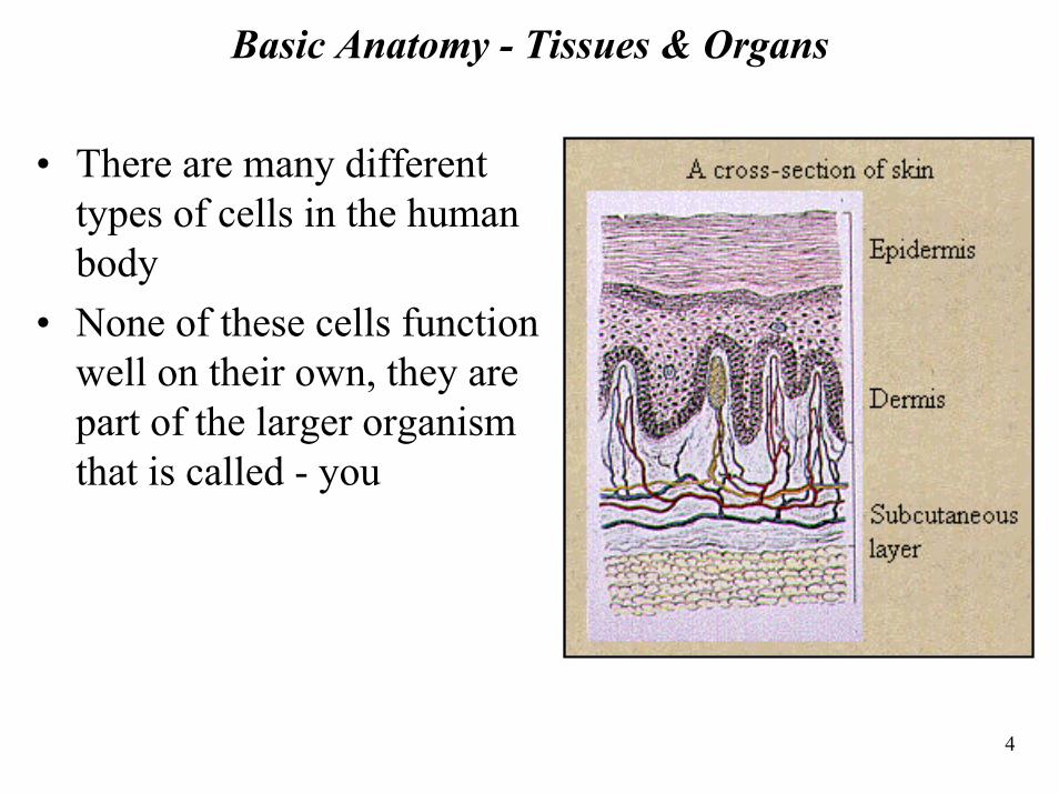
Basic Anatomy - Tissues & Organs
• There are many different types of cells in the human body
• None of these cells function well on their own, they are part of the larger organism that is called - you
5
Tissues
• Cells group together in the body to form tissues - a collection of similar cells that group together to perform a specialized function
• There are 4 primary tissue types in the human body: – Epithelial Tissue– Connective Tissue– Muscle Tissue – Nerve Tissue
6
Tissue Types • Epithelial Tissue cells pack tightly together and form continuous sheets that
serve as linings in different parts of the body– Serve as membranes lining organs helping to keep the body's organs separate, in
place and protected– Examples: outer layer of the skin, the inside of the mouth and stomach, and the
tissue surrounding the body's organs• Connective Tissue adds support and structure to the body
– Most types contain fibrous strands of the protein collagen that add strength to connective tissue
– Examples: inner layers of skin, tendons, ligaments, cartilage, bone and fat tissue – Blood is also considered a form of connective tissue
• Muscle Tissue is a specialized tissue that can contract– Contains the specialized proteins actin and myosin that slide past one another and
allow movement – Examples: contained in the muscles throughout the body
• Nerve Tissue contains two types of cells: neurons and glial cells– Has the ability to generate and conduct electrical signals in the body– These electrical messages are managed by nerve tissue in the brain and transmitted
down the spinal cord to the body
7
Organs
• An organ is a structure that contains at least two different types of tissue functioning together for a common purpose
• The integument organ is composed of skin and its associated tissues sweat glands, sebaceous glands, hair, and nails– Largest organ in the body (by weight and surface area)– Weight of your skin in adults accounts for about 16%
of total body weight– Skin is continuous with
• Digestive system (lips and anus)• Respiratory system (nose)• Urogenital system (urethra)
8
Function of Skin• Separates the internal environment from the external• Serves as a barrier to physical, biological, and chemical agents
and to radiation – Skin diseases and infections can compromise this barrier
• Regulates body temperature (thermoregulation) by :– Evaporative cooling (excretion through sweat glands )– Heat radiation at the surface of the body (blood circulation) – Serves as insulation
• Specialized pigment cells protect from ultraviolet rays of the sun• Photochemical production of vitamin D in the epidermal layer
(when it is exposed to the sun's rays)• Helps regulate metabolism• Has esthetic and beauty qualities
9
Section 2: Skin Layers
10
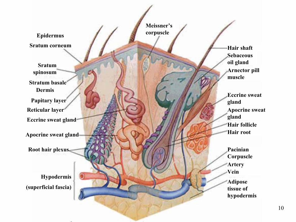
Epidermus
Sratum corneum
Stratum basale
Sratum spinosum
Dermis
Papitary layerReticular layer
Eccrine sweat gland
Apocrine sweat gland
Root hair plexus
Hypodermis
(superficial fascia)
Meissner’s corpuscle
Hair shaftSebaceous oil glandArnector pill muscle
Eccrine sweat glandApocrine sweat glandHair follicleHair root
Pacinian CorpuscleArteryVein
Adipose tissue of hypodermis
11
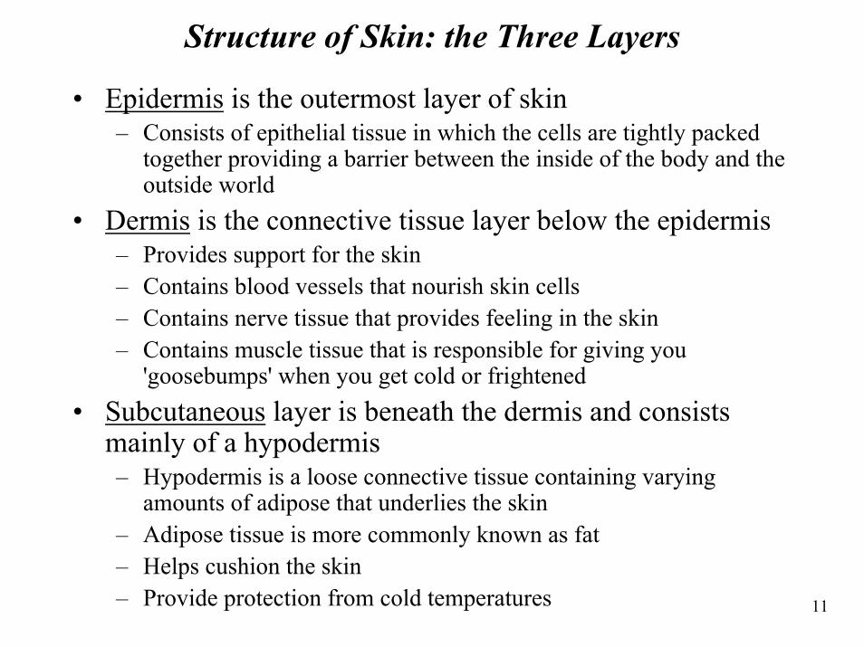
Structure of Skin: the Three Layers
• Epidermis is the outermost layer of skin– Consists of epithelial tissue in which the cells are tightly packed
together providing a barrier between the inside of the body and the outside world
• Dermis is the connective tissue layer below the epidermis – Provides support for the skin – Contains blood vessels that nourish skin cells– Contains nerve tissue that provides feeling in the skin– Contains muscle tissue that is responsible for giving you
'goosebumps' when you get cold or frightened• Subcutaneous layer is beneath the dermis and consists
mainly of a hypodermis – Hypodermis is a loose connective tissue containing varying
amounts of adipose that underlies the skin – Adipose tissue is more commonly known as fat – Helps cushion the skin– Provide protection from cold temperatures
12
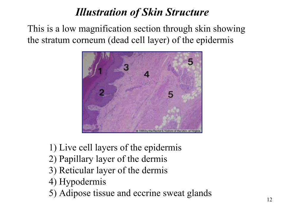
Illustration of Skin StructureThis is a low magnification section through skin showing the stratum corneum (dead cell layer) of the epidermis
1) Live cell layers of the epidermis2) Papillary layer of the dermis3) Reticular layer of the dermis 4) Hypodermis 5) Adipose tissue and eccrine sweat glands
13
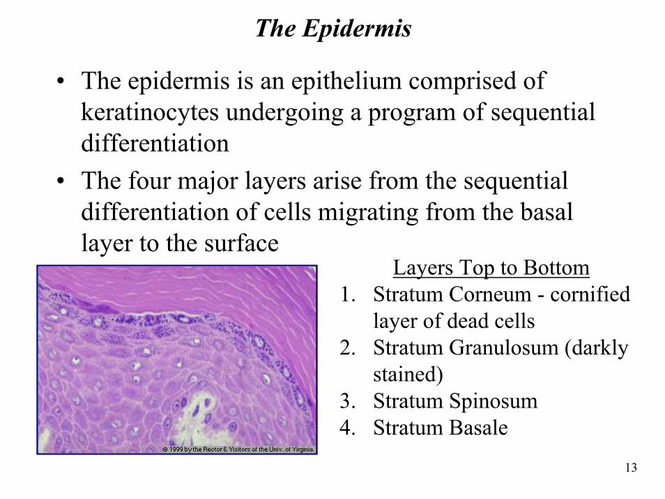
The Epidermis
• The epidermis is an epithelium comprised of keratinocytes undergoing a program of sequential differentiation
• The four major layers arise from the sequential differentiation of cells migrating from the basal layer to the surface
Layers Top to Bottom1. Stratum Corneum - cornified
layer of dead cells2. Stratum Granulosum (darkly
stained)3. Stratum Spinosum4. Stratum Basale
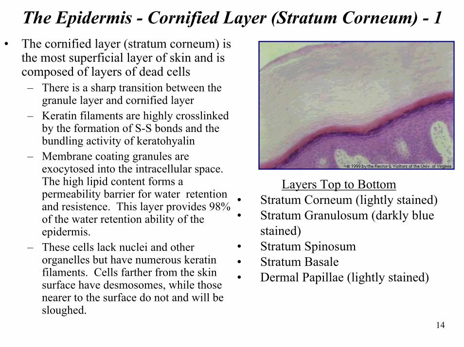
14
The Epidermis - Cornified Layer (Stratum Corneum) - 1• The cornified layer (stratum corneum) is
the most superficial layer of skin and is composed of layers of dead cells– There is a sharp transition between the
granule layer and cornified layer– Keratin filaments are highly crosslinked
by the formation of S-S bonds and the bundling activity of keratohyalin
– Membrane coating granules are exocytosed into the intracellular space. The high lipid content forms a permeability barrier for water retention and resistence. This layer provides 98% of the water retention ability of the epidermis.
– These cells lack nuclei and other organelles but have numerous keratin filaments. Cells farther from the skin surface have desmosomes, while those nearer to the surface do not and will be sloughed.
Layers Top to Bottom• Stratum Corneum (lightly stained)• Stratum Granulosum (darkly blue
stained)• Stratum Spinosum• Stratum Basale• Dermal Papillae (lightly stained)
15
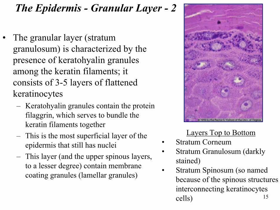
The Epidermis - Granular Layer - 2
• The granular layer (stratum granulosum) is characterized by the presence of keratohyalin granules among the keratin filaments; it consists of 3-5 layers of flattened keratinocytes– Keratohyalin granules contain the protein
filaggrin, which serves to bundle the keratin filaments together
– This is the most superficial layer of the epidermis that still has nuclei
– This layer (and the upper spinous layers, to a lesser degree) contain membrane coating granules (lamellar granules)
Layers Top to Bottom• Stratum Corneum• Stratum Granulosum (darkly
stained)• Stratum Spinosum (so named
because of the spinous structures interconnecting keratinocytescells)
16
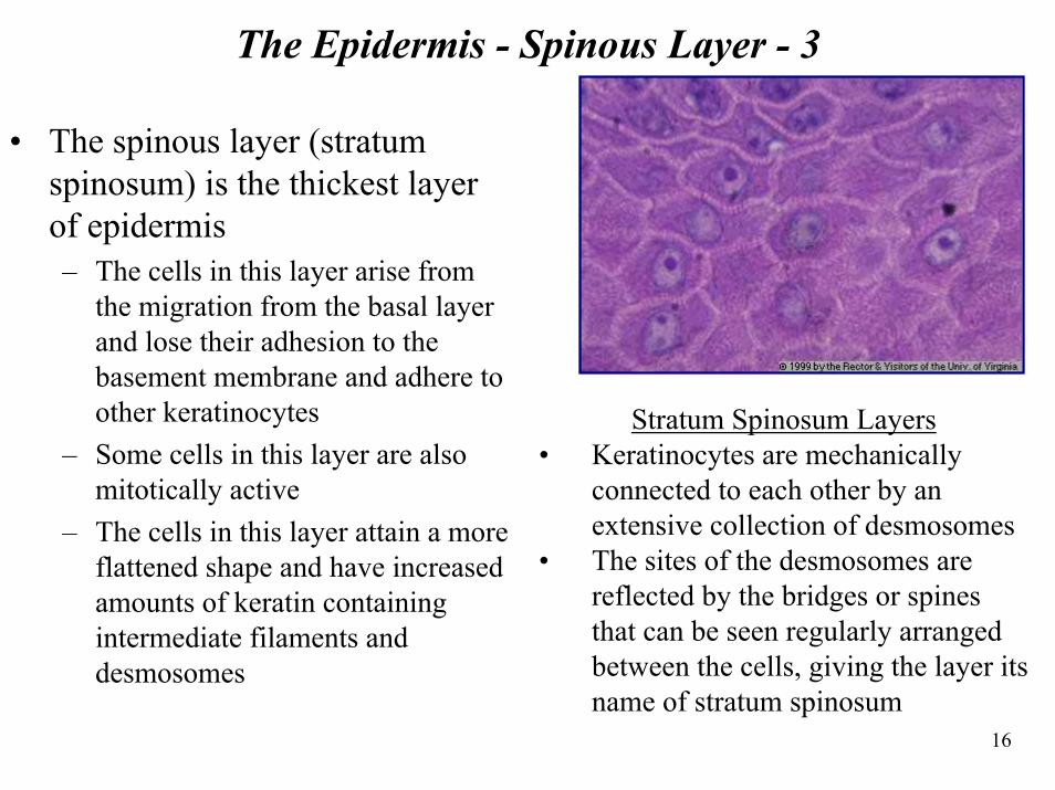
The Epidermis - Spinous Layer - 3
• The spinous layer (stratum spinosum) is the thickest layer of epidermis– The cells in this layer arise from
the migration from the basal layer and lose their adhesion to the basement membrane and adhere to other keratinocytes
– Some cells in this layer are also mitotically active
– The cells in this layer attain a more flattened shape and have increased amounts of keratin containing intermediate filaments and desmosomes
Stratum Spinosum Layers• Keratinocytes are mechanically
connected to each other by an extensive collection of desmosomes
• The sites of the desmosomes are reflected by the bridges or spines that can be seen regularly arranged between the cells, giving the layer its name of stratum spinosum
17
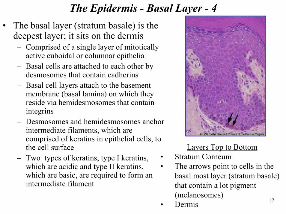
The Epidermis - Basal Layer - 4 • The basal layer (stratum basale) is the
deepest layer; it sits on the dermis– Comprised of a single layer of mitotically
active cuboidal or columnar epithelia– Basal cells are attached to each other by
desmosomes that contain cadherins– Basal cell layers attach to the basement
membrane (basal lamina) on which they reside via hemidesmosomes that contain integrins
– Desmosomes and hemidesmosomes anchor intermediate filaments, which are comprised of keratins in epithelial cells, to the cell surface
– Two types of keratins, type I keratins, which are acidic and type II keratins, which are basic, are required to form an intermediate filament
Layers Top to Bottom• Stratum Corneum• The arrows point to cells in the
basal most layer (stratum basale) that contain a lot pigment (melanosomes)
• Dermis
18
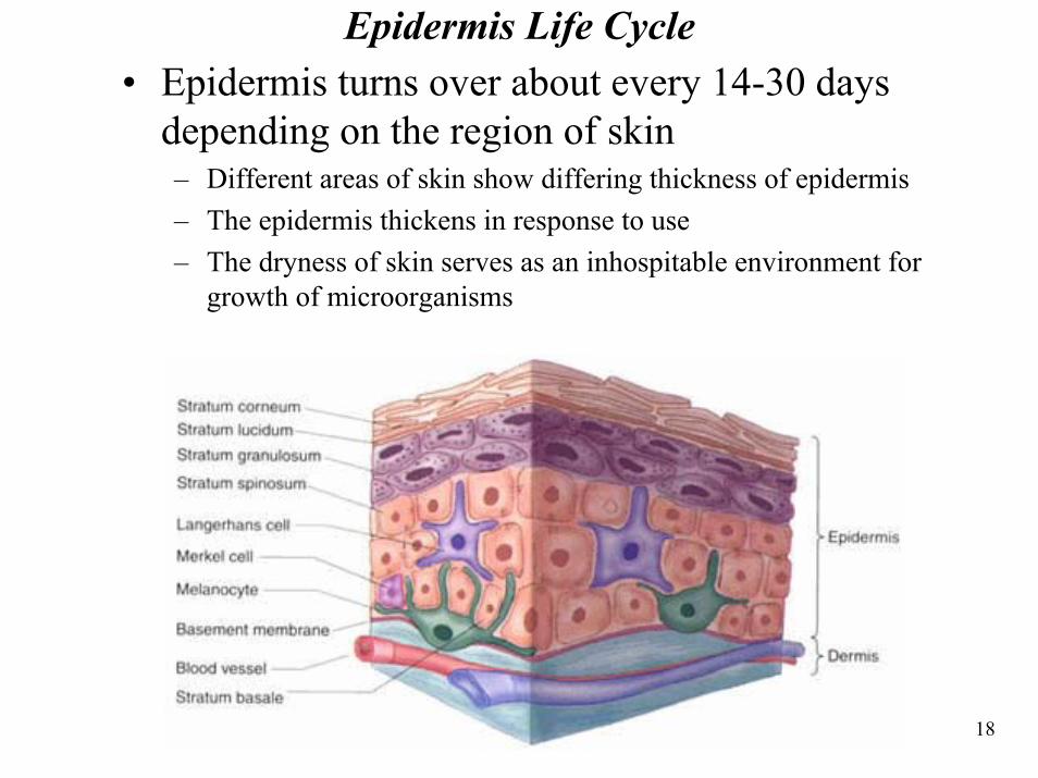
Epidermis Life Cycle• Epidermis turns over about every 14-30 days
depending on the region of skin– Different areas of skin show differing thickness of epidermis– The epidermis thickens in response to use– The dryness of skin serves as an inhospitable environment for
growth of microorganisms
19
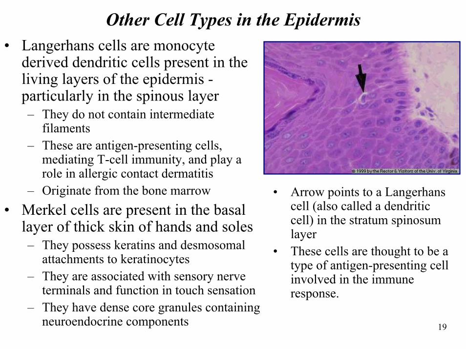
Other Cell Types in the Epidermis • Langerhans cells are monocyte
derived dendritic cells present in the living layers of the epidermis -particularly in the spinous layer– They do not contain intermediate
filaments– These are antigen-presenting cells,
mediating T-cell immunity, and play a role in allergic contact dermatitis
– Originate from the bone marrow• Merkel cells are present in the basal
layer of thick skin of hands and soles– They possess keratins and desmosomal
attachments to keratinocytes– They are associated with sensory nerve
terminals and function in touch sensation– They have dense core granules containing
neuroendocrine components
• Arrow points to a Langerhanscell (also called a dendriticcell) in the stratum spinosum layer
• These cells are thought to be a type of antigen-presenting cell involved in the immune response.
20
Other Cell Types in the Epidermis - cnt.• Melanocytes are neural crest derived cells that produce melanin,
the brown pigment that produces skin color– Melanocytes produce melanin from tyrosine in specialized organelles
called melanosomes, which contain tryosinase, an enzyme critical for melanin production
– All humans have about the same number of melanocytes• Skin color differences arise from differences in the number, size and
arrangement of melanosomes• In dark skin, there are more melanosomes, which are larger and distributed
throughout the cytoplasm rather than just in the perinuclear area as in light skin
• There are two kinds of melanin, one found in people with dark hair and the other in people with red and blond hair
– The epidermis responds to UV light• Melanin darkens via a photochemical reaction• Melanocytes increase production of melanin and the numbers of
melanosomes
21
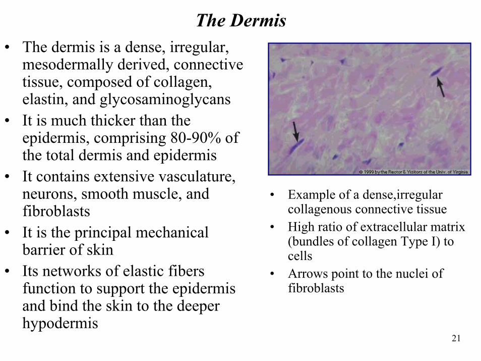
The Dermis• The dermis is a dense, irregular,
mesodermally derived, connective tissue, composed of collagen, elastin, and glycosaminoglycans
• It is much thicker than the epidermis, comprising 80-90% of the total dermis and epidermis
• It contains extensive vasculature, neurons, smooth muscle, and fibroblasts
• It is the principal mechanical barrier of skin
• Its networks of elastic fibers function to support the epidermis and bind the skin to the deeper hypodermis
• Example of a dense,irregular collagenous connective tissue
• High ratio of extracellular matrix (bundles of collagen Type I) to cells
• Arrows point to the nuclei of fibroblasts
22
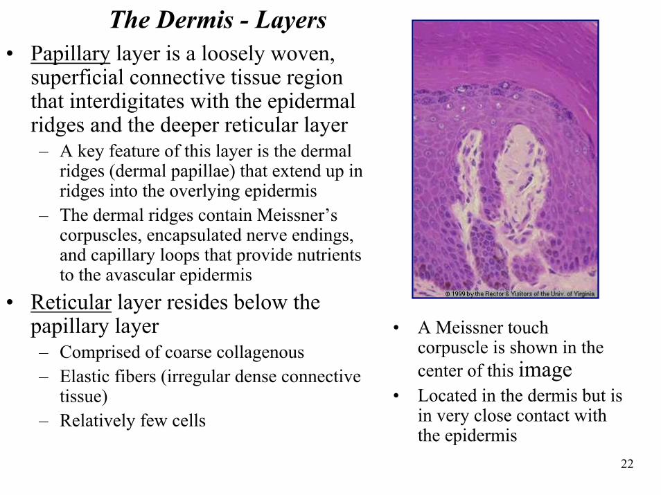
The Dermis - Layers• Papillary layer is a loosely woven,
superficial connective tissue region that interdigitates with the epidermal ridges and the deeper reticular layer– A key feature of this layer is the dermal
ridges (dermal papillae) that extend up in ridges into the overlying epidermis
– The dermal ridges contain Meissner’s corpuscles, encapsulated nerve endings, and capillary loops that provide nutrients to the avascular epidermis
• Reticular layer resides below the papillary layer – Comprised of coarse collagenous– Elastic fibers (irregular dense connective
tissue) – Relatively few cells
• A Meissner touch corpuscle is shown in the center of this image
• Located in the dermis but is in very close contact with the epidermis
23
The Dermis – Arteries, Veins, and Neurons • Arteries and veins run through the hypodermis and branch
upward to form plexuses of anastomosing vessels– This system provides nourishment to the dermis and by diffusion to the
epidermis, which is avascular– The vascular system functions in thermoregulation. Bood flow is
controlled by contraction of arterioles and venules to send blood through the capillary bed for heat radiation
• The dermis contains neuronal elements for touch, pain, itch, andtemperature reception– Some receptors are free nerve endings– Other nerve endings associate with Merkel cells in the epidermis– Meissner’s corpuscles reside in the dermal papillae and function as
mechanoreceptors in touch perception– Pacinian corpuscles are found deep in the dermis (and in the hypodermis)
and function in pressure sensation
24
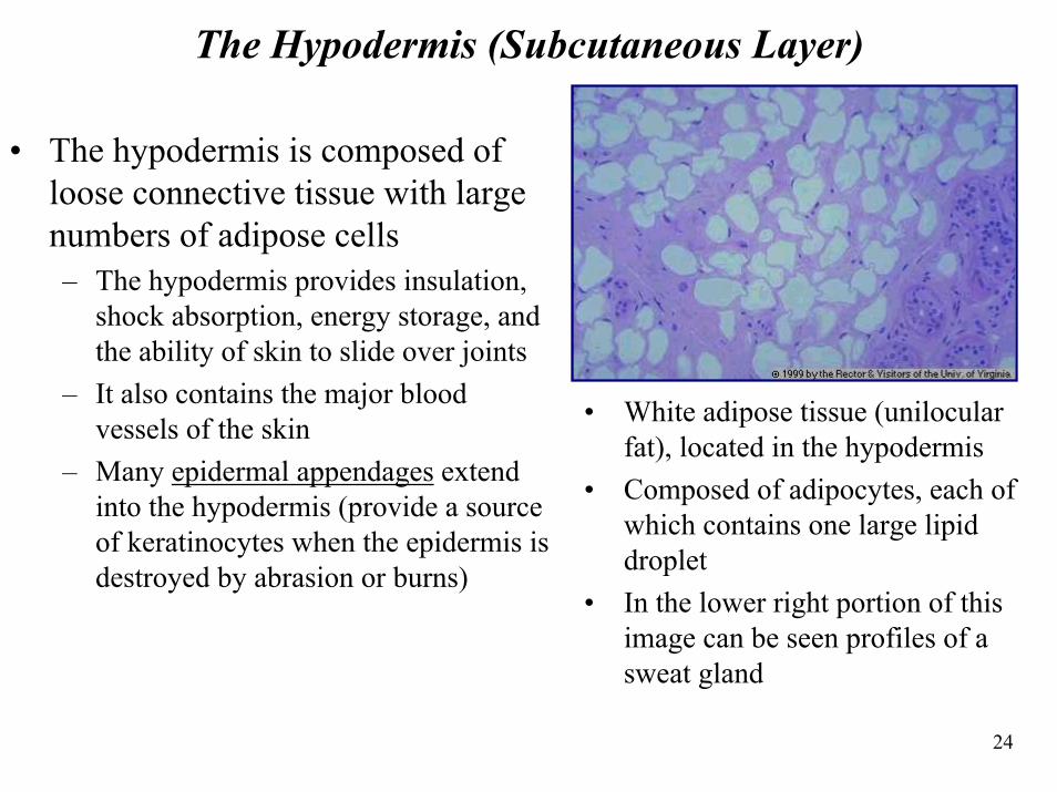
The Hypodermis (Subcutaneous Layer)
• The hypodermis is composed of loose connective tissue with large numbers of adipose cells– The hypodermis provides insulation,
shock absorption, energy storage, and the ability of skin to slide over joints
– It also contains the major blood vessels of the skin
– Many epidermal appendages extend into the hypodermis (provide a source of keratinocytes when the epidermis is destroyed by abrasion or burns)
• White adipose tissue (unilocularfat), located in the hypodermis
• Composed of adipocytes, each of which contains one large lipid droplet
• In the lower right portion of this image can be seen profiles of a sweat gland

25
Section 3: Epidermal Appendages
26
Epidermal Appendages• The epidermal appendages include hair follicles, various glands,
and nails– Hair is composed of dead epidermal cells that have undergone a modified
epidermal keratinization including the expression of specific keratin proteins that are highly crosslinked by disulfide bonds. It is derived from hair folicles, which are epidermal invaginations that project into the dermisor hypodermis.
– Sebaceous glands are appendages of hair follicles and are embedded in the dermis and hypodermis throughout the body except on the hands and soles. They are prominent in the face, neck and upper body.
– Eccrine sweat glands are simple coiled tubular glands located in the deep dermis or underlying hypodermis and are present throughout the body. Their primary function is evaporative cooling.
– Apocrine sweat glands are simple tubular glands that empty into hair follicles in axillary and anogenital regions. The secretion is a mixture of proteins, carbohydrates, and ferric ions that is odorless when secreted, but is acted on by commensal bacteria. They begin to function at puberty; but their function is unknown.
27
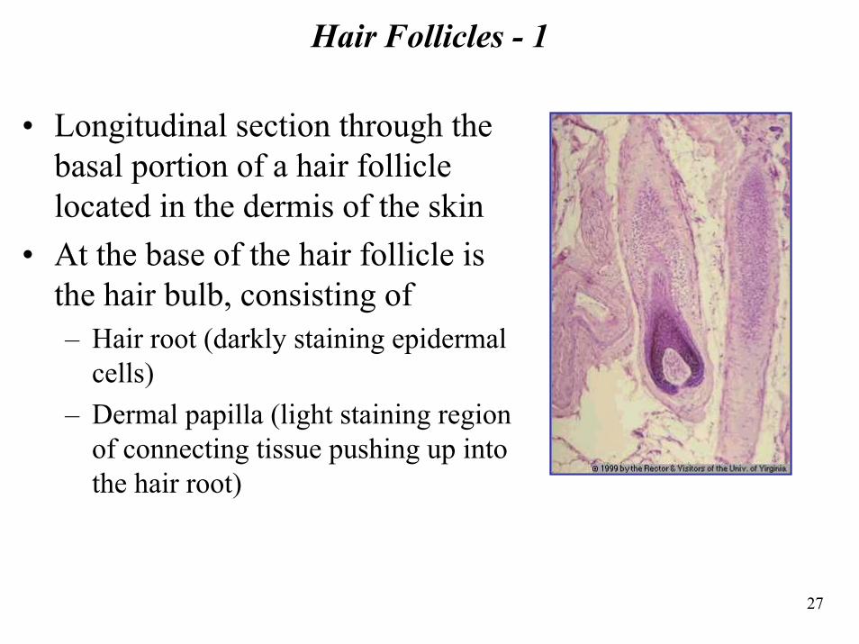
Hair Follicles - 1
• Longitudinal section through the basal portion of a hair follicle located in the dermis of the skin
• At the base of the hair follicle is the hair bulb, consisting of– Hair root (darkly staining epidermal
cells) – Dermal papilla (light staining region
of connecting tissue pushing up into the hair root)
28
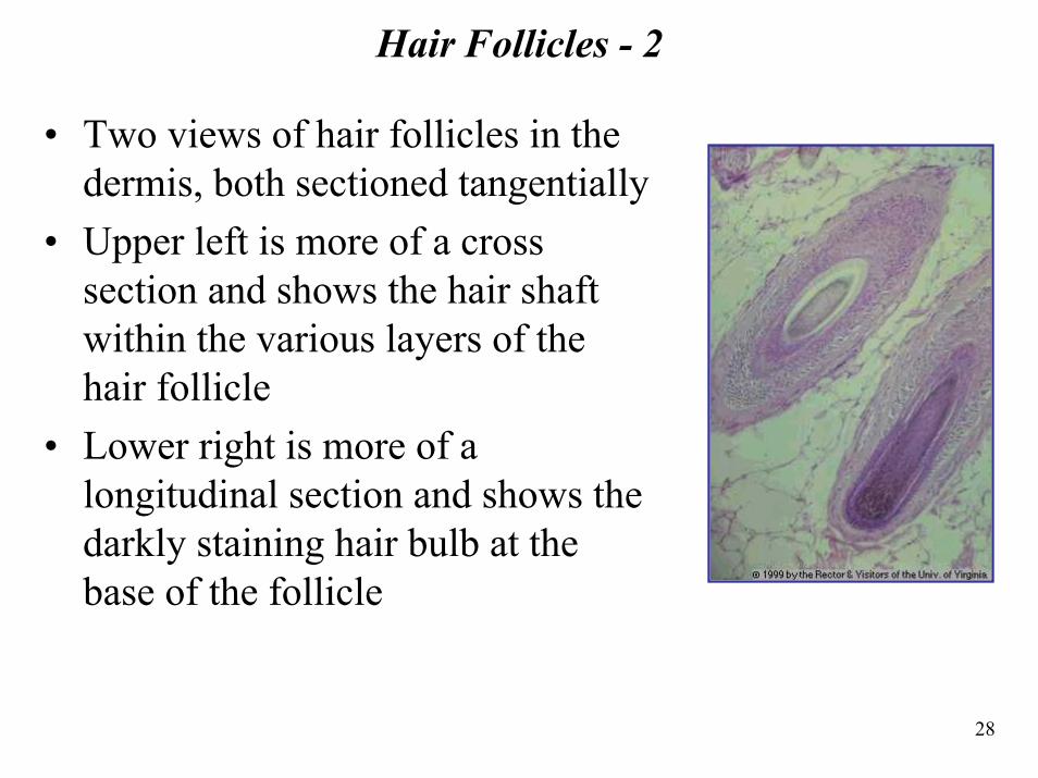
Hair Follicles - 2
• Two views of hair follicles in the dermis, both sectioned tangentially
• Upper left is more of a cross section and shows the hair shaft within the various layers of the hair follicle
• Lower right is more of a longitudinal section and shows the darkly staining hair bulb at the base of the follicle
29
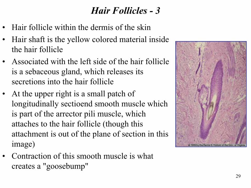
Hair Follicles - 3
• Hair follicle within the dermis of the skin• Hair shaft is the yellow colored material inside
the hair follicle• Associated with the left side of the hair follicle
is a sebaceous gland, which releases its secretions into the hair follicle
• At the upper right is a small patch of longitudinally sectioend smooth muscle which is part of the arrector pili muscle, which attaches to the hair follicle (though this attachment is out of the plane of section in this image)
• Contraction of this smooth muscle is what creates a "goosebump"
30
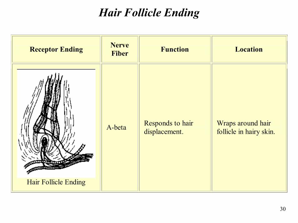
Hair Follicle Ending
Receptor Ending Nerve Fiber Function Location
Hair Follicle Ending
A-beta Responds to hair displacement.
Wraps around hair follicle in hairy skin.
31
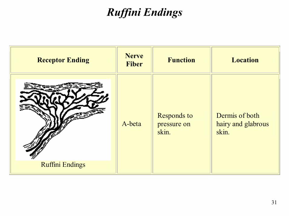
Ruffini Endings
Receptor Ending Nerve Fiber Function Location
Ruffini Endings
A-beta Responds to pressure on skin.
Dermis of both hairy and glabrous skin.
32
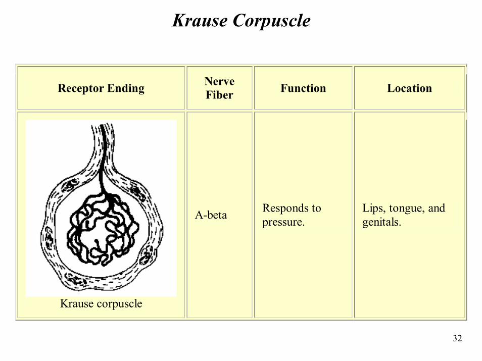
Krause Corpuscle
Receptor Ending Nerve Fiber Function Location
Krause corpuscle
A-beta Responds to pressure.
Lips, tongue, and genitals.
33
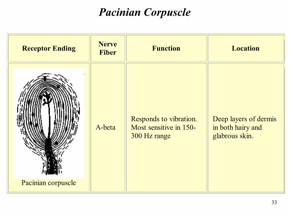
Pacinian Corpuscle
Receptor Ending Nerve Fiber Function Location
Pacinian corpuscle
A-beta Responds to vibration. Most sensitive in 150-300 Hz range
Deep layers of dermis in both hairy and glabrous skin.
34
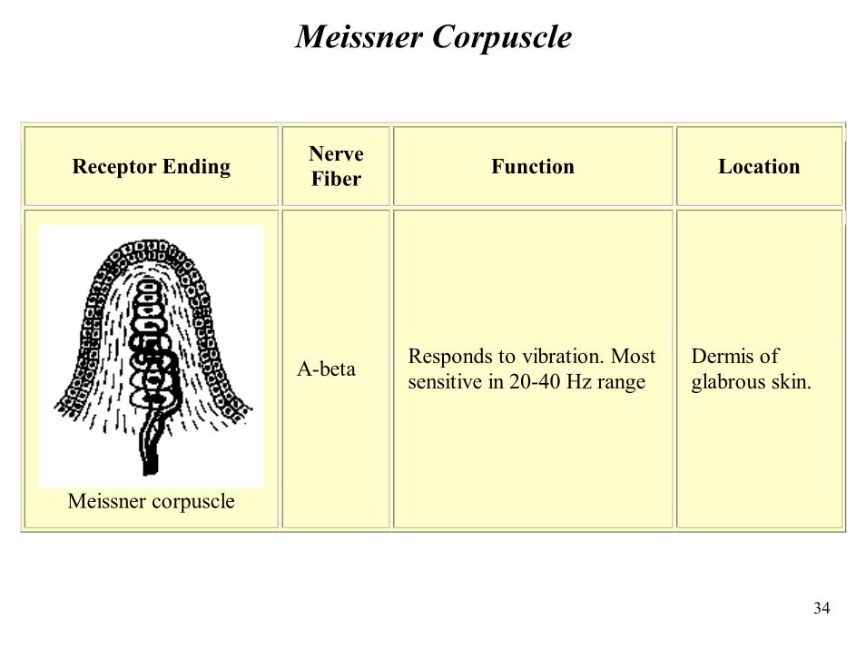
Meissner Corpuscle
Receptor Ending Nerve Fiber Function Location
Meissner corpuscle
A-beta Responds to vibration. Most sensitive in 20-40 Hz range
Dermis of glabrous skin.
35
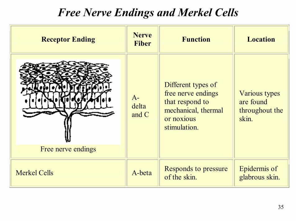
Free Nerve Endings and Merkel Cells
Receptor Ending Nerve Fiber Function Location
Free nerve endings
A-delta and C
Different types of free nerve endings that respond to mechanical, thermal or noxious stimulation.
Various types are found throughout the skin.
Merkel Cells A-beta Responds to pressure of the skin.
Epidermis of glabrous skin.
36
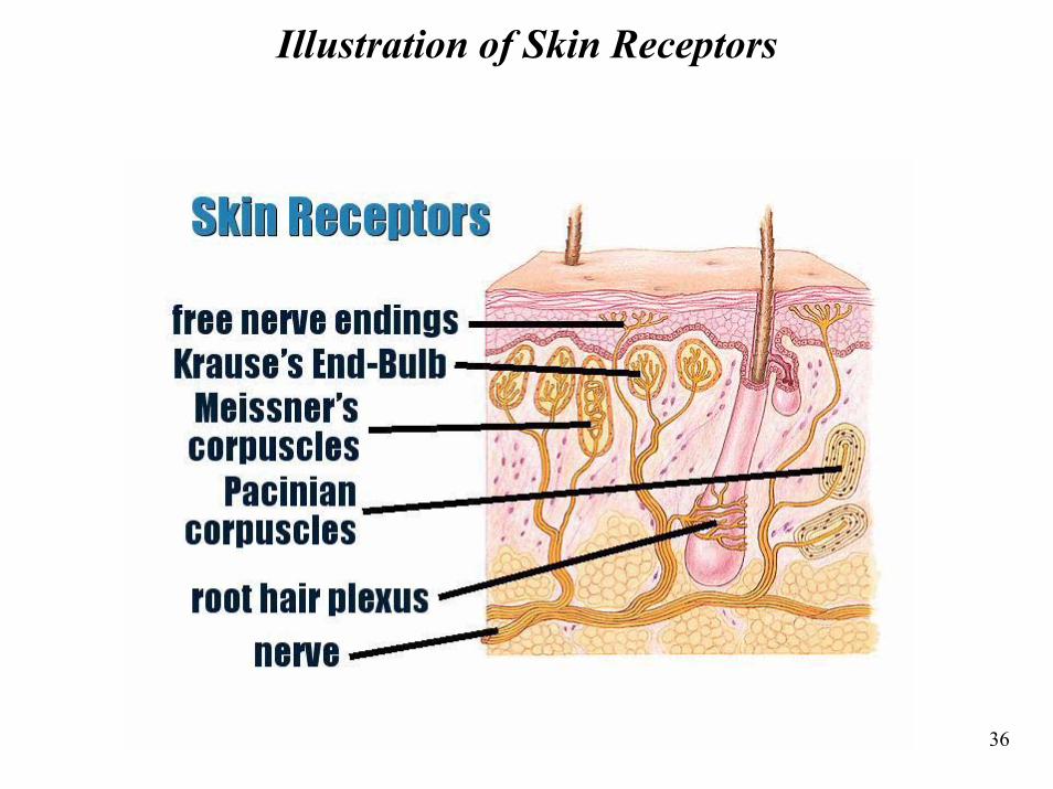
Illustration of Skin Receptors
37
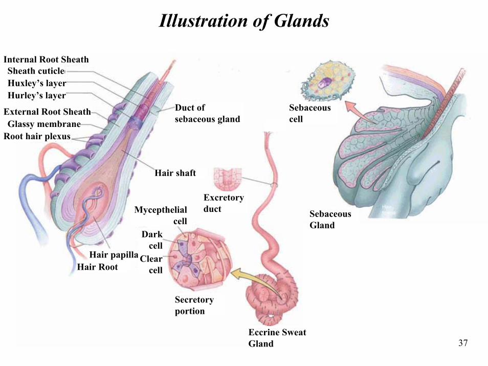
Illustration of Glands
Internal Root SheathSheath cuticleHuxley’s layerHurley’s layer
External Root SheathGlassy membrane
Root hair plexus
Duct of sebaceous gland
Secretory portion
Hair papillaHair Root
Eccrine Sweat Gland
Sebaceous Gland
Sebaceous cell
Hair shaft
Excretory duct
Clear cell
Dark cell
Mycepthelial cell
38
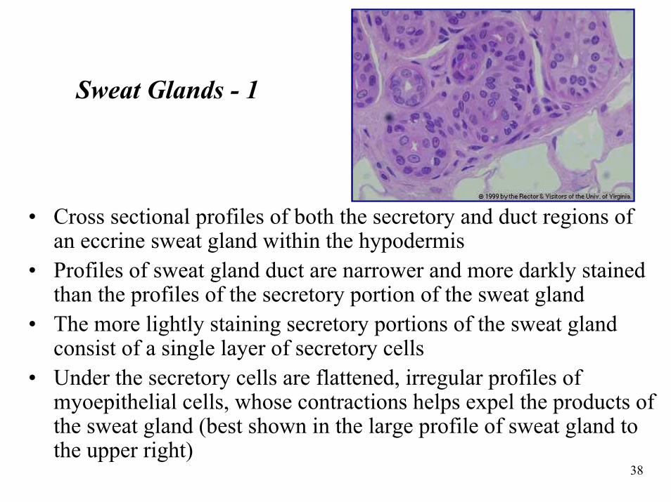
Sweat Glands - 1
• Cross sectional profiles of both the secretory and duct regions of an eccrine sweat gland within the hypodermis
• Profiles of sweat gland duct are narrower and more darkly stained than the profiles of the secretory portion of the sweat gland
• The more lightly staining secretory portions of the sweat gland consist of a single layer of secretory cells
• Under the secretory cells are flattened, irregular profiles of myoepithelial cells, whose contractions helps expel the products of the sweat gland (best shown in the large profile of sweat gland to the upper right)
39
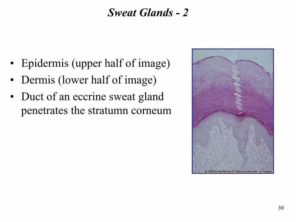
Sweat Glands - 2
• Epidermis (upper half of image)• Dermis (lower half of image)• Duct of an eccrine sweat gland
penetrates the stratumn corneum
40
Section 4: Current Melanoma Research
41
Melanoma Research
• As so many of us sadly know, melanoma affects people from all walks of life and often strikes during the prime of life
• Worldwide, the incidence of melanoma - and the number of deaths from this cancer - are rising at alarming rates
• The nonprofit Melanoma Research Foundation (MRF), founded by patients and families several years ago, believes that if we all work together, we can help find a cure for this often-fatal disease
42
Melanoma Research – Dr. Chow
• Dr. Chow's proposed research is designed to study the regulation of FasL, a protein present in melanoma tumors which is responsible for the premature death of the immune system's T-cells
• Of particular interest is how interferons act to suppress the expression of FasL, an issue of considerable therapeutic significance
• This study promises to clarify an important mechanism of interferon action which may lead to improvements in therapies to enhance the body's immune response to tumors
43
Melanoma Research – Dr. Lori White
• Dr. Lori White of Rutgers University received the Martin Schneider Memorial Research Award for her project, "Effect of Environmental Factors on Melanoma Invasion and Metastasis"
• The project seeks to discover how UV exposure and exposure to certain environmental carcinogens, especially dioxin, affects activity in the skin cells and leads to cancer progression
• Understanding such changes, she believes, will help target preventive measures, and possibly sharpen the aim of chemotherapeutics
44
Section 5: Skin Engineering
45
46
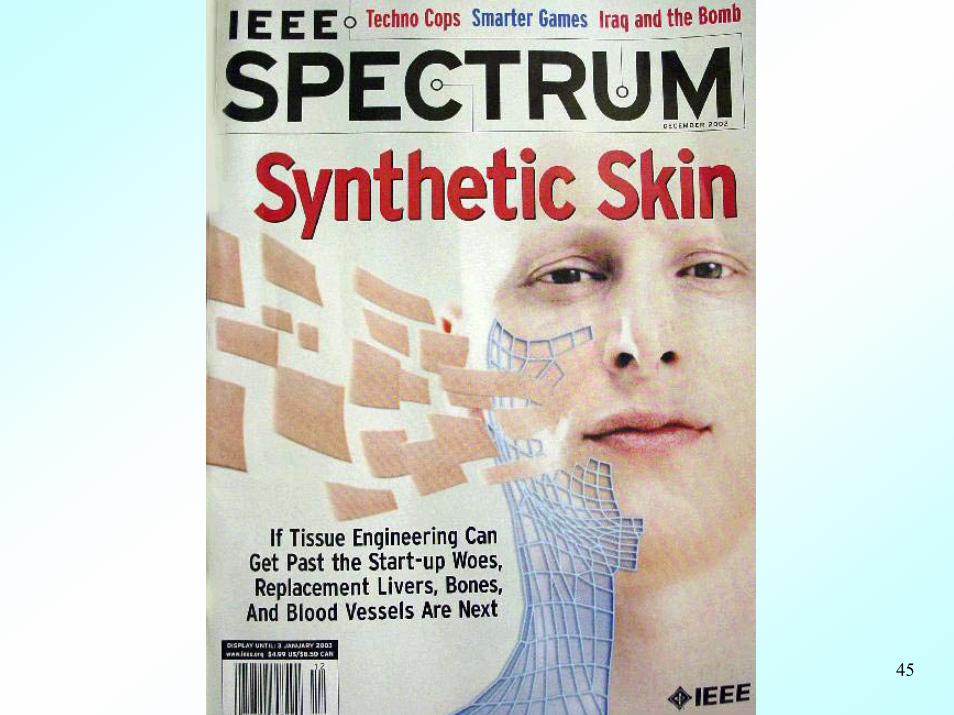
Skin Paved the Way for Tissue Engineering
• The first tissue successfully engineered and made available to people was skin
• Scientists have been developing skin substitutes for almost 20 years
• About 2 million people suffer burns each year– 13,000 require skin grafts– About 1,500 suffer burns over 20% of their bodies– With such extensive burns, skin grafting isn't
feasible and skin substitutes are required
47
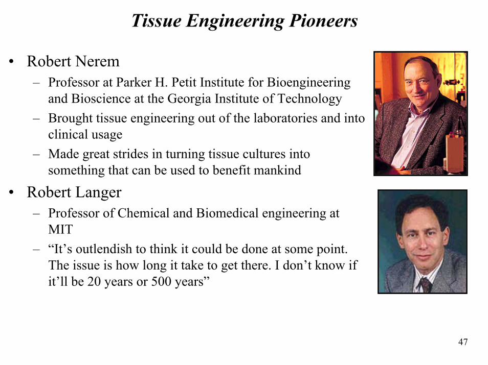
Tissue Engineering Pioneers
• Robert Nerem– Professor at Parker H. Petit Institute for Bioengineering
and Bioscience at the Georgia Institute of Technology– Brought tissue engineering out of the laboratories and into
clinical usage– Made great strides in turning tissue cultures into
something that can be used to benefit mankind
• Robert Langer– Professor of Chemical and Biomedical engineering at
MIT– “It’s outlendish to think it could be done at some point.
The issue is how long it take to get there. I don’t know if it’ll be 20 years or 500 years”
48
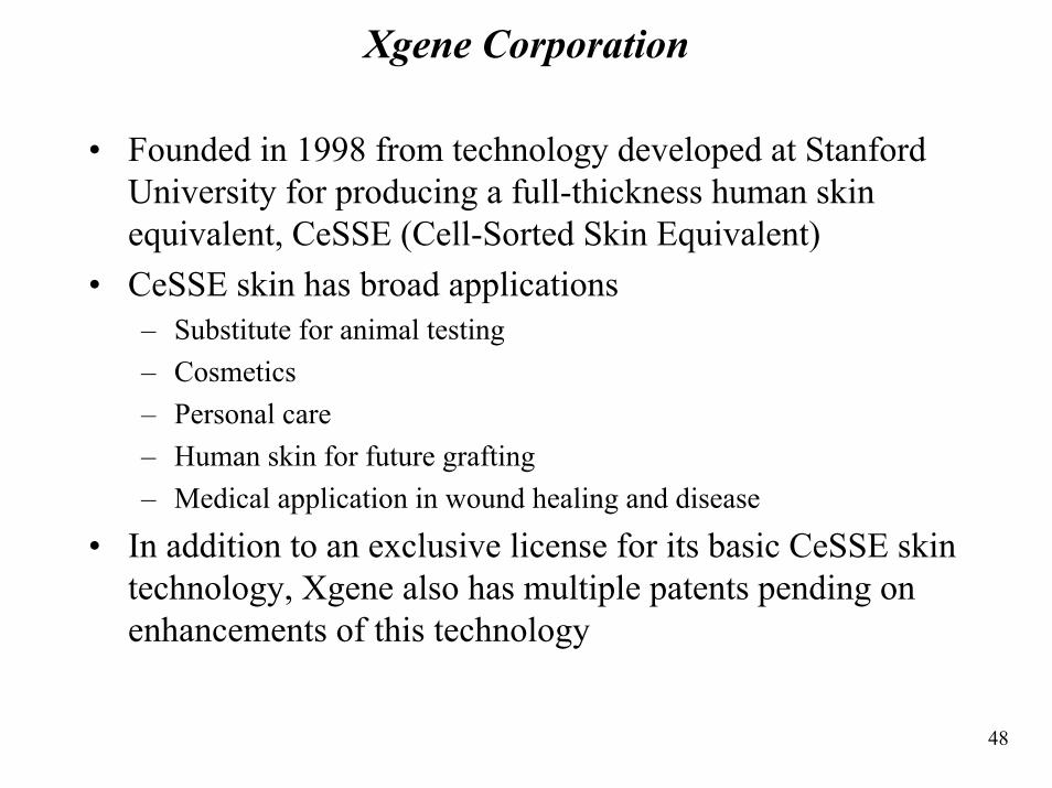
Xgene Corporation
• Founded in 1998 from technology developed at Stanford University for producing a full-thickness human skin equivalent, CeSSE (Cell-Sorted Skin Equivalent)
• CeSSE skin has broad applications– Substitute for animal testing– Cosmetics– Personal care – Human skin for future grafting– Medical application in wound healing and disease
• In addition to an exclusive license for its basic CeSSE skin technology, Xgene also has multiple patents pending on enhancements of this technology
49
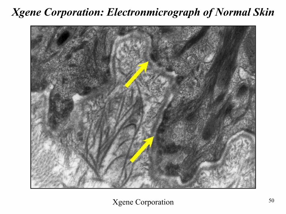
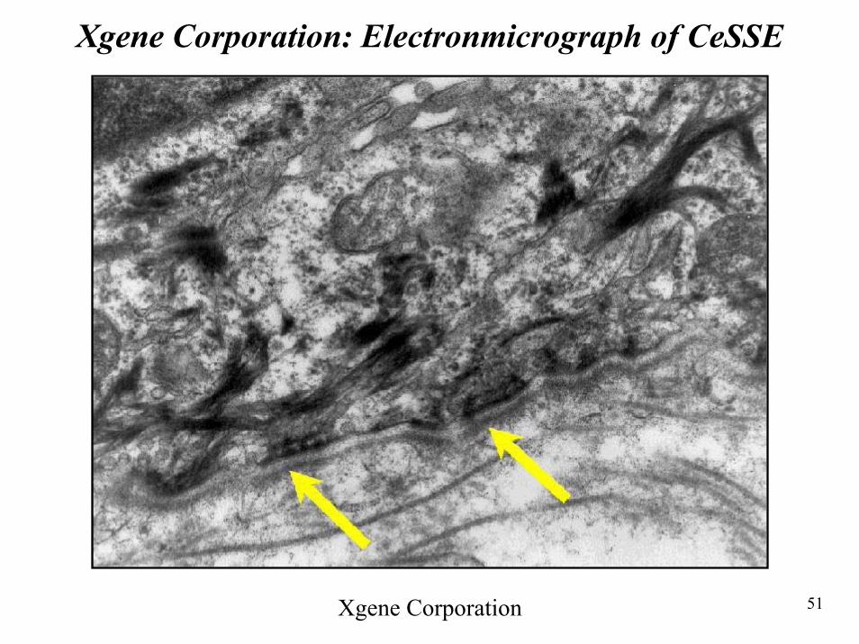
Xgene Corporation: Cell-Sorted Skin Equivalent • The following two slides show normal skin and CeSSE• Important characteristics of normal skin are recreated in two weeks
old CeSSE– Under high power examination of normal skin, the network of keratin
intermediate filaments (arrows) are seen to connect down to the hemidesmosomes (dark, electron dense structures located along the dermal-epidermal junction)
– Network of fine threads located just beneath the hemidesmosomes termed anchoring fibrils are plentiful, and larger collagen filaments in the dermis clearly evident
• The electronmicrograph of CeSSE– Keratin intermediate filaments (arrows) are seen to connect down to
hemidesmosomes located along the BMZ – At the dermal-epidermal interface, seen at high power, clearly visible collagen
filaments do develop, as seen by long filaments– Numerous anchoring fibrils were noted on the dermal side just below the
hemidesmosomes, seen as thin threads just below the hemidesmosomes– Xgene believes that the presence of these structures will give CeSSE a resistance
to the problem of blistering that afflicts most grafts on the market today
50
Xgene Corporation: Electronmicrograph of Normal Skin
Xgene Corporation
51
Xgene Corporation: Electronmicrograph of CeSSE
Xgene Corporation
52
Advanced Tissue Sciences Inc• Advanced Tissue Sciences Inc. of La Jolla, Calif., was the first
company to get a tissue-engineered skin product to the market– Approved for commercial use in March by the Food and Drug Administration– The product, Dermagraft TC, is used for severe burns as a temporary covering
for wounds– Designed as an alternative to cadaver skin (Was previously the best treatment
doctors could offer)• Dermagraft TC skin product is made from cells of the foreskins of
newborns– The cells, called dermal fibroblasts, are grown on a mesh that serves as a
three-dimensional scaffold (says Gail Naughton, co-inventor of the product and company president)
– As the cells grow over the scaffold, they secrete human skin collagen, growth factors and structural proteins; A synthetic outer skin layer is then added
– The skin substitute is placed on deep burns where the patient's skin is too badly destroyed to regenerate on its own
– The substitute helps hold in body fluids that would otherwise ooze from the exposed areas; it helps maintain body temperature and it serves as a barrier against infection
– It can be kept on the body without changing for about three months; cadaver skin is usually rejected after a week or two
53
Advanced Tissue Sciences Inc
• ATS’s skin is grown in enclosed chambers called bioreactors– Nutrients flow through them to enhance growth– Bioreactors are kept in an incubator where sensors monitor
temperature, humidity, and gas concentrations
• The company has a second skin product under expedited review by the FDA– It is intended as a permanent graft for treating diabetic foot ulcers,
which affect about 800,000 people a year– The product is designed to promote healing and help prevent
amputations
54
Organogenisis Inc
• Developed infrastructure to produce thousands of skin grafts every month
• Apligraf made in petri dishes (see slides that follow)• Grafts start as donated infant foreskin tissue taken from
circumcisions• Postage stamp size sample multiplies to such extent that
several football fields of skin graft can be crafted from it• The main difference from Dermagraft TC is that the outer
dermal layer is made from human cells called keratinocytes, which make up the epidermal layer of normal skin
55
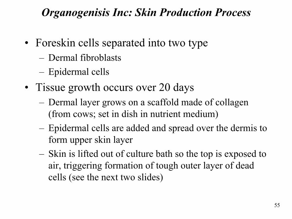
Organogenisis Inc: Skin Production Process
• Foreskin cells separated into two type– Dermal fibroblasts– Epidermal cells
• Tissue growth occurs over 20 days– Dermal layer grows on a scaffold made of collagen
(from cows; set in dish in nutrient medium)– Epidermal cells are added and spread over the dermis to
form upper skin layer– Skin is lifted out of culture bath so the top is exposed to
air, triggering formation of tough outer layer of dead cells (see the next two slides)
56
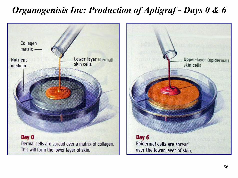
Organogenisis Inc: Production of Apligraf - Days 0 & 6
57
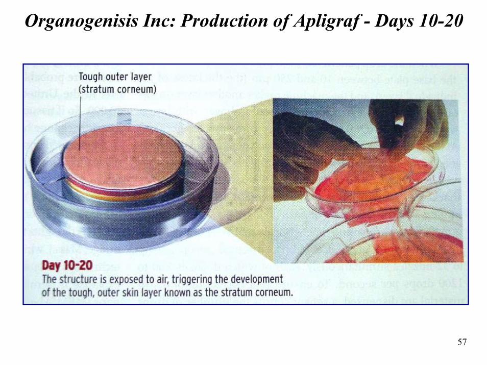
Organogenisis Inc: Production of Apligraf - Days 10-20
58
Other Companies Involved in Skin Engineering
• Integra LifeSciences Corp, Plainsboro, N.J.– Makes a skin product called Integra– Was used to treat Betty Shabazz, widow of Malcolm X,
who was badly burned over 80% of her body and later died
• Genzyme Tissue Repair, Cambridge, Mass.– Start with the patient's own cells to grow skin that can be
grafted on– Products made from foreskin cells eventually are rejected
by the patient's immune system
59
Multilayered Skin Engineering
• Multilayered skin that contains several different cell types like natural skin are expected to be developed over the next decade
• The second major tissue type to make it to market will be cartilage– Most companies are experimenting with cartilage cells– Several are only months away from human studies
• Genzyme– Has a cartilage product made from a patient's own cells
• Advanced Tissue Sciences– Developing cartilage, using cadaver cells as a source– Human studies are expected to begin in the USA and
Europe later this year
60
Role of Scaffold in Tissue Engineering
• Fabrication of specially designed porous structures (scaffolds) for tissue Engineering is one of the major focal points in current biomedical research
• Scaffolds guide the process of tissue regeneration and development
• Achieve cell delivery and provide mechanical support against compressive and tensile forces, thus maintaining the shape an integrity of the structures when implanted into the body
61
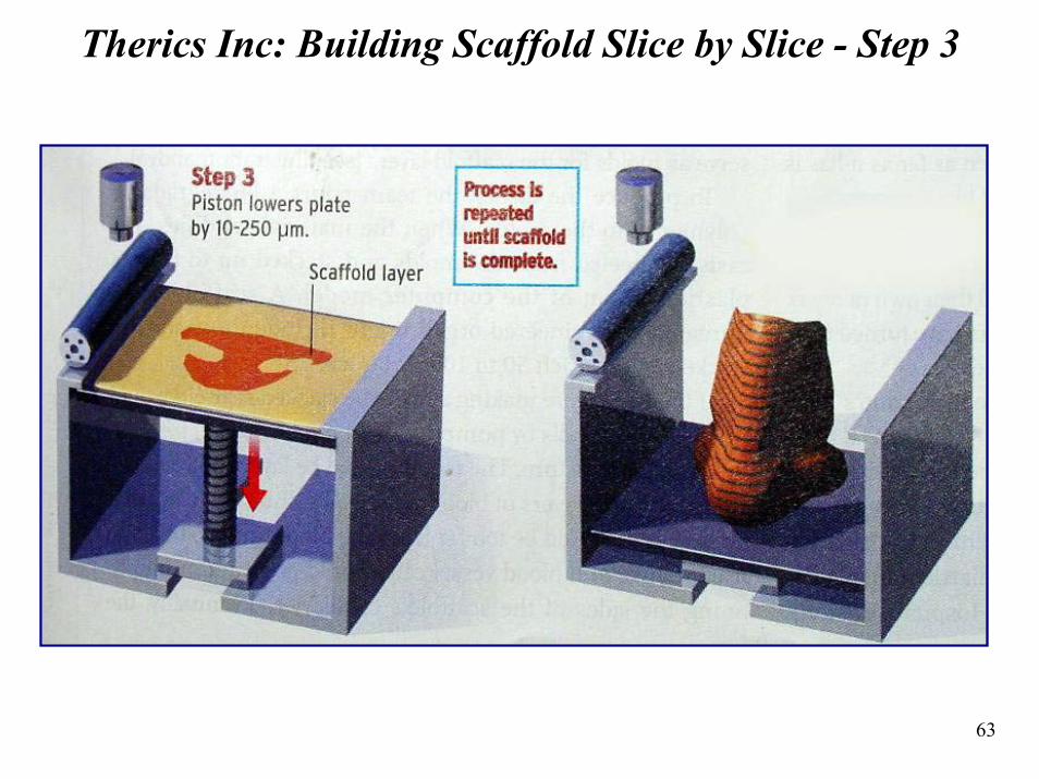
Therics Inc
• Developed a super precise fabrication machine (TheriFormprinter) to develop complex scaffolds
• Scaffold design is based on patient’s X-ray, MRI, or computer tomography scans
• CAD program is used to create 3-D computer model of scaffold (sliced into layers that are manufactured atop each other)
• TheriForm operates like a rapid prototype machine that deposits a layer of powder and then precisely binds it using a jet binder (similar to inc jet printer)
• Layers are built on top of each other until the organ is completed
• See figures on next two slides
62
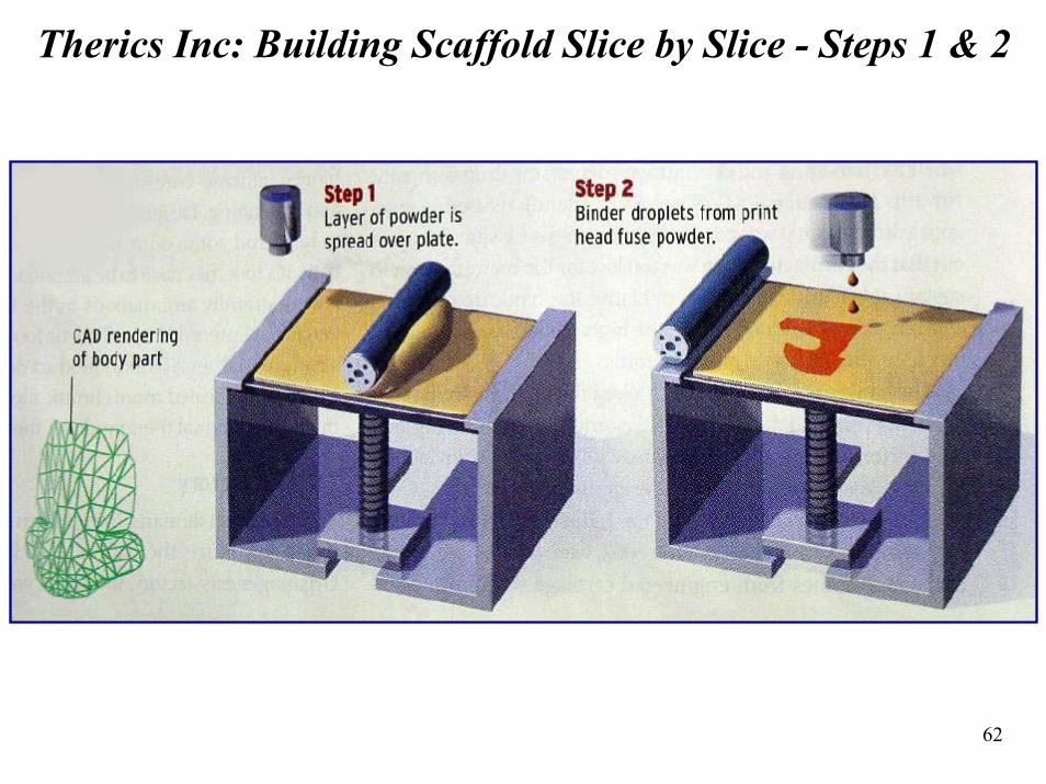
Therics Inc: Building Scaffold Slice by Slice - Steps 1 & 2
63
Therics Inc: Building Scaffold Slice by Slice - Step 3

64
Section 6: Ideas, Conclusions, and References
65
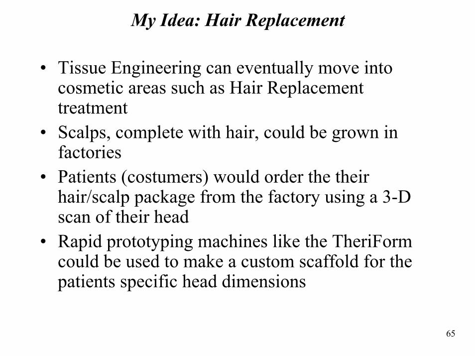
My Idea: Hair Replacement
• Tissue Engineering can eventually move into cosmetic areas such as Hair Replacement treatment
• Scalps, complete with hair, could be grown in factories
• Patients (costumers) would order the their hair/scalp package from the factory using a 3-D scan of their head
• Rapid prototyping machines like the TheriFormcould be used to make a custom scaffold for the patients specific head dimensions
66
Conclusions
• Tissue engineering will play a major role in the future of humanity
• Today, Tissue Engineering companies have an impressive array of skin replacement products
• There is uncertainty of the time frame that dictates when Tissue Engineering will produce major organs (such as the heart) that will meet the demand of patients needing organ transplants
67
References
• William Leventon, “Synthetic Skin”, IEEE Spectrum, December 2002, pp 28-33
• Andrew Y.J. Szeto, “A Tissue Engineering Pioneer”, IEEE Engineering In Medicine And Biology, July/August 2002, page 10
• Too, M.H.; Leong, K.F.; Chua, C.K.; Cheah, C.M., Intelligent Information Systems Conference, The Seventh Australian and New Zealand 2001, 2001, pp 433 -438
• faculty.washington.edu/chudler/receptor.html• krupp.wcc.hawaii.edu/BIOL100/present/senses/sld010.htm• web.jjay.cuny.edu/~acarpi/NSC/14-anatomy.htm• www.med-ed.virginia.edu/public/histology/handouts/Skin/handout.html• www.melanoma.org• www.ithaca.rice.edu• www.skin-information.com• www.xgene.com


![Untitled Document [geocities.ws]geocities.ws/aylinkrzesaj/bombas.pdf · %RPEDV Bombas são equipamentos que transformam energia mecânica em energia hidráulica, que é fornecida](https://img.pdfslide.us/doc/110x75/5a71ca837f8b9ac0538d27dc/untitled-document-geocitieswsgeocitieswsaylinkrzesajbombaspdfpdf.jpg)