Embed Size (px)
Citation preview

1
Copper-catalysed cycloaddition reactions of nitrones and alkynes for bioorthogonal labelling of living cellsAllison R. Sherratt, Mariya Chigrinova, Craig S. McKay, Louis-Philippe B. Beaulieu, Yanouchka Rouleau and John Paul Pezacki
Table of ContentsMaterials and Synthetic Methods ..............................................................................................2
Synthesis of sugar derivatives ...................................................................................................3
Synthesis of KDO-HMMPO and KDO-alkyne from KDO-azide .........................................3
Synthesis of Alexa488-CMPO ..................................................................................................7
Synthesis of Biotin-CMPO ........................................................................................................8
Experimental Methods.............................................................................................................10
Supplementary Figures ............................................................................................................12
1H and 13C NMR Spectra of KDO-HMMPO ..........................................................................14
1H and 13C NMR Spectra of KDO-alkyne...............................................................................15
1H and 13C NMR Spectra of Biotin-CMPO.............................................................................16
References................................................................................................................................17
Electronic Supplementary Material (ESI) for RSC Advances.This journal is © The Royal Society of Chemistry 2014
2
Materials and Synthetic Methods All reagents and solvents were purchased from Sigma-Aldrich, unless otherwise stated, and used
without further purification. Deuterated solvents were purchased from Cambridge Isotope
laboratories. Thin layer chromatography was performed on Analtech Uniplate® silica gel plates
(60 Å F254, layer thickness 250μm). Flash chromatography was performed using silica gel (60
Å, particle size 40–63 μm). LC-MS/MS spectra were obtained using Waters Alliance 2795 liquid
chromatograph quipped with Waters 996 PDA diode array detector and connected to Micromass
ZQ2000 mass spectrometer equipped with pneumatically assisted electrospray ionization source,
operating in both positive and negative mode. Samples were run with gradient elution of
acetonitrile/water/0.1% formic acid on the Waters SunFire C18 (2.1 x 100 mm, 3.5 µm) column
with flow rate of 0.2 mL/min. Preparatory HPLC was performed on a Waters Delta Prep 4000
equipped with Waters 996 PDA diode array detector and column Waters SunFire C18 (19 x 100
mm, 5 µm) and Waters fraction collector. Column effluents were monitored at wavelength
specified in respective procedures. Mobile phase was acetonitrile/water/0.1% formic acid,
degassed with helium sparge at flow rate of 17 mL/min. Aliquots were injected onto column via
a Pheodyne 7125 injector fitted with a 5 mL loop. All 1H and 13C NMR spectra were obtained on
a Bruker-DRX-400 spectrometer using a frequency of 400 MHz for 1H and 100 MHz for 13C and
processed using Bruker TOPSPIN 2.1 software. The following abbreviations were used to
designate chemical shift multiplicities: s = singlet, d = doublet, t = triplet, m = multiplet or
unresolved, br = broad signal and J = coupling constants in Hz.
3
Synthesis of sugar derivatives
Ac4ManN-alkyne was synthesized according to the previously reported procedure.1
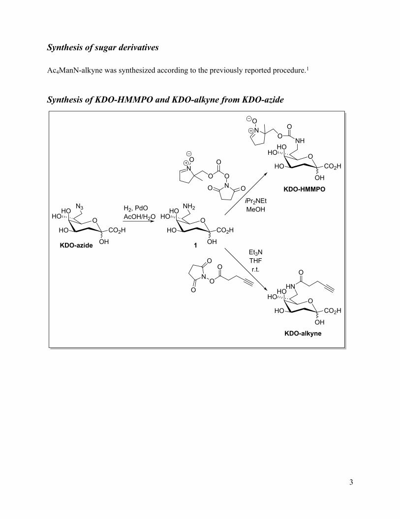
Synthesis of KDO-HMMPO and KDO-alkyne from KDO-azide
OHO
HO
OH
CO2H
HO
NH
OO
NO
O
ONO
ON OO
iPr2NEtMeOH
KDO-HMMPO
OHO
HO
OH
CO2H
HON3 H2, PdO
AcOH/H2O OHO
HO
OH
CO2H
HONH2
OHO
HO
OH
CO2H
HOHN
O
KDO-alkyne
KDO-azide 1
NO
O
O
O
Et3NTHFr.t.

4
O
ONO
ON OOOH
NO
O
O ON OO
N
O
O+
Et3NMeCN
HMMPO HMMPO-NHS
HMMPO synthesis and characterization data has been previously reported in literature.2 To a
solution of HMMPO (621 mg, 4.81 mmol, 1 eq) and disuccinimide carbonate (1.48 g, 5.77
mmol, 1.2 eq) in dry acetonitrile (50 mL) under argon atmosphere was added triethylamine (0.87
mL, 6.25 mmol, 1.3 eq). The mixture was stirred at room temperature overnight. Upon
confirmation of product formation by MS, the mixture was concentrated and the crude was used
without further purification. MS (ESI+) calcd (C11H14N2O6): 270.09 [M+H]+, found 271.2.
OHO
HO
OH
CO2H
HON3 H2, PdO
AcOH/H2O OHO
HO
OH
CO2H
HONH2
KDO-azide 1
KDO-azide was synthesized following procedures previously reported in literature.3 To a
solution of KDO-azide (433.0 mg, 1.65 mmol, 1.0 eq) in water (16.7 mL) and acetic acid (20
drops) was added palladium oxide (109.0 mg, 0.89 mmol, 0.54 eq). Hydrogen gas was bubbled
through the reaction and the solution was stirred under hydrogen gas over 23 hours. Solid was
filtered off over Celite under vacuum, the cake rinsed once with water. Filtrate was concentrated
and further dried under high vacuum to yield crude product 1 as yellow solid. (H2O:iPrOH/1:9,
Rf = 0.25). Crude 1 was immediately used in the next reaction without further purification. MS
(ESI+) calcd (C8H15NO7): 237.08 [M+H]+, found 238.2.
5
OHO
HO
OH
CO2H
HO
NH
OO
NO
OHO
HO
OH
CO2H
HO
NH2
O
ONO
ON OO
iPr2NEt, MeOH
1 KDO-HMMPO
HMMPO-NHS
To a suspension of 1 in methanol was added iPr2NEt until basic pH was achieved. HMMPO-
NHS was added drop wise over 5 minutes at room temperature. Reaction was stirred at r.t. for 4
hours. The yellow suspension was concentrated and stored under argon at -78oC. The mixture
was purified by preparatory HPLC with MeCN/H2O/FA (0.1%) as eluent, using isocratic method
with 2% MeCN for 3.5 minutes, then 5% MeCN for 4.5 minutes. Product eluted at ~6 minutes
and its presence was confirmed by MS; the fractions were pooled and concentrated under
reduced pressure. Product was obtained as off-white solid (62.0 mg, 16.5%). HRMS m/z calcd
(C15H24N2O10) [M+Na]+: 415.13293, found 415.13232; 1H-NMR (400 MHz, D2O) δ 8.21(s, 2H),
7.30 (s, 1H), 4.47 (m, 1H), 4.24 (m, 1H), 4.14 (m, 1H), 3.96 (m, 3H), 3.70 (m, 2H), 3.45 (m,
2H), 3.19 (m, 1H), 2.69 (m, 3H), 2.29 (m, 3H), 1.99 (m, 1H), 1.37 (s, 3H); 13C-NMR (100 MHz,
D2O) δ 175.6, 173.4, 165.8, 158.0, 145.1, 95.4, 76.1, 72.6, 67.6, 66.4, 66.0, 65.6, 52.3, 43.7,
33.0, 28.7, 28.2, 25.7, 19.9.
6
OHO
HO
OH
CO2H
HONH2
OHO
HO
OH
CO2H
HOHN
O
KDO-alkyne
NO
O
O
O
Et3N, THF, r.t.
1
1 (crude, 1.6 mmol) was dissolved in THF (8.24 mL), NHS-alkyne4 (320.0 mg, 1.65 mmol, 1.0
eq) and triethylamine (2.76 mL, 19.8 mmol, 12.0 eq) were added and the solution was stirred at
room temperature under argon for 43 hours. The solution was concentrated under reduced
pressure to a yellow solid, which was stored under argon at -78oC. The crude was purified by
preparatory HPLC with MeCN/H2O/FA (0.1%) as eluent, running gradient of 5 to 25%
acetonitrile over 10 minutes. The presence of product in eluted fractions was confirmed by the
MS; the fractions were pooled and concentrated under reduced pressure. The product was
obtained as red foam (71.0 mg, 13.7%). MS (ESI-) calcd (C13H19NO8): 316.10 [M-H]-, found
316.1; 1H-NMR (400 MHz, D2O) δ 4.08 (m, 2H), 3.94 (m, 1H), 3.80 (d, 1H, J=9.1 Hz), 3.60 (m,
1H), 3.30 (m, 1H), 2.79 (s, 3H), 2.50 (m, 3H), 2.37 (s, 1.5H), 1.98 (m, 2H); 13C-NMR (100
MHz, D2O) δ 176.4, 83.3, 72.6, 70.1, 69.9, 67.4, 65.9, 65.6, 48.8, 42.5, 34.4, 32.9, 25.1, 14.5,
14.2.
7
Synthesis of Alexa488-CMPO
O NH2SO3SO3
H2N
O
OH
O
HN
HN
Na
ONO
O NH2SO3SO3
H2N
O
OH
O
HNH2N
NaO
NO
OH
HATU, DIPEADMF
Alexa488-CMPO
CMPO
Alexa Fluor® 488 Cadaverine was purchased from Life Technologies, care was taken to protect
the reagent and the reaction mixture from light. Nitrone (CMPO) was synthesized according to
previously reported procedure.5 Alexa Fluor® (1 mg, 0.0016 mmol, 1 eq), CMPO (1 mg, 0.007
mmol, 4.4 eq) and HATU (1 mg, 0.0026 mmol, 1.6 eq) were dissolved in DMF (10 uL). DIPEA
(0.5 uL, 0.003 mmol, 1.76 eq) was added all at once and the mixture was stirred overnight,
protected from light. Formation of product was confirmed by LC-MS. The crude was diluted
with water (1 mL) and purified using preparatory HPLC with MeCN/H2O/FA (0.1%) as eluent,
running gradient of 10 to 95% acetonitrile over 20 minutes and monitoring at 220 nm and 498
nm. Product eluted at 6-8 minutes and its presence was confirmed by MS; the fractions were
pooled and concentrated under reduced pressure. The product was obtained as orange-red solid
(0.59 mg, 0.0008 mmol, 50 % yield), stored in freezer, protected from light. MS (ESI+) calcd
(C32H32N5O12S2-): 742.15 [M]-, found 742.4, 744.15 [M+2H]+, found 744.3.
8
Synthesis of Biotin-CMPO
S
NHHN
O
HH
O
HN O
OO NH2
HATU, DIPEADMF
NO
S
NHHN
O
HH
O
HN O
OO
HN
O
ONO
OH
CMPO
Biotin-CMPO
The Biotin-PEG6 and nitrone (CMPO)5 were synthesized according to previously reported
procedures. Biotin-PEG (91 mg, 0.20 mmol, 1 eq), CMPO (35 mg, 0.24 mmol, 1.2 eq) and
HATU (76 mg, 0.20 mmol, 1 eq) were dissolved in DMF (175 uL). DIPEA (52 uL, 0.30 mmol,
1.75 eq) was added all at once and the mixture was stirred for 45 minutes. Reaction progress was
confirmed by LC-MS. The reaction was concentrated under reduced pressure and stored at -20˚C
overnight. The crude was purified using preparatory HPLC with MeCN/H2O/FA (0.1%) as
eluent, running gradient of 10 to 60% acetonitrile over 15 minutes. The product eluted at 7.5-8
minutes and its presence was confirmed by MS; the fractions were pooled and concentrated
under reduced pressure. Some starting material was also isolated. The product was obtained as
colourless oil (20.1 mg, 0.035 mmol, 17.5 % yield). MS (ESI+) calcd (C26H45N5O7S): 572.30
[M+H]+, found 572.1; 1H-NMR (400 MHz, MeOD) δ 7.24 (s, 1H), 4.52 (dd, 1H, J=4.9, 7.7 Hz),
4.33 (dd, 1H, J=4.4, 7.8 Hz), 3.64 (m, 9H), 3.55 (dt, 4H, J=4.7, 6.0, 6.1 Hz), 3.36 (m, 2H), 3.25
(m, 3H), 2.95 (dd, 1H, J=4.9, 12.7 Hz), 2.71 (dd, 3H, J=10.6, 14.1 Hz), 2.22 (t, 3H, J=7.4, 7.4
Hz), 1.79 (ddd, 5H, J=4.0, 6.4, 12.8 Hz), 1.69 (s, 3H), 1.62 (m, 3H), 1.47 (dd, 2H, J=7.5, 15.2

9
Hz); 13C-NMR (100 MHz, MeOD) δ 174.6, 170.8, 141.1, 79.1, 70.1, 69.9, 69.8, 68.6, 68.5, 62.0,
60.2, 55.6, 39.7, 37.0, 36.4, 35.5, 31.0, 29.0, 28.8, 28.4, 28.1, 25.5, 25.0, 21.5.
10
Experimental MethodsFluorescence microscopy. All microscopy images herein were acquired using an Olympus 1 x
81 spinning-disk confocal microscope equipped with a Photometrics (Coolsnap ES) camera and
FITC filter (Semrock, Excitation: 465-499 nm, Emission: 516-556 nm). Images were acquired
using either 60X or 100X magnification using both bright field and FITC channel (4s exposure).
Images were processed using ImageJ software to apply pseudocolouring to the FITC channel,
and to apply the same pixel-intensity ranges for samples and paired controls.
CuANCR labelling of alkyne magnetic beads. Approximately 1.5 x 10⁷ alkyne magnetic
beads (Click Chemistry Tools) per sample were washed three times in PBS, then incubated with
50 µM CMPO-functionalized reporter in PBS alone, or PBS containing 0.1 mM CuSO4, 0.2 mM
L-Histidine, 2 mM sodium ascorbate for 30 minutes at 37C. Beads were subsequently washed
three times in PBS and imaged directly (for Alexa488-CMPO treated), or stained with 5 µg/ml
FITC-streptavidin (for Biotin-CMPO treated) for 30 minutes at room temperature, washed in
PBS, then imaged.
Metabolic labelling of Huh-7 cells via CuANCR. Huh-7 cells were cultured on cover slips in
Dulbecco’s modified Eagle’s medium (DMEM) supplemented with 10% fetal bovine serum
(FBS), 50 U/ml penicillin, 50 mg/ml streptomycin, and 100 nM nonessential amino acids, in the
absence or presence of 50 µM Ac4ManN-alkyne for 72 hours. Cells were then washed 3 times in
PBS and reacted with 50 µM Biotin-CMPO in PBS containing 0.1 mM CuSO4, 0.2 mM L-
Histidine, 2 mM sodium ascorbate for 30 minutes at 37C, washed 3 times in PBS, blocked in
1% BSA (in PBS) for 20 minutes at room temperature, and then stained with 5 µg/ml FITC-
streptavidin (in PBS) for 30 minutes at room temperature. Cells were then washed 3 times in

11
PBS, and fixed for 15 minutes at 4C in 4% paraformaldehyde and 4% sucrose in PBS before
imaging.
Metabolic labelling of E. coli cells via CuANCR. BL21 E. coli were inoculated into minimal
M9 medium containing 4 mM KDO or functionalized KDO derivative and cultured at 37C for
16 hours prior to washing 3 times in PBS. Cells were treated with 50 µM reporter (alkyne or
nitrone) in PBS containing 0.1 mM CuSO4, 0.2 mM L-Histidine, 2 mM sodium ascorbate for 30
minutes at 37C then washed in PBS 3 times, prior to imaging of live cells by fluorescence
microscopy.
LPS extraction, imaging and silver staining. BL21 E. coli cultured in 4 mM KDO or
functionalized KDO derivative were labelled, as above, in pre-weighed 1.5ml microfuge tubes.
After the last wash, bacterial pellets were weighted and resuspended in SDS-PAGE Laemmli
buffer (10% SDS, 50% Glycerol in 0.5M Tris-Cl pH 6.8) at a 1mg/30μl concentration. The
lysates were incubated at 100°C for 10 minutes, and then cooled to room temperature before an
equal volume of SDS-PAGE Laemmli buffer was added with 100ug of Proteinase K (Life
Technologies, Burlington, ON). After incubation for one hour at 60°C, 30μl samples containing
500μg of cell mass were run on a 15% SDS-PAGE gel. The gel was stopped when the dye front
had migrated 6-7 cm, and then immediately visualized using the ChemiDocTM MP (Bio-Rad,
Mississauga, ON) with the Blue Epi illumination source and the 530/28 excitation filter. The gel
was silver stained as previously described.7

12
Supplementary Figures
13
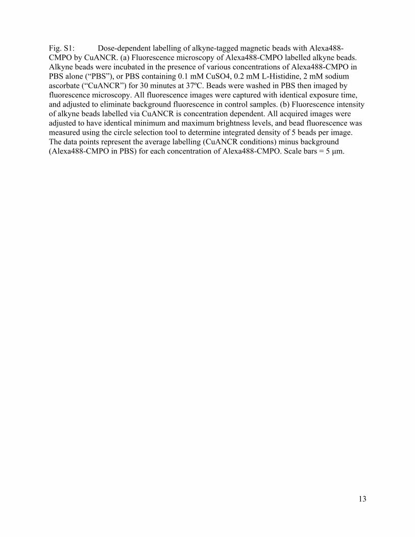
Fig. S1: Dose-dependent labelling of alkyne-tagged magnetic beads with Alexa488-CMPO by CuANCR. (a) Fluorescence microscopy of Alexa488-CMPO labelled alkyne beads. Alkyne beads were incubated in the presence of various concentrations of Alexa488-CMPO in PBS alone (“PBS”), or PBS containing 0.1 mM CuSO4, 0.2 mM L-Histidine, 2 mM sodium ascorbate (“CuANCR”) for 30 minutes at 37ºC. Beads were washed in PBS then imaged by fluorescence microscopy. All fluorescence images were captured with identical exposure time, and adjusted to eliminate background fluorescence in control samples. (b) Fluorescence intensity of alkyne beads labelled via CuANCR is concentration dependent. All acquired images were adjusted to have identical minimum and maximum brightness levels, and bead fluorescence was measured using the circle selection tool to determine integrated density of 5 beads per image. The data points represent the average labelling (CuANCR conditions) minus background (Alexa488-CMPO in PBS) for each concentration of Alexa488-CMPO. Scale bars = 5 μm.

14
1H and 13C NMR Spectra of KDO-HMMPO
OHO
HO
OH
CO2H
HO
NH
OO
NO
KDO-nitrone

15
1H and 13C NMR Spectra of KDO-alkyne
OHO
HO
OH
CO2H
HOHN
O
KDO-alkyne

16
1H and 13C NMR Spectra of Biotin-CMPO
NO
S
NHHN
O
HH
O
HN O
OO
HN
O
17
References
1 D. C. Kennedy, C. S. McKay, M. C. B. Legault, D. C. Danielson, J. A. Blake, A. F. Pegoraro, A. Stolow, Z. Mester and J. P. Pezacki, J. Am. Chem. Soc., 2011, 133, 17993.
2 K. Stolze, N. Rohr-Udilova, A. Patel and T. Rosenau, Bioorg. Med. Chem., 2011, 19, 985.
3 (a) A. Dumont, A. Malleron, M. Awwad, S. Dukan and B. Vauzeilles, Angew. Chem. Int. Ed., 2012, 51, 3143; (b) R. Winzar, J. Philips and M. J. Kiefel, Synlett, 2010, 2010, 583.
4 M. Slater, M. Snauko, F. Svec and J. M. J. Fréchet, Anal. Chem. , 2006, 78, 4969.
5 (a) P. Tsai, K. Ichikawa, C. Mailer, S. Pou, H. J. Halpern, B. H. Robinson, R. Nielsen and G. M. Rosen, J. Org. Chem., 2003, 68, 7811; (b) S.-U. Kim, Y. Liu, K. M. Nash, J. L. Zweier, A. Rockenbauer and F. A. Villamena, J. Am. Chem. Soc., 2010, 132, 17157.
6 P.-T. Doulias, J. L. Greene, T. M. Greco, M. Tenopoulou, S. H. Seeholzer, R. L. Dunbrack and H. Ischiropoulos, Proc Natl Acad Sci U S A, 2010, 107, 16958.
7 C. M. Tsai and C. E. Frasch, Analytical biochemistry, 1982, 119, 115.