Embed Size (px)
Citation preview
SI Appendix ALKBH5-dependent m6A demethylation controls splicing and stability of long 3’UTR mRNAs in male germ cells Chong Tanga,1, Rachel Klukovicha,1, Hongying Penga, Zhuqing Wanga, Tian Yua, Ying Zhanga, Huili Zhenga, Arne Klunglandb,c, and Wei Yana,d,2 aDepartment of Physiology and Cell Biology, University of Nevada, Reno School of Medicine, 1664 North Virginia Street, MS575, Reno, NV 89557, USA; bDepartment of Microbiology, Oslo University Hospital, Rikshospitalet, 0027 Oslo, Norway; cDepartment of Molecular Medicine, Institute of Basic Medical Sciences, University of Oslo, 0317 Oslo, Norway; dDepartment of Biology, University of Nevada, Reno, 1664 North Virginia Street, MS575, Reno, NV 89557, USA 1C.T. and R.K. contributed equally to this work. 2To whom correspondence should be addressed: Wei Yan M.D., Ph.D. University of Nevada Reno Foundation Professor Department of Physiology and Cell Biology University of Nevada School of Medicine Center for Molecular Medicine, Room 207B 1664 North Virginia Street, MS/0575 Reno, NV 89557 Tel: 775 784 7765 Fax: 775 784 4362 Email: [email protected] This file contains SI Materials and Methods, Fig. S1-S10, and Table S1-S3.

Materials and Methods Animal use and care. All mice used in this study were on the C57BL/6J background, and housed under specific pathogen-free conditions in a temperature- and humidity-controlled animal facility at the University of Nevada, Reno. Animal use protocol was approved by Institutional Animal Care and Use Committee (IACUC) of the University of Nevada, Reno (Protocol number 00494), and are in accordance with the “Guide for the Care and Use of Experimental Animals” established by National Institutes of Health (NIH) (1996, revised 2011). The male Alkbh5 KO mice used in this study were described previously (1). Histology. Testes were dissected from WT or KO mouse and were fixed in Bouin’s solution for 24 hours, then washed and submerged in 70% ethanol until processed and paraffin-embedded using a Leica tissue processor. Sections were cut at 5μm thick and were attached to positively-charged slides (Genesee Scientific No. 29-107). Testes sections were stained with hematoxylin and eosin and staining was performed in biological duplicates or triplicates. Cauda epididymal sperm from WT or KO were spread onto positively-charged slides (Genesee Scientific No. 29-107), fixed for 5 minutes with 4% PFA, and were stained with hematoxylin and eosin to examine morphology. Images were taken using a Keyence microscope model BZX-710. PAS staining. Periodic acid-Schiff (PAS) staining was performed on 5 μm thick paraffin sections. After deparaffinization, slides were incubated in 0.5% periodic acid for 10 min, followed by rinsing in distilled water three times. Slides were then submerged in Schiff’s reagent for 15 minutes followed by rinsing with distilled water for 10 minutes. Hematoxylin was used to counterstain the slides which were then dipped in 11% total volume HCL in 100% Ethanol until color was not rinsing off. Slides were dehydrated then mounted with Permount. Images were taken using a Keyence microscope model BzX-710. Staining was done in biological triplicates. TUNEL analyses. Terminal deoxynucleotidyl transferase dUTP nick end-labeling (TUNEL) staining was performed according to the manufacturer’s instructions with some modifications (Trevigen in situ apoptosis detection kit No. 4810-30-CK). Paraffin-embedded testis sections were 5 μm thick. Proteinase K digestion was incubated at 37°C for 20 minutes and manganese was used as the cation for the TdT reaction. Slides were counterstained with hematoxylin for 15 seconds. Staining and counts were carried out in biological triplicates and the number of TUNEL-positive cells were counted from 100 randomly selected tubules. A student’s two-tailed t-test was used to determine significance (p< 0.05). Gross morphology and sperm analysis. Testis and the whole epididymis were dissected from WT and KO mice and rinsed in PBS. After weight measurements and morphology pictures were taken, both cauda epididymides were punctured several times with a needle to release the sperm into HTF medium at 37°C for 30 minutes. Computer-assisted sperm analysis (CASA) was used to analyze motility, performed in
triplicate. A hemocytometer was used to manually count sperm concentration, measured in triplicate. Immunofluorescence staining and confocal microscopy. After dissection, testes were fixed in 4% paraformaldehyde for 1 hour at room temperature, poked several times with a 20.5 G needle, fixed 2 more hours at room temperature, then transferred to 4°C for 21 more hours. Testes were then washed 3x in 5% sucrose then dehydrated in a sucrose gradient of 5%:20% sucrose at 2:1, 1:1, 1:2 until testes sunk to the bottom of the tube. Testes were then kept in 20% sucrose for 48 hours and were perfused for 1 hour in 1:1 20% sucrose:OCT. Testes were embedded in a Tissue-Tek Cryomold in 1:1 20% sucrose:OCT and rapidly frozen using liquid nitrogen. Blocks were cut into 10μm thick sections on Fisher SuperFrost Plus Slides (No. 22-037-246), were allowed to dry for 30 minutes, and were stored at -80°C until use. Slides were permeablized by boiling in citrate buffer (pH 6.0) 5 minutes, 3 times and were blocked in 1% BSA in TBS supplemented with 5% fetal bovine serum and 5% normal goat serum at room temperature for 1 hour. Primary antibodies were diluted in 1% BSA in TBS: rabbit anti-alkbh5 (Sigma No. HPA007196, 1:150), mouse anti-GM130 (BD Biosciences No. 610822, 1:200), mouse anti-SC35 (No. ab11826, 1:500) and were incubated overnight at 4°C. After washing three times in PBS, the secondary antibody was diluted in 1% BSA in TBS: goat anti-rabbit Alexa Fluor 488 (Thermo Fisher No. A11034, 1:500) and goat anti-mouse Alexa Fluor 594 (Abcam No. ab150116, 1:400) and was incubated for 1 hour at room temperature. Slides were then washed three times in PBS, followed by submersion in 0.1% Sudan Black B in 70% ethanol. After washing three times in PBS, slides were mounted using DAPI (Abcam No. ab104139). An Olympus FV1000 confocal microscope was used to take the images of z-stacks 5μm thick. Western blots. Total protein lysates were extracted from whole testis in phosphate-buffered saline. The Pierce BCA Protein Assay Kit (Thermo Fisher, Cat#23225) was used to determine concentration. Protein (25 μg) was loaded and run on a 4-15% Mini-PROTEAN TGX Stain-free gel (Bio-Rad No. 456-8084), transferred onto a nitrocellulose membrane (0.45μm, Amersham, Cat#10600003) and blocked with Thermo Fisher Superblock for 1 hour. Primary antibodies: rabbit anti-Alkbh5 (Sigma HPA007196, 1:500) and mouse anti-β-actin (Abcam, Cat#ab8226, 1:5000) were incubated overnight at 4°C. Secondary antibodies: goat anti-rabbit HRP (Abcam, Cat#ab205718) and goat anti-mouse HRP (Southern Biotech, Cat#1030-05) were diluted 1:10,000, the membrane was washed, and StrepTactin-HRP (Bio-Rad, Cat#161-0376) was used to visualize the standards. Advanta Western Bright ECL Kit (Cat#K-12045-D20) was used for detection on the Bio-Rad ChemiDoc imager.
Purification of spermatogenic cells. Pachytene spermatocytes, round and elongating/elongated spermatids were purified from adult mouse testes using the STA-PUT method (2). The BSA gradients (0.5-4%) were prepared in the EKRB buffer (Cat#K-4002, Sigma), supplemented with sodium bicarbonate (1.26g per 1L), L-glutamine (0.29228g per 1L); Penicillin and Streptomycin mix (Thermo-Fisher, 10,000U per 1L), MEM non-essential amino acids (Thermo-Fisher, 1ml 100X per 1L), MEM amino acids (20ml 50X per 1L) and cycloheximide (100ng/ml), pH7.2-7.3). Eight testes
were pooled each time for cell purification. After being removed and decapsulated, testes were placed into 10ml of the EKRB buffer containing 5mg collagenase (Sigma) for a 12-min digestion at 32°C to disperse the testicular cells. Once dispersed, the testicular cells were washed three times using the EKRB buffer followed by trypsin digestion by incubation in 10ml EKR buffer containing trypsin (Sigma, 0.25mg/ml) and DNase I (Sigma, 20mg/ml) at 37°C for 12min with occasional pipetting to facilitate cell dispersion. Fully dispersed testicular cells were washed three times followed by centrifugation and re-suspension in 10 ml of 0.5% BSA. The cell suspension was passed through 50µm filters, and the filtrate was saved for loading onto the STA-PUT apparatus for sedimentation. After 3h sedimentation at 4°C, fractions were collected from the bottom of the sedimentation chamber. A total of 30 fractions of 15 ml each were collected. After centrifugation, the supernatants were removed, and the cells in each fraction were re-suspended, and the cell purity was determined by microscopy examination based on cell morphology, as described previously (2). Fractions containing the same cell types were pooled followed by centrifugation to collect purified pachytene spermatocytes, round spermatids, and elongating/elongated spermatids. The RNA was extracted from cells by mirVana miRNA Isolation Kit (Life Technology, Cat# AM1560). RNA extraction. RNA was extracted from HEK293 cells using the mirVana miRNA Isolation Kit (ThermoFisher, Cat#AM1560), according to the manufacturer’s instructions. Extracted RNA quantification was done using the Qubit RNA High Sensitivity Assay Kit (Invitrogen No. Q32855) measured on the Qubit 2.0 Fluorometer (Invitrogen). m6A RNA immunoprecipitation. Protein G Dynabeads (50μL, Invitrogen, Cat#10004D) were washed three times and resuspended with immunoprecipitation buffer (5mL 0.1M Sodium phosphate, 3.5mL 2M sodium chloride, 250μL 10% triton, nuclease-free water to 50mL final volume). Rabbit anti-m6A antibody (Abcam, Cat#ab151230) or normal rabbit IgG (Invitrogen, Cat#10500C) (6μg each) were added to the beads on a rocker for 1 hour at room temperature, protected from light. The beads were washed and resuspended in immunoprecipitation buffer. 200ng of RNA were heat fragmented into 200nt fragments and were added to the bead suspension on a rocker for 2 hours at room temperature, protected from light. The beads were washed, pelleted, and eluted with sodium lauryl sulfate and tween-20 for 5 minutes at 70°C. The eluate was purified with 2.2x of magnetic beads made from SpeedBeads (Sigma-Aldrich Cat#65152105050250) and run on the Agilent RNA Pico chip (Cat#5067-1513) to verify purity and RNA integrity. All immunoprecipitations were performed in triplicate. The representative results of the procedures are shown in SI Appendix, Fig. S8. RNA library construction. Immunoprecipitated RNA (1ng) and non-immunoprecipitated RNA (300ng) were constructed into next-generation sequencing libraries (Illumina) using the KAPA Stranded RNA-Seq Library Preparation Kit (KK8400) according to the manufacturer’s instructions, with some modifications. Briefly, the fragmentation, priming, and elution steps were incubated at 65°C for 3 minutes, and the adaptors and barcodes were substituted with NEBNext Multiplex Oligos for Illumina

(Cat#E7335 and E7500). Library amplification was monitored using the qPCR with the KAPA real-time library amplification kit (Cat#KK2702). All libraries were constructed in triplicate. After final amplification, libraries were size selected using magnetic beads. The eluates were quantified using Qubit DNA High Sensitivity (Invitrogen, Cat#Q32854) and the Agilent DNA High Sensitivity chip (Cat#5067-4626) was used to determine library quality. Immunoprecipitated libraries were pooled according to library quantity and their negative control counterparts were pooled with equal volume to the immunoprecipitated library. Transcriptome libraries were pooled according to quantity. All libraries were sequenced at the Genomics Center of the University of Nevada, Reno on the Illumina Next-Seq 500 using paired-end 75 sequencing.
RNA-Seq data analyses. Quality control of RNA-Seq data are shown in SI Appendix, Fig. S9. Trimmomatic was used to remove adaptor sequences, and low-quality reads from the sequencing data (3). To identify all the transcripts, we used Tophat2 and Cufflinks to assemble the sequencing reads based on the UCSC MM9 mouse genome (4). The differential expression analysis was performed by Cuffdiff (4). The global statistics and quality controls are presented in Supplemental Figure S1. The UTR and alternative splicing analyses were performed using the SpliceR pipeline (5). The sequences without identified coding frames were extracted and subjected to coding potential calculating (6). The sequencing depth and alignment ratio were listed in SI Appendix, Fig. S10. We performed the data mining using our in-house R script. Data normalization. FPKMs counts are scaled in Cuffdiff analyses via the median of the geometric means of fragment counts across all libraries, as described in (7). The principle was identical to the one used by DESeq (8). m6a RIP-Seq data analyses. Trimmomatic was used to remove adaptor sequences and low-quality reads from the sequencing data (3). To reduce bias from potential inaccurate gene structure annotation, we aligned the m6A-seq reads to the assembled gene sequences derived from RNA-Seq cufflink results, using Tophat v2.0.14 (4). The longest isoform was used if the gene had multiple isoforms. The peak-calling method was modified from published work (9). To call m6A peaks, the longest isoform of each gene was scanned using a 100nt sliding window with 10nt step. We add one count to all the windows to avoid 0 count in the windows. The peak height threshold was ten counts. For each gene, the read counts in each window were normalized by the median counts of all windows of that gene. A Fisher exact test was used to identify the differential windows between IP and input samples. The window was called positive if the count>=10 and log2(enrichment score)>0.8, which FDR is close to zero. Overlapping positive windows were merged. The following four numbers were calculated to obtain the enrichment score of each window: (a) read counts of the IP samples in the current window, (b) median read counts of the IP sample in all 100nt windows on the current mRNA, (c) read counts of the input sample in the current window, and (d) median read counts of the input sample in all 100nt windows on the current mRNA. The enrichment score of each window was calculated as (a*d)/(b*c). We used HEK293 cell m6A RIP-Seq data as the control to correlate with the 2,000bp genomic range of the published m6a nucleotide resolution site (10). The enrichment

score threshold was set as 0.8 to get the best results. If the high-confident peaks were identified in two of three, we then regarded it as a true m6A peak. We performed the data mining using our in-house R script. Sequencing data deposition. Both RNA-Seq and m6A RIP-Seq datasets have been deposited into the Sequence Read Achieve (SRA) database in the NCBI with an accession number of PRJNA420607. References 1. Zheng G, et al. (2013) ALKBH5 is a mammalian RNA demethylase that impacts RNA
metabolism and mouse fertility. Mol Cell 49(1):18-29. 2. Zhang Y, et al. (2017) MicroRNAs control mRNA fate by compartmentalization based on 3'
UTR length in male germ cells. Genome Biol 18(1):105. 3. Bolger AM, Lohse M, & Usadel B (2014) Trimmomatic: a flexible trimmer for Illumina
sequence data. Bioinformatics 30(15):2114-2120. 4. Trapnell C, et al. (2012) Differential gene and transcript expression analysis of RNA-seq
experiments with TopHat and Cufflinks. Nat Protoc 7(3):562-578. 5. Vitting-Seerup K, Porse BT, Sandelin A, & Waage J (2014) spliceR: an R package for
classification of alternative splicing and prediction of coding potential from RNA-seq data. BMC Bioinformatics 15:81.
6. Kong L, et al. (2007) CPC: assess the protein-coding potential of transcripts using sequence features and support vector machine. Nucleic Acids Res 35(Web Server issue):W345-349.
7. Anders S & Huber W (2010) Differential expression analysis for sequence count data. Genome Biol 11(10):R106.
8. Li P, Piao Y, Shon HS, & Ryu KH (2015) Comparing the normalization methods for the differential analysis of Illumina high-throughput RNA-Seq data. BMC Bioinformatics 16:347.
9. Zhao BS, et al. (2017) m6A-dependent maternal mRNA clearance facilitates zebrafish maternal-to-zygotic transition. Nature 542(7642):475-478.
10. Linder B, et al. (2015) Single-nucleotide-resolution mapping of m6A and m6Am throughout the transcriptome. Nat Methods 12(8):767-772.
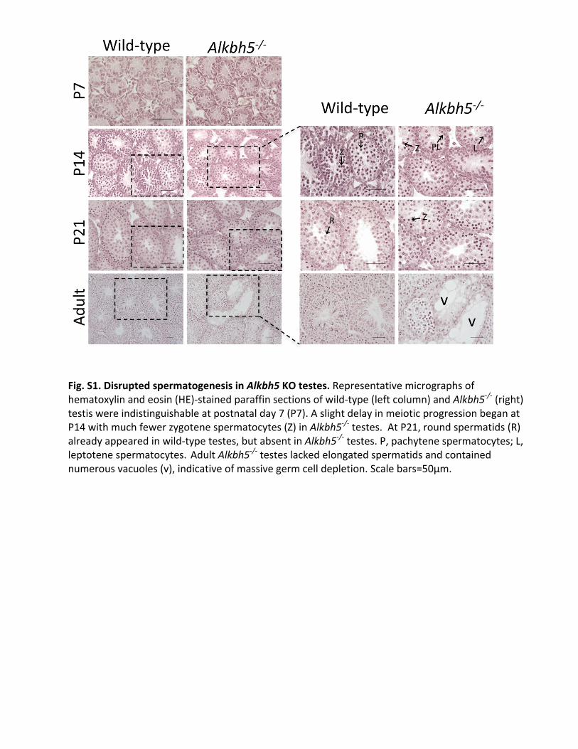
Fig.S1.DisruptedspermatogenesisinAlkbh5KOtestes.Representativemicrographsofhematoxylinandeosin(HE)-stainedparaffinsectionsofwild-type(leftcolumn)andAlkbh5-/-(right)testiswereindistinguishableatpostnatalday7(P7).AslightdelayinmeioticprogressionbeganatP14withmuchfewerzygotenespermatocytes(Z)inAlkbh5-/-testes.AtP21,roundspermatids(R)alreadyappearedinwild-typetestes,butabsentinAlkbh5-/-testes.P,pachytenespermatocytes;L,leptotenespermatocytes.AdultAlkbh5-/-testeslackedelongatedspermatidsandcontainednumerousvacuoles(v),indicativeofmassivegermcelldepletion.Scalebars=50µm.
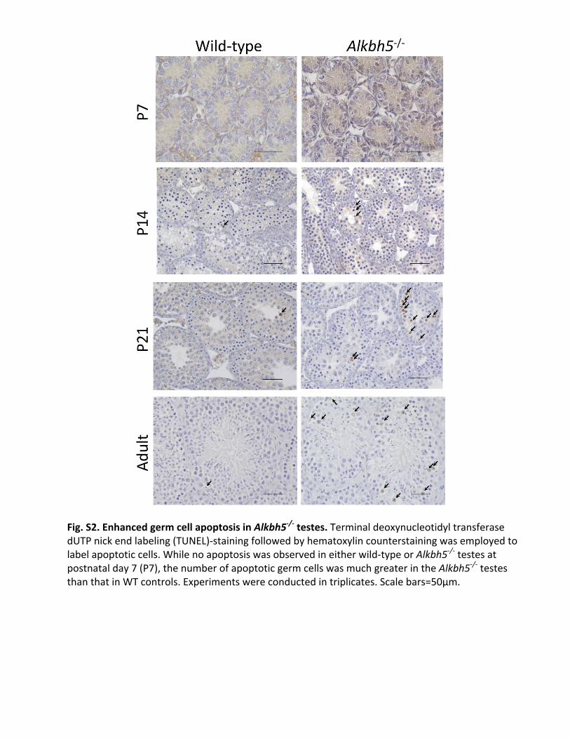
Fig.S2.EnhancedgermcellapoptosisinAlkbh5-/-testes.TerminaldeoxynucleotidyltransferasedUTPnickendlabeling(TUNEL)-stainingfollowedbyhematoxylincounterstainingwasemployedtolabelapoptoticcells.Whilenoapoptosiswasobservedineitherwild-typeorAlkbh5-/-testesatpostnatalday7(P7),thenumberofapoptoticgermcellswasmuchgreaterintheAlkbh5-/-testesthanthatinWTcontrols.Experimentswereconductedintriplicates.Scalebars=50µm.
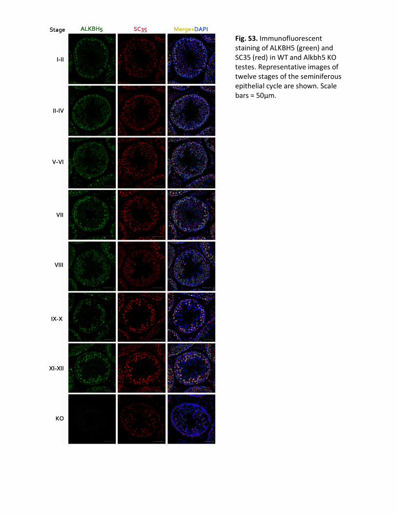
Fig.S3.ImmunofluorescentstainingofALKBH5(green)andSC35(red)inWTandAlkbh5KOtestes.Representativeimagesoftwelvestagesoftheseminiferousepithelialcycleareshown.Scalebars=50µm.
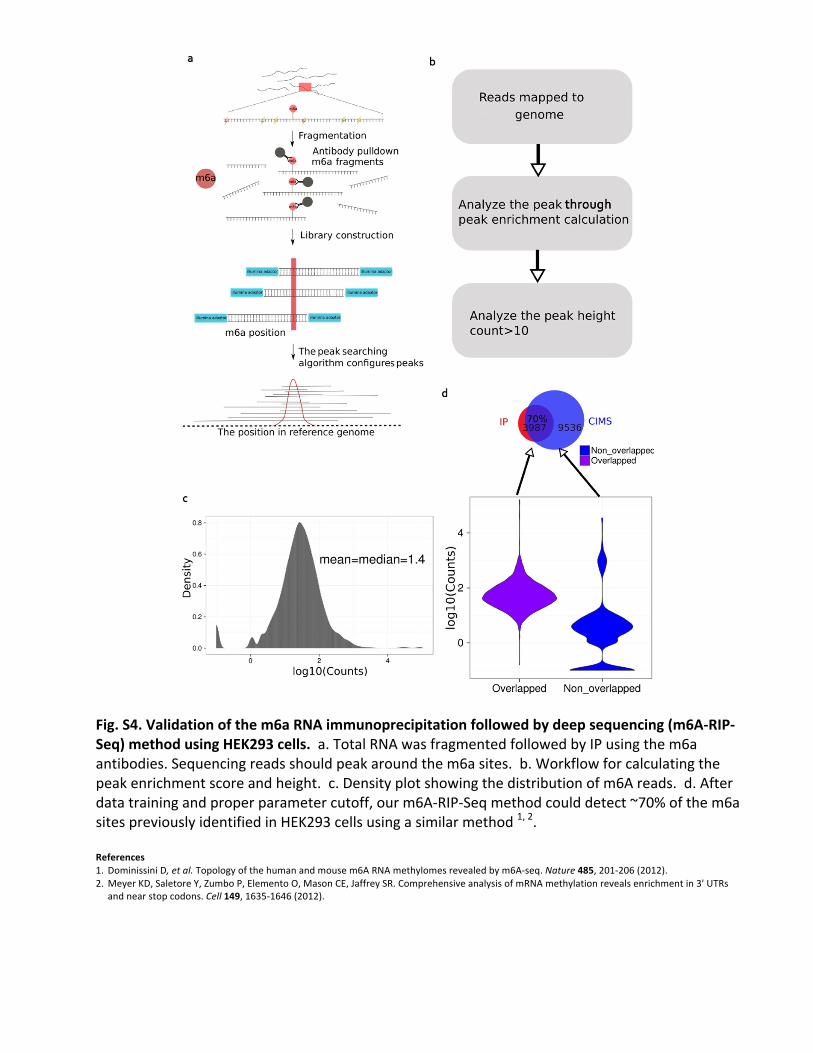
Fig.S4.Validationofthem6aRNAimmunoprecipitationfollowedbydeepsequencing(m6A-RIP-Seq)methodusingHEK293cells.a.TotalRNAwasfragmentedfollowedbyIPusingthem6aantibodies.Sequencingreadsshouldpeakaroundthem6asites.b.Workflowforcalculatingthepeakenrichmentscoreandheight.c.Densityplotshowingthedistributionofm6Areads.d.Afterdatatrainingandproperparametercutoff,ourm6A-RIP-Seqmethodcoulddetect~70%ofthem6asitespreviouslyidentifiedinHEK293cellsusingasimilarmethod1,2.References 1. DominissiniD,etal.Topologyofthehumanandmousem6ARNAmethylomesrevealedbym6A-seq.Nature485,201-206(2012).2.MeyerKD,SaletoreY,ZumboP,ElementoO,MasonCE,JaffreySR.ComprehensiveanalysisofmRNAmethylationrevealsenrichmentin3'UTRs
andnearstopcodons.Cell149,1635-1646(2012).
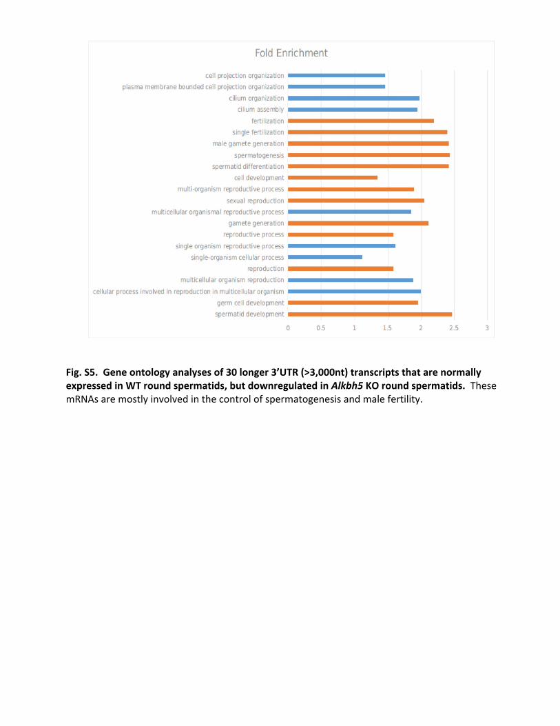
Fig.S5.Geneontologyanalysesof30longer3’UTR(>3,000nt)transcriptsthatarenormallyexpressedinWTroundspermatids,butdownregulatedinAlkbh5KOroundspermatids.ThesemRNAsaremostlyinvolvedinthecontrolofspermatogenesisandmalefertility.

Fig.S6.qPCRanalysesofexpressionlevelsofUnc50andTraf3ip1inWTandAlkbh5KOpachytenespermatocytes(pa),round(ro)andelongating(el)spermatids.Primerlocationsareshowninupperpanels,andtheprimersequencesarelistedinthetablebelow.Gapdhwasusedasaninternalcontrol.Primer Sequence(5’à3’)
GapdhF ACAGTCCATGCCATCACTGCC
GapdhR GCCTGCTTCACCACCTTCTTG
Unc50(447&452com)F GGTGTTGGCCTTCTGATCTCAACGTTA
Unc50(447&452com)R CACTGTATCCCAGGAAGGTCACATAG
Unc50(447)shortisoR CATGAGAGGTGCGAATGGATAGAGCA
Unc50(452)longisoR CATATGAGTGAACCATAACACCTCAGGCA
Traf3ip1(1506&1509com)F CAGGAGCAGAGTATCACAGACAGTGC
Traf3ip1(1506&1509com)R AGCAGGCCATTTCTCAGCAGACT
Traf3ip1(1506)longisoR CTGGAGGCTCTAAGTAGTAAGCACTGTAGC
Traf3ip1(1509)shortisoR ACTGTCCTCTTCCCAACCGAGAC

Fig.S7.GeneontologyanalysesoftheshortertranscriptsthatquicklydegradeinAlkbh5KOelongatingspermatids.NotethatmanyoftheseshortmRNAsareinvolvedintheregulationofRNAsplicing,proteinandmRNAmetabolism.
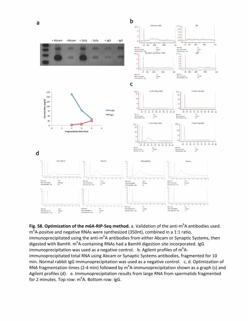
Fig.S8.Optimizationofthem6A-RIP-Seqmethod.a.Validationoftheanti-m6Aantibodiesused.m6A-positveandnegativeRNAsweresynthesized(350nt),combinedina1:1ratio,immunoprecipitatedusingtheanti-m6AantibodiesfromeitherAbcamorSynapticSystems,thendigestedwithBamHI.m6A-containingRNAshadaBamHIdigestionsiteincorporated.IgGimmunoprecipitationwasusedasanegativecontrol.b.Agilentprofilesofm6A-immunoprecipitatedtotalRNAusingAbcamorSynapticSystemsantibodies,fragmentedfor10min.NormalrabbitIgGimmunoprecipitationwasusedasanegativecontrol.c,d.OptimizationofRNAfragmentationtimes(2-4min)followedbym6A-immunoprecipitationshownasagraph(s)andAgilentprofiles(d).e.ImmunoprecipitationresultsfromlargeRNAfromspermatidsfragmentedfor2minutes.Toprow:m6A.Bottomrow:IgG.

Fig.S9.EvaluationofthequalityoftheRNA-Seqdatageneratedinthisstudy.a.Pairwisescatterplotsofthelog10normalizedFKMPscoresacrossallsixRNAsamples.b.Volcanoplotsshowingthelog10(p-value)iny-axesandlog2(foldchange)inx-axesbypairwisecomparisonacrossallsixRNAsamples.c.Densityplotsshowingthedistributionoflog10normalizedFPKMscoresacrossbiologicalreplicatesofallsixRNAsamples.d.Overdispersionplotsdemonstratingtheestimatedoverdispersionforeachsampleasaqualitycontrolmeasure.
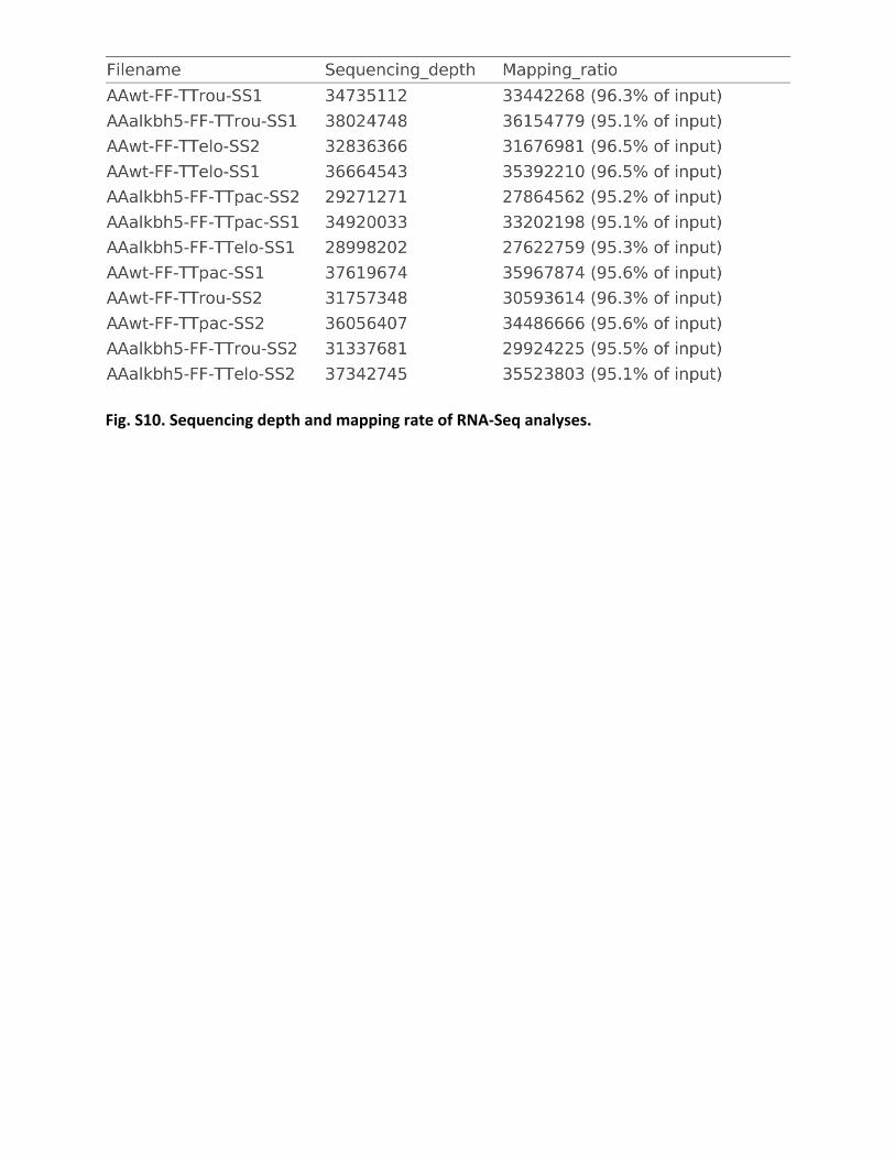
Fig.S10.SequencingdepthandmappingrateofRNA-Seqanalyses.

TableS1.DysregulatedsplicingfactorgenesinAlkbh5KOspermatogeniccells.Genesymbol Genename FunctionSfswap Splicingfactor,suppressorofwhite-apricothomolog;Sfswap;ortholog mRNAsplicingfactor(PC00148)U2af2 SplicingfactorU2AF65kDasubunit;U2af2;orthologSrsf1 Serine/arginine-richsplicingfactor1;Srsf1;orthologKhdrbs3 KHdomain-containing,RNA-binding,signaltransduction-associatedprotein3;Khdrbs3;ortholog mRNAsplicingfactor(PC00148);transcriptioncofactor(PC00217)Cdk12 Cyclin-dependentkinase12;Cdk12;ortholog non-receptorserine/threonineproteinkinase(PC00167);non-receptortyrosineproteinkinase(PC00168)Son ProteinSON;Son;orthologSnrnp70 U1smallnuclearribonucleoprotein70kDa;Snrnp70;ortholog mRNAsplicingfactor(PC00148)Brdt Bromodomaintestis-specificprotein;Brdt;orthologHnrnpl HeterogeneousnuclearribonucleoproteinL;Hnrnpl;orthologCelf5 CUGBP,Elav-likefamilymember5;Celf5;orthologTra2b Transformer-2proteinhomologbeta;Tra2b;ortholog mRNAsplicingfactor(PC00148)Srek1 Splicingregulatoryglutamine/lysine-richprotein1;Srek1;ortholog RNAbindingprotein(PC00031)Zfp326 DBIRDcomplexsubunitZNF326;Znf326;orthologSf3a1 Splicingfactor3Asubunit1;Sf3a1;ortholog mRNAsplicingfactor(PC00148)Hnrnpk HeterogeneousnuclearribonucleoproteinK;Hnrnpk;ortholog enzymemodulator(PC00095);mRNAsplicingfactor(PC00148);ribonucleoprotein(PC00201);serineprotease(PC00203)Ddx17 ProbableATP-dependentRNAhelicaseDDX17;Ddx17;orthologTmbim6 Baxinhibitor1;Tmbim6;orthologRbm25 RNA-bindingprotein25;Rbm25;ortholog

TableS2.DysregulatedcilliogenicgenesinAlkbh5KOspermatogeniccells.Genesymbol Genename FunctionLca5 Lebercilin;Lca5;orthologC2cd3 C2domain-containingprotein3;C2cd3;orthologFoxj1 ForkheadboxproteinJ1;Foxj1;orthologNek1 Serine/threonine-proteinkinaseNek1;Nek1;orthologHydin Hydrocephalus-inducingprotein;Hydin;orthologDnaaf3 Dyneinassemblyfactor3,axonemal;Dnaaf3;orthologCfap126 ProteinFlattop;Cfap126;orthologTtll8 ProteinmonoglycylaseTTLL8;Ttll8;orthologUbe2b Ubiquitin-conjugatingenzymeE2B;Ube2b;orthologTtc21a Tetratricopeptiderepeatprotein21A;Ttc21a;orthologCep89 Centrosomalproteinof89kDa;Cep89;orthologTtll3 TubulinmonoglycylaseTTLL3;Ttll3;orthologDnah7a Dynein,axonemal,heavychain7A;Dnah7a;ortholog hydrolase(PC00121);microtubulebindingmotorprotein(PC00156)Traf3ip1 TRAF3-interactingprotein1;Traf3ip1;orthologRfx4 TranscriptionfactorRFX4;Rfx4;ortholog wingedhelix/forkheadtranscriptionfactor(PC00246)Ttc26 Intraflagellartransportprotein56;Ttc26;orthologPcnt Pericentrin;Pcnt;orthologTtc21b Tetratricopeptiderepeatprotein21B;Ttc21b;orthologWdr60 WDrepeat-containingprotein60;Wdr60;ortholog microtubulefamilycytoskeletalprotein(PC00157)Ift80 Intraflagellartransportprotein80homolog;Ift80;orthologActr2 Actin-relatedprotein2;Actr2;orthologCcdc114 Coiled-coildomain-containingprotein114;Ccdc114;orthologCcdc63 Coiled-coildomain-containingprotein63;Ccdc63;orthologSclt1 Sodiumchannelandclathrinlinker1;Sclt1;orthologRabl2 Rab-likeprotein2A;Rabl2;orthologDnaic1 Dyneinintermediatechain1,axonemal;Dnai1;ortholog microtubulefamilycytoskeletalprotein(PC00157)Dnah7b Dynein,axonemal,heavychain7B;Dnah7b;ortholog hydrolase(PC00121);microtubulebindingmotorprotein(PC00156)
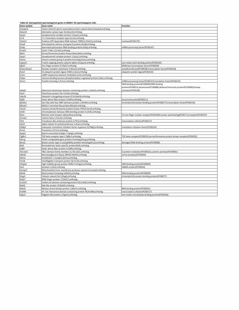
TableS3.DysregulatedspermatogensisgenesinAlkbh5 KOspermatogeniccells.Genesymbol Genename FunctionCatsperb Cationchannelsperm-associatedproteinsubunitbeta;Catsperb;orthologAdcy10 Adenylatecyclasetype10;Adcy10;orthologSycp2 Synaptonemalcomplexprotein2;Sycp2;orthologCcr6 C-Cchemokinereceptortype6;Ccr6;orthologTdrd12 PutativeATP-dependentRNAhelicaseTDRD12;Tdrd12;ortholog nuclease(PC00170)Stra8 Stimulatedbyretinoicacidgene8protein;Stra8;orthologStrbp SpermatidperinuclearRNA-bindingprotein;Strbp;ortholog mRNAprocessingfactor(PC00147)Ccnyl1 CyclinY-like1;Ccnyl1;orthologNek1 Serine/threonine-proteinkinaseNek1;Nek1;orthologSycp1 Synaptonemalcomplexprotein1;Sycp1;orthologFanca FanconianemiagroupAproteinhomolog;Fanca;orthologCapza3 F-actin-cappingproteinsubunitalpha-3;Capza3;ortholog non-motoractinbindingprotein(PC00165)Zfp37 Zincfingerprotein37;Zfp37;ortholog KRABboxtranscriptionfactor(PC00029)Grip1,Ncoa2 Nuclearreceptorcoactivator2;Ncoa2;ortholog acetyltransferase(PC00038);transcriptionfactor(PC00218)Fancl E3ubiquitin-proteinligaseFANCL;Fancl;ortholog ubiquitin-proteinligase(PC00234)Crem cAMP-responsiveelementmodulator;Crem;orthologCabyr Calcium-bindingtyrosinephosphorylation-regulatedprotein;Cabyr;orthologPum1 Pumiliohomolog1;Pum1;ortholog mRNAprocessingfactor(PC00147);translationfactor(PC00223)
Adad1 Adenosinedeaminasedomain-containingprotein1;Adad1;ortholog
DNAbindingprotein(PC00009);RNAbindingprotein(PC00031);deaminase(PC00088);defense/immunityprotein(PC00090);kinaseactivator(PC00138)
Pax5 PairedboxproteinPax-5;Pax5;orthologUbe2b Ubiquitin-conjugatingenzymeE2B;Ube2b;orthologOdf3 Outerdensefiberprotein3;Odf3;ortholog structuralprotein(PC00211)Sfmbt1 Scm-likewithfourMBTdomainsprotein1;Sfmbt1;ortholog chromatin/chromatin-bindingprotein(PC00077);transcriptionfactor(PC00218)Micalcl MICALC-terminal-likeprotein;Micalcl;orthologTex14 Inactiveserine/threonine-proteinkinaseTEX14;Tex14;orthologChd5 Chromodomain-helicase-DNA-bindingprotein5;Chd5;orthologRara Retinoicacidreceptoralpha;Rara;ortholog C4zincfingernuclearreceptor(PC00169);nucleicacidbinding(PC00171);receptor(PC00197)Ctnnb1 Cateninbeta-1;Ctnnb1;orthologTle3 Transducin-likeenhancerprotein3;Tle3;ortholog transcriptioncofactor(PC00217)Atat1 Alpha-tubulinN-acetyltransferase1;Atat1;orthologEif4g3 Eukaryotictranslationinitiationfactor4gamma3;Eif4g3;ortholog translationinitiationfactor(PC00224)Prm2 Protamine-2;Prm2;orthologSpag1 Sperm-associatedantigen1;Spag1;orthologTgfbr1 TGF-betareceptortype-1;Tgfbr1;ortholog TGF-betareceptor(PC00035);serine/threonineproteinkinasereceptor(PC00205)Pacrg Parkincoregulatedgeneproteinhomolog;Pacrg;orthologBrca2 Breastcancertype2susceptibilityproteinhomolog;Brca2;ortholog damagedDNA-bindingprotein(PC00086)Brdt Bromodomaintestis-specificprotein;Brdt;orthologOdf4 Outerdensefiberprotein4;Odf4;orthologTbc1d21 TBC1domainfamilymember21;Tbc1d21;ortholog G-proteinmodulator(PC00022);cysteineprotease(PC00081)Abhd2 MonoacylglycerollipaseABHD2;Abhd2;ortholog serineprotease(PC00203)Ednra Endothelin-1receptor;Ednra;orthologTtc26 Intraflagellartransportprotein56;Ttc26;orthologHmga2 HighmobilitygroupproteinHMGI-C;Hmga2;ortholog DNAbindingprotein(PC00009)Stx2 Syntaxin-2;Stx2;ortholog SNAREprotein(PC00034)Immp2l Mitochondrialinnermembraneproteasesubunit2;Immp2l;orthologMsh4 MutSproteinhomolog4;Msh4;ortholog DNAbindingprotein(PC00009)Stag3 CohesinsubunitSA-3;Stag3;ortholog chromatin/chromatin-bindingprotein(PC00077)Rnf17 RINGfingerprotein17;Rnf17;orthologCcdc63 Coiled-coildomain-containingprotein63;Ccdc63;orthologRabl2 Rab-likeprotein2A;Rabl2;orthologMarf1 Meiosisarrestfemaleprotein1;Marf1;ortholog RNAbindingprotein(PC00031)Arid4b AT-richinteractivedomain-containingprotein4B;Arid4b;ortholog transcriptioncofactor(PC00217)Fignl1 Fidgetin-likeprotein1;Fignl1;ortholog non-motormicrotubulebindingprotein(PC00166)