Embed Size (px)
Citation preview
Algorithm for laboratory diagnosis and treatment-monitoring of pulmonary tuberculosis and drug-resistant tuberculosis using state-of-the-art rapid molecular diagnostic technologies
Expert opinion of the European Tuberculosis Laboratory Initiative core group members for the WHO European Region

Algorithm for laboratory diagnosis and treatment-monitoring of pulmonary tuberculosis and drug-resistant tuberculosis using state-of-the-art rapid molecular diagnostic technologies
Expert opinion of the European Tuberculosis Laboratory Initiative core group members for the WHO European Region
© World Health Organization 2017All rights reserved. The Regional Office for Europe of the World Health Organization welcomes requests for permission to reproduce or translate its publications, in part or in full.
The designations employed and the presentation of the material in this publication do not imply the expression of any opinion whatsoever on the part of the World Health Organization concerning the legal status of any country, territory, city or area or of its authorities, or concerning the delimitation of its frontiers or boundaries. Dotted lines on maps represent approximate border lines for which there may not yet be full agreement.
The mention of specific companies or of certain manufacturers’ products does not imply that they are endorsed or recommended by the World Health Organization in preference to others of a similar nature that are not mentioned. Errors and omissions excepted, the names of proprietary products are distinguished by initial capital letters.
All reasonable precautions have been taken by the World Health Organization to verify the information contained in this publication. However, the published material is being distributed without warranty of any kind, either express or implied. The responsibility for the interpretation and use of the material lies with the reader. In no event shall the World Health Organization be liable for damages arising from its use. The views expressed by authors, editors, or expert groups do not necessarily represent the decisions or the stated policy of the World Health Organization.
Keywords
TUBERCULOSIS, PULMONARY – DIAGNOSIS
TUBERCULOSIS, MULTIDRUG-RESISTANT – DIAGNOSIS
CLINICAL LABORATORY TECHNIQUESDIAGNOSTIC TECHNIQUES AND
PROCEDURESEUROPE
ISBN 978 92 890 5237 5
Address requests about publications of the WHO Regional Office for Europe to:
PublicationsWHO Regional Office for EuropeUN City, Marmorvej 51
DK-2100 Copenhagen Ø, DenmarkAlternatively, complete an online request form for documentation, health information, or for permission to quote or translate, on the Regional Office website (http://www.euro.who.int/pubrequest).
Cover graphic: Venomous Vectors, ShutterstockPhoto page vi: Paulista, Shutterstock
Acknowledgements . . . . . . . . . . . . . . . . . . . . . . . . . . . . . . . . . . . . . . . . . . . . . . . . . . . . . . . . . . . . . . . . . . . . iv
Acronyms . . . . . . . . . . . . . . . . . . . . . . . . . . . . . . . . . . . . . . . . . . . . . . . . . . . . . . . . . . . . . . . . . . . . . . . . . . . . . . . iv
Abstract . . . . . . . . . . . . . . . . . . . . . . . . . . . . . . . . . . . . . . . . . . . . . . . . . . . . . . . . . . . . . . . . . . . . . . . . . . . . . . . . . . v
Introduction . . . . . . . . . . . . . . . . . . . . . . . . . . . . . . . . . . . . . . . . . . . . . . . . . . . . . . . . . . . . . . . . . . . . . . . . . . . . . . 1
Principles of laboratory diagnosis of TB . . . . . . . . . . . . . . . . . . . . . . . . . . . . . . . . . . . . . . . . . . . . . . . . 4
Prerequisites of a good laboratory network . . . . . . . . . . . . . . . . . . . . . . . . . . . . . . . . . . . . . . . . . . . . 6
Algorithms for laboratory diagnostic and follow up of TB cases . . . . . . . . . . . . . . . . . . . . . . 8
Algorithm for the initial laboratory diagnosis of individuals with symptoms
consistent with pulmonary TB . . . . . . . . . . . . . . . . . . . . . . . . . . . . . . . . . . . . . . . . . . . . . . . . . . . . . . . . . . . 8
Algorithm for monitoring the follow-up of patients with drug-sensitive pulmonary TB . . . . .11
Algorithm for the monitoring of follow-up of MDR-TB and RIF-resistant patients . . . . . . . . . .13
Practical considerations for the diagnostic algorithm . . . . . . . . . . . . . . . . . . . . . . . . . . . . . . . .15
Diagnostic methods of preference . . . . . . . . . . . . . . . . . . . . . . . . . . . . . . . . . . . . . . . . . . . . . . . . . . . . . .16
Interpretation and reporting of laboratory results . . . . . . . . . . . . . . . . . . . . . . . . . . . . . . . . . . . . . . . .18
References . . . . . . . . . . . . . . . . . . . . . . . . . . . . . . . . . . . . . . . . . . . . . . . . . . . . . . . . . . . . . . . . . . . . . . . . . . . . . .20
Annex 1 . . . . . . . . . . . . . . . . . . . . . . . . . . . . . . . . . . . . . . . . . . . . . . . . . . . . . . . . . . . . . . . . . . . . . . . . . . . . . . . . .26
Contents
AcknowledgementsThis technical document was developed as a collaborative product by the European Tuberculosis Laboratory Initiative (ELI) core group members. Document development was guided by Dr Masoud Dara (WHO Regional Office for Europe) and led by Dr Soudeh Ehsani (WHO Regional Office for Europe), with ELI core group members Professor Francis Drobniewski (Imperial College London, United Kingdom), Dr Irina Felker (Novosibirsk Tuberculosis Research Institute, WHO National Centre of Excellence, Russian Federation), Dr Sven Hoffner (the Public Health Agency of Sweden, Supranational Reference Laboratory Stockholm), Dr Gulmira I. Kalmambetova (National Reference Laboratory, Kyrgyzstan), Dr Hasmik Margaryan (Tuberculosis Laboratory Expert, Armenia), Mr Evgeni Sahalchyk (IML red GmbH, Supranational Reference Laboratory Gauting, Germany), Dr Elina V. Sevastyanova (Central Tuberculosis Research Institute, WHO National Centre of Excellence, Russian Federation), Dr Natalia Shubladze (Laboratory Consultant, National Centre of Tuberculosis and Lung Diseases and the National Reference Mycobacteriology Laboratory, Georgia), Ms Nukra Sinavbarova (National Public Health Laboratory, Tajikistan), Dr Alena Skrahina (Republican Research and Practical Centre for Pulmonology and Tuberculosis, Belarus), Dr Rasim Tahirli (WHO Collaborating Centre on Prevention and Control of Tuberculosis in the Penitentiary System, Azerbaijan) and former ELI core group secretariat Dr Kristin Kremer and former ELI core group member Dr Sabine Rüsch-Gerdes (Senior WHO consultant). Technical inputs and/or review were provided by Dr Christopher Gilpin (WHO headquarters), Mr Wayne Van Gemert (WHO headquarters) and regional Green Light Committee members Dr Gunta Dravniece (Senior Consultant, Access to Care Team, Technical Division, KNCV Tuberculosis Foundation) and Dr Elmira Gurbanova (WHO Collaborating Centre on Prevention and Control of Tuberculosis in the Penitentiary System, Azerbaijan).
Development and publication of the document were enabled with financial support from the United States Agency for International Development.
AcronymsCM common mycobacteria CRI colorimetric redox indicatorCrI credible interval DR-TB drug-resistant tuberculosisDST drug-susceptibility testingELI European Tuberculosis Laboratory
InitiativeESTC European Union Standards for
Tuberculosis CareFL first-lineFLQ fluoroquinolonegDST genotypic DST INH isoniazidISTC International Standards for
Tuberculosis CareLAMP loop-mediated isothermal
amplificationLED light-emitting diodeLJ Löwenstein-JensenLPA line probe assay MDR-TB multidrug-resistant tuberculosisMGIT mycobacteria growth indicator tubeMIC minimal inhibitory concentrationsMODS microscopic-observation
drug-susceptibility
MTB Mycobacterium tuberculosisMUT mutantM/XDR-TB multidrug and extensively drug-
resistant tuberculosisNRA nitrate reduction assayNRL National Reference LaboratoryNTM non-tuberculous mycobacteria PCR polymerase chain reactionpDST phenotypic drug susceptibility
testingPPV positive predictive value R resistant RIF rifampicinRRDR Rifampicin Resistance Determining
Region S susceptible SL second-lineSLID second-line injectable drugTB tuberculosisWT wild typeXDR-TB extensively drug-resistant
tuberculosis
iv
The European Tuberculosis Laboratory Initiative (ELI), with its
secretariat at the WHO Regional Office for Europe, has developed
this technical document to address the need in the WHO European
Region for increasing timely and accurate detection of tuberculosis
(TB) and multidrug-resistant TB (MDR-TB) through scaling-up the
appropriate use of WHO-recommended rapid molecular diagnostic
techniques. The document presents comprehensive algorithms for
diagnosis and treatment-monitoring of pulmonary TB and MDR-TB
using rapid molecular techniques recommended by WHO. With
strong commitment of the Member States and continuous support
from donors and partners, most techniques have already been
introduced to the majority of countries of the Region, particularly in
the high MDR-TB burden countries. However, to yield the maximum
benefit of each technique, the appropriate and accurately timed
sequence of different laboratory tests and correct interpretation and
communication of results between laboratories and clinicians need to
be ensured. For effective operation and efficient outcomes, sustainable
financial and human resources need to be directed towards increasing
testing capacities and optimizing sample transportation and data
communication. This document aims to address these issues, taking
the challenges and opportunities of the countries of the Region into
account.
ABSTRACT
v

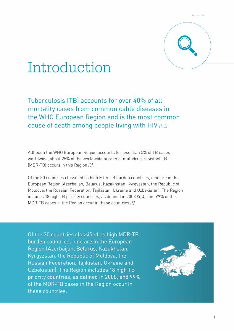
Although the WHO European Region accounts for less than 5% of TB cases worldwide, about 25% of the worldwide burden of multidrug-resistant TB (MDR-TB) occurs in this Region (3).
Of the 30 countries classified as high MDR-TB burden countries, nine are in the European Region (Azerbaijan, Belarus, Kazakhstan, Kyrgyzstan, the Republic of Moldova, the Russian Federation, Tajikistan, Ukraine and Uzbekistan). The Region includes 18 high TB priority countries, as defined in 2008 (3, 4), and 99% of the MDR-TB cases in the Region occur in these countries (5).
Introduction
Tuberculosis (TB) accounts for over 40% of all mortality cases from communicable diseases in the WHO European Region and is the most common cause of death among people living with HIV (1, 2)
Of the 30 countries classified as high MDR-TB burden countries, nine are in the European Region (Azerbaijan, Belarus, Kazakhstan, Kyrgyzstan, the Republic of Moldova, the Russian Federation, Tajikistan, Ukraine and Uzbekistan). The Region includes 18 high TB priority countries, as defined in 2008, and 99% of the MDR-TB cases in the Region occur in these countries.
Introduction
1
The Region includes high-, middle- and low-income countries with diverse national health system structures for TB control activities. Latest data from the Region on MDR-TB prevalence amount to 16% among new TB cases and 48% among previously treated cases (3, 6). Extensively drug-resistant TB (XDR-TB) is estimated to occur in 23.4% of all MDR-TB cases subjected to second-line drug-susceptibility testing (DST) (6). In 2015, treatment success rates were at 76% in new and relapse, 63% in previously treated (excluding relapses) and 51% in rifampicin-resistant/MDR-TB cohorts (6). In 2015 with the development of an ambitious post-2015 global End TB Strategy, to continue the progress and address the challenges in TB and M/XDR-TB detection, prevention and care, the Regional Office developed the TB action plan for the WHO European Region covering the period 2016–2020 (5, 6, 8).
Much work needs to be done to reach the targets set in the 2016–2020 action plan for the laboratory diagnosis of TB in the Region, particularly in the appropriate use of molecular diagnostics, and increasing the MDR-TB case-detection rate and coverage of quality-assured second-line DST among MDR-TB cases. Currently about 311 910 TB cases are registered in the 51 reporting countries of the Region (6). Bacteriological confirmation of TB diagnosis was reported for 61.4% of all new and relapse pulmonary cases in the Region and in four countries this was below 50% (5, 6). Only 57.9% of an estimated 74 000 MDR-TB cases were detected, against a regional target of diagnosing at least 85% (6, 8).
Coverage of rifampicin (RIF)-resistance testing among laboratory-confirmed pulmonary TB cases amounted to 44% of new cases and 49% of previously treated cases in 2015. Coverage of second-line DST among laboratory confirmed drug-resistant TB cases was 52.2% (3). Although DST coverage has improved significantly, scale-up of testing and the use of WHO-recommended rapid molecular tests are urgently needed to reach the target of performing DST for close to 100% of all laboratory-confirmed cases by 2020. More directive guidance and advocacy is also needed to address gaps in the laboratory diagnosis of TB in the Region, especially in the nine high MDR-TB burden countries.
In response to the need to strengthen TB laboratory capacity for accurate diagnosis and early detection of drug-resistant TB (DR-TB) in the Region and ensure implementation of the regional action plans (8, 9), the WHO Regional Office for Europe established the European Tuberculosis Laboratory Initiative (ELI) in 2012 (10). The mission of ELI is to strengthen TB laboratory capacity in the Region, with a focus on the 18 high TB priority countries. ELI members consist of national and
Algorithm for laboratory diagnosis and treatment-monitoring of pulmonary tuberculosis and drug-resistant tuberculosis using state-of-the-art rapid diagnostic technologies
2
international TB laboratory experts in the Region and international partners dedicated to accelerating and expanding access to quality-assured TB diagnostic laboratory services. ELI has a core group of members who function as an independent, technical advisory and support group for WHO and partners (10).
ELI has been working to develop a diagnostic algorithm, taking into consideration the substantial heterogeneity of the Region. The algorithm combines different types of tests synergistically by targeting patient groups appropriately and will help to make the best use of available resources by maximizing their respective strengths and mitigating their weaknesses (11, 12). The draft algorithm developed by the previous core group members (2012–2014) was revised during and after the last ELI core group meeting, held in Copenhagen, Denmark on 25 February 2016 (13). The algorithm was finalized and agreed by members during the ELI core group meeting in Tbilisi, Georgia on 30 November and 2 December 2016 and is presented here.
The recommendations presented here take into account and supplement previously published recommended standards for modern TB laboratory services, including the WHO global policy framework on implementing TB diagnostics and the European Union Standards for Tuberculosis Care (ESTC) and International Standards for Tuberculosis Care (ISTC) (11, 14, 15). The ESTC adapted the ISTC (14) to reflect the European Union setting and practices (11). The ESTC builds on previous recommendations on laboratory methods for diagnosing TB (16) to propose that:
In countries, settings or populations in which MDR-TB is suspected in a patient, rapid testing for the identification of rifampicin- and isoniazid-resistance, using validated tools in a quality-assured laboratory, should be performed.
To reassure rapid diagnosis of TB in the entire Region, ELI suggests further extension of this standard by using rapid molecular diagnosis as an initial method for all cases with clinical suspicion of TB, to be applied in all countries of the Region. With high MDR-TB rates being present in Eastern Europe, every presumptive TB case could also be an MDR-TB case.
Only 57.9% of an estimated
74 000 MDR-TB cases were
detected, against a regional
target of diagnosing at
least 85%
Introduction
3
Specific laboratory policies and diagnostic algorithms should be available at country level based on the local epidemiological situation; limited resources initially will mean that not all the required improvements can be implemented immediately, but resource limitation should not influence the recommendation, only the rate at which the recommendation is implemented.
WHO-endorsed rapid diagnostic tests should be key to the diagnostic work-up for all TB presumptive cases (17). Countries shall prioritize the use of recommended rapid molecular tests, rather than conventional microscopy, culture or DST, as the initial diagnostic test for adults and children presumed to have pulmonary TB/MDR-TB and/or HIV-associated TB and/or TB meningitis (17). This will ensure the availability of early and accurate diagnosis. Conventional microscopy should be used as an initial diagnostic test only in laboratory settings without rapid molecular tests and in the absence of systems for timely sample transportation to a setting in which these techniques are available.
In settings with high risk of transmission of TB and/or MDR-TB, such as prisons in countries of the former Soviet Union, implementation of sputum polymerase chain reaction (PCR)-based screening (GeneXpert MTB/RIF assay) as an annual screening tool has been shown to most cost-effectively reduce TB and MDR-TB when compared to more traditional interventions (18).
Principles of laboratory diagnosis of TB
Laboratory diagnosis of TB should start with appropriate clinical screening procedures to identify individuals with clinical suspicion of TB
Algorithm for laboratory diagnosis and treatment-monitoring of pulmonary tuberculosis and drug-resistant tuberculosis using state-of-the-art rapid diagnostic technologies
4
In the event of discrepancies between the results of conventional and molecular assays, it is recommended that the respective tests be repeated on the same or another sample from the same patient to exclude technical errors. If the discrepancy between microscopy and molecular tests is confirmed, the laboratory shall report the molecular test result to the clinician instead of the microscopy result due to the higher sensitivity and specificity of molecular tests compared to microscopy. If the culture becomes positive after initial molecular and microscopy tests were negative, this positive culture result should be reported as well (as this reflects the proportion of samples for which culture remains the most sensitive test). The proposed table in Annex 1 has been developed to help the interpretation and explanation of all potential discordant results that may occur, although most of these cases will be observed very rarely. The table will also assist laboratories to explain discrepant or conflicting results to clinicians.
WHO-endorsed rapid
diagnostic tests should be
key to the diagnostic work-up
for all TB presumptive cases
Principles of laboratory diagnosis of TB
5
Laboratories should have appropriate infrastructure, biosafety measures, equipment, and access to regular maintenance of equipment and infrastructure. Laboratory commodities and supplies should be managed well and laboratory data properly recorded, preferably in an electronic format. Ideally, the laboratory would have a clear and separate budget line within the programme/hospital budget.
Specimen transportation and referral mechanisms within a laboratory network should be well described, with clear transportation arrangements in place. Patient specimens kept in appropriate secure containers can be transported using most health-care vehicles to increase transport frequency and reduce delay; the quality of the specimens arriving should be monitored (15, 20, 21). A laboratory quality management system in line with WHO recommendations should be in place.
The laboratory work-up of a clinical sample should be defined by the patient’s category (new, relapse or previously treated case), the purpose of the analysis (for diagnosis or treatment success) and the patient’s risk assessment (HIV, or risk of MDR-TB, for instance). This information is to be recorded on the laboratory request form together with the laboratory tests to ensure that the most appropriate, efficient and cost-effective laboratory work-up is achieved, and to facilitate result
Prerequisites of a good laboratory network
Countries should have sufficient funding and appropriately trained human resources available for laboratories, as well as a laboratory maintenance and development plan (19)
Algorithm for laboratory diagnosis and treatment-monitoring of pulmonary tuberculosis and drug-resistant tuberculosis using state-of-the-art rapid diagnostic technologies
6
interpretation – in the case of discordant results – and communication between clinical doctors and laboratory staff. Frequent repetitions of DST are usually unnecessary and often not helpful. Once a patient has been shown to have MDR-TB, for example, subsequent repetition of isoniazid (INH) and RIF testing will be unhelpful.
An updated manual detailing recommendations for biosafety was published in 2012 by WHO (22). The current recommendations are based on assessments of the risks associated with different technical procedures performed in different types of TB laboratories. It is recommended that culture and DST be used only in laboratories at regional and central reference level that have appropriate biosafety standards (22).
Similarly, line probe assays (LPAs) should be placed at regional and central reference level laboratories (or any laboratory currently performing not automated PCR-based amplification methodologies for infections, as the infrastructure will be the same) because of the complexity of the assay and required infrastructure (23). Many countries in the Region follow the principle of patient (instead of specimen) referral to confirm the diagnosis of TB at specialized TB centres or hospitals. By using WHO recommended rapid molecular techniques for the initial diagnosis of TB and MDR-TB at district or subdistrict level (24, 25), a highly specific diagnosis of TB can be achieved at a lower level in the health system, which can help countries to reduce diagnostic delay, apply the most appropriate infection control measures and simultaneously move towards ambulatory treatment of TB cases (9).
Similarly, line probe assays
(LPAs) should be placed
at regional and central
reference level laboratories,
because of the complexity
of the assay and required
infrastructure
Prerequisites of a good laboratory network
7
Where resources and/or the availability of molecular diagnostics or other tests are limited, different algorithms could be used for different patient groups based on careful risk assessment and prioritization of molecular diagnostic tests and liquid culture for those patients presumed to have pulmonary MDR-TB and/or HIV-associated TB and/or TB meningitis.
Algorithm for the initial laboratory diagnosis of individuals with symptoms consistent with pulmonary TB
This starts by subjecting two clinical specimens (preferably including the morning sample) to a WHO-endorsed molecular diagnostic test and one specimen for culture (Fig. 1).
Algorithms for laboratory diagnostic and follow up of TB and MDR-TB cases
Three algorithms are proposed in this document:
1. for the initial diagnostics of all presumptive TB cases2. for follow-up of patients under first-line anti-TB treatment3. for follow-up of MDR-TB patients (see Fig. 1–3).
Algorithm for laboratory diagnosis and treatment-monitoring of pulmonary tuberculosis and drug-resistant tuberculosis using state-of-the-art rapid diagnostic technologies
8
Only when a positive rapid molecular test result is obtained would a subsequent microscopy test be helpful to identify the bacterial load and infectivity. Results of molecular tests should be communicated to the clinician without waiting for culture results. If GeneXpert MTB/RIF is negative, no microscopy is necessary and the sample should be sent for culture only (as culture would still be more sensitive for TB detection).
Culture-positive samples are subjected to identification, and those cultures identified as M. tuberculosis (MTB) complex should be subjected to DST. If the sample that was subjected to the molecular test is culture negative (as well as negative on the molecular test), further clinical and other non-TB specific laboratory investigations should follow.
If the molecular diagnostic test predicts susceptibility to RIF and if first-line (FL) LPA is available, it should be performed to identify INH mono-resistant cases. If INH resistance is identified, INH mono-resistant regimen should be initiated. In case FL-LPA is not available or no INH resistance is identified, the FL regimen needs to be initiated. In both cases, culture and subsequent phenotypic drug-susceptibility testing (pDST) should be done. Performing a WHO-recommended rapid molecular test for drug resistance (such as Xpert or LPA) on the primary samples reduces the delay to appropriate phenotypic resistance results.
If RIF resistance (with or without INH resistance) is confirmed, it is recommended that the patient be referred to a DR-TB treatment initiation site for appropriate therapy and to proceed with second-line (SL) LPA (26): this recommendation applies to the direct testing of sputum specimens from RIF-resistant TB or MDR-TB irrespective of the smear status, while acknowledging that the indeterminate rate is higher when testing smear-negative sputum specimens compared with smear-positive sputum specimens (26). SL-LPA is suitable for use at central or national reference laboratory level; it has the potential to be used at regional level if the appropriate infrastructure and trained staff can be ensured (26).
Once the culture becomes positive, first- and second-line pDST should be set up, irrespective of SL-LPA results, for patients with negative and positive SL-LPA results. This streamlining reduces any delay for initiating an appropriate M/XDR-TB treatment regimen. Depending on the LPA results (FL and SL), treatment should start accordingly and be adjusted if needed once pDST results are available.
SL-LPA results that lead to the exclusion of both fluoroquinolones (FLQs) and second-line injectable drug (SLID) resistance means that the use of a shorter MDR-TB regimen could be considered, providing the other criteria are met (27).
Algorithms for laboratory diagnostic and follow up of TB and MDR-TB cases
9
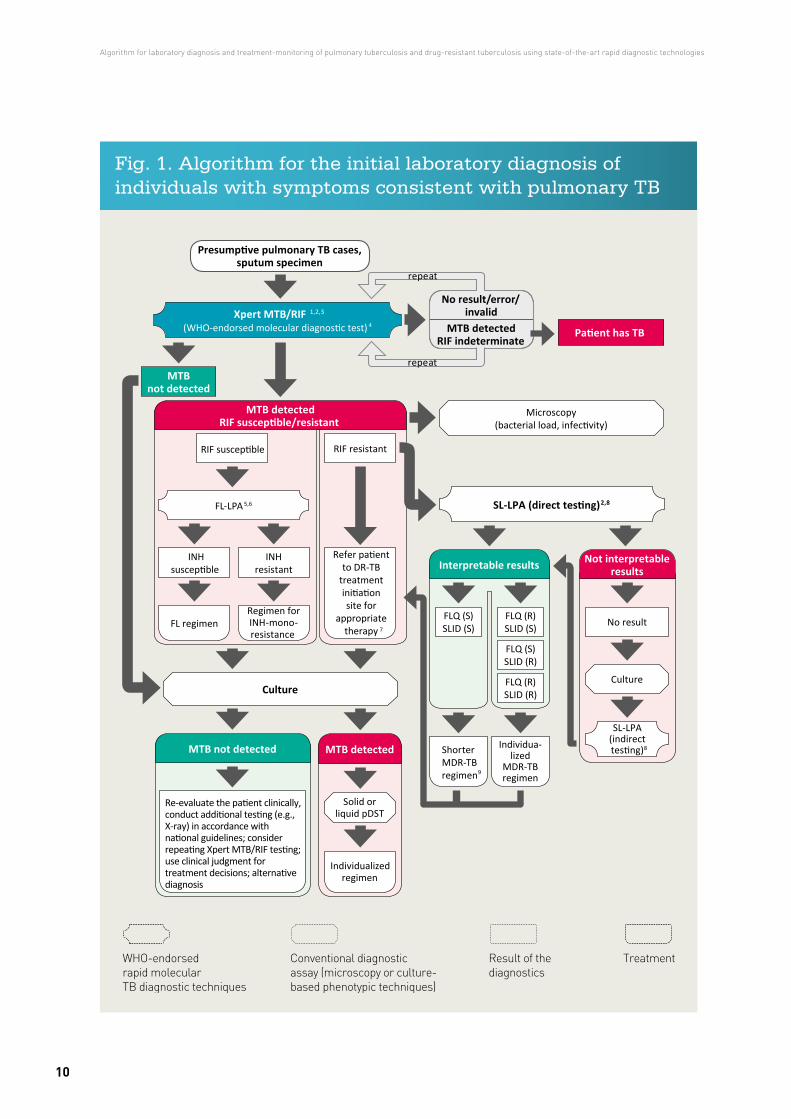
Fig. 1. Algorithm for the initial laboratory diagnosis of individuals with symptoms consistent with pulmonary TB
WHO-endorsed rapid molecular TB diagnostic techniques
Conventional diagnostic assay (microscopy or culture-based phenotypic techniques)
Result of the diagnostics
Treatment
Culture
MTB detected
Solid or liquid pDST
Individualized regimen
MTB not detected
FL regimen
Microscopy(bacterial load, infectivity)
7
RIF resistant
2,8
Not interpretable results
No result
Culture
8
Interpretable results
FLQ (S)SLID (S)
FLQ (S)SLID (R)
FLQ (R)SLID (S)
FLQ (R)SLID (R)
9
FL-LPA 5,6
Regimen for INH-mono- resistance
INHsusceptible
INH resistant
Xpert MTB/RIF 1,2,3
4
No result/error/
Patient has TB
SL-LPA (direct testing)
invalidMTB detected
RIF indeterminate
repeat
repeatMTB
not detected
Re-evaluate the patient clinically, conduct additional testing (e.g., X-ray) in accordance with national guidelines; consider repeating Xpert MTB/RIF testing; use clinical judgment for treatment decisions; alternative diagnosis
Refer patientto DR-TB
treatmentinitiationsite for
appropriatetherapy
MTB detectedRIF susceptible/resistant
RIF susceptible
SL-LPA(indirecttesting)Individua-
lizedMDR-TBregimen
(WHO-endorsed molecular diagnostic test)
ShorterMDR-TBregimen
Presumptive pulmonary TB cases,sputum specimen
Algorithm for laboratory diagnosis and treatment-monitoring of pulmonary tuberculosis and drug-resistant tuberculosis using state-of-the-art rapid diagnostic technologies
10
Algorithm for monitoring the follow-up of patients with drug-sensitive pulmonary TB
This algorithm is shown in Fig. 2.
Monthly microscopy and culture during the intensive phase of two sputum samples and immediately after completion of the intensive phase (at month two for new cases and month three for previously treated cases) is suggested, as well as during the fifth and last month of treatment, according to the timeframe of the treatment protocol in a particular country.
If microscopy and/or culture is/are positive from a sample taken after two months’ treatment, a WHO-endorsed molecular diagnostic test and DST are suggested to confirm the presence/absence of MTB and the anti-TB drug-resistance pattern.
If the WHO-endorsed molecular tests (X-pert, FL-LPA) indicate resistance to RIF with or without INH, then SL-LPA should be performed. SL-LPA results that lead to the exclusion of both FLQs and SLID resistance means that the use of a shorter MDR-TB regimen could be considered providing the other criteria are met (27).
Once culture results are available and RIF resistance is known (through WHO-endorsed molecular tests), first- and second-line pDST should be performed simultaneously.
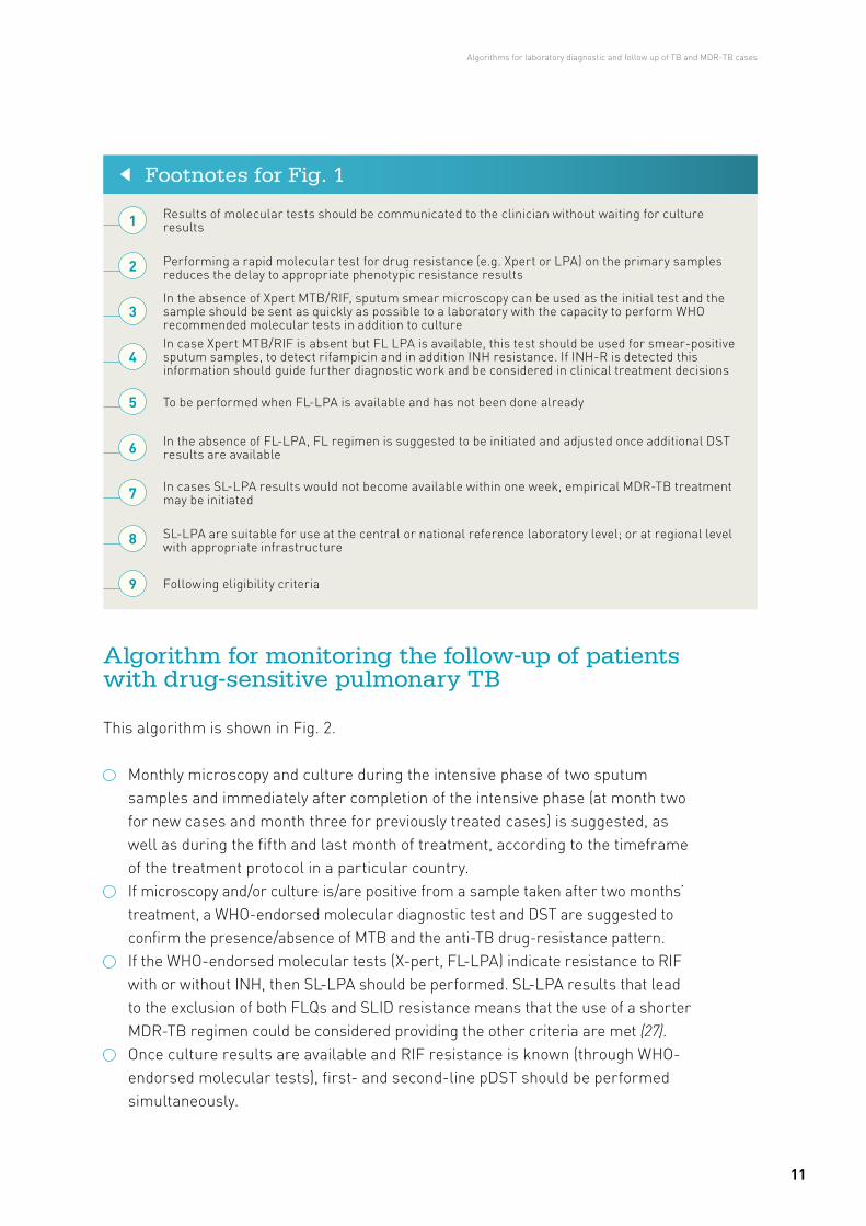
Results of molecular tests should be communicated to the clinician without waiting for culture results1
Performing a rapid molecular test for drug resistance (e.g. Xpert or LPA) on the primary samples reduces the delay to appropriate phenotypic resistance results2
In the absence of Xpert MTB/RIF, sputum smear microscopy can be used as the initial test and the sample should be sent as quickly as possible to a laboratory with the capacity to perform WHO recommended molecular tests in addition to culture
3
In case Xpert MTB/RIF is absent but FL LPA is available, this test should be used for smear-positive sputum samples, to detect rifampicin and in addition INH resistance. If INH-R is detected this information should guide further diagnostic work and be considered in clinical treatment decisions
4
To be performed when FL-LPA is available and has not been done already5
In the absence of FL-LPA, FL regimen is suggested to be initiated and adjusted once additional DST results are available6
In cases SL-LPA results would not become available within one week, empirical MDR-TB treatment may be initiated7
SL-LPA are suitable for use at the central or national reference laboratory level; or at regional level with appropriate infrastructure8
Following eligibility criteria9
Footnotes for Fig. 1
Algorithms for laboratory diagnostic and follow up of TB and MDR-TB cases
11
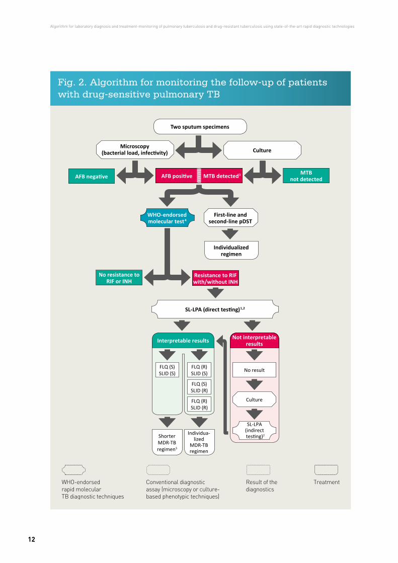
Fig. 2. Algorithm for monitoring the follow-up of patients with drug-sensitive pulmonary TB
Two sputum specimens
Microscopy(bacterial load, infectivity) Culture
Individualized regimen
MTB not detected
First-line and second-line pDST
WHO-endorsed molecular test4
3
No resistance to RIF or INH
Resistance to RIF with/without INH
Not interpretable results
No result
Culture
SL-LPA(indirecttesting)2
Interpretable results
FLQ (S)SLID (S)
FLQ (S)SLID (R)
FLQ (R)SLID (S)
FLQ (R)SLID (R)
Individua-lized
MDR-TBregimen
ShorterMDR-TB regimen5
1,2
WHO-endorsed rapid molecular TB diagnostic techniques
Conventional diagnostic assay (microscopy or culture-based phenotypic techniques)
Result of the diagnostics
Treatment
Algorithm for laboratory diagnosis and treatment-monitoring of pulmonary tuberculosis and drug-resistant tuberculosis using state-of-the-art rapid diagnostic technologies
12
The purpose of diagnostic testing here is to determine the success of treatment; repeating DST is usually not required unless the patient continues to remain smear and culture positive or there is evidence of concurrent MDR-TB exposure (that is, potential superinfection) or clinical deterioration. The laboratory needs to be told that the sample has been sent only to assess treatment success.
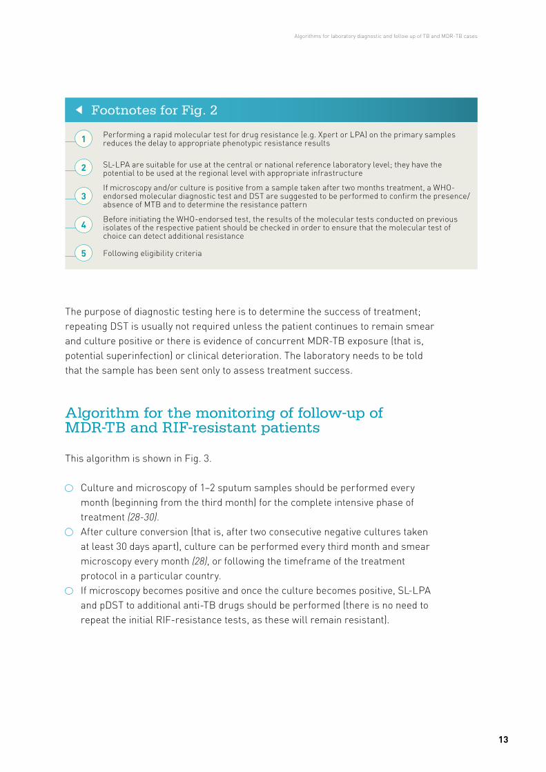
Algorithm for the monitoring of follow-up of MDR-TB and RIF-resistant patients
This algorithm is shown in Fig. 3.
Culture and microscopy of 1–2 sputum samples should be performed every month (beginning from the third month) for the complete intensive phase of treatment (28-30).
After culture conversion (that is, after two consecutive negative cultures taken at least 30 days apart), culture can be performed every third month and smear microscopy every month (28), or following the timeframe of the treatment protocol in a particular country.
If microscopy becomes positive and once the culture becomes positive, SL-LPA and pDST to additional anti-TB drugs should be performed (there is no need to repeat the initial RIF-resistance tests, as these will remain resistant).
Performing a rapid molecular test for drug resistance (e.g. Xpert or LPA) on the primary samples reduces the delay to appropriate phenotypic resistance results
If microscopy and/or culture is positive from a sample taken after two months treatment, a WHO-endorsed molecular diagnostic test and DST are suggested to be performed to confirm the presence/absence of MTB and to determine the resistance pattern
SL-LPA are suitable for use at the central or national reference laboratory level; they have the potential to be used at the regional level with appropriate infrastructure
Before initiating the WHO-endorsed test, the results of the molecular tests conducted on previous isolates of the respective patient should be checked in order to ensure that the molecular test of choice can detect additional resistance
Following eligibility criteria
Footnotes for Fig. 2
1
2
3
4
5
Algorithms for laboratory diagnostic and follow up of TB and MDR-TB cases
13
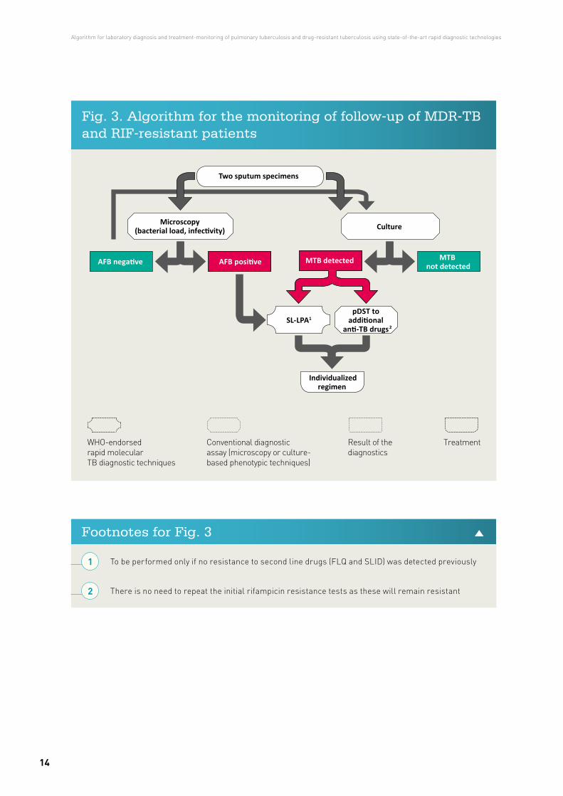
Fig. 3. Algorithm for the monitoring of follow-up of MDR-TB and RIF-resistant patients
To be performed only if no resistance to second line drugs (FLQ and SLID) was detected previously
There is no need to repeat the initial rifampicin resistance tests as these will remain resistant
Footnotes for Fig. 3
Two sputum specimens
Culture
MTB not detected
MTB detectedAFB negative AFB positive
Individualizedregimen
2SL-LPA1
Microscopy(bacterial load, infectivity)
pDST toadditional
anti-TB drugs
WHO-endorsed rapid molecular TB diagnostic techniques
Conventional diagnostic assay (microscopy or culture-based phenotypic techniques)
Result of the diagnostics
Treatment
1
2
Algorithm for laboratory diagnosis and treatment-monitoring of pulmonary tuberculosis and drug-resistant tuberculosis using state-of-the-art rapid diagnostic technologies
14
The results of all tests performed at lower levels of laboratories should be reported and referred together with the samples and/or cultured isolates to the National Reference Laboratory (NRL). This will prevent wasteful duplication of tests (except in a proportion used for quality control purposes). A handbook with the detailed methodologies needed for assay performance has been published by the European Centre for Disease Prevention and Control European Reference Laboratory Network (31).
WHO recommends replacing conventional microscopy with rapid molecular tests (such as GeneXpert MTB/RIF) for the initial diagnosis of TB (3, 17, 24, 25, 32). If resources permit, two specimens should be tested to increase the sensitivity. Microscopy would be limited to test TB positive samples detected by rapid molecular techniques to ascertain patient infectivity and infection control purposes and for treatment monitoring. Continuing with microscopy also maintains quality-assured skills in performing microscopy if molecular tests are not available.
Practical considerations for the diagnostic algorithm
Various tests are performed at different levels of laboratories and within a laboratory network. In some countries, laboratories that perform PCR-based diagnostics for infections other than TB will have the skills and infrastructure necessary for LPAs, as TB diagnosis in many countries is incorporated with other clinical microbiology tests for other diseases
Practical considerations for the diagnostic algorithm
15
Diagnostic methods of preference
Capacity for microscopy, culture and DST needs to remain, despite molecular diagnostics. Microscopy and culture are particularly important for treatment monitoring. The availability of molecular diagnostic tests does not eliminate the need for conventional microscopy, culture and DST capability; microscopy and culture remain necessary for the follow-up of treatment, and culture currently still provides maximum diagnostic sensitivity, while conventional DST is required to support a diagnosis of XDR-TB and provide a tailored patient-regime for M/XDR-TB patients. Demands for conventional techniques might change in the future based on the epidemiological situation.
Light-emitting diode (LED) fluorescence microscopy is the recommended method for microscopy at all levels of laboratory (33). Both LED microscopy and conventional fluorescence microscopy are at least 10% more sensitive than Ziehl-Neelsen microscopy. Moreover, LED microscopy is less costly compared to conventional fluorescence microscopy (33).
For culture, both solid and liquid media are recommended. Liquid culture is more sensitive and quicker than culture on solid media (increased yield 10%) (34). Liquid culture results may become available within days but are more prone to contamination, more expensive and associated with a higher biosafety risk.
Positive cultures should be differentiated into MTB complex and non-tuberculous mycobacteria. Confirmation of MTB complex is done through biochemical reaction, molecular amplification tests or immunochromatographic assays. The latter two are recommended for species identification on culture isolates, as they provide a rapid and definite identification of MTB complex (34).
Drug resistance can be detected by genotypic and phenotypic methods. Automated liquid systems are the current gold standard for FL and SL DST (34). DST should follow WHO guidelines with stringent quality assurance methods (29, 34, 35). SL DST should aim to include testing of the aminoglycosides, polypeptides and FLQs used in the country. DST results on these drugs have good reliability and reproducibility and allow a quality-assured diagnosis of XDR-TB. With the introduction of SL-LPA for detecting resistance to FLQs and SLIDs, resistant results to these drugs can be obtained more rapidly.
By direct testing, SL-LPA will detect 86% of patients with FLQ resistance, 87% with SLID resistance and 69% of XDR-TB; in all cases, the test will rarely give a false
Algorithm for laboratory diagnosis and treatment-monitoring of pulmonary tuberculosis and drug-resistant tuberculosis using state-of-the-art rapid diagnostic technologies
16
positive result (26). In interpreting the results, it should be considered that SL-LPA cannot determine resistance to individual FLQs. Resistance-conferring mutations detected by SL-LPA are highly correlated with phenotypic resistance to ofloxacin and levofloxacin. The correlation of these mutations with phenotypic resistance to moxifloxacin and gatifloxacin is unclear, however, and the inclusion of moxifloxacin or gatifloxacin in an MDR-TB regimen is best guided by pDST results (26). Mutations in some regions of the MTB complex genome (such as the eis promoter region) may be responsible for causing resistance to one drug in a class (group) more than other drugs within that class (group). For example, the eis C14T mutation is associated with kanamycin resistance in strains from eastern Europe (26).
Non-commercial methods for culture and DST, including microscopic-observation drug-susceptibility (MODS), colorimetric redox indicator (CRI) methods and the nitrate reduction assay (NRA), are also recommended by WHO (36). These tests are currently considered as an interim solution while scale-up of genotypic testing is developed. The systems are less expensive than commercial systems, but may be more prone to errors due to lack of standardization, are highly operator-dependent and only suitable for use at reference laboratory level.
Currently, the WHO-recommended molecular diagnostic tests for TB and DR-TB include LPAs and the Xpert MTB/RIF assay (as well as loop-mediated isothermal amplification (LAMP) for TB diagnosis only). Data from systematic reviews and meta-analyses show that in comparison to conventional DST, LPAs are highly sensitive (≥97%) and specific (≥99%) for the detection of RIF resistance, alone or in combination with INH (sensitivity ≥90%; specificity ≥99%), on isolates of MTB and on smear-positive sputum specimens (23, 37, 38).
An extensive review (17, 32) of the Xpert MTB/RIF assay to detect pulmonary TB disease, including studies involving almost 10 000 participants, showed the high specificity of the Xpert MTB/RIF assay to detect TB (99% (95% CrI 98–99%)). Sensitivity varied by smear status: 68% (95% CrI 61–74%) for smear-negative
By direct testing, SL-LPA will
detect 86% of patients with
FLQ resistance, 87% with
SLID resistance and 69%
of XDR-TB; in all cases, the
test will rarely give a false
positive result
Practical considerations for the diagnostic algorithm
17
culture-positive to 98% (95% CrI 97–99%) for smear-positive culture-positive samples. Performance of the Xpert MTB/RIF assay for detection of extrapulmonary TB varied by specimen type, with lower sensitivity for pleural and cerebrospinal fluid (ranging from 17% to 80%, respectively) and somewhat lower specificity (93%) for lymph node specimens, but with good sensitivity (>81%) and specificity (>98%) for other specimen types (25).
Overall, the Xpert MTB/RIF assay had 95% (95% CrI 90–97%) sensitivity and 98% (95% CrI 97–99%) specificity to detect RIF. When studies were analysed separately for settings with different levels of RIF resistance, however, the sensitivity was 96% (95% CrI 91–98%) for settings with >15% RIF resistance among the tested population, and 91% (95% CrI 79–97%) for settings with ≤ 15% RIF resistance. The corresponding pooled specificities were 97% (95% CrI 94–99%) and 99% (95% CrI 98–99%) (17, 32).
Similar data have been published in a systematic review with a detailed health economic analysis (39). The specificity of Xpert MTB/RIF for detecting TB is very high (99%), and false-positive results are likely to be linked to the detection by Xpert MTB/RIF of dead MTB bacilli that would not be detected by culture, which is the present reference standard. Given that the specificity of Xpert MTB/RIF is not 100%, the positive predictive value (PPV) of Xpert MTB/RIF testing for RIF resistance testing is adversely affected in settings with a low prevalence of drug-resistant TB disease or in populations with a low prevalence of TB. Testing for TB is not usually implemented in a general, asymptomatic population but in individuals with clinical suspicion of TB following some form of screening involving, for example, symptom assessment or chest X-ray. Such screening procedures increase the prevalence of TB in the group tested and improve the PPV of the test, reducing but not eliminating concerns related to false-positive results (32).
Interpretation and reporting of laboratory results
Results from laboratory tests, including microscopy, molecular tests, culture and DST, should be reported to clinicians as soon as possible after they become available, and all means of communication, including telephone, fax, email and SMS, should be considered to facilitate communication. Interpretation of laboratory results by laboratory doctors or equivalent is vital, especially in situations when results are apparently inconsistent or discrepant. Subsequently, appropriate
Algorithm for laboratory diagnosis and treatment-monitoring of pulmonary tuberculosis and drug-resistant tuberculosis using state-of-the-art rapid diagnostic technologies
18
isolation of the respective patients and adjustment of their treatment courses according to the anti-TB drug susceptibility pattern of the causative pathogens should be initiated promptly (29) to prevent further spread of the disease and/or the development of further resistance.
It should be noted that mutations in the rpoB gene are a very good marker for MDR-TB in the Region; the percentage of RIF mono-resistant isolates has been shown to be only 0.5% among new cases and 0.9% among previously treated cases (5). Under these circumstances, it seems reasonable to initiate patients on MDR treatment if the Xpert MTB/RIF or LPA results indicate RIF resistance. More caution is necessary when interpreting the results of molecular tests for SL anti-TB drugs.
Results from laboratory
tests, including microscopy,
molecular tests, culture and
DST, should be reported to
clinicians as soon as possible
after they become available
Practical considerations for the diagnostic algorithm
19
1. The European health report 2012: charting the way to well-being. Copenhagen: WHO Regional Office for Europe; 2012 (http://www.euro.who.int/__data/assets/pdf_file/0004/197113/EHR2012-Eng.pdf, accessed 15 February 2017).
2. Roadmap to implement the tuberculosis action plan for the WHO European Region 2016–2020. WHO Regional Office for Europe; 2016 (http://www.euro.who.int/__data/assets/pdf_file/0020/318233/Roadmap-implement-TBC-action-plan-20162020.pdf, accessed 15 February 2017).
3. Global Tuberculosis Report 2016. Geneva: World Health Organization; 2016 (WHO/HTM/TB/2016.13; http://apps.who.int/iris/bitstream/10665/250441/1/9789241565394-eng.pdf, accessed 15 February 2017).
4. Plan to Stop TB in 18 priority countries of the WHO European Region, 2007–2015. Copenhagen: WHO Regional Office for Europe; 2007 (http://www.euro.who.int/document/E91049.pdf, accessed 15 February 2017).
5. Tuberculosis surveillance and monitoring in Europe 2016. Stockholm: European Centre for Disease Prevention and Control/WHO Regional Office for Europe; 2016 (http://ecdc.europa.eu/en/publications/Publications/ecdc-tuberculosis-surveillance-monitoring-Europe-2016.pdf, accessed 15 February 2017).
6. Tuberculosis serveillance and monitoring in Europe 2017. Stockholm: European Centre for Disease Prevention and Control/WHO Regional Office for Europe; 2017.
References
Algorithm for laboratory diagnosis and treatment-monitoring of pulmonary tuberculosis and drug-resistant tuberculosis using state-of-the-art rapid diagnostic technologies
20
7. Tuberculosis surveillance and monitoring in Europe 2013. Stockholm: European Centre for Disease Prevention and Control/WHO Regional Office for Europe; 2013 (http://ecdc.europa.eu/en/publications/Publications/Tuberculosis-surveillance-monitoring-2013.pdf, accessed 15 February 2017).
8. Tuberculosis action plan for the WHO European Region 2016–2020. Copenhagen: WHO Regional Office for Europe; 2015 (http://www.euro.who.int/__data/assets/pdf_file/0007/283804/65wd17e_Rev1_TBActionPlan_150588_withCover.pdf, accessed 15 February 2017).
9. Roadmap to prevent and combat drug-resistant tuberculosis. The Consolidated Action Plan to Prevent and Combat Multidrug- and Extensively Drug-Resistant Tuberculosis in the WHO European Region, 2011–2015. Copenhagen: WHO Regional Office for Europe; 2011 (http://www.euro.who.int/__data/assets/pdf_file/0014/152015/e95786.pdf, accessed 15 February 2017).
10. European Tuberculosis Laboratory Initiative. In: Health topics [website]. Copenhagen: WHO Regional Office for Europe; 2017 (Report Number; http://www.euro.who.int/en/health-topics/communicable-diseases/tuberculosis/activities/european-tuberculosis-laboratory-initiative, accessed 15 February).
11. Migliori GB, Zellweger JP, Abubakar I, Ibraim E, Caminero JA, De Vries G, et al. European Union standards for tuberculosis care. Eur Respir J 2012;39(4):807-19; doi: 10.1183/09031936.00203811.
12. Ehsani S, van den Boom M, Gilpin C, Dara M, Europe WHOROf. The role of novel molecular techniques for tuberculosis diagnostics in the WHO European Region. J Public Health (Oxf) 2016; doi: 10.1093/pubmed/fdv200.
13. Meeting of the European Tuberculosis Laboratory Initiative (ELI) Core Group. Copenhagen: WHO Regional Office for Europe; 2016 (http://www.euro.who.int/__data/assets/pdf_file/0020/307433/ELI-TB-Core-Group-mtg.pdf, accessed 15 February 2017).
14. International Standards for Tuberculosis Care, third edition. The Hague: TB CARE I; 2014 (http://www.who.int/tb/publications/ISTC_3rdEd.pdf, accessed 15 February 2017).
References
21
15. Implementing tuberculosis diagnostics: policy framework. Geneva: World Health Organization; 2015 (WHO/HTM/TB/2015.11; http://apps.who.int/iris/bitstream/10665/162712/1/9789241508612_eng.pdf, accessed 15 February 2017).
16. Drobniewski FA, Hoffner S, Rusch-Gerdes S, Skenders G, Thomsen V, Force WHOELST. Recommended standards for modern tuberculosis laboratory services in Europe. Eur Respir J 2006;28(5):903-9; doi: 10.1183/09031936.06.00084906.
17. Automated real-time nucleic acid amplification technology for rapid and simultaneous detection of tuberculosis and rifampicin resistance: Xpert MTB/RIF assay for the diagnosis of pulmonary and extrapulmonary TB in adults and children. Policy update. Geneva: World Health Organization; 2013 (WHO/HTM/TB/2013.16; http://www.stoptb.org/wg/gli/assets/documents/WHO Policy Statement on Xpert MTB-RIF 2013 pre publication 22102013.pdf, accessed 15 February 2017).
18. Winetsky DE, Negoescu DM, DeMarchis EH, Almukhamedova O, Dooronbekova A, Pulatov D, et al. Screening and rapid molecular diagnosis of tuberculosis in prisons in Russia and Eastern Europe: a cost-effectiveness analysis. PLoS Med 2012;9(11):e1001348; doi: 10.1371/journal.pmed.1001348.
19. The European health report 2015: Targets and beyond – reaching new frontiers in evidence. Copenhagen: WHO Regional Office for Europe; 2015 (http://www.euro.who.int/__data/assets/pdf_file/0006/288645/European-health-report-2015-full-book-en.pdf, accessed 15 February 2017).
20. Policy Framework for Implementing New Tuberculosis Diagnostics. Geneva: World Health Organization; 2010 (http://www.who.int/tb/laboratory/whopolicyframework_rev_june2011.pdf, accessed 15 February 2017).
21. Guidance on regulations for the Transport of Infectious Substances 2015–2016. Geneva: World Health Organization; 2015 (WHO/HSE/GCR/2015.2; http://apps.who.int/iris/bitstream/10665/149288/1/WHO_HSE_GCR_2015.2_eng.pdf, accessed 15 February 2017).
Algorithm for laboratory diagnosis and treatment-monitoring of pulmonary tuberculosis and drug-resistant tuberculosis using state-of-the-art rapid diagnostic technologies
22
22. Tuberculosis laboratory biosafety manual. Geneva: World Health Organization; 2012 (WHO/HTM/TB/2012.11; http://apps.who.int/iris/bitstream/10665/77949/1/9789241504638_eng.pdf, accessed 15 February 2017).
23. Molecular line probe assays for rapid screening of patients at risk of multidrug-resistant tuberculosis (MDR-TB): policy statement. Geneva: World Health Organization; 2008 (http://www.who.int/tb/features_archive/policy_statement.pdf, accessed 15 February 2017).
24. Automated Real-time Nucleic Acid Amplification Technology for Rapid and Simultaneous Detection of Tuberculosis and Rifampicin Resistance: Xpert MTB/RIF System. Policy statement. Geneva: World Health Organization; 2011 (WHO/HTM/TB/2011.4; http://apps.who.int/iris/bitstream/10665/44586/1/9789241501545_eng.pdf, accessed 15 February 2017).
25. Xpert MTB/RIF implementation manual. Technical and operational ‘how-to’: practical considerations. Geneva: World Health Organization; 2014 (WHO/HTM/TB/2014.1; http://apps.who.int/iris/bitstream/10665/112469/1/9789241506700_eng.pdf, accessed 15 February 2017).
26. The use of molecular line probe assays for the detection of resistance to second-line anti-tuberculosis drugs: Policy guidance. Geneva: World Health Organization; 2016 (WHO/HTM/TB/2016.07; http://apps.who.int/iris/bitstream/10665/246131/1/9789241510561-eng.pdf, accessed 15 February 2017).
27. WHO treatment guidelines for drug-resistant tuberculosis, 2016 update. Online Annexes 4,5,6. Geneva: World Health Organization; 2016 (WHO/HTM/TB/2016.04; http://apps.who.int/iris/bitstream/10665/250125/5/9789241549639-webannexes-eng.pdf, accessed 15 February 2017).
28. Definitions and reporting framework for tuberculosis – 2013 revision. Geneva: World Health Organization; Updated December 2014 (WHO/HTM/TB/2013.2; http://apps.who.int/iris/bitstream/10665/79199/1/9789241505345_eng.pdf, accessed 15 February 2017).
References
23
29. Guidelines for the programmatic management of drug-resistant tuberculosis: update 2011. Geneva: World Health Organization; 2011 (WHO/HTM/TB/2011.6; http://apps.who.int/iris/bitstream/10665/44597/1/9789241501583_eng.pdf, accessed 15 February 2017).
30. Same-day diagnosis of tuberculosis by microscopy: policy statement. Geneva: World Health Organization; 2011 (WHO/HTM/TB/2011.7; http://apps.who.int/iris/bitstream/10665/44603/1/9789241501606_eng.pdf, accessed 15 February 2017).
31. Handbook on TB laboratory diagnostic methods for the European Union. Stockholm: ECDC: European Centre for Disease Prevention and Control; 2016 (http://ecdc.europa.eu/en/publications/publications/tuberculosis-laboratory-diagnostic-methods-eu.pdf, accessed 15 February 2017).
32. Using the Xpert MTB/RIF assay to detect pulmonary and extrapulmonary tuberculosis and rifampicin resistance in adults and children. Expert Group Meeting Report. Geneva: World Health Organization; 2013 (WHO/HTM/TB/2013.14; http://apps.who.int/iris/bitstream/10665/112659/1/WHO_HTM_TB_2013.14_eng.pdf, accessed 15 February 2017).
33. Fluorescent light-emitting diode (LED) microscopy for diagnosis of tuberculosis: policy statement. Geneva: World Health Organization; 2011 (WHO/HTM/TB/2011.8; http://apps.who.int/iris/bitstream/10665/44602/1/9789241501613_eng.pdf, accessed 15 February 2017).
34. Use of liquid TB culture and drug susceptibility testing in low- and medium-income settings. Summary report of the Expert Group Meeting on the use of liquid culture media. Geneva: World Health Organization; 2007 (http://www.who.int/tb/laboratory/use_of_liquid_tb_culture_summary_report.pdf, accessed 15 February 2017).
35. Policy guidance on drug-susceptibility testing (DST) of second-line antituberculosis drugs. Geneva: World Health Organization: The STOP TB department; 2008 (WHO/HTM/TB/2008.392; http://www.who.int/tb/publications/2008/who_htm_tb_2008_392.pdf, accessed 15 February 2017).
Algorithm for laboratory diagnosis and treatment-monitoring of pulmonary tuberculosis and drug-resistant tuberculosis using state-of-the-art rapid diagnostic technologies
24
36. Noncommercial culture and drug-susceptibility testing methods for screening patients at risk for multidrugresistant tuberculosis: policy statement. Geneva: World Health Organization; 2011 (WHO/HTM/TB/2011.9; http://apps.who.int/iris/bitstream/10665/44601/1/9789241501620_eng.pdf, accessed 15 February 2017).
37. Bwanga F, Hoffner S, Haile M, Joloba ML. Direct susceptibility testing for multi drug resistant tuberculosis: a meta-analysis. BMC Infect Dis 2009;9:67; doi: 10.1186/1471-2334-9-67.
38. Arentz M, Sorensen B, Horne DJ, Walson JL. Systematic review of the performance of rapid rifampicin resistance testing for drug-resistant tuberculosis. PLoS One 2013;8(10):e76533; doi: 10.1371/journal.pone.0076533.
39. Drobniewski F, Cooke M, Jordan J, Casali N, Mugwagwa T, Broda A, et al. Systematic review, meta-analysis and economic modelling of molecular diagnostic tests for antibiotic resistance in tuberculosis. Health Technol Assess 2015;19(34):1-188, vii-viii; doi: 10.3310/hta19340.
References
25
Both methods, Xpert MTB/RIF and first-line line probe assays (LPA), have a high sensitivity and specificity to detect tuberculosis (TB) and rifampicin resistance. The Xpert MTB/RIF assay has 95% sensitivity and 98% specificity for rifampicin detection and LPA 97% and 99% respectively (25, 26). That means there are few false susceptible and false resistant strain results reported to clinicians.
Genotypic results are recommended to be reported first to clinicians, due to the rapidity of the techniques and their high sensitivity and specificity. With this, the clinician can start treatment with first-line drugs or a multidrug-resistance (MDR) regimen based on the initial rapid molecular test results.
After repeating results with the other molecular-based technique (often LPA, as Xpert MTB/RIF is proposed to be used as the initial test) and phenotypic drug-susceptibility testing (pDST), the results can in rare cases differ. If both genotypic DST (gDST) methods show resistance but pDST results are susceptible, there is likely to be an error in performing conventional DST. Before clarification of the reasons behind the discordance, the clinician shall proceed with the initiation of treatment based on the gDST results.
Annex 1
Results obtained by genotypic (Xpert MTB/RIF and first-line LPA) and phenotypic methods (MGIT and LJ) to support primarily NRLs or regional reference laboratories for better understanding and interpreting discrepant results
Algorithm for laboratory diagnosis and treatment-monitoring of pulmonary tuberculosis and drug-resistant tuberculosis using state-of-the-art rapid diagnostic technologies
26
Silent mutations, which are rare and can be detected only with genotypic methods, do not result in amino acid changes and therefore do not confer resistance.
Rare or disputed mutations are detected by gDST, but there may be some discordance when results are compared to pDST results, as they may indicate low-level resistance that may be missed by some pDST methods.
Rare cases due to mutations outside the hotspot region will not be detected by Xpert MTB/RIF and LPA but will be detected by phenotypic methods.
Sequencing techniques would be the preferred methodology to further confirm silent, rare and disputed mutations.
Hetero-resistance can occur mainly in countries with a high MDR-TB rate, due to the fact that patients can be infected with two or more different strains (so-called mixed infections). In the LPA, heteroresistance can be visualized because both the wildtype and the mutation probes will be positive. Heteroresistance may not always be visible in the pDST, however. If in a sputum sample drug-susceptible bacteria exist at a higher concentration, pDST results would normally be “susceptible”. In this case, DST should be repeated with another sample to detect the resistant strain, preferably with all techniques.
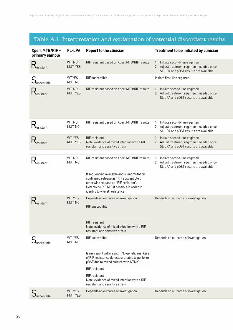
In summary: in high MDR prevalence settings, rifampicin-resistant results obtained via rapid molecular techniques (GeneXpert and first-line LPA) should be communicated to clinicians to inform the drugs used in the initiation of treatment. In case of discordance between pDST and gDST results, absence of laboratory errors should be guaranteed for both approaches (gDST and pDST) by the laboratory and as a second step the different possibilities for potential discordant results explained in Table A.1 should be considered.
Annex 1
27
Xpert MTB/RIF – primary sample
FL-LPA Report to the clinician Treatment to be initiated by clinician pDST Explanation / Possible laboratory errors
Action
ResistantWT: NO, MUT: YES
RIF resistant based on Xpert MTB/RIF results 1. Initiate second-line regimen2. Adjust treatment regimen if needed once
SL-LPA and pDST results are availableResistant
Typical mutation in the rpoB RRDR Issue report
SusceptibleWT:YES, MUT: NO
RIF susceptible Initiate first-line regimen SusceptibleIssue report
ResistantWT: NO, MUT: YES
RIF resistant based on Xpert MTB/RIF results 1. Initiate second-line regimen2. Adjust treatment regimen if needed once
SL-LPA and pDST results are availableSusceptible
Faulty pDST e.g.:• no or too little bacteria suspension added• too much RIF added• RIF stock concentration too highIf erroneous pDST can be excluded mutations that cause low level resistance could be expected.
• Repeat pDST• Review RIF stock/concentration and quality controls
ResistantWT: NO, MUT: NO
RIF resistant based on Xpert MTB/RIF results 1. Initiate second-line regimen2. Adjust treatment regimen if needed once
SL-LPA and pDST results are availableResistant
Mutation not available on HAIN strip Issue report
ResistantWT: YES, MUT: YES
RIF resistant Note: evidence of mixed infection with a RIF resistant and sensitive strain
1. Initiate second-line regimen2. Adjust treatment regimen if needed once
SL-LPA and pDST results are availableResistant
Hetero-resistance • Repeat FL-LPA from pDST• sequencing (double peaks)
ResistantWT: NO, MUT: NO
RIF resistant based on Xpert MTB/RIF results 1. Initiate second-line regimen2. Adjust treatment regimen if needed once
SL-LPA and pDST results are availableSusceptible
Faulty pDST (e.g. TB suspension not added) • Repeat pDST• Review RIF stock/concentration and quality controls
If sequencing available and silent mutation confirmed release as “RIF susceptible”, otherwise release as “RIF resistant”. Determine RIF MIC if possible in order to identify low level resistance
Silent or rare mutations Sequencing
R esistantWT: YES, MUT: NO
Depends on outcome of investigation Depends on outcome of investigation SusceptibleFaulty Xpert MTB/RIF e.g.:• sample mix up• contamination
• Repeat FL-LPA, pDST testing of another isolate of the patient
• Review samples tested before and after the sample in question
• Review maintanance record
RIF susceptible
RIF resistant Note: evidence of mixed infection with a RIF resistant and sensitive strain
Hetero-resistance • Repeat Xpert testing on another sputum sample• Repeat FL-LPA• pDST testing of another isolate of the patient
SusceptibleWT: YES, MUT: NO
RIF susceptible Depends on outcome of investigation ResistantFaulty pDST e.g.:• no drug added• contamination
• Repeat pDST• Microscopy• Blood plate from pDST
Issue report with result: “No genetic markers of RIF resistance detected; unable to perform pDST due to mixed culture with NTMs”
Mixed culture with MTB and NTM HAIN CM from RIF DST MGIT or from solid RIF DST; review original solid culture, to check for NTM, repeat pDST on another positive isolat
RIF resistant Mutation outside the rpoB 511-524 hotspot region rpoB sequencing (outside RRDR)
RIF resistant Note: evidence of mixed infection with a RIF resistant and sensitive strain
Hetero-resistance • FL-LPA: check for faint signals• Repeat FL-LPA from pDST• Sequencing (double peaks)
SusceptibleWT: YES, MUT: YES
Depends on outcome of investigation Depends on outcome of investigation S usceptibleLaboratory error (e.g. contamination of the FL-LPA)
Check negative control in the FL-LPA run. Repeat FL-LPA.
Table A.1. Interpretation and explanation of potential discordant results
Algorithm for laboratory diagnosis and treatment-monitoring of pulmonary tuberculosis and drug-resistant tuberculosis using state-of-the-art rapid diagnostic technologies
28
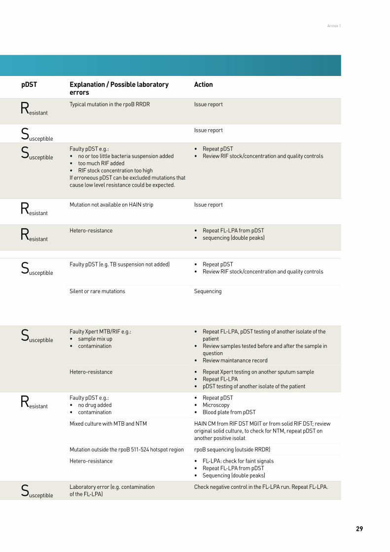
Xpert MTB/RIF – primary sample
FL-LPA Report to the clinician Treatment to be initiated by clinician pDST Explanation / Possible laboratory errors
Action
ResistantWT: NO, MUT: YES
RIF resistant based on Xpert MTB/RIF results 1. Initiate second-line regimen2. Adjust treatment regimen if needed once
SL-LPA and pDST results are availableResistant
Typical mutation in the rpoB RRDR Issue report
SusceptibleWT:YES, MUT: NO
RIF susceptible Initiate first-line regimen SusceptibleIssue report
ResistantWT: NO, MUT: YES
RIF resistant based on Xpert MTB/RIF results 1. Initiate second-line regimen2. Adjust treatment regimen if needed once
SL-LPA and pDST results are availableSusceptible
Faulty pDST e.g.:• no or too little bacteria suspension added• too much RIF added• RIF stock concentration too highIf erroneous pDST can be excluded mutations that cause low level resistance could be expected.
• Repeat pDST• Review RIF stock/concentration and quality controls
ResistantWT: NO, MUT: NO
RIF resistant based on Xpert MTB/RIF results 1. Initiate second-line regimen2. Adjust treatment regimen if needed once
SL-LPA and pDST results are availableResistant
Mutation not available on HAIN strip Issue report
ResistantWT: YES, MUT: YES
RIF resistant Note: evidence of mixed infection with a RIF resistant and sensitive strain
1. Initiate second-line regimen2. Adjust treatment regimen if needed once
SL-LPA and pDST results are availableResistant
Hetero-resistance • Repeat FL-LPA from pDST• sequencing (double peaks)
ResistantWT: NO, MUT: NO
RIF resistant based on Xpert MTB/RIF results 1. Initiate second-line regimen2. Adjust treatment regimen if needed once
SL-LPA and pDST results are availableSusceptible
Faulty pDST (e.g. TB suspension not added) • Repeat pDST• Review RIF stock/concentration and quality controls
If sequencing available and silent mutation confirmed release as “RIF susceptible”, otherwise release as “RIF resistant”. Determine RIF MIC if possible in order to identify low level resistance
Silent or rare mutations Sequencing
R esistantWT: YES, MUT: NO
Depends on outcome of investigation Depends on outcome of investigation SusceptibleFaulty Xpert MTB/RIF e.g.:• sample mix up• contamination
• Repeat FL-LPA, pDST testing of another isolate of the patient
• Review samples tested before and after the sample in question
• Review maintanance record
RIF susceptible
RIF resistant Note: evidence of mixed infection with a RIF resistant and sensitive strain
Hetero-resistance • Repeat Xpert testing on another sputum sample• Repeat FL-LPA• pDST testing of another isolate of the patient
SusceptibleWT: YES, MUT: NO
RIF susceptible Depends on outcome of investigation ResistantFaulty pDST e.g.:• no drug added• contamination
• Repeat pDST• Microscopy• Blood plate from pDST
Issue report with result: “No genetic markers of RIF resistance detected; unable to perform pDST due to mixed culture with NTMs”
Mixed culture with MTB and NTM HAIN CM from RIF DST MGIT or from solid RIF DST; review original solid culture, to check for NTM, repeat pDST on another positive isolat
RIF resistant Mutation outside the rpoB 511-524 hotspot region rpoB sequencing (outside RRDR)
RIF resistant Note: evidence of mixed infection with a RIF resistant and sensitive strain
Hetero-resistance • FL-LPA: check for faint signals• Repeat FL-LPA from pDST• Sequencing (double peaks)
SusceptibleWT: YES, MUT: YES
Depends on outcome of investigation Depends on outcome of investigation S usceptibleLaboratory error (e.g. contamination of the FL-LPA)
Check negative control in the FL-LPA run. Repeat FL-LPA.
Table A.1. Interpretation and explanation of potential discordant results
Annex 1
29


The WHO Regional Office for EuropeThe World Health Organization (WHO) is a specialized agency of the United Nations created in 1948 with the primary responsibility for international health matters and public health. The WHO Regional Office for Europe is one of six regional offices throughout the world, each with its own programme geared to the particular health conditions of the countries it serves.
Member States
AlbaniaAndorraArmeniaAustriaAzerbaijanBelarusBelgiumBosnia and HerzegovinaBulgariaCroatiaCyprusCzechiaDenmarkEstoniaFinlandFranceGeorgiaGermanyGreeceHungary
IcelandIrelandIsraelItalyKazakhstanKyrgyzstanLatviaLithuaniaLuxembourgMaltaMonacoMontenegroNetherlandsNorwayPolandPortugalRepublic of MoldovaRomaniaRussian FederationSan Marino
Serbia SlovakiaSloveniaSpainSwedenSwitzerlandTajikistanThe former Yugoslav
Republic of MacedoniaTurkeyTurkmenistanUkraineUnited KingdomUzbekistan
World Health OrganizationRegional Office for Europe
UN City, Marmorvej 51, DK-2100 Copenhagen Ø, DenmarkTel.: +45 45 33 70 00. Fax: +45 45 33 70 01. E-mail: [email protected]
Website: www.euro.who.int
9 789289 052375 >
ISBN 9789289052375